Embed Size (px)
Citation preview

CHEMICAL CONSTITUENTS FROM Crotalaria pallida, Morinda citrifolia AND Chlorophyllum molybdites
SITI RABEAH BINTI FADZIL
FACULTY OF SCIENCE UNIVERSITY OF MALAYA
KUALA LUMPUR
2018
Univers
ity of
Mala
ya

CHEMICAL CONSTITUENTS FROM Crotalaria pallida, Morinda citrifolia AND Chlorophyllum molybdites
SITI RABEAH BINTI FADZIL
DISSERTATION SUBMITTED IN FULFILMENT OF THE
REQUIREMENTS FOR THE DEGREE OF MASTER OF SCIENCE
DEPARTMENT OF CHEMISTRY FACULTY OF SCIENCE
UNIVERSITY OF MALAYA KUALA LUMPUR
2018
Univers
ity of
Mala
ya

ii
UNIVERSITY MALAYA
ORIGINAL LITERARY WORK DECLARATION
Name of Candidate: Siti Rabeah Fadzil
Registration/Matric No: SGR140008
Name of Degree: Master of Science (Except Mathematics & Science Philosophy)
Title of Project Paper/Research Report/Dissertation/Thesis (“this Work”): Chemical Constituents from Crotalaria pallida, Morinda citrifolia and Chlorophyllum molybdites
Field of Study: Organic Chemistry
I do solemnly and sincerely declare that: (1) I am the sole author/writer of this Work; (2) This Work is original; (3) Any use of any work in which copyright exists was done by way of fair dealing
and for permitted purposes and any excerpt or extract from, or reference to or reproduction of any copyright work has been disclosed expressly and sufficiently and the title of the Work and its authorship have been acknowledged in this Work;
(4) I do not have any actual knowledge nor do I ought reasonably to know that the making of this work constitutes an infringement of any copyright work;
(5) I hereby assign all and every rights in the copyright to this Work to the University of Malaya (“UM”), who henceforth shall be owner of the copyright in this Work and that any reproduction or use in any form or by any means whatsoever is prohibited without the written consent of UM having been first had and obtained;
(6) I am fully aware that if in the course of making this Work I have infringed any copyright whether intentionally or otherwise, I may be subject to legal action or any other action as may be determined by UM.
Candidate’s Signature Date:
Subscribed and solemnly declared before,
Witness’s Signature Date:
Name: Dr. Choo Yeun Mun Designation: Senior Lecturer
Univers
ity of
Mala
ya

iii
[CHEMICAL CONSTITUENTS FROM CROTALARIA PALLIDA, MORINDA
CITRIFOLIA AND CHLOROPHYLLUM MOLYBDITES]
ABSTRACT
The present dissertation work focused on the phytochemical studies of three Malaysian
plants, Crotalaria pallida, Morinda citrifolia and Chlorophyllum molybdites. C.
pallida, from the family of Fabaceae, a herbaceous legume and used traditionally for
treatment of several types of illness. This plant is also known to produce the toxic
pyrrolizidine alkaloids and flavonoids. M. citrifolia, from the family of Rubiaceae, is a
small evergreen shrub tree. It is known to produce anthraquinone compounds, which
possess various therapeutic properties including antiviral, antibacterial, antioxidant,
anticancer, antitumor, and anti-inflammatory. C. molybdites from the family of
Agaricaceae is a poisonous mushroom often involved in poisoning cases throughout
the world. This fungi is known to produce the toxic components such as a toxic
protein, molybdophyllysin. In the present study, one new cyclopentylidene, crotolidene
(CP1), and seven known compounds, i.e. hydroxydihydrobovolide (CP2), octacosane
(CP3), trans-phytyl palmitate (CP4), linoleic acid (CP5), methyl oleate (CP6), ethyl
palmitate (CP7), and palmitic acid (CP8) were isolated from the hexane extract of
C.pallida. A total of five known anthraquinones were isolated from the chloroform
extract of M. citrifolia which included damnacanthal (MC1), nordamnacanthal (MC2),
rubiadin (MC3), 1,6-dihydroxy-2-methyl-anthraquinone (MC4), 1-hydroxy-3-
methoxyanthraquinone (MC5), and 1-methoxy-2-hydroxyanthraquinone (MC6). Ethyl
acetate extracts of C. molybdites gave four known compounds which are α-D-glucose
(CM1), ethyl-β-D-glucopyranoside (CM2), 2,5-anhydro-D-hexitol (CM3), and
Univers
ity of
Mala
ya

iv
linoleic acid (CP5). All the compounds were isolated and characterized using
extensive chromatographic and spectroscopic methods.
Keywords: Phytochemical studies, Crotalaria pallida, Morinda citrifolia,
Chlorophyllum molybdites, spectroscopic analysis.
Univers
ity of
Mala
ya
v
[JUZUK KIMIA DALAM CROTALARIA PALLIDA, MORINDA CITRIFOLIA
DAN CHLOROPHYLLUM MOLYBDITES]
ABSTRAK
Disertasi ini memfokuskan kajian fitokimia ke atas tiga tumbuhan Malaysia, Crotalaria
pallida, Morinda citrifolia dan Chlorophyllum molybdites. C. pallida, dari keluarga
Fabaceae, adalah legum herba dan digunakan secara tradisional untuk rawatan beberapa
jenis penyakit. Tumbuhan ini juga dikenali dalam menghasilkan toksik alkaloid
pyrrolizidine dan flavonoid. M. citrifolia, dari keluarga Rubiaceae, adalah sejenis
pokok malar hijau renek yang kecil. Ia dikenali dalam menghasilkan sebatian
antrakuinon yang mana ianya mempunyai pelbagai ciri terapeutik termasuk antivirus,
anti bakteria, antioksida, antikanser, antitumor, dan anti-radang. C. molybdites, dari
keluarga Agaricaceaea adalah sejenis cendawan beracun yang seringkali terlibat dalam
kes-kes keracunan di serata dunia. Kulat ini dikenali dalam menghasilkan komponen
toksik seperti protin beracun, molybdophyllysin. Di dalam kajian ini, Satu
siklopentilidena baru, crotolidine (CP1) dan tujuh sebatian lain, iaitu
hidroksidihidrobovolide (CP2), octacosana (CP3), trans-phytil palmitat (CP4), asid
linoleik (CP5), metil oleat (CP6), etil palmitat (CP7), dan asid palmitik (CP8) telah
diasingkan dari ekstrak heksana C. pallida. Sejumlah lima sebatian antrakuinon yang
telah dikenali telah diasingkan dari ekstrak klorofom termasuklah damnakantal (MC1),
nordamnakantal (MC2), rubiadin (MC3), 1,6-dihidroksi-2-metilantrakuinon (MC4), 1-
hidroksi-3-metoksiantrakuinon (MC5), dan 1-metoksi-2-hidroksiantrakuinon (MC6).
Ekstrak etil asitat daripada C. molybdites telah menghasilkan pengasingan sebanyak
empat sebatian yang telah dikenali iaitu α-D-glukosa (CM1), etil-β-D-glukopiranosida
(CM2), 2,5-anhidro-D-heksitol (CM3), dan asid linoleik (CP5). Kesemua sebatian ini
Univers
ity of
Mala
ya
vi
telah diasingkan dan dikenalpasti dengan menggunakan pelbagai kaedah kromatografi
dan spektroskopi.
Kata kunci: Kajian fitokimia, Crotalaria pallida, Morinda citrifolia, Chlorophyllum
molybdites, analisis spektroskopi.
Univers
ity of
Mala
ya

vii
DEDICATION
The thesis work is dedicated to my late mother, Siti Alauwiah binti Haji Kabeb and my
lovely father, Fadzil bin Mahmood, who has been a constant support in my academic
journey. This work is also dedicated to my understanding husband, Mohd Asrul Effandi
bin Nasir and my precious son, Rayyan Abqari bin Mohd Asrul Effandi, who has been
my “big why’ to keep going and finished all my research work. Last but not least, I
dedicate this work to my younger brother, Mohammad Dzull Faqaar bin Fadzil. To all
these five names that I have mentioned above, I am truly blessed to have all of you in
my life.
Univers
ity of
Mala
ya
viii
ACKNOWLEDGEMENT
First and foremost, praise be to Allah the Almighty for giving this opportunity
and granting me the capability to proceed successfully. This thesis appears completely
due to the assistance and guidance from several helpful people. There were many
people who stand beside me during the last four years. They have guided, placed and
showed me the opportunity. Therefore, I would like to show my appreciation to all of
them.
I would like to take the opportunity in expressing my deepest appreciation and
enormous gratitude to my supervisor Dr. Choo Yeun Mun for her continually support
and convincingly advice in a way of guiding me throughout this research work.
Without her guidance and persistent help, this thesis would not have been possible to
finish during the time frame. On the other hand, a big thank you I address to the
administrative division of the Chemistry department, and also to the staff of the NMR,
MS, IR and UV team for their assistance and services.
In addition, a special thank you to my lab members Ms. Yap Ann Chee, Mrs.
Nur Atiqah Mohd Nasuha, Mrs. Nabila Elyana Adnan, Mrs. Phoebe Primus and Ms.
Noridayu Omer for their care, assistance and friendship during the ups and downs in
the lab. I am also grateful for having many friends whom share their friendship,
insights and experiences, especially Ms. Amy Nuzwir Wateh, Mrs. Fatirah Muhamad
Sarih, Ms. Hazira Hussin, and Ms. Irma Zulayka Mohamad Ahad. Finally, my heartiest
thanks go to my family for their valuable support and constant encouragement
throughout my study.
Univers
ity of
Mala
ya

ix
TABLE OF CONTENTS
Abstract………………………………………………………………………… iii
Abstrak…………………………………………………………………………. v
Dedication……………………………………………………………………… vii
Acknowledgements…………………………………………………………….. viii
Table of Contents………………………………………………………………. ix
List of Figures………………………………………………………………….. xiii
List of Tables…………………………………………………………………… xvii
List of Abbreviations…………………………………………………………… xix
List of Symbols………………………………………………………………… xxi
CHAPTER 1: INTRODUCTION……………………………………………
1.1 Natural product: modern drugs from natural sources………………….
1.2 Objective……………………………………………………………….
1
1
11
CHAPTER 2: LITERATURE REVIEW……………………………….…..
2.1 Crotalaria pallida………………………………………………….…..
2.1.1 General…………………………………………………………
2.1.2 Compounds isolated from the genus Crotalaria ………………
2.2 Morinda citrifolia………………………………………………….…..
2.2.1 General…………………………………………………………
2.2.2 Compounds isolated from Morinda citrifolia…………….……
12
12
12
16
36
36
42
Univers
ity of
Mala
ya

x
2.3 Chlorophyllum molybdites……………………………………..………
2.3.1 General…………………………………………………...…….
2.3.2 Compounds isolated from Chlorophyllum molybdites………...
63
63
68
CHAPTER 3: METHODOLOGY…………………………………………..
3.1 Source and authentication……………………………………….…..…
3.1.1 Crotalaria pallida………………………………………...……
3.1.2 Morinda citrifolia…………………………………….….……
3.1.3 Chlorophyllum molybdites…………………………….….……
3.2 General…………………………………………………………………
3.3 Plant extraction………………………………………………...………
3.3.1 Crotalaria pallida…………………………………..….………
3.3.2 Morinda citrifolia…………………………………….…..……
3.3.3 Chlorophyllum molybdites…………………………….………
3.4 Chromatographic method…………………………………..…………
3.4.1 Thin Layer Chromatographic (TLC)…………………..………
3.4.2 Column Chromatographic (CC)……………………………….
3.4.3 Centrifugal Thin Layer Chormatographic (CTCL)……………
3.4.4 Preparative Thin Layer Chromatogaphic (PTLC)……..………
3.4.5 High-Performance Liquid Chromatography (HPLC)…….……
3.5 Staining reagents for TLC…………………………………..…………
3.5.1 Iodine staining reagent…………………………………………
3.5.2 Anisaldehyde staining reagent…………………………………
73
73
73
74
74
75
75
75
76
76
77
77
77
78
78
79
79
79
80
Univers
ity of
Mala
ya
xi
3.6 Isolation of compounds………………………………..………………
3.6.1 Isolation of compounds from the hexane extract of Crotalaria
pallida………………………………………………………….
3.6.2 Isolation of compounds from the chloroform extract of Morinda
citrifolia…………………………………………..……………..
3.6.3 Isolation of compounds from the ethyl acetate extract of
Chlorophyllum molybdites………………………….…………
3.7 Compound data…………………………………………………..…… 3.7.1 Compound isolated from Crotalaria pallida……………….…
3.7.2 Compound isolated from Morinda citrifolia……………….….
3.7.3 Compound isolated from Chlorophyllum molybdites……….….
3.8 Table of Rf values………………………………………………………
80
80
80
81
87
87
90
93
94
CHAPTER 4: RESULTS AND DISCUSSION…………………………….
4.1 Compound isolated from Crotalaria Pallida………………………….
4.1.1 CP1: Crotolidene……………………………………………… 4.1.2 CP2: Hydroxydihydrobovolide………………………………..
4.1.3 CP3: Octacosane………………………………………………
4.1.4 CP4: Trans-phytyl palmitate…………………………………..
4.1.5 CP5: Linoleic acid…………………………………………….
4.1.6 CP6: Methyl oleate…………………………………………….
4.1.7 CP7: Ethyl palmitate…………………………………………..
4.1.8 CP8: Palmitic acid…………………………………………….
95
95
96
102
107
111
116
121
125
129
Univers
ity of
Mala
ya

xii
4.2 Compound isolated from Morinda citrifolia…………………………..
4.2.1 MC1: Damnachantal…………………………………………..
4.2.2 MC2: Nordamnachantal……………………………………….
4.2.3 MC3: Rubiadin………………………………………………..
4.2.4 MC4: 1,6-Dihydroxy-2-Methylanthraquinone……………….
4.2.5 MC5: 1-Hydroxy-3-Methoxyanthraquinone………………….
4.2.6 MC6: 1-Methoxy-2-Hydroxyanthraquinone………………….
4.3 Compound isolated from Chlorophyllum molybdites………………….
4.3.1 CM1: α-D-glucose……………………………………………..
4.3.2 CM2: Ethyl-β-D-glucopyranoside……………………………..
4.3.3 CM3: 2,5-Anhydro-D-hexitol…………………………………
133
134
139
144
149
154
157
163
164
168
172
CHAPTER 5: CONCLUSION……………………………………………… 176
References……………………………………………….…………………….. 180
List of Publications and Papers Presented…………………………………….. 202
Appendix……………………………………………………………………… 203
Univers
ity of
Mala
ya

xiii
LIST OF FIGURES
Figure 1.1 : Structure of compounds isolated from plants 2
Figure 1.2 : Structure of compounds isolated from microorganism 4
Figure 1.3 : Structure of compounds isolated from animal sources 5
Figure 1.4 : Structure of compounds isolated from marine sources 6
Figure 1.5 : Structure of new approved drugs based on Natural Products
(NPs)
8
Figure 1.6 : Structure of new approved drugs based on semi-synthetic
NPs
9
Figure 1.7 : Structure of new approved drugs based on NP-derived
compounds
10
Figure 2.1 : Structure of compounds isolated from the genus Crotalaria 16
Figure 2.2 : Structure of compounds isolated from Morinda citrifolia 42
Figure 2.3 : Structure of compounds isolated from Chlorophyllum molybdites
68
Figure 3.1 : The whole plant of Crotalaria pallida 73
Figure 3.2 : The roots of Morinda citrifolia 74
Figure 3.3 : The fruit bodies of Chlorophyllum molybdites 74
Figure 3.4 : Isolation of compounds from the hexane extract of Crotalaria pallida
82
Figure 3.5 : Isolation of compounds from the chloroform extract of Morinda citrifolia
85
Figure 3.6 : Isolation of compounds from the ethyl acetate extract of Chlorophyllum molybdites
86
Figure 4.1 : Selected (a) HMBC (b) NOESY of CP1 (Crotolidene) 98
Figure 4.2 : 1H NMR spectrum (CDCl3, 600 MHz) of CP1 (Crotolidene) 100
Univers
ity of
Mala
ya

xiv
Figure 4.3 : 13C NMR spectrum (CDCl3, 150 MHz) of CP1 (Crotolidene)
101
Figure 4.4 : Selected HMBC of CP2 (Hydroxydihydrobovolide) 103
Figure 4.5
:
1H NMR spectrum (CDCl3, 600 MHz) of CP2 (Hydroxydihydrobovolide)
105
Figure 4.6 : 13C NMR spectrum (CDCl3, 150 MHz) of CP2 (Hydroxydihydrobovolide)
106
Figure 4.7 : 1H NMR spectrum (CDCl3, 600 MHz) of CP3 (Octacosane) 109
Figure 4.8 : 13C NMR spectrum (CDCl3, 150 MHz) of CP3 (Octacosane) 110
Figure 4.9 : Selected HMBC of CP4 (Trans-phytyl palmitate) 112
Figure 4.10 : 1H NMR spectrum (CDCl3, 600 MHz) of CP4 (Trans-phytyl palmitate)
114
Figure 4.11 : 13C NMR spectrum (CDCl3, 150 MHz) of CP4 (Trans-phytyl palmitate)
115
Figure 4.12 : Selected HMBC of CP5 (Linoleic acid) 117
Figure 4.13 : 1H NMR spectrum (CDCl3, 600 MHz) of CP5 (Linoleic acid)
119
Figure 4.14 : 13C NMR spectrum (CDCl3, 150 MHz) of CP5 (Linoleic acid)
120
Figure 4.15 : Selected HMBC of CP6 (Methyl oleate) 122
Figure 4.16 : 1H NMR spectrum (CDCl3, 600 MHz) of CP6 (Methyl oleate)
123
Figure 4.17 : 13C NMR spectrum (CDCl3, 150 MHz) of CP6 (Methyl oleate)
124
Figure 4.18 : Selected HMBC of CP7 (Ethyl palmitate) 126
Figure 4.19 : 1H NMR spectrum (CDCl3, 600 MHz) of CP7 (Ethyl palmitate)
127
Figure 4.20 : DEPT-135 NMR spectrum (CDCl3, 150 MHz) of CP7 (Ethyl palmitate)
128
Figure 4.21 : Selected HMBC of CP8 (Palmitic acid) 130
Figure 4.22 : 1H NMR spectrum (CDCl3, 600 MHz) of CP8 (Palmitic acid)
131
Univers
ity of
Mala
ya

xv
Figure 4.23 :
13C NMR spectrum (CDCl3, 150 MHz) of CP8 (Palmitic acid)
132
Figure 4.24 : Selected HMBC correlations of MC1 (Damnachantal) 135
Figure 4.25
:
1H NMR spectrum (CDCl3, 600 MHz) of MC1 (Damnachantal)
137
Figure 4.26 : 13C NMR spectrum (CDCl3, 150 MHz) of MC1 (Damnachantal)
138
Figure 4.27 : Selected HMBC correlations of MC2 (Nordamnachantal) 140
Figure 4.28 : 1H NMR spectrum (CDCl3, 600 MHz) of MC2 (Nordamnachantal)
142
Figure 4.29 : 13C NMR spectrum (CDCl3, 150 MHz) of MC2 (Nordamnachantal)
143
Figure 4.30 : Selected HMBC correlations of MC3 (Rubiadin) 145
Figure 4.31 : 1H NMR spectrum (CDCl3, 600 MHz) of MC3 (Rubiadin) 147
Figure 4.32 : 13C NMR spectrum (CDCl3, 150 MHz) of MC3 (Rubiadin) 148
Figure 4.33 : Selected HMBC correlations of MC4 (1,6-Dihydroxy-2- methylanthraquinone)
150
Figure 4.34 : 1H NMR spectrum (CDCl3, 600 MHz) of MC4 (1,6-Dihydroxy-2-methylanthraquinone)
152
Figure 4.35 : 13C NMR spectrum (CDCl3, 150 MHz) of MC4 (1,6-Dihydroxy-2-methylanthraquinone)
153
Figure 4.36 : 1H NMR spectrum (CDCl3, 150 MHz) of MC5 (1-Hydroxy-3-methoxyanthraquinone)
156
Figure 4.37 : Selected HMBC correlations of MC6 (1-Methoxy-2-hydroxyanthraquinone)
158
Figure 4.38 : 1H NMR spectrum (CDCl3, 150 MHz) of MC6 (1-Methoxy-2-hydroxyanthraquinone)
160
Figure 4.39
:
13C NMR spectrum (CDCl3, 150 MHz) of MC6 (1-Methoxy-2-hydroxyanthraquinone)
161
Figure 4.40 : HMBC spectrum (CDCl3, 600 MHz) of MC6 (1-Methoxy-2-hydroxyanthraquinone)
162
Figure 4.41 : Selected HMBC correlations of CM1 (α-D-glucose) 165
Univers
ity of
Mala
ya

xvi
Figure 4.42 : 1H NMR spectrum (CDCl3, 600 MHz) of CM1 (α-D-glucose)
166
Figure 4.43 : 13C NMR spectrum (CDCl3, 150 MHz) of CM1 (α-D-glucose)
167
Figure 4.44 : Selected HMBC correlations of CM2 (Ethyl-β-D-glucopyranoside)
169
Figure 4.45 : 1H NMR spectrum (CDCl3, 600 MHz) of CM2 (Ethyl-β-D-glucopyranoside)
170
Figure 4.46 : 13C NMR spectrum (CDCl3, 150 MHz) of CM2 (Ethyl-β-D-glucopyranoside)
171
Figure 4.47 : Selected HMBC correlations of CM3 (2,5-Anhydro-D-hexitol)
172
Figure 4.48 : 1H NMR spectrum (CDCl3, 600 MHz) of CM3 (2,5-Anhydro-D-hexitol)
174
Figure 4.49 : 13C NMR spectrum (CDCL3, 150 MHz) of CM3 (2,5-Anhydro-D-hexitol)
175
Univers
ity of
Mala
ya

xvii
LIST OF TABLES
Table 2.1 : Name of compounds isolated from different Crotalaria sp. 27
Table 2.2 : Name of compounds isolated from Morinda citrifolia 55
Table 2.3 : Name of compounds isolated from Chlorophyllum molybdites
71
Table 3.1 : The Rf values 94
Table 4.1 : The NMR (CDCl3, 600 MHz) data of CP1 (Crotolidene) 99
Table 4.2 : The NMR (CDCl3, 600 MHz) data of CP2 (Hydroxydihydrobovolide)
104
Table 4.3 : The NMR (CDCl3, 600 MHz) data of CP3 (Octacosane) 108
Table 4.4 : The NMR (CDCl3, 600 MHz) data of CP4 (Trans-phytyl palmitate)
113
Table 4.5 : The NMR (CDCl3, 600 MHz) data of CP5 (Linoleic acid) 118
Table 4.6 : The NMR (CDCl3, 600 MHz) data of CP6 (Methyl oleate) 122
Table 4.7 : The NMR (CDCl3, 600 MHz) data of CP7 (Ethyl palmitate)
126
Table 4.8 : The NMR (CDCl3, 600 MHz) data of CP8 (Palmitic acid) 130
Table 4.9 : The NMR (CDCl3, 600 MHz) data of MC1 (Damnachantal)
136
Table 4.10 : The NMR (CDCl3, 600 MHz) data of MC2 (Nordamnachantal)
137
Table 4.11 : The NMR (CDCl3, 600 MHz) data of MC3 (Rubiadin) 146
Table 4.12 : The NMR (CDCl3, 600 MHz) data of MC4 (1,6-Dihydroxy-2-methylanthraquinone)
151
Table 4.13 : The NMR (CDCl3, 600 MHz) data of MC5 (1-Hydroxy-3-methoxyanthraquinone)
155
Table 4.14 : The NMR (CDCl3, 600 MHz) data of MC6 (1-Methoxy-2- hydroxyanthraquinone)
159
Table 4.15 : The NMR (CDCl3, 600 MHz) data of CM1 (α-D-glucose)
165
Univers
ity of
Mala
ya

xviii
Table 4.16 : The NMR (CDCl3, 600 MHz) data of CM2 (Ethyl-β-D-glucopyranoside)
169
Table 4.17 : The NMR (CDCl3, 600 MHz) data of CM3 (2,5-Anhydro-D-hexitol)
173
Table 5.1 : Compounds isolated from Crotalaria pallida, Morinda citrifolia and Chlorophyllum molybdites
177
Univers
ity of
Mala
ya
xix
LIST OF ABBREVIATION
AD : Anno Domini
ADHD : Attention-Deficit Hyperactivity Disorder
BC : Before Christ
CC : Column Chromatography
COSY : Correlation Spectroscopy
CTLC : Centrifugal Thin Layer Chromatography
DEPT : Distortionless Enhancement by Polarization Transfer
DNA : Deoxyribonucleic Acid
EGF : Epidermal Growth Factor
ESI : Electrospray Ionization
EtOH : Ethanol
FRAP : Ferric Reducing Antioxidant Power
HIV : Human Immunodeficiency Virus
HMBC : Heteronuclear Multiple Bond Correlation
HPLC : High Performance Liquid Chromatography
HRESIMS : High Resolution Electrospray Ionization Mass Spectrometry
HSQC : Heteronuclear Single Quantum Coherence
IR : Infrared Spectroscopy
ITSrDNA : Internal Transcribed Space Ribosomal Deoxyribonucleic Acid
LCMS : Liquid Chromatography Mass Spectrometry
MAA : Marketing Authorization Application
MEPs : Metalloendopeptidase
MS : Mass Spectrometry
NaCl : Natrium chloride
NFE : Nitrogen Free Extract
Univers
ity of
Mala
ya
xx
NMR : Nuclear Magnetic Resonance
NPs : Natural Products
ORAC : Oxygen Radical Absorbance Capacity
PAs : Pyrolizidine Alkaloid
PTLC : Preparative Thin Layer Chromatography
Q-TOF : Quadrapole Time-of-Flight
RBCs : Red Blood Cells
TCM : Traditional Chinese Medicine
TLC : Thin Layer Chromatography
UV : Ultraviolet Spectroscopy
Vpr : Viral Protein R
WHO : World Health Organization
Univers
ity of
Mala
ya

xxi
LIST OF SYMBOLS
α : Alpha
β : Beta
J : Coupling Constant
δ : Chemical shift
ε : Epsilon
g : Gram
Hz : Hertz
Kg : Kilogram
λ : Lambda
MHz : Megahertz
nm : Nanometer
% : Percentage
Rf : Retention Factor
Univers
ity of
Mala
ya
1
CHAPTER 1: INTRODUCTION
1.1 Natural products: modern drugs from natural sources
Throughout the ages, humans have relied on nature for their basic needs,
especially the medicines that has been used for treatment of many diseases. Natural
products have been a source of medicines to cure diseases since early human history.
Before the “Synthetic Era” around the early 1900s, 80% of all medicines were obtained
from plants either from roots, bark, or leaves. In more recent time, natural products
have continued to be significant sources of drugs and drug-leads. Over 80% of the
drugs developed for treatment of diseases were reported from or inspired by natural
product compounds (Cragg & Newman, 2013; Harvey, 2008; McChessney et al.,
2007).
The early records of natural products were written on clay tablets in cuneiform
from the Mesopotamia (2,600 BC). The best known record of the Egyptian medicine
the “Ebers Papyrus” (1,500 BC) while the best documentation of Indian Ayurvedic
system is perhaps the most ancient of all medicinal traditions which dates before 1,000
BC. The Chinese Materia Medica (1,100 BC), with the first record from Wu Shi Er
Bing Fang reported 52 prescriptions, Shennong Herbal (~ 100 BC) reported 365 drugs,
and the Tang Herbal (659 AD) reported 850 drugs. In the ancient Western world, the
Greeks contributed significantly by the work of the philosopher and natural scientist
Theophratus (~ 33 BC) and physician Dioscorides (100 AD) who authored History of
plants and De Materia Medica respectively (Atanasov et al., 2015; Cragg & Newman,
2013; Dias et al., 2012; Gumani et al., 2014; Gurib-Fakim, 2006).
Univers
ity of
Mala
ya

2
Natural sources can be divided into a group of four, i. e. plant, marine, animal
and microbial resources. In 1985, William Withering published his treatment for heart
patients using diatonic foxglove extract that is also known as digitalis. This treatment
has led to the discovery of digoxin, which has been used for the treatment of
arrhythmia and congestive heart failure. At the end of the 18th century, Felix Hoffmann
has synthesized aspirin from salicylic acid obtained from willow bark (Salix alba). This
is an example of a synthetic drug that had been isolated from a plant. In early 19th
century, Freidrich Serturner isolated morphine from opium poppy, Papaver
somniferum. The finding of morphine has led to the discovery of dose-controlled
medicine for pain. In 1820, the French pharmacists, Caventou and Pelletier isolated the
antimalarial drug, quinine from the bark of Cinchona species (Balunas & Kinghom,
2005; Cragg & Newman, 2013; Rishton, 2008).
O
O
HO
OH
O O
OHO
O
OH
O O
OHOH
H
H
H
OH N
H
CH3
HO
HO
Morphine
Digoxin
HO
N
N
O
HH
Quinine
O OH
O
O
Aspirin
OHO
OH
Salicylic acid
Figure 1.1: Structure of compounds isolated from plants.
Univers
ity of
Mala
ya

3
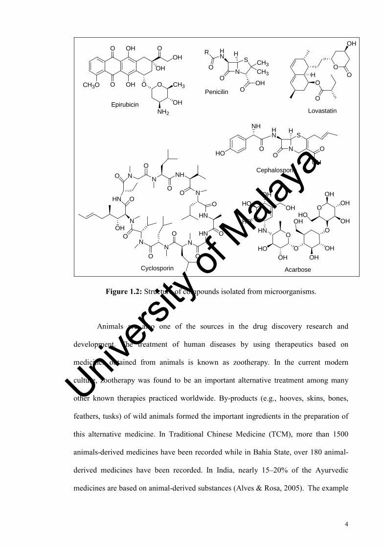
The studies of microorganism as sources of drugs started when Professor
Alexander Fleming published his findings on an active agent named penicillin from
Penicillium notatum in the British Journal of Experimental Pathology in June 1929.
Penicillin was isolated in a yellow powder form and used as a potent antibacterial
compound during World War II. The huge success of penicillin encouraged a
worldwide effort to assemble a large collection of microorganism for research purpose
to discover new drugs and it finally lead to the discovery of antibacterial agents
(cephalosporins), antidiabetic agents (acarbose), and anticancer agents (epirubicin)
(Demain & Sanchez, 2009). Other than penicilin, there are many other example of
drugs from microorganism sources. For example, the isolation of lovastatin
from Pleurotus ostreatus which is capable not only to help in reducing blood
cholesterol, but it also has an anti-fungal property and anti-carcinogenic effects
(Lakshmanan & Radha, 2012). Also the discovery of Cyclosporin A (CyA) from
Tolypocladium inflatum as a promising drug was attributed to its immunosuppressive
and its antifungal activities (Survase et al., 2009).
Univers
ity of
Mala
ya
4
OH
OH
O
O O
OH
OOH
O CH3
NH2
OH
CH3O
Epirubicin
N
S
CH3
CH3
HNR
O
O
H
OHOPenicilin
HNO
O
O
OHOOH
HOOH
HO
OHOH
OH
OH
OHHO
OH
OH
Acarbose
N
S
O
HHN
O
NH
HOO
OHCephalosporin
O
O
OO
OH
H
Lovastatin
HN O
OHN
NO
NO NH
ON
HN
HNN
N
O
O
O
O
ON
O
O
Cyclosporin
Figure 1.2: Structure of compounds isolated from microorganisms.
Animals are also one of the sources in the drug discovery research and
development. The treatment of human diseases by using therapeutics based on
medicines obtained from animals is known as zootherapy. In the current modern
culture, zootherapy was found to be an important alternative treatment among many
other known therapies practiced worldwide. By-products (e.g., hooves, skins, bones,
feathers, tusks) of wild animals formed the important ingredients in the preparation of
this alternative medicine. In Traditional Chinese Medicine (TCM), more than 1500
animals-derived medicines have been recorded while in Bahia State, over 180 animal-
derived medicines have been recorded. In India, nearly 15–20% of the Ayurvedic
medicines are based on animal-derived substances (Alves & Rosa, 2005). The example
Univers
ity of
Mala
ya

5
of animal-derived natural products includes epibatidine, derived from the skin of an
Ecuadorian poison frog. Epibatidine was reported to be ten times more potent than
morphine (Dias et al., 2012; Omprakash, 2013). Other example including teprotide
from a Brazilian pit viper Bothrops jararaca which has led to the development of
antihypertensive agent cilazapril and captopril, and echistatin, a disintegrin from the
venom of saw-scaled viper Echis carinatus which then lead to the development of
the antiplatelet drug tirofiban (Patlak, 2003; Pedrosa et al., 2013).
Captopril Epibatidine
Teprotide
HN
N
Cl
H
H
H
NN
OHO
OHN
O O
Cilazapril
N
OHOO
HS
HN
NH
O
O N
O
NH
O NH N
NH
O
NH2HN
O NHNH
NH2O
OCH3
O
CH3
N
O N
COOH
Figure 1.3: Structure of compounds isolated from animal sources.
In contrast with other sources of natural products, marine organisms never have
a significant history of usage in traditional medicines. The organised research of marine
environments only began in earnest in the mid-1970s. During the decade (1977–1987),
about 2,500 new metabolites were reported from a variety of marine organisms (Cragg
& Newman, 2013). In the early 1950s, the first notable discovery is the isolation of C-
nucleosides, spongouridine or uridine and spongothymidine from the Caribbean
Univers
ity of
Mala
ya

6
sponge, Cryptotheca crypta. The synthesis of structural analogues of the compounds
have led to the discovery of cytosine arabinoside (Ara-C) as a clinical anticancer agent,
and (Ara-A) as an antiviral agent. In the other hand, isolation from the mediterranean
tunicate Aplidium albicans has resulted to the discovery of a depsipeptide named
plitidepsin (Aplidin®,PharmaMa). Plitidepsin is effective in treating various cancers,
including small cell and nonsmall cell lung, melanoma, bladder as well as non-hodgkin
lymphoma and acute lymphoblastic leukemia (Omprakash, 2013; Schwartsmann et al.,
2001).
NHN
O
O
OHO
HO
OH
Uridine
NH
N O
O
O OH
OHOH
Spongothymidine
NN
OOHO
HO
OH NH2
O
OHHO
HO
NN
N N
NH2
H3C
O
NHO
O
N
O
O
N
O
O
NHOH
O
NHN N O
OO
OO
O
Plitidepsin
Vidarabine or (Ara-A) Cytarabine or (Ara-C)
Figure 1.4: Structure of compounds isolated from marine sources.
Natural product compounds that have undergone the clinical evaluation for
marketing purpose can be classified into three groups which is; natural products (NPs),
semi-synthetic NPs, and NP-derived compounds. The NPs group compounds are
derived from natural sources such as plant, animals and microorganism as discussed
Univers
ity of
Mala
ya

7
above, which have biological activities. While the semi-synthetic NPs group
compounds are derived from NP template but using semi-synthetic, and the compounds
that synthetically derived from NP template are grouped into NP-derived compounds.
Between the years of 2005 to 2010, a total 19 NP based drugs were approved for
marketing worldwide from Marketing Authorization Application (MAA) in Europe.
Among that, seven are classified as NPs, 10 as semi-synthetic NPs, and two as NP-
derived drugs (Mishra & Tiwari, 2011).
Sativex ®, a mixture of dronabinol and cannabinol was derived from the
cannabis plant is an example of NPs drug. It is the first pharmaceutical prescription
medicine in the world. Sativex ® was launched in Canada on April 2005 for
neuropathic pain relief in multiple sclerosis. In 2007, Health Canada approved Sativex
® as an adjunctive analgesis for severe pain in advanced cancer patients, reducing the
depending of opioid medications (Nurmikko et al., 2007; Wade et al., 2003). Other NPs
drug includes Fumagilin (Flisint ®), which was isolated from Aspergillus fumigatus. It
is an antimicrobial that capable on inhibiting the proliferation of endothelial cells. In
2005, fumagilin was approved for used against intestinal microsporidiosis, a disease
from Enterocytozoon bieneusi parasite that is causing chronic diarrhea (McCowen et
al., 1951). Exenatide (Byetta ®) was isolated from the oral secretions of the poisonous
lizard Heloderma suspectum (Gila monster). Exenatide was approved as an adjunctive
therapy in type 2 diabetis mellitus in 2006 (Cvetković & Plosker, 2007).
In 2007, the marine alkaloid named trabectedin (Yondelis®), isolated from the
ascidian Ecteinascidia turbinate became the first marine anticancer drug to be approved
in the European Union. It was approved for used against ovarian cancer and soft tissue
Univers
ity of
Mala
ya

8
sarcomas. In present, trabectedin is in trials against breast cancer, pediatric sarcomas
and prostate cancers (Montaser & Luesch, 2011; Soares et al., 2007).
O
HOO
O
O
OO
Fumagilin
HO
OH
Cannabidol
O
OH
Dronabinol
HGEGTFTSDLSKQMEEEAVRLFIEWLKNGGPSSGAPPPS
Exenatide
NH
HO
H3CO
O
OS
N
OH3C
O
H3C
OO
N CH3
HOOCH3
CH3
TrabectedinOH
H
Figure 1.5: Structure of new approved drugs based on Natural products (NPs).
Ixabepilone (Ixempra ®) is a semi-synthetic derivative of epothilone B
produced by Somngium cellulosum and it was developed as anticancer drug. It also has
a unique antibacterial and antifungal activities. In the preclinical study, it is shown that
natural epothilones A and B have potent antineoplastic activity against a wide range of
tumor cell (Lee et al., 2008). Tigecycline (Tygacil ®) is an antibiotic structurally
similar to tetracycline. Its capability to inhibit protein translation is by blocking the
entry of amino-acyl molecules into the ribosome. In 2005, tigecycline was approved for
the treatment of complicated skin and skin structure infections, and intra-abdominal
infections (Nishamin, 2006). Anidulafungin (Eraxis ™ in US, Ecalta ™ in Europe) is a
Univers
ity of
Mala
ya

9
semi-synthetic derivative of the fungal metabolite named echinocandin B. It was used
against invasive and oesophageal candidiasis and candidemia (Debono et al., 1995).
Telavancin (Vibativ ™) is a semi-synthetic analogue of vancomycin. It is capable to
inhibit the growth of bacterial for the treatment of nosocomial pneumonia (Judice &
Pace, 2003; Laohavaleeson & Nicolau, 2007).
ONH
O
NH
HOHO
N
HHO
HN
OHH
NH OHH
OHHO
OO
O NO
HOH
O
HN
O
OHH
Anidulafungin
NS
HN
O OH
OH
O
OH
Ixabepilone
OHN
NH
O O
O
OHOH
OH
NN
OH
NH2
H H
Tigecycline
Figure 1.6: Structure of new approved drugs based on semi-synthetic NPs.
Presently, there are nine β-lactams (one penem, two cephalosporins, and six
carbapenems) in clinical trials undergoing the drug registration. Among them,
doripenem is one of the ultra-broad spectrum injectable antibiotics. Doripenem
(Finibax ®) was launched in 2005. It showing a wide-range of antibacterial spectrum
and getting the approval for use in intra-abdominal and urinary tract infections (Keam,
2008; Mishra & Tiwari, 2011).
Univers
ity of
Mala
ya

10
Attention-Deficit Hyperactivity Disorder (ADHD), is a neuro-developmental
disorder. For many years, methylphenidate and amphetamines have been used for
ADHD management. But due to abuse potential, both drugs are being controlled and
limited. Lisdexamfetamine (Vyvanse ™) consist of dextroamphetamine. It was
designed to help ADHD patients when combined with essential amino acid (Biljana et
al., 2009; Blick & Keating, 2007).
NH2
CH3
Dextroamphetamine
N
OOHS
HNHNS
O
O
H2N
O
HO
H
Doripenem
O
NH
NH2NH2
Lisdexamfetamine
HN
O O
Methylphenidate
NH2
CH3
Amphetamine
Figure 1.7: Structure of new approved drugs based on NP-derived compounds.
Despite from all the successes from natural products history, many large
pharmaceutical companies have limited usage of natural products in the drug discovery
screening processes. The main factors that cause this problem are the difficulties in
access and supply, also the complexities of natural product chemistry itself. So, as a
natural product chemist, we need to improve the access to natural products and make it
look interesting to be exploring (Harvey, 2008).
Univers
ity of
Mala
ya

11
1.2 Objectives
This work aimed on the study of chemical constituents from three Malayan
plant species, i.e. Crotalaria pallida, Morinda citrifolia and Chlorophyllum molybdite.
The objectives of this study were to isolate and characterize new compounds from the
above mentioned plants by using extensive chromatographic and spectroscopic
methods.
Univers
ity of
Mala
ya
12
CHAPTER 2: LITERATURE REVIEW
2.1 Crotalaria pallida Aiton
2.1.1 General
The genus Crotalaria L. (Leguminosae, Papilionoideae, and Crotalarieae)
comprising about 600 species is widely distributed throughout tropical, neutropical and
subtropical regions. The centres of diversity are on eastern and southern tropical Africa
and India, and two other new centres found in Mexico and Brazil (Flores et al., 2009).
The genus Crotalaria is commonly known as devil-bean, shake shake, rattlebox or
rattlepod. It gets the name from the sound made when their pod-like fruit is shaken and
the seeds will “rattle” around inside (Wunderlin & Hansen, 2008). Although Crotalaria
species is a part of human diet, many species are known to be toxic to man and
livestock (Culvenor & Smith, 1962). It is also reported to be poisonous and is a death
cause (crotalism) in grazing cattle, horses, sheep or livestock fed with grains containing
seeds from this plant (Fletcher et al., 2009).
According to the International Legume DATABASE & Information Service,
Crotalaria Pallida Aiton are now considered as one single species together with the
other ten, which are Crotalaria Brownei, Crotalaria falcate, Crotalaria fertilis,
Crotalaria Hookeri, Crotalaria mucronata, Crotalaria pallida Klotzsch, Crotalaria
pisiformis, Crotalaria striata, Crotalaria striata var. acutifolia, Crotalaria tinctoria,
and Crotalaria zuccarininana Crotalaria pallida (White, 2017). C. pallida, from
Fabaceae family, popularly known as “rattle” or “rattlesnake” is a perennial herb or
sub-shrub, with multi-branched stems ascending or erect, 1-2 m tall (Morad, 2017). In
Malaysia, C. pallida is known as kiri-kiri, tirik-irik, giring-giring, or kacang kayu
Univers
ity of
Mala
ya

13
(Abdullah, 1990; Morad, 2017). C. pallida is a species native to Africa and usually
grows in warm, open areas and in arid and semiarid regions (Fonseca et al., 2006).
Various parts of C. pallida are used in folk medicines to treat several type of
illness such as urinary problems and fever. The leaves of C. pallida were used as
vermifuge (Jain & Borthakur, 1980). The Karbi people mentioned as Mikir, are one of
the major ethnic groups in Northest India that live in the hill areas of Assam take the
extract of C. pallida leave to kill intestinal worms (Sharma & Kumar, 2013). The leave
powder and root bark of C. pallida added with the leaf of Wrightia tinctoria and Tragia
involucrata has been used for treatment of skin disease (Ayyuanar & Ignacimuthu,
2005). In the other hand, the roots of C. pallida are used as poultice and applied to
swelling of joints (Jain & Borthakur, 1980). Chakma tribe also known as Daingnet
people used the roots and leaves of C. pallida to treat stomach pain and urination
problem (Roy et al., 2008).
Many researchers have reported their findings on C. pallida. It possesses
various therapeutic properties including antidiabetic, antibacterial, antimicrobial, anti-
inflammatory, anthelmintic, and antioxidant. Ethanol extract from leaves C. pallida
showed highest antimicrobial activity than other solvent extracts towards bacterial
strains X. axanopodis pv. malvacearum, Vibrio cholere, Shigella flexneri, Shigella
dysenteriae, E. coli and C. michiganensis sub spp. michiganensis. Antifungal activity
also shows the ethanol extract was more active against all bacterial strains followed by
petroleum ether, ethyl acetate, water and chloroform extracts. Phytochemical screening
showed that the ethanol extract yielded strongly the presence of combined reducing
sugar, glycosides, alkaloids, flavonoids, terpenoids, saponins, phenols, steroid and
tannins. Strong occurrence of all these polyphenolic compounds confirmed the
Univers
ity of
Mala
ya
14
antioxidant and anti-inflammatory activity in C. pallida extract while the presence of
flavonoid compounds confirmed the antibacterial activity. The ethanol extract also
showed a maximum inhibition of heat induced hemolysis of red blood cells (RBCs) and
have highest membrane stabilization activity. In addition, it is also showed anti-
lipoxygenase activity and anthelmintic activity (Alam et al., 2014; Govindappa et al.,
2011). Anthelmintic activity was done on adult worm Paramhistoma cervi (trematoda)
and the extract showed dose dependent decrease in paralysis and death time. This result
suggests the possible used of C. pallida extract as a vermifuge previously (Alam et al.,
2014).
Diabetes mellitus is the most common endocrine disorder with more than 150
million people suffering worldwide. It is predicted to increase to 300 million people by
the year 2025. Thus, the World Health Organization (WHO) has recommended the use
of traditional plant as alternative method of treatment due to their perceived
effectiveness with minimal side effects in clinical experiments. In 2005, Panda and his
colleagues respond to this problem with their studies on screening the antidiabetic
activity of leaf extract of C. pallida in alloxan induced diabetic rats. They reported that
ethanol extract of C. pallida leaves showed most potent antidiabetic activity and this
effect are comparable with the standard drug (Glibenclamide). However, further studies
are required to examine the mechanism of antidiabetic activity and to isolate the active
compound responsible for this pharmacological activity (Panda et al., 2005). Other
studies has shown ethanol extract of C. pallida leaves is more significant in wound
healing process in excision and incision wound models. This is due to the presence of
the phytoconstituent compounds that may exhibit the synergistic effect towards healing
of wounds. With all the results from that study, it supports the traditional use of the
leaves from C. pallida by folks previously (Panda et al., 2015).
Univers
ity of
Mala
ya

15
On the other hand, a novel antimicrobial peptide named Cp-AMP has been
isolated and characterized from the seed of C. pallida which showed promising
bioactivity against the gram-negative bacterium Proteus sp. as well as a potent
phytopathogenic fungi Fusarium osysporum (Pelegrini et al., 2009). A proteinaceous
trypsin inhibitor named CpaTI was purified from the seed of C. pallida and its
deleterious effects against insect pests Callosobruchus maculatus (cowpea weevil) and
Ceratitis capitata (fruit fly) were examined. In vitro and in vivo susceptibility of C.
maculatus and C. capitata enzymes to CpaTI shows a strong susceptibility for both
insect larvae but when CpaTI was added to artificial diets, C. maculatus still shows its
susceptibility while C. capitata shows disagreement towards CpaTI. The occurrence of
a high inhibition of CpaTI in vitro activity indicated a possibility to use CpaTI as an
insecticidal agent in insect control strategies (Gomes et al., 2005).
Seeds of Crotalaria sp. also contain lectin. Lectins are carbohydrate-binding
proteins in many plants, animals and also other organism. A lectin from the seed of C.
pallida dubbed (CPL) showed hemagglutination activity specific to types A and B of
human erythrocytes which inhibited by raffinose and galactose (Rego et al., 2002).
Other than that, flower extracts from C. pallida which is rich in coumarins shows in
vitro anti-HIV properties on phytochemical screening (Govindappa et al., 2013). The
genus Crotalaria is a well-known plant that contains the toxic pyrrolizidine alkaloid
(PAs), and non-proteic aminoacids, mainly in the seeds. Monocrotaline is a
pyrrolizidine alkaloid that commonly found in the seed of many Crotalaria sp.
(Pilbeam & Joyce, 1983). Monocrotaline is proven to be a neurotoxin by inducing
cytotoxicity in the central nervous system and also cause genotoxic effects (Pitanga et
al., 2011; Silva-Neto et al., 2010). In the other hand, some of the pyrrolizidine alkaloids
Univers
ity of
Mala
ya

16
such as madurensine and doronecine show anticancer properties on cancerous U-937
cells (Roux et al., 2011).
2.1.2 Compounds isolated from the genus Crotalaria
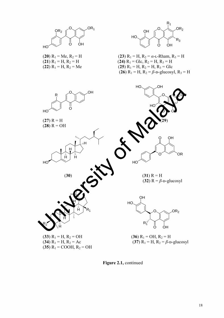
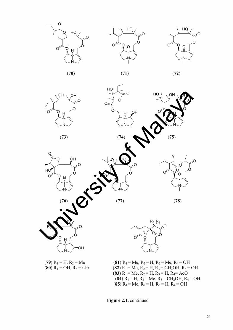
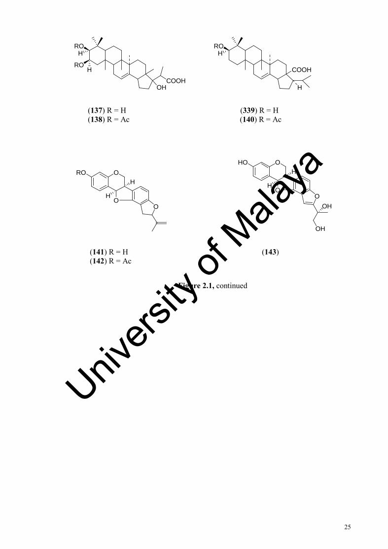
Table 2.1 lists the compounds isolated from the Crotalaria sp. and Figure 2.1
shows the structures of compounds previously isolated from Crotalaria sp.
N
HOO
O
OO
O
N
HOO
O
OOHHO
N
HOO
O
OHO
OH HO
(1) (2) (3)
N
HOO
OO
O
MeMeMe
N
HOO
OO
O
MeMeMe
N
HOO
O
OHO
HO HO
(4) (5) (6)
Figure 2.2: Structure of compounds isolated from the genus Crotalaria
Univers
ity of
Mala
ya

17
N
HOO
O
OHO
N
OH
N
OOO
O
OR1
R3 R4R2
(7) (8) (9) R1 = Me R2 = H, R3 = CH2OH, R4 = OH (10) R1 = Me R2 = H, R3 = Me, R4 = OH
(11) R1 = H, R2 = Me, R3 = OH, R4 = Me (12) R1 = H, R2 = Me, R3 = OAc, R4 = Me
HO
O
O
OHOH
O
HO
O O O
OHO (13) (14)
HO
O
O
O
OH
O
O OH
OR1
R2O (15) (16) R1 = Me, R2 = H
O
O OH
OR1
HO
R2R3
O
OHHO
OO
O (17) R1 = H, R2 = H, R3 = OMe (19) R1 = H, R2 = Me (18) R1 = Me, R2 = OMe, R3 = H
Figure 2.1, continued
Univers
ity of
Mala
ya
18
O
O OH
OR1
HO
OR2
O
O OH
OR2OH
R1
R3HO
(20) R1 = Me, R2 = H (23) R1 = H, R2 = α-ʟ-Rham, R3 = H (21) R1 = H, R2 = H (24) R1 = Glc, R2 = H, R3 = H (22) R1 = H, R2 = Me (25) R1 = H, R2 = H, R3 = Glc
(26) R1 = H, R2 = β-ᴅ-glucosyl, R3 = H
O
O
OHR
HO
O
O
OH
OH
OHHO
HO
(27) R = H (29) (28) R = OH
HO
HH
HH
O
O
OR
OH
HO
(30) (31) R = H (32) R = β-ᴅ-glucosyl
R2
H
H
H
H R1
O
O
OR2
OH
HOOH
R1
(33) R1 = H, R2 = OH (36) R1 = OH, R2 = H (34) R1 = H, R2 = Ac (37) R1 = H, R2 = β-ᴅ-glucosyl (35) R1 = COOH, R2 = OH
Figure 2.1, continued
Univers
ity of
Mala
ya

19
O O
O
HO
R
H
O O
O
O
HO
R
H
(38) R = H (40) R = H (39) R = OH (41) R = OH
O
O
OR1
OHR2O
OHHO
O
O
OH
OH
R3OR1
R2
(42) R1 = β-ᴅ-glucosyl, R2 = H (45) R1 = Glc, R2 = H, R3 = H (43) R1 = H, R2 = H (46) R1 = H, R2 = Glc, R3 = H (44) R1 = H, R2 = Gal (47) R1 = Glc, R2 = H, R3 = Xyl
OHHO
HOH
H HCOOHOHCOOH
H
H
HOOH
H HCOOHOHCOOH
H
H
(48) (49) (50)
O
O
OR2
OH
HO
R1O
O
O
OR1
R2
R3O
(51) R1 = H, R2 = H (56) R1 = H, R2 = OH, R3 = H (52) R1 = Me, R2 = H (57) R1 = Glu-apiosyl, R2 = OH, R3 = H (53) R1 = Xyl, R2 = Rham (58) R1 = Glu-apiosyl, R2 = OH, R3 = Me (54) R1 = Rham, R2 = Rham (59) R1 = Me, R2 = H, R3 = H (55) R1 = Gal-Rham, R2 = Rham
Figure 2.1, continued
Univers
ity of
Mala
ya

20
OH
OHHO O
OHHO
O
O
O
O
(60) (61)
OO
HO OH
OO
HOOHO
(62) (63)
O
O
OHHO
N
OO
O
O H
R1 R2
(64) (65) R1 = OH, R2 = H (66) R1 = H, R2 = OH
O
O
R2
OO
H
H
R1 N
OO
O
OO OH
HO
(67) R1 = O-β-ᴅ-glucopyranoside, R2 = H (68) R1 = H, R2 = O-β-ᴅ-glucopyranoside (69)
Figure 2.1, continued
Univers
ity of
Mala
ya
21
N
OO
O
O H
O HO
O
N
OO
O
O
HO
O
N
OO
O
O
HO
O
(70) (71) (72)
N
OO
O
O
OH
H
OH
N
O
O
HOO
O
OHH
N
OO
O
O
OH
H
OHHO
(73) (74) (75)
N
HOO
O
O
O
O
HO
OH
N
OOO
OO HO
N
OO
O
O O
OO
(76) (77) (78)
N
OO
O
O H
HOR1
OH
R2
N
OOO
O
R1 R2
R3R4
(79) R1 = H, R2 = Me (81) R1 = Me, R2 = H, R3 = Me, R4 = OH (80) R1 = OH, R2 = i-Pr (82) R1 = Me, R2 = H, R3 = CH2OH, R4 = OH (83) R1 = Me, R2 = H, R3 = H, R4= AcO
(84) R1 = H, R2 = Me, R3 = CH2OH, R4 = OH (85) R1 = Me, R2 = H, R3 = H, R4 = OH
Figure 2.1, continued
Univers
ity of
Mala
ya

22
N
OOO
ORHO
OHHOOH
O OH O
HO OH
OH
OH
(86) R = Me (88) (87) R = CH2OH
N
HHO
O
N
H
OH
OHHO
N
H
HO
OHHO
(89) (90) (91)
N
H R1O
R2
N
H R1HO
R2
OHO
O
O
(92) R1 = H, R2 = OAc (95) R1 = H, R2 = OH (97) (93) R1 = OH, R2 = H (96) R1 = OH, R2 = H (94) R1 = H, R2 = H
O
O
H
H
O
O
O
OH
(98) (99)
Figure 2.1, continued
Univers
ity of
Mala
ya

23
O O
HOO OH
O
HO OH
CH3HO
OHO
HOHO OH
O
HOO OH
OOH
(100) (101) (102)
N
OO
O
O
R3R2 R4R1
O
OOH
R1OR2
R3
(103) R1 = H, R2 = OH, R3 = H, R4 = H (110) R1 = H, R2 = H, R3 = H (104) R1 = H, R2 = OAc, R3 = H, R4 = H (111) R1 = H, R2 = Me, R3 = H (105) R1 = Me, R2 = OH, R3 = Me, R4 = H (112) R1 = H, R2 = H, R3 = OH (106) R1 = Me, R2 = OAc, R3 = OH, R4 = Me (113) R1 = OMe, R2 = Me, R3 = OMe (107) R1 = Me, R2 = OH, R3 = OH, R4 = Me (108) R1 = i-Pr, R2 = OH, R3 = OH, R4 = Me (109) R1 = i-Pr, R2 = OH, R3 = OH, R4 = CH2OH
N
OOO
OHO R
N
OO
O
O
OH
R
HO
H
N
OOO
R2O
HO
HO
R1
(114) R = Me (1) R = O (118) R1 = Me, R2 = H (115) R = CH2OH (2) R = CH2 (119) R1 = H, R2 = Me
Figure 2.1, continued
Univers
ity of
Mala
ya

24
N
OOO
OOH
NO
O
HO H
HO O
O
N
O HO
O
OHO
(120) (121) (122)
O OHO
OHO N
HR2R1
CH2
OHO
OO
O
(123) (124) R1 = OH, R2 = H (126) (125) R1 = H, R2 = OH
N
HO
O
OO
OH
N
O O
O
O
HO H
OH
OH
O
O
(127) (128) (129)
N
OO
O
OHR2 O
R1
R2 R3HO
R1
O
R4
OH
OH
(130) R1 = OH, R2 = H (133) R1 = OH, R2 = OH, R3 = H, R4 = H (131) R1 = OH, R2 = OH (134) R1 = OMe, R2 = H, R3 = H, R4 = H (132) R1 = H, R2 = OH (135) R1 = H, R2 = H, R3 = H, R4 = Prenyl (136) R1 = H, R2 = H, R3 = OH, R4 = Prenyl
Figure 2.1, continued
Univers
ity of
Mala
ya
25
OH
RO
ROH
H
COOH H
ROH
COOH
(137) R = H (339) R = H (138) R = Ac (140) R = Ac
O
OO
ROH
H
O
OO
HOH
H
OH
OH
(141) R = H (143) (142) R = Ac
Figure 2.1, continued
Univers
ity of
Mala
ya

26
O
OH OH
OO
OOHOH
OO
O
O
OHOH
OH
HO
H
OH
OH
O OO
OH
OH
(144)
OH
O
OHOH
O
OOH
OH
O OO
OH
HO
OH
OH
(145)
Univers
ity of
Mala
ya

27
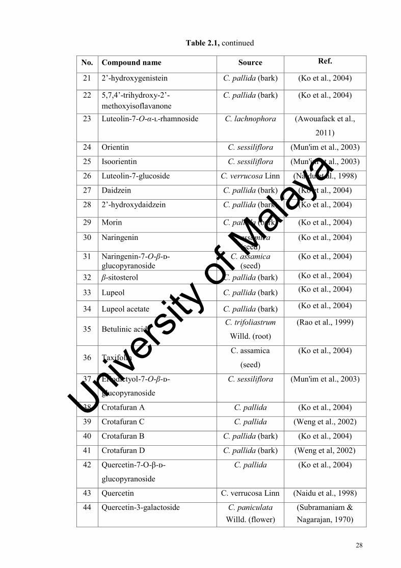
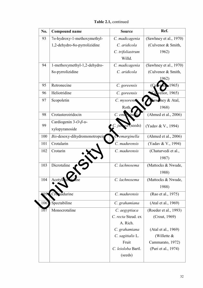
Table 2.1: Name of compounds isolated from different Crotalaria sp.
No. Compound name Source Ref.
1 Acetylsenecivernine C. gillettii (Asres et al., 2004)
2 Gynuramine C. gillettii (Asres et al., 2004)
3 Jacoline C. fascicularis (Asres et al., 2004)
4 Retroisosenine C. gillettii (Asres et al., 2004)
5 Nemorensine C. incana subsp. purpurascens
(Asres et al., 2004)
6 Scleratine C. fascicularis (Asres et al., 2004)
7 Dihydrosenecionine C. incana subsp. purpurascens
(Asres et al., 2004)
8
Tashiromine C. emarginella C. philipsiae
(Asres et al., 2004)
9 Senkirkine C. laburnifolia C. phillipsiae
(Asres et al., 2004)
10 Hydroxysenkirkine C. laburnifolia (Asres et al., 2004)
11 Crotaverrine C. verrucosa Linn (Suri et al., 1976)
12 O12-Acetylcrotaverrine C. verrucosa Linn (Suri et al., 1976)
13 Lachnoisoflavone A C. lachnophora (Awouafack et al.,
2011)
14 Lachnoisoflavone B C. lachnophora (Awouafack et al., 2011)
15 Licoagroisoflavone C. lachnophora (Awouafack et al., 2011)
16 Prunetin C. lachnophora (Awouafack et al., 2011)
17 3’-O-methylorobol C. lachnophora (Awouafack et al., 2011)
18 7-O-methyltectorigenin C. lachnophora (Awouafack et al., 2011)
19 Cajanol C. lachnophora (Awouafack et al., 2011)
20 Cajanin C. lachnophora (Awouafack et al., 2011)
Univers
ity of
Mala
ya
28
Table 2.1, continued
No. Compound name Source Ref.
21 2’-hydroxygenistein C. pallida (bark) (Ko et al., 2004)
22 5,7,4’-trihydroxy-2’-methoxyisoflavanone
C. pallida (bark) (Ko et al., 2004)
23 Luteolin-7-O-α-ʟ-rhamnoside C. lachnophora (Awouafack et al.,
2011)
24 Orientin C. sessiliflora (Mun'im et al., 2003)
25 Isoorientin C. sessiliflora (Mun'im et al., 2003)
26 Luteolin-7-glucoside C. verrucosa Linn (Naidu et al., 1998)
27 Daidzein C. pallida (bark) (Ko et al., 2004)
28 2’-hydroxydaidzein C. pallida (bark) (Ko et al., 2004)
29 Morin C. pallida (bark) (Ko et al., 2004)
30 Naringenin C. assamica (seed)
(Ko et al., 2004)
31 Naringenin-7-O-β-ᴅ-glucopyranoside
C. assamica (seed)
(Ko et al., 2004)
32 β-sitosterol C. pallida (bark) (Ko et al., 2004)
33 Lupeol C. pallida (bark) (Ko et al., 2004)
34 Lupeol acetate C. pallida (bark) (Ko et al., 2004)
35 Betulinic acid C. trifoliastrum
Willd. (root)
(Rao et al., 1999)
36 Taxifolin C. assamica
(seed)
(Ko et al., 2004)
37 Eriodictyol-7-O-β-ᴅ-
glucopyranoside
C. sessiliflora (Mun'im et al., 2003)
38 Crotafuran A C. pallida (Ko et al., 2004)
39 Crotafuran C C. pallida (Weng et al., 2002)
40 Crotafuran B C. pallida (bark) (Ko et al., 2004)
41 Crotafuran D C. pallida (bark) (Weng et al, 2002)
42 Quercetin-7-O-β-ᴅ-
glucopyranoside
C. pallida (Ko et al., 2004)
43 Quercetin C. verrucosa Linn (Naidu et al., 1998)
44 Quercetin-3-galactoside C. paniculata Willd. (flower)
(Subramaniam & Nagarajan, 1970)
Univers
ity of
Mala
ya

29
Table 2.1, continued
No. Compound name Source Ref.
45 Vitexin C. sessiliflora (Mun'im et al., 2003)
46 Isovitexin C. sessiliflora (Yoo et al., 2004)
47 Vitexin-4’-O-xyloside C. striata DC (Subramaniam & Nagarajan, 1970)
48 Hydroquinone C. sessiliflora (Mun'im et al., 2003)
49 Eucomic acid C. sessiliflora (Mun'im et al., 2003)
50 Hydroxyeucomic acid C. sessiliflora (Mun'im et al., 2003)
51 Kaempferol C. verrucosa Linn (Naidu et al., 1998)
52 4’,5,7-trihydroxy-3-methoxyflavone
C. madurensis (Bhakuni & Chaturvedi, (1984)
53 Lepidoside C. semperflorens
Vent.
(Dhasmana & Garg,
1991)
54 Kaempferitin C. semperflorens
Vent.
(Dhasmana & Garg,
1991)
55 Robinin C. zanziriba
(C. usaramoensis)
(Shitamoto et al.,
2010)
56 Apigenin C. pallida (bark) (Subramaniam & Nagarajan, 1970)
57 Apigenin-7-O-β-ᴅ-apiofuranosyl-(16)-glucopyranoside
C. podocarpa (Wanjala & Majinda, 1999)
58 Acacetin-7- O-β-ᴅ-apiofuranosyl-(16)-glucopyranoside
C. podocarpa (Wanjala & Majinda, 1999)
59 4’,7-dihydroxyflavone C. sessiliflora (Yoo et al., 2004)
60 1-O-methyl-myo-inositol C. trifoliastrum
(root)
(Rao et al., 1999)
61 Methoxystilbene C. madurensis (Bhakuni &
Chaturvedi, (1984)
62 Crotmadine C. madurensis (Bhakuni &
Chaturvedi, (1984)
63 Dihydroxyalpinumisoflavone C. madurensis (Bhakuni & Chaturvedi, (1984)
64
Crotmarine C. madurensis (Bhakuni & Chaturvedi, (1984)
Univers
ity of
Mala
ya
30
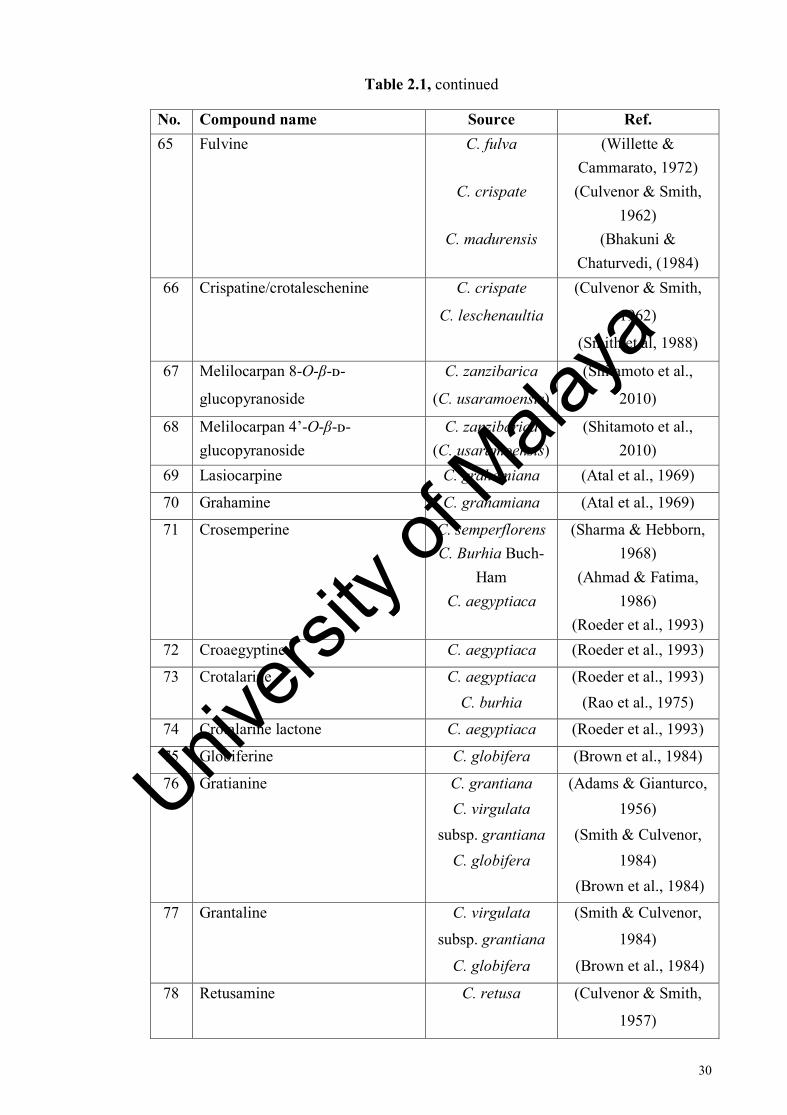
Table 2.1, continued
No. Compound name Source Ref. 65 Fulvine C. fulva
C. crispate
C. madurensis
(Willette & Cammarato, 1972) (Culvenor & Smith,
1962) (Bhakuni &
Chaturvedi, (1984) 66 Crispatine/crotaleschenine C. crispate
C. leschenaultia
(Culvenor & Smith,
1962)
(Smith et al, 1988)
67 Melilocarpan 8-O-β-ᴅ-
glucopyranoside
C. zanzibarica
(C. usaramoensis)
(Shitamoto et al.,
2010)
68 Melilocarpan 4’-O-β-ᴅ-glucopyranoside
C. zanzibarica (C. usaramoensis)
(Shitamoto et al., 2010)
69 Lasiocarpine C. grahamiana (Atal et al., 1969)
70 Grahamine C. grahamiana (Atal et al., 1969)
71 Crosemperine C. semperflorens
C. Burhia Buch-Ham
C. aegyptiaca
(Sharma & Hebborn, 1968)
(Ahmad & Fatima, 1986)
(Roeder et al., 1993) 72 Croaegyptine C. aegyptiaca (Roeder et al., 1993)
73 Crotalarine C. aegyptiaca
C. burhia (Roeder et al., 1993)
(Rao et al., 1975)
74 Crotalarine lactone C. aegyptiaca (Roeder et al., 1993)
75 Globiferine C. globifera (Brown et al., 1984)
76 Gratianine C. grantiana
C. virgulata subsp. grantiana
C. globifera
(Adams & Gianturco, 1956)
(Smith & Culvenor, 1984)
(Brown et al., 1984)
77 Grantaline C. virgulata
subsp. grantiana
C. globifera
(Smith & Culvenor,
1984)
(Brown et al., 1984)
78 Retusamine C. retusa (Culvenor & Smith,
1957)
Univers
ity of
Mala
ya

31
Table 2.1, continued
No. Compound name Source Ref.
79 Retusine C. retusa (Culvenor & Smith,
1957)
80 Croalbidine C. albida (Sawhney & Atal,
1973)
81 Integerrimine C. incana Linn (seed)
C. tetragona
C. narahutensis
(Sawhney & Atal, 1970)
(Puri et al., 1974) (Mattocks & Nwude,
1988) 82 Usaramine C. mucronata
Desv.
C. usaramoensis E. G. Baker
C. incana Linn
C naragutensis
(Atal & Sawhney, 1968)
(Culvenor & Smith, 1966)
(Sawhney & Atal, 1970)
(Mattocks & Nwude, 1988)
83 Crotastriatine C. striata (Gandhi et al., 1968)
84 Mucronatinine C. mucronata (Bhacca & Sharma,
1968)
85 Nilgrine C. mucronata (Atal & Sawhney,
1968)
86 Senecionine C. usaramoensis (Culvenor & Smith,
1966)
87 Retrorsine C. usaramoensis (Culvenor & Smith,
1966)
88 Coatline A isomer (α-S-hydroxyl) C. zanzibarica (C. usaramoensis)
(Shitamoto et al., 2010)
89 1β,2β-epoxy-1α-hydroxymethyl-8α-pyrrolizidine
C. trifoliastrum (Culvenor et al., 1967)
90 Crotanecine Crotalaria sp. (Atal & Kapur, 1966)
91 Croalbinecine C. albida Heyne
ex Roth.
(Sawhney et al., 1974)
92 7β-acetoxyy-1-methoxymethyl-1,2-dehydro-8α-pyrrolizidine
C. aridicola (Culvenor et al., 1967)
Univers
ity of
Mala
ya
32
Table 2.1, continued
No. Compound name Source Ref.
93 7α-hydroxy-1-methoxymethyl-1,2-dehydro-8α-pyrrolizidine
C. madicagenia
C. aridicola C. trifoliastrum
Willd.
(Sawhney et al., 1970) (Culvenor & Smith,
1962)
94 1-methoxymethyl-1,2-dehydro-8α-pyrrolizidine
C. madicagenia
C. aridicola (Sawhney et al., 1970)
(Culvenor & Smith, 1962)
95 Retronecine C. goreensis (Culvenor, 1965)
96 Heliotridine C. goreensis (Culvenor, 1965)
97 Scopoletin C. mysorensis
Roth.
(Sawhney & Atal,
1968)
98 Crotasteroiridocin C. emarginella (Ahmed et al., 2006)
99 Cardiogenin 3-O-β-ᴅ-
xylopyranoside C. juncea (seeds) (Yadav & V., 1994)
100 Bis-desoxy-dihydromonotropein C. emarginella (Ahmed et al., 2006)
101 Crotalarin C. madurensis (Yadav & V., 1994)
102 Crotarin C. madurensis (Chaturvedi et al.,
1987)
103 Dicrotaline C. lachnosema (Mattocks & Nwude,
1988)
104 Acetyldicrotaline C. lachnosema (Mattocks & Nwude,
1988)
105 Cromadurine C. madurensis (Rao et al., 1975)
106 Spectabiline C. grahamiana (Atal et al., 1969)
107 Monocrotaline C. aegyptiaca
C. recta Steud. ex A. Rich.
C. grahamiana
C. sagittalis L. Fruit
C. leioloba Bartl. (seeds)
(Roeder et al., 1993) (Crout, 1969)
(Atal et al., 1969)
(Willette & Cammarato, 1972) (Puri et al., 1974)
Univers
ity of
Mala
ya

33
Table 2.1, continued
No. Compound name Source Ref. 108 Trichodesmine C. rubiginosa
Willd.
C. recta Steud. ex A. Rich.
C. tetragona Roxb.
C. globifera
(Atal et al., 1966)
(Crout, 1969)
(Puri et al., 1974)
(Brown et al., 1984)
109 Junceine C. juncea (seed)
C. rubiginosa Willd.
(Adams & Gianturco, 1956)
(Atal et al., 1966)
110 Crotaramosmin C. ramosissima (Kumar et al., 1999)
111 Crotaramin C. ramosissima (Kumar et al., 1999) 112 Crotin C. ramosissima (Kumar et al., 1999)
113 Trimethoxychalcone C. ramosissima (Rao & Narukulla,
2007)
114 Seneciphylline C. juncea (Adams & Gianturco,
1956)
115 Ridelliine C. juncea (Adams & Gianturco, 1956)
116 Neocroalbidinone C. albida (Sun et al., 2013)
117 Neocroalbidine C. albida (Sun et al., 2013)
118 Anacrotine (crotalaburnine) C. incana
C. laburnifolia (seeds)
(Mattocks, 1968) (Sawhney & Atal,
1971) 119 Trans-anacrotine C. capensis (Verdon & van Wyk,
1992)
120 Crotananine C. nana (Siddiqi et al., 1978)
121 Madurensine C. agatiflora subsp. agatiflora
Schweinf
(Roux et al., 2011)
122
Doronenine C. agatiflora subsp. agatiflora
Schweinf
(Roux et al., 2011)
Univers
ity of
Mala
ya

34
Table 2.1, continued
No. Compound name Source Ref. 123 2-methyl-3-(2-oxo-[5H]-5-
hydroxymethyl-5-methylfuran-3-yl)-propanoic acid
C. verrucosa (Suri et al., 1989)
124 7β-hydroxy-1-methylene-8α-pyrrolizidine
C. goreensis (Culvenor & Smith, 1961)
125 7β-hydroxy-1-methylene-8β-pyrrolizidine
C. goreensis
C. maypurensis H. B. & K.
(Culvenor & Smith, 1961)
(Culvenor et al., 1968) 126 Crotaoprostrin C. prostrate (Krohn et al., 2002)
127 Cronaburmine C. nana (Siddiqi et al., 1978)
128 Assamicadine C. assamica (Cheng et al., 1989)
129 Munchiwarin C. medicagenia (Narender et al., 2005)
130 Axillaridine C. scassellatii
(Seed)
(Wiedenfeld et al.,
1985)
131 Axillarine C. scassellatii
(Seed)
(Wiedenfeld et al.,
1985)
132 Desoxyaxillarine C. scassellatii
(Seed)
(Wiedenfeld et al.,
1985)
133 Ramosismin C. ramosissima (Khalilullah et al.,
1993)
134 Crotaorixin C. orixensis
(aerial)
(Narender et al., 2005)
135 Medicagenin C. medicagenia
DC (root)
(Narender et al., 2005)
136 3’,5’-di-C-prenyl-2,4’,4-trihydroxy chalcone
C. medicagenia (root)
(Rao et al., 1987)
137 Emarginellic acid C. emarginella (Ahmed et al., 2006)
138 Emarginellic acetate C. emarginella (Ahmed et al., 2006)
139 Crotalic acid C. emarginella (Ahmed et al., 2006)
140 Crotalic acetate C. emarginella (Ahmed et al., 2006) 141 Barbacarpan C. barbata
(aerial)
(Babu et al., 1998)
142
Barbacarpan acetate
C. barbata
(aerial)
(Babu et al., 1998)
Univers
ity of
Mala
ya

35
Table 2.1, continued
No. Compound name Source Ref.
143 Crotafuran E C. pallida (bark) (Weng et al., 2003)
144
3-O-α-ʟ-rhamnopyranosyl(12)[β-ᴅ-glucopyranosyl(16)]-β-ᴅ-galactopyranosyl(12)-6-O-methyl-β-ᴅ-glucuronopyranosyl soyasapogenol B
C. albida
(Ding et al., 1991)
145
3-O-β-ᴅ-xylopyranosyl(12)-β-ᴅ-galactopyranosyl(12)-6-O-methyl-β-ᴅ-glucuronopyranosyl sophoradiol
C. albida
(Ding et al., 1991)
Univers
ity of
Mala
ya
36
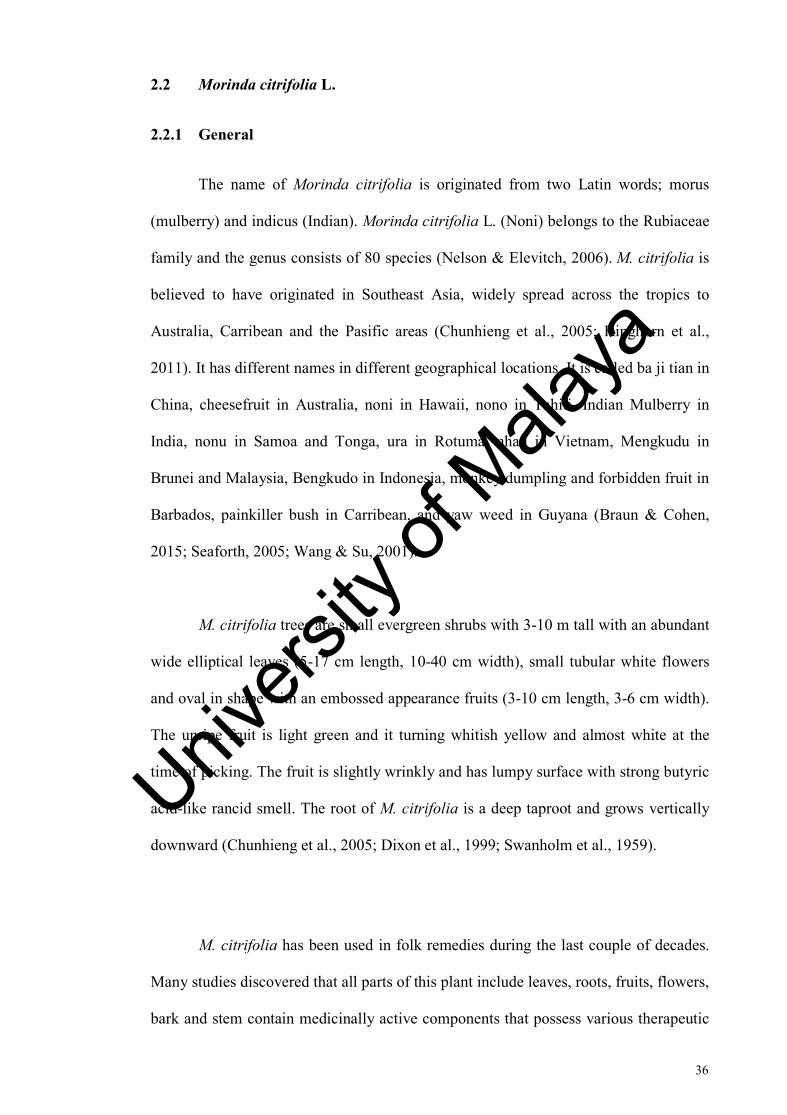
2.2 Morinda citrifolia L.
2.2.1 General
The name of Morinda citrifolia is originated from two Latin words; morus
(mulberry) and indicus (Indian). Morinda citrifolia L. (Noni) belongs to the Rubiaceae
family and the genus consists of 80 species (Nelson & Elevitch, 2006). M. citrifolia is
believed to have originated in Southeast Asia, widely spread across the tropics to
Australia, Carribean and the Pasific areas (Chunhieng et al., 2005; Kinghorn et al.,
2011). It has different names in different geographical locations. It is called ba ji tian in
China, cheesefruit in Australia, noni in Hawaii, nono in Tahiti, Indian Mulberry in
India, nonu in Samoa and Tonga, ura in Rotuma, nhau in Vietnam, Mengkudu in
Brunei and Malaysia, Bengkudo in Indonesia, monkey dumpling and forbidden fruit in
Barbados, painkiller bush in Carribean, and yaw weed in Guyana (Braun & Cohen,
2015; Seaforth, 2005; Wang & Su, 2001).
M. citrifolia trees are small evergreen shrubs with 3-10 m tall with an abundant
wide elliptical leaves (5-17 cm length, 10-40 cm width), small tubular white flowers
and oval in shape with an embossed appearance fruits (3-10 cm length, 3-6 cm width).
The unripe fruit is light green and it turning whitish yellow and almost white at the
time of picking. The fruit is slightly wrinkly and has lumpy surface with strong butyric
acid-like rancid smell. The root of M. citrifolia is a deep taproot and grows vertically
downward (Chunhieng et al., 2005; Dixon et al., 1999; Swanholm et al., 1959).
M. citrifolia has been used in folk remedies during the last couple of decades.
Many studies discovered that all parts of this plant include leaves, roots, fruits, flowers,
bark and stem contain medicinally active components that possess various therapeutic
Univers
ity of
Mala
ya

37
properties (Chan-Blanco et al., 2006; Zin & Abdul-Hamid, 2002). M. citrifolia is also
popular as a source of red, yellow, and purple dyes (Singh, 2012). In mid-1950s, M.
citrifolia is more popular as a dye. In Polynesia, they get the yellow dye from the trunk
bark, while a red dye was made by mixing the root of M. citrifolia with lime that
derived from coral. Javanese people employed the root of M. citrifolia for dyeing batik,
Australian Aborigines for dyeing cotton and wool, Indians for dyeing yarn, carpets and
turban, and Polynesians for dyeing kapa (bark cloth) (Chan-Blanco et al., 2006).
Various publications have shown that M. citrifolia can be consumed to treat,
relieve and as an alternative medicines for different kind of illnesses. These include
hiccough, hoarseness, gingivitis, gastric ulcers, sprains, headaches, atheroselerosis,
blood vessel problems, heartburn, arthritis, antiemetic, diarrhea, infertility, postpartum
haemorrhage, cancer, vaginal bleeding, menorrhagia, mental depression, poor
digestion, secondary amenorrhoea, high blood pressure, menstrual difficulties, muscle
ache and pain, uterine haemorrhage, coryza, neuralgia, oedema, carcinomas,
induration, pain of breast, heart trouble, ostcodynia and many other ailments
(Bushnell, 1993; Dittmar, 1993; Dixon et al., 1999).
In India, the leaves of this plant is make into juice and applied externally for
gout, and used internally as a tonic and to treat fever. The leaves are also used to apply
wounds and ulcers for treatment purpose. For the throat and gum complaints, dysentery
and leucorrhoea, they used the fruits of M. citrifolia as treatment (Jain & DeFilipps,
1991; Kamboj, 1988; Moorthy & Reddy, 1970; Morton, 1992; Ross, 2001). While the
root of M. citrifolia is used for cathartic and febrifuge (Hu, 2005; Li, 2002).
Univers
ity of
Mala
ya

38
In New Guinea the leaves are used to relieve headaches, treat sores of leprosy
and sometimes it was taken internally for stomach ache (Cambie & Brewis, 1997;
Weiner, 1976). They also used the root of M. citrifolia to make a juice and consume
internally for fevers and skin disorders (Hu, 2005; Li, 2002). People in Guyana
macerated the leaves of M. citrifolia alone or mixed with Pothomorphe peltata, in
coconut oil for an external rub to relieve rheumatic pains and arthritic, also chewed the
fruits to heal mouth ulceration (DeFilipps et al., 2004).
In Hawaii, leaves and bark are pounded, cooked, strained and used as a tonic,
take orally as abortifacient. The fruits are taken orally for asthma, insecticide for hair,
and used as a poultice to heal broken bones and deep cuts and bruises. The immature
fruit juice are taken internally to treat diabetes, menstrual cramp, hypertension,
digestive disorders, and as a general tonic. While the mash green fruit is used for skin
condition, and the rotten-ripe fruit is used for lassitude of old age (Degener, 1973;
Tabrah & Eveleth, 1966; Wagner et al., 1990).
In West Indies, the leaves are used as a poultice to wrap around the rheumatic
joints and treatment for pain while the fruits are heated to treat sores of inflammation
(Ayensu, 1981). While in Fiji, the leaves were consumed with leaves of Epipremnum
pinnatum to relieve pregnancy pains or with Psychotria sp. to heal haemorrhoids.
People in Australia used the roots of M. citrifolia to relieve fever, toothache,
inflammation, malaria, skin disorder, jaundice, sore throat, diarrhea and dysentery. The
root was grated to use as treatment to heal the stings from insects and stonefish. The
roots of M. citrifolia are also used with Euodia hortensis and Geniostoma vitiense to
treat malnutrition (Pawlus & Kinghorn, 2007). In China, the roots of M. citrifolia are
believed to be able to treat cancer, beri-beri, cholecystitis, lumbago, stimulate the
Univers
ity of
Mala
ya

39
endocrine system and increase the leucocyte count. The whole plant extract is taken to
relieve aching bones (Hu, 2005; Li, 2002).
The main group of active compounds in this plant is the anthraquinones, which
possess various therapeutic properties including antiviral, antibacterial, antioxidant,
anticancer, antitumor, anti-tubercular effects, and anti-inflammatory (Singh, 2012).
Damnacanthal (287), a component from M. citrifolia fruit was identified as an inhibitor
of Viral protein R (Vpr) induced cell death. But the mechanism of damnachantal
inhibits Vpr induced apoptosis is still under the research. Damnacanthal (287) was also
reported as a unique anthraquinones with anti-cancer and anti-HIV activity (Hiramitsu
et al., 1993). In the other hand, 1-methoxy-2-formyl-3-hydroxy anthraquinone obtained
from the roots suppressed the cytopathic effect of HIV infected MT-4 cells, without
inhibiting cell growth (Umezawa, 1992).
The presence of phenolic compounds such as L-asperuloside (203), acubin
(309) and alizarin (301) in M. citrifolia fruits, and the anthraquinones in the roots, are
accounted for the antimicrobial activities in this plant. All these compounds have been
shown to have activity against infectious bacteria strains such as Pseudomonas
aeruginosa, Shigela sp., Staphylococcus aureus, Escherichia coli, Proteus morgaii,
Bacillus subtilis, and Salmonella sp. These antibacterial compounds are effective for
skin infection treatment, fever, colds, and other bacterial-caused health problems
(Atkinson, 1956). The acetonitrile extracts of the dried fruits inhibited the growth of
Escherichia coli, Bacillus subtilis, Pseudomonas aeruginosa, and Streptococcus
pyrogene (Locher et al., 1995). It is also helps in stomach ulcer through inhibition of
the bacteria, H. pylori (Duncan et al., 1998). Lee and colleagues reported the methanol
and aqueous crude of fruits extract inhibited the growth of Vibrio harveyi, Vibrio
Univers
ity of
Mala
ya

40
alginolyticus, Streptoccus sp. and E. coli. They also found that hexane and ethanol
extract of the fruits are able to provide protection against Mycobacterium tuberculosiss
(Lee at al., 2008). In another studies, Murray et. al reported that the antibacterial effect
of this plant is greater when the fruit is ripe.
Ethyl acetate extract of the fruits from this plant has been reported to exhibit
higher antioxidative activity and this lead to the isolation of three antioxidant phenolic
compounds; aesculetin (308), 3, 3’,4’,5, 7-pentahydroxyflavone or (quercetin) (271)
and isoscopoletin (307) by Chang and his colleagues (Chang-hong et al., 2007). Other
researchers have been reporting that the seed extract from this plant exhibit significant
antioxidant activity in the Ferric Reducing Antioxidant Power (FRAP) and Oxygen
Radical Absorbance Capacity (ORAC) (Brett et al., 2011).
The methanol extract of fruits were reported to exhibit cytotoxic activity
against neuroblastoma (LAN5) cell lines and breast cancer (MCF7) in 3-(4,5-
dimethythiazol-. 2-yl)-2,5-diphenyl tetrazolium bromide (MTT) assay (Arpornsuwan
& Punjanon, 2006). The ethanol precipitate of M. citrifolia juice possess antitumor and
immunomodulatory activity against Sarcoma 180 ascites tumor in mice (Furusawa et
al., 2003). In 1993, Hiramitsu and his colleagues found that damnacanthal (287) is a
new inhibitor of ras function and thus suppress ras-expressing tumors (Hiramitsu et
al., 1993). This anthraquinone also showed potent inhibitory activity towards tyrosine
kinase such as Src, Lyn, Lck and EGF receptor (Bowie & Cooke, 1962). Other studies
has reported two novel glycosides; 6-O-(β-D-glucopyranosyl)-1-O-octanoyl-β-D-
glucopyranose (167) and asperulosidic acid (305) that obtained from isolation of the
fruits extract showed effectiveness in suppressing 12-O-tedtradecanoylphorbol-13-
acetate (TPA) or epidermal growth factor (EGF), thereby inducing the cell
Univers
ity of
Mala
ya

41
transformation and associated AP-1 activity (Guangming et al., 2001). Moon and
colleagues have reported that scopoletin (242) has a regulatory effect in the
inflammatory reactions mediated by the human mast cell line HMC-1. Scopoletin
(242) is a phenolic coumarin that is medicinally active and showed positive potential
as antitumor against skin cancer, antihyperlipidemic, antiulcer, antiviral and
antimicrobial (Moon et al., 2007; Nam & Kim, 2015).
Researchers from Philippines reported that M. citrifolia has been found to kill
Mycobacterium tuberculosis. It was also found that the extract of M. citrifolia leaves
have an antitubercular effect which inhibits the growth of bacteria in a test tube. Ralph
Heinicke state that the fruits of M. citirfolia contain a natural precursor for Xeronine,
named Proxeroninase (Heinicke, 1985). Proxeronine is converted to the alkaloid in the
body by enzymes, Proxeroninase. He claims that Xeronine has a capasity to modify the
molecular structure of protein. Thus, Xeronine has a wide range of biological activities
especially for a problem in the cell due to protein structural (Heinicke, 2001).
Approximately 200 phytochemicals have been isolated and identified from
different parts of M. citrifolia and different location all over the world. Anthraquinones
are the major phenolic compounds that have been isolated from this plant (Bowie &
Cooke, 1962).
Univers
ity of
Mala
ya

42
2.2.2 Compounds isolated from Morinda citrifolia
Figure 2.2 shows the structures of compounds previously isolated from M.
citrifolia. Table 2.2 lists the compounds in Figure 2.2, which were separated from
different parts of the plant.
OH
OHO
O O
O
O
OH
(146) (147)
OH
OH
HO
OO OH
O
OHO
O OHO
O
HO HO
(148) (149)
O
O
HO R1
OH O
O
R2R1
OH
(150) R1 = OH (152) R1 = OH, R2 = OH (MC4) R1 = H (153) R1 = H, R2 = H
O
HO
HO
O
O
O
OH
O
O
HO
H
HO
H
H
O
OH
(154) (155)
Figure 2.2: Structure of compounds isolated from Morinda citrifolia
Univers
ity of
Mala
ya

43
OH
HO
R1
R3
R2
O
O
R2
R1
R3O
O
OH
O
(156) R1 = H, R2 = OCH3, R3 = OH (158) R1 = CH3, R2 = O, R3 = H (157) R1 = OCH3, R2 = H, R3 = H (159) R1 = CH2OCH3, R2 = H, R3 = OCH3
OO
O
OH
HOOH
O
HO
HOH
OH
R
O
OH
OO
O
OH
HOOH
O
HO
HOH
O
R
O
OHO
OH
OH
OH
OH
(160) R = C5H11 (162) R = C5H11 (161) R = C7H15 (163) R = C7H15
O
OHOH
HO
OHO
O
OHOH
HO
OO
OHHO
HOO
HO
(164) (165)
O
OHOH
HO
OO
OHHO
HOO
HO
R
O
H
HO
OH
O
HOR H
H
HO
(166) R = C5H11 (168) R = CH3 (167) R = C7H15 (169) R = CH2OH
Figure 2.2, continued
Univers
ity of
Mala
ya

44
O O
O
OO
H
O
O
O O
O
OO
O
O
O
O
O
HOH
H
OH
OH
HO
HO
O O
(170) (171) (172)
O O
OHO
OH
HO
HO OH
HO
H
H
HOH
O
(173) (174)
R2R3
R1
H
OH
OHH H
HO O O
H
HO
H
H
OH
O O
(175) R1 = CH3, R2 = H, R3 = H (177) (176) R1 = H, R2 = OH, R3 = CH3
Figure 2.2, continued
Univers
ity of
Mala
ya
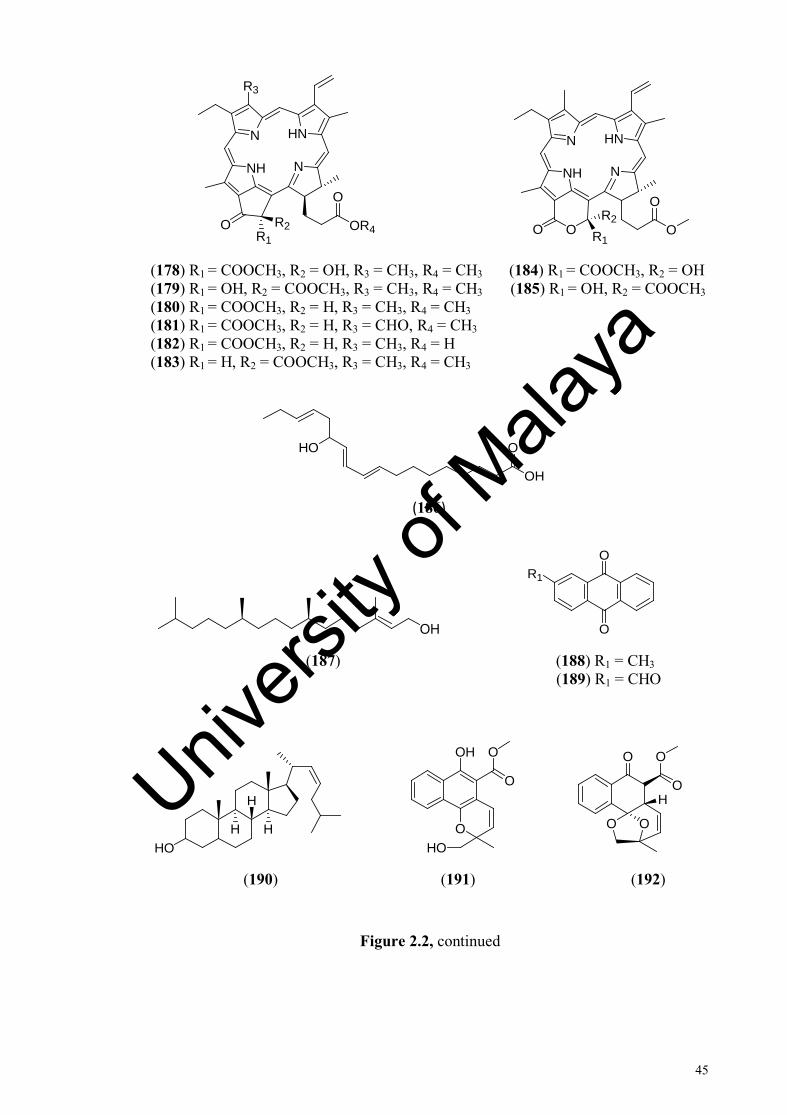
45
N
NH
O
N
HN
R3
OR4OR1
R2
N
NH
O
O
N
HN
OO R1
R2
(178) R1 = COOCH3, R2 = OH, R3 = CH3, R4 = CH3 (184) R1 = COOCH3, R2 = OH (179) R1 = OH, R2 = COOCH3, R3 = CH3, R4 = CH3 (185) R1 = OH, R2 = COOCH3 (180) R1 = COOCH3, R2 = H, R3 = CH3, R4 = CH3 (181) R1 = COOCH3, R2 = H, R3 = CHO, R4 = CH3 (182) R1 = COOCH3, R2 = H, R3 = CH3, R4 = H (183) R1 = H, R2 = COOCH3, R3 = CH3, R4 = CH3
O
OH
HO
(186)
OH
R1
O
O
(187) (188) R1 = CH3 (189) R1 = CHO
HOH
H
H
O
OH O
HO
O
O
O
O
O O
H
(190) (191) (192)
Figure 2.2, continued
Univers
ity of
Mala
ya
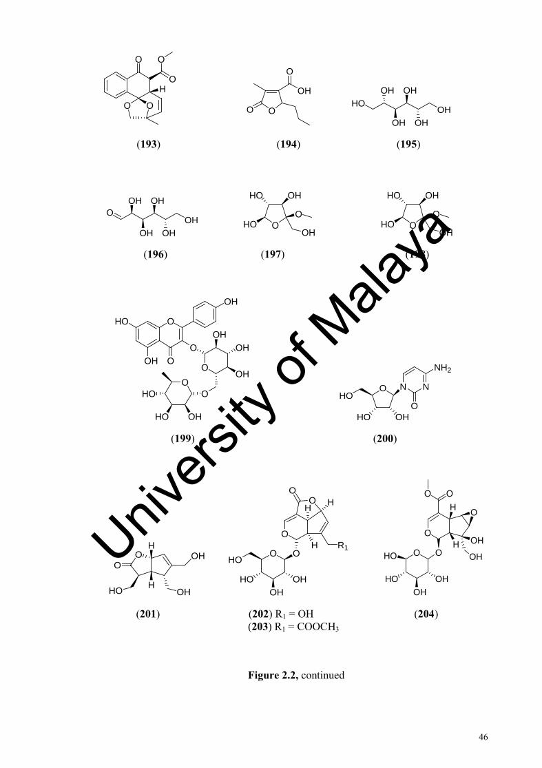
46
O
O
O
O O
H
OO
OH
O
OH
OH
OHHO
OH
OH
(193) (194) (195)
OH
OH
OHO
OH
OH OHO
OH
O
HO
OH OHO
OH
O
HO
OH
(196) (197) (198)
O
O
HO
OH
OH
OO
OO
HO OH
HO
OH
OH
OH
O N
HO OH
HON
O
NH2
(199) (200)
O OH
OHHO
O
H
H
O O
O
O
OHHO OH
HOR1
O
H
H
H
O O
O
OOH
H OH
O
OHHO
OHHO OH
(201) (202) R1 = OH (204) (203) R1 = COOCH3
Figure 2.2, continued
Univers
ity of
Mala
ya
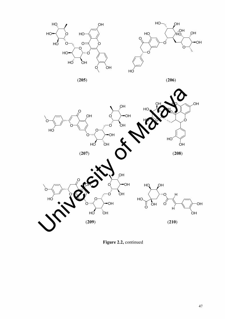
47
O
O
OH
HO
OHO
OO
OH
HO
HO
O
OHO
HO
HO
OO
O
HO
OHHOOH
OH
OH
HOO
HO
(205) (206)
O
OO
OHO
HO OO
O
HO
OH
OH
OH
OH
OH
OH
O
OH
HO
O
HO
O
OHOH
HO
HO
(207) (208)
O
OO
OHO
HO OO
O
HO
OH
OH
OH
OH
OH
HO
OH
HO
OOH
OHOH
OHHO
(209) (210)
Figure 2.2, continued
Univers
ity of
Mala
ya

48
O
O
OH
OH
HO
HO
HO
O
OO
OHO
OH
OH
OHO
OH
O
OH
OH
OH
(211) (212)
O
OHR4
OH
OHR1
R2
R3
O
O
OH
R2
O
R1
R3
(213) R1 = OH, R2 = OH, R3 = OH, R4 = H (216) R1 = H, R2 = H, R3 = OH (214) R1 = H, R2 = OH, R3 = H, R4 = OH (217) R1 = OCH3, R2 = H, R3 = OH (215) R1 = OCH3, R2 = OH, R3 = H, R4 = H (218) R1 = H, R2 = OH, R3 = OH
R1R2
R3R4
OH
O
R3
R2R1
OH
OH
H
(219) R1 = OH, R2 = OH, R3 = OH, R4 = H (222) R1 = H, R2 = OH, R3 = H (220) R1 = OH, R2 = H, R3 = H, R4 = OH (223) R1 = H, R2 = OH, R3 = OCH3 (221) R1 = H, R2 = OH, R3 = OCH3, R4 = H (224) R1 = OCH3, R2 = OH, R3 = OCH3
OH
OH
O
O
HO
R1 OH
OH
O
O
R2
R1
(225) R1 = H (227) R1 = OH, R2 = OH (226) R1 = OH (228) R1 = OH, R2 = OCH3
Figure 2.2, continued
Univers
ity of
Mala
ya

49
OH
OH
OHO
HO
HO
HO
O
OH
OH
O
O
OO
(229) (230) (231)
HOO
O
OHO OH
OH
OHO
HO
HO
O
HO
H
O
O
(232) (233) (234)
OHH
H
H
OHH
H
HH
(235) (236)
OHH
H
H
O OH
OH
OHOH
OHH
H
H
(237) (238)
OOOH
HO OHOH
OOO
HO OHOH
O
OHOH
OH
OH
(239) (240)
Figure 2.2, continued
Univers
ity of
Mala
ya

50
O
HO
O
O
O
HO O
HO
OHO (CH)14
OCH3
(241) (242) (243)
O
OR2
OOH
R1
O
OO
R2
R1
O
(244) R1 = OH, R2 = OH (246) R1 = OH, R2 = H (245) R1 = H, R2 = H (247) R1 = H, R2 = OH
OO
OO
H O
O O
OHO
H
HOOH
OH
(248) (249)
O
O
HO
HO
OH
OH
O
OHO
HO
OH
OH
HO HO
(250) (251)
OO
HO
O
OHO
O
HO
O
O
OH
HO
OH
OH
HO
(252) (253)
Figure 2.2, continued
Univers
ity of
Mala
ya

51
O OH
OH
O
O HO
OH
O
(254) (255)
R2
OOH
OR1
R3OH
O
OH
OO
OO
HO OH
(256) R1 = OH, R2 = CH2OH, R3 = OH (259) (257) R1 = OH, R2 = CH3, R3 = OH (258) R1 = H, R2 = CH3, R3 = OH
O
HO
O
OH
HOOH
O OO
OOH
OHHO
OHHO
OH
O
HOO
O
O
OO
O
O
OH
O
OH
O
O
H
H
HO
(260) (261)
O O
O O
O
OHO
HOOH
HO
O
O OH
O
O
OHOH
OH
OH
O
(262) (263)
Figure 2.2, continued
Univers
ity of
Mala
ya
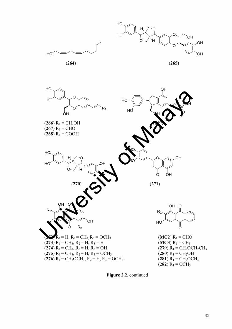
52
HO
O
O
O
O
OH
OH
OH
HO
HOH
H
(264) (265)
O
O
HO
HO
OHR1
HO
HO
OH
OH
OOH
(266) R1 = CH2OH (269) (267) R1 = CHO (268) R1 = COOH
O
O
OH
OH
HO
HOH
H
O
O OH
HO
OHHO
(270) (271)
R1
OOH
OOH
R2R3
R1
O
OOH
HO
(272) R1 = H, R2 = CH3, R3 = OCH3 (MC2) R1 = CHO (273) R1 = CH3, R2 = H, R3 = H (MC3) R1 = CH3 (274) R1 = CH3, R2 = H, R3 = OH (279) R1 = CH2OCH2CH3 (275) R1 = CH3, R2 = H, R3 = OCH3 (280) R1 = CH2OH (276) R1 = CH2OCH3, R2 = H, R3 = OCH3 (281) R1 = CH2OCH3 (282) R1 = OCH3
Figure 2.2, continued
Univers
ity of
Mala
ya

53
R2
O
OR1
R3
R1
O
OO
HO
(283) R1 = OCH3, R2 = CH2O-primeverose, R3 = O- (MC1) R1 = CHO
(284) R1 = OH, R2 = CH2O-primeverose, R3 = O- (288) R1 = CH2-primeverose (285) R1 = CH3, R2 = H, R3 = OH (289) R1 = CH3 (286) R1 = OCH3, R2 = CH2OCH3, R3 = OCH3 (290) R1 = CH2OH (291) R1 = CH2OCH3
R1
O
OO
R2
R1
O
OOH
R2
(292) R1 = CH2OH, R2 = O-primeverose (297) R1 = CH2OH, R2 = O-primeverose (293) R1 = CH2O-gentiobiose, R2 = H (298) R1 = CH2O-primeverose, R2 = H (MC6) R1 = OH, R2 = OH (MC5) R1 = H, R2 = OCH3 (295) R1 = H, R2 = OH (300) R1 = CHO, R2 = H (296) R1 = OH, R2 = H (301) R1 = OH, R2 = H
O
OOHR3
R2R1
OO
O
OHOH
HO
HO
HO
HR1O
O
OR2
(302) R1 = OH, R2 = O-primeverose, R3 = H (304) R1 = H, R2 = H (303) R1 = OCH3, R2 = OCH3, R3 = O-primeverose (305) R1 = H, R2 = COCH3 (306) R1 = CH3, R2 = H
O OR
OH
O
OO
OHHO
OH
OH
HO
HH
HOOH
(307) R = CH3 (309) (308) R = H
Figure 2.2, continued
Univers
ity of
Mala
ya

54
R2
O
OR1
R3
O
OO
O
OHOH
OHO
OHHO
HO
OH
OHH
OH
OR2
R1O
(310) R1 = OCH3, R2 = OCH3, R3 = OH (314) R1 = C5H11, R2 = H (311) R1 = OCH3, R2 = OH, R3 = OCH3 (315) R1 = C7H15, R2 = H (312) R1 = OH, R2 = OCH3, R3 = OCH3 (316) R1 = C7H15, R2 = C7H15 (313) R1 = OH, R2 = OCH3, R3 = OH (317) R1 = C7H15, R2 = C5H11
R2
O
OR1
R3R4
OH
(318) R1 = OH, R2 = CH2OCH3, R3 = H, R4 = OCH3 (319) R1 = OCH3, R2 = OH, R3 = OCH3, R4 = H (320) R1 = OH, R2 = CH3, R3 = H, R4 = OCH3
R2
O
OR1
R3R4
R6
R5
(321) R1 = OH, R2 = OCH3, R3 = H, R4 = OH, R5 = H, R6 = H (322) R1 = OCH3, R2 = OH, R3 = H, R4 = H, R5 = H, R6 = H (323) R1 = OCH3, R2 = H, R3 = OCH3, R4 = H, R5 = OH, R6 = H (324) R1 = OH, R2 = OCH3, R3 = H, R4 = H, R5 = H, R6 = OH
Univers
ity of
Mala
ya
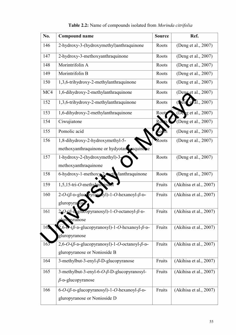
55
Table 2.2: Name of compounds isolated from Morinda citrifolia
No. Compound name Source Ref.
146 2-hydroxy-3-(hydroxymethyl)anthraquinone Roots (Deng et al., 2007)
147 2-hydroxy-3-methoxyanthraquinone Roots (Deng et al., 2007)
148 Morintrifolin A Roots (Deng et al., 2007)
149 Morintrifolin B Roots (Deng et al., 2007)
150 1,3,6-trihydroxy-2-methylanthraquinone Roots (Deng et al., 2007)
MC4 1,6-dihydroxy-2-methylanthraquinone Roots (Deng et al., 2007)
152 1,3,6-trihydroxy-2-methylanthraquinone Roots (Deng et al., 2007)
153 1,6-dihydroxy-2-methylanthraquinone Roots (Deng et al., 2007)
154 Ciwujiatone Roots (Deng et al., 2007)
155 Pomolic acid Roots (Deng et al., 2007)
156 1,8-dihydroxy-2-hydroxymethyl-5-
methoxyanthraquinone or hydyotanthraquinone
Roots (Deng et al., 2007)
157 1-hydroxy-2-(hydroxymethyl)-3-
methoxyanthraquinone
Roots (Deng et al., 2007)
158 6-hydroxy-1-methoxy-2-methylanthraquinone Roots (Deng et al., 2007)
159 1,5,15-tri-O-methylmorindol Fruits (Akihisa et al., 2007)
160 2-O-(β-ᴅ-glucopyranosyl)-1-O-hexanoyl-β-ᴅ-
gluropyranose
Fruits (Akihisa et al., 2007)
161 2-O-(β-ᴅ-glucopyranosyl)-1-O-octanoyl-β-ᴅ-
gluropyranose
Fruits (Akihisa et al., 2007)
162 2,6-O-(β-ᴅ-glucopyranosyl)-1-O-hexanoyl-β-ᴅ-
gluropyranose
Fruits (Akihisa et al., 2007)
163 2,6-O-(β-ᴅ-glucopyranosyl)-1-O-octanoyl-β-ᴅ-
gluropyranose or Nonioside B
Fruits (Akihisa et al., 2007)
164 3-methylbut-3-enyl-β-D-glucopyranose Fruits (Akihisa et al., 2007)
165 3-methylbut-3-enyl-6-O-β-D-glucopyranosyl-
β-ᴅ-glucopyranose
Fruits (Akihisa et al., 2007)
166 6-O-(β-ᴅ-glucopyranosyl)-1-O-hexanoyl-β-ᴅ-
gluropyranose or Nonioside D
Fruits (Akihisa et al., 2007)
Univers
ity of
Mala
ya

56
Table 2.2, continued
No. Compound name Source Ref.
167 6-O-(β-ᴅ-glucopyranosyl)-1-O-octanoyl-β-ᴅ-
gluropyranose or Nonioside C
Fruits (Akihisa et al., 2007)
168 Barbinervic acid Leaves (Takashima et al.,
2007)
169 Clethric acid Leaves (Takashima et al.,
2007)
170 Pteryxin Leaves (Takashima et al.,
2007)
171 Peucedanocoumarin III Leaves (Takashima et al.,
2007)
172 Citrifoside Leaves (Takashima et al.,
2007)
173 Roseoside II Leaves (Takashima et al., 2007)
174 Oleanolic acid Leaves (Takashima et al., 2007)
175 Hederagenin Leaves (Takashima et al., 2007)
176 Rotungenic acid Leaves (Takashima et al., 2007)
177 3-O-acetylpomolic acid Leaves (Takashima et al., 2007)
178 132(R)-hydroxypheophorbide a methyl ester Leaves (Takashima et al., 2007)
179 132(S)-hydroxypheophorbide a methyl ester Leaves (Takashima et al., 2007)
180 Methyl pheophorbide a Leaves (Takashima et al., 2007)
181 Methyl pheophorbide b Leaves (Takashima et al., 2007)
182 Pheophorbide a Leaves (Takashima et al., 2007)
Univers
ity of
Mala
ya

57
Table 2.2, continued
No. Compound name Source Ref. 183 13-epi-phaeophorbide a methyl ester Leaves (Takashima et al.,
2007)
184 151(R)-hydroxypurpurin-7 lactone dimethyl ester
Leaves (Takashima et al., 2007)
185 151(S)-hydroxypurpurin-7 lactone dimethyl ester
Leaves (Takashima et al., 2007)
186 13-hydroxy-9,11,15-octadecatrienoic acid Leaves (Takashima et al., 2007)
187 Phytol Leaves (Takashima et al., 2007)
188 2-methylanthraquinone or tectoquine Roots (Lv et al., 2011)
189 2-formylanthraquinone Roots (Lv et al., 2011)
190 Cholest-22-en-3-ol Roots (Lv et al., 2011)
191 Nonin A Roots (Lv et al., 2011)
192 Nonin B Roots (Lv et al., 2011)
193 Nonin C Roots (Lv et al., 2011)
194 Decumbic acid Roots (Lv et al., 2011)
195 D-mannitol Fruits (Su et al., 2005)
196 D-glucose Fruits (Su et al., 2005)
197 Methyl β-ᴅ-fructanoside Fruits (Su et al., 2005)
198 Methyl α-ᴅ-fructanoside Fruits (Su et al., 2005)
199 Nicotifloroside Fruits (Su et al., 2005)
200 Cytidine Fruits (Su et al., 2005)
201 Borreriagenin or morindacin Fruits (Su et al., 2005) (Tang et al., 2009)
202 Deacetylasperuloside Fruits (Su et al., 2005) (Tang et al., 2009)
203 Asperuloside Leaves Fruits
(Sang et al., 2001) (Tang et al., 2009)
204 6β,7β-epoxy-8-epi-splendoside Fruits (Su et al., 2005)
205 Narcissoside Fruits (Su et al., 2005)
206 Naringin Fruits (Lin et al., 2013)
207 Diosmine Fruits (Lin et al., 2013)
208 Quercitrin Fruits (Lin et al., 2013)
Univers
ity of
Mala
ya

58
Table 2.2, continued
No. Compound name Source Ref. 209 Hesperidin Fruits (Lin et al., 2013)
210 Chlorogenic acid Fruits (Lin et al., 2013)
211 Rosmarinic acid Fruits (Lin et al., 2013)
212 Neohesperidin Fruits (Lin et al., 2013)
213 Myricetin Fruits (Lin et al., 2013)
214 Morin Fruits (Lin et al., 2013)
215 Isorhamnetin Fruits (Lin et al., 2013)
216 Daidzein Fruits (Lin et al., 2013)
217 Glycitein Fruits (Lin et al., 2013)
218 Genistein Fruits (Lin et al., 2013)
219 Gallic acid Fruits (Lin et al., 2013)
220 Gentisic acid Fruits (Lin et al., 2013)
221 Vanilic acid Fruits (Lin et al., 2013)
222 p-coumaric acid or 4-coumaric acid Fruits (Lin et al., 2013)
223 Ferulic acid Fruits (Lin et al., 2013)
224 Sinapic acid Fruits (Lin et al., 2013)
225 Apigenin Fruits (Lin et al., 2013)
226 Luteolin Fruits (Lin et al., 2013)
227 Eriodictyol Fruits (Lin et al., 2013)
228 Hesperetin Fruits (Lin et al., 2013)
229 Catechin Fruits (Lin et al., 2013)
230 Caffeic acid Fruits (Lin et al., 2013)
231 Dimethyl phthalate Fruits (He at al., 2012)
232 Syringic acid Fruits (Lin et al., 2013)
233 Epicatechin Fruits (Lin et al., 2013)
234 4-epi-dunnisinin Fruis (Tang et al., 2009) (He at al., 2012)
235 19-hydroxyl-ursolic acid Fruits (He at al., 2012)
236 (24S)-ergost-7-en-3β-ol Fruits (He at al., 2012)
237 Daucosterol Fruits (He at al., 2012)
238 β-sitosterol Fruits (He at al., 2012)
239 1-O-(3’-methylbut-3’-enyl)-β-ᴅ-glucopyranose Fruits (He at al., 2012)
240
3-methylbut-3-enyl-6-O-β-ᴅ-glucopyranosyl-β-ᴅ-glucopyranoside or Nonioside A
Fruits
(Wang et al., 2000) (He at al., 2012)
Univers
ity of
Mala
ya
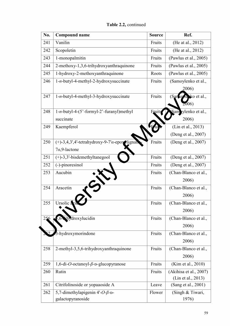
59
Table 2.2, continued
No. Compound name Source Ref. 241 Vanilin Fruits (He at al., 2012)
242 Scopoletin Fruits (He at al., 2012)
243 1-monopalmitin Fruits (Pawlus et al., 2005)
244 2-methoxy-1,3,6-trihydroxyanthraquinone Fruits (Pawlus et al., 2005)
245 1-hydroxy-2-methoxyanthraquinone Roots (Pawlus et al., 2005)
246 1-n-butyl-4-methyl-2-hydroxysuccinate Fruits (Samoylenko et al., 2006)
247 1-n-butyl-4-methyl-3-hydroxysuccinate Fruits (Samoylenko et al., 2006)
248 1-n-butyl-4-(5’-formyl-2’-furanyl)methyl succinate
Fruits (Samoylenko et al., 2006)
249 Kaempferol Fruits (Lin et al., 2013) (Deng et al., 2007)
250 (+)-3,4,3',4'-tetrahydroxy-9-7'α-epoxylignano-7α,9-lactone
Fruits (Deng et al., 2007)
251 (+)-3,3'-bisdemethyltanegool Fruits (Deng et al., 2007)
252 (-)-pinoresinol Fruits (Deng et al., 2007)
253 Aucubin Fruits (Chan-Blanco et al., 2006)
254 Aracetin Fruits (Chan-Blanco et al., 2006)
255 Ursolic acid Fruits (Chan-Blanco et al., 2006)
256 5,6-dihydroxylucidin Fruits (Chan-Blanco et al., 2006)
257 3-hydroxymorindone Fruits (Chan-Blanco et al., 2006)
258 2-methyl-3,5,6-trihydroxyanthraquinone Fruits (Chan-Blanco et al., 2006)
259 1,6-di-O-octanoyl-β-ᴅ-glucopyranose Fruits (Kim et al., 2010)
260 Rutin Fruits (Akihisa et al., 2007) (Lin et al., 2013)
261 Citrifolinoside or yopaaoside A Leave (Sang et al., 2001)
262 5,7-dimethylapigenin 4'-O-β-ᴅ-galactopyranoside
Flower (Singh & Tiwari, 1976)
Univers
ity of
Mala
ya
60
Table 2.2, continued
No. Compound name Source Ref. 263 Acacetin 7-O-β-ᴅ-glucopyranoside Flower (Singh & Tiwari,
1976)
264 (Z,Z)-2,5-Undecadien-1-ol Fruits (Farine et al., 1996) 265 Isoprincepin Fruits (Kamiya et al., 2004)
266 Americanol A Fruits (Kamiya et al., 2004)
267 Americanin A Fruits (Kamiya et al., 2004)
268 Americanoic acid A Fruits (Kamiya et al., 2004)
269 Morindolin Fruits (Kamiya et al., 2004)
270 3,3'-bisdemethylpinoresinol Fruits (Deng et al., 2007) (Kamiya et al., 2004)
271 3,3',4',5,7-pentahydroxyflavone or quercetin Fruits (Deng et al., 2007) (Liu et al., 2007) (Lin et al., 2013)
272 1,7-dihydroxy-8-methoxy-2-methylanthraquinone or morindone-5-methylether
Roots (Lv et al., 2011)
273 Soranjidiol Roots (Kamiya et al., 2010)
274 Morindone Roots (Kamiya et al., 2010)
275 1,6-dihydroxy-5-methoxy-2-methylanthraquinone
Fruits (Pawlus et al., 2005)
276 5,15-di-O-methylmorindol Fruits (Akihisa et al., 2007) (Takashima et al.,
2007) MC2 1,3-dihydroxy-2-formylanthraquinone or
nordamnacanthal Roots (Lv et al., 2011)
(Kamiya et al., 2010) MC3 1,3-dihydroxy-2-methylanthraquinone or
rubiadin Roots (Lv et al., 2011)
279 Ibericin Roots (Kamiya et al., 2010)
280 Lucidin Roots (Kamiya et al., 2010)
281 1,3-dihydroxy-2-methoxymethylanthraquinone Roots (Kamiya et al., 2010)
282 1,3-dihydroxy-2-methoxyanthraquinone Roots (Pawlus et al., 2005)
283 1-methoxy-2-primeverosyloxymethyl-anthraquinone-3-olate
Roots (Kamiya et al., 2009)
284 1-hydroxy-2-primeverosyloxymethyl-anthraquinone-3-olate
Roots (Kamiya et al., 2009)
Univers
ity of
Mala
ya
61
Table 2.2, continued
No. Compound name Source Ref.
285 1-methyl-3-hydroxyanthraquinone Roots (Lv et al., 2011)
286 1,3-dimethoxy-2-methoxymethylanthraquinone Roots (Lv et al., 2011)
MC1 1-methoxy-3-hydroxy-2-formylanthraquinone or damnacanthal
Roots (Lv et al., 2011) (Kamiya et al., 2010)
288 Damnacanthol-11-O-β-primeveroside Roots (Kamiya et al., 2009)
289 Rubiadin 1-methylether or rubiadin
monomethyl ether
Roots (Kamiya et al., 2010)
290 3-hydroxy-2-(hydroxymethyl)-1-methoxyanthraquinone or damnacanthol
Roots (Kamiya et al., 2010)
291 3-hydroxy-1-methoxy-2-
methoxymethylanthraquinone
Roots (Kamiya et al., 2010)
292 Damnacanthol-3-O-β-ᴅ-primeveroside Roots (Kamiya et al., 2008)
293 Digiferruginol-1-methylether-11-O-β-
gentibiose
Roots (Kamiya et al., 2009)
MC6 1-methoxy-2-hydroxyanthraquinone or alizarin 1-methyl ether
Roots (Lv et al., 2011)
295 1-methoxy-3-hydroxynthraquinone Roots (Lv et al., 2011) 296 2-hydroxy-1-methoxyanthraquinone Fruits (Pawlus et al., 2005)
297 Lucidin-3-O-β-ᴅ-primeveroside Roots (Kamiya et al., 2008) 298 Digiferruginol-11-O-β-primeveroside Roots (Kamiya et al., 2009) MC5 1-hydroxy-3-methoxyanthraquinone Roots (Lv et al., 2011) 300 2-formyl-1-hydroxyanthraquinone Roots (Iwao & Kuraishi,
1987) 301 1,2-dihydroxyanthraquinone or alizarin Roots
Fruits (Deng et al., 2007) (Wang et al., 2002)
302 Morindone-6-O-β-ᴅ-primeveroside Roots (Kamiya et al., 2008) 303 1-hydroxy-5,6-dimethoxy-2-methyl-7-
primeverosyloxyanthraquinone Roots (Kamiya et al., 2009)
304 Deacetylasperulosidic acid Roots Fruits
(Kamiya et al., 2008) (Samoylenko et al.,
2006) 305 Asperulosidic acid Roots
Leaves Fruits
(Kamiya et al., 2008) (Akihisa et al., 2007)
(Su et al., 2005) (Tang et al., 2009)
Univers
ity of
Mala
ya

62
Table 2.2, continued
No. Compound name Source Ref.
306 6α-hydroxyadoxoside Fruits (Su et al., 2005)
307 Isoscopoletin Fruits (Deng et al., 2007) (Liu et al., 2007)
308 Aesculetin Fruits (Liu et al., 2007) 309 Acubin Fruits (Leach et al., 1998) 310 Anthragallol 1,2-di-O-methyl ether Roots (Dalsgaard et al.,
2006)
311 Anthragallol 1,3-di-O-methyl ether Fruits (Kamiya et al., 2005)
312 Anthragallol 2,3-di-O-methyl ether Heartw
ood
(Dalsgaard et al.,
2006)
313 Anthragallol 2-O-methyl ether Fruits (Kamiya et al., 2005)
314 Nonioside E Fruits (Kim et al., 2005)
315 Nonioside F Fruits (Kim et al., 2005)
316 Nonioside G Fruits (Kim et al., 2005)
317 Nonioside H Fruits (Kim et al., 2005)
318 5,15-dimethylmorindol Fruits (Takashima et al.,
2007)
319 6-hydroxy-anthragallol-1,3-dimethylether Fruits (Kamiya et al., 2005)
320 Morindone-5-methylether Fruits (Kamiya et al., 2005)
321 1,4-dihydroxy-2-methoxy-7-
methylanthraquinone or austrocortinin
Fruits (Pawlus & Kinghorn,
2007)
322 2-hydroxy-1-methoxy-7-methylanthraquinone Roots (Sang & Ho, 2009)
323 Morenone-1 Roots (Jain & Srivastava,
1992)
324 Morenone-2 Roots (Jain & Srivastava,
1992)
Univers
ity of
Mala
ya
63
2.3 Chlorophyllum molybdites
2.3.1 General
Mushrooms belong to the class of basidomycetes which consist of fruits bodies
and mycelium. Generally, the edible species are called mushrooms while the poisonous
species called toadstool (Adedayo, 2011). The terms toadstool comes from the German
word todestuhl meaning death’s stool and has been used when referring to the
poisonous mushroom. There are about 5000 mushrooms species was reported in the
world and approximately only 50-100 are known to be poisonous to human (Barbato,
1993; Bryson, 1996).
Mycotoxins refer to any toxic substance by a fungus that capable to poison
other organisms (Bennett, 1987). The toxic components of poisonous mushrooms have
been identified as amatoxins, phallatoxins, gyromitrin, orellanine, muscarine,
tricholomic acid, ibotenic acid, muscinol, psilocybin, psilocin, and tetraethyl thiuram
disulphate (Benedict, 2013; Buku et al., 1980; Fasidi & Kadiri, 1995; Saupe, 1981). In
clinical practice, only amatoxins are topic of interest for chemical study because it is
thermostable and cannot be removed or destroyed by cooking. Amatoxins can be
analyzed in urine, blood and various organs and it is extremely powerful poisons that
can cause fatality (Faulstich et al., 1980; Faulstich et al., 1982; Vellinga, 2006).
Chlorophyllum species are characterized by their spores. The spore can be
green, or without any colour. Based on parsimony analysis of ITS rDNA (Internal
Transcribed Space Ribosomal DNA) sequences from European collections, four
Chlorophyllum species that is Chlorophyllum rachodes, Chlorophyllum brunneum,
Chlorophyllum olivieri and Chlorophyllum molybdites has been carried out (Vellinga,
2006). While in China, they have discovered other species of this genus which is
Univers
ity of
Mala
ya
64
Chlorophyllum agaricoides, Chlorophyllum hortense, and Chlorophyllum
sphaerosporum (Ge & Yang, 2006).
Chlorophyllum molybdites (Meyer: Fr) Massee previously known as Lepiota
Morgani or the ‘green parasol’ mushroom (Blayney et al., 2006). It is also known by a
variety of common names such as Lepiota esculenta, Lepiota molybdites, Morgan’s
Lepiota, False parasol, and Green-Lined Parasol. It has a green spores which are a bit
wider than those of the other species, but the rest are looks very much like
Chlorophyllum rachodes and it has very similar appearance to Amanitas except for the
gills (Berger & Guss, 2005; Watling, 1991). The Mushroom Poisoning Case Registry
of North American Mycological Association’s Toxicology Committee lists C.
molybdites as the most common mushroom poisoning reported because of the
misidentification (Augenstein, 1994). C. molybdites can be found either alone or in
partial or fairy rings (up to 16 feet in diameter) on grasslands, gardens, lawns, and
occasionally in open woods during the wet periods of spring, summer and fall
(Augenstein, 1994; Berger & Guss, 2005; Vellinga, 2006).
C. molybdites is a common species that is widely distributed in the eastern
states of North America, and also has been found in Hawaii, Tahiti, the Philippines,
New Guinea, Australia, India, east and central Africa, the West Indies, China and
much of South America including Brazil and Argentina, and with continuing high
summer temperatures it might well spread northwards in Europe (Arora, 1986; Bessey,
1939; Ge & Yang, 2006; Vellinga, 2006). C. molybdites are often involved in
poisonings throughout the world. The consumption and ingestion of this C. molybdites
is almost never resulting any fatality. But, it can causes discomfort situation to the
consumer such as intoxication, dizziness, diarrhea, nausea was followed by profuse
Univers
ity of
Mala
ya
65
continuous vomiting, convulsion, intestinal pain, and also griping stomach pain
(Blayney et al., 1980; Fasidi & Kadiri, 1995; Gosh et al., 1977; Gray, 1973; Lehmann
& Khazan, 1992; Wieland, 1968).
Many researchers have reported their findings on this species either on clinical
studies or in the other fields. In 1996, Floch and his colleagues reported a series of in
vivo and in vitro experiments using extracts from C. molybdites. In the experiment, it is
found that the extract was innocuous when administered orally in mice, but when
injected intraperitoneally it resulted in fatality. The results also indicated that the
extract will lost its effectiveness over the time (Floch et al., 1966).
In 1974, Eilers and Nelson published a report on the toxicity of C. molybdites,
in mice, chicks and dogs. The force feeding and administration via subcutaneous routes
produced a little toxicity, while intraperitoneal injection produced significant toxicity
for both mice and chicks. In the other hand, intravenous injections of toxin extracts
was given to the dogs and it result in a decrease in mean blood pressure. The report
also stated that the toxins in C. molybdites was found in all parts of the mushrooms but
the most concentrated part is in the cap followed by the stripe, gills and spores, in
mature or immature specimens of the species (Eilers & Nelson, 1974).
The study on chemical composition and toxic trace element composition in C.
molybdites indicated the presence of higher tannin and trypsin contents in this species
(Falade et al., 2008). Tannins are known to retard growth through reduced digestion
and/or absorption (Laurena et al., 1984). While the results obtain for all of the trace
elements (Lead, Cadmium, Nickel, Arsenic Chromium and Mercury) shows that
mercury has the lowest while lead has the highest. Lead has been reported to cause
Univers
ity of
Mala
ya
66
irreversible damage to the central nervous system and permanent mental retardation,
while Mercury poison was reported to cause fetal malformation, kidney and liver
damage (Chisolm, 1965; Falade et al., 2008; Weiss & Landrigan, 2000; Yilmaz et al.,
2003).
Pharmacological and toxicological studies have shown the reduction severity of
toxic signs in mice poisoned with the lyophilized extract of C. molybdites when tested
with Penicilin G although it doesn’t completely eliminate the toxins. Penicilin G in this
case is used as a drug that block the transport system responsible for the amatoxin
uptake by the hepatocytes. Administration of penicillin G was shown to have
significant prophylactic and therapeutic effect to reduce the severity in mice dose but it
did not confer significant protection from injury induced by the mushroom toxins on
tissue and organs in the mice (Ambali et al., 2008; Rumack & Spoerke, 1994).
The studies on proximate, mineral and phytate composition of this mushrooms
have indicates that the crude of C. molybdites have very low content in that three
variables. It had a lowest content of crude fat, protein, sodium, potassium, phosphorus,
zinc, copper, and the chloride content. But in the other hand, the mushroom has the
highest Nitrogen free extract (NFE) content (Ndamitso & Abulude, 2013).
Previous studies on mushroom toxins suggested the presence of a cholinergic
compound in this species. However, Eilers and Nelson reported that the toxic
component in this fungus is a polymeric protein. In a later study, Yamada and
colleagues isolated a toxic metalloendopeptidase (MEPs) named molybdophyllysin
from C. molybdites. Molybdophyllysin was found as a relatively small protein with
proteolytic activity and this shows a contrast finding from Eilers and Nelson. It was
Univers
ity of
Mala
ya
67
reported that molybdophyllysin is a member of the deuterolysin family of MEPs that
contain aspzincin zinc-binding motif (Yamada et al., 2012).
In a subsequent study, Kobayashi and his colleagues isolated the new lectin
from C. molybdites known as Chlorophyllum molybdites lectin (CML). In this
research, it was discovered that CML exhibited N-glycolylneuraminic acid (NeuGc)
binding specificity and showed strong hemagglutination of Pronase-treated porcine
erythrocytes. NeuGc is a common sialic acid found in most mammals and lectins that
can recognize sialic acids have been used as a medical and biological probe for sugar
or sugar chain detector. NeuGc-specific lectins are rare and difficult to get
commercially (Kobayashi et al., 2004).
In 2001, Yoshikawa and his colleagues isolated two new steroidal derivatives
from this fungus. Both (22E,24R)-3α-ureido-ergosta-4,6,8(14),22-tetraene (325) and
(22E,24R)-5α,8α-epidioxyergosta-6,22-diene-3β-ol-3-О-β-D-glucopyranoside (326)
have indicated the moderate cytotoxicity against the human stomach cancer cell Kato-
III (Yoshikawa et al., 2001).
Univers
ity of
Mala
ya

68
2.3.2 Compounds isolated from Chlorophyllum molybdites
Table 2.3 lists the compounds isolated from the C. molybdites and Figure 2.3
shows the structures of compounds previously isolated from C. molybdites.
HNH2N
O
H
ORO
O
(325) (326) R = β-D glucopyranosyl (327) R = H (328) R = Glc
H
OHHO HO OH
O
H
(329) (330)
H
OHHO
OH
H
O
O
HO
OHHO
HOH
HOO
(331) (332)
Figure 2.3: Structure of compounds isolated from Chlorophyllum molybdites
Univers
ity of
Mala
ya

69
O
O
HO
OHHO
HOH H
HO
(333) (334)
N
NH
O O
HOOH
NH
NH
O
O
NH
NH
O
O N
NH2
O
(335) (336) (337) (338)
HH
RO
OOH
(339) R =H (341) (340) R = Glc
O OH
OOR
(342) (CP5) 9Z, 12Z; R = H (344) 9Z, 12Z; R = CH2CH (OH) CH2OH (345) 9Z, 12Z; R = Glc
Figure 2.3, continued
Univers
ity of
Mala
ya

70
O OR
N
O OH
O
(346) R = H (348) (347) R = Glc
Univers
ity of
Mala
ya
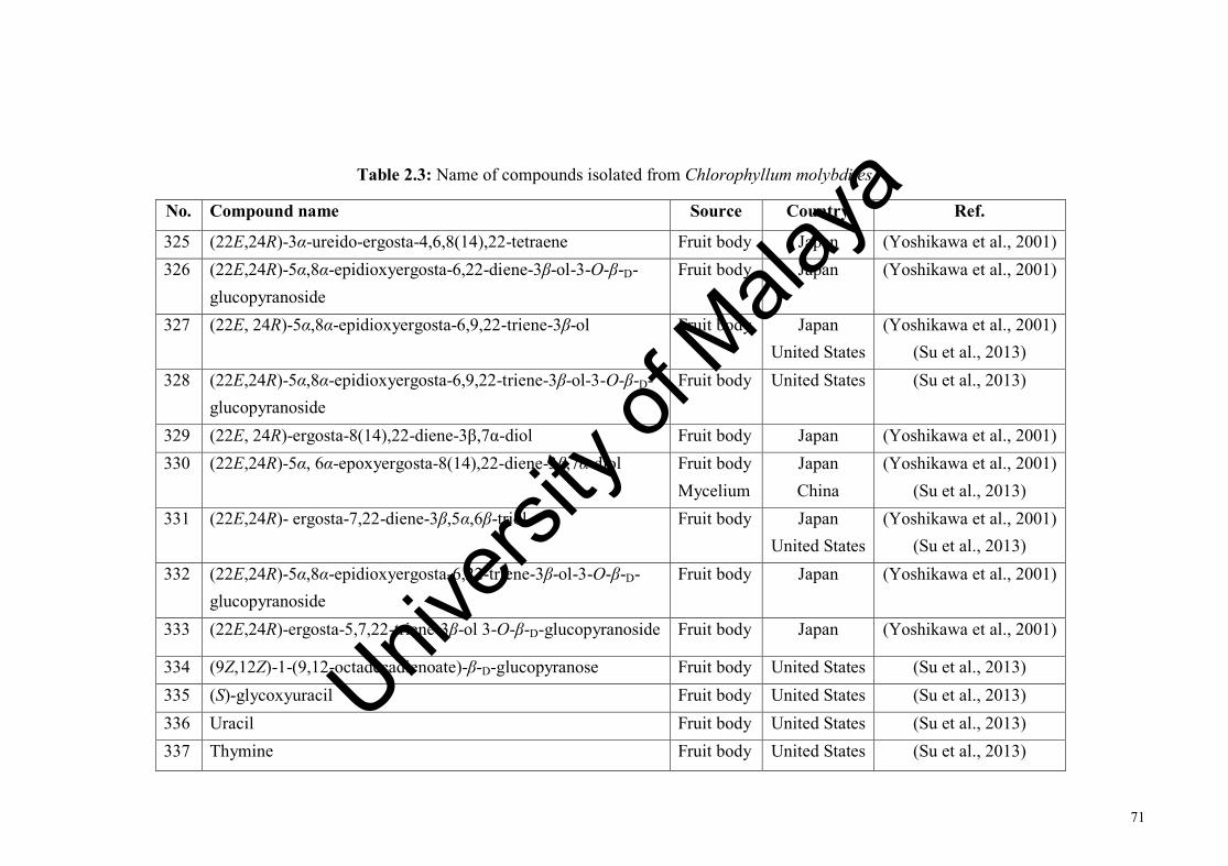
71
Table 2.3: Name of compounds isolated from Chlorophyllum molybdites
No. Compound name Source Country Ref.
325 (22E,24R)-3α-ureido-ergosta-4,6,8(14),22-tetraene Fruit body Japan (Yoshikawa et al., 2001) 326 (22E,24R)-5α,8α-epidioxyergosta-6,22-diene-3β-ol-3-О-β-D-
glucopyranoside Fruit body Japan (Yoshikawa et al., 2001)
327 (22E, 24R)-5α,8α-epidioxyergosta-6,9,22-triene-3β-ol Fruit body Japan United States
(Yoshikawa et al., 2001) (Su et al., 2013)
328 (22E,24R)-5α,8α-epidioxyergosta-6,9,22-triene-3β-ol-3-О-β-D-glucopyranoside
Fruit body United States (Su et al., 2013)
329 (22E, 24R)-ergosta-8(14),22-diene-3β,7α-diol Fruit body Japan (Yoshikawa et al., 2001) 330 (22E,24R)-5α, 6α-epoxyergosta-8(14),22-diene-3β,7α-diol Fruit body
Mycelium Japan China
(Yoshikawa et al., 2001) (Su et al., 2013)
331 (22E,24R)- ergosta-7,22-diene-3β,5α,6β-triol Fruit body Japan United States
(Yoshikawa et al., 2001) (Su et al., 2013)
332 (22E,24R)-5α,8α-epidioxyergosta-6,22-triene-3β-ol-3-О-β-D-glucopyranoside
Fruit body Japan (Yoshikawa et al., 2001)
333 (22E,24R)-ergosta-5,7,22-triene-3β-ol 3-О-β-D-glucopyranoside Fruit body Japan (Yoshikawa et al., 2001)
334 (9Z,12Z)-1-(9,12-octadecadienoate)-β-D-glucopyranose Fruit body United States (Su et al., 2013) 335 (S)-glycoxyuracil Fruit body United States (Su et al., 2013) 336 Uracil Fruit body United States (Su et al., 2013) 337 Thymine Fruit body United States (Su et al., 2013)
Univers
ity of
Mala
ya
72
Table 2.3, continued
No. Compound name Source Country Ref.
338 Nicotinamide Fruit body United States (Su et al., 2013)
339 Ergosterol Fruit body United States (Su et al., 2013)
340 Ergosterol-3-О-β-D-glucopyranoside Fruit body United States (Su et al., 2013)
341 Oleic acid Fruit body United States (Su et al., 2013)
342 Palmityl-1-О-β-D-glucopyranoside Fruit body United States (Su et al., 2013)
CP5 Linoleic acid Fruit body United States (Su et al., 2013)
344 1-linoleylglycerol Fruit body United States (Su et al., 2013)
345 (22E,24R)- ergosta-7,22-diene-3β-ol Mycelium China (Gong et al., 2010)
346 Palmitoleic acid Fruit body United States (Su et al., 2013)
347 Palmitic acid Fruit body United States (Su et al., 2013)
348 Lepiotin B Fruit body Japan (Ohta et al., 1998)
Univers
ity of
Mala
ya
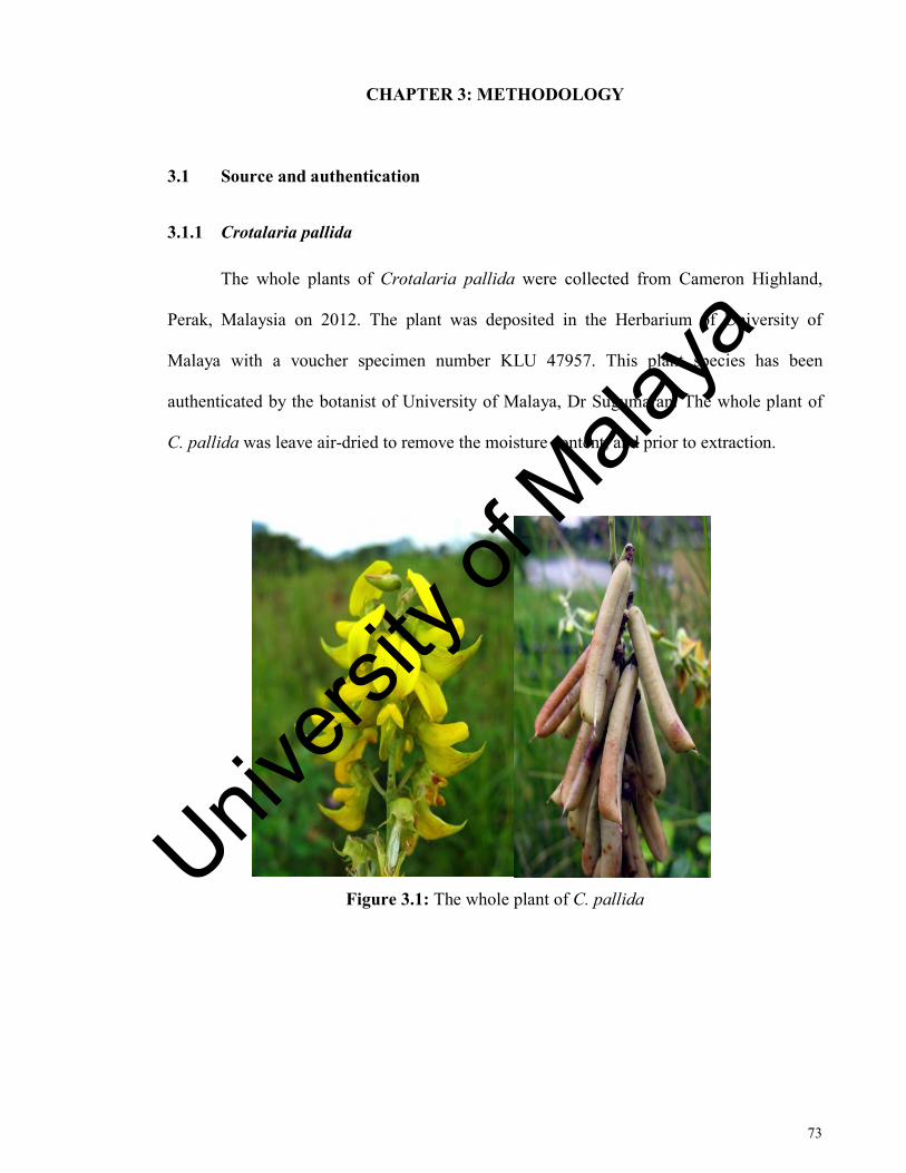
73
CHAPTER 3: METHODOLOGY
3.1 Source and authentication
3.1.1 Crotalaria pallida
The whole plants of Crotalaria pallida were collected from Cameron Highland,
Perak, Malaysia on 2012. The plant was deposited in the Herbarium of University of
Malaya with a voucher specimen number KLU 47957. This plant species has been
authenticated by the botanist of University of Malaya, Dr Sugumaran. The whole plant of
C. pallida was leave air-dried to remove the moisture content, and prior to extraction.
Figure 3.1: The whole plant of C. pallida
Univers
ity of
Mala
ya

74
3.1.2 Morinda citrifolia
The roots (with root barks) of Morinda citrifolia were collected from Kuala Lipis,
Pahang, Malaysia on June 2013. A voucher specimen is deposited at the Herbarium of
Chemistry Department, University of Malaya. The roots were cut into smaller pieces, air-
dried to remove the moisture content, and finally subjected to extraction.
Figure 3.2: The roots of M. citrifolia
3.1.3 Chlorophyllum molybdites
The fruit bodies of Chlorophyllum molybdites were collected in University of
Malaya, Kuala Lumpur on January 2013. A voucher specimen is deposited at the
Herbarium of Chemistry Department, University of Malaya. The mushroom was cleaned in
the laboratory, leave air-dried to remove the moisture content, and prior to extraction.
Figure 3.3: The fruit bodies of C. molybdites
Univers
ity of
Mala
ya
75
3.2 General
NMR spectral data were obtained using 600 MHz Bruker AVANCE III (Bruker,
Fallanden, Switzerland) NMR spectrometers with chemical shifts (δ) expressed in ppm and
TMS as an internal standard in CDCl3 or CD3OD. The coupling constants (J) are reported in
Hz. The HRESIMS data were obtained from the Agilent 6530 Q-TOF (Agilent
Technologies, Santa Clara, CA, USA) mass-spectrometer equipped with the Agilent 1200
series Rapid Resolution LC system. The ESI-MS data were obtained from the Agilent 6490
Triple Quad (Agilent Technologies, Santa Clara, CA, USA) mass-spectrometer equipped
with Agilent 1290 Infinity u-HPLC system. The UV measurement was carried out using the
Agilent Cary 60 UV Vis spectrophotometer (Agilent Technologies, Santa Clara, CA, USA).
The IR measurement was carried out on the Perkin-Elmer RX1 FT-IR (Perkin Elmer,
Waltham, MA, USA) spectrophotometer using NaCl cell.
3.3 Plant Extraction
3.3.1 Crotalaria pallida
1.8 kg whole plant of dried C. pallida were grounded and extracted with 95%
ethanol to give 195 g of crude ethanolic extract. The ethanolic extract was further
partitioned into n-hexane, chloroform and ethanol extract by sequential solvent-solvent
extraction respectively, with each solvent extraction repeating for three times. 23 g of n-
hexane, 50 g of chloroform and 118 g of ethanol crude were obtained from the evaporation
of solvent using rotary-evaporator. Preliminary TLC screening indicated that the most
promising extract is the n-hexane extract, and the chloroform and ethanol extracts do not
Univers
ity of
Mala
ya

76
have as many spots as the n-hexane extract. The n-hexane crude extracts were later
subjected to various chromatographic methods to isolate pure compounds.
3.3.2 Morinda citrifolia
0.85 kg of the dry M. citrifolia roots were extracted with chloroform and denatured
ethanol consecutively, and each solvent extraction was repeated three times. 8.6 g of
chloroform crude extract and 15.4 g of ethanol crude extract was yielded from evaporation
of solvent using rotary-evaporator. Preliminary TLC screening indicated that the most
promising extract is the chloroform extract and the ethanol extracts do not have as many
spots as the chloroform extract. The chloroform crude extract was later subjected to various
chromatographic methods for isolation of pure compounds.
3.3.3 Chlorophyllum molybdites
2.2 kg of the dry fruit bodies of C. molybdites were crushed and extracted with
hexane, ethyl acetate and denatured ethanol consecutively, with each solvent extraction
repeating for three times. The extracts were concentrated to dryness under reduced pressure
which gives 1.1 g of hexane, 16.2 g of ethyl acetate and 67.9 g of ethanol crude extract
consecutively. Preliminary TLC screening indicated that the most promising extract is the
ethyl acetate extract and hexane and ethanol extracts do not have as many spots as the ethyl
acetate extract. The ethyl acetate crude extract was later subjected to various
chromatographic methods to isolate pure compounds.
Univers
ity of
Mala
ya

77
3.4 Chromatographic methods
3.4.1 Thin Layer Chromatography (TLC)
TLC was extensively used in the isolation process of pure compounds. TLC were
performed on pre-coated silica gel plates (Kieselgel 60 F24, Merck, Darmstadt, Germany),
and spotted using a fine glass capillary tube, followed by development in a solvent
saturated TLC tank with various solvents. The developed TLC plate was then viewed under
short wave and/or long wave UV light, and stained with iodine and anisaldehyde staining
reagents.
3.4.2 Column Chromatography (CC)
CC was carried out using silica gel 60 (0.063-0.200 and 0.040-0.063 mm, Merck,
Darmstadt, Germany). The column was packed by preparing silica gel slurry in desired
eluting solvent and then settling the gel into the column. The solvent systems used were
hexane, ethyl acetate, acetone, chloroform and diethyl ether, in different mixing
compositions and gradients. TLC was used to aid in monitoring the fractions.
CC was also carried out using Sephadex LH20 (silica gel 60, 0.040-0.063 mm,
Merck, Darmstadt, Germany). The column was packed by firstly preparing Sephadex LH20
slurry in desired eluting solvent before settling the gel into the column. The solvent system
used is methanol and water, in different mixing compositions and gradients. This material
is designed to be used for organic solvents and water mixtures. TLC was used to aid in
monitoring the fractions.
Univers
ity of
Mala
ya
78
3.4.3 Centrifugal Thin Layer Chromatography (CTLC)
CTLC was carried out by using the Kieselgel 60 with gypsum silica gel (Merck,
Darmstadt, Germany) prepared on a round glass plate of 24 cm in diameter. The silica gel
slurry was prepared by using about 30 g of silica gel dissolved in 90 mL of distilled water
(to prepare for 1 cm thickness on the plate). The glass plate was taped on the edge by using
cellophane tape before pouring the silica gel slurry evenly on the glass plate. The slurry was
air-dried before removing the cellophane tape and the plate was subsequently baked in an
oven at 80 °C for 2 hours to remove any water remained. Then, the plate was allowed to
cool at room temperature for 6 hours before scrapping the plate 5 mm from the edge and 30
mm from the centre. Chromatography was carried out by first wetting the spinning plate
mounted on the machine with eluting solvent, and then the sample was loaded on the center
to form a thin band before eluting with the desired solvent system. TLC was used to aid in
monitoring the fractions obtained.
3.4.4 Preparative Thin Layer Chromatography (PTLC)
Prep TLC is a useful technique for the purification of the small quantities of sample
because it allows rapid separation of a number of components in a reaction mixture. It was
carried out using the TLC silica gel 60 F254 (Merck, Darmstadt, Germany). The TLC plate
were gently marked roughly 1.5 cm from one side of the plate. This is the “origin” line.
Using a fine glass capillary tube, a thin line of sample was deposited across the pencil line.
After obtained the prep TLC chamber, pour in the eluent, approximately about 150 mL.
Then, the plate was placed in and the top sealed with lid. A typical run takes about 1 hour
to 2 hours. After that, the plate was removed and visualized under UV. The bands that
appear were marked with pencil, cut into pieces by using a scissor and put it in a small
Univers
ity of
Mala
ya
79
bottom flask, then soaked in a mixture of methanol, chloroform, and acetone. The sample
then was concentrated to dryness by using the rotary evaporator or speed evaporator. To
separate the sample with the silica gel, the sample was soaked with the solvent that the
sample can dissolved in (e.g. chloroform and acetone). The supernatant was taken out and
concentrated to dryness, and then it was subjected to 1H NMR experiment.
3.4.5 High-Performance Liquid Chromatography (HPLC)
HPLC was carried out through an Agilent ZORBAX Eclipse XDB-C18 (9.4 x 250
mm; 5 micron) column, on Waters 600E series HPLC equipped with Waters UV detector.
Reverse Phase HPLC (RP-HPLC) was carried out by using a general gradient solvent
system of 95% water: 5% acetonitrile to 5% water: 95% acetonitrile. The solvents used
were HPLC grade.
3.5 Staining reagents for TLC
3.5.1 Iodine staining reagent
A 100 mL wide mouth chamber (with cap) was assembled with a piece of filter
paper and a few iodine crystals were added to saturate the chamber with iodine vapor. TLC
plate was then placed in the chamber and incubated for a few minutes to allow reaction
between the iodine vapor and the compounds on the TLC plate. When the entire TLC plate
turned brownish, the TLC was removed to identify the dark brown spots on the brownish
TLC plate. The spots were marked with a pencil. This reagent is normally used to detect
unsaturated compounds.
Univers
ity of
Mala
ya

80
3.5.2 Anisaldehyde staining reagent
A 100 mL anisaldehyde staining reagent was prepared by mixing 1 mL of
anisaldehyde, 100 mL of denatured ethanol, and 2 mL of 98% sulphuric acid together. The
staining was carried out by flooding the TLC plate with the stain solution and immediately
heated using dryer until the TLC plate turned pink or the maximal visualization of the
reacted compounds. The colour changes to violet, blue, red, grey or green according to
functional groups namely lichen constituents, phenols, terpenes, sugars and steroids.
3.6 Isolation of compounds
3.6.1 Isolation of compounds from the hexane extract of Crotalaria pallida
The hexane extract that obtained from the whole plant of C. pallida (23 g) was
subjected to extensive chromatographic separation as summarized in the flow diagram
shown in Figure 3.4 to yield eight compounds.
3.6.2 Isolation of compounds from the chloroform extract of Morinda citrifolia
The chloroform extract that obtained from the roots of M. citrifolia (2.3 g) was
subjected to extensive chromatographic separation as summarized in the flow diagram
shown in Figure 3.5 to yield six compounds.
Univers
ity of
Mala
ya

81
3.6.3 Isolation of compounds from the ethyl acetate extract of Chlorophyllum
molybdites
The ethyl acetate extract that obtained from the fruit bodies of C. molybdites (16.2
g) was subjected to extensive chromatographic separation as summarized in the flow
diagram shown in Figure 3.6 to yield four compounds.
Univers
ity of
Mala
ya

82
Figure 3.4: Isolation of compounds from the hexane extract of Crotalaria pallida
Crotalaria pallida Hexane extract
(23.0 g)
Fraction 2 ( 5.7 g)
Fraction 2 (86.9 mg)
Fraction 4 (244.9 mg)
Fraction 7 (670.9 mg)
Fraction 32 (3.9 g)
CC 9: 1 = H: E
Fraction 3 (551.8 mg)
Fraction 13 (2.6 g)
Fraction 4 (38.1 mg)
Fraction 5 (996.7 mg)
CC 100% C
CC 1: 1 = H: C
CC = Column Chromatography C = Chloroform H = Hexane E = Ethyl acetate
Univers
ity of
Mala
ya

83
Figure 3.4, continued
Fraction 2 5.7 g
Fraction 2 (86.9 mg)
Fraction 1 (7.2 mg)
Fraction 1 (15.0 mg)
CTLC 8: 8: 1 = H: C: M
CP3 (9.5 mg)
CTLC 1: 1 = H: E
CTLC 100% H
Fraction 4 (244.9 mg)
Fraction 1 (154.8 mg)
Fraction 1 (143.4 mg)
CP5 (3.8 mg)
CTLC 49: 1 = H: E
CTLC 100% H
CTLC 100% H
Fraction 7 (670.9 mg)
Fraction 4 (550.6 mg)
CTLC 19: 1 = H: E
Fraction 6 (176.2 mg)
CP6 (3.9 mg)
CTLC 1: 1 = H: E
Fraction 2 (39.4 mg)
CP4 (34.0 mg)
CTLC 19: 1 = H: E
CC 49: 1 = H: E
Fraction 32 ( 3.9 g)
Fraction 1 (3.5 g)
Fraction 2 (2.0 g)
Fraction 4 (189.0 mg)
CTLC 19: 1 = H: E
CP7 (41.5 mg)
CTLC 49: 1 = H: E
CTLC 49: 1 = H: E
CTLC 1: 1 = H: E
CC 9: 1 = H: C
CC = Column chromatography CTLC = Centrifugal Thin Layer Chromatography C = Chloroform H = Hexane E = Ethyl acetate
Univers
ity of
Mala
ya

84
Figure 3.4, continued
Fraction 13 2.6 g
Fraction 4 (38.1 mg)
Fraction 4 (6.0 mg)
CP1 (0.9 mg)
CTLC 1: 4 = H: C
Fraction 6 (15.3 mg)
CP2 (2.4 mg)
CTLC 100% C
CTLC 19: 1 = H: E
Fraction 5 ( 996.7 mg)
Fraction 13 (594.4 mg)
Fraction 1 (46.2 mg)
CP8 (3.5 mg)
CTLC 1: 4 = H: C
CTLC 3: 7 = H: C
CTLC 1: 1 = H: C
CC 100% C
CC = Column chromatography CTLC = Centrifugal Thin Layer Chromatography C = Chloroform H = Hexane E = Ethyl acetate Univ
ersity
of M
alaya

85
Figure 3.5: Isolation of compounds from the chloroform extract of Morinda citrifolia
Morinda citrifolia Chloroform extract
2.3 g
Fraction 4 (486.5 mg)
Fraction 3 (38.7 mg)
MC5 (3.2 mg)
CC 4: 1 = H: C
Fraction 19 (318.4 mg)
MC1 (39.2 mg)
MC2 (17.6 mg)
Fraction 15 (65.2 mg)
PTLC 1: 1 = H: C
CC 1: 1 = H: C
CC 1: 1 = H: C
Fraction 17 (168.4 mg)
Fraction 14 (100.9 mg)
Fraction 3 (74.8 mg)
Fraction 19 (10.9 mg)
MC4 (4.3 mg)
CTLC 1: 9 = H: C
CC 1: 9 = H: C
CC 100% C
Fraction 11 (43.1 mg)
Fraction 5 (20.3 mg)
Fraction 2 (13.1 mg)
CTLC 2: 8 = A: H
MC3 (1.7 mg)
CTLC 100% C
Fraction 7 (15.3 mg)
Fraction 5 1: 1 = H: C
PTLC 1: 1 = H: C
MC6 (2.8 mg)
CTLC 1: 1 = H: C
CTLC 1: 4 =H: C
CC 100% C
CC 100% C
CC = Column chromatography CTLC = Centrifugal Thin Layer Chromatography PTLC = Preparative Thin Layer Chromatography C = Chloroform H = Hexane A = Acetone
Univers
ity of
Mala
ya

86
Figure 3.6: Isolation of compounds from the ethyl acetate extract of Chlorophyllum molybdites
Chlorophyllum molybdites Ethyl acetate extract
16.2 g
Vial 1 (9.6 g)
Fraction 1a (266.4 mg)
Fraction 8 (82.8 mg)
Fraction 4 (27.5 mg)
Fraction 5 (17.8 mg)
CTLC 9: 1 = C: M
CM2 (5.8 mg)
CTLC 9: 1 = C: M
Fraction 15 (35.5 mg)
CM1 (3.4 mg)
CTLC 9: 1 = C: M
LH 20 1: 1 = C: M
CC 100% C
Fraction 1c (2.2 g)
CC 100% C
Vial 2 (1.8 g)
Fraction 2a (242.8 mg)
Fraction 9 (91.1 mg)
Fraction 1 (67.2 mg)
CP5 (12.0 mg)
HPLC 9: 1 = W: ACN
CTLC 100% C
CC 100% C
Fraction 2b (710.6 mg)
Fraction 16 (214.9 mg)
Fraction 15 (24.6 mg)
CM3 (2.8 mg)
CTLC 9: 1 = C: M
CTLC 9: 1 = C: M
CC 100% C
CC 100% C
CC 100% C
CC = Column chromatography CTLC = Centrifugal Thin Layer Chromatography PTLC = Preparative Thin Layer Chromatography HPLC = High Performance Liquid Chromatography C = Chloroform W = Water M = Methanol ACN = Acetonitrile
Univers
ity of
Mala
ya
87
3.7 Compound data
3.7.1 Compounds isolated from Crotalaria pallida
Crotolidene (CP1): Colourless oil; molecular formula C14H22O2; [α]D -66.7 (c 0.237,
CHCl3); UV (EtOH) λmax (log ε) 205 (1.70), 276 (0.56) nm; IR (NaCl) vmax 3250, 3010,
2959, 2937, 2870, 1674, 1653, 1608, 1457, 1363, 1259, 1175, 1049, 982, 803 and 771
cm-1; 1H NMR (CDCl3, 600 MHz) δ 7.19 (1H, d, J = 16 Hz, H-3), 6.09 (1H, d, J = 16
Hz, H-4), 3.99 (1H, m, H-8), 2.41 (1H, dd, J = 17 and 6 Hz, H-7a), 2.27 (3H, s, Me-1),
2.06 (1H, dd, J = 17 and 11 Hz, H-7b), 1.78 (1H, m, H-9a), 1.75 (3H, s, Me-11), 1.47
(1H, t, J = 12 Hz, H-9b), 1.10 (3H, s, Me-13), 1.09 (3H, s, Me-12). 13C NMR (CDCl3,
150 MHz) δ 198.7 (C, C-2), 142.4 (CH, C-3), 135.9 (C, C-6), 132.6 (CH, C-4), 132.4
(C, C-5), 64.8 (CH, C-8), 48.6 (CH2, C-9), 43.0 (CH2, C-7), 37.1 (C, C-10), 30.3 (CH3,
C-12), 28.8 (CH3, C-13), 27.5 (CH3, C-1), 21.8 (CH3, C-11). HMBC: 2J C-2 to Me-1
and H-3; C-4 to H-3; C-5 to Me-11; C-8 to H-7b and H-9b; C-10 to H-9a and H-9b; C-
12 to Me-13; C-13 to Me-12; 3J C-3 to Me-1; C-4 to Me-11; C-5 to H-7a and H-7b; C-6
to H-4, Me-11, Me-12 and Me-13; C-7 to H-9a and H-9b; C-9 to Me-12 and Me-13; C-
13 to H-9a and H-9b. HRESIMS m/z 223.1497 [M+H]+ (calcd for C14H22O2+H,
223.1698) and m/z 205.1316 [M-OH]- (calcd for C14H22O2-OH, 205.1592).
Hydroxydihydrobovolide (CP2): Colorless oil; molecular formula C11H18O3; [α]D -
13.3 (c 0.015, CHCl3); UV (EtOH) λmax (log ε) 220 (3.49), 291 (0.23) nm; IR (NaCl)
vmax 3369, 2956, 2926, 2859, 1741, 1694, 1458, 1438, 1381, 1287, 1260, 1133, 1101,
1056, 974, 956, 903, 767 cm-1; 1H NMR (CDCl3, 600 MHz) δ 1.96 (1H, m, H-6a), 1.92
(3H, s, 4-Me), 1.80 (3H, s, 3-Me), 1.74 (1H, m, H-6b), 1.29 (2H, m, CH2-9), 1.27 (2H,
m, CH2-8), 1.15 (2H, m, CH2-7), 0.86 (3H, t, J = 7 Hz, CH3-10). 13C NMR (CDCl3, 150
MHz) δ 172.2 (C, C-2), 157.8 (C, C-3), 125.6 (C, C-4), 107.0 (C, C-5), 36.2 (CH2, C-6),
31.7 (CH2, C-8), 22.8 (CH2, C-9), 22.6 (CH2, C-7), 14.1 (CH3, C-10), 10.9 (CH3, 4-Me),
Univers
ity of
Mala
ya
88
8.7 (CH3, 3-Me); HRESIMS m/z 199.1335 [M+H]+ (calcd for C11H18O3+H, 199.1329)
and m/z 181.1233 [M+H - H2O]+ (calcd for C11H18O3-OH, 181.1223).
Octacosane (CP3): White amorphous solid; molecular formula C28H58; UV (Hex) λmax
(log ε) 207 (3.11) nm; IR (NaCl) vmax 2926, 2854, 1459, 908, 734 cm-1; 1H NMR
(CDCl3, 600 MHz) δ 1.26-1.24 (48H, m, CH2-3 to CH2-26), 1.11 (4H, m, CH2-2 and
CH2-27), 0.88 (6H, t, J = 7 Hz, CH3-1 and CH3-28).13C NMR (CDCl3, 150 MHz) δ 32.3
(CH2, C-3 and C-26), 30.0 (CH2, C-5 to C-24), 29.6 (CH2, C-4 and C-25), 23.0 (CH2, C-
2 and C-27), 14.4 (CH3, C-1 and C-28).
Trans-phytyl palmitate (CP4): White amorphous solid; molecular formula C36H70O2;
[α]D -1.4 (c 0.237, CHCl3); UV (Hex) λmax (log ε) 210 (3.33) nm; IR (NaCl) vmax 2916,
2849, 1738, 1732, 1641, 1462, 1456, 1372, 1236, 1165, 1114, 1046, 958, 756, 729, and
719 cm-1; 1H NMR (CDCl3, 600 MHz) δ 5.30 (1H, t, J = 7 Hz, H2’), 4.56 (2H, d, J = 7
Hz, CH2-1’), 2.28 (2H, t, J = 8 Hz, CH2-2), 1.97 (2H, t, J = 7 Hz, CH2-4’), 1.68 (3H, s,
3’-Me), 1.58 (2H, m, CH2-3), 1.50 (1H, m, H-15’), 1.38-1.25 (36H, m, CH2-4 to CH2-
15, CH2-5’, CH2-7’, CH2-9’ and CH2-11’ to CH2-13’), 1.13 (2H, m, CH2-14’), 1.05 (6H,
m, CH2-6’, CH2-8’ and CH2-10’), 0.88 (3H, m, CH3-16), 0.86 (6H, m, 15’-CH3 and
CH3-16’), 0.84 (6H, m, 7’-CH3 and 11’-CH3). 13C NMR (CDCl3, 150 MHz) δ 174.1 (C,
C-1), 142.7 (C, C-3’), 118.5 (CH, C-2’), 61.4 (CH2, C-1’), 40.1 (CH2, C-4’), 39.6 (CH2,
C-14’), 37.5-37.3 (CH2, C-6’, C-8’ and C-10’), 36.8 (CH2, C-12’), 34.6 (CH2, C-2), 33.0
(CH, C7’ and C-11’), 32.2 (CH2, C-14), 29.9-29.4 (CH2, C-4 to C-13), 28.2 (CH, C-
15’), 25.2-24.7 (CH2, C-3, C-5’, C-9’ and C-13’), 22.9 (CH2, C-15), 22.8 (CH3, 15’-CH3
and C-16’), 19.9 (CH3, 7’-CH3 and 9’-CH3), 16.6 (CH3, 3’-CH3), 14.3 (CH3, C-16);
GCMS m/z 57 (79), 68 (100) [C5H8]+, 71 (43), 82 (74), 95 (83), 123 (36), 137 (4).
Univers
ity of
Mala
ya

89
Linoleic acid (CP5): Light yellowish amorphous solid; molecular formula C18H32O2;
UV (EtOH) λmax (log ε) 224 (1.28), 272 (0.36) nm; IR (NaCl) vmax 3436, 3024, 2984,
2918, 2850, 1741, 1710, 1648, 1465, 1374, 1243, 1047 and 758 cm-1; 1H NMR (CDCl3,
600 MHz) δ 5.32-5.36 (4H, m, H-9, H-10, H-12, and H-13), 2.79 (1H, t, J = 7, H-11a),
2.75 (1H, t, J = 7, H-11b), 2.32 (2H, t, J = 8 Hz, CH2-2), 2.03 (4H, m, CH2-8 and CH2-
14), 1.61 (2H, m, CH2-3), 1.26 (14H, m, CH2-4 to CH2-7 and CH2-15 to CH2-17), 0.87
(3H, t, J = 7 Hz, CH3-18). 13C NMR (CDCl3, 150 MHz) δ 179.8 (C, C-1), 130.5 (CH, C-
13), 130.3 (CH, C-9), 128.3 (CH, C-12), 128.1 (CH, C-10), 34.2 (CH2, C-2), 31.8 (CH2,
C-16), 29.9-29.3 (CH2, C-4 to C-7 and C-15), 27.4 (CH2, C-8 and C-14), 25.9 (CH2, C-
11), 24.9 (CH2, C-3), 22.8 (CH2, C-17), 14.3 (CH3, C-18); HRESIMS with m/z
279.2371 [M-H]- (calcd for C18H32O2-H, m/z 279.2330).
Methyl oleate (CP6): Yellowish oil; molecular formula C19H36O2; UV (Hex) λmax (log
ε) 202 (2.80), 222 (0.68), 277 (0.11) nm; IR (NaCl) vmax 2926, 2855, 1744, 1458, 1437,
1376, 1364, 1245, 1196, 1171, 1018, 882 and 722 cm-1; 1H NMR (CDCl3, 600 MHz) δ
5.32 (2H, m, H-9 and H-10), 3.64 (3H, s, 1-OCH3), 2.28 (2H, t, J = 8 Hz, CH2-2), 1.98
(4H, m, CH2-8 and CH2-11), 1.59 (2H, m, CH2-3), 1.28 (20H, m, CH2-4 to CH2-7 and
CH2-12 to CH2-17), 0.86 (3H, t, J = 7 Hz, CH3-18). 13C NMR (CDCl3, 150 MHz) δ
174.6 (C, C-1), 130.2 (CH, C-10), 130.0 (CH, C-9), 52.7 (CH3, 1-OCH3), 34.3 (CH2, C-
2), 32.1 (CH2, C-16), 30.0-29.3 (CH2, C-4 to C-7 and C-12 to C-15), 27.4 (CH2, C-8 and
C-11), 25.2 (CH2, C-3), 22.9 (CH2, C-17), 14.3 (CH3, C-18); GCMS 296 (3) [M]+, 264
(14), 222 (10), 180 (8), 137 (9), 123 (17), 110 (27), 97 (52), 83 (61), 73 (60), 69 (74), 55
(100).
Ethyl palmitate (CP7): Yellowish oil; molecular formula C18H36O2; UV (Hex) λmax
(log ε) 206 (3.10), 274 (0.11) nm; IR (NaCl) vmax 2924, 2853, 1740, 1465, 1420, 1373,
Univers
ity of
Mala
ya

90
1349, 1302, 1244, 1177, 1116, 1098, 1036, 859, 804, 721 cm-1; 1H NMR (CDCl3, 600
MHz) δ 4.08 (2H, q, J = 7 Hz, 1-OCH2CH3), 2.38 (2H, t, J = 8 Hz, CH2-2), 1.57 (2H, m,
CH2-3), 1.24 (24H, m, CH2-4 to CH2-15), 1.21 (3H, t, J = 7, 1-OCH2CH3), 0.84 (3H, t, J
= 7 Hz, CH3-16). 13C NMR (CDCl3, 150 MHz) δ 173.8 (C, C-1), 60.4 (CH2, 1-
OCH2CH3), 34.6 (CH2, C-2), 32.2 (CH2, C-14), 30.0-29.4 (CH2, C-4 to C-13), 25.3
(CH2, C-3), 23.0 (CH2, C-15), 14.5 (CH3, 1-OCH2CH3), 14.4 (CH3, C-16); GCMS m/z
284 (4) [M]+, 255 (2), 241 (6), 157 (11), 101 (53), 88 (100), 70 (26), 55 (27).
Palmitic acid (CP8): White amorphous solid; molecular formula C16H32O2; UV (EtOH)
λmax (log ε) 208 (0.73), 268 (0.18) nm; IR (NaCl) vmax 3409, 2917, 2850, 1707, 1464,
1411, 1297, 908, 735 and 650 cm-1; 1H NMR (CDCl3, 600 MHz) δ 2.33 (2H, t, J = 8 Hz,
CH2-2), 1.61 (2H, m, CH2-3), 1.28 (24H, m, CH2-4 to CH2-15), 0.86 (3H, t, J = 7 Hz,
CH3-16). 13C NMR (CDCl3, 150 MHz) δ 178.7 (C, C-1), 34.0 (CH2, C-2), 32.1 (CH2, C-
14), 29.9-29.3 (CH2, C-4 to C-13), 24.9 (CH2, C-3), 22.9 (CH2, C-15), 14.2 (CH3, C-
16); HRESIMS with m/z 255.2369 [M-H]- (calcd for C16H32O2-H, m/z 255.2330).
3.7.2 Compounds isolated from Morinda citrifolia
Damnachantal (MC1): Yellow amorphous powder; molecular formula C16H10O5; UV
(EtOH) λmax (log ε) 210 (2.58), 255 (3.60), 285 (4.21), 400 (0.33) nm; IR (NaCl) vmax
3462, 2922, 1678, 1646, 1575, 1457, 1388, 1345, 1261, and 715 cm-1; 1H NMR
(CDCl3, 600 MHz) δ 12.27 (1H, s, 3-OH), 10.46 (1H, s, 2-CHO), 8.29 (1H, dd, J = 8
and 1 Hz, H-8), 8.24 (1H, dd, J = 8 and 1 Hz, H-5), 7.82 (1H, td, J = 8 and 1 Hz, H-7),
7.77 (1H, td, J = 8 and 1 Hz, H-6), 7.67 (1H, s, H-4), 4.13 (1H, s, 1-OMe). 13C NMR
(CDCl3, 150 MHz) δ 195.5 (C, 2-CHO), 181.9 (C, C-10), 180.2 (C, C-9), 166.9 (C, C-
1), 166.7 (C, C-3), 141.6 (C, C-4a), 134.9 (C, C-8a), 134.9 (CH, C-7), 133.7 (CH, C-6),
132.5 (C, C-10a), 127.4 (CH, C-5), 127.2 (CH, C-8), 118.1 (C, C-2), 117.7 (C, C-9a),
Univers
ity of
Mala
ya

91
113.1 (CH, C-4), 64.8 (C, 1-OMe) ; HRESIMS with m/z 281.0 [M-H]- (calcd for
C16H10O5-H, 281.0).
Nordamnachantal (MC2): Yellow-orange needles; molecular formula C15H8O5; UV
(EtOH) λmax (log ε) 210 (2.08), 265 (4.26), 290 (3.26), 430 (0.89) nm; IR (NaCl) vmax
3075, 1674, 1593, 1574, 1269, 1191, and 714 cm-1; 1H NMR (CDCl3, 600 MHz) δ 14.05
(1H, s, 1-OH), 12.67 (1H, s, 3-OH), 10.50 (1H, s, 2-CHO), 8.31 (1H, dd, J = 8 and 1
Hz, H-8), 8.28 (1H, dd, J = 8 and 1 Hz, H-5), 7.83 (1H, m, H-7), 7.83 (1H, m, H-6),
7.33 (1H, s, H-4). 13C NMR (CDCl3, 150 MHz) δ 194.1 (C, 2-CHO), 186.9 (C, C-9),
181.5 (C, C-10), 169.2 (C, C-1), 168.3 (C, C-3), 139.5 (C, C-4a), 134.8 (CH, C-7),
134.7 (CH, C-6), 133.3 (C, C-10a), 133.2 (C, C-8a), 127.8 (CH, C-5), 127.0 (CH, C-8),
112.2 (C, C-2), 109.4 (CH, C-4), 109.1 (C, C-9a) ; HRESIMS with m/z 267.0 [M-H]-
(calcd for C15H8O5-H, 267.0).
Rubiadin (MC3): Yellow amorphous powder; molecular formula C15H10O4; UV
(EtOH) λmax (log ε) 210 (2.87), 245 (2.77), 280 (3.33), 425 (0.69) nm; IR (NaCl) vmax
3391, 2923, 1629, 1593, 1336, 1121, and 710 cm-1; 1H NMR (CDCl3, 600 MHz) δ 13.07
(1H, s, 1-OH), 8.18 (1H, dd, J = 8 Hz, H-8), 8.11 (1H, dd, J = 5 Hz, H-5), 7.68 (1H, td,
J = 8 Hz, H-7), 7.64 (1H, td, J = 8 Hz, H-6), 7.13 (1H, s, H-4), 2.11 (1H, s, 2-Me). 13C
NMR (CDCl3, 150 MHz) δ 186.8 (C, C-9), 183.7 (C, C-10), 163.3 (C, C-1), 162.7 (C,
C-3), 134.4 (C, C-8a), 134.4 (CH, C-7), 134.0 (CH, C-6), 133.5 (C, C-10a), 132.1 (C,
C-4a), 127.2 (CH, C-5), 126.9 (CH, C-8), 119.2 (C, C-9a), 109.9 (C, C-2), 107.8 (CH,
C-4), 8.3 (C, 2-Me) ; ESI-MS m/z 253.0 [M-H]- (calcd for C15H10O4-H, 253.1).
1,6-Dihydroxy-2-methylanthraquinone (MC4): Yellow-orange amorphous powder;
molecular formula C15H10O4; UV (EtOH) λmax (log ε) 220 (4.51), 270 (5.50), 420 (1.30)
Univers
ity of
Mala
ya
92
nm; IR (NaCl) vmax 3419, 1641, 1596, 1451, 1361, 1282, 1259, and 1033 cm-1; 1H NMR
(CDCl3, 600 MHz) δ 13.11 (1H, s, 1-OH), 8.13 (1H, d, J = 8, H-8), 7.63 (1H, d, J = 8,
H-4), 7.49 (1H, s, H-5), 7.41 (1H, d, J = 8, H-8), 7.11 (1H, d, J = 8, H-7), 2.31 (1H, s, 2-
Me). 13C NMR (CDCl3, 150 MHz) δ 187.5 (C, C-9), 170.8 (C, C-6), 169.1 (C, C-1),
136.7 (C, C-10a), 136.6 (CH, C-3), 136.6 (C, C-2), 131.3 (C, C-4a), 130.0 (CH, C-8),
121.7 (C, C-7), 119.3 (CH, C-4), 118.5 (CH, C-9a), 113.2 (CH, C-5), 16.4 (C, 2-Me);
HRESIMS m/z 253.0 [M-H]- (calcd for C15H10O4-H, 253.1).
1-Hydroxy-3-methoxyanthraquinone (MC5): Yellow amorphous powder; molecular
formula C15H10O4; UV (EtOH) λmax (log ε) 205 (2.37), 255 (2.43), 340 (0.21), 420
(0.45) nm; IR (NaCl) vmax 3400, 2926, 2855, 1730, 1674, 1593, 1455, and 709 cm-1; 1H
NMR (CDCl3, 600 MHz) δ 13.33 (1H, s, 1-OH), 8.33 (1H, m, H-5), 8.33 (1H, m, H-8),
7.85 (1H, m, H-6), 7.85 (1H, m, H-7), 7.53 (1H, s, H-4), 6.26 (1H, s, H-2), 3.42 (1H, s,
3-OMe). 13C NMR (CDCl3, 150 MHz) δ 134.9 (CH, C-7), 134.9 (CH, C-6), 127.5 (CH,
C-5), 127.5 (CH, C-8), 108.5 (CH, C-4); HRESIMS with m/z 253.0 [M-H]- (calcd for
C15H10O4-H, 253.1).
1-Methoxy-2-hydroxyanthraquinone (MC6): Yellow amorphous powder; molecular
formula C15H10O4; UV (EtOH) λmax (log ε) 205 (2.82), 245 (3.67), 275 (3.93), 405
(0.75) nm; IR (NaCl) vmax 3370, 2927, 1573, 1285, and 716 cm-1; 1H NMR (CDCl3, 600
MHz) δ 13.01 (1H, s, 2-OH), 8.24 (1H, m, H-5), 8.24 (1H, m, H-8), 8.12 (1H, d, J = 9,
H-4), 7.75 (1H, m, H-6), 7.75 (1H, m, H-7), 7.34 (1H, d, J = 9, H-3), 4.01 (1H, s, 1-
OMe). 13C NMR (CDCl3, 150 MHz) δ 181.7 (C, C-10), 155.3 (C, C-2), 146.6 (C, C-1),
134.2 (CH, C-6), 134.1 (CH, C-7), 134.1 (CH, C-8a), 134.1 (C, C-10a), 127.2 (CH, C-
4a), 127.4 (C, C-8), 126.9 (CH, C-5), 125.9 (CH, C-4), 125.9 (C, C-9a), 120.4 (CH, C-
3), 62.6 (C, 1-OMe) ; HRESIMS with m/z 253.0 [M-H]- (calcd for C15H10O4-H, 253.1).
Univers
ity of
Mala
ya

93
3.7.3 Compounds isolated from Chlorophyllum molybdites
α-D-glucose (CM1): White powder; molecular formula C6H12O6; UV (EtOH) λmax (log
ε) 220 (2.49), 260 (1.19) nm; IR (NaCl) vmax 3362, 2937, 1661, 1600, 1416, 1149, 1049,
993, 710 cm-1; 1H NMR (CDCl3, 600 MHz) δ 5.13 (1H, d, J = 4 Hz, H-1), 3.84 (2H, m,
H-6b), 3.82 (1H, m, H-5), 3.80 (2H, m, H-6a), 3.70 (1H, dd, J = 11 and 5 Hz, H-3), 3.50
(1H, dd, J = 10 and 4 Hz, H-2), 3.32 (1H, m, H-4).13C NMR (CDCl3, 150 MHz) δ 93.6
(CH, C-1), 73.1 (CH, C-3), 72.3 (CH, C-2), 71.8 (CH, C-5), 70.5 (CH, C-4), 61.2 (CH2,
C-6); HRESIMS with m/z 197.1 [M-OH]+ (calcd for C6H12O6+OH, 197.0).
Ethyl-β-D-glucopyranoside (CM2): White powder; molecular formula C8H16O6; UV
(EtOH) λmax (log ε) 223 (1.26), 256 (0.92) nm; IR (NaCl) vmax 3352, 2979, 2933, 1715,
1695, 1417, 1077, 900, 768 cm-1; 1H NMR (CDCl3, 600 MHz) δ 4.28 (1H, d, J = 8 Hz,
H-1), 3.97 (2H, dd, J = 10 and 2 Hz, H-7a), 3.87 (1H, dd, J = 12 and 3 Hz, H-5a), 3.68
(1H, dd, J = 12 and 6 Hz, H-5b), 3.63 (2H, dd, J = 7 and 3 Hz, H-7b), 3.36 (1H, t, J = 9
Hz, H-3), 3.30 (2H, m, H-6), 3.28 (1H, m, H-4), 3.18 (1H, dd, J = 17 and 1 Hz, H-2),
1.25 (3H, t, J = 7 Hz, CH3-7); 13C NMR (CDCl3, 150 MHz) δ 102.78 (CH, C-1), 76.78
(CH, C-4), 76.59 (CH, C-3), 73.72 (CH, C-2), 70.27 (CH2, C-6), 64.82 (CH2, C-7),
61.38 (CH, C-5), 14.05 (CH3, 7-Me); HRESIMS with m/z 225.2 [M-OH]+ (calcd for
C8H16O6+OH, 225.2).
2,5-Anhydro-D-hexitol (CM3): White powder; molecular formula C16H12O5; UV
(EtOH) λmax (log ε) 220 (3.66), 262 (1.56) nm; IR (NaCl) vmax 3334, 2939, 1602, 1360,
1081, 1042, cm-1; 1H NMR (CDCl3, 600 MHz) δ 3.80 (2H, dd, J = 11 and 6 Hz, H-1 and
H-6), 3.77 (1H, m, H-2 and H-5), 3.69 (1H, m, H-3 and H-4), 3.64 (2H, dd, J = 11 and 6
Hz, H-1 and H-6) ; 13C NMR (CDCl3, 150 MHz) δ 71.6 (CH, C-2), 71.6 (CH, C-2), 69.9
(CH, C-3), 69.9 (CH, C-4), 63.7 (CH2, C-1), 63.7 (CH2, C-6).
Univers
ity of
Mala
ya
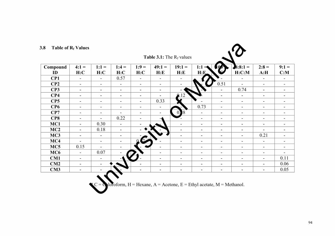
94
3.8 Table of Rf Values
Table 3.1: The Rf values
* C = Chloroform, H = Hexane, A = Acetone, E = Ethyl acetate, M = Methanol.
Compound ID
4:1 = H:C
1:1 = H:C
1:4 = H:C
1:9 = H:C
49:1 = H:E
19:1 = H:E
1:1 = H:E
100% C
8:8:1 = H:C:M
2:8 = A:H
9:1 = C:M
CP1 - - 0.57 - - - - - - - - CP2 - - - - - - - 0.51 - - - CP3 - - - - - - - - 0.74 - - CP4 - - - - - 0.12 - - - - - CP5 - - - - 0.33 - - - - - - CP6 - - - - - - 0.73 - - - - CP7 - - - - - 0.18 - - - - - CP8 - - 0.22 - - - - - - - - MC1 - 0.30 - - - - - - - - - MC2 - 0.18 - - - - - - - - - MC3 - - - - - - - - - 0.21 - MC4 - - - 0.07 - - - - - - - MC5 0.15 - - - - - - - - - - MC6 - 0.07 - - - - - - - - - CM1 - - - - - - - - - - 0.11 CM2 - - - - - - - - - - 0.06 CM3 - - - - - - - - - - 0.05
Univers
ity of
Mala
ya

95
CHAPTER 4: RESULTS AND DISCUSSION
4.1 Compound isolated from Crotalaria pallida
Eight compounds have been isolated from the hexane extract of the C. pallida.
The isolated compounds are summarized below.
HO
O
O
OHO
Crotolidene (CP1) Hydroxydihydrobovolide (CP2)
20
O
O
Octacosane (CP3) Trans-phytyl palmitate (CP4)
HO
O
Linoleic acid (CP5)
O
O
Methyl oleate (CP6)
O
O
Ethyl palmitate (CP7)
HO
O
Palmitic acid (CP8)
Univers
ity of
Mala
ya

96
4.1.1 CP1: Crotolidene (New)
HO
O1
23
4
56
78
9 10
11
1213
CP1 was isolated as colourless oil with molecular formula C14H22O2, as
determined by HRESIMS with m/z 223.1497 [M+H]+ (calculated for C14H22O2+H,
223.1698) and m/z 205.1316 [M-OH]- (calculated for C14H22O2-OH, 205.1592). CP1
showed an optical activity [α]D -66.7 (c 0.237, CHCl3). The UV spectrum showed
absorption maxima at 205 and 276 nm. The IR absorptions indicated the presence of
hydroxyl group (3250 cm-1), aliphatic group (2959, 2937 and 2870 cm-1), conjugated
ketone (1674 cm-1), and double bond (3010 cm-1).
The 1H NMR spectrum (Figure 4.2, Table 4.1) indicated the presence of four
methyl singlets at δH 1.09 (Me-12), 1.10 (Me-13), 1.75 (Me-11) and 2.27 (Me-1); one
sp3 oxymethine at δH 3.99 (H-8), two sp2 methines at δH 6.09 (H-4) and 7.19 (H-3),
and two set of sp3 methylenes at δH 2.41, 2.06 (H-7a and H-7b), and δH 1.47, 1.78 (H-
9b and H-9a). The methyl signal at δH 2.27 (δC 27.5) is typical of the methyl ketone
functionality.
The 13C NMR spectrum (Figure 4.3, Table 4.1) showed the presence of 13
carbon signals which are attributed to four methyls, two methylenes, three methines
and four quaternary carbons. The observations of the methine signal at δC 64.8 (δH
3.99) suggested an oxymethine functionality consistent with the IR results. The
quaternary carbons signal at δC 198.7 (C-2) suggested the presence of a carbonyl
functionality in agreement with the IR results.
Univers
ity of
Mala
ya

97
The COSY spectrum gave two partial structures consisted of CH2-CH(OH)-
CH2 and CH=CH, consistent with the IR, 13C and 1H NMR data as discussed above.
The first partial structure CH2-CH(OH)-CH2 containing two methylenes and an
oxymethine corresponds to C(7)-C(8)-C(9), while the second alkene-methine partial
structure CH=CH is assigned to C(3)-C(4). The observed coupling constant of 16 Hz
for the sp2 methines suggested H-3 and H-4 are arranged in trans-configuration (3E).
The HMBC spectrum (Figure 4.1) showed J2 correlations from carbonyl C-2 to
Me-1, and H-3 and J3 correlation from C-3 to Me-1 connecting the methyl ketone
functionality to the CH=CH (i.e. C(3)-C(4)) partial structure providing the Me-CO-
CH=CH- fragment. The J2 HMBC correlations from C-5 to Me-11, and J3 HMBC
correlations from C-4 to Me-11 and C-6 to H-4 and Me-11, extended the conjugation
of the fragment to Me-CO-CH=CH-C(Me)=C- (methyl-hexadienone) corresponding to
C(1)-C(2)-C(3)-C(4)-C(5)C(11)-C(6)-. Hence, three of the four degrees of unsaturation
indicated by the molecular formula of CP1 can be attributed to the C(2) ketone, and
C(3)-C(4) and C(5)-C(6) double bonds. The C-5 of the methyl-hexadienone fragment
showed J3 HMBC correlation to H-7, connecting the Me-CO-CH=CH-C(Me)=C-
(methyl-hexadienone) to the CH2-CH(OH)-CH2 partial structures together. The vice
versa J3 HMBC correlation between Me-12 and Me-13 suggested both the methyls
group are connected to the same carbon center (C-10). The observation of J3 HMBC
correlation from C-6 to Me-12 and Me-13 suggested C-6 is α positioned to the C-10
carbon center which is connecting to both Me-12 and Me-13. The final key HMBC J2
correlation from C-10 to H-9 and J3 correlations from C-9 to Me-12 and Me-13 formed
the cyclopentyl ring of structure CP1. The cyclopentyl ring accounted for the last
degree of unsaturation in the structure of CP1.
Univers
ity of
Mala
ya

98
The configuration of the C-5 and C-6 alkene is assigned as Z. In the 5Z
configuration, the Me-11 is located in further distance from other protons such as CH2-
7 and H-3. However, in the case of 5E configuration, the Me-11 is located in near
proximity with Me-12 and Me-13. The NOESY experiments of CP1 show no
correlations between Me-11 and other protons suggested no proximate protons in the
Me-11 vicinity supporting the 5Z configuration. Due to the minute amount of
compound CP1, we were not able to assign the configuration of OH-8. The
enantiomeric relationship of α-OH-8 and β-OH-8 structures was not resolvable in the
NMR experiments. Hence, the structure of crotolidene (CP1) is (3E,5Z)-5-(4-hydroxy-
2,2-dimethylcyclopentylidene)hex-3-en-2-one. This is the first report for CP1.
HO
O1
2
34
56
78
9 10
11
1213
(a)
O
HO
4
1213
(b)
Figure 4.3: Selected (a) HMBC (b) NOESY of CP1 (Crotolidene)
Univers
ity of
Mala
ya

99
Table 4.2: The NMR (CDCl3, 600 MHz) data of CP1 (Crotolidene)
Position δH δC HMBC NOESY 1 2.27 (s) 27.5 2, 3, 4 2 - 198.7 3, 11 3 7.19 (d, 16) 142.5 2, 4 1, 4, 11, 12 4 6.09 (d, 16) 132.6 3, 11 1, 3, 5, 11, 12 5 - 132.4 7, 11 6 - 135.9 4, 11, 12, 13 7
2.41 (dd, 6, 17) 2.06 (dd, 11, 17)
43.0 43.0
5, 9 5, 9
8 3.99 (m) 64.8 7, 9 8, 10, 11, 12 9
1.78 (m) 1.47 (t, 12)
48.6 48.6
7, 8, 10, 12, 13 7, 8, 10, 12, 13
10 - 37.1 9 11 12
1.75 (s) 1.09 (s)
21.8 30.3
4, 5, 6 6, 9, 13
13 1.10 (s) 28.8 6, 9, 12
Univers
ity of
Mala
ya
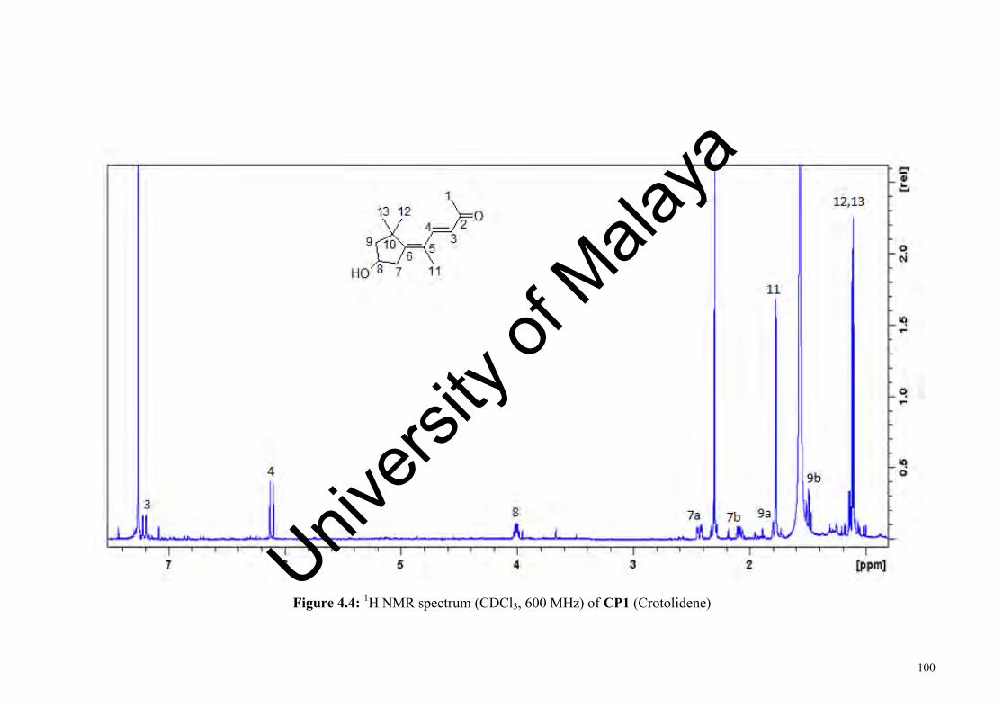
100
Figure 4.4: 1H NMR spectrum (CDCl3, 600 MHz) of CP1 (Crotolidene) Univ
ersity
of M
alaya

101
Figure 4.5: 13C NMR spectrum (CDCl3, 150 MHz) of CP1 (Crotolidene)
Univers
ity of
Mala
ya

102
4.1.2 CP2: Hydroxydihydrobovolide
O
OHO
10
23 4
56
7
8
9
CP2 was isolated as colourless oil with the molecular formula C11H18O3, as
determined by HRESIMS m/z 199.1335 [M+H]+ (calculated for C11H18O3 + H
199.1329) and m/z 181.1233 [M+H-H2O]+ (calculated for C11H18O3 - OH 181.1223).
CP2 showed an optical activity [α]D -13.3 (c 0.02, CHCl3). The UV showed absorption
maxima at 220 and 291 nm. The IR spectrum showed absorptions for hydroxyl group
(3369 cm-1), aliphatic group (2956, 2927 and 2859 cm-1), conjugated ester (1741 cm-1),
and the conjugated alkene (1694 cm-1) functionalities.
The 1H NMR spectrum (Figure 4.5, Table 4.2) showed the presence of three
methyl signals at δH 1.92 (4-Me), 1.80 (3-Me) and 0.86 (CH3-10), and three set of
methylene signals. The 13C NMR spectrum (Figure 4.6, Table 4.2) showed the
presence of eleven carbon signals consisting of three methyls, four methylenes and
four quaternary carbons. The carbon signals at δC 172.2 is typical of the carbonyl
carbon (C-2) of the 2-furanone moiety, while the carbon signal at δC 107.0 (C-5)
suggested that this carbon is flanked between two oxygen atoms.
The COSY spectrum showed only one partial structure, such as
CH3CH2CH2CH2 corresponding to C(6)-C(7)-C(8)-C(9)-C(10) pentyl side chain. The
HMBC (Figure 4.4) showed J3 correlations from C-2 and C-4 to 3-Me, and C-3 and C-
5 to 4-Me confirming the presence of the dimethyl-2-furanone moiety. The observation
13C NMR chemical shift of C-5 at δC 107.0 suggested that the hydroxyl group is
attached to this carbon. The pentyl side chain is connected to the hydroxyl-dimethyl-2-
Univers
ity of
Mala
ya
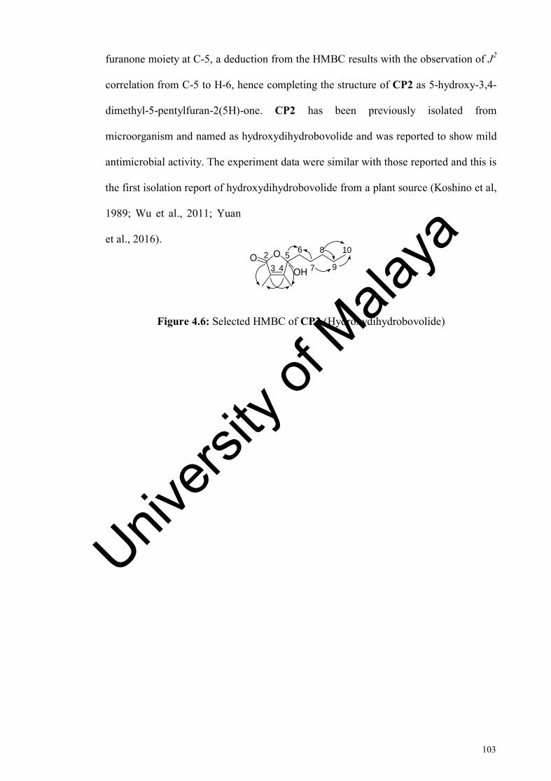
103
furanone moiety at C-5, a deduction from the HMBC results with the observation of J2
correlation from C-5 to H-6, hence completing the structure of CP2 as 5-hydroxy-3,4-
dimethyl-5-pentylfuran-2(5H)-one. CP2 has been previously isolated from
microorganism and named as hydroxydihydrobovolide and was reported to show mild
antimicrobial activity. The experiment data were similar with those reported and this is
the first isolation report of hydroxydihydrobovolide from a plant source (Koshino et al,
1989; Wu et al., 2011; Yuan
et al., 2016).
Figure 4.6: Selected HMBC of CP2 (Hydroxydihydrobovolide)
O
OHO
1023 4
5 6
7
8
9
Univers
ity of
Mala
ya
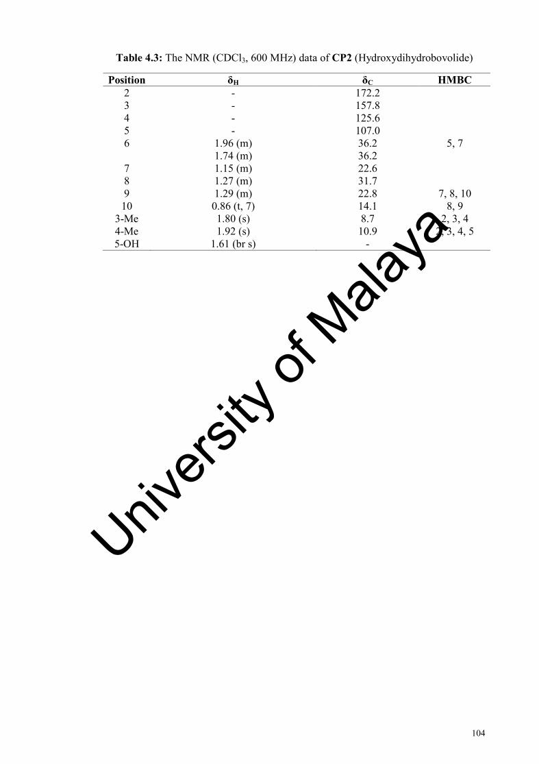
104
Table 4.3: The NMR (CDCl3, 600 MHz) data of CP2 (Hydroxydihydrobovolide)
Position δH δC HMBC 2 - 172.2 3 - 157.8 4 - 125.6 5 - 107.0 6 1.96 (m)
1.74 (m) 36.2 36.2
5, 7
7 1.15 (m) 22.6 8 1.27 (m) 31.7 9 1.29 (m) 22.8 7, 8, 10 10 0.86 (t, 7) 14.1 8, 9
3-Me 1.80 (s) 8.7 2, 3, 4 4-Me 5-OH
1.92 (s) 1.61 (br s)
10.9 -
2, 3, 4, 5
Univers
ity of
Mala
ya
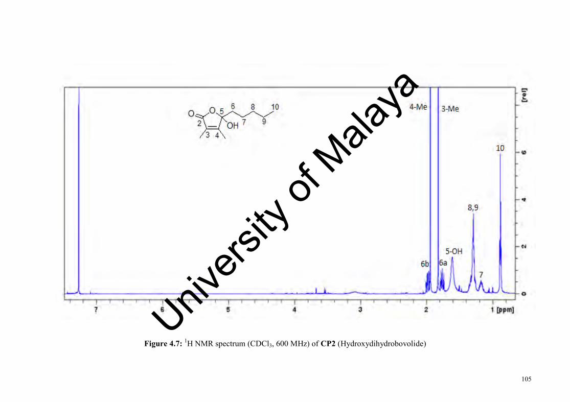
105
Figure 4.7: 1H NMR spectrum (CDCl3, 600 MHz) of CP2 (Hydroxydihydrobovolide)
Univers
ity of
Mala
ya

106
Figure 4.8: 13C NMR spectrum (CDCl3, 150 MHz) of CP2 (Hydroxydihydrobovolide)
Univers
ity of
Mala
ya
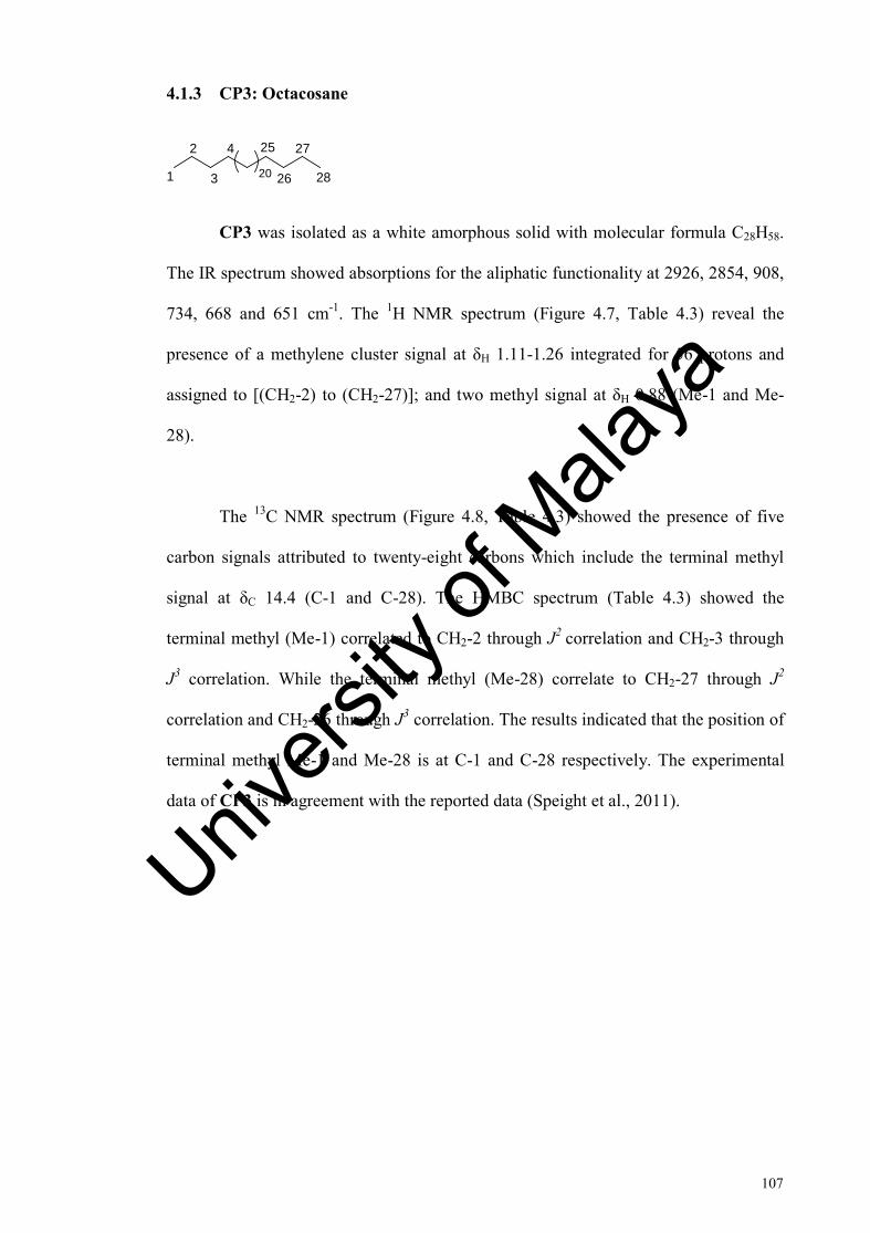
107
4.1.3 CP3: Octacosane
4
3
25
26
27
28
2
1 20
CP3 was isolated as a white amorphous solid with molecular formula C28H58.
The IR spectrum showed absorptions for the aliphatic functionality at 2926, 2854, 908,
734, 668 and 651 cm-1. The 1H NMR spectrum (Figure 4.7, Table 4.3) reveal the
presence of a methylene cluster signal at δH 1.11-1.26 integrated for 56 protons and
assigned to [(CH2-2) to (CH2-27)]; and two methyl signal at δH 0.88 (Me-1 and Me-
28).
The 13C NMR spectrum (Figure 4.8, Table 4.3) showed the presence of five
carbon signals attributed to twenty-eight carbons which include the terminal methyl
signal at δC 14.4 (C-1 and C-28). The HMBC spectrum (Table 4.3) showed the
terminal methyl (Me-1) correlated to CH2-2 through J2 correlation and CH2-3 through
J3 correlation. While the terminal methyl (Me-28) correlate to CH2-27 through J2
correlation and CH2-26 through J3 correlation. The results indicated that the position of
terminal methyl Me-1 and Me-28 is at C-1 and C-28 respectively. The experimental
data of CP3 is in agreement with the reported data (Speight et al., 2011).
Univers
ity of
Mala
ya

108
Table 4.4: The NMR (CDCl3, 400 MHz) of CP3 (Octacosane)
Position δH δC HMBC 1 0.88 (t,7) 14.4 2, 3 2 1.11 (m) 23.0 3, 4 3 1.24 (m) 32.2 2, 4 4 1.26 (m) 29.6 3, 5-24
5-24 1.26 (m) 30.0 3, 5-24 25 1.26 (m) 29.6 5-24, 26 26 1.24 (m) 32.2 25, 27 27 1.11 (m) 23.0 25, 26 28 0.88 (t, 7) 14.4 26, 27
Univers
ity of
Mala
ya
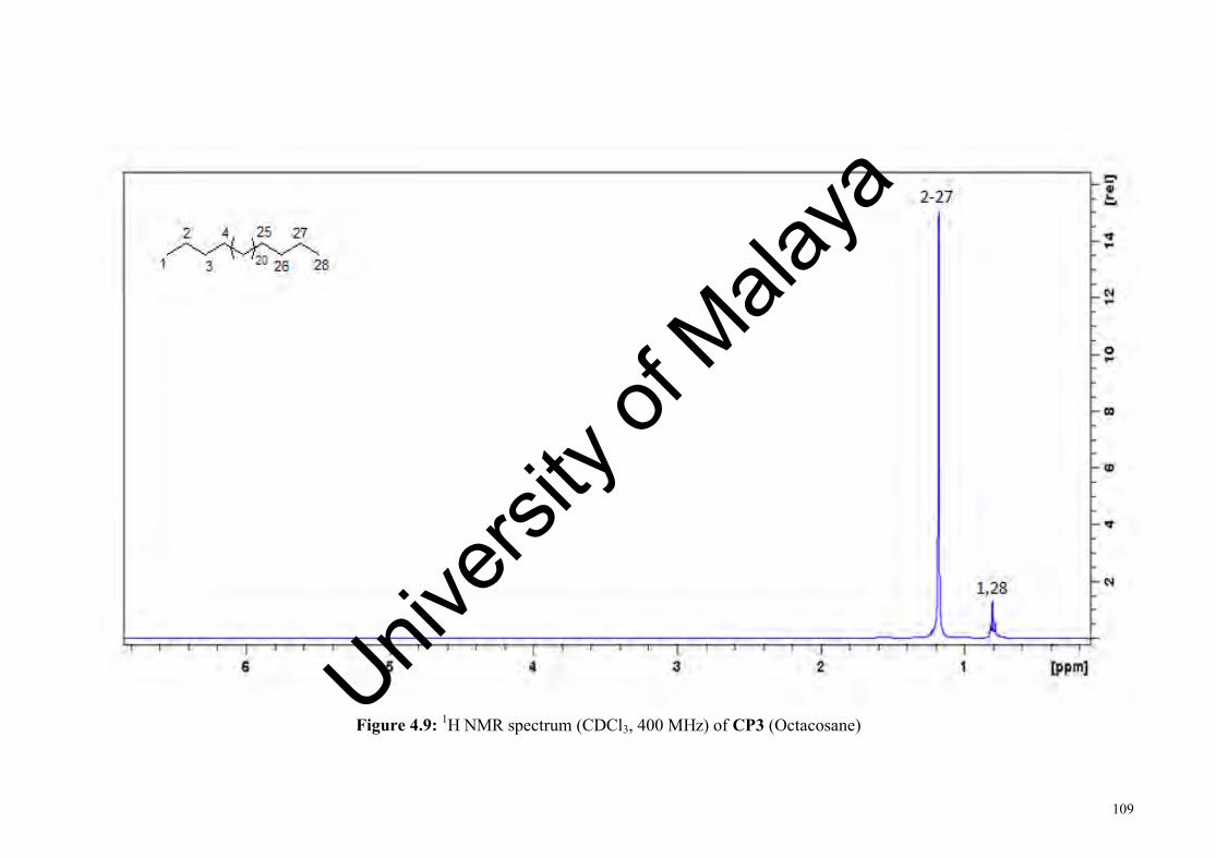
109
Figure 4.9: 1H NMR spectrum (CDCl3, 400 MHz) of CP3 (Octacosane)
Univers
ity of
Mala
ya
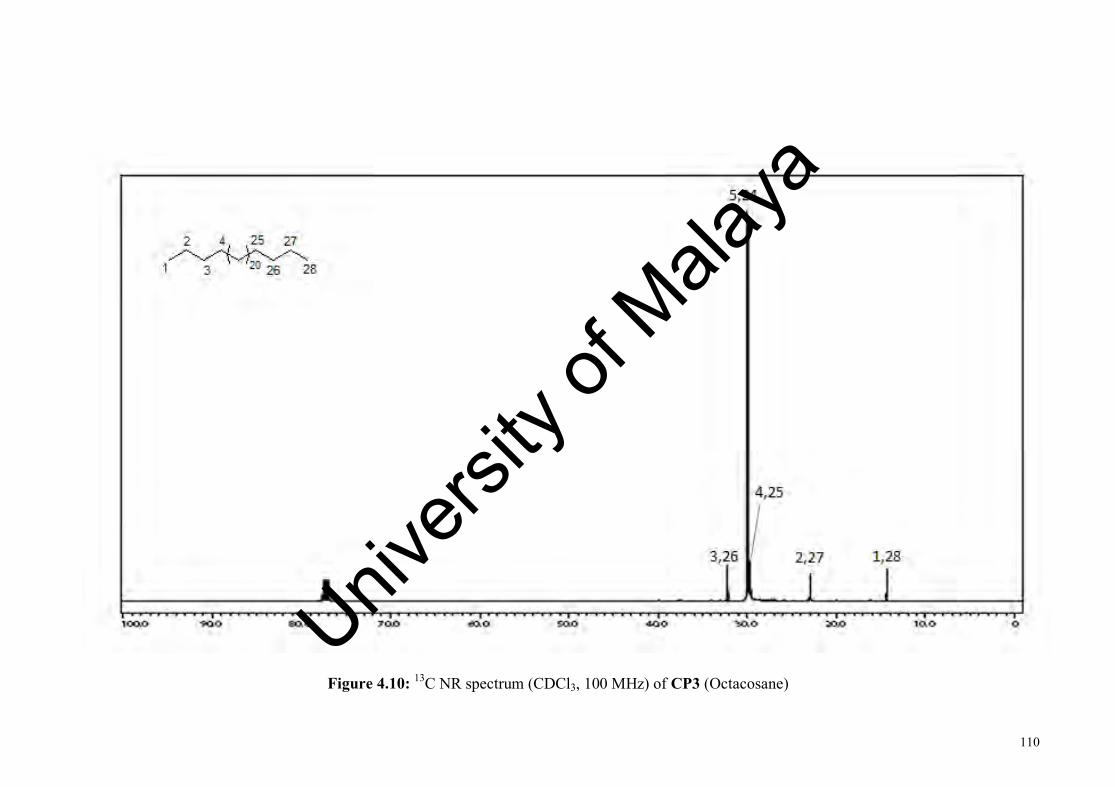
110
Figure 4.10: 13C NR spectrum (CDCl3, 100 MHz) of CP3 (Octacosane)
Univers
ity of
Mala
ya

111
4.1.4 CP4: Trans-phytyl palmitate
O
O1
14
13
12
13'
10
14'
11
8
15'
16'
16
15 9 7
6
5
12'
4
2
11'
3
10'
9'
6'8' 4'
7' 5' 3'
2'
1'
CP4 was isolated as white amorphous solid with molecular formula C36H70O2
as determined by GCMS which give m/z 57 (79), 68 (100) [C5H8]+, 71 (43), 82 (74), 95
(83), 123 (36), 137 (4). CP4 showed an optical activity of [α]D -1.4 (c 0.237, CHCl3).
The UV spectrum showed absorption maximum at 210 nm. The IR spectrum showed
absorptions for aliphatic (2916 and 2849 cm-1), carbonyl (1732 cm-1) and vinyl (1641
cm-1) functionalities.
The 1H NMR spectrum (Figure 4.10, Table 4.4) revealed the presence of an
olefinic proton signal at δH 5.30 (H-2’), methine signals at δH 1.50 (H-15’), 1.35 (H-7’
and H-11’); methylene signals at δH 4.56 (CH2-1’), δH 2.28 (CH2-2), 1.97 (CH2-4’),
1.58 (CH2-3), 1.38 (CH2-5’), 1.25 [CH2-(4-15), CH2-9’, CH2-12’ and CH2-13’], 1.13
(CH2-14’) and 1.05 (CH2-6’, CH2-8’ and CH2-10’); a methyl group attached to a
quaternary carbon at δH 1.68 (3’-Me); a methyl group attached to a secondary carbon at
δH 0.88 (Me-16); and four methyl groups attached to tertiary carbon at δH 0.84-0.86
(Me-16’, 7’-Me, 11’-Me and 15’-Me). The methylene signal at δH 4.56 (CH2-1’)
appeared at the most deshielded region due to its location which is adjacent to the ester
oxygen. The 13C NMR spectrum (Figure 4.11, Table 4.4) showed the presence of 36
carbon atoms which include a carbonyl carbon at δC 174.1 (C-1), and a quaternary
olefinic carbon at δC 142.7 (C-3’).
Univers
ity of
Mala
ya

112
The HMBC spectrum (Figure 4.9, Table 4.4) showed J3 correlation from C-1 to
CH2-1’ indicated the connectivity between ester bond from the phytyl moiety to the
fatty acid moiety. 3’-Me showed HMBC correlations to H-2’ and CH2-4’; 7’-Me
showed HMBC correlations to CH2-6’, H-7’and CH2-8’; 11’-Me showed HMBC
correlation to H-11’; 15’-Me showed HMBC correlations to H-15’ and Me-16’; and
Me-16’ showed HMBC correlations to H-15’ and 15’-Me confirming the position of
branched methyl of phytyl moiety are at C-3’, C-7’, C-11’ and C-15’ respectively. The
HMBC correlations between C-2’ with CH2-1’ and C-3’ with CH2-1’ indicated the
presence of a double bond at C(2’)=C(3’). The J3 HMBC correlations from C-16’ to
15’-Me and vice versa indicated the position of terminal methyl from phytyl moiety is
at C-16’ (Asand et al., 1991).
O
O
Figure 4.11: Selected HMBC of CP4 (Trans-phytyl palmitate)
Univers
ity of
Mala
ya

113
Table 4.5: The NMR (CDCl3, 400 MHz) data of CP4 (Trans-phytyl palmitate)
Position δH δC HMBC 1 - 174.1 2 2.28 (t, 8) 34.6 1, 3, 4, 3 1.58 (m) 25.2 1, 2, 4 4 1.25 (m) 29.4
5 1.25 (m) 29.5
6 1.25 (m) 29.7
7-10 1.25 (m) 29.9
11 1.25 (m) 29.8 12 1.25 (m) 29.6
13 1.25 (m) 29.6
14 1.25 (m) 32.2
15 16
1.25 (m) 0.88 (m)
22.9 14.3
14
1’ 4.56 (d, 7) 61.4 1, 2’, 3’ 2’ 5.30 (t, 7) 118.5 1’,3’-Me, 4’ 3’ - 142.7 4’ 1.97 (t, 7) 40.1 2’, 3’, 3’-Me, 5’, 6’ 5’ 1.38 (m) 25.2 4’, 6’ 6’ 1.05 (m) 37.7 5’ 7’ 1.35 (m) 33.0 5’, 9’ 8’ 1.05 (m) 37.6 9’ 9’ 1.25 (m) 24.7
10’ 1.05 (m) 37.5 9’ 11’ 1.35 (m) 33.0 9’, 13’ 12’ 1.25 (m) 36.8
13’ 1.25 (m) 25.0 12’, 14’ 14’ 1.13 (m) 39.6
15’ 16’
1.50 (m) 0.86 (m)
28.2 22.8
13’,15’-Me, 16’ 15’-Me
3’-Me 7’-Me 11’-Me 15’-Me
1.68 (s) 0.84 (m) 0.84 (m) 0.86 (m)
16.6 19.9 19.9 22.8
1’, 3’, 4’ 6’, 7’, 8’
11’ 16’
Univers
ity of
Mala
ya

114
Figure 4.12: 1H NMR spectrum (CDCl3, 400 MHz) of CP4 (Trans-phytyl palmitate) Univ
ersity
of M
alaya

115
Figure 4.13: 13C NMR spectrum (CDCl3, 100 MHz) of CP4 (Trans-phytyl palmitate) Univ
ersity
of M
alaya
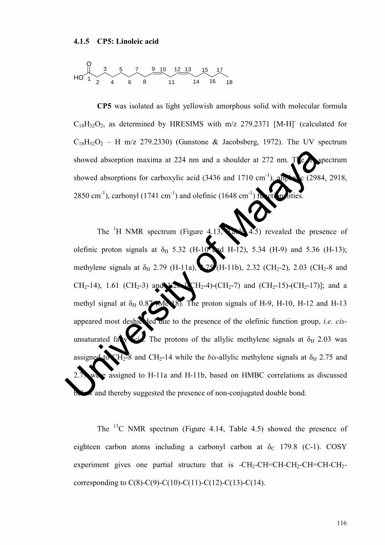
116
4.1.5 CP5: Linoleic acid
3
4
5
6
7
8
9 10
11
12 13
14
15HO
O
16
17
181 2
CP5 was isolated as light yellowish amorphous solid with molecular formula
C18H32O2, as determined by HRESIMS with m/z 279.2371 [M-H]- (calculated for
C18H32O2 – H m/z 279.2330) (Gunstone & Jacobsberg, 1972). The UV spectrum
showed absorption maxima at 224 nm and a shoulder at 272 nm. The IR spectrum
showed absorptions for carboxylic acid (3436 and 1710 cm-1), aliphatic (2984, 2918,
2850 cm-1), carbonyl (1741 cm-1) and olefinic (1648 cm-1) functionalities.
The 1H NMR spectrum (Figure 4.13, Table 4.5) revealed the presence of
olefinic proton signals at δH 5.32 (H-10 and H-12), 5.34 (H-9) and 5.36 (H-13);
methylene signals at δH 2.79 (H-11a), 2.75 (H-11b), 2.32 (CH2-2), 2.03 (CH2-8 and
CH2-14), 1.61 (CH2-3) and 1.26 [(CH2-4)-(CH2-7) and (CH2-15)-(CH2-17)]; and a
methyl signal at δH 0.87 (Me-18). The proton signals of H-9, H-10, H-12 and H-13
appeared most deshielded due to the presence of the olefinic function group, i.e. cis-
unsaturated fatty acid. The protons of the allylic methylene signals at δH 2.03 was
assigned to CH2-8 and CH2-14 while the bis-allylic methylene signals at δH 2.75 and
2.79 were assigned to H-11a and H-11b, based on HMBC correlations as discussed
below and thereby suggested the presence of non-conjugated double bond.
The 13C NMR spectrum (Figure 4.14, Table 4.5) showed the presence of
eighteen carbon atoms including a carbonyl carbon at δC 179.8 (C-1). COSY
experiment gives one partial structure that is -CH2-CH=CH-CH2-CH=CH-CH2-
corresponding to C(8)-C(9)-C(10)-C(11)-C(12)-C(13)-C(14).
Univers
ity of
Mala
ya

117
The HMBC spectrum (Figure 4.12, Table 4.5) showed correlations of carbonyl
carbon to the methylene protons through J2 correlation (C-1 to CH2-2) and J3
correlation (C-1 and CH2-3). The J2 HMBC correlations from C-9 to CH2-8 and C-13
to CH2-14; J3 HMBC correlations from C-10 to CH2-8 and C-12 to CH2-14 indicated
that the position of allylic methylene is at C-8 and C-14. The HMBC correlations from
C-9, C-10, C-12 and C-13 to H-11a and H-11b confirmed the position of bis-allylic
methylene is at C-11. The J2 HMBC correlations between C-14 and C-16 to CH2-15,
and J3 HMBC correlation from C-17 to CH2-15 suggested that the position of the
double bonds were between C-9/C-10 and C-12/C-13. The terminal methyl (Me-18)
showed J2 HMBC correlation with CH2-17 and J3 HMBC correlation with CH2-16
confirmed the position of the terminal methyl is at C-18. The experimental data for
CP5 is in agreement with the reported data (Gunstone & Jacobsberg, 1972; Vlahov,
1999).
9 13HO
O
18
1
Figure 4.12: Selected HMBC of CP5 (Linoleic acid)
Univers
ity of
Mala
ya

118
Table 4.5: The NMR (CDCl3, 600 MHz) data of CP5 (Linoleic acid)
Position δH δC HMBC
1 - 179.8 2 2.32 (t, 8) 34.2 1, 3, 4 3 1.61 (m) 24.9 1, 2, 4 4 1.26 (m) 29.3 5 1.26 (m) 29.3 6 1.26 (m) 29.6 7 1.26 (m) 29.8 8 2.03 (m) 27.4 7, 9, 10 9 5.34 (m) 130.3 8, 10, 11 10 5.32 (m) 128.1 8, 9, 11 11 2.75 (t, 7)
2.79 (t, 7) 25.9 9, 10, 12, 13
12 5.32 (m) 128.3 11, 13, 14 13 5.36 (m) 130.5 11, 12, 14 14 2.03 (m) 27.4 12, 13, 15 15 1.26 (m) 29.9 14, 16, 17 16 1.26 (m) 31.8 17 1.26 (m) 22.8 18, 18 0.87 (t, 7) 14.3 16, 17
Univers
ity of
Mala
ya

119
Figure 4.13: 1H NMR spectrum (CDCl3, 600 MHz) of CP5 (Linoleic acid)
Univers
ity of
Mala
ya

120
Figure 4.14: 13C NMR spectrum (CDCl3, 150 MHz) of CP5 (Linoleic acid)
Univers
ity of
Mala
ya

121
4.1.6 CP6: Methyl oleate
3
4
5
6
7
8
9 10
11
12
13
14
15O
O16
17
181
2
1'
CP6 was isolated as yellowish oil with molecular formula C19H36O2, as
determined by GCMS with m/z 296 (3) [M+], 264 (14), 222 (10), 180 (8), 137 (9), 123
(17), 110 (27), 97 (52), 83 (61), 73 (60), 69 (74), 55 (100). The UV spectrum showed
absorption maxima at 202 nm with two shoulders at 222 and 277 nm. The IR spectrum
showed absorptions for aliphatic (2926 and 2855 cm-1) and carbonyl (1744 cm-1)
functionalities (Rakoff et al., 1979).
The 1H NMR spectrum (Figure 4.16, Table 4.6) revealed the presence of an
olefinic proton signal at δH 5.32 which was integrated for two protons and attributed to
H-9 and H-10; a methoxyl signal at δH 3.64 (OMe-1’); four group of methylene signals
at δH 2.28 (CH2-2), δH 1.98 (CH2-8 and CH2-11), δH 1.59 (CH2-3), δH 1.28 [(CH2-4)-
(CH2-7) and (CH2-12)-(CH2-17)]; and methyl signals at δH 0.86 (Me-18). The 13C
NMR spectrum (Figure 4.17, Table 4.6) showed the presence of 19 carbon signal
including a carbonyl signal at δC 174.6 (C-1). COSY experiment give one partial
structure that is CH2-CH=CH-CH2 corresponding to C(8)-C(9)-C(10)-C(11).
The HMBC spectrum (Figure 4.15, Table 4.6) showed J3 correlation from C-1
to OMe-1’, hence confirming the position of the terminal methyl ester. The carbonyl
carbon at C-1 showed J2 and J3 HMBC correlations with the α-methylene proton (CH2-
2) and β-methylene proton (CH2-3), respectively. The J2 and J3 HMBC correlations
from C-2 to CH2-3 and CH2-4, respectively indicated the position of α-methylene
signal is at C-2. While the J2 and J3 HMBC correlations from C-3 to CH2-2 and CH2-4
indicated the position of β-methylene signal is at C-3. The J2 HMBC correlations from
Univers
ity of
Mala
ya

122
C-8 to CH2-7 and H-9, C-11 to H-10 and CH2-12; J3 HMBC correlations from C-8 to
H-10 and C-11 to H-9 indicated that the position of allylic methylene is at C-8 and C-
11. The partial structure from COSY experiment (CH2-CH=CH-CH2) connecting with
C-7 and C-12 providing the fragment of CH2-CH2-CH=CH-CH2-CH2. The terminal
methyl (Me-18) showed J2 HMBC correlation with CH2-17 and J3 HMBC correlation
with CH2-16 confirmed the position of the terminal methyl is at C-18. The
experimental data of CP6 is in agreement with the reported data (Knothe et al., 1995;
Lie et al., 1995; Rakoff et al., 1979).
O
O18
1
9 101'
Figure 4.15: Selected HMBC of CP6 (Methyl oleate)
Table 4.6: The NMR (CDCl3, 600 MHz) data of CP6 (Methyl oleate)
Position δH δC HMBC
1’ 3.64 (s) 52.7 1 1 - 174.6 2 2.28 (t, 8) 34.3 1, 3, 4 3 1.59 (m) 25.2 1, 2, 4 4 1.28 (m) 29.4 5 1.28 (m) 29.3 6 1.28 (m) 29.3 7 1.28 (m) 29.9 8 1.98 (m) 27.4 7, 9, 10 9 5.32 (m) 130.0 8, 11 10 5.32 (m) 130.2 8, 11 11 1.98 (m) 27.4 9, 10, 12 12 1.28 (m) 30.0 13 1.28 (m) 29.5 14 1.28 (m) 29.7 15 1.28 (m) 29.5 16 1.28 (m) 32.1 17 1.28 (m) 22.9 16 18 0.86 (t, 7) 14.3 16, 17
Univers
ity of
Mala
ya

123
Figure 4.16: 1H NMR spectrum (CDCl3, 600 MHz) of CP6 (Methyl oleate)
Univers
ity of
Mala
ya

124
Figure 4.17: 13C NMR spectrum (CDCl3, 150 MHz) of CP6 (Methyl oleate)
Univers
ity of
Mala
ya

125
4.1.7 CP7: Ethyl palmitate
3
4
5
6
7
8
9
10
11
12
13
14
15
161
2
1'O
O
2'
CP7 was isolated as yellowish oil with molecular formula C18H36O2,
determined by GCMS m/z 284 (4) [M]+, 255 (2), 241 (6), 157 (11), 101 (53), 88 (100),
70 (26), 55 (27) (Joshi et al., 2009). The UV spectrum showed absorptions at 206 and
274 nm. The IR spectrum showed absorptions for aliphatic (2924 and 2853 cm-1) and
carbonyl (1740 cm-1) functional groups.
The 1H NMR spectrum (Figure 4.19, Table 4.7) showed OCH2- signal at δH
4.08 (CH2-1’); methylene signals at δH 2.38 (CH2-2), 1.57 (CH2-3), 1.24 [(CH2-4)-
(CH2-15)], and two methyl signals at δH 1.21 (Me-2’) and 0.84 (Me-16). The DEPT-
135 NMR spectrum (Figure 4.20, Table 4.7) showed the presence of 17 carbon signals
which corresponded to the two methyl and fifteen methylene functionalities. COSY
experiment gives two partial structures that are OCH2-CH3 corresponding to C(1’)-
C(2’) and CH2-CH2-(CH2)12-CH3 corresponding to -C(2)-C(3)-C(4-15)-C(16). The
observe pattern of these protons suggested the presence of ethyl ester functionality in
the compound.
The presence of the ethyl ester moiety was confirmed by the HMBC
experiment. The HMBC spectrum (Figure 4.18, Table 4.7) indicated the presence of
the ethyl ester bond through J3 correlation from C-1 to CH2-1’. Meanwhile, the
presence of the ethyl moiety was indicated through J2 HMBC correlation from C-1’ to
Me-2’.In addition, the carbonyl carbon at C-1 showed J2 and J3 HMBC correlations to
CH2-2 and CH2-3. The C-2 methylene showed J2 and J3 HMBC correlations to CH2-3
and CH2-4, respectively; C-3 methylene carbon showed J2 HMBC correlations to CH2-
Univers
ity of
Mala
ya

126
2 and CH2-4. The terminal methyl (Me-16) showed J2 and J3 HMBC correlations to
CH2-15 and CH2-14, respectively, suggesting the position of the terminal methyl is at
C-16. The experimental data of CP7 is in agreement with the reported data (Joshi et
al., 2009).
O
O
2' 1 16
1'
Figure 4.18: Selected HMBC of CP7 (Ethyl palmitate)
Table 4.7: The NMR (CDCl3) data of CP7 (Ethyl palmitate)
a Value obtained from HMBC spectrum.
Position δH δC HMBC 1 1’
- 4.08 (q, 7)
173.8a
60.4 1
2 2’
2.38 (t, 8) 1.21 (t, 7))
34.6 14.5
1, 3,4 1’
3 1.57 (p, 8) 25.3 2, 4 4 1.24 (m) 29.4 5 1.24 (m) 29.6 6 1.24 (m) 29.8 7 1.24 (m) 29.9 8 1.24 (m) 30.0 9 1.24 (m) 30.0 10 1.24 (m) 30.0 11 1.24 (m) 29.9 12 1.24 (m) 29.9 13 1.24 (m) 29.7 14 1.24 (m) 32.2 15 1.24 (m) 23.0 14 16 0.84 (t, 7) 14.4 14, 15
Univers
ity of
Mala
ya

127
Figure 4.19: 1H NMR spectrum (CDCl3, 600 MHz) of CP7 (Ethyl palmitate)
Univers
ity of
Mala
ya

128
Figure 4.20: DEPT-135 NMR spectrum (CDCl3, 150 MHz) of CP7 (Ethyl palmitate) Univ
ersity
of M
alaya

129
4.1.8 CP8: Palmitic acid
3
4
5
6
7
8
9
10
11
12
13
14
15
161
2HO
O
CP8 was isolated as white amorphous solid with molecular formula C16H32O2
as determined by HRESIMS with m/z 255.2369 [M-H]- (calculated for C16H32O2 - H
m/z 255.2330) (Miranda-Vilela & Grisolia, 2009). The UV spectrum showed
absorption maxima at 208 and 268 nm. The IR spectrum showed absorptions for
carboxylic acid (3409 cm-1), aliphatic (2917 cm-1 and 2850 cm-1) and carbonyl (1707
cm-1) functionalities.
The 1H NMR spectrum (Figure 4.22, Table 4.8) revealed the presence of
methylene signals at δH 2.33 (CH2-2), 1.61 (CH2-3), 1.28 [(CH2-4)-(CH2-15)], and
methyl signal at δH 0.86 (Me-16). The 13C NMR spectrum (Figure 4.23, Table 4.8)
showed the presence of 16 carbon signals corresponded to a methyl and fourteen
methylene and a carbonyl carbon at δC 178.7 (C-1).
The HMBC spectrum (Figure 4.21, Table 4.8) showed J2 correlation from the
carbonyl C-1 to CH2-2. The C-2 methylene carbon showed J2 and J3 HMBC
correlations to CH2-3 and CH2-4; and the C-3 methylene carbon showed J2 HMBC
correlation to CH2-2. The terminal Me-16 showed J2 and J3 HMBC correlations to
CH2-15 and CH2-14, respectively, suggesting the position of terminal methyl is at C-
16. The experimental data of CP8 is in agreement with the reported data (Miranda-
Vilela & Grisolia, 2009).
Univers
ity of
Mala
ya
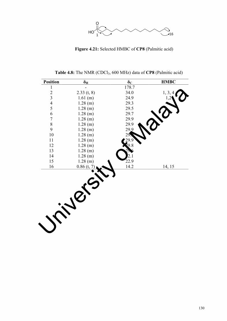
130
161HO
O
Figure 4.21: Selected HMBC of CP8 (Palmitic acid)
Table 4.8: The NMR (CDCl3, 600 MHz) data of CP8 (Palmitic acid)
Position δH δC HMBC 1 - 178.7 2 2.33 (t, 8) 34.0 1, 3, 4 3 1.61 (m) 24.9 1,2 4 1.28 (m) 29.3 5 1.28 (m) 29.5 6 1.28 (m) 29.7 7 1.28 (m) 29.9 8 1.28 (m) 29.9 9 1.28 (m) 29.9 10 1.28 (m) 29.9 11 1.28 (m) 29.9 12 1.28 (m) 29.8 13 1.28 (m) 29.6 14 1.28 (m) 32.1 15 1.28 (m) 22.9 16 0.86 (t, 7) 14.2 14, 15
Univers
ity of
Mala
ya

131
Figure 4.22: 1H NMR spectrum (CDCl3, 600 MHz) of CP8 (Palmitic acid)
Univers
ity of
Mala
ya

132
Figure 4.23: 13C NMR spectrum (CDCl3, 150 MHz) of CP8 (Palmitic acid)
Univers
ity of
Mala
ya

133
4.2 Compounds isolated from Morinda citrifolia
Six compounds were isolated from the chloroform extract of the roots of M.
citrifolia. The isolated compounds are summarized below.
OH
O
OMeO
O OH
O
OHO
O Damnacanthal (MC1) Nordamnacanthal (MC2)
OH
MeOHO
O
MeOHO
OHO
Rubiadin (MC3) 1,6-Dihydroxy-2-methylanthraquinone (MC4)
OHO
OOMe
OHOMeO
O
1-Hydroxy-3- 1-Methoxy-2- methoxyanthraquinone (MC5) hydroxyanthraquinone (MC6)
Univers
ity of
Mala
ya

134
4.2.1 MC1: Damnacanthal
OMeO
OOH
O
345
6
78
9
1010a
9a
4a
8a 1 2
A B C
MC1 was isolated as yellow amorphous powder with molecular formula
C16H10O5 as determined by ESI-MS with m/z 281.0 [M-H]- (calculated for C16H10O5-
H, 281.0). The UV spectrum showed absorption maxima at 400, 285, 255, and 210 nm.
The IR spectrum indicated the presence of hydroxyl group (3462 cm-1), conjugated
carbonyl group (1678 cm-1), and aromatic ring (1575, 1457 cm-1) functionalities.
The 1H NMR spectrum (Figure 4.25 and Table 4.9) revealed the presence of
one methoxyl signal at δH 4.13 (1-OMe), five resolved aromatic signals at δH 7.67 (H-
4), 7.77 (H-6), 7.82 (H-7), 8.24 (H-5) and 8.29 (H-8); an aldehyde signal at δH 10.46
(2-CHO), and a hydroxyl signal at δH 12.27 (3-OH). A set of doublet of doublet (dd) at
δH 8.24 and δH 8.29, integrating for two protons and were assigned to H-5 and H-8,
respectively. Another set of triplet of doublets (td) at δH 7.77 and δH 7.82, integrating
for two protons and were assigned to H-6 and H-7, respectively. The COSY
experiment gave a partial structure of -CH=CH-CH=CH-, corresponding to (C-5)-(C-
6)-(C-7)-(C-8). The observed pattern of these four proton signals in the aromatic
region suggested the presence of unsubstituted aromatic moiety at ring A. The isolated
aromatic proton at δH 7.67 was assigned to H-4 of ring C. Meanwhile, a hydroxyl, a
methoxyl and a formyl group were accounted as substituents in ring C.
The 13C NMR spectrum (Figure 4.26 and Table 4.9) showed the presence of 16
carbon signals which included three carbonyl signals at δC 195.5 (2-CHO), 181.9 (C-
10), and 180.2 (C-9); and one methoxy carbon at δC 64.8 (1-OMe). The carbon signal
Univers
ity of
Mala
ya

135
at δC 166.9 (C-1) and 166.7 (C-3) suggested the position of these carbons were
adjacent to an oxygen atom.
The HMBC spectrum (Figure 4.24 and Table 4.9) showed the connectivity
between ring A and ring B through J3 correlations from C-5 to H-7, C-6 to H-8, C-7 to
H-5, C-8 to H-6, C-8a to H-7, C-9 to H-8, C-10 to H-5, C-10a to H-6; indicating that
the connection points of benzene ring with the aliphatic ring were at C-8a and C-10a.
While the connectivity between ring C and ring B were observed through J2 HMBC
correlations from C-3 to H-4 and C-4a to H-4; and J3 HMBC correlations from C-10 to
H-4, C-9a to H-4, C-2 to H-4; indicating that the connection points of benzene ring
with the aliphatic ring were at C-4a and C-9a. The J3 HMBC correlations between C-1
and C-3 to 2-CHO, and J4 HMBC correlation from C-4 to 2-CHO suggested that the
position of the carbonyl group were at C-2. The J3 HMBC correlation from C-1 to 1-
OMe suggested that the position of the methoxyl group was at C-1. The experimental
data of MC1 is in agreement with the reported data (Kamiya et al., 2010).
OMeO
OOH
O
345
6
78
9
10
12
Figure 4.24: Selected HMBC correlations of MC1 (Damnachantal)
Univers
ity of
Mala
ya

136
Table 4.9: The NMR (CDCl3, 600 MHz) data of MC1 (Damnacanthal)
Position δH δC HMBC 1 - 166.9 1-OMe, 3-OH, 2-CHO 2 - 118.1 4, 3-OH 3 - 166.7 4, 2-CHO 4 7.67 (s) 113.1 3-OH, 2-CHO 4a - 141.6 4, 3-OH 5 8.24 (dd, 8, 1) 127.4 7 6 7.77 (td, 8, 1) 133.7 8 7 7.82 (td, 8, 1) 134.9 5 8 8.29 (dd, 8, 1) 127.2 6 8a - 134.9 7 9 - 180.2 8 9a - 117.7 4 10 - 181.9 4, 5 10a - 132.5 6
1-OMe 4.13 (s) 64.8 3-OH 12.27( s) -
2-CHO 10.46 (s) 195.5 3-OH
Univers
ity of
Mala
ya
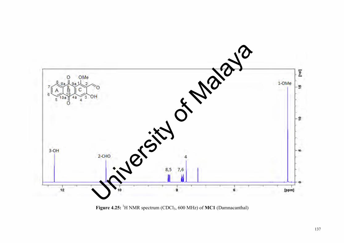
137
Figure 4.25: 1H NMR spectrum (CDCl3, 600 MHz) of MC1 (Damnacanthal)
Univers
ity of
Mala
ya

138
Figure 4.26: 13C NMR spectrum (CDCl3, 150 MHz) of MC1 (Damnacanthal)Univ
ersity
of M
alaya

139
4.2.2 MC2: Nordamnacanthal
OHO
OOH
O
345
6
78
9
104a
8a 1 29a
10a
A B C
MC2 was isolated as yellowish-orange needles with molecular formula
C15H8O5 as determined by ESI-MS with m/z 267.0 [M-H]- (calculated for C15H8O5-H,
267.0). The UV spectrum showed absorption maxima at 430, 290, 265, and 210 nm.
The IR spectrum indicated the presence of hydroxyl group (3075 cm-1), conjugated
carbonyl group (1674 cm-1), and aromatic ring (1593, 1574 cm-1) functionalities.
The 1H NMR spectrum (Figure 4.28 and Table 4.10) revealed the presence of
four sets of resolved aromatic signals: Two sets of doublet of doublet (dd) at δH 8.28
and δH 8.31 integrating for two protons and were assigned to H-5 and H-8,
respectively; multiplet (m) at δH 7.83, integrating for two protons and were assigned to
H-6 and H-7, respectively; singlet at δH 7.33 (H-4). In addition, an aldehyde proton
was observed at δH 10.50 (2-CHO), and two hydroxyl signals at δH 12.67 (3-OH) and
14.05 (1-OH). The COSY experiment gave a partial structure of -CH=CH-CH=CH-,
which corresponds to (C-5)-(C-6)-(C-7)-(C-8). The observed pattern of these four
proton signals in the aromatic region suggested the presence of unsubstituted aromatic
moiety at ring A. The isolated aromatic proton at δH 7.33 (H-4) was assigned to ring C,
as well as two hydroxyls and a formyl group. The 13C NMR spectrum (Figure 4.29 and
Table 4.10) showed the presence of 15 carbon signals which include three carbonyl
signals at at δC 194.1 (2-CHO), 186.9 (C-9), and 181.5 (C-10). The carbon signal at δC
169.2 (C-1) and 168.3 (C-3) suggested the position of these carbons were adjacent to
an oxygen atom.
Univers
ity of
Mala
ya

140
The HMBC spectrum (Figure 4.27 and Table 4.10) showed the connectivity
between ring A and ring B through J3 correlations from C-5 to H-7, C-6 to H-8, C-7 to
H-5, C-8 to H-6, C-8a to H-7, C-8a to H-5, C-9 to H-8, C-10 to H-5, C-10a to H-6 and
C-10a to H-8; indicating that the connection points of benzene ring with the aliphatic
ring were at C-8a and C-10a. While the connectivity between ring C and ring B were
observed through J3 HMBC correlations from C-10 to H-4, C-9a to H-4, and C-2 to H-
4; indicating that the connection points of benzene ring with the aliphatic ring were at
C-4a and C-9a. The J3 HMBC correlations between C-1 and C-3 to 2-CHO; and J4
HMBC correlations from C-4 and C-9a to 2-CHO suggested that the position of the
carbonyl group were at C-2. The experimental data of MC2 is in agreement with the
reported data (Kamiya et al., 2010).
OHO
OOH
O
345
6
78
9
10
12
Figure 4.27: Selected HMBC correlations of MC2 (Nordamnachantal)
Univers
ity of
Mala
ya
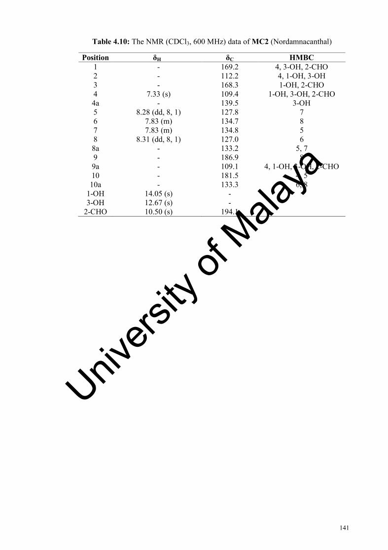
141
Table 4.10: The NMR (CDCl3, 600 MHz) data of MC2 (Nordamnacanthal)
Position δH δC HMBC 1 - 169.2 4, 3-OH, 2-CHO 2 - 112.2 4, 1-OH, 3-OH 3 - 168.3 1-OH, 2-CHO 4 7.33 (s) 109.4 1-OH, 3-OH, 2-CHO 4a - 139.5 3-OH 5 8.28 (dd, 8, 1) 127.8 7 6 7.83 (m) 134.7 8 7 7.83 (m) 134.8 5 8 8.31 (dd, 8, 1) 127.0 6 8a - 133.2 5, 7 9 - 186.9 8 9a - 109.1 4, 1-OH, 3-OH, 2-CHO 10 - 181.5 4, 5 10a - 133.3 6, 8
1-OH 14.05 (s) - 3-OH 12.67 (s) -
2-CHO 10.50 (s) 194.1
Univers
ity of
Mala
ya

142
Figure 4.28: 1H NMR spectrum (CDCl3, 600 MHz) of MC2 (Nordamnacanthal)
Univers
ity of
Mala
ya

143
Figure 4.29: 13C NMR spectrum (CDCl3, 150 MHz) of MC2 (Nordamnacanthal) Univ
ersity
of M
alaya

144
4.2.3 MC3: Rubiadin
OHO
OOH
Me
345
6
78
9
104a
8a 1 29a
10a
A B C
MC3 was isolated as yellow amorphous powder with molecular formula
C15H10O4 as determined by ESI-MS with m/z 253.0 [M-H]- (calculated for C15H10O4-H,
253.1). The UV spectrum showed absorption maxima at 425, 280, 245, and 210 nm.
The IR spectrum indicated the presence of hydroxyl group (3391 cm-1), conjugated
carbonyl group (1629 cm-1), and the aromatic ring (1593 cm-1) functionalities.
The 1H NMR spectrum (Figure 4.31 and Table 4.11) revealed the presence a
methyl signal at δH 2.11(2-Me) and one hydroxyl signal at δH 13.07 (1-OH). In
addition, two sets of doublet of doublet (dd) at δH 8.11 and δH 8.18, integrating for two
protons and were assigned to H-5 and H-8, respectively. Another set of triplet of
doublet (td) at δH 7.64 and δH 7.68, integrating for two protons and were assigned to H-
6 and H-7, respectively. The COSY experiment gave a partial structure of -CH=CH-
CH=CH-, corresponding to (C-5)-(C-6)-(C-7)-(C-8). The observed pattern of these
four proton signals in the aromatic region suggested the presence of unsubstituted
aromatic moiety at ring A.
The 13C NMR spectrum (Figure 4.32 and Table 4.11) showed the presence of
15 carbon signals which include two carbonyl signals at δC 186.8 (C-9) and 183.7 (C-
10); and one methyl signal at δC 8.3 (2-Me). The carbon signal at δC 163.3 (C-1) and
162.7 (C-3) suggested the position of these carbons were adjacent to an oxygen atom.
Univers
ity of
Mala
ya
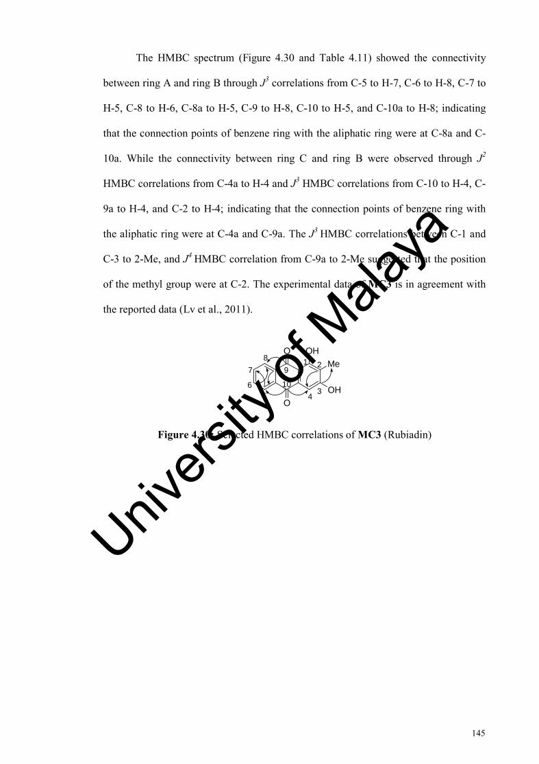
145
The HMBC spectrum (Figure 4.30 and Table 4.11) showed the connectivity
between ring A and ring B through J3 correlations from C-5 to H-7, C-6 to H-8, C-7 to
H-5, C-8 to H-6, C-8a to H-5, C-9 to H-8, C-10 to H-5, and C-10a to H-8; indicating
that the connection points of benzene ring with the aliphatic ring were at C-8a and C-
10a. While the connectivity between ring C and ring B were observed through J2
HMBC correlations from C-4a to H-4 and J3 HMBC correlations from C-10 to H-4, C-
9a to H-4, and C-2 to H-4; indicating that the connection points of benzene ring with
the aliphatic ring were at C-4a and C-9a. The J3 HMBC correlations between C-1 and
C-3 to 2-Me, and J4 HMBC correlation from C-9a to 2-Me suggested that the position
of the methyl group were at C-2. The experimental data of MC3 is in agreement with
the reported data (Lv et al., 2011).
OHO
OOH
Me
345
6
78
9
10
1 2
Figure 4.30: Selected HMBC correlations of MC3 (Rubiadin)
Univers
ity of
Mala
ya

146
Table 4.11: The NMR (CDCl3, 600 MHz) data of MC3 (Rubiadin)
Position δH δC HMBC 1 - 163.3 2-Me 2 - 109.9 4 3 - 162.7 2-Me 4 7.13 (s) 107.8 4a - 132.1 4 5 8.11 (dd, 8, 1) 127.2 7 6 7.64 (td, 8, 1) 134.0 8 7 7.68 (td, 8, 1) 134.4 5 8 8.18 (dd, 8, 1) 126.9 6 8a - 134.4 5 9 - 186.8 8 9a - 119.2 4, 2-Me 10 - 183.7 4, 5 10a - 133.5 8
1-OH 13.07 (s) - 2-Me 2.11 (s) 8.3
Univers
ity of
Mala
ya
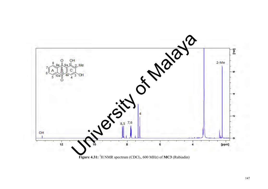
147
Figure 4.31: 1H NMR spectrum (CDCl3, 600 MHz) of MC3 (Rubiadin)
Univers
ity of
Mala
ya

148
Figure 4.32: 13C NMR spectrum (CDCl3, 150 MHz) of MC3 (Rubiadin) Univers
ity of
Mala
ya

149
4.2.4 MC4: 1,6-Dihydroxy-2-methylanthraquinone
OHO
O
Me
3456
78
9
104a
8a 1 29a
10a
A B CHO
MC4 was isolated as yellowish-orange amorphous powder with molecular
formula C15H10O4 as determined by ESI-MS with m/z 253.0 [M-H]- (calculated for
C15H10O4-H, 253.1). The UV spectrum showed absorption maxima at 420, 270, and
220 nm. The IR spectrum indicated the presence of hydroxyl group (3419 cm-1),
conjugated carbonyl group (1641 cm-1), and aromatic ring (1596, 1451 cm-1)
functionalities.
The 1H NMR spectrum (Figure 4.34 and Table 4.12) revealed the presence a
methyl signal at δH 2.31 (2-Me) and a hydroxyl signal at δH 13.11 (1-OH). Two
doublets (d) at δH 7.11 and δH 8.3, integrating for two protons and were assigned to H-
7 and H-8, respectively. Two other doublets (d) at δH 7.41 and δH 7.63, integrating for
two protons and were assigned to H-3 and H-4, respectively. The aromatic proton at δH
7.49 was assigned to H-5. The COSY experiment confirmed the presence of two
partial structures of -CH=CH-, corresponding to (C-3)-(C-4) and (C-7)-(C-8).
The 13C NMR spectrum (Figure 4.35 and Table 4.12) showed the presence of
13 carbon signals which include one carbonyl signal at δC 187.5 (C-9); and one methyl
signals at δC 16.4 (2-Me). The C-8a and C-10 quaternary carbons were not detected in
the spectrum due to the minute amount of MC4 sample. The carbon signals at δC 169.1
(C-1) and 170.8 (C-6) suggested that the hydroxyl groups are attached to these
carbons.
Univers
ity of
Mala
ya
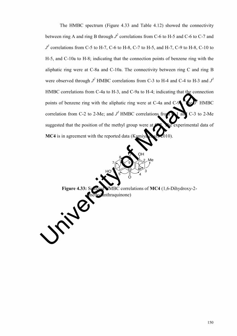
150
The HMBC spectrum (Figure 4.33 and Table 4.12) showed the connectivity
between ring A and ring B through J2 correlations from C-6 to H-5 and C-6 to C-7 and
J3 correlations from C-5 to H-7, C-6 to H-8, C-7 to H-5, and H-7, C-9 to H-8, C-10 to
H-5, and C-10a to H-8; indicating that the connection points of benzene ring with the
aliphatic ring were at C-8a and C-10a. The connectivity between ring C and ring B
were observed through J2 HMBC correlations from C-3 to H-4 and C-4 to H-3 and J3
HMBC correlations from C-4a to H-3, and C-9a to H-4; indicating that the connection
points of benzene ring with the aliphatic ring were at C-4a and C-9a. The J2 HMBC
correlation from C-2 to 2-Me; and J3 HMBC correlations from C-1 and C-3 to 2-Me
suggested that the position of the methyl group were at C-2. The experimental data of
MC4 is in agreement with the reported data (Kamiya et al., 2010).
OHO
O
Me
345
6
78
9
10
1 2
HO
Figure 4.33: Selected HMBC correlations of MC4 (1,6-Dihydroxy-2- methylanthraquinone)
Univers
ity of
Mala
ya
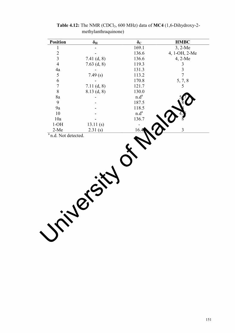
151
Table 4.12: The NMR (CDCl3, 600 MHz) data of MC4 (1,6-Dihydroxy-2- methylanthraquinone)
Position δH δC HMBC 1 2
- -
169.1 136.6
3, 2-Me 4, 1-OH, 2-Me
3 7.41 (d, 8) 136.6 4, 2-Me 4 7.63 (d, 8) 119.3 3 4a - 131.3 3 5 7.49 (s) 113.2 7 6 - 170.8 5, 7, 8 7 7.11 (d, 8) 121.7 5 8 8.13 (d, 8) 130.0 8a - n.da 5, 7 9 - 187.5 8 9a - 118.5 4 10 - n.da 4, 5 10a - 136.7 8
1-OH 13.11 (s) - 2-Me 2.31 (s) 16.4 3
a n.d. Not detected.
Univers
ity of
Mala
ya

152
Figure 4.34: 1H NMR spectrum (CDCl3, 600 MHz) of MC4 (1,6-Dihydroxy-2-methylanthraquinone)
Univers
ity of
Mala
ya

153
Figure 4.35: 13C NMR spectrum (CDCl3, 150 MHz) of MC4 (1,6-Dihydroxy-2-methylanthraquinone) Univ
ersity
of M
alaya

154
4.2.5 MC5: 1-Hydroxy-3-methoxyanthraquinone
OHO
O
3
456
78
9
104a
8a 1 29a
10a
A B COMe
MC5 was isolated as yellow amorphous powder with molecular formula
C15H10O4 as determined by ESI-MS with m/z 253.0 [M-H]- (calculated for C15H10O4-H,
253.1). The UV spectrum showed absorption maxima at 420, 340, 255, and 205 nm.
The IR spectrum indicated the presence of hydroxyl group (3400 cm-1), methyl group
(2926 cm-1), conjugated carbonyl group (1674 cm-1), and aromatic ring (1455 cm-1)
functionalities.
The 1H NMR spectrum (Figure 4.36 and Table 4.13) revealed the presence a
methoxyl signal at δH 3.42 (3-OMe) and a hydroxyl signal at δH 13.33 (1-OH). A
multiplet (m) signal at δH 8.33, integrating for two protons and were assigned to H-5
and H-8. Another multiplet (m) signal at δH 7.85, integrating for two protons were
assigned to H-6 and H-7. The observed pattern of these four proton signals in the
aromatic region suggested the presence of unsubstituted aromatic moiety at ring A.
Two aromatic protons at δH 6.26 and 7.53 were assigned to H-2 and H-4, respectively
on ring C. The hydroxyl and a methoxyl groups were accounted as substituents on ring
C. Due to the minute amount of MC5 sample, a number of the carbon signals were not
detected in the 13C NMR spectrum (Table 4.13). Out of 15 carbon atoms, only five
carbon signals were detected in the spectrum which are C-4, C-5, C-6, C-7 and C-8.
But due to the limited amount of the compound, the authenticity of this
compound was then determined by comparison of the NMR data with the literature.
Univers
ity of
Mala
ya
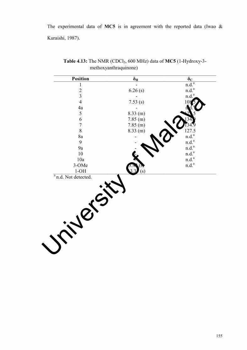
155
The experimental data of MC5 is in agreement with the reported data (Iwao &
Kuraishi, 1987).
Table 4.13: The NMR (CDCl3, 600 MHz) data of MC5 (1-Hydroxy-3- methoxyanthraquinone)
Position δH δC 1 - n.d.a 2 6.26 (s) n.d.a 3 - n.d.a 4 7.53 (s) 108.5 4a - n.d.a 5 8.33 (m) 127.5 6 7.85 (m) 134.9 7 7.85 (m) 134.9 8 8.33 (m) 127.5 8a - n.d.a 9 - n.d.a 9a - n.d.a 10 - n.d.a 10a - n.d.a
3-OMe 3.42 (s) n.d.a 1-OH 13.33 (s)
a n.d. Not detected.
Univers
ity of
Mala
ya

156
Figure 4.36: 1H NMR spectrum (CDCl3, 600 MHz) of MC5 (1-Hydroxy-3-methoxyanthraquinone) Univ
ersity
of M
alaya

157
4.2.6 MC6: 1-Methoxy-2-hydroxyanthraquinone
OMeO
O
345
6
78
9
104a
8a 1 29a
10a
A B COH
MC6 was isolated as yellow amorphous powder with molecular formula
C15H10O4 as determined by ESI-MS with m/z 253.0 [M-H]- (calculated for C15H10O4-H,
253.1). The UV spectrum showed absorption maxima at 405, 275, 245, and 205 nm.
The IR spectrum indicated the presence of hydroxyl group (3370 cm-1), conjugated
carbonyl group (1671 cm-1), and aromatic ring (1573 cm-1) functionalities.
The 1H NMR spectrum (Figure 4.38 and Table 4.14) revealed the presence a
methoxyl signal at δH 4.01 (1-OMe) and a hydroxyl proton signal at δH 13.01 (2-OH).
In addition, a set of multiplet (m) at δH 8.24, integrating for two protons and were
assigned to H-5 and H-8, respectively. Another set of multiplet (m) at δH 7.75,
integrating for two protons and were assigned to H-6 and H-7, respectively. The COSY
experiment gave partial structure -CH=CH-CH=CH-, corresponding to (C-5)-(C-6)-(C-
7)-(C-8), suggested the presence of unsubstituted aromatic moiety at ring A. Two sets
of doublet (d) aromatic protons at δH 7.34 and 8.12 were assigned to H-3 and H-4,
respectively on ring C. While the hydroxyl and a methoxyl group were accounted as
substituents on ring C. The 13C NMR spectrum (Figure 4.39 and Table 4.14) showed
the presence of carbonyl signal at δC 181.7 (C-10); and a methoxyl signal at δC 62.5 (1-
OMe). Carbon signal for C-9 was not detected in the 13C NMR spectrum due to minute
amount of MC6 sample. The carbon signal at δC 146.6 (C-1) and 155.3 (C-2)
suggested that these carbons are located adjacent to an oxygen atom.
Univers
ity of
Mala
ya

158
The HMBC spectrum (Figure 4.37, Figure 4.40, and Table 4.14) showed the
connectivity between ring A and B through J3 correlations from C-8a to H-8; and J3
correlations from the C-5 to H-7, C-6 to H-8, C-8a to H-5, C-10 to H-5, and C-10a to
H-8; indicating that the connection points of benzene ring with the aliphatic ring were
at C-8a and C-10a. The connectivity between ring C and ring B were observed through
J3 HMBC correlations from C-10 to H-4, C-9a to H-4, and C-2 to H-4; indicating that
the connection points of benzene ring with the aliphatic ring were at C-4a and C-9a.
The J3 HMBC correlation from C-1 to 1-OMe suggested that the position of the
methoxyl group were at C-1. The experimental data of MC6 is in agreement with the
reported data (Lv et al., 2009).
OMeO
O
OH
35
6
78
9
10
1 2
Figure 4.37: Selected HMBC correlations of MC6 (1-Methoxy-2-
hydroxyanthraquinone)
Univers
ity of
Mala
ya

159
Table 4.14: The NMR (CDCl3, 600 MHz) data of MC6 (1-Methoxy-2- hydroxyanthraquinone)
Position δH δC HMBC 1 - 146.6 3, 1-OMe 2 - 155.3a 3, 4 3 7.34 (d, 9) 120.4 4 8.12 (d, 9) 125.9 4a - 127.2 3 5 8.24 (m) 126.9 6 7.75 (m) 134.1 8 7 7.75 (m) 134.2 5 8 8.24 (m) 127.7 8a - 134.1 5, 8 9 - n.d.b 9a - 125.9 4 10 - 181.7a 4 10a - 134.1 8
1-OMe 4.01 (s) 62.6 2-OH 13.01 (s) -
a Value obtained from HMBC spectrum. b n.d. Not detected.
Univers
ity of
Mala
ya

160
Figure 4.38: 1H NMR spectrum (CDCl3, 600 MHz) of MC6 (1-Methoxy-2-hydroxyanthraquinone)
Univers
ity of
Mala
ya
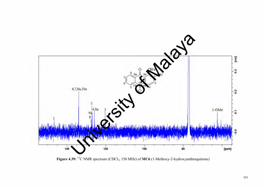
161
Figure 4.39: 13C NMR spectrum (CDCl3, 150 MHz) of MC6 (1-Methoxy-2-hydroxyanthraquinone)
Univers
ity of
Mala
ya
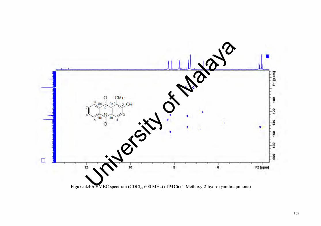
162
Figure 4.40: HMBC spectrum (CDCl3, 600 MHz) of MC6 (1-Methoxy-2-hydroxyanthraquinone) Univers
ity of
Mala
ya

163
4.3 Compounds isolated from Chlorophyllum molybdites
Four compounds were isolated from the ethyl acetate extract of the fruit bodies
of C. molybdites. The isolated compounds are summarized below.
OH
HO
H
HOH
OHOHH H
OH
OH
HO
H
HOH
OOHH H
OH
Me α-D-glucose (CM1) Ethyl-β-D-glucopyranoside (CM2)
O
HO OH
OHOH
HO
O
2,5-Anhydro-D-hexitol (CM3) Linoleic acid (CP5)
*Note: Linoleic acid (CP5) was isolated from both Crotalaria pallida and Chlorophyllum molybdites. Compound description and data of CP5 are presented in section 4.1.5 under Crotalaria pallida section.
Univers
ity of
Mala
ya

164
4.3.1 CM1: α-D-glucose
3
4 56
12
CM1 was isolated as white amorphous powder with molecular formula
C6H12O6 as determined by ESI-MS with m/z 197.1 [M-OH]+ (calculated for
C6H12O6+OH, 197.0). The UV spectrum showed absorption maxima at 220 and 260
nm. The IR spectrum indicated the presence of hydroxyl group (3362 cm-1), and
hemiacetal group (1049 cm-1) functionalities.
The 1H NMR spectrum (Figure 4.42 and Table 4.15) showed the presence of
five methine signals, including an anomeric proton at δH 5.13, and two methylene
protons at δH 3.80 (H-6a) and δH 3.84 (H-6b). Of the five methine signals, one signal
was observed as doublet (d) at δH 5.13 (H-1), two were observed as multiplet (m) at δH
3.82 (H-5) and δH 3.32 (H-4); and the last two signals were observed as doublet of
doublet (dd) at δH 3.70 (H-3) and 3.50 (H-2). The 13C NMR spectrum (Figure 4.43 and
Table 4.15) showed the presence of six carbon signals consisted of five methine and
one methylene carbons. The carbon signal at δC 93.61 is typical of an anomeric carbon
(C-1) of the glucose moiety. This carbon signal appears as most the deshielded signal
which suggested that this carbon is attached to two oxygen atoms.
COSY experiment give a partial structure of -CH(OH)-CH(OH)-CH(OH)-CH-
CH(OH)- corresponding to C(2)-C(3)-C(4)-C(5)-C(6). The observed pattern of these
protons indicated the presence of pyranosyl ring. The HMBC spectrum (Figure
4.41and Table 4.15) showed J2 correlations from C-2 to H-1, C-3 to H-4, C-4 to H-5,
and C-5 to H-6; and J3 correlations from C-1 to H-5, C-2 to H-4, C-3 to H-1, C-4 to H-
Univers
ity of
Mala
ya
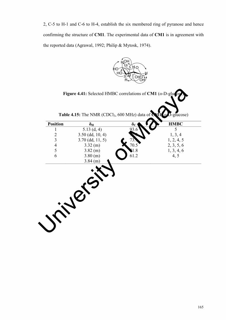
165
2, C-5 to H-1 and C-6 to H-4, establish the six membered ring of pyranose and hence
confirming the structure of CM1. The experimental data of CM1 is in agreement with
the reported data (Agrawal, 1992; Philip & Mytosk, 1974).
OH
HO
H
HOH
OHOHH H
OH
123
456
Figure 4.41: Selected HMBC correlations of CM1 (α-D-glucose)
Table 4.15: The NMR (CDCl3, 600 MHz) data of CM1 (α-D-glucose)
Position δH δC HMBC 1 5.13 (d, 4) 93.6 5 2 3.50 (dd, 10, 4) 72.3 1, 3, 4 3 3.70 (dd, 11, 5) 73.1 1, 2, 4, 5 4 3.32 (m) 70.5 2, 3, 5, 6 5 3.82 (m) 71.8 1, 3, 4, 6 6 3.80 (m)
3.84 (m) 61.2 4, 5
Univers
ity of
Mala
ya
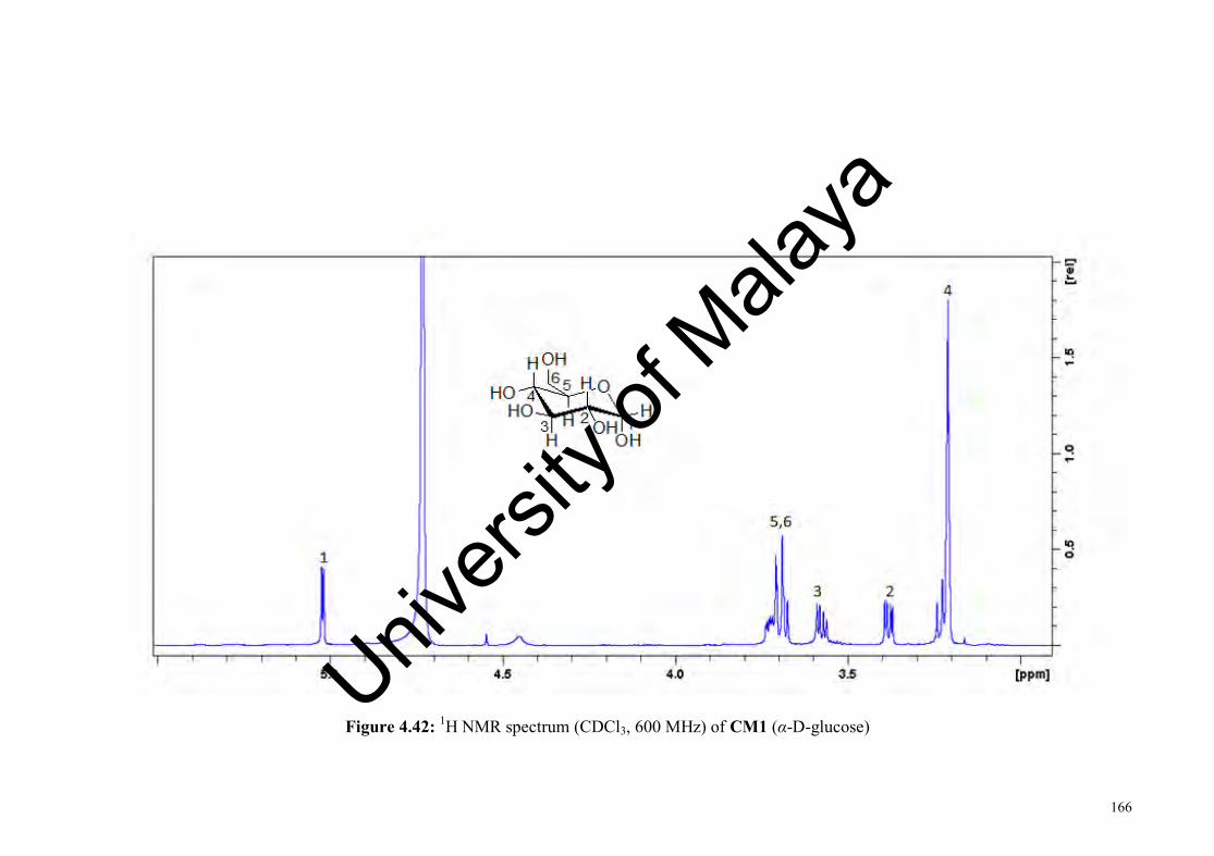
166
Figure 4.42: 1H NMR spectrum (CDCl3, 600 MHz) of CM1 (α-D-glucose)
Univers
ity of
Mala
ya

167
Figure 4.43: 13C NMR spectrum (CDCl3, 150 MHz) of CM1 (α-D-glucose)
Univers
ity of
Mala
ya

168
4.3.2 CM2: Ethyl-β-D-glucopyranoside
OH
HO
H
HOH
HOHH O
OH
23
45
6
7Me
1
CM2 was isolated as white amorphous powder with molecular formula
C8H16O6 as determined by ESI-MS with m/z 225.2 [M-OH]+ (calculated for
C8H16O6+OH, 225.2). The UV spectrum showed absorption maxima at 223 and 256
nm. The IR spectrum indicated the presence of hydroxyl group (3352 cm-1), and
hemiacetal group (1077 cm-1) functionalities.
The 1H NMR spectrum (Figure 4.45 and Table 4.16) showed the presence of
five methines including an anomeric proton at δH 4.28, four methylene protons at δH
3.97 (H-7a), δH 3.63 (H-7b), δH 3.87 (H-6a), δH 3.68 (H-6b), and one methyl at δH 1.25
(7-Me). Of the methine, one signals were observed as doublet (d) at δH 4.28 (H-1), one
triplet (t) at δH 3.36 (H-3), two signals were observed as multiplet (m) at δH 3.30 (H-2)
and δH 3.28 (H-5); and one signal were observed as doublet of doublet (dd) at δH 3.18
(H-4).
The 13C NMR spectrum (Figure 4.46 and Table 4.16) showed the presence of
eight carbon signals consisted of five methine, two methylenes and one methyl. The
carbon signal at δC 102.78 is typical of an anomeric carbon (C-1) of the glucose
moiety. This carbon appears as the most deshielded carbon which is flanked by two
oxygen atoms. The carbon signal at δC 14.05 (7-Me) suggested the presence of a
methyl group.
Univers
ity of
Mala
ya

169
COSY experiment give a partial structure of -CH(OH)-CH(OH)-CH(OH)-CH-
corresponding to C(2)-C(3)-C(4)-C(5). The observed pattern of these protons indicated
the presence of pyranosyl ring. The HMBC spectrum (Figure 4.44 and Table 4.16)
showed J2 correlations from C-1 to H-2, C-2 to H-3, C-3 to H-4, C-4 to H-5, and C-5
to H-6; and J3 correlations from C-1 to H-5, C-2 to H-4, C-3 to H-1, C-4 to H-2, C-5 to
H-1, and C-6 to H-4, establish the six membered ring of pyranose and hence
confirming the structure of CM2. The J2 HMBC correlation from C-7 to 7-Me
suggested that the position of the methyl group were at C-7. The experimental data of
CM2 is in agreement with the reported data (Agrawal, 1992; Philip & Mytosk, 1974).
OH
HO
H
HOH
HOHH O
OH
23
45
6
7Me
Figure 4.44: Selected HMBC correlations of CM2 (Ethyl-β-D-glucopyranoside)
Table 4.16: The NMR (CDCl3, 600 MHz) data of CM2 (Ethyl-β-D-glucopyranoside)
Position δH δC HMBC 1 4.28(d, 8) 102.7 2, 5, 7 2 3.30 (m) 73.7 1, 3, 4 3 3.36 (t, 9) 76.7 1, 2, 4, 5 4 3.18 (dd, 9, 1) 70.2 2, 3, 5 5 3.28 (m) 76.5 1, 4, 6 6 3.87 (dd, 12, 3)
3.68 (dd 12, 6) 64.8 4, 5
7 3.97 (dd, 10, 2) 3.63(dd, 7, 3)
61.3 1, 7-Me
7-Me 1.25 (t, 7) 14.0 7
Univers
ity of
Mala
ya

170
Figure 4.45: 1H NMR spectrum (CDCl3, 600 MHz) of CM2 (Ethyl-β-D-glucopyranoside)
Univers
ity of
Mala
ya
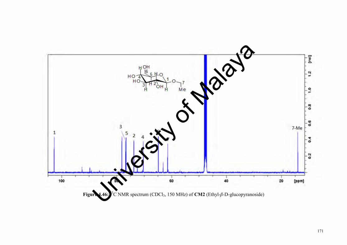
171
Figure 4.46: 13C NMR spectrum (CDCl3, 150 MHz) of CM2 (Ethyl-β-D-glucopyranoside) Univ
ersity
of M
alaya

172
4.3.3 CM3: 2,5-Anhydro-D-hexitol
O
OHHO
OH OH1
234
56
CM3 was isolated as white amorphous powder with molecular formula
C16H12O5. The UV spectrum showed absorption maxima at 220 and 262 nm. The IR
spectrum showed absorptions for ether (1081 cm-1) and hydroxyl group (3334 cm-1).
The 1H NMR spectrum (Figure 4.48 and Table 4.17) showed the presence of
two set of methylene signals at δH 3.80 (H-1a and H-6a), δH 3.64 (H-1b and H-6b) and
two methine signals at δH 3.77 (H-2 and H-5), δH 3.69 (H-3 and H-4). Both methine
signals were observed as multiplet (m) while the two methylene signals were observed
as doublet of doublet (dd). The 13C NMR spectrum (Figure 4.49 and Table 4.17)
showed the presence of three carbon signals consisted of two methines and a
methylene. Only three out of the six carbon resonances are visible suggesting CM3
possesses an axis of symmetry. The HMBC NMR spectrum (Figure 4.47 and Table
4.17) showed J2 correlations from C-1 to H-2, C-2 to H-1, C-2 to H-3, C-3 to H-2; and
J3 correlations from C-1 to H-3, C-3 to H-1, establish half structure of the CM3. The
experimental data of CM3 is in agreement with the reported data (Bock et al., 1981).
O
OHHO
OH OH1
234
56
Figure 4.47: Selected HMBC correlations of CM3 (2,5-Anhydro-D-hexitol)
Univers
ity of
Mala
ya

173
Table 4.17: The NMR (CDCl3, 600 MHz) data of CM3 (2,5-Anhydro-D-hexitol)
Position δH δC HMBC 1 3.80 (dd, 11, 6)
3.64 (dd, 11, 6) 63.7 2, 3, 4, 5
2 3.77 (m) 71.6 1, 3, 4, 6 3 3.69 (m) 69.9 1, 2, 5, 6 4 3.69 (m) 69.9 1, 2, 5, 6 5 3.77 (m) 71.6 1, 3, 4, 6 6 3.80 (dd, 11,6)
3.64 (dd, 11,6) 63.7 2, 3, 4, 5
Univers
ity of
Mala
ya

174
Figure 4.48: 1H NMR spectrum (CDCl3, 600 MHz) of CM3 (2,5-Anhydro-D-hexitol) Univ
ersity
of M
alaya

175
Figure 4.49: 13C NMR spectrum (CDCl3, 150 MHz) of CM3 (2,5-Anhydro-D-hexitol) Univ
ersity
of M
alaya
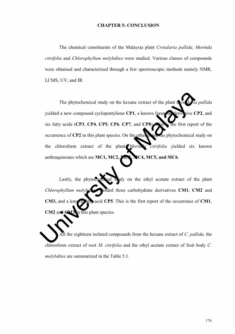
176
CHAPTER 5: CONCLUSION
The chemical constituents of the Malaysia plant Crotalaria pallida, Morinda
citrifolia and Chlorophyllum molybdites were studied. Various classes of compounds
were obtained and characterized through a few spectroscopic methods namely NMR,
LCMS, UV, and IR.
The phytochemical study on the hexane extract of the plant Crotalaria pallida
yielded a new compound cyclopentyliene CP1, a known furanone derivative CP2, and
six fatty acids (CP3, CP4, CP5, CP6, CP7, and CP8). This is the first report of the
occurrence of CP2 in this plant species. On the other hand, the phytochemical study on
the chloroform extract of the plant Morinda citrifolia yielded six known
anthraquinones which are MC1, MC2, MC3, MC4, MC5, and MC6.
Lastly, the phytochemical study on the ethyl acetate extract of the plant
Chlorophyllum molybdites yielded three carbohydrate derivatives CM1, CM2 and
CM3, and a known fatty acid CP5. This is the first report of the occurrence of CM1,
CM2 and CM3 in this plant species.
All the eightteen isolated compounds from the hexane extract of C. pallida, the
chloroform extract of root M. citrifolia and the ethyl acetate extract of fruit body C.
molybdites are summarized in the Table 5.1.
Univers
ity of
Mala
ya

177
Table 5.1: Compounds isolated from Crotalaria pallida, Morinda citrifolia and Chlorophyllum molybdites.
Comp. ID.
Source Compound structure Compound name
CP1 C. pallida
HO
O
Crotolidene
CP2
O
OHO
Hydroxydihydrobovolide
CP3
20
Octacosane
CP4
O
O
Trans-phytyl palmitate
CP5
HO
O
Linoleic acid
CP6
O
O
Methyl oleate
CP7
O
O
Ethyl palmitate
CP8
HO
O
Palmitic acid
Univers
ity of
Mala
ya

178
Table 5.1, continued
MC1 M. citrifolia
OH
O
OMeO
O
Damnacanthal
MC2
OH
O
OHO
O
Nordamnacanthal
MC3
OH
MeOHO
O
Rubiadin
MC4
MeOHO
OHO
1,6-Dihydroxy-2- methylanthraquinone
MC5
OHO
OOMe
1-Hydroxy-3- methoxyanthraquinone
MC6
OHOMeO
O
1-Methoxy-2- hydroxyanthraquinone
Univers
ity of
Mala
ya

179
Table 5.1, continued
CM1 C. molybdites
OH
HO
H
HOH
OHOHH H
OH
α-D-glucose
CM2
OH
HO
H
HOH
OOHH H
OH
Me
Ethyl-β-D-glucopyranoside
CM3
O
HO OH
OHOH
2,5-Anhydro-D-hexitol
CP5
HO
O
Linoleic acid
Univers
ity of
Mala
ya
180
REFERENCES
Abdullah, A. S. (1990). Poisonous plants of Malaysia. Kuala Lumpur: Tropical Press.
Adams, R., & Gianturco, M. (1956). The structures of grantianine and sceleratine. A
suggested biogenesis of the acids in the alkaloids from Senecio and Crotalaria species. Journal of the American Chemical Society, 78(17), 4458-4464.
Adedayo, M. R. (2011). Proximate analysis on four edible mushrooms. Journal of
Applied Sciences and Environmental Management, 15(1), 9-11.
Ahmad, V. U., & Fatima, I. (1986). Isolation and 13C NMR of crosemperine from Crotalaria Burhia Buch-Ham. Journal of the Chemical Society of Pakistan, 8(1), 89-90.
Ahmed, B., Al-Howiriny, T. A., & Mossa, J. S. (2006). Crotalic and emarginellic
acids: Two triterpenes from Crotalaria emarginella and anti-inflammatory and anti-hepatotoxic activity of crotalic acid. Phytochemistry, 67, 956-964.
Agrawal, P., K, (1992). NMR spectroscopy in the structural elucidation of
oligosaccharides and glycosides. Phytochemistry, 31(10), 3307-3330.
Akihisa, T., Matsumoto, K., Tokuda, H., Yasukawa, K., Seino, K., Nakamoto, K.,
Kuninaga, H., Suzuki, T., & Kimura, Y. (2007). Anti-inflammatory and potential cancer chemopreventive constituents of the fruits of Morinda citrifolia (Noni). Journal of Natural Products, 70(5), 754-757.
Alam, M. H., Murshid, G. M. M., Bokshi, B., Suma, S. A., Sathi, S. I., & Rahman, A.
(2014). Assessment of antioxidant, antibacterial and anthelmintic activities of ethanol extract of leaves of Crotalaria pallida (Aiton). International Journal of Pharmacognosy, 1(7), 438-444.
Alves, R. R. N., & Rosa, I. L. (2005) Why study the use of animal products in
traditional medicines? Journal of Ethnobiology and Ethnomedicine, 5, 1-5. Ambali, S. F., Mamman, M., Adaudi, A. O., Esievo, K. A. N., Ayo, J. O., & Abubakar,
M. S. (2008). Curative and protective effects of Penicilin G on experimental Chlorophyllum molybdites poisoning in mice. Journal of Pharmacology and Toxicology, 3(3), 241-245.
Arora, D., (1986). Mushrooms demystified. Berkeley: Ten Speed Press.
Univers
ity of
Mala
ya

181
Arpornsuwan, T., & Punjanon, T. (2006). Tumor cell-selective antiproliferative effect of the extract from Morinda citrifolia fruits. Phytotherapy Research., 20(6), 515-517.
Asand, M., Urix, A. H., & Ahmad, V. U. (1991). Diterpene fatty acid ester from
Leucas nutans. Journal of Natural Products, 54(5): 1444-1446.
Asres, K., Sporer, F., & Wink, M. (2004). Patterns of pyrrolizidine alkaloids in 12
Ethiopian Crotalaria species. Biochemical Systematics and Ecology, 32, 915-930.
Atal, C. K., & Kapur, K. K. (1966). A new pyrrolizidine aminoalcohol in alkaloids
from Crotalaria species. Tetrahedron Letters, 7(6), 537-544.
Atal, C. K., Sharma, R. K., Culvenor, C. C. J., & Smith, L. W. (1966). Alkaloids of
Crotalaria rubiginosa Willd. trichodesmine and junceine. Australian Journal of Chemistry, 19, 2189-2191.
Atal, C. K., & Sawhney, R. S. (1968). Nilgirine, a new crotalaria alkaloid of
senecionine type lacking the 1'-carbon atom. Tetrahedron Letters, 9(54), 5605-5608.
Atal, C. K., Culvenor, C. C. J., Sawhney, R. S., & Smith, L. W. (1969). The alkaloids
of Crotalaria grahamiana. Grahamine, the 3'-[(-)-2-methylbutyryl] ester of monocrotaline. Australian Journal of Chemistry, 22, 1773-1777.
Atanasov, A. G., Waltenberger, B., Pferschy-Wenzig, E. M., Linder, T., Wawrosch, C.,
Uhrin, P., Temml, V., Wang, L., Schwaiger, S., Heiss, E. H., Rollinger, J. M., Schuster, D., Breuss, J. M., Bochkov, V., Mihovilovic, M. D., Kopp, B., Bauer, R., Dirsch, V. M., & Stuppner, H. (2015). Discovery and resupply of pharmacologically active plant-derived natural products: A review. Biotechnology Advances, 33(8), 1582-1614.
Atkinson, N. (1956). Antibacterial substances from flowering plants. 3 Antibacterial activity of dried Australian plants by rapid direct plate test. Australian Journal of Experimental Biology and Medical Science, 34, 17-26.
Augenstein, W. L. (1994). Chlorophyllum molybdites. Handbook of Mushroom Poisoning: Diagnosis and Treatment.
Awouafack, M. D., Spiteller, P., Lamshöft, M., Kusari, S., Ivanova, B., Tane, P., &
Spiteller, M. (2011). Antimicrobial isopropenyl-dihydrofuranoisoflavones from Crotalaria lachnophora. Journal of Natural Products, 74, 272-278.
Univers
ity of
Mala
ya

182
Ayensu, E. S. (1981). Medicinal plants of the West Indies. Algonac, MI: Reference Publications.
Ayyuanar, M., & Ignacimuthu, S. (2005). Medicinal plants used by tribals of
tirunelveli hills, Tamil nadu to treat poisonous bites and skin diseases. Indian Journal of Traditional Knowledge, 4(3), 229-236.
Babu, U. V., Bhandari, S. P. S., & Garg, H. S. (1998). Barbacarpan, a pterocarpan
from Crotalaria barbata. Phytochemistry, 48(8), 1457-1459.
Balunas, M. J., Kinghorm, A. D. (2005). Drug discovery from medicinal plants. Life
Sciences, 78(5), 431-441.
Barbato, M. P. (1993). Poisoning from accidental ingestion of mushrooms. The Medical Journal of Australia, 158(12), 842-847.
Benedict, R. G. (2013). Mushroom toxins other than Amanita. Microbial Toxins, 6, 281-320.
Bennett, J. (1987). Mycotoxins, mycotoxicoses, mycotoxicology and Mycopathologia. Mycopathologia, 100(1), 3-5.
Berger, K. J., & Guss, D. A. (2005). Mycotoxins revisited: part II. The Journal of Emergency Medicine, 28(2), 175-183.
Bessey, E. A. (1939). A case of poisoning by Lepiota morgani. Mycologia, 31(1), 1 09-110.
Bhacca, N. S., & Sharma, R. K. (1968). Mucronatinine, a new alkaloid from Crotalaria mucronata Desv.-I. Tetrahedron, 24, 6319-6326.
Bhakuni, D. S., & Chaturvedi, R. (1984). Chemical constituents of Crotalaria
madurensis. Journal of Natural Products, 47(4), 585-591.
Biljana, P., Pratik, B., & Lalitha, S. (2009). Lisdexamfetamine: A prodrug for the treatment of attention-deficit/hyperactivity disorder. American Journal of Health-System Pharmacy, 66(22), 2005-2012.
Blayney, D., Rosenkranz, E., & Zettner, A. (1980). Mushroom poisoning from Chlorophyllum molybdites. Western Journal of Medicine, 132(1), 74.
Univers
ity of
Mala
ya
183
Blick, S. K. A., & Keating, G. M. (2007). Lisdexamfetamine. Pediatric Drugs, 9(2), 129-135.
Bock, K., Pedersen, C., & Thogersen, H. (1981). Acid catalysed dehydration of Alditols. Part I. D-Glucitol and D-Mannitol. Acta Chemica Scandinavica B, 35, 441-449.
Bowie, J. H., & Cooke, R. G. (1962). Colouring matters of Australian plants. IX.
Anthraquinones from Morinda species. Australian Journal of Chemistry, 15(2), 332-335.
Braun, L., & Cohen, M. (2015). Herbs and natural supplements: An evidence-based
guide. Elsevier, 4(2), 471-475.
Brett, J. W., Jarakae, C. J., Afa, K. P., & Shixin, D. (2011). Toxicity and antioxidant
tests of Morinda citrifolia (noni) seed extract. Advance Journal of Food Science and Technology, 3(4), 303-307.
Brown, K., Devlin, J. A., & Robins, D. J. (1984). Globiferine, a pyrrolizidine alkaloid
from Crotalaria globifera. Phytochemistry, 23(2), 457-459.
Bryson, P. D., (1996). Comprehensive reviews in toxicology: for emergency clinicians.
Washington, DC: CRC press.
Buku, A., Wieland, T., Bodenmüller, H., & Faulstich, H. (1980). Amaninamide, a new
toxin ofAmanita virosa mushrooms. Experientia, 36(1), 33-34.
Bushnell, O. (1993). The gifts of civilization: gerams and genocide in Hawaii.
Honolulu: University of Hawaii Press.
Cambie, R. C., & Brewis, A. A. (1997). Anti-fertility plants of the Pacific. Victoria,
Australia: CSIRO Publishing Collingwood.
Chan-Blanco, Y., Vaillant, A. M., Perez, M., Reynes, J. M., & Brillouet, P. B. (2006).
The noni fruits (Morinda citrifolia L.): a review of agricultural research, nutritional and therapeutic properties. Journal of Food Composition and Analysis, 19, 645-654.
Chang-hong, L., Ya-rong, X., Yong-hang, Y., Feng-feng, Y., Jun-yan, L. & Jing-lei, S.
(2007). Extraction and characterization of antioxidant compositions from fermented juice of Morinda citrifolia. Planta Medica, 56, 430-434.
Univers
ity of
Mala
ya

184
Chaturvedi, R., Pant, N., Garg, H. S., & Bhakuni, D. S. (1987). Isoflavonoids of Crotalaria madurensis. Journal of Natural Products, 50(2), 266-269.
Cheng, D., Liu, Y., & Chu, T. T. (1989). Assamicadine, a new pyrrolizidine alkaloid
from Crotalaria assamica. Journal of Natural Products, 52(5), 1153-1155.
Chisolm, J. J. (1965). Chronic lead intoxication in children. Developmental Medicine
& Child Neurology, 7(5), 529-536.
Chunhieng, T., Hay, L., & Montet, D. (2005). Detailed study of the juice composition of noni (Morinda citrifolia) fruits from Cambodia. Fruits, 60(1), 13-24.
Chesnut, V. K. (1900). Poisonous properties of the green-spored Lepiota. Ecological Society of America. 8(5), 87-93.
Cragg, G. M., & Newman, D. J. (2013). Natural products: a continuing source of novel drug leads. Biochimica et Biophysica Acta, 1830(6), 3670-3695.
Crout, D. H. G. (1969). Pyrrolizidine alkaloids. The co-occurrence of monocrotaline and trichodesmine in Crotalaria recta Steud. ex A. Rich. Phytochemistry, 7, 1425-1427.
Culvenor, C. C. J., & Smith, L. W. (1957). The alkaloids of Crotalaria retusa L. Australian Journal of Chemistry, 10(4), 464-473.
Culvenor, C. C. J., & Smith, L. W. (1961). The alkaloids of Crotalaria goreensis: 7β-
hydroxy-1-methylene-8β- and 7β-hydroxy-1-methylene-8α-pyrrolizidine. Australian Journal of Chemistry, 14, 284-294.
Culvenor, C. C. J., & Smith, L. W. (1962). Alkaloids of Crotalaria trifoliastrum Willd.
and C. aridicola Domin. I. Methyl ethers of supinidine and retronecine. Australian Journal of Chemistry, 15(1), 121-129.
Culvenor, C. C. J., Smith, L. W., & Woods, W. G. (1965). An unusual
dichlorocyclopropyl artefact in alkaloidal extracts of Crotalaria goreensis Guill. et Perr. Tetrahedron Letters, 25, 2025-2029.
Culvenor, C. C., & Smith, L. W. (1966). Usaramine, a new pyrrolizidine alkaloid from
Crotalaria usaramoensis E. G. Baker. Australian Journal of Chemistry, 19, 2127-2131.
Univers
ity of
Mala
ya

185
Culvenor, C. C. J., O'Donovan, G. M., & Smith, L. W. (1967). Alkaloids of Crotalaria trifoliastrum and C. aridicola. III. Additional pyrrolizidine derivatives. Australian Journal of Chemistry, 20(4), 757-768.
Culvenor, C. C. J., Sawhney, R. S., & Smith, L. W. (1968). The alkaloids of Crotalaria
maypurensis H. B. & K. Australian Journal of Chemistry, 21, 2135-2136.
Cvetković, R. S., & Plosker, G. L. (2007). Exenatide: a review of its use in patients with type 2 diabetes mellitus (as an adjunct to metformin and/or a sulfonylurea). Drugs. 67(6), 935-954.
Dalsgaard, P. W., Potterat, O., Dieterle, F., Paululat, T., Kuhn, T., & Hamburger, M. (2006). New trisaccharide fatty acid esters from the fruit of Morinda citrifolia (Noni). Planta Medica, 72(14), 1322-1327.
Debono, M., Turner, W. W., LaGrandeur, L., Burkhardt, F. J., Nissen, J. S., Nichols,
K. K., Rodriguez, M. J., Zweifel, M. J., & Zeckner, D. J. (1995). Semisynthetic chemical modification of the antifungal lipopeptide echinocandin B (ECB): structure-activity studies of the lipophilic and geometric parameters of polyarylated acyl analogs of ECB. Journal of Medicinal Chemistry, 38(17), 3271–3281.
DeFilipps, R. A., Maina, S. L., & Crepin, J. (2004). Medicinal plants of the Guianas (Guyana, Surinam, French Guiana). Washington, DC: Smithsonian Institution.
Degener, O. (1973). Plants of Hawaii National Park illustrative of plants and customs
of the South sea. Ann Abor, MI: Braun-Brumfield.
Deng, Y., Chin, Y. W., Chai, H., Keller, W. J., & Kinghorn, A. D. (2007).
Anthraquinones with quinone reductase-inducing activity and benzophenones from Morinda citrifolia (Noni) roots. Journal of Natural Products, 70, 2049-2052.
Dhasmana, H. & Garg, H. S. (1991). Lepidoside - A rare flavonol diglycoside from Crotalaria semperflorens Vent. Journal of Indian Chemical Society, 68, 476-477.
Demain, A. L., & Sanchez, S. (2009). Microbial drug discovery: 80 years of progress.
The Journal of Antibiotic, 62(1), 5-16.
Dias, D. A., Urban, S., & Roessner, U. (2012). A historical overview of natural products in drug discovery. Metabolites, 2(2), 303-306.
Univers
ity of
Mala
ya

186
Ding, Y., Kinjo, J. E., Yang, C. R., & Nohara, T. (1991). Oleanene glycosides from Crotalaria albida. Chemical and Pharmaceutical Bulletin, 39(2), 496-498.
Dittmar, A. (1993). Morinda citrifolia L.-use in indigenous Samoan medicine. Journal
of Herbs, Spices and Medicinal Plants, 1(3), 77-92.
Dixon, A. R., McMillen, H., & Etkin, N. L. (1999). Fement this: the transformation of
Noni, a traditional Polynesian medicine (Morinda citrifolia, Rubiaceae). Ecological Botony, 53, 51-68.
Duncan, S. H., Flint, H. J., & Stewart, C. S. (1998). Inhibitory activity of gut bacteria against Escherichia Coli O157 mediated by dietary plant metabolites. FEMS Microbiology Letters, 164, 258-283.
Eilers, F. I., & Nelson, L. R. (1974). Characterization and partial purification of the
toxin of Lepiota morganii. Toxicon, 12(6), 557-563. Falade, O. S., Adepoju, O. O., Owoyomi, O., & Adewusi, S. R. (2008). Chemical
composition and toxic trace element composition of some Nigerian edible wild mushrooms. International Journal of Food Science & Technology, 43(1), 24-29.
Farine, J. P., Legaal, L., Moreteau, B., & Quere, J. L. L. (1996). Volatile components
of ripe fruit of Morinda citrifolia and their effects on Drosophila. Phytochemistry Letters, 41(2), 433-438.
Fasidi, I. O., & Kadiri, M. (1995). Toxicological screening of seven Nigerian mushrooms. Food Chemistry, 52(4), 419-422.
Faulstich, H., Trischmann, H., & Zobeley, S. (1975). A radioimmunoassay for amanitin. Federation of European Biochemical Societies Letters, 56(2), 312-315.
Faulstich, H. (1980). Mushroom poisoning. The Lancet, 316(8198), 794-795.
Faulstich, H., Zobeley, S., & Trischmann, H. (1982). A rapid radioimmunoassay, using a nylon support, for amatoxins from Amanita mushrooms. Toxicon, 20(5), 913-924.
Fletcher, M. T., McKenzie, R. A., Blaney, B. J., & Reichmann, K. G. (2009). Pyrrolizidine alkaloids in Crotalariataxa from Northen Australia: Risk of grazing livestock. Journal of Agriculture and Food Chemistry, 57, 311-319.
Univers
ity of
Mala
ya

187
Floch, H., Labarbe, C., & Roffi, J. (1966). Étude expérimentale de la toxicité de la Lépiote de Morgan. Rev. Mycology, 31, 317-322.
Flores, A. S., de Azevado Tozzi, A. M. G., & Trigo, J. R. (2009). Pyrrolizidine alkaloid profiles in Crotalaria species from Brazil: Chemotaxonomic significance. Biochemical Systematic and Ecology, 37, 459-469.
Fonseca, R. L., Guimarães, P. R., Rodrigues, S., Scachetti-Pereira, R., & Townsend, A. (2006). Predicting invasive potential of smooth crotalaria (Crotalaria pallida) in Brazillian national parks based on African records. Weed Science, 54, 458-463.
Furusawa, E., Hirazumi, A., Story, S., & Jensen J. (2003). Antitumor potential of a polysaccharide-rich substance from the fruit juice of Morinda citrifolia (noni) on sarcoma 180 ascites tumor in mice. Phytotherapy Research, 17(10), 1158-1164.
Gandhi, R. N., Rajagopalan, T. R., & Seshadri, T. R. (1968). A new alkaloid from the
seeds of Crotalaria striata. Current Science, 37(10), 285-286.
Ge, Z., & Yang, Z. L. (2006). The genus Chlorophyllum (Basidiomycetes) in China.
Mycotaxon, 96, 181-192.
Ghosh, R., Pathak, N., & Singh, M. (1977). The genus chlorophyllum in India. Indian Phytopathology. 29, 50-53.
Gomes, C. E. M., Barbosa, A. E. A. D., Macedo, L. L. P., Pitanga, J. C. M., Moura, F. T., Oliveira, A. S., Moura, R. M., Queiroz, A. F. S., Macedo, F. P., Andrade, L. B. C., Vidal, M. S., & Sales, M. P. (2005). Effect of trypsin inhibitor from Crotalaria pallida seeds on Callosobrunchus maculatus (cowpea weevil) and Ceratitis capitata (fruit fly). Plant Physiology and Biochemistry, 43, 1095-1102.
Gong, Q. F., Zhang, Y. M., Tan, N. H., & Chen, Z. H. (2010). Chemical constituents
of three poisonous mushrooms. Natural Product Research and Development, 22, 185-188.
Govindappa, M., Bharath, N., Shruthi, H. B., Sadananda, T. S., & Sharanappa, P. (2011). Antimicrobial, antioxidant, and in vitro anti-inflammatory activity and phytochemical screening of Crotalaria pallida Aiton. African Journal of Pharmacy and Pharmacology, 5(21), 2359-2371.
Govindappa, M., Umashankar, T., Ramachandra, Y. L., & Chen, C. J. (2013). In vitro
anti-HIV properties of coumarins from Crotalaria Pallida Aiton. Biochemistry: An Indian Journal, 7(3), 114-121.
Univers
ity of
Mala
ya

188
Gray, W. D. (1973). The use of fungi as food and in food processing. Part II. CRC Critical Reviews in Food Technology, 3(2), 121-215.
Guangming, L., Ann, B., Wei-Ya, M., Shengmin, S., Chi-Tang, H., & Zigang, D.
(2001). Two novel glycosides from the fruits of Morinda citrifolia (noni) inhibit AP-1 transactivation and cell transformation in the mouse epidermal JB6 cell line. Cancer Research, 61, 5749-5756.
Gurnani, N., Mehta, D., Gupta, M., & Mehta, B. K. (2014). Natural products: sources
of potential drugs. African Journal of Basic & Applied Sciences, 6(6), 171-186.
Gunstone, F. D., & Jacobsberg, F. R. (1972). Fatty acids, part 36 the synthesis, silver ion chromatographic, and NMR spectroscopic properties of the nine 9,12-diunsaturated n-C18 acids. Chemistry and Physics of Lipids, 9(2), 112-122.
Gurib-Fakim, A. (2006). Medicinal plants: traditions of yesterday and drugs of
tomorrow. Molecular Aspects of Medicine, 27(1), 1-93.
Harvey, A. L. (2008). Natural products in drug discovery. Drug Discovery Today, 13 (19-20), 894-901.
He, Q. W., Wu, P. Y., Liang, Y. M., Xiang, C., Du, Z. W., & Wang, G. (2012). Chemical constituents of the sarcocarp of Morinda citrifolia. Chinese Traditional Patent Medicine, 34(9), 1729-1722.
Heinicke, R. M. (1985). The pharmacologically active ingredients of Noni. Bulletin of the National Tropical Botanical Garden, 15(1), 1-3.
Heinicke, R. M. (2001). The xeronine system: a new cellular mechanism that explains the health promoting action of NONI and Bromelian. Orem, Utah: Direct Source Publishing.
Hiramitsu, T., Imoto, M., Koyano, T., & Umezawa, K. (1993). Introduction of normal phenotypes in ras-transformed cells by damnavhantal from Morinda citrifolia. Cancer Letters, 73(2-3), 161-166.
Hu, S. Y. (2005). Food plants of China. Hong Kong: Chinese University Press.
Iwao, M., & Kuraishi, T. (1987). Utilization of sulfide, sulfoxide, and sulfone groups
as regiochemical control elements in the diels-alder reaction of naphthoquinones. Bulletin of the Chemical Society of Japan., 60, 4051-4060.
Univers
ity of
Mala
ya
189
Jain, R. K., & Srivastava, S. D. (1992). Two new anthraquinones in the roots of Morinda citrifolia. Proceedings of the National Academy of Sciences India, 62, 11-13.
Jain, S. K., & Borthakur, S. K. (1980). Ethnobotany of the Mikers of India. Economic
Botany, 34(3), 264-272.
Jain, S. K., & DeFilipps, R. A. (1991). Medicinal plants of India. Algonac, MI:
Reference Publications.
Joshi, H., Joshi, A. B., Sati, H., Gururaja, M. P., Shetty, P. R., Subrahmanyam, E. V.
S., & Satyanaryana, D. (2009). Fatty acids from Memecylon umbellatum (Burm.). Asian Journal of Organic Chemistry, 2(2), 178-180.
Judice, J. K., & Pace, J. L. (2003). Semi-synthetic glycopeptide antibacterials.
Bioorganic & Medicinal Chemistry Letters, 13(23), 4165-4168.
Kamboj, V. P. (1988). A review on Indian medicinal plants with interceptive activity. Indian Journal of Medicinal Research, 87, 336-355.
Kamiya, K., Tanaka, Y., Endang, H., Umar, M., & Satake, T. (2004). Chemical
constituents of Morinda citrifolia fruits inhibit copper-induced low-density lipoprotein oxidation. Journal of Agricultural and Food Chemistry, 52, 5843-5848.
Kamiya, K., Tanaka, Y., Hanani, E., Umar, M., Satake, T. (2005). New Anthraquinone
and Iridoid from the Fruits of Morinda citrifolia. Chemical and Pharmaceutical Bulletin, 53, (12), 1597-1599.
Kamiya, K., Hamabe, W., Harada, S., Murakami, R., Tokuyama, S., & Satake, T. (2008). Chemical constituents of Morinda citrifolia roots exhibit hypoglycemic effects in streptozotocin-induced diabetic mice. Biological and Pharmaceutical Bulletin, 31, 935-938.
Kamiya, K., Hamabe, W., Tokuyama, S., & Satake, T. (2009). New anthraquinone
glycosides from the root of Morinda cotrifolia. Fitoterapia, 80, 196-199.
Kamiya, K., Hamabe, W., Tokuyama, S., Hirano, K., Satake, T., Kumamoto-Yonezawa, Y., Yoshida, H., & Mizushina, Y. (2010). Inhibitory effect of anthraquinones isolated from the Noni (Morinda citrifolia) root on animal A-, B-, Y-families of DNA polerases and human cancer cell proliferation. Food Chemistry, 118, 725-730.
Keam, S. J. (2008). Doripenem A Review of its use in the treatment of bacterial infections. Drugs, 68 (14), 2021-2057.
Univers
ity of
Mala
ya
190
Khalilullah, M., Sharma, V. M., & Rao, P. S. (1993). Ramosismin, a new prenylated chalcone from Crotalaria ramosissima. Fitoterapia, 44, 232-239.
Kim, H. K., Kwon, M. K., Kim, J. N., Kim, C. K., Lee, Y. J., Shin, H. J., Lee, J., &
Lee, H. S. (2010). Identification of novel fatty acid glucosides from the tropical fruit Morinda citrifolia L.. Phytochemistry Letters, 3, 238-241.
Kim, S. W., Jo, B. K., Jeong, J. H., Choi, S. U., & Hwang, Y. I. (2005). Induction of
extracellular matrix synthesis in normal human fibroblasts by anthraquinone isolated from Morinda citrifolia (Noni) fruit. Journal of Medicinal Food, 8(4), 552-555.
Kinghorn, A. D., Chai, H. B., Sung, C. K., & Keller, W. J. (2011). The classical drug discovery approach to defining bioactive constituents of botanicals. Fitoterapia, 82, 71-79.
Knothe, G., Lie Ken Jie, M. S. F., Lam, C. C., & Bagby, M. O. (1995). Evaluation of
the 13C-NMR signals of the unsaturated carbons of triacylglycerols. Chemistry and Physics of Lipids, 77, 187-191.
Ko, H. H., Weng, J. R., Tsao, L. T., Yen, M. H., Wang, J. P., & Lin, C. N. (2004).
Anti-inflammatory flavonoids and pterocarpanoid from Crotalaria pallida and C. assamica. Bioorganic and Medicinal Chemistry Letters, 14, 1011-1014.
Kobayashi, Y., Kobayashi, K., Umehara, K., Dohra, H., Murata, T., Usui, T., &
Kawagishi, H. (2004). Purification, characterization, and sugar binding specificity of an N-glycolylneuraminic acid-specific lectin from the mushroom Chlorophyllum molybdites. Journal of Biological Chemistry, 279(51), 53048-53055.
Koshino, H., Yoshihara, T., Sakamura, S., Shimanuki, T., Sato, T., & Tajimi, A. (1989). Novel C-ll epoxy fatty acid from Stromata of Epichloe typhina on Phleum pretense. Agriultural and Biological Chemistry, 53(9): 2527-2528.
Krohn, K., Steingröver, K., & Rao, M. S. (2002). Isolation and synthesis of chalcones
with different degress of saturation. Phytochemistry, 61, 931-936.
Kumar, J. K., Narender, T., Rao, M. S., Rao, P. S., Toth, G., Balazs, B., & Duddeck,
H. (1999). Further dihydrochalcones from Crotalaria ramosissima. Journal of Brazilian Chemical Society, 10(4), 278-280.
Lakshmanan, D., & Radha, K. V. (2012). An effective quantitative estimation
lovastatin from Pleurotus ostreatus using UV and HPLC. International Journal of Pharmacy and Pharmaceutical Sciences, 4, 462-464.
Univers
ity of
Mala
ya
191
Laohavaleeson, S., Kuti, J. L., & Nicolau, D. P. (2007). Telavancin: a novel lipoglycopeptide for serious gram-positive infections. Expert Opinion on Investigational Drugs, 16(3), 347-357.
Laurena, A. C., Den Truong, V., & Mendoza, E. M. T. (1984). Effects of condensed
tannins on the in vitro protein digestibility of cowpea [Vigna unguiculata (L.) Walp.]. Journal of Agricultural and Food Chemistry, 32(5), 1045-1048.
Leach, A. J., Leach, D. N., & Leach, G. J. (1998). Antibacterial activity of some medicinal plants of Papua New Guinea. Science in New Guinea, 1(1), 1-7.
Lee, F. Y. F., Borzilleri, R., Fairchild, C. R., Kamath, A., Smykla, R., Kramer, R., & Vite, G. (2008). Preclinical discovery of ixabepilone, a highly active antineoplastic agent. Cancer Chemotherapy Pharmacology, 63, 157–166.
Lehmann, P. F., & Khazan, U. (1992). Mushroom poisoning by Chlorophyllum molybdites in the Midwest United States. Mycopathologia, 118(1), 3-13.
Li, T. S. C. (2002). Chinese and related North American herbs; phytopharmacology and therapeutic values. Boca Raton, Florida: CRC Press.
Lie Ken Jie, M. S. F., & Lam, C. C. (1995). 13C-Nuclear magnetic resonance
spectroscopic studies of triacylglycerols of type AAA containing (Z)- and (E)-monoethylenic acyl groups. Chemistry and Physics of Lipids, 78, 15-27.
Lin, Y. L., Chang, Y. Y., Yang, D. J., Tzang, B. S., & Chen, Y. C. (2013). Beneficial
effects of noni (Morinda citrifolia L.) juice on livers of high-fat. Food Chemistry, 140, 31-38.
Liu, C. H., Xue, Y. R., Ye, Y. H., Yuan, F. F., Liu, J. Y., & Shuang, J. L. (2007). Extraction and characterization of antioxidant compositions from fermented fruit juice of Morinda citrifolia (Noni). Agricultural Sciences in China, 6(12), 1494-1501.
Locher, C. P., Burch, M. T., Mower, H. F., Berestecky, H., Davis, H., Van Polel, B., Lasure, A., Vander, B. D. A., & Vlieti-Nick, A. J. (1995). Anti-microbial activity and anti-complement activity of extracts obtained from selected Hawaiian medicinal plants. Journal of Ethnopharmacology, 49, 23-32.
Lv, L., Chen, H., Ho, C. T., & Sang, S. (2011). Chemical components of the roots of
Noni (Morinda citrifolia) and their cytotoxic effects. Fitoterapia, 82, 704-708.
Mattocks, A. R. (1968). Anacrotine, from Crotalaria incana. Journal of Chemical Society (C), 235-237.
Univers
ity of
Mala
ya
192
Mattocks, A. R., & Nwude, N. (1988). Pyrrolizidine alkaloids from Crotalaria lachnosema and C. naragutensis. Phytochemistry, 27(10), 3289-3291.
McChesney, J. D., Venkataraman, S. K., Henri, J. T. (2007). Plant natural products:
Back to the future or into extinction? Phytochemistry, 68, 2015-2022.
McCowen, M. C., Callender, M. E., & Lawlis, J. F. Jr. (1951). Fumagillin (H-3), a new antibiotic with amebicidal properties. Science, 113, 202–203.
Miranda-Vilela, A. L., & Grisolia, C. K. (2009). Characterization of the major nutritional components of Caryocar brasiliense fruit pulp by NMR spectroscopy. Quim Nova, 32(9), 2310-2313.
Mishra, B. B., & Tiwari, V. K. (2011). Natural products: An evolving role in future
drug discovery. European Journal of Medicinal Chemistry, 46, 4769-4807.
Montaser, R., & Luesch, H. (2011). Marine natural products: a new wave of drugs? Future Medicinal Chemistry, 3(12), 1475-89.
Moon, P. D., Lee, B. H., Jeong, H. J., An, H. J., Park, S. J., Kim, H. R., Ko, S. G., Um, J. Y., Hong, S. H., & Kim, H. M. (2007). Use of scopoletin to inhibit the production of inflammatory cytokines through inhibition of the IκB/NF-κB signal cascade in the human mast cell line HMC-1. European Journal of Pharmacology, 555(2-3), 218-225.
Moorthy, N. K., & Reddy, G. S. (1970). Preliminary phytochemical and pharmacological study of Morinda citrifolia, Linn. Antiseptic, 67, 167-171.
Morad, A. F. Crotalaria pallida Aiton. Retrieve from
http://eol.org/data_objects/25764063 (accessed August, 2017).
Morton, J. F. (1992). The ocean-going noni, or Indian mulberry (Morinda citrifolia,
Rubiaceae) and some of its “colourful” relatives. Economic Botany, 46, 241-256.
Mun'im, A., Negishi, O., & Ozawa, T. (2003). Antioxidative compounds from
Crotalaria sessiliflora. Bioscience, Biotechnology and Biochemistry, 67(2), 410-414.
Murray, P. E., Farber, R. M., Namerow, K. N., Kuttler, S., & Garcia-Godoy, F. (2008).
Evaluation of Morinda citrifolia as an endodontic irrigant. Journal of Endodontics, 34(1), 66-70.
Univers
ity of
Mala
ya

193
Naidu, K. C., Aruna, V., & Satyanarayana, T. (1998). Phytoconstituents from leaves of Crotalaria verrucosa Linn. Indian Journal of Natural Products, 14(1), 17-19.
Nam, H. & Kim, M. M. (2015). Scopoletin has a potential activity for anti-aging via
autophagy in human lung fibroblasts. Phytomedicine, 22(3), 362-368.
Narender, T., Shweta, Tanvir, K., Rao, M. S., Srivastava, K., & Puri, S. K. (2005). Prenylated chalcones isolated from Crotalaria genus inhibits in vitro growth of the human malaria parasite Plasmodium falciparum. Bioorganic and Medicinal Chemistry Letters, 15, 2453-2455.
Natarajan, K., & Kaviyarasan, V. (1991). Chlorophyllum molybdites poisoning in
India—A case study. Mycologist, 5(2), 70.
Nelson, S. C., & Elevitch, C. R. (2006). Noni: The Complete Guide for Consumers and Growers. Hong Kong: Colorcraft Ltd.
Ndamitso, M., & Abulude, F. (2013). Nutritional assessment of some mushroom species. Electronic Journal of Polish Agricultural Universities. Series: Horticulture, 4(16).
Nishamin, K. (2006). Tigecycline: A new glycylcycline antimicrobial agent. American Journal of Health-System Pharmacy, 63(13), 1235-1243.
Nurmikko, T. J., Serpell, M. G., Hoggart, B., Toomey, P. J., Morlion, B. J., & Haines, D. (2007). Sativex successfully treats neuropathic pain characterised by allodynia: a randomised, double-blind, placebo-controlled clinical trial. Pain, 133, 210-220.
Ohta, T., Inoue, H., Kusano, G., & Oshima, Y. (1998). Lepiotins A and B, new
alkaloids from the mushrooms, Macrolepiota neomastoidea and Chlorophyllum molybdites. Heterocycles, 47(2), 883-891.
Omprakash, H. N. (2013). Natural Products fromplant, microbial and marine species. International Journal of Science and Technology, 10(1), 611-646.
Panda, S. K., Das, D., & Tripathy, N. K. (2005). Screening of antidiabetic activity of
leaf extracts of Crotalaria pallida in alloxan induced diabetic rats. Der Pharmacia Sinica, 6(2), 41-44.
Panda, S. K., Das, D., Tripthaty, N. K., Mishra, S. R., & Padhy, R. P. (2015).
Evaluation of wound healing potential of crude leave extracts of Crotalaria pallida Aiton, in wistar rats. World Journal of Pharmacy and Pharmaceutical Sciences, 4(9), 959-965.
Univers
ity of
Mala
ya

194
Patlak, M. (2003). From viper's venom to drug design: treating hypertension. Breakthroughs in Bioscience, 9-10.
Pawlus, A. D., Su, B. N., Keller, W. J., & Kinghorn, A. D. (2005). An anthraquinone witn potent quinone reductase-inducing activity and other constituents of the fruits of Morinda citrifolia (Noni). Journal of Natural Products, 68, 1720-1722.
Pawlus, A. D. & Kinghorn, A. D. (2007). Review of the ethnobotany, chemistry,
biological activity and safety of the botanical dietary supplement Morinda citrifolia (noni). Journal of Pharmacy and Pharmacology, 59, 1587-1609.
Pedrosa, M. F. F., Silva, J. F., & Menezes, Y. A. S. (2013). Toxins from Venomous
Animals: Gene Cloning, Protein Expression and Biotechnological Applications. Brazil. Analytical Procedures to Biomedical Applications, 2, 23-72.
Pelegrini, P. B., Farias, L. R., Saude, A. C. M., Costa, F. B., Jr, C. B., Silva, L. P., Oliveira, A. S., Comes, C. E. M., Sales, M. P., & Franco, O. L. (2009). A novel antimicrobial peptide from Crotalaria pallida seeds with activity against human and phytopathogens. Current Microbiology, 59, 400-404.
Pilbeam, D. J., & Joyce, L. (1983). Occurence of the pyrrolizidine alkaloid
monocrotaline in Crotalaria seeds. Journal of Natural Products, 46(5), 601-605.
Pitanga, B. P. S., Silva, V. D. A., Souza, C. S., Junqueira, H. A., Fragomeni, B. O. N.,
Nascimento, R. P., Silva, A. R., de Fátima D., Costa, M., El-Bachá, R. S., & Costa, S. L. (2011). Assessment of neurotoxicity of monocrotaline, an alkaloid extracted from Crotalaria retusa in astrocyte/neuron co-culture system. NeuroToxicology, 32(6), 776-784.
Philip, A. J. G., & Mytosk, M. (1974). Further studies on the assignment of signals in
13C magnetic resonance spectra of aldoses and derived methyl glycosides. Canadian Journal of Chemistry, 53, 1212-1223.
Puri, S. C., Sawhney, R. S., & Atal, C. K. (1974). Genus Crotalaria XVI. Pyrrolizidine
alkaloids of C. leioloba Bartl., C. stipularia Desv. and C. tetragona Roxb. Journal of Indian Chemical Society, 11, 628.
Rakoff, H., Weisleder, D., & Emken, E. A. (1979). 13C-Nuclear magnetic resonance
of mono- and dihydroxy saturated and unsaturated fatty methyl esters. Lipids, 14(1), 81-83.
Rao, G. V. R., Rao, P. S., & Raju, K. R. (1987). A prenylated chalcone from
Crotalaria medicaginea. Phytochemistry, 26(10), 2866-2868.
Univers
ity of
Mala
ya

195
Rao, M. S., Kumar, J. K., Rao, P. S., Tóth, G., Simon, A., Balázs, B., & Duddeck, H. (1999). Constituents of Crotalaria trifoliastrum roots. Fitoterapia, 70, 200-202.
Rao, M. S., & Narukulla, R. (2007). A new trimethoxychalcone from Crotalaria
ramosissima. Fitoterapia, 78, 446-447.
Rao, P. G., Sawhney, R. S., & Atal, C. K. (1975). Genus Crotalaria. XXIII.
Croburhine, a novel pyrrolizidine alkaloid from Crotalaria burhia. Indian Journal of Chemistry, 13(8), 835-836.
Rego, E. J. L., de Carvalho, D. D., Marangoni, S., de Oliveira, B., & Novello, J. C.
(2002). Lectins from seeds of Crotalaria pallida (smooth rattlebox). Phytochemistry, 60, 441-446.
Rishton, G. M. (2008). Natural products as a robust source of new drugs and drug
leads: past successes and present day issues. The American journal of cardiology, 101(10A), 43-49.
Roeder, E., Sarg, T., El-Dahmy, S., & Ghani, A. A. (1993). Pyrrolizidin alkaloids from Crotalaria aegyptiaca. Phytochemistry, 34(5), 1421-1423.
Rogers, D. P. (1957). Division of mycology: identification of edible and poisonous
mushrooms. Transactions of the New York Academy of Sciences, 19(6 Series II), 545-547.
Ross, I. A. (2001). Medicinal plants of the world. Totowa, New Jersey: Humana Press.
Ross, I. A. (2003). Medicinal Plants of the World: Chemical Constituents, Traditional
and Modern Medicinal Uses. Totowa, New Jersey: Humana Press, Vol. 3.
Roux, K. L., Hussein, A. A., & Lall, N. (2011). In vitro chemo-preventive activity of Crotalaria agatiflora subspecies agatiflora Schweinf. Journal of Ethnopharmacology, 138, 748-755.
Roy, S., Uddin, M. Z., Hassan, M. A., & Rahman, M. M. (2008). Medico-botanical report on the Chakma community of bangladesh. Bangladesh Journal of Plant Taxonomy, 15(1), 67-72.
Rumack, B. H., & Spoerke, D. G. (1994). Handbook of mushroom poisoning:
diagnosis and treatment. Boca Raton, Florida: CRC Press.
Univers
ity of
Mala
ya

196
Samoylenko, V., Zhao, J., Dunbar, D. C., Khan, I. A., Rushing, J. W., & Muhammad, I. (2006). New constituents from Noni (Morinda citrifolia) fruit juice. Journal of Agricultural Food Chemistry, 54, 6398-6402.
Sang, S., Cheng, X., Zhu, N., Wang, M., Jhoo, J. W., Stark, R. E., Badmaev, V., Ghai, G., Rosen, R. T., & Ho, C. T. (2001). Iridoids glycosides from the leaves of Morinda citrifolia. Journal of Natural Products, 64, 799-800.
Sang, S., & Ho, C. T. (2009). Chemical Components of Noni (Morinda citrifolia L.) Root. Herbs: Challenges in Chemistry and Biology, 14, 185-194.
Saupe, S. G. (1981). Occurrence of psilocybin/psilocin in Pluteus salicinus
(Pluteaceae). Mycologia, 781-784.
Sawhney, R. S., & Atal, C. K. (1968). Phytochemical studies on the genus Crotalaria: Part VIII. Chemical constituents of Crotalaria mysorensis Roth. Journal of Indian Chemical Society, 45(11), 1052-1053.
Sawhney, R. S., & Atal, C. K. (1970). Phytochemical studies on the genus Crotalaria:
Part IX. Integerrimine and usaramine from Crotalaria incana Linn. Journal of Indian Chemical Society, 47(7), 667-668.
Sawhney, R. S., & Atal, C. K. (1971). Genus Crotalaria XII. Identity of crotalaburnine
as anacrotine. Journal of Indian Chemical Society, 48(9), 887-888.
Sawhney, R. S., & Atal, C. K. (1973). Croalbidine, a new pyrrolizidine alkaloid from
Crotalaria albida. Indian Journal of Chemistry, 11, 88-89.
Sawhney, R. S., Atal, C. K., Culvenor, C. C. J., & Smith, L. W. (1974). Genus
Crotalaria. XVII. The stereochemistry of croalbinecine. Australian Journal of Chemistry, 27, 1805-1808.
Schwartsmann, G., da Rocha, A. B., Berlinck, R. G., & Jimeno, J. (2001). Marine
organisms as a source of new anticancer agent. Lancet Oncology, 2, 221-225.
Seaforth, C. A. (2005). Commentary on Noni. Tropical Fruits Newsletter, 11, 10-11.
Sharma, R. K., & Hebborn, P. (1968). Antitumor and antimicrobial screening of crosemperine, the otonecine ester occuring in Crotalaria semperflorens Vent. II. Journal of Medicinal Chemistry, 11(3), 620-621.
Sharma, M., & Kumar, A. (2013). Journal of pharmacognosy and phytochemistry Leguminosae (Fabaceae). Tribal Medicines, 2(1), 276-283.
Univers
ity of
Mala
ya

197
Shitamoto, J., Matsunami, K., Otsuka, H., Shinzato, T., & Takeda, Y. (2010). Crotalionosides A-C, three new megastigmane glucosides, two new pterocarpan glucosides and a chalcone C-glucoside from the whole plants of Crotalaria zanzibarica. Chemical and Pharmaceutical Bulletin, 58(8), 1026-1032.
Siddiqi, M. A., Suri, K. A., & Atal, C. K. (1978). Genus Crotalaria: Part XXXIV.
Cronaburmine, a new pyrrolizidine alkaloid from Crotalaria nana Burm. Indian Journal of Chemistry (B), 16B(12), 1132-1133.
Silva-Neto, J. P., Barreto, R. A., Pitanga, B. P., Souza, C. S., Silva, V. D., Silva, A. R.,
Velozo, E. S., Cunha, S. D., Batatinha, M. J., Tardy, M., Ribeiro, C. S., Costa, M. F., El-Bachá, R. S., & Costa, S. L. (2010). Genotoxicity and morphologiccal changes induced by the alkaloid monocrotaline, extracted from Crotalaria retusa, in a model of glial cells. Toxicon, 55(1), 105-117.
Singh, D. R. (2012). Morinda citrifolia L. (noni): a review of the scientific validation
for its nutritional and therapeutic properties. Journal of Diabetis and Endocrinology, 3(6), 77-91.
Singh, J. & Mehrota, B. (1974). A survey of the gilled mushroom in India. Beihefte
Nova Hedwigia, 47, 511-529.
Singh, J., & Tiwari, R. D. (1976). Flavone glycosides from the flowers of Morinda
citrifolia. Journal of the Indian Chemical Society, 53(4), 424. Smith, C. O. (1936). Lepiota morgani in Southern California. Mycological Society of
America, 28, 86-86. Smith, L. W., & Culvenor, C. C. J. (1984). Grantianine and grantaline alkaloids of
Crotalaria virgulata subsp. grantiana. Phytochemistry, 23(2), 473-474.
Smith, L. W., Edgar, J. A., Gable, R. W., Mackay, M. F., Suri, O. P., Atal, C. K., &
Culvenor, C. C. J. (1988). Crotaleschenine, an alkaloid of Crotalaria leschenaultii. Australian Journal of Chemistry, 41(4), 429-436.
Soares, D. G., Escargueil, A. E., Poindessous, V., Sarasin, A., Gramont, A., Bonatto,
D. Henriques, J. A. P., Larsen, A. K. (2007). Replication and homologous recombination repair regulate DNA double-strand break formation by the antitumor alkylator ecteinascidin 743. Proceedings of the National Academy of Sciences of the United States of America, 104(32), 13062-13067.
Univers
ity of
Mala
ya

198
Speight, R. J., Rourke, J. P., Wong, A., Barrow, N. S., Ellis, P. R., Bishop, P. T., & Smith, M. E. (2011). 1H and 13C solution- and solid-state NMR investigation into wax products from the Fischer-Tropsch process. Solid State Nuclear Magnetic Resonance, 39(3-4), 58-64.
Su, B. N., Pawlus, A. D., Jung, H. A., Keller, W. J., McLaughlin, J. L., & Kinghorn, A.
D. (2005). Chemical constituents of the fruits of Morinda citrifolia (Noni) and their antioxidant activity. Journal of Natural Products, 68, 592-595.
Su, Z., Wang, P., Yuan, W., & Li, S. (2013). Chemical constituents from the fruit body of Chlorophyllum molybdites. Natural Product Communications, 8(9), 1227-1228.
Subramanian, S. S., & Nagarajan, S. (1970). Flavonoids of three Crotalaria species.
Phytochemistry, 9, 2581-2584.
Sun, Q. H., Yang, J. J., Wei, X. H., Xu, H., & Chou, G. X. (2013). Two new
pyrrolizidine alkaloids from Crotalaria albida. Phytochemistry, 6(3), 449-452.
Suri, O. P., Sawhney, R. S., Bhatia, M. S., & Atal, C. K. (1976). Novel
secopyrrolizidine alkaloids from Crotalaria verrucosa. Phytochemistry, 15, 1061-1063.
Suri, O. P., Suri, K. A., & Dhar, K. L. (1989). 2-methyl-3-(2-oxo-[5H]-5-
hydroxylmethyl-5-methylfuran-3-yl)-propanoic acid, a new necic acid lactone from Crotalaria verrucosa. Journal of Natural Products, 52(1), 178-179.
Survase. S. A., Shaligram, N. S., Pansuriya, R. C., Annapure, U. S., & Singhal, R. S.
(2009). A novel medium for the enhanced production of cyclosporin A by Tolypocladium inflatum MTCC 557 using solid state fermentation. Journal of Microbiology and Biotechnology, 19(5), 462-467.
Swanholm, C. E., St John, H., & Scheuer, P. J. (1959). A survey of alkaloids in
Hawaiian plants. Pacific Sciences, 13, 295-305.
Tabrah, F. L., & Eveleth, B. M. (1966). Evaluation of the effectiveness of ancient
Hawaiian medicine. Hawaii Medical Journal, 25, 223-230.
Takashima, J., Ikeda, Y., Komiyama, K., Hayashi, M., Kishida, A., & Ohsaki, A.
(2007). New Constituents from the Leaves of Morinda citrifolia. Chemical and Pharmaceutical Bulletin, 55(2), 343-345.
Univers
ity of
Mala
ya

199
Tang, J., Liu, H., & Liu, J. (2009). Iridoids from Morinda citrifolia. Journal of Natural Products, 19(5), 379-381.
Torrelio, M., & Izquierdo, J. (1970). Cholinergic-like effects of Chlorophyllum molybdites (Meyer, ex Fr.) Mass.(Agaricaceae). Archives Internationales de Pharmacodynamie et de Therapie, 185(1), 185-190.
Trestail, J. (1992). Mushroom poisoning case registry: North American mycological association report 1992. McIlvainea, 10(2), 51-59.
Umezawa, K. (1992). Isolation of 1-methoxy-2-foremyl-3-hydroxy-anthraquinone from M citrifolia and neoplasm inhibitors containing the same. Journal of Japanese Kokai Tokkyo Koho, 736, 94-87.
Vellinga, E. C. (2006). Chlorophyllum in Great Britain. Field Mycology, 7(4), 136-140.
Verdoorn, G. H., & van Wyk, B. E. (1992). Pyrrolizidine alkaloids from seeds of Crotalaria capensis. Phytochemistry, 31(1), 369-371.
Vlahov, G. (1999). Application of NMR to the study of olive oils. Progress in Nuclear
Magnetic Resonance Spectroscopy, 35, 341-357.
Wade, D. T., Robson, P., House, H., Makela, P., Aram, J. (2003). A preliminary
controlled study to determine whether whole-plant cannabis extracts can improve intractable neurogenic symptoms. Clinical Rehabilitation, 17, 21-29.
Wagner, W. L., Herbst, D. R., & Sohmer, S. H. (1990). Manual of the flowering plants of Hawaii. Honolulu: University of Hawaii Press.
Wang, M., Kikuzaki, H., Csiszar, K., Boyd, C. D., Maunakea, A., Fong, S. F. T., Ghai,
G., Rosen, R. T., Nakatani, N., & Ho, C. T. (1999). Novel Trisaccharide fatty acid ester from the fruits of Morinda citrifolia (Noni). Journal of Agricultural and Food Chemistry, 47, 4880-4882.
Wang, M., Kikuzaki, H., Jin, Y., Nakatani, N., Zhu, N., Csiszar, K. C. B., Rosen, R. T., Ghai, G., & Ho, C. T. (2000). Novel Glycosides from Noni (Morinda citrifolia). Journal of Natural Products, 63, 1182-1183.
Wang, M. Y., & Su, C. (2001). Cancer preventive effect of Morinda citrifolia (noni). Annals of the New York Academy of Sciences, 95(2), 161-168.
Univers
ity of
Mala
ya

200
Wang, M. Y., West, B. J., Jensen, C. J., Nowicki, D., Su, C., Palu, A. K., & Anderson, G. (2002). Morinda citrifolia (Noni): A literature review and recent advances in Noni research. Acta Pharmacologica Sinica, 23(12), 1127-1141.
Wanjala, C. C. W., & Majinda, R. R. T. (1999). Flavonoid glycosides from Crotalaria
podocarpa. Phytochemistry, 51, 705-707.
Watling, R. (1991). A striking addition to the British mycoflora. Mycologist, 5(1), 23.
Weiner, M. A. (1976). Secrets of Fijian medicine. Berkeley: University of California Press.
Weiss, B., & Landrigan, P. J. (2000). The developing brain and the environment: an
introduction. Environmental Health Perspectives, 108(3), 373.
Weng, J. R., Yen, M. H., & Lin, C. N. (2002). New pterocarpanoids of Crotalaria pallida and Crotalaria assamica. Helvetica Chimca Acta, 85, 847-851.
Weng, J. R., Tsao, L. T., Yen, M. H., Wang, J. P., & Lin. C. N. (2003). Anti-
inflammatory constituents and new pterocarpanoid of Crotalaria pallida. Journal of Natural Products, 6, 404-407.
White, R. Crotalaria pallida. Retrieved from
http://www.ildis.org/LegumeWeb?version~10.01&LegumeWeb&tno~3708&genus~Crotalaria&species~pallida (accessed August, 2017).
Wiedenfeld, H., Roeder, E., & Anders, E. (1985). Pyrrolizidine alkaloids from seeds of
Crotalaria scassellatii. Phytochemistry, 24(2), 376-378.
Wieland, T. (1968). Poisonous principles of mushrooms of the genus Amanita Four-
carbon amines acting on the central nervous system and cell-destroying cyclic peptides are produced. Science, 159(3818), 946-952.
Willette, R. E., & Cammarato, L. V. (1972). Phytochemical survey of Connecticut I: Isolation of monocrotaline from Crotalaria sagittalis L. Fruit. Journal of Pharmaceutical Sciences, 61(1), 122.
Wu, J., Tsujimori, M., Hirai, H., & Kawagishi, H. (2011). Novel compounds from the
mycelia and fruiting bodies of Climacodon septentrionalis. Bioscience Biotechnolology and Biochemistry, 75(4): 783−785.
Wunderlin, R. P., & Hansen, B. F. (2008). Atlas of florida vascular plants
(http://www.plantatlas.usf.edu./).
Univers
ity of
Mala
ya
201
Yadav, R. N., & V., T. (1994). A cardenolide cardiogenin 3-O-β-ᴅ-xylopyranoside from the seeds of Crotalaria juncea. Phytochemistry, 35(5), 1375-1377.
Yamada, M., Tokumitsu, N., Saikawa, Y., Nakata, M., Asano, J., Miyairi, K., Okuno,
T., Konno, K., & Hashimoto, K. (2012). Molybdophyllysin, a toxic metalloendopeptidase from the tropical toadstool, Chlorophyllum molybdites. Bioorganic & Medicinal Chemistry, 20(22), 6583-6588.
Yilmaz, F., Isiloglu, M., & Merdivan, M. (2003). Heavy metal levels in some
macrofungi. Turkish Journal of Botany, 27(1), 45-56.
Yoo, H. S., Lee, J. S., Kim, C. Y., & Kim, J. (2004). Flavonoids of Crotalaria sessiliflora. Archives of Pharmacal Research, 27(5), 544-546.
Yoshikawa, K., Ikuta, M., Arihara, S., Matsumura, E., & Katayama, S. (2001). Two
new steroidal derivatives from the fruit body of Chlorophyllum molybdites. Chemical and Pharmaceutical Bulletin, 49(8), 1030-1032.
Young, T. (1989). Poisonings by Chlorophyllum molybdites in Australia. Mycologist, 3(1), 11-12.
Yuan, W., Cheng, A., Fu, W., Zhao, M., Li, X., Cai, Y., Dong, J., Huang, K., Gustafson, K. R., & Yan, P. (2016). Structurally diverse metabolites from the soft coral Sinularia verruca collected in the South China sea. Journal of Natural Products, 79(4): 1124−1131.
Zhu, L., Li, H., Liang, Y., Wang, X., Xie, H., Zhang, T., & Ito, Y. (2009). Application of high-speed counter-current chromatography and preparative high-performance liquid chromatography mode for rapid isolation of anthraquinones from Morinda officinalis How. Separation and Purification Technology, 70(2), 147-152.
Zin, Z. M., & Abdul-Hamid, A. O. (2002). Antioxidative activity of extracts from
Mengkudu (Morinda citrifolia L.) root, fruit, and leaf. Food Chemistry, 78, 231-277.
Univers
ity of
Mala
ya

202
LIST OF PUBLICATIONS AND PAPERS PRESENTED
(a) List of publications
Fadzil, S. R., Yap, A. C., Choo, Y. M. (2017). A new cyclopentylidene and other
chemical constituents from Malaysian Crotalaria pallida. Sains Malaysiana, 46(9),
1581-1586.
(b) List of papers presented
Siti Rabeah Fadzil, Choo Yeun Mun. Chemical constituents from Malaysia
Chlorophyllum molybdites. 5th International Conference for Young Chemists (ICYC),
August 5-7, Penang, Malaysia.
Univers
ity of
Mala
ya