Embed Size (px)
Citation preview

fcell-08-601376 November 12, 2020 Time: 15:27 # 1
ORIGINAL RESEARCHpublished: 19 November 2020
doi: 10.3389/fcell.2020.601376
Edited by:Claudia Tanja Mierke,
Leipzig University, Germany
Reviewed by:Peter Veranic,
University of Ljubljana, SloveniaHiroshi Miyamoto,
University of Rochester MedicalCenter, United States
*Correspondence:James K. Gimzewski
[email protected];[email protected]
Jianyu [email protected]
Specialty section:This article was submitted toCell Adhesion and Migration,
a section of the journalFrontiers in Cell and Developmental
Biology
Received: 31 August 2020Accepted: 23 October 2020
Published: 19 November 2020
Citation:Yu W, Lu Q-Y, Sharma S, Ly C,
Di Carlo D, Rowat AC, LeClaire M,Kim D, Chow C, Gimzewski JK and
Rao J (2020) Single CellMechanotype and Associated
Molecular Changes in Urothelial CellTransformation and Progression.
Front. Cell Dev. Biol. 8:601376.doi: 10.3389/fcell.2020.601376
Single Cell Mechanotype andAssociated Molecular Changes inUrothelial Cell Transformation andProgressionWeibo Yu1, Qing-Yi Lu2, Shivani Sharma1, Chau Ly3, Dino Di Carlo4, Amy C. Rowat3,Michael LeClaire5, Donghyuk Kim4, Christine Chow1, James K. Gimzewski5* andJianyu Rao1*
1 Department of Pathology and Laboratory Medicine, University of California, Los Angeles, Los Angeles, CA, United States,2 Department of Medicine, University of California, Los Angeles, Los Angeles, CA, United States, 3 Department of IntegrativeBiology and Physiology, University of California, Los Angeles, Los Angeles, CA, United States, 4 Departmentof Bioengineering, University of California, Los Angeles, Los Angeles, CA, United States, 5 Department of Chemistryand Biochemistry, University of California, Los Angeles, Los Angeles, CA, United States
Cancer cell mechanotype changes are newly recognized cancer phenotypic events,whereas metastatic cancer cells show decreased cell stiffness and increaseddeformability relative to normal cells. To further examine how cell mechanotype changesin early stages of cancer transformation and progression, an in vitro multi-step humanurothelial cell carcinogenic model was used to measure cellular Young’s modulus,deformability, and transit time using single-cell atomic force microscopy, microfluidic-based deformability cytometry, and quantitative deformability cytometry, respectively.Measurable cell mechanotype changes of stiffness, deformability, and cell transit timeoccur early in the transformation process. As cells progress from normal, to preinvasive,to invasive cells, Young’s modulus of stiffness decreases and deformability increasesgradually. These changes were confirmed in three-dimensional cultured microtumormasses and urine exfoliated cells directly from patients. Using gene screening andproteomics approaches, we found that the main molecular pathway implicated in cellmechanotype changes appears to be epithelial to mesenchymal transition.
Keywords: cell mechanotype, bladder cancer, cell stiffness, cell deformability, cancer progression, EMT
INTRODUCTION
Urothelial carcinoma (UC) of bladder is the fifth most common cancer in the United States (Siegelet al., 2018). Because of the high rate of disease recurrence and progression, lifelong continuedmonitoring is an essential part of management, which places a heavy burden on patients andhealthcare services (Leal et al., 2016). Urine cytology is the most accessible method to examinepotential UC cells. Morphologically, malignant cells present increased nuclear-to-cytoplasmic(N/C) ratio and abnormal nuclear architecture, which provides the basis of cytology diagnosis.However, relying on morphology alone has many limitations. Some high-grade carcinoma cellsalso have abundant cytoplasm (Renshaw and Gould, 2018). Benign reactive urothelial cells, tissuecluster, viral effect, post-treatment effect, and inflammation could increase the ambiguity andsubjectivity. In addition, although in cystoscopy, high-grade invasive carcinoma often appearswith large or multiple lesions, there is no specific morphological or molecular features for
Frontiers in Cell and Developmental Biology | www.frontiersin.org 1 November 2020 | Volume 8 | Article 601376
fcell-08-601376 November 12, 2020 Time: 15:27 # 2
Yu et al. Mechanobiology in Urothelial Malignancy
distinguishing invasive lesions from non-invasive tumors. Thishas a major clinical implication as invasive tumors, especiallymuscle invasive tumors, typically require aggressive treatmentincluding radical cystectomy.
Genetically, urothelial bladder cancer has a remarkablepropensity for divergent differentiation in association withadvanced disease and aggressive behavior and demonstratesheavy mutational burden with an extensive heterogeneity incarcinogenesis (Nordentoft et al., 2014). These changes canbe traced for developing molecular diagnostic biomarkers.Currently, FDA has approved NMP22, NMP22 BladderChek,and UroVysion urine assay for bladder cancer diagnosis andsurveillance, and immunocytology (uCyt+), BTA-TRAK, andBTA-STAT for surveillance (Santoni et al., 2018). Studies haveshown that single or combined molecular tests improve overallsensitivity of cytology to more than 70% (Todenhofer et al., 2013;He et al., 2016). Other DNA-based tests, including Telomerasereverse transcriptase (TERT) mutation assay, DNA methylation,microRNA, and transcriptomic biomarkers exhibited variedsensitivity between 50 and 80% (Beukers et al., 2017; Tan et al.,2018). However, tumor heterogeneity constitutes a main hurdleto the development of robust molecular biomarkers for bladdercancer (Lamy et al., 2016). It is still ambitious to capture the wholecomplex molecular heterogeneity landscape at cell level for thedistinguishing of different malignant stages.
Indeed, the hallmark, and probably the deadliest aspect ofcancer, is the invasive and metastatic nature of the disease. Cancercell invasive and metastatic behaviors are likely the result ofaltered molecular, biochemical, and biophysical properties thatare brought by the complex interplay of activation/inactivationof multiple signaling pathways regulating these cellular events.Recent emerging evidence has indicated that cellular mechanicalproperties, or mechanotype, is directly relevant to cell malignantphenotype, especially invasion, and metastasis. Our previousstudy showed that metastatic cancer cells from patients withvarious types of cancers (lung, breast, and pancreas) are lessstiff than benign reactive mesothelial cells from human pleuralfluid samples based on Young’s modulus of elasticity determinedby atomic force microscopy (AFM) (Cross et al., 2007). Thereactive mesothelial cells and metastatic cancer cells oftenshare very similar morphological features, creating difficultiesin routine clinical diagnosis. Further studies showed that AFMmeasurements could be used to predict the response of tumorcells to the treatment of therapeutic drugs (Cross et al., 2008,2011; Sharma et al., 2012, 2014). In addition, the cancer cellmechanotype evaluated by quantitative deformability cytometry(q-DC) can also be used to predict their invasion across breastand ovarian cancer cell lines (Nyberg et al., 2018). Usingmicrofluidic inertial focusing, hydrodynamic stretching, andhigh-speed image analysis, we have also demonstrated that celldeformability (i.e., the ability to change shape under load)provides a quantitative marker for objective algorithmic-baseddiagnoses of malignant pleural effusion cells (Gossett et al., 2012;Tse et al., 2013). Measurements of circulating tumor cells usingthis technique also revealed a more deformable phenotype thanother large cells present in blood (Che et al., 2017). UC of thebladder has a well-defined multi-step nature of development.
Urinary exfoliated cells, derived from primary UC tumors,provide a unique living model for the study of UC. However, thecell mechanotype changes of UC cells and urinary exfoliated cellshave not previously been systematically studied.
In the present study, we characterized the changes incellular mechanotype in a well-established multi-step urothelialcancer progression model and clinical urinary specimensusing an array of techniques including single cell AFMindentation method, microfluidic-based deformability cytometry(DC) analysis, and q-DC analysis. Gene expression andproteomics analysis were performed to investigate the underlyingmolecular events associated with malignant phenotype and cellmechanotype changes.
MATERIALS AND METHODS
Cell CultureA human UC in vitro model included HUC-BC, HUC-PC, and MCT-11 cell lines were from the Pathology andLaboratory Medicine Department at the University of California,Los Angeles (UCLA) (Bookland et al., 1992a,b). Cells weregrown in Dulbecco’s Modified Eagle’s Medium (DMEM)containing 10% (v/v) fetal bovine serum (FBS) and 1% (v/v)streptomycin/penicillin (S/P), and maintained at 37.0◦C with 5%CO2. Medium was replaced every 2–3 days depending on celldensity. For three-dimensional (3D) cell culture, 2 × 10³ cellsin a 200 µL DMEM containing 10% FBS were seeded in 96-well spheroid microplates (corning). The plate was incubatedfor 48 h at 37◦C, 5% CO2 to allow the formation of cellspheroid. Cultured HUC-BC, HUC-PC, and MCT-11 cells in50% confluency were treated with 200 µM 4-ABP or 60 µg/mLGTE (both from Sigma-Aldrich), which were determined bycell proliferation assay. Cells were exposed for 48 h prior toharvesting for mechanotype analysis.
Cell ImageStream Morphology AnalysisWe used the ImageStreamx MarkII imaging flow cytometer todiscriminate subtle morphologic or signal distribution changeswithin cell populations. Treated and untreated HUC cellsuspensions with a concentration of 2 × 10⁷ cells/mL inPBS/2%FBS were labeled with Texas red and DAPI. For each cell,a side-scatter (darkfield) image, a transmitted light (brightfield)image, and two fluorescence images of G-actin and nuclearDNA were acquired to analyze the changes of cell diameterand nuclear area.
Urinary Specimen Collection andProcessingUrinary exfoliated cells were collected from a 20 mL urinaryspecimen after centrifugation and then attached on slides throughcytospinning at 100 rpm for 5 min. We previously used short-term ex vivo culture to allow cell attachment (Cross et al., 2007),but the culturing step is time consuming and introduces artifacts.The cytospin method is fast and preserves the morphologyof urine cells well, which has been verified in our laboratory.
Frontiers in Cell and Developmental Biology | www.frontiersin.org 2 November 2020 | Volume 8 | Article 601376

fcell-08-601376 November 12, 2020 Time: 15:27 # 3
Yu et al. Mechanobiology in Urothelial Malignancy
Cytospun cells were covered with DMEM/F-12 medium, scannedunder 200X microscope field, and measured Young’s modulus onuroepithelial cells, which can be distinguished from squamousepithelial cells and cells of hematologic origin.
Analysis of Cell Young’s Modulus UsingAFMTreated and untreated HUC-BC, HUC-PC, and MCT-11 cells(1 × 10⁵ cell/mL) were seeded in 60 × 15 mm petri dishes. AFMmeasurement was performed when cells completely attachedon the surface using a Catalyst Bioscope (Bruker) with acombined inverted optical/confocal microscope (Zeiss). Thiscombination permits lateral positioning of the AFM tip overthe nuclear region of the cell with micrometer to nanometerprecision. Mechanical measurements were carried out at 37◦Cusing silicon nitride cantilevers with experimentally determinedspring constants. Force–displacement curves were recorded at1 KHz for determination of Young’s modulus. The moduluswas calculated by converting the force curves into force–indentation curves and fitting with the Hertz–Sneddon model,which describes the indentation of an elastic sample using astiff conical indenter on cell nuclear area. To prevent damageto the cell surface and to reduce any possible substrate-inducedeffects, measurements were performed in force ranges resultingin shallow indentations of the cell (< 400 nm). We measuredabout 15 cells in each sample. Data were plotted as histogramsof Young’s modulus (E, KPa) vs. relative frequency for eachmeasured sample.
Analysis of Cell Deformability Using DCTreated and untreated HUC-BC, HUC-PC, and MCT-11were detached and suspended at 1 × 10⁵ cells/mL forDC measurement. Microfluidic devices were fabricated usingstandard photolithographic methods and polydimethylsiloxane(PDMS) replica molding techniques. Cell suspensions werepumped through PEEK tubing inserted into the DC microfluidicchip by a syringe pump with a volumetric flow rate rangingfrom 700 to 1,075 µL/min to test various stresses on cellresponse. High-speed (350,000 frames/s) video was acquired,and an automated image analysis algorithm was used to extractcell size and shape metrics. Automated image analysis softwarewas used to extract a host of independent physical parametersfrom these images. The software stores and graphs strain metrics(deformability = a/b, where a is the long axis dimension of thecell and b is the short axis) properties of 1,000 of cells in a densityscatter plot format.
Analysis of Cell Transit Time Using q-DCMonolayer cells in culture were detached and suspended at1 × 105 cells/mL for q-DC experiment. Q-DC microfluidicdevices were mounted onto an inverted microscope (ZeissObserver, Zeiss, Oberkochen, Germany) that was equipped witha 20/0.40 NA objective. Cell suspensions were driven by aconstant air pressure (69 kPa) to flow through the channels. Ascells deformed through microfluidic constrictions with 7 µmheight and 7 µm width, a CMOS camera (MicroRNAcoEx4,
Vision Research, Wayne, NJ, United States) was used to capturebrightfield images at rates of 600–2,000 frames per second. Usingcustom software1 (MATLAB), we analyzed the displacements ofsingle cells through the microfluidic channel and extracted transittime based on the time a cell entered and exited a constriction(Nyberg et al., 2016).
Gene Expression AnalysisTotal RNA was extracted from treated and untreated cells usinga TRIzol reagent (Thermo Fisher Scientific, United States).The RNA samples were then applied to a RNeasy Mini spincolumn for purification (RNeasy Miniprep Kit, Qiagen). Globalgene expression profiles of the cells were generated using theAffymetrix GeneChip Human Transcriptome Array (HTA) 2.0system. An Affymetrix WT PLUS Reagent Kit was used toprepare the RNA for hybridization to the HTA. Data analysis wasperformed using the Affymetrix Expression Console software andTranscriptome Analysis Console software. Ingenuity PathwayAnalysis (IPA) was followed to compare the changes in geneexpression in different signaling pathways.
Quantitative Analysis of Protein Level ofUrothelial Cells by Mass SpectrometryPeptide sample preparation, LC-MS acquisition, and analysiswere carried out at the UCLA Proteome Research Center.Briefly, protein aliquots (50 µg) were reduced and alkylated viasequential incubations of 5 mM tris(2-carboxyethyl)phosphinehydrochloride and 10 mM iodoacetamide at room temperature,allowed to bound to beads after the addition of 10 µL ofcarboxylate-modified magnetic beads (Hughes et al., 2014),and then digested by sequential addition of lys-C and trypsinproteases. Peptide samples were separated on a 75 µm i.d.,25 cm C18 column packed with 1.9 µm C18 particles(Dr. Maisch GmbH HPLC) using a gradient of increasingacetonitrile concentration and injected into an Orbitrap-FusionLumos Tribrid mass spectrometer (Thermo Fisher Scientific,United States), on which MS/MS spectra were acquired byData Dependent Acquisition (DDA) mode. Database searchingwas performed using the MaxQuant (1.6.10.43) against thehuman reference proteome from EMBL (UP000005640_9606HUMAN Homo sapiens, 20,874 entries). The search includedcarbamidomethylation on cysteine as a fixed modification andmethionine oxidation and N-terminal acetylation as variablemodifications. The digestion mode was set to trypsin andallowed a maximum of two missed cleavages. The precursormass tolerances were to 20 and 4.5 ppm for the first and secondsearches, respectively, while a 20 ppm mass tolerance was used forfragment ions. Datasets were filtered at 1% FDR at both the PSMand protein-level. Peptide quantitation was performed using theMaxQuant’s LFQ mode.
Immunohistochemical Staining AnalysisCells were harvested, washed, and then fixed to prepare forImmunohistochemical staining (IHC) analysis. Cells were placed
1https://github.com/rowatlab
Frontiers in Cell and Developmental Biology | www.frontiersin.org 3 November 2020 | Volume 8 | Article 601376

fcell-08-601376 November 12, 2020 Time: 15:27 # 4
Yu et al. Mechanobiology in Urothelial Malignancy
on slides through the cytospin procedure. We used Citratebuffer PH 6.0 to perform antigen retrieval. The antibodies usedwere M0725 anti-vimentin IgG (Dako), and pan-cytokeratin(AE1/AE3) (Cell Marque) mouse monoclonal antibody with aconcentration of 1/100. We incubated cell slides with the firstantibody at 4◦C overnight. Then second antibody incubation andDAB process were performed to develop stain color.
Statistical AnalysisStatistical analysis was performed using the SAS version9.2 software (SAS Institute Inc., Cary, NC, United States).Continuous data were presented as means plus/minus thestandard deviation (± SD) and compared with the Student’st-test and one-way analysis of variance. For cell transit time,we used the Mann-Whitney U test to determine statisticalsignificance. Categorical variables were compared by the chi-square (χ2) test. Statistical significance was defined by a two-tailed p-value of ≤ 0.05.
RESULTS
Characterization of Urothelial CellMechanotype During MalignantTransformation and Progression UsingAFM, DC, and q-DCUC, like many other epithelial malignancies, typically followsa multi-step development and progression from normal tonon-invasive in situ to invasive carcinoma (Humphrey et al.,2016; Wang and Mckenney, 2019). To recapitulate the multi-step nature, we used a unique carcinogenic human urothelialcarcinoma (HUC) model. The HUC model includes three celllines. These cell lines, derived originally from the same progenitorcells, undergo different malignant transformation, and showdistinct progression potentials in response to the treatment ofcarcinogen 4-aminobiphenyl (4-ABP) (Bookland et al., 1992a,b).The HUC-BC is untransformed cells and not tumorigenic (hencesimulating “normal urothelial cells”), the HUC-PC exhibits non-invasive transformation phenotype (simulating “preinvasive”urothelial cells), and the MCT-11 is transformed carcinoma cellsand exhibits typical invasive and aggressive phenotype.
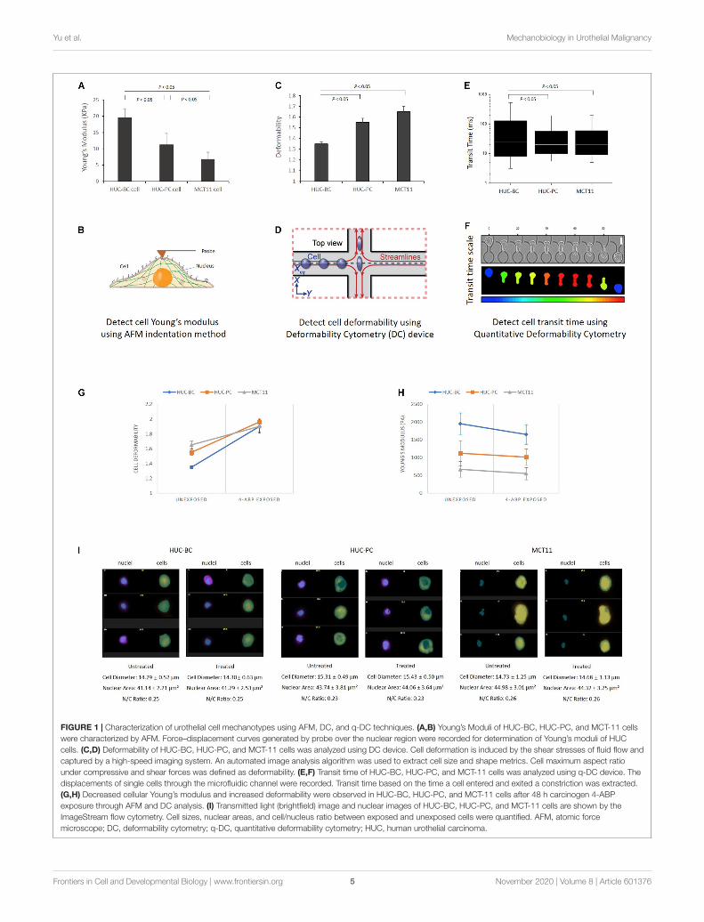
To investigate the cellular mechanotype changes, threepreviously established platforms were used (Cross et al., 2007;Gossett et al., 2012; Nyberg et al., 2017). As shown inFigures 1A,B, AFM characterizes elasticity, or stiffness, of livingcells through measuring force–displacement as the tip is pushedtoward the cell, indented into the sample, and subsequentlyretracted (Cross et al., 2007). The stiffness of cells is quantifiedby the value of Young’s modulus. From untransformed HUC-BCcells, HUC-PC cells to transformed MCT-11 cells, the stiffnessgradually decreases (HUC-BC vs. HUC-PC, cell modulus:19.5 ± 2.6 KPa vs. 11.2 ± 4.5 KPa, P < 0.05, t-test; HUC-BC vs.MCT-11, cell modulus: 19.5± 2.6 KPa vs. 6.7± 2.2 KPa, P< 0.05,t-test) (Figure 1A). The inversely related trend was observed incell deformability measurement by DC in which a continuousstream of single cells is created, where each cell’s deformation
under a high-speed microfluidic flow is measured with high-speed imaging (Gossett et al., 2012). DC enables high-throughputsingle-cell mechanotyping. Cellullar deformation is induced bythe shear and inertial stresses of fluid flow (Figure 1D). DCmeasurements show that in the order of HUC-BC, HUC-PC,and MCT-11 cells, deformability progressively increases (HUC-BC vs. HUC-PC, deformability: 1.35 ± 0.02 vs. 1.55 ± 0.04,P < 0.05, t-test; HUC-BC vs. MCY-11, deformability: 1.35± 0.02vs. 1.65 ± 0.05, P < 0.05, t-test) (Figure 1C). To confirmthis finding, we analyzed cellular deformability using anothertechnique, q-DC (Nyberg et al., 2017). By applying a pressuregradient across the microfluidic device, cells were driven todeform through micron-scale constrictions (Figure 1F). Wetracked the time scale for single cells to transit through micron-scale gaps in the microfluidic channel; the resultant “transit time”reflects cell mechanotype, whereby stiffer cells and particles havea longer transit time (Nyberg et al., 2016). In the pooled datasetthat includes three replicates, HUC-PC and MCT-11 cells showeda statistically significant decrease in transit time compared toHUC-BC cells (HUC-BC vs. HUC-PC, median transit time: 25.6vs. 20.0 ms, P < 0.05, U-test; HUC-BC vs. MCT-11, mediantransit time: 25.6 vs. 20.6 ms, P < 0.05, U-test), substantiatingan increase in the compliance of highly malignant UC cellscompared to the untransformed urothelial cells (Figure 1E).These results indicate that highly malignant carcinoma cells areless stiff and more deformable than untransformed cells.
Decreased Stiffness in HUC Cells From3D Cultured Microtumor Mass and FromPatient Urinary Cytology SpecimensTo better mimic in vivo tumor growth, we used a scaffold-free spheroid cell culture condition to grow bladder cancercells as micro tumor masses (Figure 2A). Prior to AFMmeasurement, cells were dissociated from tumor masses andplaced on the slides after cytospin (Figure 2B). Again, weobserved that untransformed HUC-BC cells display significantlyhigher stiffness than HUC-PC and MCT-11 cells (Figure 2C).
Based on our previously published methods (Cross et al., 2007;Gossett et al., 2012), we developed a practical protocol to preparecells directly from urinary cytology specimens for mechanotypeanalyses described in the section “Materials and Methods”. Usingthis method, we tested nine clinical urinary specimens, whichwere divided into three groups. Group one and two included sixspecimens from patients with high-grade urothelial carcinoma(HGUC) confirmed by cystoscopy biopsy diagnosis. The threespecimens of group one had positive cytology findings and thethree specimens of group two had atypical cytology findings.Group three included another three from patients with no UCas confirmed by both cystoscopy and cytology examination.As shown in Figure 2D, a pooled data analysis demonstratedthe same mechanotype change pattern as we observed in 3Dcultured microtumor masses, where urinary exfoliated cellsfrom group three had the highest Young’s modulus. Whilecertainly more samples will be needed to be conclusive, thepreliminary finding shows the feasibility of measuring single-cellmechanotype changes in clinical urinary samples.
Frontiers in Cell and Developmental Biology | www.frontiersin.org 4 November 2020 | Volume 8 | Article 601376
fcell-08-601376 November 12, 2020 Time: 15:27 # 5
Yu et al. Mechanobiology in Urothelial Malignancy
FIGURE 1 | Characterization of urothelial cell mechanotypes using AFM, DC, and q-DC techniques. (A,B) Young’s Moduli of HUC-BC, HUC-PC, and MCT-11 cellswere characterized by AFM. Force–displacement curves generated by probe over the nuclear region were recorded for determination of Young’s moduli of HUCcells. (C,D) Deformability of HUC-BC, HUC-PC, and MCT-11 cells was analyzed using DC device. Cell deformation is induced by the shear stresses of fluid flow andcaptured by a high-speed imaging system. An automated image analysis algorithm was used to extract cell size and shape metrics. Cell maximum aspect ratiounder compressive and shear forces was defined as deformability. (E,F) Transit time of HUC-BC, HUC-PC, and MCT-11 cells was analyzed using q-DC device. Thedisplacements of single cells through the microfluidic channel were recorded. Transit time based on the time a cell entered and exited a constriction was extracted.(G,H) Decreased cellular Young’s modulus and increased deformability were observed in HUC-BC, HUC-PC, and MCT-11 cells after 48 h carcinogen 4-ABPexposure through AFM and DC analysis. (I) Transmitted light (brightfield) image and nuclear images of HUC-BC, HUC-PC, and MCT-11 cells are shown by theImageStream flow cytometry. Cell sizes, nuclear areas, and cell/nucleus ratio between exposed and unexposed cells were quantified. AFM, atomic forcemicroscope; DC, deformability cytometry; q-DC, quantitative deformability cytometry; HUC, human urothelial carcinoma.
Frontiers in Cell and Developmental Biology | www.frontiersin.org 5 November 2020 | Volume 8 | Article 601376

fcell-08-601376 November 12, 2020 Time: 15:27 # 6
Yu et al. Mechanobiology in Urothelial Malignancy
FIGURE 2 | Change in Young’s modulus of cells from 3D cultured microtumor masses and from patient urinary cytology specimens. (A) A 3D cell culture conditionwas used to grow HUC-BC, HUC-PC, MCT-11 cells as micro tumor masses. Spheroid formations of HUC cells were shown over 72 h after seeding. HE stainsdemonstrate dense, rapid-growing cell masses. (B) Dissociated HUC-BC, HUC-PC, and MCT-11 cells were under AFM measuring. (C) Untransformed HUC-BCcells display significant higher cellular Young’s modulus than HUC-PC and MCT-11 cells. (D) Pooled data analysis of Young’s moduli of urinary exfoliated cells fromnine urine cytology specimens. Group one included three cytology-positive specimens from patients with HGUC confirmed by cystoscopy biopsy diagnosis. Grouptwo included thee cytology-atypical specimens from patients with HGUC confirmed by cystoscopy biopsy diagnosis. Group three included three cytology-negativespecimens from patients with no UC. The number of measured uroepithelial cells in each case ranged from 3 to 10. The average number of force-displacementcurve obtained from each cell was three. AFM, atomic force microscope; HUC, human urothelial carcinoma.
Decreasing Stiffness and IncreasingDeformability During MalignantTransformation and Progression Inducedby 4-ABPAs shown in Figure 2C, the magnitude of difference betweenHUC-BC vs. HUC-PC and MCT-11 cells cultured from 3Dcondition appeared to be larger than the differences in cellularstiffness measured by AFM on 2D cultured cells. We suspectedthat in microtumor masses, HUC cells were grown without
losing cell-cell interaction in all directions, central hypoxia, andcell response and resistance to the tumor microenvironment.The malignant potential can be fully expressed and captured bymechanotype analysis. Previously, it has been shown that uponexposure to carcinogen 4-ABP, the malignant phenotypes, namelythe ability of cells to form tumor nodules upon injected to thenude mice, can be enhanced (Bookland et al., 1992a,b). Thus, tofurther characterize the link between mechanotype change andurothelial transformation and progression, we exposed three celllines to 4-ABP. After 48-h exposure, we quantified morphological
Frontiers in Cell and Developmental Biology | www.frontiersin.org 6 November 2020 | Volume 8 | Article 601376

fcell-08-601376 November 12, 2020 Time: 15:27 # 7
Yu et al. Mechanobiology in Urothelial Malignancy
changes of cells using the ImageStream flow cytometry analysis.No significant differences were observed in cell sizes, nuclearareas, and cell/nucleus ratio between treated and untreatedcells (Figure 1I). However, decreased cell Young’s modulus wasobserved in HUC-BC, HUC-PC, and MCT-11 cells throughAFM measurement (Figure 1H). A similar trend in the changesof cell deformability was seen by DC, whereby all three celllines displayed increased deformability compared to untreatedcontrols (Figure 1G). Altogether, mechanotype characterizationof the urothelial cells demonstrated specifically decreasedcellular modulus and increased cell deformability as malignancyincreases, indicating that changes in cellular mechanotypeis associated with urothelial cell malignant transformationand progression.
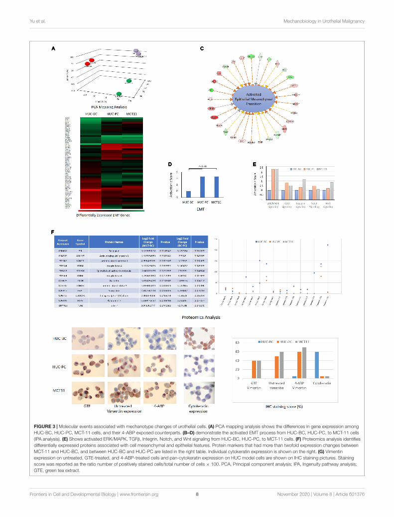
Correlation of ActivatedEpithelial-Mesenchymal TransitionPathway With the Change of CellularMechanotypeTo explore the underlying molecular events associated with thesespecific mechanotype changes, we screened the transcriptomeof HUC-BC, HUC-PC, and MCT-11 cells. The 3D portrayaldiagram (Figure 3A) demonstrates the differences in geneexpression among the three cell lines and their carcinogen-induced counterparts. Using IPA, we identified the associationof many differentially expressed genes with epithelial tomesenchymal transition (EMT). Compared to HUC-BC cells,HUC-PC, and MCT-11 cells have more than two foldactivation in the EMT pathway (activation z score: 2.319,P < 0.001) (Figures 3B–D). Upstream regulator analysisindicated several molecular signaling pathways, includingERK/MAPK, TGFβ, Integrin, Notch, and Wnt signaling, allof which are important contributors of the EMT pathway,were activated (Figure 3E). The EMT process was originallyfound in tissue repair and fibrosis, in which epithelial cells losetheir junctions and apical-basal polarity and motile behavioras they differentiate into mesenchymal cells. Importantly, theEMT process involves gene expression reprogramming, signalingchanges and cytoskeleton reorganization, and plays a pivotalrole in malignancy progression (Sarrio et al., 2008; Thieryet al., 2009). Recent genome-wide characterization study on 112bladder cancer cases revealed that highly malignant sarcomatoidurothelial bladder cancer is largely driven by dysregulation ofthe EMT network (Guo et al., 2019). However, the evidenceof a full EMT phenotype in clinical carcinomas and metastasesis frequently questioned because EMT can be reversible andtransient (Brabletz, 2012). Also, the role of the EMT process asa driving force on cell mechanotype regulation is not clear. Toconfirm the change between activated EMT and decreased cell’smodulus and increased deformability, we induced malignantprogression on HUC-BC, HUC-PC, and MCT-11 cells with 4-ABP. As shown in Figures 1G,H, 4-ABP exposure reducedYoung’s modulus and increased deformability in all three celllines, especially in HUC-BC and HUC-PC cells. We thenperformed an IPA canonical pathway analysis on differentiallyexpressed genes. Again, activated EMT pathway was found in
4-ABP treated HUC-BC and HUC-PC cells in comparison totheir untreated counterparts (HUC-BC vs. treated HUC-BC:Activation z score: 0.1, P < 0.001; HUC-PC vs. treated HUC-PC:Activation z score 0.08, P < 0.001). These results suggest that theEMT pathway may be correlated with mechanotype dynamics inurothelial cells.
Identifying Protein Targets inEpithelial-Mesenchymal TransitionPathway Associated With MechanotypeChangesWe then interrogated the differences in protein content usingthe proteomics approach (Michalski et al., 2011). A number ofdifferentially expressed proteins associated with EMT, includingpeiplakin, a component of desmosomes and of the epidermalcornified envelope in keratinocytes, E cadherin, an epithelialmarker localized at cell–cell contacts, mesenchymal markervimentin, fibronectin, and many other regulator proteins(Kokkinos et al., 2007) were identified. The table of Figure 3Flists a few protein markers that had more than twofoldexpression changes between MCT-11 and HUC-BC, and betweenHUC-BC and HUC-PC. The change trends of both epithelialmarks and mesenchymal markers demonstrated urothelial cellsobtained mesenchymal features while losing epithelial makerswith malignant progression, which is consistent with the resultsfrom gene expression analysis.
Cytokeratins are a class of intermediate filamentsdemonstrating epithelial differentiation. Individual cytokeratinsare commonly used markers for determining the grade ofbladder cancer cells (Kim et al., 2015; Hashmi et al., 2018).As illustrated in Figure 3F, protein abundance of individualcytokeratins showed the expression of most cytokeratin familymolecules decrease from HUC-BC, to HUC-PC, and MCT-11cells. Among them, cytokeratin 5, 7, 17, and 19 have remarkabledecreasing trend. This trend was confirmed by pan-cytokeratinIHC staining, where HUC-BC cells demonstrated significantlyhigher pan-cytokeratin expression than HUC-PC and MCT-11cells (Figure 3G). On the other hand, vimentin, an EMT marker,is one of the intermediate filaments that mainly functions tomaintain cell integrity and involved in the cell migration andinvasion of metastasizing cancer cells (Coulombe and Wong,2004; Mendez et al., 2010). Our previous study showed thatovarian cancer cells induced to have mesenchymal phenotypeeither by overexpression of EMT transcription factors (ZEB1,SNAI1, and SNAI2), oncogenes (H-Ras v12), or by drugresistance were all consistently more deformable than cellswith epithelial phenotype (Qi et al., 2015). In these processes,increased vimentin and reduced E-Cadherin were indicated.Over-expression of vimentin correlates with increased tumorgrowth and invasiveness, and as well as with poor clinicaloutcomes in several cancers (Maier et al., 2016). Anotherstudy reported that intracellular mechanical homeostasiswas interrupted in vimentin-knockdown breast cancer cells.Overexpressing vimentin in MCF7 breast cancer cells reorientedmicrotubule polarity, increased cell directional migration, andEMT phenotypes (Liu et al., 2015). To confirm the upregulation
Frontiers in Cell and Developmental Biology | www.frontiersin.org 7 November 2020 | Volume 8 | Article 601376
fcell-08-601376 November 12, 2020 Time: 15:27 # 8
Yu et al. Mechanobiology in Urothelial Malignancy
FIGURE 3 | Molecular events associated with mechanotype changes of urothelial cells. (A) PCA mapping analysis shows the differences in gene expression amongHUC-BC, HUC-PC, MCT-11 cells, and their 4-ABP exposed counterparts. (B–D) demonstrate the activated EMT process from HUC-BC, HUC-PC, to MCT-11 cells(IPA analysis). (E) Shows activated ERK/MAPK, TGFβ, Integrin, Notch, and Wnt signaling from HUC-BC, HUC-PC, to MCT-11 cells. (F) Proteomics analysis identifiesdifferentially expressed proteins associated with cell mesenchymal and epithelial features. Protein markers that had more than twofold expression changes betweenMCT-11 and HUC-BC, and between HUC-BC and HUC-PC are listed in the right table. Individual cytokeratin expression is shown on the right. (G) Vimentinexpression on untreated, GTE-treated, and 4-ABP-treated cells and pan-cytokeratin expression on HUC model cells are shown on IHC staining pictures. Stainingscore was reported as the ratio number of positively stained cells/total number of cells × 100. PCA, Principal component analysis; IPA, Ingenuity pathway analysis;GTE, green tea extract.
Frontiers in Cell and Developmental Biology | www.frontiersin.org 8 November 2020 | Volume 8 | Article 601376
fcell-08-601376 November 12, 2020 Time: 15:27 # 9
Yu et al. Mechanobiology in Urothelial Malignancy
of vimentin in UC cells, we performed an IHC analysis. HUC-BCcells have the lowest expression of vimentin whereas MCT-11cells have the highest, in line with the activation status of EMTand urothelial cell mechanotype changes (Figure 3G). Previously,we also reported that chemopreventive agent green tea extract(GTE) modulated cytoskeletal actin remodeling via Rho activityin the same UC cells and significantly increased the stiffness ofGTE-treated metastatic tumor cells compared to normal cells(Lu et al., 2005; Cross et al., 2011). In the present study, wefound that GTE counteracts the effect of carcinogen 4-ABPby reversing the increase in vimentin expression (Figure 3G),further corroborating epithelial-mesenchymal transition playsan important role in urothelial cell mechanotype changes.
DISCUSSION
While metastatic tumor cells show a distinctive cell mechanotyperelative to normal cells, the specific pattern of changes in cellmechanotype during the early stages of cancer transformationand progression is not well studied. Identifying metastaticcancer cells may be clinically too late to save patients’ lives.Therefore, there is a great need for developing biomarkersthat can be used to determine the invasive and metastaticpotential of malignant cells before the invasion or metastasisactually occur. Moreover, the molecular mechanism underlyingthe changes in cellular mechanotype is poorly studied andunderstood. Recent evidence suggests that the regulation ofcellular mechanotype may be the result of alterations ofmultiple genes and signal transcription pathways (Way andWeeds, 1990; Provenzano et al., 2009; Lammerding, 2011;
De Las Heras et al., 2013; Fedorchak et al., 2014). Thus,knowledge of cell mechanotype changes in the early stages ofcancer transformation and progression, and the mechanism ormolecular pathways associated with these biomechanical changesmay have a significant impact on not only developing biomarkersthat can be utilized to distinguish invasive cancer, but alsofinding drug targets for disrupting, or inhibiting cancer cellinvasion or metastasis.
We, here, report the cell mechanotype profiles in relation toUC utilizing an in vitro human urothelial carcinogenesis modelthat recapitulates the multistep process of cancer progression.Cellular Young’s modulus and deformability were analyzed usingAFM indentation, microfluidic-based DC, and q-DC analyses.From normal, to preinvasive, to invasive cells, Young’s modulus,or cell stiffness, progressively decreases. Cellular deformabilitysignificantly increases. Previous studies indicated that changesin cell mechanotype could be detected in precancerous cervicalintraepithelial lesions as early as CIN II (Ding et al., 2015), andalso in precancerous esophageal cells (Fuhrmann et al., 2011).Our malignant transformation/progression experiments impliedthat cellular mechanotype changes may start at the early stage ofmalignant progression. To verify this finding in a disease-specificsetting, we integrated cell mechanotyping technology into clinicalsamples. Currently, a variety of platforms have been developed tomeasure cellular mechanical properties, and different techniquesare known to probe mechanical properties at different timescalesand depths of the cell (Urbanska et al., 2020). The mostcommonly explored method is to use biomechanical probes,represented by AFM and magnetic tweezer (Yang et al., 2011),whereas cell deformability can be optically probed underthe force induced on the whole cells. These methods also
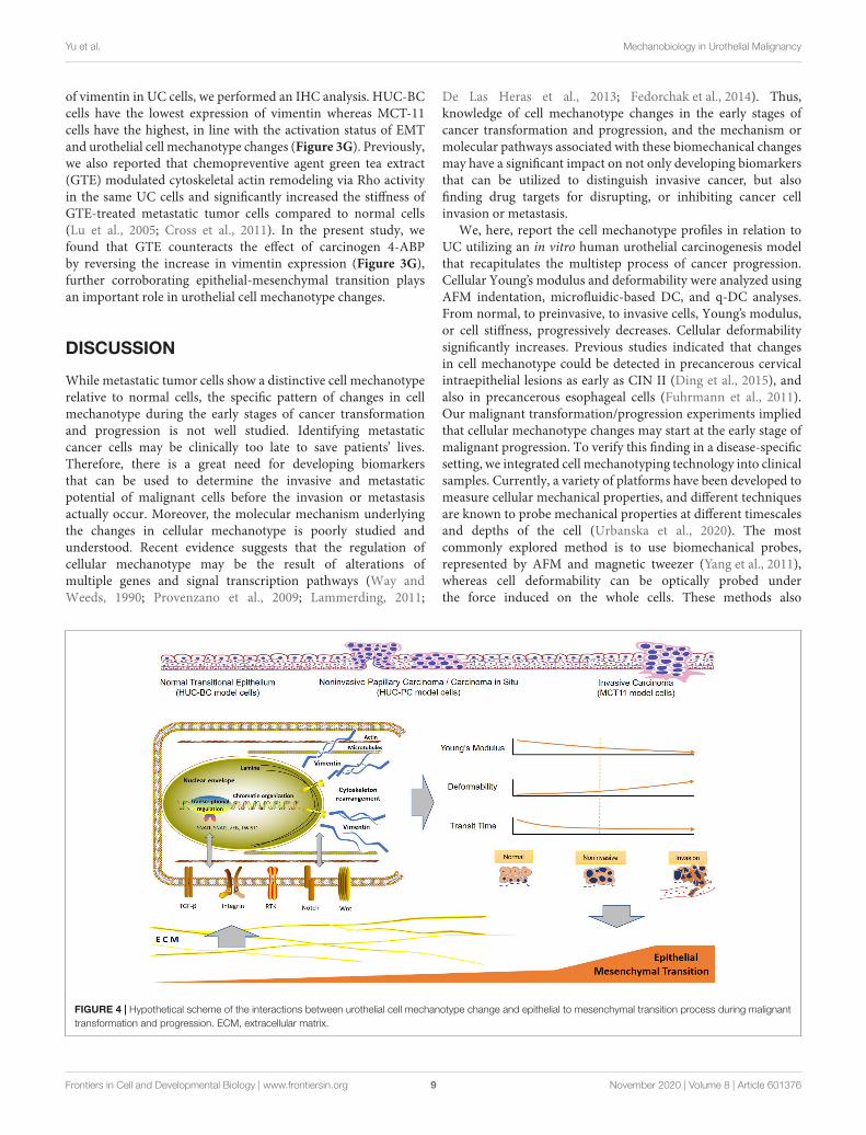
FIGURE 4 | Hypothetical scheme of the interactions between urothelial cell mechanotype change and epithelial to mesenchymal transition process during malignanttransformation and progression. ECM, extracellular matrix.
Frontiers in Cell and Developmental Biology | www.frontiersin.org 9 November 2020 | Volume 8 | Article 601376

fcell-08-601376 November 12, 2020 Time: 15:27 # 10
Yu et al. Mechanobiology in Urothelial Malignancy
include micropipette aspiration (Oh et al., 2012), microfluidicassays (Guo et al., 2014), DC (Gossett et al., 2012), andmicroplate stretcher (Thoumine et al., 1999). More recently,high-throughput techniques have also been explored (Darlingand Di Carlo, 2015). Most of these techniques were developed ina laboratory setting and are still in a pre-clinical stage. Urinaryexfoliated cells, which are directly from the primary tumor,provide a unique living model for the study of UC. However, forclinical specimens, variability is common in terms of cell numberand type. Under AFM, uroepithelial cells, squamous epithelialcells, and cell of hematologic origin can be easily distinguished.Although only nine specimens were preliminarily analyzed inthe current study, the findings were consistent with those fromin vitro condition. Additionally, it presented a proof of conceptfor the development of mechanotype signature for urothelialcancer early detection.
It has become increasingly clear that EMT is driven by atleast four fundamental regulatory network layers. The first layeris EMT-inducing transcription factor control, such as SNAI1,ZEB, and TWIST1. Another three layers are the expression ofsmall non-coding RNAs, differential splicing, and translationaland post-translational control, which determine the stabilizationand localization of network proteins (De Craene and Berx,2013). The dynamic and transient nature of EMT comprisinga broad spectrum of intermediate phenotypes adds furthercomplexity to capture EMT molecular status to determine thediagnosis and progression of cancer (Santamaria et al., 2017).Our findings indicate a close correlation between EMT and cellmechanotype in the process of malignant progression, whichprovides new insight for developing novel biomarkers for cell-based UC early detection. The underlying molecular events andthe specific mechanotype changes during UC development andprogression can be summarized in Figure 4. One may expectthat progression of EMT drives cell mechanotype changes duringmalignant transformation and progression. Conversely, it is alsopossible that change in cell mechanotype, such as decreasing incell stiffness and increasing in deformability that occurs duringmalignant progression, enables tumor cells to detach from aprimary tumor, squeeze through confined spaces, and facilitatecells to invade the surrounding tissue that affects EMT.
In summary, we described the specific mechanotype changesof urothelial cells in malignant transformation and progression.Measurable cell mechanotype changes of stiffness, deformability,and cell transit time occur early in the transformation process.
As cells progress from normal, to preinvasive, to invasivecancer cells, Young’s modulus of stiffness decreases anddeformability increases gradually. The key molecular pathwayimplicated in urothelial cell mechanotype changes appears to beassociated with EMT.
DATA AVAILABILITY STATEMENT
The datasets presented in this study can be found in onlinerepositories. The names of the repository/repositories andaccession number(s) can be found below: http://www.ebi.ac.uk/arrayexpress/experiments/E-MTAB-9699, accession no:E-MTAB-9699.
ETHICS STATEMENT
The studies involving human participants were reviewed andapproved by UCLA-MIRB2. Written informed consent forparticipation was not required for this study in accordance withthe national legislation and the institutional requirements.
AUTHOR CONTRIBUTIONS
WY: cell experiment, clinical sample processing, gene expressionanalysis, protein expression experiment, and manuscript writing.Q-YL: proteomics analysis, data interpretation, manuscriptcritical review, and revision. SS: AFM experiment, study design,data analysis, and manuscript revision. CL: qDC experiment. DD:DC cell deformability analysis and manuscript revision. AR: qDCanalysis and manuscript revision. ML: AFM experiment. DK: DCexperiment. CC: manuscript review. JG: study design and AFMcell stiffness analysis. JR: study overall conception and design,data analysis and interpretation. All authors contributed to thearticle and approved the submitted version.
FUNDING
This study was supported by the Jonsson Comprehensive CancerCenter Impact Grant, and NCI/NIH R21 CA208196: Cancer CellMechanical Profiling Analysis.
REFERENCESBeukers, W., Van Der Keur, K. A., Kandimalla, R., Vergouwe, Y., Steyerberg,
E. W., Boormans, J. L., et al. (2017). FGFR3, TERT and OTX1 as a urinarybiomarker combination for surveillance of patients with bladder cancer in alarge prospective multicenter study. J. Urol. 197, 1410–1418. doi: 10.1016/j.juro.2016.12.096
Bookland, E. A., Reznikoff, C. A., Lindstrom, M., and Swaminathan, S.(1992a). Induction of thioguanine-resistant mutations in human uroepithelialcells by 4-aminobiphenyl and its N-hydroxy derivatives. Cancer Res. 52,1615–1621.
Bookland, E. A., Swaminathan, S., Oyasu, R., Gilchrist, K. W., Lindstrom, M.,and Reznikoff, C. A. (1992b). Tumorigenic transformation and neoplastic
progression of human uroepithelial cells after exposure in vitro to 4-aminobiphenyl or its metabolites. Cancer Res. 52, 1606–1614.
Brabletz, T. (2012). To differentiate or not–routes towards metastasis. Nat. Rev.Cancer 12, 425–436. doi: 10.1038/nrc3265
Che, J., Yu, V., Garon, E. B., Goldman, J. W., and Di Carlo, D. (2017). Biophysicalisolation and identification of circulating tumor cells. Lab Chip 17, 1452–1461.doi: 10.1039/c7lc00038c
Coulombe, P. A., and Wong, P. (2004). Cytoplasmic intermediate filamentsrevealed as dynamic and multipurpose scaffolds. Nat. Cell Biol. 6, 699–706.doi: 10.1038/ncb0804-699
Cross, S. E., Jin, Y. S., Lu, Q. Y., Rao, J., and Gimzewski, J. K. (2011). Greentea extract selectively targets nanomechanics of live metastatic cancer cells.Nanotechnology 22:215101. doi: 10.1088/0957-4484/22/21/215101
Frontiers in Cell and Developmental Biology | www.frontiersin.org 10 November 2020 | Volume 8 | Article 601376

fcell-08-601376 November 12, 2020 Time: 15:27 # 11
Yu et al. Mechanobiology in Urothelial Malignancy
Cross, S. E., Jin, Y. S., Rao, J., and Gimzewski, J. K. (2007). Nanomechanical analysisof cells from cancer patients.Nat. Nanotechnol. 2, 780–783. doi: 10.1038/nnano.2007.388
Cross, S. E., Jin, Y. S., Tondre, J., Wong, R., Rao, J., and Gimzewski, J. K.(2008). AFM-based analysis of human metastatic cancer cells. Nanotechnology19:384003. doi: 10.1088/0957-4484/19/38/384003
Darling, E. M., and Di Carlo, D. (2015). High-throughput assessment of cellularmechanical properties. Annu. Rev. Biomed. Eng. 17, 35–62. doi: 10.1146/annurev-bioeng-071114-040545
De Craene, B., and Berx, G. (2013). Regulatory networks defining EMT duringcancer initiation and progression. Nat. Rev. Cancer 13, 97–110. doi: 10.1038/nrc3447
De Las Heras, J. I., Batrakou, D. G., and Schirmer, E. C. (2013). Cancer biologyand the nuclear envelope: a convoluted relationship. Semin. Cancer Biol. 23,125–137. doi: 10.1016/j.semcancer.2012.01.008
Ding, Y. X., Cheng, Y., Sun, Q. M., Zhang, Y. Y., You, K., Guo, Y. L., et al. (2015).Mechanical characterization of cervical squamous carcinoma cells by atomicforce microscopy at nanoscale. Med. Oncol. 32, 71.
Fedorchak, G. R., Kaminski, A., and Lammerding, J. (2014). Cellularmechanosensing: getting to the nucleus of it all. Prog. Biophys. Mol. Biol.115, 76–92. doi: 10.1016/j.pbiomolbio.2014.06.009
Fuhrmann, A., Staunton, J. R., Nandakumar, V., Banyai, N., Davies, P. C., andRos, R. (2011). AFM stiffness nanotomography of normal, metaplastic anddysplastic human esophageal cells. Phys. Biol. 8:015007. doi: 10.1088/1478-3975/8/1/015007
Gossett, D. R., Tse, H. T., Lee, S. A., Ying, Y., Lindgren, A. G., Yang, O. O., et al.(2012). Hydrodynamic stretching of single cells for large population mechanicalphenotyping. Proc. Nat.l Acad. Sci. U.S.A. 109, 7630–7635. doi: 10.1073/pnas.1200107109
Guo, C. C., Majewski, T., Zhang, L., Yao, H., Bondaruk, J., Wang, Y., et al.(2019). Dysregulation of EMT drives the progression to clinically aggressivesarcomatoid Bladder Cancer. Cell Rep. 27, 1781.e4–1793.e4.
Guo, Q., Duffy, S. P., Matthews, K., Santoso, A. T., Scott, M. D., and Ma, H. (2014).Microfluidic analysis of red blood cell deformability. J. Biomech. 47, 1767–1776.doi: 10.1016/j.jbiomech.2014.03.038
Hashmi, A. A., Hussain, Z. F., Irfan, M., Edhi, M. M., Kanwal, S., Faridi, N.,et al. (2018). Cytokeratin 5/6 expression in bladder cancer: association withclinicopathologic parameters and prognosis. BMC Res. Notes 11:207. doi: 10.1186/s13104-018-3319-4
He, H., Han, C., Hao, L., and Zang, G. (2016). ImmunoCyt test compared tocytology in the diagnosis of bladder cancer: a meta-analysis. Oncol. Lett. 12,83–88. doi: 10.3892/ol.2016.4556
Hughes, C. S., Foehr, S., Garfield, D. A., Furlong, E. E., Steinmetz, L. M., andKrijgsveld, J. (2014). Ultrasensitive proteome analysis using paramagnetic beadtechnology. Mol. Syst. Biol. 10:757. doi: 10.15252/msb.20145625
Humphrey, P. A., Moch, H., Cubilla, A. L., Ulbright, T. M., and Reuter, V. E.(2016). The 2016 WHO classification of tumours of the urinary system and malegenital organs-Part B: prostate and bladder tumours. Eur. Urol. 70, 106–119.doi: 10.1016/j.eururo.2016.02.028
Kim, J., Akbani, R., Creighton, C. J., Lerner, S. P., Weinstein, J. N., Getz, G., et al.(2015). Invasive bladder cancer: genomic insights and therapeutic promise.Clin. Cancer Res. 21, 4514–4524. doi: 10.1158/1078-0432.ccr-14-1215
Kokkinos, M. I., Wafai, R., Wong, M. K., Newgreen, D. F., Thompson, E. W.,and Waltham, M. (2007). Vimentin and epithelial-mesenchymal transition inhuman breast cancer–observations in vitro and in vivo.Cells Tissues Organs 185,191–203. doi: 10.1159/000101320
Lammerding, J. (2011). Mechanics of the nucleus. Compr. Physiol. 1, 783–807.Lamy, P., Nordentoft, I., Birkenkamp-Demtroder, K., Thomsen, M. B., Villesen,
P., Vang, S., et al. (2016). Paired exome analysis reveals clonal evolution andpotential therapeutic targets in urothelial carcinoma. Cancer Res. 76, 5894–5906. doi: 10.1158/0008-5472.can-16-0436
Leal, J., Luengo-Fernandez, R., Sullivan, R., and Witjes, J. A. (2016). Economicburden of bladder cancer across the european union. Eur. Urol. 69, 438–447.doi: 10.1016/j.eururo.2015.10.024
Liu, C. Y., Lin, H. H., Tang, M. J., and Wang, Y. K. (2015). Vimentin contributesto epithelial-mesenchymal transition cancer cell mechanics by mediatingcytoskeletal organization and focal adhesion maturation. Oncotarget 6, 15966–15983. doi: 10.18632/oncotarget.3862
Lu, Q. Y., Jin, Y. S., Pantuck, A., Zhang, Z. F., Heber, D., Belldegrun, A., et al.(2005). Green tea extract modulates actin remodeling via Rho activity in anin vitro multistep carcinogenic model. Clin. Cancer Res. 11, 1675–1683. doi:10.1158/1078-0432.ccr-04-1608
Maier, J., Traenkle, B., and Rothbauer, U. (2016). Visualizing epithelial-mesenchymal transition using the chromobody technology. Cancer Res. 76,5592–5596. doi: 10.1158/0008-5472.can-15-3419
Mendez, M. G., Kojima, S., and Goldman, R. D. (2010). Vimentin induces changesin cell shape, motility, and adhesion during the epithelial to mesenchymaltransition. FASEB J. 24, 1838–1851. doi: 10.1096/fj.09-151639
Michalski, A., Damoc, E., Hauschild, J. P., Lange, O., Wieghaus, A., Makarov,A., et al. (2011). Mass spectrometry-based proteomics using Q Exactive, ahigh-performance benchtop quadrupole Orbitrap mass spectrometer. Mol. CellProteomics 10:M111011015.
Nordentoft, I., Lamy, P., Birkenkamp-Demtroder, K., Shumansky, K., Vang, S.,and Hornshoj, H. (2014). Mutational context and diverse clonal developmentin early and late bladder cancer. Cell Rep. 7, 1649–1663. doi: 10.1016/j.celrep.2014.04.038
Nyberg, K. D., Bruce, S. L., Nguyen, A. V., Chan, C. K., Gill, N. K., Kim, T. H.,et al. (2018). Predicting cancer cell invasion by single-cell physical phenotyping.Integr. Biol. 10, 218–231. doi: 10.1039/c7ib00222j
Nyberg, K. D., Hu, K. H., Kleinman, S. H., Khismatullin, D. B., Butte, M. J., andRowat, A. C. (2017). Quantitative deformability cytometry: rapid, calibratedmeasurements of cell mechanical properties. Biophys. J. 113, 1574–1584. doi:10.1016/j.bpj.2017.06.073
Nyberg, K. D., Scott, M. B., Bruce, S. L., Gopinath, A. B., Bikos, D., Mason,T. G., et al. (2016). The physical origins of transit time measurements forrapid, single cell mechanotyping. Lab Chip 16, 3330–3339. doi: 10.1039/c6lc00169f
Oh, M. J., Kuhr, F., Byfield, F., and Levitan, I. (2012). Micropipette aspirationof substrate-attached cells to estimate cell stiffness. Jove J. Vis. Exp. 27:3886.
Provenzano, P. P., Inman, D. R., Eliceiri, K. W., and Keely, P. J. (2009). Matrixdensity-induced mechanoregulation of breast cell phenotype, signaling andgene expression through a FAK-ERK linkage. Oncogene 28, 4326–4343. doi:10.1038/onc.2009.299
Qi, D., Kaur Gill, N., Santiskulvong, C., Sifuentes, J., Dorigo, O., Rao, J.,et al. (2015). Screening cell mechanotype by parallel microfiltration. Sci. Rep.5:17595.
Renshaw, A. A., and Gould, E. W. (2018). High-grade urothelial carcinoma onurine cytology resembling umbrella cells. Acta Cytol. 62, 62–67. doi: 10.1159/000481908
Santamaria, P. G., Moreno-Bueno, G., Portillo, F., and Cano, A. (2017). EMT:present and future in clinical oncology. Mol. Oncol. 11, 718–738. doi: 10.1002/1878-0261.12091
Santoni, G., Morelli, M. B., Amantini, C., and Battelli, N. (2018). Urinary markersin bladder cancer: an update. Front. Oncol. 8:362. doi: 10.3389/fonc.2018.00362
Sarrio, D., Rodriguez-Pinilla, S. M., Hardisson, D., Cano, A., Moreno-Bueno,G., and Palacios, J. (2008). Epithelial-mesenchymal transition in breast cancerrelates to the basal-like phenotype. Cancer Res. 68, 989–997. doi: 10.1158/0008-5472.can-07-2017
Sharma, S., Santiskulvong, C., Bentolila, L. A., Rao, J., Dorigo, O., and Gimzewski,J. K. (2012). Correlative nanomechanical profiling with super-resolution F-actinimaging reveals novel insights into mechanisms of cisplatin resistance inovarian cancer cells. Nanomedicine 8, 757–766. doi: 10.1016/j.nano.2011.09.015
Sharma, S., Santiskulvong, C., Rao, J., Gimzewski, J. K., and Dorigo, O. (2014). Therole of Rho GTPase in cell stiffness and cisplatin resistance in ovarian cancercells. Integr. Biol. 6, 611–617. doi: 10.1039/c3ib40246k
Siegel, R. L., Miller, K. D., and Jemal, A. (2018). Cancer statistics, 2018. CA CancerJ. Clin. 68, 7–30. doi: 10.3322/caac.21442
Tan, W. S., Tan, W. P., Tan, M. Y., Khetrapal, P., Dong, L., Dewinter, P., et al. (2018).Novel urinary biomarkers for the detection of bladder cancer: a systematicreview. Cancer Treat Rev. 69, 39–52. doi: 10.1016/j.ctrv.2018.05.012
Thiery, J. P., Acloque, H., Huang, R. Y., and Nieto, M. A. (2009). Epithelial-mesenchymal transitions in development and disease. Cell 139, 871–890. doi:10.1016/j.cell.2009.11.007
Frontiers in Cell and Developmental Biology | www.frontiersin.org 11 November 2020 | Volume 8 | Article 601376
fcell-08-601376 November 12, 2020 Time: 15:27 # 12
Yu et al. Mechanobiology in Urothelial Malignancy
Thoumine, O., Ott, A., Cardoso, O., and Meister, J. J. (1999). Microplates: anew tool for manipulation and mechanical perturbation of individual cells.J. Biochem. Biophys. Methods 39, 47–62. doi: 10.1016/s0165-022x(98)00052-9
Todenhofer, T., Hennenlotter, J., Esser, M., Mohrhardt, S., Tews, V., Aufderklamm,S., et al. (2013). Combined application of cytology and molecular urine markersto improve the detection of urothelial carcinoma. Cancer Cytopathol. 121,252–260. doi: 10.1002/cncy.21247
Tse, H. T., Gossett, D. R., Moon, Y. S., Masaeli, M., Sohsman, M., Ying, Y., et al.(2013). Quantitative diagnosis of malignant pleural effusions by single-cellmechanophenotyping. Sci. Transl. Med. 5:212ra163. doi: 10.1126/scitranslmed.3006559
Urbanska, M., Munoz, H. E., Shaw Bagnall, J., Otto, O., Manalis, S. R., Di Carlo, D.,et al. (2020). A comparison of microfluidic methods for high-throughput celldeformability measurements. Nat. Methods 17, 587–593. doi: 10.1038/s41592-020-0818-8
Wang, G., and Mckenney, J. K. (2019). Urinary bladder pathology world healthorganization classification and american joint committee on Cancer stagingupdate. Arch. Pathol. Lab. Med. 143, 571–577. doi: 10.5858/arpa.2017-0539-ra
Way, M., and Weeds, A. (1990). Actin-binding proteins. Cytoskeletal ups anddowns. Nature 344, 292–294. doi: 10.1038/344292a0
Yang, R., Xi, N., Fung, C. K., Seiffert-Sinha, K., Lai, K. W., and Sinha, A. A. (2011).The emergence of AFM applications to cell biology: how new technologies arefacilitating investigation of human cells in health and disease at the nanoscale.J. Nanosci. Lett. 1, 87–101.
Conflict of Interest: The authors declare that the research was conducted in theabsence of any commercial or financial relationships that could be construed as apotential conflict of interest.
Copyright © 2020 Yu, Lu, Sharma, Ly, Di Carlo, Rowat, LeClaire, Kim, Chow,Gimzewski and Rao. This is an open-access article distributed under the termsof the Creative Commons Attribution License (CC BY). The use, distribution orreproduction in other forums is permitted, provided the original author(s) and thecopyright owner(s) are credited and that the original publication in this journalis cited, in accordance with accepted academic practice. No use, distribution orreproduction is permitted which does not comply with these terms.
Frontiers in Cell and Developmental Biology | www.frontiersin.org 12 November 2020 | Volume 8 | Article 601376