Embed Size (px)
Citation preview

University of KentuckyUKnowledge
Physiology Faculty Publications Physiology
11-4-2011
Age-Associated Disruption of Molecular ClockExpression in Skeletal Muscle of the SpontaneouslyHypertensive RatMitsunori MiyazakiUniversity of Kentucky
Elizabeth SchroderUniversity of Kentucky, [email protected]
Stephanie E. EdelmannUniversity of Kentucky, [email protected]
Michael E. HughesYale University
Karl KornackerOhio State University - Main Campus
See next page for additional authors
Right click to open a feedback form in a new tab to let us know how this document benefits you.Follow this and additional works at: https://uknowledge.uky.edu/physiology_facpub
Part of the Physiology Commons
This Article is brought to you for free and open access by the Physiology at UKnowledge. It has been accepted for inclusion in Physiology FacultyPublications by an authorized administrator of UKnowledge. For more information, please contact [email protected].
Repository CitationMiyazaki, Mitsunori; Schroder, Elizabeth; Edelmann, Stephanie E.; Hughes, Michael E.; Kornacker, Karl; Balke, C. William; and Esser,Karyn A., "Age-Associated Disruption of Molecular Clock Expression in Skeletal Muscle of the Spontaneously Hypertensive Rat"(2011). Physiology Faculty Publications. 11.https://uknowledge.uky.edu/physiology_facpub/11
AuthorsMitsunori Miyazaki, Elizabeth Schroder, Stephanie E. Edelmann, Michael E. Hughes, Karl Kornacker, C.William Balke, and Karyn A. Esser
Age-Associated Disruption of Molecular Clock Expression in Skeletal Muscle of the Spontaneously HypertensiveRat
Notes/Citation InformationPublished in PLoS ONE, v. 6, no. 11, e27168.
© 2011 Miyazaki et al. This is an open-access article distributed under the terms of the Creative CommonsAttribution License, which permits unrestricted use, distribution, and reproduction in any medium, providedthe original author and source are credited.
Digital Object Identifier (DOI)http://dx.doi.org/10.1371/journal.pone.0027168
This article is available at UKnowledge: https://uknowledge.uky.edu/physiology_facpub/11

Age-Associated Disruption of Molecular Clock Expressionin Skeletal Muscle of the Spontaneously HypertensiveRatMitsunori Miyazaki1., Elizabeth Schroder1*., Stephanie E. Edelmann1, Michael E. Hughes2, Karl
Kornacker3, C. William Balke4, Karyn A. Esser1
1 Department of Physiology, Center for Muscle Biology, University of Kentucky, Lexington, Kentucky, United States of America, 2 Department of Cellular and Molecular
Physiology, Yale School of Medicine, New Haven, Connecticut, United States of America, 3 Division of Sensory Biophysics, Ohio State University, Columbus, Ohio, United
States of America, 4 Clinical and Translational Science Institute and the Department of Medicine, University of California San Francisco, San Francisco, California, United
States of America
Abstract
It is well known that spontaneously hypertensive rats (SHR) develop muscle pathologies with hypertension and heart failure,though the mechanism remains poorly understood. Woon et al. (2007) linked the circadian clock gene Bmal1 tohypertension and metabolic dysfunction in the SHR. Building on these findings, we compared the expression pattern ofseveral core-clock genes in the gastrocnemius muscle of aged SHR (80 weeks; overt heart failure) compared to aged-matched control WKY strain. Heart failure was associated with marked effects on the expression of Bmal1, Clock and Rora inaddition to several non-circadian genes important in regulating skeletal muscle phenotype including Mck, Ttn and Mef2c.We next performed circadian time-course collections at a young age (8 weeks; pre-hypertensive) and adult age (22 weeks;hypertensive) to determine if clock gene expression was disrupted in gastrocnemius, heart and liver tissues prior to or afterthe rats became hypertensive. We found that hypertensive/hypertrophic SHR showed a dampening of peak Bmal1 and Rev-erb expression in the liver, and the clock-controlled gene Pgc1a in the gastrocnemius. In addition, the core-clock gene Clockand the muscle-specific, clock-controlled gene Myod1, no longer maintained a circadian pattern of expression ingastrocnemius from the hypertensive SHR. These findings provide a framework to suggest a mechanism whereby chronicheart failure leads to skeletal muscle pathologies; prolonged dysregulation of the molecular clock in skeletal muscle resultsin altered Clock, Pgc1a and Myod1 expression which in turn leads to the mis-regulation of target genes important formechanical and metabolic function of skeletal muscle.
Citation: Miyazaki M, Schroder E, Edelmann SE, Hughes ME, Kornacker K, et al. (2011) Age-Associated Disruption of Molecular Clock Expression in Skeletal Muscleof the Spontaneously Hypertensive Rat. PLoS ONE 6(11): e27168. doi:10.1371/journal.pone.0027168
Editor: Shin Yamazaki, Vanderbilt University, United States of America
Received May 12, 2011; Accepted October 11, 2011; Published November 4, 2011
Copyright: � 2011 Miyazaki et al. This is an open-access article distributed under the terms of the Creative Commons Attribution License, which permitsunrestricted use, distribution, and reproduction in any medium, provided the original author and source are credited.
Funding: This work was supported by R01AR055246 from NIAMS, RC1ES018636 from NIEHS, and a postdoctoral fellowship provided by the American HeartAssociation to Mitsunori Miyazaki (0825668D), http://www.heart.org/HEARTORG/. The funders had no role in study design, data collection and analysis, decisionto publish, or preparation of the manuscript.
Competing Interests: The authors have declared that no competing interests exist.
* E-mail: [email protected]
. These authors contributed equally to this work.
Introduction
The role of the molecular clock as an underlying factor
contributing to cardiovascular and skeletal muscle disease is a new
but growing area of research. It is now recognized that most, if not
all, cells in the body contain a self-sustaining molecular circadian
clock [1]. In general, the synchronization of all the body’s clocks is
orchestrated by a central clock (SCN: suprachiasmatic nucleus)
located in the hypothalamus acting through neurohumoral
mechanisms [2]. The synchronization of circadian clocks has
been experimentally shown to provide an adaptive advantage by
enhancing an organisms ability to respond to daily changes in
light, temperature and humidity [3]. Pathologies emerge when
there is a misalignment between internal circadian rhythms and
daily cycles in environmental cues such as light which can occur
when the expression of the molecular clock becomes shifted or
dampened [4].
Circadian rhythms, endogenously generated rhythms with
<24 hour-period, are driven by an intrinsic molecular clock
which works as a transcription/translation feedback system. In
mammals, the proteins encoded by core-clock genes, Bmal1 and
Clock, dimerize to drive transcription of Period (Per) and Cryptochrome
(Cry) and the protein products of these genes down-regulate
BMAL1 and CLOCK function. BMAL1:CLOCK heterodimers
directly regulate the expression of a group of genes referred to as
primary clock-controlled genes (CCGs). Many other genes are
expressed in a circadian manner and these genes are indirectly
influenced by the core clock factors as well as environmental cues,
such as hormones like melatonin and cortisol [5–13].
Woon and colleagues (2007) identified polymorphisms in the
promoter region of Bmal1 (Brain and muscle Arnt-like protein-1)
that were within the same congenic interval associated with
hypertension in the spontaneously hypertensive rat (SHR) [14].
Furthermore, the same group reported a significant genetic
PLoS ONE | www.plosone.org 1 November 2011 | Volume 6 | Issue 11 | e27168

association of Bmal1 polymorphisms and hypertension and type II
diabetes in humans [14]. These findings implicate the regulation of
Bmal1 expression and proper molecular clock function in the
development of cardiometabolic pathologies. Consistent with these
findings, Andrews and coworkers (2010) identified a common
skeletal muscle pathology in two clock-compromised mouse strains
that were characterized by diminished force capacity and reduced
mitochondrial content and function [5].
It is well recognized that chronic heart failure (CHF) and
cardiovascular disease are often associated with distinct skeletal
muscle pathologies [15,16]. This association has been shown in
different rodent models of cardiovascular disease as well as in
humans. These studies have reported the presence of a number of
pathologies in skeletal muscle related to both function and
metabolism. Specifically, structural and biochemical alterations
have been demonstrated in patients with CHF, including fiber
atrophy [17], fiber type transformation [18] reduced sensitivity to
insulin, decrease in oxidative capacity [19] and abnormalities in
mitochondrial structure [20–22]. The mechanism(s) underlying
the development of these skeletal muscle pathologies associated
with cardiovascular disease remain undefined.
The purpose of this study was to determine if changes occurred
in the expression of core clock factors and CCGs in peripheral
tissues either prior to or following the development of
cardiovascular disease and insulin resistance. Our working
hypothesis was that clock gene expression would change after
the development of hypertension in muscle and non-muscle
tissues. In this study we found that circadian gene expression was
altered across all tissues studied (gastrocnemius, liver, heart) in the
young pre-hypertensive SHR. Analysis of gene expression at the
adult hypertensive/hypertrophic stage (22 week old), found that
the disruption/dysregulation of clock gene expression persisted
primarily in skeletal muscle of SHR compared to WKY rats. In
addition, there were changes in expression of the core clock gene
Clock and two clock-controlled genes in skeletal muscle, Myod1
and Pgc1a of adult SHR. Further examination revealed, that the
expression of molecular clock factors were significantly altered in
the skeletal muscle of SHR at end stage heart failure. Collectively,
these results suggest that genetic difference(s) in the SHR vs.
WKY are associated with altered expression of the core clock
factors and downstream clock controlled genes in peripheral
tissues prior to the development of hypertension. We also propose
that prolonged alterations of the molecular clock factors in
skeletal muscle may lead to a loss of regulation of downstream
targets, Myod1 and Pgc1a with subsequent effects on skeletal
muscle functional and metabolic pathologies described in the
SHR.
Results
Dysregulation of core-clock genes is evident in thegastrocnemius of 8 week old SHR rats
Preliminary experiments in 80 week old, overt heart failure
rats demonstrated a diminished expression of several core
clock genes as well as several non-circadian genes important in
regulating skeletal muscle phenotype and function (Figures S1and S2) in the gastrocnemius of SHR compared to age-
matched WKY rats. While it was impossible to perform a
circadian time course collection on the heart failure rats because
of high mortality, we were able to perform a time course on
8 week old and 22 week old SHR to determine if the onset of
hypertension was associated with disruption of the circadian clock.
Tissues were collected from rats every 4 hours for 40 hours.
Table 1 provides the results of the statistical analysis with the
circadian parameters of the mRNA data for the core-clock genes
Bmal1, Clock and Per2 in the gastrocnemius and liver of the young
WKY and SHR rats using JTK_CYCLE [23]. The core
molecular clock factors, Bmal1, Clock and Per2 exhibited circadian
expression in both gastrocnemius and liver tissues (Figure 1A–F).
Peak expression of Per2 was reduced in the gastrocnemius
(Figure 1C) of the young SHR. Peak Per2 expression was
unaffected in the liver and (Figure 1F) heart (Figure S3C) of the
young SHR.
The core-clock genes Bmal1 and Clock heterodimerize and
transcriptionally regulate a group of direct clock-controlled genes
which are thought to be necessary for the maintenance of normal
Table 1. The circadian parameters of the core-clock genes Bmal1, Clock, and Per2 in the gastrocnemius and liver of young WKYand SHR calculated using JTK_CYCLE analysis.
Circadian Statistics Core Clock Genes
Strain Gene JTK _CYCLE p value Circadian JTK_CYCLE JTK_CYCLE Amplitude
Gastrocnemius
WKY Young Bmal1 1.21E-11 Yes 1.54
SHR Young Bmal1 5.73E-08 Yes 1.48
WKY Young Clock 1.29E-05 Yes 0.39
SHR Young Clock 3.74E-04 Yes 0.75
WKY Young Per2 3.13E-09 Yes 0.73
SHR Young Per2 6.20E-09 Yes 0.28
Liver
WKY Young Bmal1 2.20E-09 Yes 1.45
SHR Young Bmal1 2.26E-06 Yes 1.49
WKY Young Clock 2.43E-07 Yes 0.24
SHR Young Clock 5.95E-03 Yes 0.48
WKY Young Per2 8.65E-09 Yes 0.2
SHR Young Per2 2.00E-05 Yes 0.11
doi:10.1371/journal.pone.0027168.t001
Clock Disruption in Skeletal Muscle of SHR
PLoS ONE | www.plosone.org 2 November 2011 | Volume 6 | Issue 11 | e27168
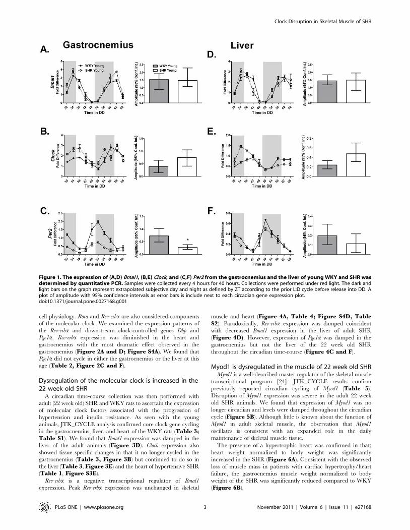
cell physiology. Rora and Rev-erba are also considered components
of the molecular clock. We examined the expression patterns of
the Rev-erba and downstream clock-controlled genes Dbp and
Pgc1a. Rev-erba expression was diminished in the heart and
gastrocnemius with the most dramatic effect observed in the
gastrocnemius (Figure 2A and D; Figure S4A). We found that
Pgc1a did not cycle in either the gastrocnemius or the liver at this
age (Table 2, Figure 2C and F).
Dysregulation of the molecular clock is increased in the22 week old SHR
A circadian time-course collection was then performed with
adult (22 week old) SHR and WKY rats to ascertain the expression
of molecular clock factors associated with the progression of
hypertension and insulin resistance. As seen with the young
animals, JTK_CYCLE analysis confirmed core clock gene cycling
in the gastrocnemius, liver, and heart of the WKY rats (Table 3;Table S1). We found that Bmal1 expression was damped in the
liver of the adult animals (Figure 3D). Clock expression also
showed tissue specific changes in that it no longer cycled in the
gastrocnemius (Table 3, Figure 3B) but continued to do so in
the liver (Table 3, Figure 3E) and the heart of hypertensive SHR
(Table 1, Figure S3E).
Rev-erba is a negative transcriptional regulator of Bmal1
expression. Peak Rev-erba expression was unchanged in skeletal
muscle and heart (Figure 4A, Table 4; Figure S4D, TableS2). Paradoxically, Rev-erba expression was damped coincident
with decreased Bmal1 expression in the liver of adult SHR
(Figure 4D). However, expression of Pgc1a was damped in the
gastrocnemius but not the liver of the 22 week old SHR
throughout the circadian time-course (Figure 4C and F).
Myod1 is dysregulated in the muscle of 22 week old SHRMyod1 is a well-described master regulator of the skeletal muscle
transcriptional program [24]. JTK_CYCLE results confirm
previously reported circadian cycling of Myod1 (Table 5).
Disruption of Myod1 expression was severe in the adult 22 week
old SHR animals. We found that expression of Myod1 was no
longer circadian and levels were damped throughout the circadian
cycle (Figure 5B). Although little is known about the function of
Myod1 in adult skeletal muscle, the observation that Myod1
oscillates is consistent with an expanded role in the daily
maintenance of skeletal muscle tissue.
The presence of a hypertrophic heart was confirmed in that;
heart weight normalized to body weight was significantly
increased in the SHR (Figure 6A). Consistent with the observed
loss of muscle mass in patients with cardiac hypertrophy/heart
failure, the gastrocnemius muscle weight normalized to body
weight of the SHR was significantly reduced compared to WKY
(Figure 6B).
Figure 1. The expression of (A,D) Bmal1, (B,E) Clock, and (C,F) Per2 from the gastrocnemius and the liver of young WKY and SHR wasdetermined by quantitative PCR. Samples were collected every 4 hours for 40 hours. Collections were performed under red light. The dark andlight bars on the graph represent extrapolated subjective day and night as defined by ZT according to the prior L:D cycle before release into DD. Aplot of amplitude with 95% confidence intervals as error bars is include next to each circadian gene expression plot.doi:10.1371/journal.pone.0027168.g001
Clock Disruption in Skeletal Muscle of SHR
PLoS ONE | www.plosone.org 3 November 2011 | Volume 6 | Issue 11 | e27168

Figure 2. Rev-erba is a core clock gene and Dbp, and Pgc1a are clock-controlled genes. The expression of (A,D) Rev-erb, (B,E) Dbp, and (C,F)Pgc1a in the gastrocnemius and liver of young WKY and SHR was measured by quantitative PCR. Samples were collected every 4 hours for 40 hours.Collections were performed under red light. The dark and light bars on the graph represent extrapolated subjective day and night as defined by ZTaccording to the prior L:D cycle before release into DD. A plot of amplitude with 95% confidence intervals as error bars is include next to eachcircadian gene expression plot.doi:10.1371/journal.pone.0027168.g002
Table 2. The circadian parameters of the core-clock gene Rev-erb and the clock-controlled genes Dbp and Pgc1a in thegastrocnemius and liver of young WKY and SHR calculated using JTK_CYCLE analysis.
Circadian Statistics
Strain Gene JTK _CYCLE p value Circadian JTK_CYCLE JTK_CYCLE Amplitude
Gastrocnemius
WKY Young Rev erb 3.74E-11 Yes 0.66
SHR Young Rev erb 1.55E-06 Yes 0.21
WKY Young Dbp 2.28E-08 Yes 2.01
SHR Young Dbp 2.70E-08 Yes 3.88
WKY Young Pgc1a 7.40E-02 No 0.19
SHR Young Pgc1a 1.24E-01 No 0.57
Liver
WKY Young Rev erb 3.74E-11 Yes 0.15
SHR Young Rev erb 1.04E-07 Yes 0.09
WKY Young Dbp 1.04E-07 Yes 4.19
SHR Young Dbp 5.19E-06 Yes 3.28
WKY Young Pgc1a 1 No 0.02
SHR Young Pgc1a 1 No 0.04
doi:10.1371/journal.pone.0027168.t002
Clock Disruption in Skeletal Muscle of SHR
PLoS ONE | www.plosone.org 4 November 2011 | Volume 6 | Issue 11 | e27168

Figure 3. The expression of (A,D) Bmal1, (B,E) Clock, and (C,F) Per2 from the gastrocnemius and the liver of adult WKY and SHR wasdetermined by quantitative PCR. Samples were collected every 4 hours for 40 hours. Collections were performed under red light. The dark andlight bars on the graph represent extrapolated subjective day and night as defined by ZT according to the prior L:D cycle before release into DD. Aplot of amplitude with 95% confidence intervals as error bars is include next to each circadian gene expression plot.doi:10.1371/journal.pone.0027168.g003
Table 3. The circadian parameters of the core-clock genes Bmal1, Clock, and Per2 in the gastrocnemius and liver of adult WKY andSHR calculated using JTK_CYCLE analysis.
Circadian Statistics Core Clock Genes
Strain Gene JTK _CYCLE p value Circadian JTK_CYCLE JTK_CYCLE Amplitude
Gastrocnemius
WKY Adult Bmal1 7.40E-10 Yes 2.52
SHR Adult Bmal1 1.20E-06 Yes 1.91
WKY Adult Clock 1.68E-03 Yes 0.80
SHR Adult Clock 7.82E-02 No 0.69
WKY Adult Per2 1.24E-13 Yes 0.80
SHR Adult Per2 3.92E-09 Yes 0.61
Liver
WKY Adult Bmal1 9.87E-12 Yes 1.95
SHR Adult Bmal1 4.42E-09 Yes 0.75
WKY Adult Clock 2.47E-05 Yes 0.55
SHR Adult Clock 2.20E-09 Yes 0.74
WKY Adult Per2 3.13E-09 Yes 0.40
SHR Adult Per2 3.11E-08 Yes 0.29
doi:10.1371/journal.pone.0027168.t003
Clock Disruption in Skeletal Muscle of SHR
PLoS ONE | www.plosone.org 5 November 2011 | Volume 6 | Issue 11 | e27168

Figure 4. The expression of (A,D) Rev-erba, (B) Dbp, and (C) Pgc1a in the gastrocnemius and liver of adult WKY and SHR wasmeasured by quantitative PCR. Samples were collected every 4 hours for 40 hours. Collections were performed under red light. The dark andlight bars on the graph represent extrapolated subjective day and night as defined by ZT according to the prior L:D cycle before release into DD. Aplot of amplitude with 95% confidence intervals as error bars is include next to each circadian gene expression plot.doi:10.1371/journal.pone.0027168.g004
Table 4. The circadian parameters of the core-clock gene Rev-erb and the clock-controlled genes Dbp and Pgc1a in thegastrocnemius and liver of adult WKY and SHR calculated using JTK_CYCLE analysis.
Circadian Statistics
Strain Gene JTK _CYCLE p value Circadian JTK_CYCLE JTK_CYCLE Amplitude
Gastrocnemius
WKY Adult Rev erb 2.66E-09 Yes 1.32
SHR Adult Rev erb 5.87E-06 Yes 0.93
WKY Adult Dbp 5.22E-13 Yes 5.45
SHR Adult Dbp 6.20E-09 Yes 5.00
WKY Adult Pgc1a 5.87E-06 Yes 6.67
SHR Adult Pgc1a 3.76E-05 Yes 0.86
Liver
WKY Adult Rev erb 2.00E-05 Yes 0.70
SHR Adult Rev erb 4.78E-12 Yes 0.18
WKY Adult Dbp 5.07E-10 Yes 4.07
SHR Adult Dbp 1.20E-06 Yes 2.24
WKY Adult Pgc1a 5.19E-06 Yes 0.06
SHR Adult Pgc1a 2.66E-03 Yes 0.04
doi:10.1371/journal.pone.0027168.t004
Clock Disruption in Skeletal Muscle of SHR
PLoS ONE | www.plosone.org 6 November 2011 | Volume 6 | Issue 11 | e27168

Discussion
In this study, we examined a circadian time-course expression
pattern of the molecular clock and clock-controlled genes in the
gastrocnemius and liver in WKY and SHR rats at different disease
stages to determine if polymorphisms upstream of the Bmal1 gene
in the SHR were associated with effects on the molecular clock
either before or after disease onset. Overall, we found that there
were differences in the expression of the molecular clock factors
both pre- and post-disease onset but the patterns in different tissues
varied across ages. The observation that molecular clock factor
expression was altered across all peripheral tissues studied (skeletal
muscle, liver and heart) prior to disease onset suggests that an
underlying problem with the molecular clock may contribute to
the both the hypertension and metabolic disease in the SHR rat.
When looking at clock gene expression after disease onset, we
found more pronounced effects in skeletal muscle with a loss of
Clock cycling and a dampening of the clock-controlled genes Pgc1aand Myod1. The damped expression of Myod1 and Pgc1a in the
hypertensive and insulin resistant SHR was associated with a
significant loss of muscle mass. Examination of single time point
gene expression in tissues from heart failure SHR rats revealed
significant down-regulation of several molecular clock genes in
skeletal muscle as well as genes responsible for daily muscle
maintenance and function. In summary, these findings confirm
that molecular clock expression in the SHR is disrupted prior to
the onset of cardiovascular and metabolic disease. The continued
disruption in clock and clock-controlled genes in the skeletal
Figure 5. Myod1 is a skeletal muscle specific clock-controlled gene. The expression of Myod1 in the gastrocnemius of (A) young and (B) adultWKY and SHR was determined by quantitative PCR. Samples were collected every 4 hours for 40 hours. Collections were performed under red light.The dark and light bars on the graph represent extrapolated subjective day and night as defined by ZT according to the prior L:D cycle before releaseinto DD. Note that Myod1 does not cycle in the gastrocnemius from adult SHR rats. A plot of amplitude with 95% confidence intervals as error bars isinclude next to each circadian gene expression plot.doi:10.1371/journal.pone.0027168.g005
Table 5. The circadian parameters of the skeletal muscle specific clock-controlled gene Myod1 in the gastrocnemius of young andadult WKY and SHR calculated using JTK_CYCLE analysis.
Circadian Statistics
Strain Gene JTK _CYCLE p value Circadian JTK_CYCLE JTK_CYCLE Amplitude
Gastrocnemius
WKY Young Myod1 2.87E-04 Yes 1.25
SHR Young Myod1 2.54E-06 Yes 2.68
WKY Adult Myod1 8.79E-04 Yes 11.86
SHR Adult Myod1 5.65E-02 No 1.52
doi:10.1371/journal.pone.0027168.t005
Clock Disruption in Skeletal Muscle of SHR
PLoS ONE | www.plosone.org 7 November 2011 | Volume 6 | Issue 11 | e27168

muscle following disease onset suggests that the expression and/or
stability of the molecular clock may contribute to the pathophys-
iology phenotype seen in this model.
Core-clock disruption precedes the development ofhypertension
Our studies in the WKY and SHR demonstrate a pattern of
core-clock disruption beginning in the pre-hypertensive SHR.
Hypertension and metabolic disease are systemic diseases
involving many organs beyond vasculature and heart. Subtle
disruption of the molecular clock through changing peak
amplitude (Per2, Rev-erba) may be characteristic of an unstable
clock mechanism in these tissues thus, making the tissues/organs
more prone to disease triggers and increasing the probability of
disease onset. Since the clock mechanism is not totally lost upon
disease onset we favor a model in which there is a delicate time-
dependent balance of expression of core-clock genes necessary to
diminish the potential for developing pathology. An example of
subtle changes in core-clock expression patterns contributing to
pathology occurs in human breast cancer. Dysregulation or loss of
PER1 and/or PER2 has been shown in human breast cancer
tumor cells when compared with normal cells from adjacent tissue
[25]. Lack of PER synchrony was even observed in the expression
patterns of PER in separate cancer cell populations of the same
cancer tissue. In addition, many clinical studies have associated
circadian regulation/synchrony with cancer [25–27]. Studies in
liver and adipose tissue of ob/ob mice have also shown that
dysregulation of Per1, Per2 and Dbp occurs prior to the
development of metabolic abnormalities suggesting a causal link
[28]. These data under-score the need for future studies focusing
on targeting the molecular clock to stabilize phase and amplitude
and coordinate the circadian system.
Hypertension and insulin resistance is associated withpronounced changes of the core-clock in thegastrocnemius muscle
As the SHR progressed from pre-hypertensive to hypertensive
and insulin resistant (22 Weeks, adult), the disruption of the
molecular clock was more pronounced in the gastrocnemius
compared to the liver. The tissue specificity of the clock disruption
in skeletal muscle suggests 1) there is likely a gene – environment
interaction leading to tissue specific disruption of the molecular
clock; 2) clock gene disruption in skeletal muscle is associated with
insulin resistance and 3) clock disruption of skeletal muscle likely
contributes significantly to the whole body insulin resistance in this
model. The SHR strain has been used extensively as a genetic
model of essential hypertension. Skeletal muscle is a primary site of
insulin resistance in essential hypertension [29,30] It is interesting
to note that SHR exhibit several skeletal muscle abnormalities and
dysfunctions when compared to WKY counterparts including
decreased fatigue resistance [31], insulin resistance [32], develop-
ment of less contractile force [33], increased interstitial norepi-
nephrine levels [34], altered sodium pump number and activity
[35], elevated intracellular free calcium [36], fiber type transfor-
mation [37], and decreased capillary density [38]. Major
alterations in skeletal muscle ultrastructure and biochemical
properties have also been demonstrated in patients with chronic
heart failure including fiber atrophy [17], transformation of fast-
twitch type I to slow-twitch type II fibers [18,39], decrease in
oxidative enzyme capacity [19] and abnormal mitochondrial
structure [20–22]. Recent studies in two clock-compromised
mouse strains identified a common skeletal muscle pathology that
was characterized by diminished force capacity and reduced
mitochondrial content and function [5] both of which have been
observed in SHR. Loss of Clock cycling in the hypertensive SHR
provide insights that that modification of core-clock components
may contribute to the functional and metabolic phenotype
observed in SHR skeletal muscle. Metabolic dysfunction may also
manifest through decreased expression of Rev-erb in the SHR
gastrocnemius. The orphan nuclear receptors Rev-erb and Rora
(RAR-related orphan receptor) link the feedback loops of the clock
by repressing and activating Bmal1 gene transcription, respectively
[40–43]. Rora and Rev-erb have also been shown to regulate lipid
homeostasis in skeletal muscle [44,45]. Changes in the expression
of these genes could have a broad impact beyond circadian
regulation, in that skeletal muscle relies heavily on fatty acids as a
fuel source.
Circadian expression of Myod1 and Pgc1a is disrupted inthe hypertensive gastrocnemius
Recent work from our lab has demonstrated that expression of
Myod1 oscillates in a circadian pattern in skeletal muscle [10] and
is a direct clock-controlled gene [5]. Cycling of Myod1, a direct
target of the core-clock genes Bmal1 and Clock in skeletal muscle,
ceased in the gastrocnemius muscle of the hypertensive and insulin
resistant SHR. While speculative, the loss in gastrocnemius muscle
weight in the hypertensive SHR may be connected to the
Figure 6. Heart weight normalized to body weight in the WKYand SHR demonstrating the presence of cardiac hypertrophyin the SHR (A). Gastrocnemius wet weight normalized to body weightmeasured from the hypertensive WKY and SHR demonstrating a loss ofmuscle mass in the SHR (B).doi:10.1371/journal.pone.0027168.g006
Clock Disruption in Skeletal Muscle of SHR
PLoS ONE | www.plosone.org 8 November 2011 | Volume 6 | Issue 11 | e27168

disruption in Myod1 expression [46]. Another gene, Pgc1a, known
to play a critical role in regulating the expression of metabolic and
mitochondrial genes in skeletal muscle was damped in the
gastrocnemius of the hypertensive rats throughout the circadian
collection time-course. Data from Patti and colleagues is highly
suggestive of a potential link between PGC1 expression and insulin
resistance in diabetic and non-diabetic individuals with a family
history of diabetes [47]. A reduction of the transcriptional co-
activator Pgc1a in the gastrocnemius of the hypertensive SHR is
indicative of a role for the molecular clock in the insulin resistance
observed in these hypertensive animals. This work is in agreement
with previous work from Andrews et al., (2010) demonstrating a
down-regulation of both Pgc1a and Pgc1ß in mice with disruption
of the core-clock genes ClockD19 or Bmal12/2. These mice exhibit
altered mitochondrial structure and function.
Multiple core-clock components are down regulated inskeletal muscle of heart failure SHR
Our data revealed a pattern of down-regulation of several core-
clock genes, including Clock and Bmal1, and non-circadian skeletal
muscle genes, such as Mef2 and Ttn, responsible for daily muscle
maintenance in the gastrocnemius of the SHR during overt heart
failure. Work from this lab recently reported that muscle from
Bmal12/2 and ClockD19 mice exhibit disrupted myofilament
architecture and exhibit reduced normalized maximal force [5]
demonstrating a role for the molecular clock in maintaining
structure and function at the cellular level in skeletal muscle.
However, the skeletal muscle specific circadian gene Myod1 was
unchanged in the heart failure animals (Figure S1A). This finding
may more reflect a limitation of the experimental design rather
than the biology. Given the high mortality of the heart failure
animals, it was only possible to collect at a single time point. Tissue
for heart failure experiments were collected during the daylight
hours and this lack of change may be more a result of the time of
day of collection. As seen in Figure 5B, Myod1 expression in the
gastrocnemius from the WKY and hypertensive SHR shows that
Myod1 expression levels were not different during the daylight
hours. Our observation that Myod1 target genes Mck and Ttn, were
down-regulated is suggestive that the loss of oscillation of Myod1 in
the SHR is maintained in the heart failure rats.
Patterns of dysregulation in the molecular clock including
diminished amplitude, phase shifts, and period changes have been
observed in many disease states like diabetes, cardiovascular
disease and cancer [4,48–52]. Dyssynchrony can be the result of
genetic alterations or changes in environmental cues. As a whole,
our data demonstrate a pattern of molecular clock dysregulation
that begins prior to the development of hypertension and insulin
resistance and may be linked to the genetic background of the
SHR progressing independent of the development of hypertension
or worsening because of the development of hypertension. In
association with previous studies, these data are highly suggestive
of a role for the molecular clock in skeletal muscle pathology
associated with hypertension/hypertrophy in SHR. Whether these
genetic variables are identical or overlap with those in hyperten-
sive patients is unknown. Future studies will need to examine other
models of hypertension in order to better understand the
mechanism behind hypertension, heart failure and circadian gene
regulation in skeletal muscle.
Methods
AnimalsAll animal procedures were conducted in accordance with
institutional guidelines for the care and use of laboratory animals
as approved by the University of Kentucky Institutional Animal
Care and Use Committees. This work was approved by the
Institutional Animal Care and Use Committee (IACUC) at the
University of Kentucky, protocol number 00890M2005. WKY
and SHR rats were obtained from Harlan Laboratories. Rats were
housed in a temperature- and humidity-controlled room main-
tained on a 12 h light–12 h dark cycle with food and water ad
libitum.
Tissue Collections (80 wks old)Due to the high mortality of the heart failure rats (80 wks old)
tissue samples were collected at a single time point, Tissue samples
from old rats (80 wks old) were collected at the same time of day,
during 8AM and 9AM. This corresponds to time between ZT1
and ZT2.
Circadian CollectionsAll rats were maintained in the 12 h L/D cycles, then released
into DD. Starting 30 h after entry into DD skeletal muscle, heart
and liver from three WKY and three SHR rats were collected
every 4 h for 40 h under dim red light (,5 lux). The muscles, liver
and heart were removed from each rat and frozen in liquid
nitrogen.
RNA isolationTotal RNA was prepared from frozen tissue samples using
TRIzol (Invitrogen) according to the manufacturer’s directions.
RNA samples were treated with TURBO DNase (Ambion, Austin,
TX) to remove genomic DNA contamination. Isolated RNA was
quantified by spectrophotometry (l= 260 nm). First-strand cDNA
synthesis from total RNA was performed with a mixture of oligo(dT)
primer and random hexamers using SuperScript III First-Strand
Synthesis SuperMix (Invitrogen). All isolated RNA and cDNA
samples were stored at 280uC until further analysis. Real-time
quantitative PCR using TaqMan (Applied Biosystems) assays was
used to examine the gene expression of several core-clock, clock-
controlled, and skeletal muscle specific genes. The gene expression
assays were as follows: Bmal1 Rn00562847_m1*; Per2
Rn01427704_m1*; Clock Rn00573120_m1*; Rora Rn01173769_
m1*; Rev- erb Rn00595671_m1*; Myod Rn00591291_m1*; Pgc1aRn00580241_m1*; Rpl26 Rn02127510_s1*; Mck Rn01644605_
m1*; Dbp Rn00497539_m1*. RPL26 was used as an internal
calibration control [5,10,11,53]. The DDCT method was used for
the quantification of real-time PCR data in the circadian collections.
Gene expression in each sample was shown as the relative value
compared to the WKY soleus sample No. 1 from DD 30 hours
group. This enabled us to compare not only inter-species differences
but also inter-organ differences in the expression pattern/amplitude
of each gene.
Statistics. A new version of JTK_CYCLE was used to
identify and characterize cycling variables in our datasets [23].
JTK_CYCLE analyses were performed using the R statistical
package, version 2.12.1 to look specifically for 24 hour rhythms.
Briefly, the original version of JTK_CYCLE estimated the
amplitude of the most probable period/lag combination by
calculating the median sign-adjusted deviation from the median
over the first complete cycle. The new version-2 of JTK_CYCLE
provides a more precise estimate of amplitude by replacing the
median with the ‘‘pseudo median’’ (Hodges-Lehmann estimator)
and additionally reports the associated confidence interval
calculated by the Wilcox test function in the standard R stats
package. A clear explanation of the mathematical basis for these
new features is available online [54]. Differences in heart weight/
body weight ratios and gastrocnemius wet weight/body weight
Clock Disruption in Skeletal Muscle of SHR
PLoS ONE | www.plosone.org 9 November 2011 | Volume 6 | Issue 11 | e27168

were calculated with Graphpad Prism software using a two-tailed
student t test.
Supporting Information
Figure S1 Bmal1, Clock, Per2 and Rora are core-clockgenes. Expression of (A) Bmal1, (B) Clock, (C) Per2, and (D) Rora
from the gastrocnemius of WKY and SHR in heart failure (80
weeks) was determined by quantitative PCR. (*p,0.05).
(TIF)
Figure S2 The expression of the skeletal muscle genes(A) Myod1, (B) Mck, (C) Mef2c, and (D) Ttn from thegastrocnemius of WKY and SHR in heart failure (80weeks) was determined by quantitative PCR. (*p,0.05).(TIF)
Figure S3 The expression of (A,D) Bmal1, (B,E) Clock,and (C,F) Per2 from the heart of young and adult WKYand SHR was determined by quantitative PCR. Samples
were collected every 4 hours for 40 hours. Collections were
performed under total red light. The dark and light bars on the
graph represent extrapolated subjective day and night as defined
by ZT according to the prior L:D cycle before release into DD. A
plot of amplitude with 95% confidence intervals as error bars is
include next to each circadian gene expression plot.
(TIF)
Figure S4 Rev-erba is a core-clock gene and Dbp, andPgc1a are clock-controlled genes. The expression of (A,D)
Rev-erb, (B,E) Dbp, and (C,F) Pgc1a in the heart of young and adult
WKY and SHR rats was measured by quantitative PCR. Samples
were collected every 4 hours for 40 hours. Collections were
performed under red light. The dark and light bars on the graph
represent extrapolated subjective day and night as defined by ZT
according to the prior L:D cycle before release into DD. A plot of
amplitude with 95% confidence intervals as error bars is include
next to each circadian gene expression plot.
(TIF)
Table S1 The circadian parameters of the core-clockgenes Bmal1, Clock, and Per2 in the heart of young andadult WKY and SHR calculated using JTK_CYCLEanalysis.
(TIF)
Table S2 The circadian parameters of the clock-con-trolled genes Rev-erb, Dbp, and Pgc1a in the heart ofyoung and adult WKY and SHR calculated usingJTK_CYCLE analysis.
(TIF)
Acknowledgments
We would like to thank John Hogenesch for kindly sharing and offering
technical assistance in the use of the JTK_CYCLE software.
Author Contributions
Conceived and designed the experiments: KAE CWB MM. Performed the
experiments: MM SEE. Analyzed the data: MM ES MEH KK.
Contributed reagents/materials/analysis tools: KAE CWB. Wrote the
paper: ES KAE.
References
1. Yoo SH, Ko CH, Lowrey PL, Buhr ED, Song EJ, et al. (2005) A noncanonical
E-box enhancer drives mouse Period2 circadian oscillations in vivo. Proc Natl
Acad Sci U S A 102: 2608–2613.
2. Guo H, Brewer JM, Champhekar A, Harris RBS, Bittman EL (2005)
Differential control of peripheral circadian rhythms by suprachiasmatic-
dependent neural signals. Proc Natl Acad Sci U S A 102: 3111–3116.
3. Woelfle MA, Ouyang Y, Phanvijhitsiri K, Johnson CH (2004) The Adaptive
Value of Circadian Clocks: An Experimental Assessment in Cyanobacteria. Curr
Biol 14: 1481–1486.
4. Martino TA, Oudit GY, Herzenberg AM, Tata N, Koletar MM, et al. (2008)
Circadian rhythm disorganization produces profound cardiovascular and renal
disease in hamsters. Am J Physiol Regul Integr Comp Physiol 294: R1675–R1683.
5. Andrews JL, Zhang X, McCarthy JJ, McDearmon EL, Hornberger TA, et al.
CLOCK and BMAL1 regulate MyoD and are necessary for maintenance of
skeletal muscle phenotype and function. Proc Natl Acad Sci U S A 107:
19090–19095.
6. Panda S, Antoch M, Miller B, Su A, Schook A, et al. (2002) Coordinated
transcription of key pathways in the mouse by the circadian clock. Cell 109:
307–320.
7. Weger BD, Sahinbas M, Otto GW, Mracek P, Armant O, et al. (2011) The
Light Responsive Transcriptome of the Zebrafish: Function and Regulation.
PLoS ONE 6(2): e17080.
8. Rey G, Cesbron F, Rougemont J, Reinke H, Brunner M, et al. (2011) Genome-
Wide and Phase-Specific DNA-Binding Rhythms of BMAL1 Control Circadian
Output Functions in Mouse Liver. PLoS Biol 9: e1000595.
9. Bur IM, Zouaoui S, Fontanaud P, Coutry N, Molino F, et al. (2010) The
Comparison between Circadian Oscillators in Mouse Liver and Pituitary Gland
Reveals Different Integration of Feeding and Light Schedules. PLoS ONE 5:e15316.
10. McCarthy JJ, Andrews JL, McDearmon EL, Campbell KS, Barber BK, et al.
(2007) Identification of the circadian transcriptome in adult mouse skeletalmuscle. Physiol Genomics 31: 86–95.
11. Miller BH, McDearmon EL, Panda S, Hayes KR, Zhang J, et al. (2007)
Circadian and CLOCK-controlled regulation of the mouse transcriptome andcell proliferation. Proc Natl Acad Sci U S A 104: 3342–3347.
12. Rudic RD, Curtis AM, Cheng Y, FitzGerald G, Michael WY (2005) Peripheral
Clocks and the Regulation of Cardiovascular and Metabolic Function. MethodsEnzymol Academic Press. pp 524–539.
13. Hughes ME, DiTacchio L, Hayes KR, Vollmers C, Pulivarthy S, et al. (2009)
Harmonics of Circadian Gene Transcription in Mammals. PLoS Genet 5:e1000442.
14. Woon PY, Kaisaki PJ, Braganca J, Bihoreau MT, Levy JC, et al. (2007) Aryl
hydrocarbon receptor nuclear translocator-like (BMAL1) is associated withsusceptibility to hypertension and type 2 diabetes. Proc Natl Acad Sci U S A 104:
14412–14417.
15. Mettauer B, Zoll J, Garnier A, Ventura-Clapier R (2006) Heart failure: a model
of cardiac and skeletal muscle energetic failure. Pflugers Arch European 452:653–666.
16. Lunde PK, Sjaastad I, Schiøtz Thorud HM, Sejersted OM (2001) Skeletalmuscle disorders in heart failure. Acta Physiol Scand 171: 277–294.
17. Harrington D, Anker SD, Chua TP, Webb-Peploe KM, Ponikowski PP, et al.(1997) Skeletal Muscle Function and Its Relation to Exercise Tolerance in
Chronic Heart Failure. J Am Coll Cardiol 30: 1758–1764.
18. Lipkin DP, Jones DA, Round JM, Poole-Wilson PA (1988) Abnormalities of
Skeletal Muscle in Patients with Chronic Heart Failure. Int J Cardiol 20:161.
19. Harridge SD, Magnusson G, Gordon A (1996) Skeletal muscle contractilecharacteristics and fatigue resistance in patients with chronic heart failure. Eur
Heart J 17: 896–901.
20. Drexler H, Riede U, Munzel T, Konig H, Funke E, et al. (1992) Alterations of
skeletal muscle in chronic heart failure. Circ 85: 1751–1759.
21. Lampert E, Mettauer B, Hoppeler H, Charloux A, Charpentier A, et al. (1996)
Structure of Skeletal Muscle in Heart Transplant Recipients. J Amer CollCardiol 28: 980–984.
22. Hambrecht R, Fiehn E, Yu J, Niebauer J, Weigl C, et al. (1997) Effects ofEndurance Training on Mitochondrial Ultrastructure and Fiber Type
Distribution in Skeletal Muscle of Patients With Stable Chronic Heart Failure.J Amer Coll Cardiol 29: 1067–1073.
23. Hughes ME, Hogenesch JB, Kornacker K (2010) JTK_CYCLE: An EfficientNonparametric Algorithm for Detecting Rhythmic Components in Genome-
Scale Data Sets. J Biol Rhythms 25: 372–380.
24. Tapscott SJ (2005) The circuitry of a master switch: Myod and the regulation of
skeletal muscle gene transcription. Devel 132: 2685–2695.
25. Chen ST, Choo KB, Hou MF, Yeh KT, Kuo SJ, et al. (2005) Deregulated
expression of the PER1, PER2 and PER3 genes in breast cancers.Carcinogenesis 26: 1241–1246.
26. Gery S, Komatsu N, Baldjyan L, Yu A, Koo D, et al. (2006) The CircadianGene Per1 Plays an Important Role in Cell Growth and DNA Damage Control
in Human Cancer Cells. Mol Cell 22: 375–382.
27. Pogue-Geile KL, Lyons-Weiler J, Whitcomb DC (2006) Molecular overlap of
fly circadian rhythms and human pancreatic cancer. Cancer Letters 243:55–57.
Clock Disruption in Skeletal Muscle of SHR
PLoS ONE | www.plosone.org 10 November 2011 | Volume 6 | Issue 11 | e27168
28. Ando H, Kumazaki M, Motosugi Y, Ushijima K, Maekawa T, et al. (2011)
Impairment of Peripheral Circadian Clocks Precedes Metabolic Abnormalitiesin ob/ob Mice. Endocrinology 152: 1347–1354.
29. Capaldo B, Lembo G, Napoli R, Rendina V, Albano G, et al. (1991) Skeletal
muscle is a primary site of insulin resistance in essential hypertension.Metabolism 40: 1320–1322.
30. Wolfe RR (2006) The underappreciated role of muscle in health and disease.The American Journal of Clinical Nutrition 84: 475–482. DOI Electronic
Resource Number.
31. Gray SD, Carlsen RC, Deng J (1994) Soleus muscle contractile properties inhypertensive rats. Amer J Physiol - Regulatory, Integrative and Comparative
Physiology 267: R735–R739.32. Hulman S, Falkner B, Freyvogel N (1993) Insulin resistance in the conscious
spontaneously hypertensive rat: Euglycemic hyperinsulinemic clamp study.Metabolism 42: 14–18.
33. Atrakchi A, Gray SD, Carlsen RC (1994) Development of soleus muscles in
SHR: relationship of muscle deficits to rise in blood pressure. Amer J Physiol -Cell Physiol 267: C827–C835.
34. Cabassi A, Vinci S, Quartieri F, Moschini L, Borghetti A (2001) NorepinephrineReuptake Is Impaired in Skeletal Muscle of Hypertensive Rats In Vivo.
Hypertension 37: 698–702.
35. Pickar JG, Carlsen RC, Atrakchi A, Gray SD (1994) Increased Na(+)-K+ pumpnumber and decreased pump activity in soleus muscles in SHR. Amer J Physiol -
Cell Physiol 267: C836–C844.36. Ameen M, Davies JE, Ng LL (1991) A Comparison of Free Intracellular
Calcium and Magnesium Levels in the Vascular Smooth Muscle and StriatedMuscle Cells of the Spontaneously Hypertensive and Wistar Kyoto Normoten-
sive Rat. Ann N Y Acad Sci 639: 550–553.
37. Bortolotto SK, Stephenson DG, Stephenson GM (1999) Fiber type populationsand Ca2+-activation properties of single fibers in soleus muscles from SHR and
WKY rats. Amer J Physiol - Cell Physiol 276: C628–C637.38. Kobayashi N, DeLano FA, Schmid-Schonbein GW (2005) Oxidative Stress
Promotes Endothelial Cell Apoptosis and Loss of Microvessels in the
Spontaneously Hypertensive Rats. Arterioscler Thromb Vasc Biol 25:2114–2121.
39. Sullivan MJ, Duscha BD, Klitgaard H, Kraus WE, Cobb FR, et al. (1997)Altered expression of myosin heavy chain in human skeletal muscle in chronic
heart failure. Med Sci Sports Exerc 29: 860–866.40. Lowrey PL, Takahashi JS (2004) Mammalian Circadian Biology: Elucidating
Genome-Wide Levels of Temporal Organization. Annu Rev Genomics Hum
Genet 5: 407–441.
41. Akashi M, Takumi T (2005) The orphan nuclear receptor RORa regulates
circadian transcription of the mammalian core-clock Bmal1. Nat Struct Mol Biol
12: 441–448.
42. Sato TK, Panda S, Miraglia LJ, Reyes TM, Rudic RD, et al. (2004) A
Functional Genomics Strategy Reveals Rora as a Component of the
Mammalian Circadian Clock. Neuron 43: 527–537.
43. Ueda HR, Hayashi S, Chen W, Sano M, Machida M, et al. (2005) System-level
identification of transcriptional circuits underlying mammalian circadian clocks.
Nat Genet 37: 187–192.
44. Lau P, Nixon SJ, Parton RG, Muscat GE (2004) RORa Regulates the
Expression of Genes Involved in Lipid Homeostasis in Skeletal Muscle Cells.
J Biol Chem 279: 36828–36840.
45. Ramakrishnan SN, Lau P, Burke LJ, Muscat GE (2005) Rev-erba Regulates the
Expression of Genes Involved in Lipid Absorption in Skeletal Muscle Cells. J Biol
Chem 280: 8651–8659.
46. Vissing K, Andersen JL, Harridge SD, Sandri C, Hartkopp A, et al. (2005) Gene
expression of myogenic factors and phenotype-specific markers in electrically
stimulated muscle of paraplegics. J Appl Physiol 99: 164–172.
47. Patti ME, Butte AJ, Crunkhorn S, Cusi K, Berria R, et al. (2003) Coordinated
reduction of genes of oxidative metabolism in humans with insulin resistance and
diabetes: Potential role of PGC1 and NRF1. Proc Natl Acad Sci U S A 100:
8466–8471.
48. Martino TA, Tata N, Belsham DD, Chalmers J, Straume M, et al. (2007)
Disturbed Diurnal Rhythm Alters Gene Expression and Exacerbates Cardio-
vascular Disease With Rescue by Resynchronization. Hypertension 49:
1104–1113.
49. Maywood ES, O’Neill J, Wong GK, Reddy AB, Hastings MH, et al. (2006)
Circadian timing in health and disease Prog Brain Res Elsevier. pp 253–269.
50. Filipski E, Delaunay F, King VM, Wu MW, Claustrat B, et al. (2004) Effects of
Chronic Jet Lag on Tumor Progression in Mice. Cancer Res 64: 7879–7885.
51. Yasuniwa Y, Izumi H, Wang KY, Shimajiri S, Sasaguri Y, et al. (2010)
Circadian Disruption Accelerates Tumor Growth and Angio/Stromagenesis
through a Wnt Signaling Pathway. PLoS ONE 5: e15330.
52. Durgan DJ, Young ME (2010) The Cardiomyocyte Circadian Clock: Emerging
Roles in Health and Disease. Circ Res 106: 647–658.
53. Thorrez L, Van Deun K, Tranchevent LC, Van Lommel L, Engelen K, et al.
(2008) Using Ribosomal Protein Genes as Reference: A Tale of Caution. PLoS
ONE 3: e1854.
54. Geyer CJ (4/13/2003) Stat 5102 Notes: Nonparametric Tests and Confidence
Intervals. from,http://www.stat.umn.edu/geyer/old03/5102/notes/rank.pdf.
Clock Disruption in Skeletal Muscle of SHR
PLoS ONE | www.plosone.org 11 November 2011 | Volume 6 | Issue 11 | e27168
![Review the circadian clock: Gene specific effects on aging, … · 2016. 5. 24. · the animals age [1]. Notably, however, targeted disruption of the Clock gene does not lead to the](https://img.dokumen.tips/doc/110x75/5fc56be7522e8701bf63ee4c/review-the-circadian-clock-gene-specific-effects-on-aging-2016-5-24-the-animals.jpg)