Embed Size (px)
Citation preview
JOURNAL OF BACTERIOLOGY, Dec. 1984, p. 1181-1183 Vol. 160, No. 30021-9193/84/121181-03$02.00/0Copyright © 1984, American Society for Microbiology
Simple, Rapid, and Quantitative Release of Periplasmic Proteins byChloroform
GIOVANNA FERRO-LUZZI AMES,'* CATHERINE PRODY,1 AND SYDNEY KUSTU2
Department of Biochemistry, University of California, Berkeley, California 94720,1 and Department of Bacteriology,University of California, Davis, California 956162
Received 16 July 1984/Accepted 5 September 1984
We introduce a method by which periplasmic proteins can be released rapidly, simply, and quantitatively bytreating cells with chloroform. All the amino acid-binding proteins tested maintained their activity duringchloroform treatment. This method makes practical the analysis of the periplasmic protein complement of alarge number of strains.
The periplasmic proteins of gram-negative bacteria aredefined operationally as those proteins which are releasedinto the medium by mild osmotic shock (2). These proteinscomprise ca. 10 to 15% of the total cell protein and includenumerous transport-related binding proteins and a variety ofenzymes, which appear to lie outside the cytoplasmic mem-brane layer of the cell envelope (2). The cell compartment inwhich these proteins are located is called the periplasm, andit constitutes a sizable fraction of the total cell volume (14).Studies involving periplasmic proteins often require thatthey be initially separated from the bulk of the cell protein byosmotic shock. This is accomplished by first suspendingwashed cells in a concentrated sucrose solution in thepresence of EDTA and then subjecting them to osmoticshock in cold distilled water. The procedure is cumbersomebecause it requires a number of centrifugation steps. There-fore, it has been difficult to use this procedure to screen largenumbers of strains rapidly for the presence or absence,nature (such as altered mobility), and activity of periplasmicproteins (see, e.g., references 5 and 6). In some cases, theprotein of interest may be visible in a sodium dodecyl sulfategel electrophoretogram of intact cells (e.g., if its size is suchthat it moves to a relatively uncrowded region of the gel andif it is sufficiently abundant). However, the protein ofinterest often cannot be seen. In such instances, simplyseparating the periplasmic proteins from the rest of thecellular protein achieves an approximate 10-fold purifica-tion. The use of a rapid cell permeabilization procedure (10)allows the activity of certain periplasmic proteins to beassayed. However, this procedure is not useful if the onlymeasurable activity of the periplasmic protein of interest issubstrate binding, nor is it useful for ascertaining the pres-ence of inactive proteins or determining the specific locali-zation in the periplasm of either active or inactive proteins.Thus, the separation of periplasmic proteins from the bulk ofthe cell protein is a very important step in characterizingmany mutations which affect periplasmic proteins and mu-tants which may have been collected by a variety of selec-tion methods.We describe here a method for specifically releasing
periplasmic proteins rapidly, simply, and reproducibly byexposing cells to chloroform. (We refer to chloroform shockand chloroform shock fluid in this study, although weassume that the proteins released by treating cells withchloroform are liberated by a process unrelated to osmotic
* Corresponding author.
shock.) In our standard procedure, 2-ml bacterial culturesare grown to saturation overnight in culture tubes (13 by 100mm). The cells are collected by centrifugation for 10 min at1,100 x g. The use of rubber centrifuge adapters allows,centrifugation of the culture tubes directly, without break-age. The supernatant is decanted thoroughly. When manytubes are handled simultaneously, it is necessary to decantthem rapidly to avoid resuspension of the cells. The cellpellet is resuspended by brief vortexing in the residualmedium, and 20 ,ul of CHCl3 is then added. After briefvortexing, the tubes are maintained at room temperature forabout 15 min, and then 0.2 ml of 0.01 M Tris hydrochloride(pH 8.0) is added. The cells are separated by centrifugationat 6,000 x g for 20 min, and the supernatant fractioncontaining the periplasmic proteins is withdrawn with aPasteur pipette. It is necessary to obtain a tightly packed cellpellet at this stage because this is the final supernatant,which should be as free as possible from contamination bycells. It is advisable to withdraw only a portion of thesupernatant. We found that it is possible to centrifugedisposable culture tubes at 7,000 rpm in an SS34 Sorvallrotor without incurring any breakage if a no. 366 rubberSorvall adapter is used. Thus, the entire operation is carriedout without any transfer of the cultures. Because the oper-ations required are so simple, the only limit is dictated by thenumber of tubes that can be centrifuged at one time. The no.369 Sorvall adapter within a no. 456 adapter allows centrif-ugation of 30 samples in the GSA rotor simultaneously.
Figure 1 shows an acrylamide gel electrophoretogram ofthe proteins liberated by this method, by a few variations ofthis technique, and by the standard osmotic shock proce-dure. The patterns of bands obtained by chloroform shockand osmotic shock are very similar (Fig. 1, lanes 5, 6, and 8),and the time of incubation with chloroform has little effect(compare lanes 5 and 6). It is interesting that toluene, whichis often used in cell permeabilization procedures, not onlyfails to improve protein release, but actually interferes withit (compare lanes 1, 2, 3, and 7 with lanes 5 and 6). A mixtureof chloroform and toluene has been used to release peri-plasmic proteins from colonies on a petri plate (3), but nooptimization of the procedure was presented in that study.Chloroform shock releases the major periplasmic compo-nents as effectively as osmotic shock does (Fig. 1). Toidentify other solvents which would also release periplasmicproteins, we replaced the chloroform with phenethyl alco-hol, ether, ether-chloroform (1:1), or dimethyl sulfoxide,keeping other conditions essentially the same, except that
1181
on June 25, 2018 by guesthttp://jb.asm
.org/D
ownloaded from

1182 NOTES
1 2 3 4 5 6 7 8 MW
- -94K
:7 -60K
_ -36K
__l
_-40204-25K
CT CT T PA C C CT 0
1:1 1:9 1:1FIG. 1. Electrophoretogram of periplasmic proteins released
from cells of strain TA831 (AhisF645) by various treatments. Lanes:1, chloroform-toluene (1:1); 2, chloroform-toluene (1:9); 3, toluene;4, phenethyl alcohol; 5 and 6, chloroform; 7, chloroform-toluene(1:1); 8, osmotic shock fluid; MW, molecular weight standards (94K= 94,000 molecular weight). Lanes 1 to 5, 20 ,ul of solvent(s) addedand standard procedure used, except for the sample in lane 4, whichwas incubated at 37°C for 15 min before buffer was added. Lanes 6and 7, solvent (20 ,ul) was added immediately before 200 p.l of 0.01M Trishydrochloride (pH 8); incubation was for 30 min. The strainwas cultured to saturation in minimal salts medium with 0.4%glucose as the carbon source (12). Shock fluids were diluted twofoldwith twice-concentrated Laemmli sample buffer (8) and boiled for 2min, and 15 ,ul was electrophoresed on 10% acrylamide gels asdescribed previously (1). Gels were stained with Coomassie blue.
phenethyl alcohol-treated cells were incubated for 15 min at37°C before addition of the buffer (Fig. 1, lane 4). All of thesetreatments caused release of lesser amounts of proteins andso were not investigated further (data not shown). Adding0.01 M MgCl2 to the cells before chloroform treatmentdecreased the amount of protein released, but the pattern ofproteins was qualitatively the same (data not shown). Theorder in which the chloroform and the buffer are addedseems to have little effect on the nature and amount ofproteins released. However, adding the buffer a short time
after the chloroform gives more reproducible results; per-haps this sequence allows some of the excess chloroform toevaporate and thus prevents it from interfering with the tightpacking of the cell pellet. The amount of chloroform added isalso important for the same reason. Amounts greater than 20,ul interfere with cell packing and should be avoided.Many periplasmic proteins are found to be unusually
stable to heat and proteolytic digestion; they may also beparticularly resistant to chloroform. We found this to be truefor all of the periplasmic binding proteins we assayed. Thus,these proteins can be readily assayed for activity in achloroform shock fluid. The amounts of several differentperiplasmic binding proteins (i.e., the glutamine-bindingprotein, the lysine-arginine-ornithine-binding protein, andother arginine-binding proteins) released were essentiallythe same with both chloroform and osmotic shock, as shownin Table 1 for two different strains. Similar results wereobtained for the histidine-binding protein (data not shown).The total amount of protein released by the two procedureswas also comparable (ca. 16% of the total cell protein).Perhaps some periplasmic proteins cannot withstand expo-sure to chloroform, and so each of these proteins will have tobe tested individually. When the released periplasmic pro-teins only need to be examined by sodium dodecyl sulfategel electrophoresis or immunological techniques (6), suchinactivation would be irrelevant. To demonstrate that chloro-form released mainly periplasmic proteins and left the innermembrane essentially impermeable to cytoplasmic proteins,we assayed the chloroform shock fluid for the presence ofcytoplasmic enzymes. Glutamine synthetase, which is notinactivated by chloroform, was present in very small, com-parable amounts in chloroform and osmotic shock fluids(Table 1). In a strain grown under derepressing conditions,only about 1% of the total glutamine synthetase activityappeared in either shock fluid. Similar results were obtainedwith 3-galactosidase (10) in strain TA2365 (pho-25), whichcarries an F' lac+ phoA+ plasmid (data not shown).To determine whether the chloroform shock procedure
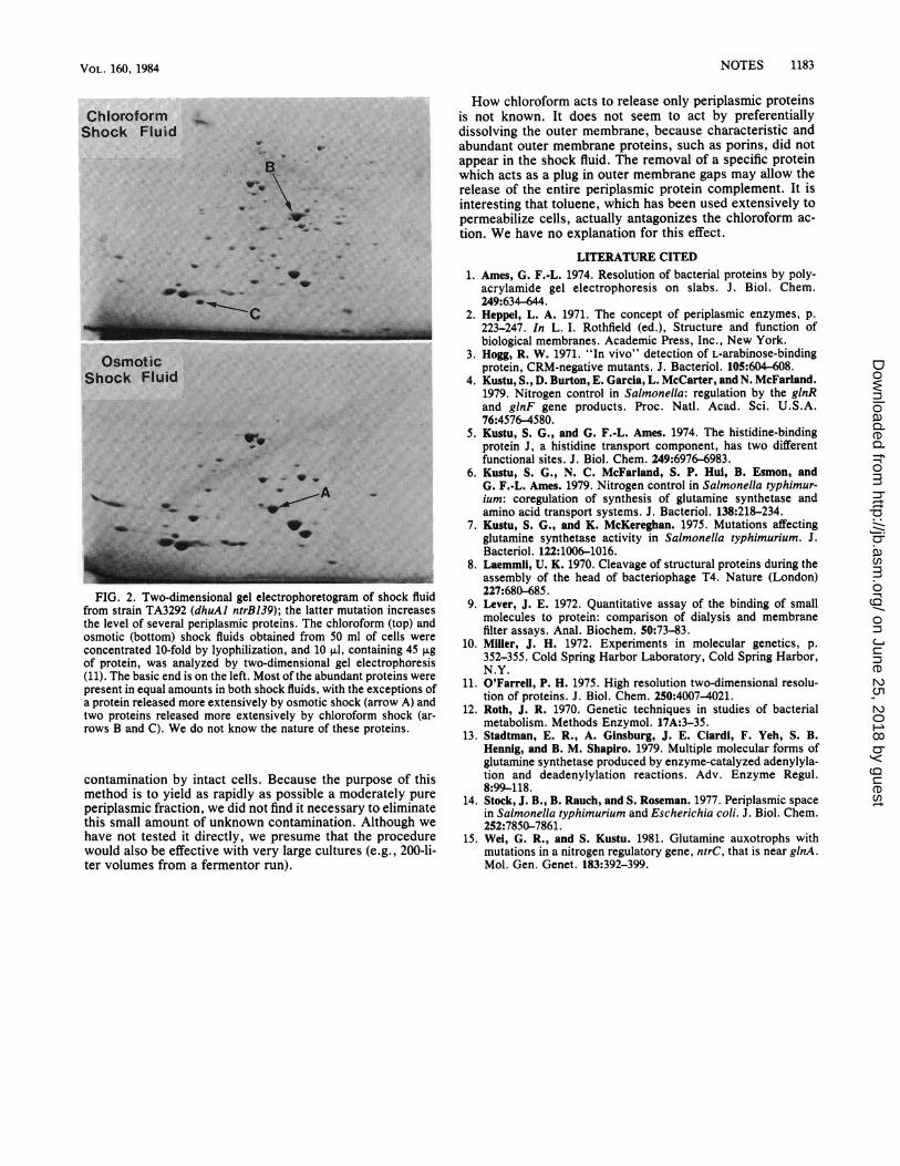
would be effective on larger volumes of cells, we treated50-ml cultures in an identical, scaled-up way and obtainedidentical results by one-dimensional gel electrophoresis (datanot shown). A more detailed comparison of the proteinsreleased by the large-scale chloroform shock method withthose released by the osmotic shock method was done bytwo-dimensional gel electrophoresis (Fig. 2). The two pro-cedures gave similar patterns for most of the abundantproteins. The chloroform shock fluid had a background ofmany minor spots, which may represent either a smallamount of cytoplasmic proteins being released or minor
TABLE 1. Proteins released into shock fluidsBinding (pmol of substrate/mg Glutamine
Straina Shock [dry wt] of cells) of': synthetase Total protein (mg)treatment (to/om)Gln Lys-Arg-Orn Arg (,umol/10 ml)c
TA831 Chloroform 11.9 20.7 16.5 0.4 3.7Osmotic 7.5 20.3 15.8 0.6 4.0
SK97 Chloroform 0.7 <2 10.7 <0.2 2.7Osmotic 0.6 <2 10.8 <0.2 3.0
a Cells were grown under nitrogen-limiting conditions (4). Strain SK97 carries AhisF645 ntrC73. This strain cannot increase the level of glutamine synthetase orof the glutamine-binding or lysine-arginine-ornithine-binding protein under nitrogen limitation (15).
b See reference 9 for methods aid calculations.C Units represent total glutamine synthetase in the chloroform and osmotic shock fluids from 100 ml of cells (7, 13). In strain TA831 this activity amounted toabout 1% of the total glutamine synthetase since the activity in crude extracts, prepared by disrupting an equivalent number of cells at 18,000 lb/in2 in a Frenchpressure cell, was -46 U. The activity was unaffected by the addition of chloroform to either the crude extract or the shock fluid.
J. BACTERIOL.
on June 25, 2018 by guesthttp://jb.asm
.org/D
ownloaded from
VOL. 160, 1984
ChloroformShock Fluid
.:willW ....
l4i. 0.:r 5 5 .:. e.:.
s, C'
OsmoticShock Fluid
FIG. 2. Two-dimensional gel electrophoretogram of shock fluidfrom strain TA3292 (dhuAI ntrB139); the latter mutation increasesthe level of several periplasmic proteins. The chloroform (top) andosmotic (bottom) shock fluids obtained from 50 ml of cells wereconcentrated 10-fold by lyophilization, and 10 ,ul, containing 45 ,ugof protein, was analyzed by two-dimensional gel electrophoresis(11). The basic end is on the left. Most of the abundant proteins werepresent in equal amounts in both shock fluids, with the exceptions ofa protein released more extensively by osmotic shock (arrow A) andtwo proteins released more extensively by chloroform shock (ar-rows B and C). We do not know the nature of these proteins.
contamination by intact cells. Because the purpose of thismethod is to yield as rapidly as possible a moderately pureperiplasmic fraction, we did not find it necessary to eliminatethis small amount of unknown contamination. Although wehave not tested it directly, we presume that the procedurewould also be effective with very large cultures (e.g., 200-li-ter volumes from a fermentor run).
How chloroform acts to release only periplasmic proteinsis not known. It does not seem to act by preferentiallydissolving the outer membrane, because characteristic andabundant outer membrane proteins, such as porins, did notappear in the shock fluid. The removal of a specific proteinwhich acts as a plug in outer membrane gaps may allow therelease of the entire periplasmic protein complement. It isinteresting that toluene, which has been used extensively topermeabilize cells, actually antagonizes the chloroform ac-tion. We have no explanation for this effect.
LITERATURE CITED1. Ames, G. F.-L. 1974. Resolution of bacterial proteins by poly-
acrylamide gel electrophoresis on slabs. J. Biol. Chem.249:634-644.
2. Heppel, L. A. 1971. The concept of periplasmic enzymes, p.223-247. In L. I. Rothfield (ed.), Structure and function ofbiological membranes. Academic Press, Inc., New York.
3. Hogg, R. W. 1971. "In vivo" detection of L-arabinose-bindingprotein, CRM-negative mutants. J. Bacteriol. 105:604-608.
4. Kustu, S., D. Burton, E. Garcia, L. McCarter, and N. McFarland.1979. Nitrogen control in Salmonella: regulation by the glnRand glnF gene products. Proc. Natl. Acad. Sci. U.S.A.76:4576-4580.
5. Kustu, S. G., and G. F.-L. Ames. 1974. The histidine-bindingprotein J, a histidine transport component, has two differentfunctional sites. J. Biol. Chem. 249:6976-6983.
6. Kustu, S. G., N. C. McFarland, S. P. Hui, B. Esmon, andG. F.-L. Ames. 1979. Nitrogen control in Salmonella typhimur-ium: coregulation of synthesis of glutamine synthetase andamino acid transport systems. J. Bacteriol. 138:218-234.
7. Kustu, S. G., and K. McKereghan. 1975. Mutations affectingglutamine synthetase activity in Salmonella typhimurium. J.Bacteriol. 122:1006-1016.
8. Laemmli, U. K. 1970. Cleavage of structural proteins during theassembly of the head of bacteriophage T4. Nature (London)227:680-685.
9. Lever, J. E. 1972. Quantitative assay of the binding of smallmolecules to protein: comparison of dialysis and membranefilter assays. Anal. Biochem. 50:73-83.
10. Miller, J. H. 1972. Experiments in molecular genetics, p.352-355. Cold Spring Harbor Laboratory, Cold Spring Harbor,N.Y.
11. O'Farrell, P. H. 1975. High resolution two-dimensional resolu-tion of proteins. J. Biol. Chem. 250:4007-4021.
12. Roth, J. R. 1970. Genetic techniques in studies of bacterialmetabolism. Methods Enzymol. 17A:3-35.
13. Stadtman, E. R., A. Ginsburg, J. E. Ciardi, F. Yeh, S. B.Hennig, and B. M. Shapiro. 1979. Multiple molecular forms ofglutamine synthetase produced by enzyme-catalyzed adenylyla-tion and deadenylylation reactions. Adv. Enzyme Regul.8:99-118.
14. Stock, J. B., B. Rauch, and S. Roseman. 1977. Periplasmic spacein Salmonella typhimurium and Escherichia coli. J. Biol. Chem.252:7850-7861.
15. Wei, G. R., and S. Kustu. 1981. Glutamine auxotrophs withmutations in a nitrogen regulatory gene, ntrC, that is near glnA.Mol. Gen. Genet. 183:392-399.
NOTES 1183
on June 25, 2018 by guesthttp://jb.asm
.org/D
ownloaded from