Embed Size (px)
Citation preview
VirK Is a Periplasmic Protein Required for Efficient Secretion ofPlasmid-Encoded Toxin from Enteroaggregative Escherichia coli
Gabriela Tapia-Pastrana,a Lucia Chavez-Dueñas,a Humberto Lanz-Mendoza,c Ken Teter,b and Fernando Navarro-Garcíaa
Department of Cell Biology, Centro de Investigación y de Estudios Avanzados del IPN (CINVESTAV-IPN), México DF, Mexicoa; Burnett School of Biomedical Sciences,College of Medicine, University of Central Florida, Orlando, Florida, USAb; and Centro de Investigaciones sobre Enfermedades Infecciosas, Instituto Nacional de SaludPública, Cuernavaca, Morelos, Mexicoc
Despite the autotransporter (AT) moniker, AT secretion appears to involve the function of periplasmic chaperones. We identi-fied four periplasmic proteins that specifically bound to plasmid-encoded toxin (Pet), an AT produced by enteroaggregativeEscherichia coli (EAEC). These proteins include the 17-kDa Skp chaperone and the 37-kDa VirK protein. We found that the virKgene is present in different Enterobacteriaceae. VirK bound to misfolded conformations of the Pet passenger domain, but it didnot bind to the folded passenger domain or to the � domain of Pet. Assays with an EAEC�virK mutant and its complementedversion showed that, in the absence of VirK, Pet was not secreted but was instead retained in the periplasm as proteolytic frag-ments. In contrast, Pet was secreted from a �skp mutant. VirK was not required for the insertion of porin proteins into the outermembrane but assisted with insertion of the Pet � domain into the outer membrane. Loss of VirK function blocked the EAEC-mediated cytotoxic effect against HEp-2 cells. Thus, VirK facilitates the secretion of the AT Pet by maintaining the passengerdomain in a conformation that both avoids periplasmic proteolysis and facilitates �-domain insertion into the outer membrane.
Secretion systems are used by many bacterial pathogens for thedelivery of virulence factors to the extracellular space or di-
rectly into host cells. With their complex cell envelope, Gram-negative bacteria have evolved at least six general secretion sys-tems to enable protein transfer across the inner membrane (IM),the periplasm, and the outer membrane (OM) (11). Some se-creted proteins are exported across the IM and OM in a single stepvia the type I, type III, type IV, or type VI pathway. Other secretedproteins are first exported into the periplasmic space via the uni-versal Sec-dependent or twin-arginine translocation pathway.These proteins are then translocated across the OM via type II,type V, or, less commonly, type I or type IV pathway (35). Amongthese pathways, one branch of the type V secretion system is no-table for its apparent simplicity, the autotransporter (AT) mech-anism. This pathway derives its name from the notion that ATproteins can, after Sec-dependent transfer to the periplasm, me-diate their own translocation across the OM. The AT structure iscomposed of three major domains that contribute to its secretion;(i) a cleavable N-terminal signal sequence directs the protein toSec-dependent passage into the periplasm, (ii) a passenger do-main contains the effector function of the protein, and (iii) a C-terminal translocation unit forms a �-barrel pore in the OM thatfacilitates passenger domain export to the extracellular milieu(11). Although ATs constitute the largest family of secreted pro-teins in Gram-negative bacteria (26), the molecular mechanismsinvolved in AT secretion are not completely understood. Recentstudies indicate that, contrary to the AT name, a variety of acces-sory factors are necessary for release of the AT passenger domaininto the extracellular milieu.
AT secretion requires insertion of the AT �-barrel transloca-tion unit into the OM. OM proteins (OMPs) are synthesized in thecytosol and translocated to the periplasm through the SecYEGtranslocon. OMPs pass through the translocon and enter theperiplasm in an unfolded state before they form a �-barrel struc-ture which inserts itself into the OM. In recent years, several stud-ies have demonstrated that periplasmic proteins can serve as mo-
lecular chaperones in the assembly pathway of OMPs. Forexample, deletion of the genes encoding the 17-kDa Skp proteinor the 48-kDa protein survival factor A (SurA) resulted in reducedconcentrations of OMPs in the OM (17). Skp, SurA, and DegP areparts of a functional network that is vital for proper protein fold-ing and degradation in the cell envelope (29). Skp and DegP func-tion in one pathway, whereas SurA belongs to a separate, parallelpathway (29). In general, it seems that many periplasmic chaper-ones function in OMP biogenesis by preventing the misfoldingand aggregation of a mostly unstructured protein folding inter-mediate. These chaperones and the �-barrel assembly machinery(BAM) system also appear to be involved in the OM insertion ofthe AT �-barrel translocation unit (12, 30, 31, 36).
Classical AT (monomeric) biogenesis likely involves an un-folded periplasmic intermediate of the passenger domain as well.An AT proprotein must pass through the narrow (�2-nm-diam-eter) pore of the Sec translocon en route to the periplasm, whichsuggests that the passenger domain enters the periplasm in anunfolded state (11). Once in the periplasm, the N-terminal signalsequence is proteolytically removed and the C-terminal domainforms a �-barrel pore which inserts itself into the OM. Transloca-tion through an OM pore(s) then exposes the passenger domainto the extracellular milieu, where it may remain associated withthe extracellular face of the OM or be cleaved and released into themedium. The nature of the OM translocation pore remains un-known, but it could involve the monomeric � domain, an oligo-
Received 16 February 2012 Returned for modification 29 March 2012Accepted 24 April 2012
Published ahead of print 30 April 2012
Editor: A. Camilli
Address correspondence to Fernando Navarro-García, [email protected].
Copyright © 2012, American Society for Microbiology. All Rights Reserved.
doi:10.1128/IAI.00167-12
2276 iai.asm.org Infection and Immunity p. 2276–2285 July 2012 Volume 80 Number 7
Dow
nloa
ded
from
http
s://j
ourn
als.
asm
.org
/jour
nal/i
ai o
n 14
Nov
embe
r 20
21 b
y 87
.198
.122
.225
.
meric �-domain complex, or the Omp85 pore (7, 38). In any case,it appears likely that the passenger domain would exit theperiplasm and pass through the translocation pore in an unfoldedor partially folded state. ATs engineered to express folded domainsin the periplasmic space are not efficiently secreted through theOM translocation channel, providing further evidence that thesecretion-competent passenger domain maintains a largely un-folded conformation while in the periplasm (14, 15). The struc-tural state of the unfolded passenger domain would render it sus-ceptible to proteolytic attack by periplasmic proteases, thusrequiring a protective but transient interaction with periplasmicchaperones. Indeed, several studies have reported that the twoparallel pathways of chaperone activity (i.e., Skp/DegP and SurA)for OMPs are also involved in passenger domain translocationacross the OM (12, 14, 28, 30, 31, 36).
In this work, we identified a new component of the chaperonepathways involved with AT secretion. VirK was identified as aperiplasmic protein that specifically binds to the passenger do-main of plasmid-encoded toxin (Pet), a serine protease AT of theEnterobacteriaceae (SPATE) produced by enteroaggregative Esch-erichia coli (EAEC). VirK bound to misfolded but not folded con-formations of the Pet passenger domain. VirK was required for thesecretion of this SPATE protein from EAEC. In the absence ofVirK, Pet degradation products accumulated in the periplasmicspace. However, OM insertion of porin proteins was not affectedby the loss of VirK. Our data suggest that VirK facilitates Pet se-cretion by protecting the unfolded AT passenger domain fromproteolysis during transit through the periplasmic space.
MATERIALS AND METHODSBacterial strains and plasmids. The bacterial strains and plasmids used inthis study are described in Table 1. All strains were routinely grown aero-bically at 37°C in Luria-Bertani (LB) broth. When necessary, the mediumwas supplemented with ampicillin (100 �g/ml), tetracycline (15 �g/ml),kanamycin (50 �g/ml), or chloramphenicol (20 �g/ml).
Cellular fractionation. Bacterial strains were grown overnight at 37°Cat 150 rpm in 50 ml of LB broth supplemented with the appropriateantibiotic. Supernatants were filtered through a 0.45-�m filter, and theproteins in the supernatant were precipitated with 10% (vol/vol) trichlo-
roacetic acid (TCA) on ice for 1 h. The supernatants were then centrifugedat 14,000 �g for 15 min, washed with acetone for 15 min, dried, andsuspended in 100 �l of Tris-Laemmli buffer. A 10-�l portion of eachsample was separated by sodium dodecyl sulfate-polyacrylamide gel elec-trophoresis (SDS-PAGE) as described by Laemmli (19) and visualized byCoomassie blue staining.
Periplasmic proteins were obtained from bacterial cultures in the log-arithmic phase of growth. Bacterial pellets were resuspended in 10 ml of30 mM Tris-HCl with 20% sucrose and 1 mM EDTA (pH 8.0), incubatedfor 10 min, and centrifuged at 8,000 �g for 10 min. Bacterial pellets wereresuspended in 500 �l of ice-cold 5 mM MgSO4 and shaken for 10 min.The suspension was centrifuged at 8,000 �g for 10 min at 4°C, and thesupernatants containing the periplasmic fractions were collected and im-mediately analyzed by SDS-PAGE or stored at �20°C in Completeprotease inhibitor cocktail (Boehringer Mannheim). The quality of thepurified periplasmic fractions was tested by Western blot assay using anti-GroEL (kindly donated by Mario Cancino) and anti-�-lactamase(Chemicon, Temecula, CA) antibodies to detect GroEL (a cytoplasmicprotein) and �-lactamase (a periplasmic protein), respectively.
OMs were obtained by the Sarkosyl extraction method as previouslydescribed (18). The bacterial cultures were grown as indicated above.Cultures were harvested by centrifugation at 7,000 �g for 20 min at 4°C.The pellet was resuspended in 10 ml of 10 mM HEPES (pH 7.4), and thecells were lysed by sonication for 60 s (five times) in ice water using aSoniprep sonicator at 50% amplitude. Unbroken cells were removed bycentrifugation at 2,500 �g for 30 min at 4°C. The supernatant was centri-fuged at 100,000 �g for 1 h at 4°C. The resulting pellet was suspended in10 ml of 1% Sarkosyl in 0.1 M Tris-HCl (pH 7.2) and incubated atroom temperature for 30 min. The suspension was then centrifuged at100,000 �g for 1 h at 4°C. The pellet was suspended in 10 mM HEPES (pH7.4). All samples were analyzed by SDS-PAGE.
Translocation unit purification and antibody production. OMPsfrom HB101(pCEFN1) were separated by preparative 12% SDS-PAGEand then stained with Coomassie blue. The protein band of the translo-cation unit was excised from the gel and electroeluted using an Electro-Eluter system (10 mA for 1 h) from Bio-Rad (Hercules, CA) and a proteinelution buffer (25 mM Tris-base, 192 mM glycine, 0.1% SDS). SDS wasremoved from the protein samples by electrodialysis; the buffer in theelectroelution chamber was replaced with fresh SDS-free elution buffer.To remove glycine from the buffer, the samples were dialyzed againstphosphate-buffered saline (PBS) for 12 h. The protein was used in overlayexperiments and to produce antibodies against the translocation unit.
Antibodies against the translocation unit were elicited by using thepure protein obtained from electroelution as described above and inject-ing the protein into female BALB/c mice. Antibody response and speci-ficity were determined by immunoblotting, and the antisera were dilutedaccording to their sensitivity.
SDS-PAGE and Western blotting. Periplasmic fractions and super-natant proteins from bacterial cells were quantified by the micro-Brad-ford method (4) and resolved by SDS-PAGE with 12% polyacrylamidegels. The proteins were transferred to nitrocellulose membranes for West-ern blot analysis (34). Immunoblotting was performed using rabbit pri-mary antibodies against Pet and a horseradish peroxidase (HRP)-conju-gated goat anti-rabbit IgG secondary antibody (Zymed, Grand Island,NY) as indicated by the manufacturer. HRP was detected with the ECLreagent from Amersham (Piscataway, NJ).
Coimmunoprecipitation assays. Periplasmic proteins (800 �g) ob-tained from bacterial cultures expressing Pet (pCEFN1), the PetS260Imutant protein (pCEFN2), or Pet without the �-barrel translocation unit(pJPN205) were used for immunoprecipitation assays. A 5-�g sample of apolyclonal anti-Pet antibody and protein A-agarose suspension (RocheDiagnostics, Mannheim, Germany) was incubated with periplasmic pro-teins for 3 h at 4°C to precipitate the protein-antibody complex. The beadswere washed three times with radioimmunoprecipitation assay (RIPA)buffer (1% Nonidet P-40, 50 mM Tris-HCl [pH 7.5], 150 mM NaCl,
TABLE 1 Bacterial strains and plasmids used in this study
Strain or plasmid Description Reference
StrainsHB101 Nonpathogenic strain, K-12/B hybrid 3042 Wild-type EAEC strain from Peru 22UT5600 Nonpathogenic strain, K-12 hybrid 16MC4100 Nonpathogenic K-12 strain 32MC4100�skp �skp mutant 32BL21/pLysDE3 Expression strain 33EAEC�virK::kam �virK isogenic mutant This workEAEC�virK::kam/
pVirK�virK isogenic mutant transformed
with pVirKThis work
PlasmidspCEFN-1 3.9-kb fragment expressing Pet protein
cloned into pSPORT1 (Ampr)9
pCEFN-2 Pet serine protease motif mutant 24pRSET-A/virK 0.95-kb fragment of virK gene cloned
into pRSET-AThis work
pKD46 Red recombinase expression 6pKD4 Template for kanamycin resistance gene 6
Role of VirK in Pet Biogenesis
July 2012 Volume 80 Number 7 iai.asm.org 2277
Dow
nloa
ded
from
http
s://j
ourn
als.
asm
.org
/jour
nal/i
ai o
n 14
Nov
embe
r 20
21 b
y 87
.198
.122
.225
.

0.02% SDS) and resuspended in loading buffer before the immunocom-plexes were resolved by SDS-PAGE.
Overlay assay. Overlay assays were performed as previously reported(5). Periplasmic proteins (30 �g) were separated by SDS-PAGE with 12%polyacrylamide gels. The proteins were transferred to nitrocellulose mem-branes (Bio-Rad), which were blocked overnight at 4°C in blocking buffer(150 mM NaCl, 8 mM Na2HPO4, 2 mM NaH2PO4 [pH 7.3], 2 mM CaCl2,5% nonfat dry milk). The membranes were then incubated for 1 h inbinding buffer (20 mM Tris-HCl, 150 mM NaCl, 0.1% Tween 20, 2 mMCaCl2, 5% bovine serum albumin) with either a 5-�g/ml concentration ofPetS260I or the �-barrel translocation unit. The membranes were washedand then incubated for 1 h in blocking buffer with rabbit polyclonal anti-Pet antibodies (1:500 dilution) or mouse polyclonal anti-�-barrel-trans-location-unit antibodies (1:500 dilution). Following another wash step,the membranes were incubated for 1 h in blocking buffer with an HRP-conjugated goat anti-rabbit IgG antibody or an HRP-conjugated goatanti-mouse IgG antibody (Zymed) as indicated by the manufacturer.HRP was detected with the ECL reagent from Amersham.
Pulldown assay. The periplasmic proteins (3 mg) from E. coli HB101or UT5600 were incubated with PetS260I (60 �g) or the Pet �-barreltranslocation unit (60 �g) for 3 h at 4°C. Anti-Pet or anti-translocation-unit antibodies were then added to the samples, followed by a proteinA-agarose suspension (Roche Diagnostics, Mannheim, Germany). Afteranother 3 h at 4°C, the complexes were collected by centrifugation and thesupernatant was removed. The pellet was washed five times with RIPAbuffer and resuspended in loading buffer before the immunocomplexeswere resolved by SDS-PAGE.
Protein identification by MALDI-TOF. Candidate proteins isolatedby pulldown assay were resolved by SDS-PAGE and stained with Coomas-sie blue. Excised gel slices were destained overnight (50% methanol, 5%acetic acid). After being washed with acetonitrile, proteins in the gel sliceswere reduced and alkylated with 10 mM dithiothreitol and 50 mM iodo-acetamide, respectively. The gel slices were then treated with 50 mM am-monium bicarbonate and washed with acetonitrile. Protein was extractedfrom the gel slice using 5% formic acid and 50% acetonitrile solution.Enzymatic digestion of the extracted protein was performed with trypsin,and matrix-assisted laser desorption ionization–time of flight (MALDI-TOF) analysis was performed with a Voyager-DE PRO mass spectrome-ter. Samples were processed in reflectron mode and calibrated externallywith a mixture of known peptide standards in a molecular mass range of900 to 1,600 Da (Applied Biosystems, Carlsbad, CA). Identification ofproteins was performed using ALDENTE (http://web.expasy.org/cgi-bin/aldente/help.pl?intro.html) and MASCOT databases. Proteins with thehighest score, higher statistical significance (P � 0.05), a minimum ofmissed cleavages, a minimum of delta parts per million between the mo-lecular masses of the experimental peptides and those of the correspond-ing theoretical peptides, a theoretical pI/molecular weight (MW) ratioclose to the experimental pI/MW ratio, and more than 20% coverage wereretained.
Construction of a virK mutant. To generate the isogenic virK mutantof EAEC, the virK gene was interrupted with a gene encoding kanamycinresistance by use of the lambda red recombinase system (6). The kanamy-cin resistance gene was amplified from pKD4 by PCR with primers virK-FRT forward (5=-ATG TTT TCT ATA AGT AAC TTA TCA TTT ATCGGT TTC CTT AAA AGG ATT GTT GTG TAG GCT GGA GCT GCTT-3=) and reverse (5=-TTT ACC AAA ATT ATC ATT ACT GTT TTA TTCAGA GGA TAC ATC AAA TTT TAC ATA TGA ATA TCC TCC TTAG-3=). The product was treated with DpnI and introduced into EAECcarrying pKD46. Colonies containing the virK::Km interrupted gene (re-ferred to as EAEC�virK) were then selected as previously described (6).
Expression and purification of VirK. The virK gene (GenBank acces-sion no. AF134403) was amplified from plasmid DNA by PCR using thefollowing primers: forward, 5=-GGA TCC ATG TTT TCT ATA AGT AACTTATC ATT-3=; reverse, 5=-CTG CAG ATT TGA ATT TTG ATG TTTTGA GTG T ACA-3= (BamHI and PstI restriction sites are underlined).
The product was cloned into the PGEM-T Easy vector system kit as rec-ommended by the manufacturer (Promega Corporation, Madison, WI).Subsequently, the insert was obtained through digestion with restrictionenzymes BamHI and PstI. The insert was purified from an agarose gel withthe QIAquick gel extraction kit and DNA purification system accordingto the technique established by the manufacturer (Qiagen, Valencia, CA).The pRSET-A expression vector was used to subclone the virK gene inorder to get pRSET-A-pVirK-His, after which E. coli BL21/pLysDE3 wastransformed with this construct. The construct was verified by DNA se-quencing (ABI Prism 310 automated sequencer; Perkin-Elmer, AppliedBiosystems), and overexpression of the fusion protein was induced byadding 1 mM isopropyl-�-D-thiogalactopyranoside (IPTG; Invitrogen,Carlsbad, CA) to the transformed bacterial culture. VirK-His6 was puri-fied by Ni-nitrilotriacetic acid (NTA) agarose affinity chromatography inaccordance with the manufacturer’s instructions (Qiagen). Protein con-centration was determined by the Bradford assay, and purity was assessedby SDS-PAGE with 12% polyacrylamide gels.
Cell culture. The human cell line HEp-2 (ATCC CCL-23) was grownin minimum essential medium (MEM), supplemented with L-glutamine,nonessential amino acids, sodium pyruvate, penicillin, streptomycin (In-vitrogen), and 10% fetal bovine serum (PAA Cell Culture Co., Piscataway,NJ). Cells were incubated at 37°C with 5% CO2. Cells were routinelyharvested with 10 mM EDTA and 0.25% trypsin (Invitrogen) in PBS (pH7.4), resuspended in the supplemented MEM, and incubated at 37°C in ahumidified atmosphere of 5% CO2. For all experiments, cells were usedonly during five consecutive passages.
Immunofluorescence microscopy. After bacterial interaction, cellswere washed with PBS, fixed with 4% paraformaldehyde in PBS, washed,and permeabilized when required by the addition of 0.1% Triton X-100 inPBS. The actin cytoskeleton was detected by staining with 0.05 mg/mltetramethyl rhodamine isothiocyanate-phalloidin. Slides were mountedwith Vectashield (Vector Laboratories, Burlingame, CA) and coveredwith glass coverslips. Preparations were analyzed and registered using aLeica TCS SP2 confocal microscope and prepared digitally with ImageJSoftware (NIH).
Infection model. HEp-2 cells adjusted to 5 � 104 per well in LabTekslides (VWR, Radnor, PA) were incubated for 24 h at 37°C in a humidifiedatmosphere of 5% CO2. The medium was then replaced with fresh Dul-becco’s modified Eagle medium (DMEM; Invitrogen) lacking fetal bovineserum and antibiotics but with 1% tryptone, which enhances the expres-sion and secretion of Pet from EAEC grown in DMEM (1). EAEC strain042 was grown overnight at 37°C in LB broth. Kanamycin and/or ampi-cillin and 1 mM IPTG were added when required. EAEC bacterial culturesused to inoculate fresh DMEM–1% tryptone medium (multiplicity ofinfection, 20) were incubated with cells for 2 h.
RESULTSPet binds to periplasmic proteins. To determine whether Pet as-sociates with periplasmic proteins during secretion, we performedcoimmunoprecipitation assays using polyclonal antibodiesagainst the passenger domain of Pet and the periplasmic fractionof Pet-expressing E. coli. Full-length, unprocessed Pet is a 140-kDaprotein. Cleavage of the unusually long signal sequence generatesa 134-kDa intermediate consisting of the passenger domain andthe �-barrel translocation domain. Proteolytic nicking of this in-termediate on the extracellular face of the OM releases the maturepassenger domain into the medium as a 104-kDa toxin, while the�-barrel translocation domain remains embedded in the OM as a30-kDa protein (23). Western blot analysis confirmed the purity ofour periplasmic fraction, which contained �-lactamase but lackedcytoplasmic protein GroEL and OM protein OmpA (Fig. 1A).
AT translocation is very fast (15, 20), so we utilized two strat-egies to capture the transient interaction between Pet and poten-tial chaperones. First, coimmunoprecipitation assays were per-
Tapia-Pastrana et al.
2278 iai.asm.org Infection and Immunity
Dow
nloa
ded
from
http
s://j
ourn
als.
asm
.org
/jour
nal/i
ai o
n 14
Nov
embe
r 20
21 b
y 87
.198
.122
.225
.
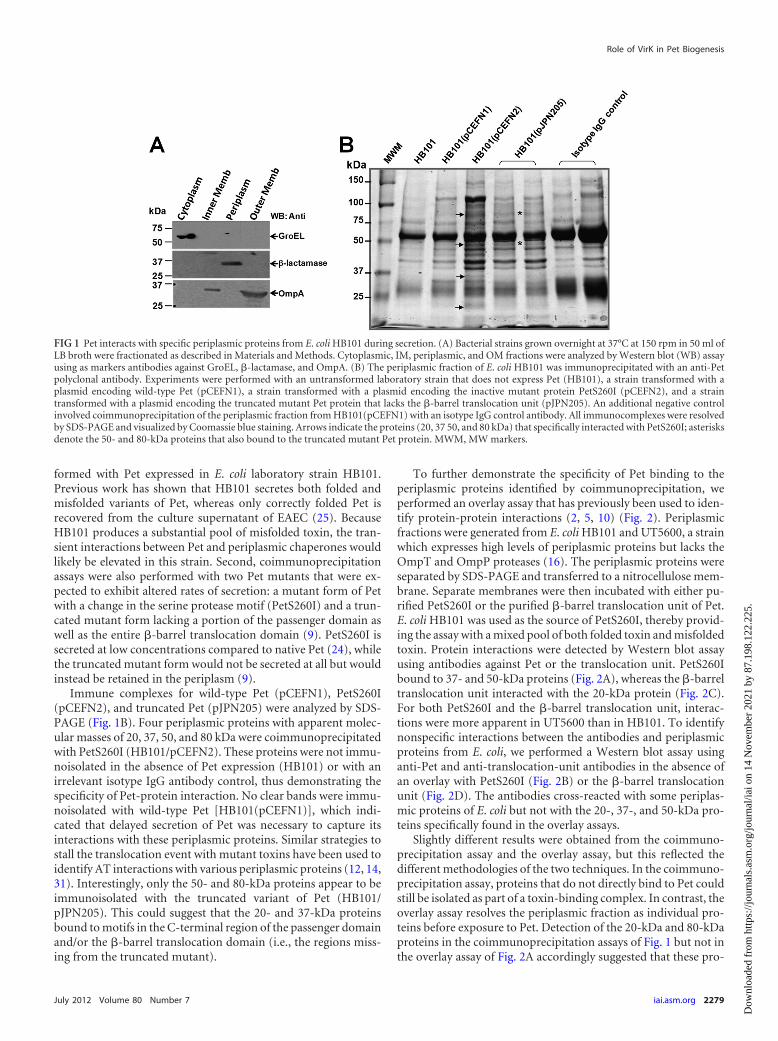
formed with Pet expressed in E. coli laboratory strain HB101.Previous work has shown that HB101 secretes both folded andmisfolded variants of Pet, whereas only correctly folded Pet isrecovered from the culture supernatant of EAEC (25). BecauseHB101 produces a substantial pool of misfolded toxin, the tran-sient interactions between Pet and periplasmic chaperones wouldlikely be elevated in this strain. Second, coimmunoprecipitationassays were also performed with two Pet mutants that were ex-pected to exhibit altered rates of secretion: a mutant form of Petwith a change in the serine protease motif (PetS260I) and a trun-cated mutant form lacking a portion of the passenger domain aswell as the entire �-barrel translocation domain (9). PetS260I issecreted at low concentrations compared to native Pet (24), whilethe truncated mutant form would not be secreted at all but wouldinstead be retained in the periplasm (9).
Immune complexes for wild-type Pet (pCEFN1), PetS260I(pCEFN2), and truncated Pet (pJPN205) were analyzed by SDS-PAGE (Fig. 1B). Four periplasmic proteins with apparent molec-ular masses of 20, 37, 50, and 80 kDa were coimmunoprecipitatedwith PetS260I (HB101/pCEFN2). These proteins were not immu-noisolated in the absence of Pet expression (HB101) or with anirrelevant isotype IgG antibody control, thus demonstrating thespecificity of Pet-protein interaction. No clear bands were immu-noisolated with wild-type Pet [HB101(pCEFN1)], which indi-cated that delayed secretion of Pet was necessary to capture itsinteractions with these periplasmic proteins. Similar strategies tostall the translocation event with mutant toxins have been used toidentify AT interactions with various periplasmic proteins (12, 14,31). Interestingly, only the 50- and 80-kDa proteins appear to beimmunoisolated with the truncated variant of Pet (HB101/pJPN205). This could suggest that the 20- and 37-kDa proteinsbound to motifs in the C-terminal region of the passenger domainand/or the �-barrel translocation domain (i.e., the regions miss-ing from the truncated mutant).
To further demonstrate the specificity of Pet binding to theperiplasmic proteins identified by coimmunoprecipitation, weperformed an overlay assay that has previously been used to iden-tify protein-protein interactions (2, 5, 10) (Fig. 2). Periplasmicfractions were generated from E. coli HB101 and UT5600, a strainwhich expresses high levels of periplasmic proteins but lacks theOmpT and OmpP proteases (16). The periplasmic proteins wereseparated by SDS-PAGE and transferred to a nitrocellulose mem-brane. Separate membranes were then incubated with either pu-rified PetS260I or the purified �-barrel translocation unit of Pet.E. coli HB101 was used as the source of PetS260I, thereby provid-ing the assay with a mixed pool of both folded toxin and misfoldedtoxin. Protein interactions were detected by Western blot assayusing antibodies against Pet or the translocation unit. PetS260Ibound to 37- and 50-kDa proteins (Fig. 2A), whereas the �-barreltranslocation unit interacted with the 20-kDa protein (Fig. 2C).For both PetS260I and the �-barrel translocation unit, interac-tions were more apparent in UT5600 than in HB101. To identifynonspecific interactions between the antibodies and periplasmicproteins from E. coli, we performed a Western blot assay usinganti-Pet and anti-translocation-unit antibodies in the absence ofan overlay with PetS260I (Fig. 2B) or the �-barrel translocationunit (Fig. 2D). The antibodies cross-reacted with some periplas-mic proteins of E. coli but not with the 20-, 37-, and 50-kDa pro-teins specifically found in the overlay assays.
Slightly different results were obtained from the coimmuno-precipitation assay and the overlay assay, but this reflected thedifferent methodologies of the two techniques. In the coimmuno-precipitation assay, proteins that do not directly bind to Pet couldstill be isolated as part of a toxin-binding complex. In contrast, theoverlay assay resolves the periplasmic fraction as individual pro-teins before exposure to Pet. Detection of the 20-kDa and 80-kDaproteins in the coimmunoprecipitation assays of Fig. 1 but not inthe overlay assay of Fig. 2A accordingly suggested that these pro-
FIG 1 Pet interacts with specific periplasmic proteins from E. coli HB101 during secretion. (A) Bacterial strains grown overnight at 37°C at 150 rpm in 50 ml ofLB broth were fractionated as described in Materials and Methods. Cytoplasmic, IM, periplasmic, and OM fractions were analyzed by Western blot (WB) assayusing as markers antibodies against GroEL, �-lactamase, and OmpA. (B) The periplasmic fraction of E. coli HB101 was immunoprecipitated with an anti-Petpolyclonal antibody. Experiments were performed with an untransformed laboratory strain that does not express Pet (HB101), a strain transformed with aplasmid encoding wild-type Pet (pCEFN1), a strain transformed with a plasmid encoding the inactive mutant protein PetS260I (pCEFN2), and a straintransformed with a plasmid encoding the truncated mutant Pet protein that lacks the �-barrel translocation unit (pJPN205). An additional negative controlinvolved coimmunoprecipitation of the periplasmic fraction from HB101(pCEFN1) with an isotype IgG control antibody. All immunocomplexes were resolvedby SDS-PAGE and visualized by Coomassie blue staining. Arrows indicate the proteins (20, 37 50, and 80 kDa) that specifically interacted with PetS260I; asterisksdenote the 50- and 80-kDa proteins that also bound to the truncated mutant Pet protein. MWM, MW markers.
Role of VirK in Pet Biogenesis
July 2012 Volume 80 Number 7 iai.asm.org 2279
Dow
nloa
ded
from
http
s://j
ourn
als.
asm
.org
/jour
nal/i
ai o
n 14
Nov
embe
r 20
21 b
y 87
.198
.122
.225
.
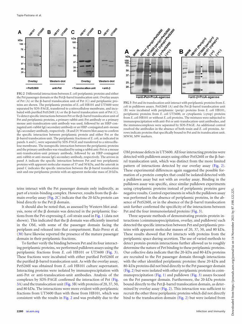
teins interact with the Pet passenger domain only indirectly, aspart of a toxin-binding complex. However, results from the �-do-main overlay assay (Fig. 2C) indicate that the 20-kDa protein canbind directly to the Pet � domain.
It should also be noted that, as assessed by Western blot anal-ysis, none of the � domain was detected in the periplasmic frac-tions from the Pet-expressing E. coli strain used in Fig. 1 (data notshown). This indicated that the � domain was efficiently insertedin the OM, with some of the passenger domain facing theperiplasm and released into that compartment. Ruiz-Perez et al.(30) have likewise reported the presence of the mature passengerdomain in their periplasmic fractions.
To further verify the binding between Pet and its four interact-ing periplasmic proteins, we performed pulldown assays using theperiplasmic fractions from E. coli HB101 or UT5600 (Fig. 3).These fractions were incubated with either purified PetS260I orthe purified �-barrel translocation unit. As with the overlay assay,PetS260I was obtained from E. coli HB101 culture supernatant.Interacting proteins were isolated by immunoprecipitation withanti-Pet or anti-translocation-unit antibodies. Analysis of thecomplexes by SDS-PAGE confirmed the interaction of Pet (Fig.3A) and the translocation unit (Fig. 3B) with proteins of 20, 37, 50,and 80 kDa. The interactions were more evident with periplasmicfractions from UT5600 than with those from HB101, which wasconsistent with the results in Fig. 2 and was probably due to the
OM protease defects in UT5600. All four interacting proteins weredetected with pulldown assays using either PetS260I or the �-bar-rel translocation unit, which was distinct from the more limitedpattern of interactions detected by our overlay assay (Fig. 2).These experimental differences again suggested the possible for-mation of a protein complex that could be isolated/detected witha pulldown assay but not with an overlay assay. Binding in thepulldown assay was specific, since similar pulldown experimentsusing cytoplasmic proteins instead of periplasmic proteins gavenegative results. Control experiments in which the pulldown assaywas performed in the absence of periplasmic proteins, in the ab-sence of PetS260I, or in the absence of the �-barrel translocationunit further confirmed the specificity of the interactions betweenPet and the four immunoisolated proteins (Fig. 3).
Three separate methods of demonstrating protein-protein in-teractions (coimmunoprecipitation, overlay, and pulldown) eachdetected the specific interaction between Pet and periplasmic pro-teins with apparent molecular masses of 20, 37, 50, and 80 kDa.These results showed that Pet interacts with proteins from theperiplasmic space during secretion. The use of varied methods todetect protein-protein interactions further allowed us to roughlydetermine the nature of Pet binding to these periplasmic proteins.Our collective data indicate that the 20-kDa and 80-kDa proteinsare recruited to the Pet passenger domain through interactionswith the other identified periplasmic proteins: these 20-kDa and80-kDa proteins did not bind directly to the Pet passenger domain(Fig. 2) but were isolated with other periplasmic proteins in coim-munoprecipitation (Fig. 1) and pulldown (Fig. 3) assays focusedon the Pet passenger domain. Furthermore, the 20-kDa proteinbound directly to the Pet �-barrel translocation domain, as deter-mined by overlay assay (Fig. 2). This interaction was sufficient torecruit the other three periplasmic proteins which did not directlybind to the translocation domain (Fig. 2) but were isolated from
FIG 2 Differential interactions between E. coli periplasmic proteins and eitherthe Pet passenger domain or the Pet �-barrel translocation unit. Overlay assaysof Pet (A) or the �-barrel translocation unit of Pet (C) and periplasmic pro-teins are shown. The periplasmic proteins of E. coli HB101 and UT5600 wereseparated by SDS-PAGE, transferred to a nitrocellulose membrane, and incu-bated with purified PetS260I (A) or the �-barrel translocation unit of Pet (C).To detect specific interactions between Pet or the �-barrel translocation unit ofPet and periplasmic proteins, a primary rabbit anti-Pet antibody or a primarymouse anti-translocation-unit antibody was used, followed by an HRP-con-jugated anti-rabbit IgG secondary antibody or an HRP-conjugated anti-mouseIgG secondary antibody, respectively. (B and D) Western blot assay to confirmthe specific interaction between periplasmic protein and either Pet or the�-barrel translocation unit. The periplasmic fractions of E. coli, as indicated inpanels A and C, were separated by SDS-PAGE and transferred to a nitrocellu-lose membrane. The nonspecific interaction between the periplasmic proteinsand the primary antibodies was visualized by using a rabbit anti-Pet or a mouseanti-translocation-unit primary antibody, followed by an HRP-conjugatedanti-rabbit or anti-mouse IgG secondary antibody, respectively. The arrows inpanel A indicate the specific interaction between Pet and two periplasmicproteins with apparent molecular masses of 37 and 50 kDa, and the asterisk inpanel C indicates the specific interaction between the �-barrel translocationunit and one periplasmic protein with an apparent molecular mass of 20 kDa.
FIG 3 Pet and its translocation unit interact with periplasmic proteins from E.coli in pulldown assays. PetS260I (A) and the Pet �-barrel translocation unit(B) were incubated with periplasmic (perip) proteins from E. coli HB101,periplasmic proteins from E. coli UT5600, or cytoplasmic (cytop) proteinsfrom E. coli HB101 or without E. coli proteins. The mixtures were subjected toimmunoprecipitation with anti-Pet or anti-translocation-unit antibodies, andthe immunocomplexes were separated by SDS-PAGE. An additional controlresolved the antibodies in the absence of both toxin and E. coli proteins. Ar-rows indicate proteins that specifically bound to Pet and its translocation unit.MWM, MW markers.
Tapia-Pastrana et al.
2280 iai.asm.org Infection and Immunity
Dow
nloa
ded
from
http
s://j
ourn
als.
asm
.org
/jour
nal/i
ai o
n 14
Nov
embe
r 20
21 b
y 87
.198
.122
.225
.
the pulldown assay (Fig. 3). All four identified proteins thus ap-pear to form a toxin-binding complex, with two proteins directlyrecognizing the Pet passenger domain and one protein directlyrecognizing the Pet �-barrel translocation domain.
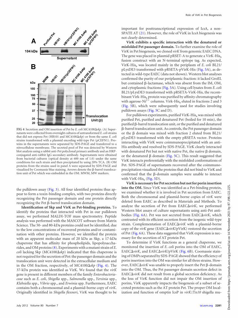
Identification of Skp and VirK as Pet-binding proteins. Toidentify the proteins that interacted with Pet in our pulldownassay, we performed MALDI-TOF mass spectrometry. Peptideanalysis was performed with the MASCOT software from MatrixScience. The 50- and 80-kDa proteins could not be identified, dueto the low concentrations of recovered proteins and/or contami-nation with other proteins. However, we identified the proteinwith an apparent molecular mass of 20 kDa as Skp, a 17-kDachaperone that has affinity for phospholipids, lipopolysaccha-rides, and OM proteins (8). Experiments with a mutant strain of E.coli lacking Skp (MC4100�skp) indicated that this chaperone isnot required for the secretion of Pet: the passenger domain and thetranslocation unit were detected in the extracellular medium andin the OM fraction, respectively, of MC4100�skp (Fig. 4). The37-kDa protein was identified as VirK. We found that the virKgene is present in different members of the family Enterobacteria-ceae such as E. coli, Shigella spp., Salmonella spp., Yersinia spp.,Klebsiella spp., Vibrio spp., and Erwinia spp. Furthermore, EAECcontains both a chromosomal and a plasmid-borne copy of virK.Originally identified in Shigella flexneri, VirK was thought to be
important for posttranscriptional expression of IcsA, a non-SPATE AT (21). However, the role of VirK in IcsA biogenesis wasnot clearly determined.
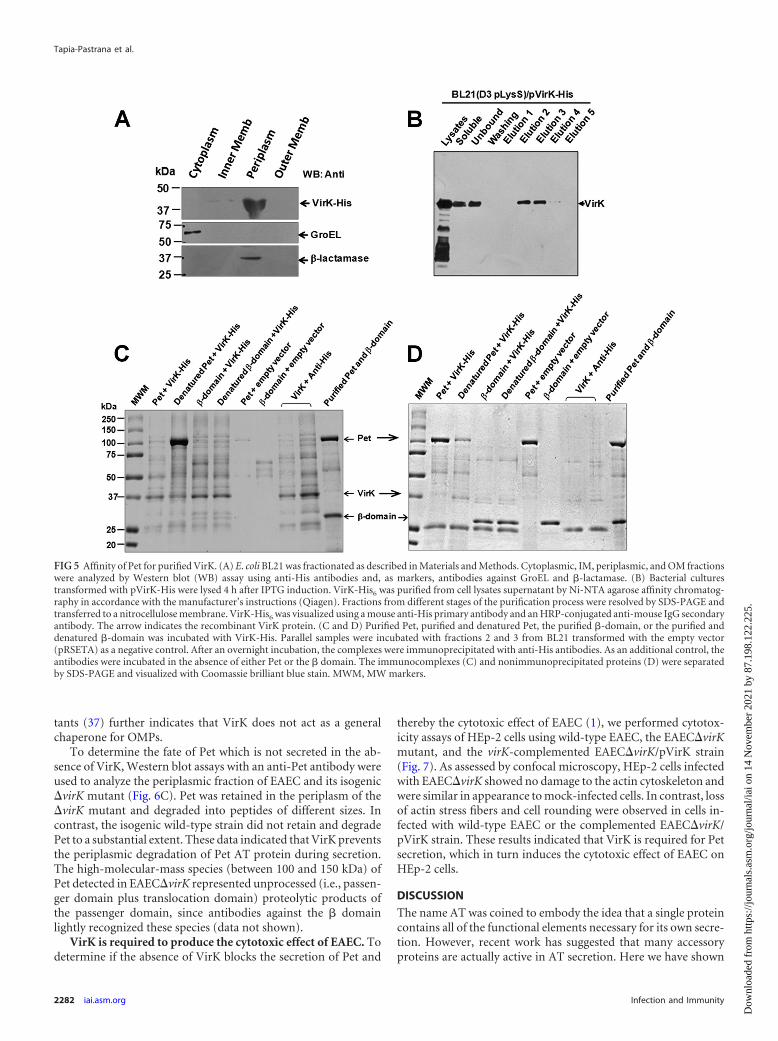
VirK exhibits a specific interaction with the denatured ormisfolded Pet passenger domain. To further examine the role ofVirK in Pet biogenesis, we cloned virK from genomic EAEC DNA.The gene was placed in plasmid pRSET-A to generate a VirK-His6
fusion construct with an N-terminal epitope tag. As expected,VirK-His6 was located mainly in the periplasm of E. coli BL21/pLysDE3 transformed with pRSETA-pVirK-His (Fig. 5A), as de-tected in wild-type EAEC (data not shown). Western blot analysesconfirmed the purity of our periplasmic fraction: it lacked GroELbut contained �-lactamase, which was absent from the IM, OM,and cytoplasmic fractions (Fig. 5A). Using cell lysates from E. coliBL21/pLysDE3 transformed with pRSETA-VirK-His, the recom-binant Virk-His6 protein was purified by affinity chromatographywith agarose-Ni2� columns. Virk-His6 eluted in fractions 2 and 3(Fig. 5B), which were subsequently used for studies involvingpulldown assays (Fig. 5C and D).
For pulldown experiments, purified VirK-His6 was mixed withpurified Pet, purified and denatured Pet (boiled for 10 min), thepurified �-barrel translocation unit, or the purified and denatured�-barrel translocation unit. As controls, the Pet passenger domainor the � domain was mixed with fraction 2 eluted from BL21/pLysDE3 transformed with the empty pRSETA vector. Proteinsinteracting with VirK were coimmunoprecipitated with an anti-His antibody and resolved by SDS-PAGE. VirK clearly interactedwith denatured Pet but not with native Pet, the native � domain,or the denatured � domain (Fig. 5C). This result suggested thatVirK interacts preferentially with the misfolded conformations ofPet. SDS-PAGE of supernatants recovered after the coimmuno-precipitation visualized the proteins that did not bind to VirK andconfirmed that the �-domain samples were unable to interactwith VirK-His6 (Fig. 5D).
VirK is necessary for Pet secretion but not for porin insertioninto the OM. Since VirK was identified as a Pet-binding protein,we examined whether it is involved in Pet secretion from EAEC.Both the chromosomal and plasmid-borne copies of virK weredeleted from EAEC as described in Materials and Methods. Toanalyze the secretion of Pet from EAEC�virK, we performedWestern blot assays of culture supernatants using anti-Pet anti-bodies (Fig. 6A). Pet was not secreted from EAEC�virK, whichcontrasted with its efficient secretion from the isogenic wild-typestrain. Complementation of EAEC�virK with a plasmid-bornecopy of the virK gene (EAEC�virK/pVirK) restored the secretionof Pet (Fig. 6A). These data suggested that VirK expression is nec-essary for the secretion of AT protein Pet.
To determine if VirK functions as a general chaperone, wemonitored the insertion of E. coli porins into the OM of EAEC,EAEC�virK, and EAEC�virK/pVirK (Fig. 6B). Coomassie stain-ing of OMPs separated by SDS-PAGE showed that the efficiency ofporin insertion into the OM was similar for all three strains. How-ever, EAEC�virK was unable to properly insert the Pet �-domaininto the OM. Thus, the Pet passenger domain secretion defect inEAEC�virK did not result from a global secretion deficiency. Asthe loss of VirK function did not impair the OM insertion ofporins, VirK apparently impacts the biogenesis of a subset of se-creted proteins such as the AT protein Pet. The proper OM local-ization and function of omptin IcsP in virK::Tn10 shigella mu-
FIG 4 Secretion and OM insertion of Pet by E. coli MC4100�skp. (A) Super-natants were collected from overnight cultures of untransformed E. coli strainsthat did not express Pet (HB101 and MC4100�skp) or from the same E. colistrains transformed with a plasmid encoding wild-type Pet (pCEFN1). Pro-teins in the supernatants were separated by SDS-PAGE and transferred to anitrocellulose membrane. The secreted pool of Pet was detected by Westernblot analysis using a rabbit anti-Pet polyclonal primary antibody and an HRP-conjugated anti-rabbit IgG secondary antibody. Supernatants were obtainedfrom bacterial cultures (optical density at 600 nm of 1.0) under the sameconditions for each strain and then precipitated by using 20% TCA. (B) OMproteins from the strains used in panel A were separated by SDS-PAGE andvisualized by Coomassie blue staining. Arrows denote the �-barrel transloca-tion unit of Pet which was embedded in the OM. MWM, MW markers.
Role of VirK in Pet Biogenesis
July 2012 Volume 80 Number 7 iai.asm.org 2281
Dow
nloa
ded
from
http
s://j
ourn
als.
asm
.org
/jour
nal/i
ai o
n 14
Nov
embe
r 20
21 b
y 87
.198
.122
.225
.
tants (37) further indicates that VirK does not act as a generalchaperone for OMPs.
To determine the fate of Pet which is not secreted in the ab-sence of VirK, Western blot assays with an anti-Pet antibody wereused to analyze the periplasmic fraction of EAEC and its isogenic�virK mutant (Fig. 6C). Pet was retained in the periplasm of the�virK mutant and degraded into peptides of different sizes. Incontrast, the isogenic wild-type strain did not retain and degradePet to a substantial extent. These data indicated that VirK preventsthe periplasmic degradation of Pet AT protein during secretion.The high-molecular-mass species (between 100 and 150 kDa) ofPet detected in EAEC�virK represented unprocessed (i.e., passen-ger domain plus translocation domain) proteolytic products ofthe passenger domain, since antibodies against the � domainlightly recognized these species (data not shown).
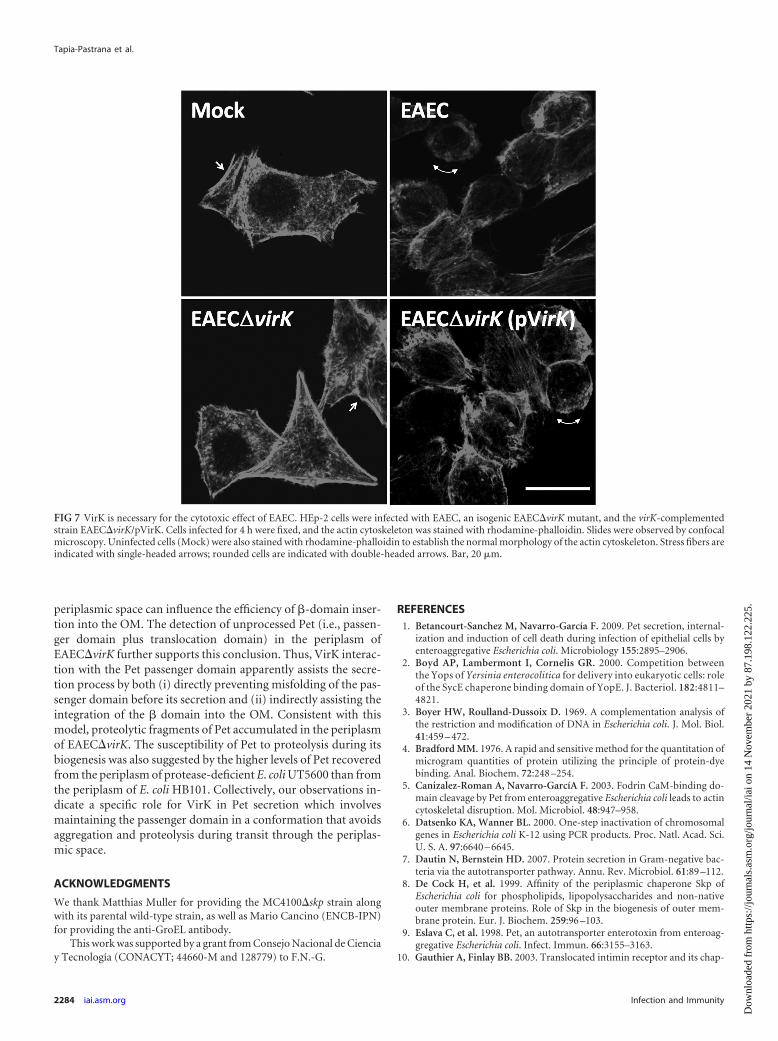
VirK is required to produce the cytotoxic effect of EAEC. Todetermine if the absence of VirK blocks the secretion of Pet and
thereby the cytotoxic effect of EAEC (1), we performed cytotox-icity assays of HEp-2 cells using wild-type EAEC, the EAEC�virKmutant, and the virK-complemented EAEC�virK/pVirK strain(Fig. 7). As assessed by confocal microscopy, HEp-2 cells infectedwith EAEC�virK showed no damage to the actin cytoskeleton andwere similar in appearance to mock-infected cells. In contrast, lossof actin stress fibers and cell rounding were observed in cells in-fected with wild-type EAEC or the complemented EAEC�virK/pVirK strain. These results indicated that VirK is required for Petsecretion, which in turn induces the cytotoxic effect of EAEC onHEp-2 cells.
DISCUSSION
The name AT was coined to embody the idea that a single proteincontains all of the functional elements necessary for its own secre-tion. However, recent work has suggested that many accessoryproteins are actually active in AT secretion. Here we have shown
FIG 5 Affinity of Pet for purified VirK. (A) E. coli BL21 was fractionated as described in Materials and Methods. Cytoplasmic, IM, periplasmic, and OM fractionswere analyzed by Western blot (WB) assay using anti-His antibodies and, as markers, antibodies against GroEL and �-lactamase. (B) Bacterial culturestransformed with pVirK-His were lysed 4 h after IPTG induction. VirK-His6 was purified from cell lysates supernatant by Ni-NTA agarose affinity chromatog-raphy in accordance with the manufacturer’s instructions (Qiagen). Fractions from different stages of the purification process were resolved by SDS-PAGE andtransferred to a nitrocellulose membrane. VirK-His6 was visualized using a mouse anti-His primary antibody and an HRP-conjugated anti-mouse IgG secondaryantibody. The arrow indicates the recombinant VirK protein. (C and D) Purified Pet, purified and denatured Pet, the purified �-domain, or the purified anddenatured �-domain was incubated with VirK-His. Parallel samples were incubated with fractions 2 and 3 from BL21 transformed with the empty vector(pRSETA) as a negative control. After an overnight incubation, the complexes were immunoprecipitated with anti-His antibodies. As an additional control, theantibodies were incubated in the absence of either Pet or the � domain. The immunocomplexes (C) and nonimmunoprecipitated proteins (D) were separatedby SDS-PAGE and visualized with Coomassie brilliant blue stain. MWM, MW markers.
Tapia-Pastrana et al.
2282 iai.asm.org Infection and Immunity
Dow
nloa
ded
from
http
s://j
ourn
als.
asm
.org
/jour
nal/i
ai o
n 14
Nov
embe
r 20
21 b
y 87
.198
.122
.225
.

that the periplasmic protein VirK is needed to secrete Pet proteinfrom EAEC. VirK was not required for the OM insertion ofnon-AT proteins. These results suggest that VirK is an accessoryfactor specifically involved with the biogenesis of a subset of se-creted proteins such as the AT Pet.
Other accessory proteins are also known to operate in AT se-cretion. Release of the SPATE hemoglobin protease (Hbp) in-volves SurA and subunits of the BAM complex that promote theinsertion and assembly of OM proteins (31). BamA (YaeT/Omp85), SurA, and Skp interact with the SPATE EspP (12). BamA(YaeT/Omp85) also appears to function in the OM assembly ofmultiple AT proteins (13). Finally, the DegP protease will degradeAT folding intermediates that accumulate in the periplasmicspace (14). These observations support a model of AT biogen-esis in which periplasmic proteins and the BAM complex col-lectively act to (i) prevent misfolding of the passenger domainbefore its secretion, (ii) catalyze the integration of the � do-main into the OM, and (iii) facilitate the translocation of thepassenger domain across the OM.
Here we report that the biogenesis of the AT Pet also involvesthe periplasmic protein VirK. Four periplasmic proteins with ap-parent molecular masses of 20, 37, 50, and 80 kDa were identifiedas Pet binding partners by coimmunoprecipitation, overlay, andpulldown studies. These proteins were identified with a strategythat involved delayed AT secretion as a means to capture the tran-sient interactions between Pet and its chaperones. Similar strate-gies have been used to identify the roles of other periplasmic pro-teins in AT secretion (12, 14, 31). Protein sequencing identifiedthe 20-kDa protein as Skp and the 37-kDa protein as VirK. Sub-sequent experiments using purified, His-tagged VirK confirmedthe direct interaction between VirK and the Pet passenger domain.The 50- and 80-kDa Pet binding partners have yet to be identified,although it is interesting that SurA is a 48-kDa protein and BamAis an 86-kDa protein (12, 14, 31).
Skp bound to Pet in our coimmunoprecipitation assay, andan additional overlay assay indicated that Skp interacts specif-ically with the �-barrel translocation domain of Pet. This sug-gests that Skp, as previously reported for the AT Hbp (31),helps to insert the �-barrel domain into the OM. The collectivedata of the coimmunoprecipitation, overlay, and pulldown as-says also indicate that Skp forms one component of a chaper-one complex involving VirK, the 50-kDa protein, and the 80-kDa protein. However, a skp deletion mutant still secreted Petinto the medium and inserted the Pet �-barrel domain into theOM, albeit with lower efficiency than the isogenic wild-typestrain. The release of Pet from MC4100�skp supports a modelof AT biogenesis in which there are redundant, parallel path-ways for AT secretion (28, 29).
The phenotype of the virK deletion strain in regard to bacterialgrowth and OMP insertion was distinct from the �skp mutantstrain’s phenotype and the previously described phenotypes ofstrains with other AT accessory factors deleted (27, 29). surA anddegP mutant strains exhibited retarded growth rates and apparentcell lysis when expressing EspP (30). Furthermore, SurA, DegP,and Skp appear to function in the targeting and/or insertion ofOMPs (28). In contrast, the �virK mutation did not affect growthrates (data not shown) and did not substantially inhibit the inser-tion of porin proteins into the OM. The lack of VirK interactionwith the Pet �-barrel translocation domain also argues against adirect role for VirK in OM insertion. These collective observationssuggest that the role of VirK in Pet AT biogenesis is distinct fromthe functions performed by AT accessory factors Skp, SurA, andDegP. The role(s) of VirK in the biogenesis of other classical (mo-nomeric) ATs, as well as trimeric ATs and TpsA proteins, remainsto be established.
Unlike Skp, VirK is essential for Pet secretion. The OM inser-tion of omptin IcsP (37) and porins was not disrupted in virK nullmutants, but a virK deletion mutant of EAEC was unable to secretePet. The functional consequences of the Pet secretion defects weredemonstrated with EAEC because EAEC�virK did not display cy-totoxicity against HEp-2 cells. Complementation of virK restoredcytotoxicity and the mutant’s capacity for secretion of the Petpassenger domain.
Multiple assays demonstrated that VirK recognizes misfolded/unfolded conformations of the Pet passenger domain but does notbind to the folded passenger domain, the native � domain, or thedenatured � domain. However, OM insertion of the Pet � domaindid not occur in the absence of VirK. This suggested that the fold-ing state of the passenger domain during transit through the
FIG 6 Inhibition of Pet AT but not porin secretion from virK null mutants.(A) Supernatants were collected from overnight cultures of an E. coli labora-tory strain that does not express Pet (HB101), a pathogenic E. coli strain thatexpresses Pet (EAEC), isogenic strain EAEC�virK, and virK-complementedstrain EAEC�virK/pVirK. Proteins in the supernatants were separated by SDS-PAGE and transferred to a nitrocellulose membrane. The secreted pools of Petwere detected by Western blot (WB) analysis using a rabbit anti-Pet primaryantibody, followed by an HRP-conjugated anti-rabbit IgG secondary anti-body. (B) OMPs were obtained from cultures of EAEC, EAEC�virK, andEAEC�virK/pVirK; the purified � domain was used as a control. The OMPswere separated by SDS-PAGE and visualized with Coomassie brilliant bluestain. Arrows indicate the porins and the �-barrel translocation unit. For allexperiments, supernatants from the bacterial strains were normalized bygrowth under equal conditions and sample harvesting at the same growthphase (optical density at 600 nm of 1.0) before precipitation with 20% TCA.(C) Retention and degradation of Pet AT protein in a virK mutant strain.Periplasmic fractions were obtained from overnight cultures of EAEC and itsisogenic virK mutant. Samples were resolved by SDS-PAGE and transferred tonitrocellulose membranes. The periplasmic pool of Pet was detected by West-ern blot analysis using a rabbit anti-Pet primary antibody, followed by anHRP-conjugated anti-rabbit IgG secondary antibody.
Role of VirK in Pet Biogenesis
July 2012 Volume 80 Number 7 iai.asm.org 2283
Dow
nloa
ded
from
http
s://j
ourn
als.
asm
.org
/jour
nal/i
ai o
n 14
Nov
embe
r 20
21 b
y 87
.198
.122
.225
.
periplasmic space can influence the efficiency of �-domain inser-tion into the OM. The detection of unprocessed Pet (i.e., passen-ger domain plus translocation domain) in the periplasm ofEAEC�virK further supports this conclusion. Thus, VirK interac-tion with the Pet passenger domain apparently assists the secre-tion process by both (i) directly preventing misfolding of the pas-senger domain before its secretion and (ii) indirectly assisting theintegration of the � domain into the OM. Consistent with thismodel, proteolytic fragments of Pet accumulated in the periplasmof EAEC�virK. The susceptibility of Pet to proteolysis during itsbiogenesis was also suggested by the higher levels of Pet recoveredfrom the periplasm of protease-deficient E. coli UT5600 than fromthe periplasm of E. coli HB101. Collectively, our observations in-dicate a specific role for VirK in Pet secretion which involvesmaintaining the passenger domain in a conformation that avoidsaggregation and proteolysis during transit through the periplas-mic space.
ACKNOWLEDGMENTS
We thank Matthias Muller for providing the MC4100�skp strain alongwith its parental wild-type strain, as well as Mario Cancino (ENCB-IPN)for providing the anti-GroEL antibody.
This work was supported by a grant from Consejo Nacional de Cienciay Tecnología (CONACYT; 44660-M and 128779) to F.N.-G.
REFERENCES1. Betancourt-Sanchez M, Navarro-García F. 2009. Pet secretion, internal-
ization and induction of cell death during infection of epithelial cells byenteroaggregative Escherichia coli. Microbiology 155:2895–2906.
2. Boyd AP, Lambermont I, Cornelis GR. 2000. Competition betweenthe Yops of Yersinia enterocolitica for delivery into eukaryotic cells: roleof the SycE chaperone binding domain of YopE. J. Bacteriol. 182:4811–4821.
3. Boyer HW, Roulland-Dussoix D. 1969. A complementation analysis ofthe restriction and modification of DNA in Escherichia coli. J. Mol. Biol.41:459 – 472.
4. Bradford MM. 1976. A rapid and sensitive method for the quantitation ofmicrogram quantities of protein utilizing the principle of protein-dyebinding. Anal. Biochem. 72:248 –254.
5. Canizalez-Roman A, Navarro-GarcíA F. 2003. Fodrin CaM-binding do-main cleavage by Pet from enteroaggregative Escherichia coli leads to actincytoskeletal disruption. Mol. Microbiol. 48:947–958.
6. Datsenko KA, Wanner BL. 2000. One-step inactivation of chromosomalgenes in Escherichia coli K-12 using PCR products. Proc. Natl. Acad. Sci.U. S. A. 97:6640 – 6645.
7. Dautin N, Bernstein HD. 2007. Protein secretion in Gram-negative bac-teria via the autotransporter pathway. Annu. Rev. Microbiol. 61:89 –112.
8. De Cock H, et al. 1999. Affinity of the periplasmic chaperone Skp ofEscherichia coli for phospholipids, lipopolysaccharides and non-nativeouter membrane proteins. Role of Skp in the biogenesis of outer mem-brane protein. Eur. J. Biochem. 259:96 –103.
9. Eslava C, et al. 1998. Pet, an autotransporter enterotoxin from enteroag-gregative Escherichia coli. Infect. Immun. 66:3155–3163.
10. Gauthier A, Finlay BB. 2003. Translocated intimin receptor and its chap-
FIG 7 VirK is necessary for the cytotoxic effect of EAEC. HEp-2 cells were infected with EAEC, an isogenic EAEC�virK mutant, and the virK-complementedstrain EAEC�virK/pVirK. Cells infected for 4 h were fixed, and the actin cytoskeleton was stained with rhodamine-phalloidin. Slides were observed by confocalmicroscopy. Uninfected cells (Mock) were also stained with rhodamine-phalloidin to establish the normal morphology of the actin cytoskeleton. Stress fibers areindicated with single-headed arrows; rounded cells are indicated with double-headed arrows. Bar, 20 �m.
Tapia-Pastrana et al.
2284 iai.asm.org Infection and Immunity
Dow
nloa
ded
from
http
s://j
ourn
als.
asm
.org
/jour
nal/i
ai o
n 14
Nov
embe
r 20
21 b
y 87
.198
.122
.225
.

erone interact with ATPase of the type III secretion apparatus of entero-pathogenic Escherichia coli. J. Bacteriol. 185:6747– 6755.
11. Henderson IR, Navarro-García F, Desvaux M, Fernandez RC,Ala’Aldeen D. 2004. Type V protein secretion pathway: the autotrans-porter story. Microbiol. Mol. Biol. Rev. 68:692–744.
12. Ieva R, Bernstein HD. 2009. Interaction of an autotransporter passengerdomain with BamA during its translocation across the bacterial outermembrane. Proc. Natl. Acad. Sci. U. S. A. 106:19120 –19125.
13. Jain S, Goldberg MB. 2007. Requirement for YaeT in the outer membraneassembly of autotransporter proteins. J. Bacteriol. 189:5393–5398.
14. Jong WS, et al. 2007. Limited tolerance towards folded elements duringsecretion of the autotransporter Hbp. Mol. Microbiol. 63:1524 –1536.
15. Junker M, Besingi RN, Clark PL. 2009. Vectorial transport and folding ofan autotransporter virulence protein during outer membrane secretion.Mol. Microbiol. 71:1323–1332.
16. Kaufmann A, Stierhof YD, Henning U. 1994. New outer membrane-associated protease of Escherichia coli K-12. J. Bacteriol. 176:359 –367.
17. Kleinschmidt JH. 2006. Folding kinetics of the outer membrane pro-teins OmpA and FomA into phospholipid bilayers. Chem. Phys. Lipids141:30 – 47.
18. Klingman KL, Jansen EM, Murphy TF. 1988. Nearest neighbor analysisof outer membrane proteins of nontypeable Haemophilus influenzae. In-fect. Immun. 56:3058 –3063.
19. Laemmli UK. 1970. Cleavage of structural proteins during the assembly ofthe head of bacteriophage T4. Nature 227:680 – 685.
20. Mogensen JE, Kleinschmidt JH, Schmidt MA, Otzen DE. 2005. Mis-folding of a bacterial autotransporter. Protein Sci. 14:2814 –2827.
21. Nakata N, et al. 1992. Identification and characterization of virK, a viru-lence-associated large plasmid gene essential for intercellular spreading ofShigella flexneri. Mol. Microbiol. 6:2387–2395.
22. Nataro JP, et al. 1995. Heterogeneity of enteroaggregative Escherichia colivirulence demonstrated in volunteers. J. Infect. Dis. 171:465– 468.
23. Navarro-GarcíA F, Canizalez-Roman A, Luna J, Sears C, Nataro JP.2001. Plasmid-encoded toxin of enteroaggregative Escherichia coli is inter-nalized by epithelial cells. Infect. Immun. 69:1053–1060.
24. Navarro-GarcíA F, Sears C, Eslava C, Cravioto A, Nataro JP. 1999.Cytoskeletal effects induced by pet, the serine protease enterotoxin ofenteroaggregative Escherichia coli. Infect. Immun. 67:2184 –2192.
25. Nemec KN, et al. 2010. A host-specific factor is necessary for efficientfolding of the autotransporter plasmid-encoded toxin. Biochimie 92:171–177.
26. Pallen MJ, Chaudhuri RR, Henderson IR. 2003. Genomic analysis ofsecretion systems. Curr. Opin. Microbiol. 6:519 –527.
27. Purdy GE, Fisher CR, Payne SM. 2007. IcsA surface presentation inShigella flexneri requires the periplasmic chaperones DegP, Skp, and SurA.J. Bacteriol. 189:5566 –5573.
28. Purdy GE, Hong M, Payne SM. 2002. Shigella flexneri DegP facilitatesIcsA surface expression and is required for efficient intercellular spread.Infect. Immun. 70:6355– 6364.
29. Rizzitello AE, Harper JR, Silhavy TJ. 2001. Genetic evidence for parallelpathways of chaperone activity in the periplasm of Escherichia coli. J. Bac-teriol. 183:6794 – 6800.
30. Ruiz-Perez F, et al. 2009. Roles of periplasmic chaperone proteins in thebiogenesis of serine protease autotransporters of Enterobacteriaceae. J.Bacteriol. 191:6571– 6583.
31. Sauri A, et al. 2009. The Bam (Omp85) complex is involved in secretionof the autotransporter haemoglobin protease. Microbiology 155:3982–3991.
32. Schäfer U, Beck K, Muller M. 1999. Skp, a molecular chaperone ofGram-negative bacteria, is required for the formation of soluble periplas-mic intermediates of outer membrane proteins. J. Biol. Chem. 274:24567–24574.
33. Studier FW, Rosenberg AH, Dunn JJ, Dubendorff JW. 1990. Use of T7RNA polymerase to direct expression of cloned genes. Methods Enzymol.185:60 – 89.
34. Towbin H, Staehelin T, Gordon J. 1979. Electrophoretic transfer ofproteins from polyacrylamide gels to nitrocellulose sheets: procedure andsome applications. Proc. Natl. Acad. Sci. U. S. A. 76:4350 – 4354.
35. Tseng TT, Tyler BM, Setubal JC. 2009. Protein secretion systems inbacterial-host associations, and their description in the Gene Ontology.BMC Microbiol. 9(Suppl 1):S2. doi:10.1186/1471-2180-9-S1-S2.
36. Wagner JK, Heindl JE, Gray AN, Jain S, Goldberg MB. 2009. Contri-bution of the periplasmic chaperone Skp to efficient presentation of theautotransporter IcsA on the surface of Shigella flexneri. J. Bacteriol. 191:815– 821.
37. Wing HJ, Goldman SR, Ally S, Goldberg MB. 2005. Modulation of anouter membrane protease contributes to the virulence defect of Shigellaflexneri strains carrying a mutation in the virK locus. Infect. Immun. 73:1217–1220.
38. Yen YT, et al. 2010. Importance of conserved residues of the serineprotease autotransporter beta-domain in passenger domain processingand beta-barrel assembly. Infect. Immun. 78:3516 –3528.
Role of VirK in Pet Biogenesis
July 2012 Volume 80 Number 7 iai.asm.org 2285
Dow
nloa
ded
from
http
s://j
ourn
als.
asm
.org
/jour
nal/i
ai o
n 14
Nov
embe
r 20
21 b
y 87
.198
.122
.225
.