Embed Size (px)
Citation preview

Fax +41 61 306 12 34E-Mail [email protected]
Research Paper
J Vasc Res 2011;48:453–464 DOI: 10.1159/000327009
Shear Stress Induces the Release ofan Endothelial Elastase: Role in Integrin � v � 3 -Mediated FGF-2 Release
Theres Hennig Christina Mogensen Julian Kirsch Ulrich Pohl Torsten Gloe
Walter Brendel Centre of Experimental Medicine, Ludwig-Maximilians-University München, Munich , Germany
icked by application of pancreatic elastase to static endothe-lial cells. Conclusion: By inducing the release of an endothe-lial elastase, shear stress induces an integrin-dependent re-lease of FGF-2 from endothelial cells.
Copyright © 2011 S. Karger AG, Basel
Introduction
Fluid shear stress is probably the most significant physiological stimulus for endothelial cells to produce the endothelium-derived autacoids nitric oxide, prostacyclin and endothelium-derived hyperpolarizing factor [1, 2] . Functionally, this shear stress-induced release of auta-coids elicits a flow-dependent dilation allowing the acute adaptation of vascular conductivity to an augmented flow load. Moreover, shear stress controls the expression of various genes involved in autacoid synthesis and, in addition, cell survival, cell growth and cell cycle control [3–5] . Consequently, shear stress is also a pivotal stimulus for structural arterial remodeling following chronic al-teration of blood flow [6–8] .
Key Words
Shear stress � Mechanotransduction � Elastase � Integrin � v � 3 � Fibroblast growth factor 2
Abstract
Background/Aims: Laminar shear stress is an important stimulus in the endothelium-dependent control of vascular tone and of vascular remodeling processes. Based on previ-ous studies demonstrating integrin-mediated release of fi-broblast growth factor 2 (FGF-2), we investigated whether shear stress-induced integrin activation requires the involve-ment of an extracellular protease. Methods: Cultured por-cine aortic endothelial cells (PAEC) were exposed to laminar shear stress (16 dyn/cm 2 ), whereas static cells served as con-trols. Results: Exposure of PAEC to shear stress led to an in-creased activity of a protease in supernatants. This protease could be characterized as elastase but was different from neutrophil and pancreatic elastases. The enhanced activity was accompanied by the activation of integrin � v � 3 and p38 MAPK, and followed by an increased FGF-2 concentration in the supernatant. Pretreatment with inhibitors of either elas-tase or integrin � v � 3 resulted in a reduction of FGF-2 release. The observed effects of shear stress on integrin � v � 3 and p38 MAPK activation, as well as on FGF-2 release could be mim-
Received: November 17, 2010 Accepted after revision: February 17, 2011 Published online: June 17, 2011
Prof. Dr. Ulrich Pohl Walter Brendel Centre of Experimental Medicine Ludwig-Maximilians-Universität München, Marchioninistrasse 27DE–81377 Munich (Germany) Tel. +49 89 21807 6500, E-Mail upohl @ lmu.de
© 2011 S. Karger AG, Basel1018–1172/11/0486–0453$38.00/0
Accessible online at:www.karger.com/jvr
T.H. and C.M. contributed equally to this work.
Hennig/Mogensen/Kirsch/Pohl/Gloe
J Vasc Res 2011;48:453–464 454
There is ample evidence that shear stress stimulates en-dothelial cells via multiple pathways, involving the activa-tion of ion channels, G protein-coupled receptors, inte-grins as well as receptor tyrosine kinases, ultimately lead-ing to the activation of mitogen-activated protein kinases (MAPK) and downstream transcription factors. Inte-grins, membrane caveolae, receptor tyrosine kinases, G proteins, ion channels as well as glycocalyx components have been reported to act as mechanotransducers upon endothelial stimulation by shear stress [9–11] . Consider-ing that a mechanical deformation of the cells occurs dur-ing exposure to shear forces, the cytoskeleton and its plas-malemmal anchoring complexes also play a role in the mechanotransduction of shear stress [12] . In particular, transmembrane proteins such as platelet endothelial cell adhesion molecule (PECAM-1) and integrins potentially act as mechanotransducers. With regard to integrins, there is evidence for inside-out signaling during shear stress which may support endothelial cell adherence under conditions of elevated external forces. Indeed, PECAM-1, using vascular endothelial cell cadherin as an adaptor molecule, elicits activation of integrins in a clas-sical inside-out manner [13] . However, there is also evi-dence that shear stress leads to outside-in integrin signal-ing. Several reports on altered expression, activity and release of metalloproteinases and cysteine proteases (ca-thepsins L and K) under shear stress strongly suggest that modulation of the extracellular matrix (ECM) by these enzymes contributes to activation of cellular adhesion molecules as part of mechanotransduction [14–17] . More-over, a modulation of ECM protein expression under chronic shear stress has been reported [18, 19] . Besides, various different ECM proteins or their fragments can lead to specific integrin and MAP kinase signaling during flow [20] . Therefore, the production, composition and in particular the enzymatic modification of the ECM may be pivotal in the response of endothelial cells to shear stress.
We have shown previously that endothelial adhesion molecules, in particular laminin-binding protein and in-tegrin � v � 3 , mediate shear stress responses in cultured endothelial cells [21, 22] . Laminin-binding protein has been shown to act as an essential mediator of shear stress-induced endothelial nitric oxide synthase expression in porcine aortic endothelial cells. In addition, integrin � v � 3 was found to be an integral part of the signaling cas-cade leading to shear stress-induced release of fibroblast growth factor 2 (FGF-2) from endothelial cells, a poten-tially significant process in flow-related vascular remod-eling. However, the mechanism of integrin activation re-mained unclear.
Based on previous observations by others on the oc-currence of proteases or their inhibitors during shear stress [14–17] , we hypothesized that the activation of the integrin � v � 3 , and hence the FGF-2 release, is due to the action of an endothelium-derived protease. Therefore, we studied potential alterations of matrix composition and of proteolytic activities in endothelial cell cultures ex-posed to arterial levels of shear stress. Since we found evidence for the release of a yet unidentified elastase dur-ing shear stress, we also studied whether exogenously ap-plied elastase could mimic the effects of shear stress on integrin � v � 3 activation and FGF-2 release in static endo-thelial cells.
Methods
Unless stated otherwise, all reagents were purchased from Ap-plichem.
Cell Culture Porcine aortic endothelial cells (PAEC) were isolated as de-
scribed previously by Gloe et al. [21] . Cells were cultured on stan-dard plastic culture dishes in DMEM (Gibco) containing 10% FCS (Biochrom) and 1% of a mixture of amphotericin B, penicillin and streptomycin (Sigma). They were subcultured using trypsin/EDTA (Sigma). Cultured cells were identified as endothelial cells by the presence of PECAM-1 antigen. PAEC at passages 3–5 were used in all experiments.
Shear Stress Experiments A laminar shear stress of 16 dyn/cm 2 was applied using a cone
and plate apparatus as described before [21] . Cells were trans-ferred onto glass plates coated with collagen G (10 � g/ml; Bio-chrom). After 16 h, the medium was changed to DMEM (without phenol red; Sigma) containing 1% FCS. The inhibitors chymo-statin (CHY; Sigma), methoxysuccinyl-Ala-Ala-Pro-Val-chloro-methylketone (MEO; Bachem) and Abciximab (ABC; Lilly) were added 15 min prior to the exposure to shear stress and left in the medium throughout the experiment. Static controls were taken from the same culture and also grown on glass plates. Afterwards, the supernatant was collected for quantitative FGF-2 and elastase activity measurements. The cells were immediately lysed for Western blot analysis or fixed for immunofluorescence experi-ments as described below. Protein extracts were frozen after prep-aration and stored at –20 ° C until further analysis. Cell lysates were analyzed for equal protein levels using the bicinchoninic acid assay (Perbio).
Assessment of FGF-2 FGF-2 in cell supernatants was measured using a commercial-
ly available ELISA kit (R&D Systems) according to the manufac-turer’s instructions. Immunostaining of FGF-2 was performed as follows: PAEC were fixed with 3.7% formaldehyde, blocked using 1% bovine serum albumin in PBS, and incubated with the pri-mary FGF-2 antibody (Sigma) overnight at 4 ° C. The FITC-la-
Flow-Induced FGF-2 Release Mediated by an Elastase
J Vasc Res 2011;48:453–464 455
beled secondary antibody (Invitrogen) was applied for 30 min at room temperature. The samples were analyzed using a standard fluorescence microscope (Axiovert 200M; Zeiss). FGF-2 trans-location to the cell surface was analyzed by flow cytometry (FACScan; Becton Dickinson) using a FGF-2 primary antibody (Sigma) and an FITC-labeled secondary antibody (Invitrogen). Suspended cells were detached using a citrate buffer composed of 135 m M KCl and 15 m M sodium citrate.
Determination of Elastase Activity Elastase activity measurements were performed using the spe-
cific substrate methoxysuccinyl-Ala-Ala-Pro-Val-p-nitroaniline (MeOSuc-Ala-Ala-Pro-Val-pNA, 20 � g/ml; Bachem). Fifty mi-croliters of each sample was incubated with 1 � l of substrate for 30 min and absorbance detected using a spectrophotometer. A calibration curve for each individual set of experiments was gen-erated using porcine pancreatic elastase.
Zymography Supernatants from cells exposed to shear stress and static con-
trols were analyzed by SDS-PAGE (gels supplemented with 10 mg/ml gelatine) using denaturing but nonreducing conditions. SDS was removed from the gel by 3 subsequent washings with 2.5% Triton X-100 for 30 min each. Proteases were allowed to react with the gelatine in the gel for 16 h at 37 ° C in a bathing solution con-taining 50 m M Tris (pH 7.5), 200 m M NaCl, 5 m M CaCl 2 and 0.02% Tween 20. Finally, the gel was stained with Coomassie blue G250 (0.7 m M in 10% acetic acid) and destained (7% acetic acid in 5% methanol) until areas of proteolytic activity were detectable as white bands.
Immunoblotting and Immunoprecipitation The buffer used for cell lysis was composed as follows: 20 m M
Tris, 137 m M NaCl, 2 m M EDTA (pH 8.0), 10% glycerol and 0.1% sodium deoxylcholate. The protease and phosphatase inhibitors aprotinin (10 � g/ml), leupeptin (10 � g/ml), phenylmethylsulfonyl fluoride (1 m M ), sodium fluoride (0.5 m M ) and sodium orthovan-adate (0.5 m M ) were added to the buffer prior to each experiment. Protein content was assessed using a bicinchoninic acid assay (Perbio). Equal amounts of total protein were separated by SDS-PAGE and transferred to a nitrocellulose membrane using stan-dard techniques. The membrane was incubated overnight at 4 ° C with antibodies against neutrophil elastase (Santa Cruz), Shc (Up-state), phospho-p38 (Cell Signaling), phospho-ERK1/ERK2 (Cell Signaling), GAPDH (Chemicon), p38 (Cell Signaling) and phos-pho-Hsp27 (Cell Signaling). Incubation with the secondary anti-body (Calbiochem) was performed subsequently for 1.5 h at room temperature. Protein bands were detected using a horseradish peroxide chemiluminescence detection kit.
For immunoprecipitation cell lysates were incubated for 2 h at 4 ° C with LM609 (Chemicon), an antibody to integrin � v � 3 , and magnetic beads coated with protein G (Miltenyi Biotec). Precipi-tates were separated using a magnetic separation unit (Miltenyi Biotec). Immunoblotting was performed as described above.
Elastase Experiments Static cells were treated with porcine pancreatic elastase (0.5
U/ml; SERVA). The elastase was applied to the supernatant of stat-ic endothelial cells for 5, 10, 30 or 60 min. Cells were then imme-diately fixed or lysed as described above. MAP kinase inhibition
was performed using the p38 MAPK inhibitor SB201290 (10 � M ; Upstate) or U0126 (10 � M ; Calbiochem) to block the activity of MEK1/2. These substances were added to the medium 30 min prior to elastase treatment.
Statistical Analysis All quantitative data are expressed as means 8 standard error
of the mean. Statistical tests were performed using the SigmaStat statistic software (SPSS Inc.). If not stated otherwise, differences between treatment groups or changes over time were analyzed us-ing one-way analysis of variance for repeated measures (ANOVA RM) followed by post hoc analysis using the Student Newman-Keuls method. If data were not normally distributed, ANOVA RM on ranks was used but mean values are presented nevertheless for comparability of data. Representative immunoblots and fluo-rescence images are shown. The number of experiments is indi-cated in the figure legends. p values less than 0.05 were considered significant.
Results
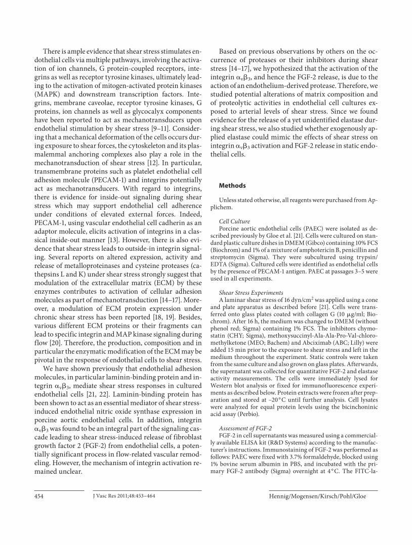
Effects of Shear Stress An Elastase Is Released by Endothelial Cells after Exposure to Shear Stress Zymographic analysis revealed a marked proteolytic
activity at 28 kDa in supernatants from cells exposed to shear stress but not in static cells ( fig. 1 a). Supernatants of static and cells exposed to shear stress were additionally analyzed by immunoblotting using an antibody raised against neutrophil elastase. As in zymograms, a band was observed at 28 kDa only after shear stress exposure ( fig. 1 b). To further characterize this protease activity we used the specific elastase substrate MeOSuc-Ala-Ala-Pro-Val-pNA. Elastase activity in the supernatant of cells ex-posed to shear stress was more than 3-fold higher than in the supernatant of static controls (static 0.06 8 0.01 U/ml, shear 0.20 8 0.03 U/ml) ( fig. 1 c). An increase in elas-tase activity could still be observed after pretreatment with ABC, an antibody fragment that blocks the integrin � v � 3 (static + ABC 0.04 8 0.01 U/ml, shear + ABC 0.15 8 0.01 U/ml). To assess how quickly this protease is re-leased, time course experiments were performed. Signif-icantly increased elastase activity was already detectable after 5 min of exposure to shear stress. During the follow-ing 30 min, the elastase concentration in the supernatant increased further, after which it reached a plateau ( fig. 1 d). In order to investigate whether neutrophil or pancreatic elastases are expressed in endothelial cells, we performed RT-PCR. However, neither neutrophil nor pancreatic elastases were revealed in endothelial cells at the mRNA level (data not shown).

Hennig/Mogensen/Kirsch/Pohl/Gloe
J Vasc Res 2011;48:453–464 456
Shear Stress-Induced FGF-2 Release Is Elastase Dependent Shear stress not only increased elastase activity in the
supernatant but also significantly augmented the con-centration of FGF-2 in the supernatant more than 4-fold (static 78.5 8 17.9 pg/ml, shear 365.5 8 45.1 pg/ml; fig. 2 a), which is in line with our previous findings [22] . This increase in FGF-2 concentration was partly an elas-tase-dependent effect, since pretreatment with both the unspecific serine protease inhibitor CHY (10 � g/ml,
a
ABC
†
†
†
0
100
200
300
400
500
MEO CHY
FGF-
2 in
the
supe
rnat
ant (
pg/m
l)
*
ShearStatic
b
ShearStatic
FGF-2
StaticShear
StaticShear
28 kDa
a
28 kDa
b
0
0.1
0.2
0.3
c
n.s.
n.s.
ABC ABC
ShearStatic
Elas
tase
act
ivity
(U/m
l) *
*
d
00 20 40 60 80 100 120
0.1
0.2
0.3
Shear
Static
Time (min)
Elas
tase
act
ivity
(U/m
l)
*
***
*
Fig. 1. Endothelial shear stress is associated with an increased elastase activity in the supernatant. a Supernatants of static and cells exposed to 16 dyn/cm 2 shear stress (2 h) were analyzed by zymography (n = 3). White bands represent areas of protease ac-tivity. b Supernatants were analyzed by immunoblotting using an anti-neutrophil elastase antibody (n = 3). c Enzymatic activity in supernatants was determined using MeOSuc-Ala-Ala-Pro-Val-pNA (ABC, 1 � g/ml; n = 20). * p ! 0.05 vs. static, ANOVA RM on ranks, Student-Newman-Keuls. n.s. = Not significant. d A sig-nificant increase of elastase activity occurred already after 5 min of shear stress exposure (n = 4–6). * p ! 0.05 vs. static.
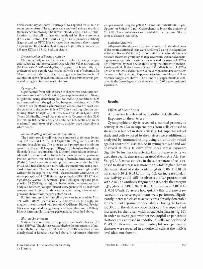
Fig. 2. The effect of shear stress on FGF-2 release is dependent on elastase and the integrin � v � 3 . a PAEC were either left static or exposed to 16 dyn/cm 2 shear stress for 2 h. Serine protease inhibition was performed using CHY (10 � g/ml), specific elas-tase inhibition with MEO (50 � M ) and integrin � v � 3 was inhib-ited with ABC (1 � g/ml) (n = 7–12). * p ! 0.001 vs. static; † p ! 0.05 vs. shear. b FGF-2 immunofluorescence images of static cells and cells exposed to shear stress show a translocation of FGF-2 to the cell membrane (16 dyn/cm 2 , 2 h, n = 3). Scale bar = 10 � m. ! 63.
Flow-Induced FGF-2 Release Mediated by an Elastase
J Vasc Res 2011;48:453–464 457
shear + CHY 205.3 8 33.7 pg/ml) as well as the specific elastase inhibitor MEO (shear + MEO 250.8 8 48.2 pg/ml) significantly attenuated the shear stress-induced FGF-2 increase. In accordance with our earlier study [22] , blocking the integrin � v � 3 with ABC abolished the in-crease in FGF-2 in supernatants during shear stress (shear + ABC 124.9 8 8.4 pg/ml; fig. 2 a).
Immunofluorescence labeling of static cells showed FGF-2 mainly in nuclear and perinuclear regions. In con-trast, shear stress-treated cells additionally exhibited ag-gregate-like staining of FGF-2 at the plasma membrane, suggesting that part of the intracellular FGF-2 had been redistributed towards the membrane for release upon shear stress exposure ( fig. 2 b).
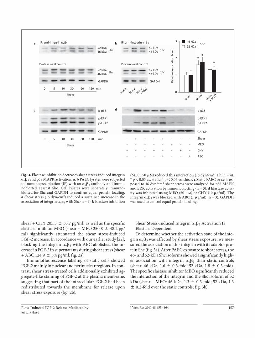
Shear Stress-Induced Integrin � v � 3 Activation Is Elastase Dependent To determine whether the activation state of the inte-
grin � v � 3 was affected by shear stress exposure, we mea-sured the association of this integrin with its adaptor pro-tein Shc ( fig. 3 a). After PAEC exposure to shear stress, the 46- and 52-kDa Shc isoforms showed a significantly high-er association with integrin � v � 3 than static controls (shear: 46 kDa, 1.6 8 0.3-fold; 52 kDa, 1.8 8 0.3-fold). The specific elastase inhibitor MEO significantly reduced the interaction of the integrin and the Shc isoform of 52 kDa (shear + MEO: 46 kDa, 1.3 8 0.3-fold; 52 kDa, 1.3 8 0.2-fold over the static controls; fig. 3 b).
+––+––––
–+––+–––
––+––+––
–––++++–
Shear
CHY
ABC
MEO
0
1
2
3
Shear
GAPDH
IP: anti-integrin �v�3IP: anti-integrin �v�3a
GAPDH
Protein level control
p-p38
p-ERK1
p-ERK2
p-ERK1
p-ERK2
p-p38
GAPDH
Shc
c d
**
Rela
tive
asso
ciat
ion
leve
l
Shear
0 5 10 30 60 120
0 5 10 30 60 120
Protein level control
GAPDH
min
min
b Shc52 kDa46 kDa
Shc52 kDa46 kDa
Shc52 kDa46 kDa
Shc52 kDa46 kDa
†
StaticShear
Shear
+ MEO
StaticShear
Shear
+ MEO
52 kDa
46 kDa
Fig. 3. Elastase inhibition decreases shear stress-induced integrin � v � 3 and p38 MAPK activation. a , b PAEC lysates were subjected to immunoprecipitation (IP) with an � v � 3 antibody and immu-noblotted against Shc. Cell lysates were separately immuno-blotted for Shc and GAPDH to confirm equal protein loading. a Shear stress (16 dyn/cm 2 ) induced a sustained increase in the association of integrin � v � 3 with Shc (n = 3). b Elastase inhibition
(MEO, 50 � M ) reduced this interaction (16 dyn/cm 2 , 1 h; n = 4). * p ! 0.05 vs. static; † p ! 0.05 vs. shear. c Static PAEC or cells ex-posed to 16 dyn/cm 2 shear stress were analyzed for p38 MAPK and ERK activation by immunoblotting (n = 3). d Elastase activ-ity was inhibited using MEO (50 � M ) or CHY (10 � g/ml). The integrin � v � 3 was blocked with ABC (1 � g/ml) (n = 3). GAPDH was used to control equal protein loading.
Hennig/Mogensen/Kirsch/Pohl/Gloe
J Vasc Res 2011;48:453–464 458
0
0.2
0.4
0.6
0.8
1.0
0
0.05
0.10
0.15
0.20
0.25
0
50
100
150
200
250
300
0
50
100
150
0
50
100
150
200
Control HEPELA ELA +HEP
a 5 0 2 4 6 8 1010 30 60
*
FGF-2
**
b
c
Abs
orpt
ion
Abs
orpt
ion
*
**
MEO (μM)
Elastase activity (U/ml)d
e
f
w/o
w/o 0.05 0.5 5 50
0.05 μM
0.5 μM
50 μM
Control
**
MEO
w/o0.05 μM
0.5 μM
5 μM
50 μM
MEO
min
†
† †
Elastase
Control Elastase
Control Elastase
FGF-
2 in
the
supe
rnat
ant (
pg/m
l)FG
F-2
in th
e su
pern
atan
t (pg
/ml)
FGF-
2 in
the
supe
rnat
ant
(% o
f con
trol
, w/o
)
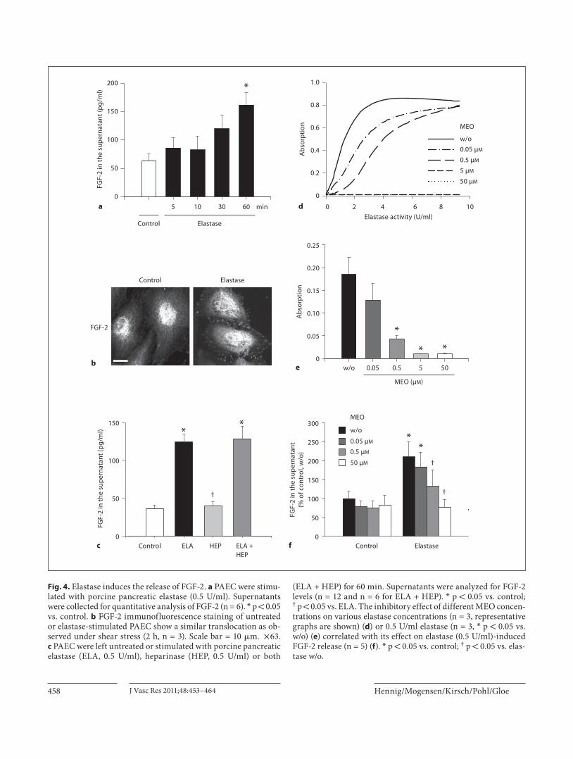
Fig. 4. Elastase induces the release of FGF-2. a PAEC were stimu-lated with porcine pancreatic elastase (0.5 U/ml). Supernatants were collected for quantitative analysis of FGF-2 (n = 6). * p ! 0.05 vs. control. b FGF-2 immunofluorescence staining of untreated or elastase-stimulated PAEC show a similar translocation as ob-served under shear stress (2 h, n = 3). Scale bar = 10 � m. ! 63. c PAEC were left untreated or stimulated with porcine pancreatic elastase (ELA, 0.5 U/ml), heparinase (HEP, 0.5 U/ml) or both
(ELA + HEP) for 60 min. Supernatants were analyzed for FGF-2 levels (n = 12 and n = 6 for ELA + HEP). * p ! 0.05 vs. control; † p ! 0.05 vs. ELA. The inhibitory effect of different MEO concen-trations on various elastase concentrations (n = 3, representative graphs are shown) ( d ) or 0.5 U/ml elastase (n = 3, * p ! 0.05 vs. w/o) ( e ) correlated with its effect on elastase (0.5 U/ml)-induced FGF-2 release (n = 5) ( f ). * p ! 0.05 vs. control; † p ! 0.05 vs. elas-tase w/o.
Flow-Induced FGF-2 Release Mediated by an Elastase
J Vasc Res 2011;48:453–464 459
Shear Stress-Induced p38 MAPK Activation Is Blocked by Elastase Inhibition Shear stress application to PAEC resulted in an en-
hanced phosphorylation of p38 MAPK and ERK1/2 ( fig. 3 c). The inhibition of elastase with both the specific inhibitor MEO as well as the unspecific inhibitor CHY prevented the shear stress-induced p38 MAPK phosphor-ylation but not ERK1/2 activation. The integrin � v � 3 blocker ABC also reduced the activation of p38 MAPK ( fig. 3 d). In contrast, the phosphorylation of ERK1/2 was not reduced after inhibition of elastase or the integrin � v � 3 in cells exposed to shear stress. Neither of the in-hibitors affected p38 MAPK and ERK1/2 phosphoryla-tion levels in static control cells.
Effects of External Elastase on Static Cells Elastase Treatment Induces an Increase in FGF-2 in the Supernatant Since the data obtained in this study suggest that an
endogenous, as yet not molecularly characterized elas-tase plays a causal role in the shear stress-induced in-crease in the extracellular concentration of FGF-2, we further investigated whether external elastase treat-ment could mimic this effect in non-shear-stressed cells. For this purpose, static PAEC were treated with 0.5 U/ml of porcine pancreatic elastase. Application of elastase for 5, 10, 30 or 60 min increased the concentra-tion of FGF-2 in the supernatant in a time-dependent manner, reaching a significant difference after 60 min [control 63.2 8 12.0 pg/ml, elastase (60 min) 161.7 8 21.4 pg/ml; fig. 4 a]. Immunofluorescence revealed a similar translocation of FGF-2 to the cell membrane as observed under shear stress ( fig. 4 b). As measured by flow cytometry, elastase did not induce FGF-2 release from endothelial cells kept in suspension: whereas there was a significant translocation of FGF-2 to the cell sur-face in cells attached to their matrix by 50% relative to control values (control 1.00 8 0.05 vs. elastase 1.50 8 0.15, n = 13, p = 0.005), the same treatment was without effect in cells kept in suspension (control 1.00 8 0.10 vs. elastase 1.00 8 0.07, n = 7).
In contrast to the treatment with elastase, treatment with heparinase I (0.5 U/ml) for 60 min did not alter the concentration of FGF-2 in the supernatant (control 35.8 8 5.3 pg/ml vs. heparinase 39.5 8 5.8 pg/ml). Hepari-nase I cleaves heparin and heparan sulfates on the cell membrane, known to be extracellular binding sites for FGF-2 [23] . Simultaneous application of elastase and hep-arinase did not further increase the FGF-2 concentration in the supernatant when compared to elastase treatment alone (elastase 124.3 8 10.6 pg/ml vs. elastase + hepari-nase 128.7 8 16.2 pg/ml; fig. 4 c).
MEO inhibited the activity of pancreatic elastase in a concentration-dependent manner. Total inhibition of elastase activity was achieved using 5 or 50 � M of the in-hibitor ( fig. 4 d, e). The inhibitory effect of different con-centrations of MEO on the elastase activity correlated well with its inhibitory effects on FGF-2 release ( fig. 4 f).
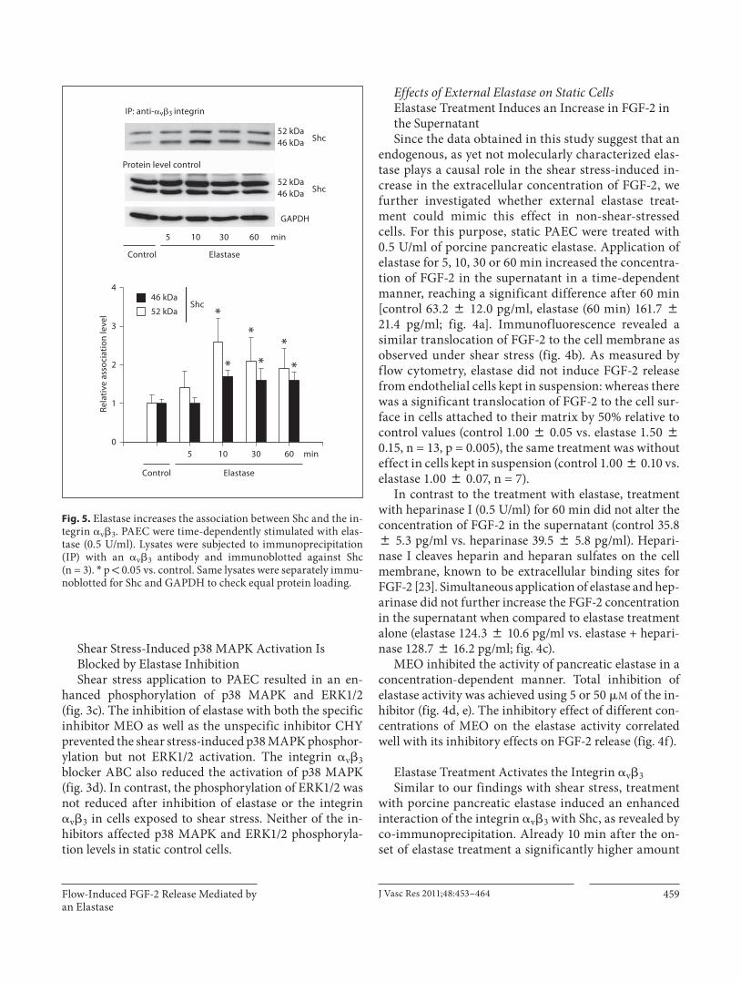
Elastase Treatment Activates the Integrin � v � 3 Similar to our findings with shear stress, treatment
with porcine pancreatic elastase induced an enhanced interaction of the integrin � v � 3 with Shc, as revealed by co-immunoprecipitation. Already 10 min after the on-set of elastase treatment a significantly higher amount
0
1
2
3
4
* **
*
**
Rela
tive
asso
ciat
ion
leve
l
GAPDH
Protein level control
Shc52 kDa46 kDa
Shc52 kDa46 kDa
Shc
Control Elastase
Control Elastase
5 10 30 60 min
5 10 30 60 min
52 kDa
46 kDa
IP: anti-�v�3 integrin
Fig. 5. Elastase increases the association between Shc and the in-tegrin � v � 3 . PAEC were time-dependently stimulated with elas-tase (0.5 U/ml). Lysates were subjected to immunoprecipitation (IP) with an � v � 3 antibody and immunoblotted against Shc(n = 3). * p ! 0.05 vs. control. Same lysates were separately immu-noblotted for Shc and GAPDH to check equal protein loading.

Hennig/Mogensen/Kirsch/Pohl/Gloe
J Vasc Res 2011;48:453–464 460
of the Shc isoforms was associated with the integrin [elastase (10 min): 46 kDa, 2.6 8 0.6-fold; 52 kDa, 1.7 8 0.2-fold over controls]. This interaction was main-tained over 60 min of elastase treatment [elastase (60 min): 46 kDa, 1.9 8 0.5-fold; 52 kDa, 1.6 8 0.2-fold over controls; fig. 5 ].
Pharmacological Inhibition of p38 MAPK Blocks Elastase-Induced FGF-2 Release To analyze whether elastase treatment activated MAP
kinases in a similar manner as shear stress, p38 MAPK and ERK activation was analyzed. Application of porcine pancreatic elastase led to an increased phosphorylation of both p38 MAPK and ERK in a time-dependent manner ( fig. 6 a).
To study the impact of MAP kinase activation on the elastase-induced FGF-2 release, cells were treated with the specific inhibitors SB202190 (for p38 MAPK) or U0126 (for MEK1/2). The effective inhibition was moni-tored by Western blot analysis. The effect of SB202190 was examined by analyzing the phosphorylation of Hsp27, a downstream target of p38. Whereas p38 MAPK inhibition significantly reduced the FGF-2 increase in the supernatant (to 57 8 15%), ERK inhibition had no influ-ence on the elastase-induced release of this growth factor ( fig. 6 b).
Discussion
The main finding of this study is that shear stress in-duces integrin activation and FGF-2 release partly via a novel protease-dependent pathway. Due to its design, which focused on the comparability with our own earlier study [22] and others [24–26] , the experiments cannot answer the question whether this protease activation re-quired a minimum level of shear stress or whether the protease release would have gradually changed with dif-ferent levels of shear stress.
However, our results clearly show that exposure of resting endothelial cells to arterial levels of laminar shear stress results in an enhanced enzymatic activity in their supernatant that can be characterized as an elastase. An increased concentration of this elastase was already mea-surable after 5 min of shear stress application, indicating that the endothelium is able to respond very quickly to increased levels of flow. The enzyme is, however, different from pancreatic and neutrophil elastases and other pro-teases with elastolytic activity. This elastase mediates the shear stress-induced activation of the integrin � v � 3 and subsequently of the p38 MAPK pathway, leading to the release of FGF-2. Furthermore, we were able to demon-strate that treatment of PAEC with exogenously applied porcine pancreatic elastase induced virtually the same ef-
5 10 30 60 min0
50
100
150
SB SB
Elastase
*
ba
p-p38
p38
p-p38
p-Hsp27
p-ERK-1
p-ERK-2
GAPDH
U0U0
ElastaseControl Elastase
FGF-
2 in
the
supe
rnat
ant
(% o
f ela
stas
e)
p-ERK1
p-ERK2
GAPDH
Fig. 6. Elastase causes activation of p38 MAPK and ERK. P38 MAPK inhibition decreases elastase-induced FGF-2 release. a Treatment with porcine pancreatic elastase activates p38 MAPK (n = 6) and ERK (n = 6). Equal protein loading was confirmed with total p38 MAPK and GAPDH. b PAEC were treated with the p38 MAPK inhibitor SB202190 (SB) or the MEK1/2 inhibitor U0126 (U0) (10 � M each). Cell supernatants were analyzed for FGF-2 re-
lease using an ELISA (n = 12, ANOVA RM on ranks, Student-Newman-Keuls) * p ! 0.05 vs. elastase. Immunoblotting of cell lysates was performed to confirm inhibitory effects of the phar-macological substances used. Since SB202190 does not block p38 MAPK phosphorylation directly, the activation of the down-stream target protein Hsp27 was studied instead. Equal protein loading was controlled with GAPDH.

Flow-Induced FGF-2 Release Mediated by an Elastase
J Vasc Res 2011;48:453–464 461
fects as observed under shear stress. The present study extends previous work from our laboratory demonstrat-ing that shear stress induces the release of FGF-2 from endothelial cells [22] . This process was found to be spe-cifically dependent on the integrin � v � 3 but the connec-tion between shear stress and integrin activation re-mained unclear.
The endothelial FGF-2 release was reduced by the elas-tase inhibitor MEO which is specific for enzymes of the elastase family [27] . Moreover, the substrate MeOSuc-Ala-Ala-Pro-Val-pNA, also considered to be specific for neutrophil and pancreatic elastase, was cleaved by this enzyme [28, 29] . The presence of enzymes which have an elastolytic activity in intact vessels has been reported pre-viously: Hornebeck et al. [30] isolated an elastase from pig aortas that exhibited an elastolytic activity similar to pancreatic elastase. It is unlikely, however, that the en-zyme is identical with neutrophil and pancreatic elastase,
since we found no transcripts of these elastases in our cells. Of note, the specific substrate is not cleaved by oth-er proteases, like cathepsins or MMPs, which previously also have been found to be activated, released or inhibited after shear stress exposure [15, 16] . Thus, the involvement of proteases from the cathepsin and MMP family can be excluded in the present setting. Although some of these proteases also cleave elastin and can therefore be termed elastolytic enzymes, the activity of the endothelial en-zyme which we detected was comparable solely to neutro-phil and pancreatic elastase because they, in contrast to the other elastolytic enzymes, degrade the same specific substrate. Therefore, we propose that this endothelial ser-ine protease with true elastase activity is a not yet de-scribed mediator of endothelial shear stress responses. The major evidence for this conclusion is the following: (1) susceptibility to specific elastase inhibitors, (2) cleav-age of the specific elastase substrate and (3) appropriate molecular weight.
The characterization of the enzyme as an elastase based on the specific substrate does not mean that elastin is the only matrix protein targeted by the enzyme. Neu-trophil elastase, for example, has a broad spectrum of ma-trix substrates such as collagens, fibronectin, laminin and proteoglycans, and also cleaves some cytokines [31, 32] . Further studies are required to identify the matrix effects of the endothelial elastase in more detail and also to clarify whether its activity in the intact vessel wall is further controlled by serine protease inhibitors. There-fore, the mechanism of action of the elastase needs fur-ther characterization. Presently, it is unknown whether the elastase is acutely released in its active form from the endothelium in response to shear stress or whether it is activated by another enzyme or signal. Since shear stress still increased elastase activity when integrin � v � 3 was blocked, we can exclude that the elastase release is prefer-entially due to a direct activation of the integrin � v � 3 by shear forces.
As we were currently not able to identify the exact nature of the endothelial elastase which also precluded silencing of the respective gene, we sought to support our conclusions by comparing the effects of shear stress with those in response to exogenously applied pancre-atic elastase. Although this enzyme most likely does not possess an identical substrate spectrum, the consistency of the findings was remarkable. Both treatments in-duced activation of the integrin � v � 3 and p38 MAPK and, as a consequence, increased FGF-2 levels in the su-pernatant. A similar pancreatic elastase-mediated re-lease of FGF-2 has previously been described in cultures
p38
FGF-2
Shearstress
Endothelial cells
ECM
ERK
Elastase
Integrin
Elastase
SHC
�v �3 �3�v
Fig. 7. Proposed model for shear stress-induced FGF-2 release from endothelial cells. Shear stress induces the release of an en-dothelial elastase, activating integrin � v � 3 outside-in signaling and thereby intracellular signaling cascades. FGF-2 is released via p38 MAPK.

Hennig/Mogensen/Kirsch/Pohl/Gloe
J Vasc Res 2011;48:453–464 462
of pulmonary fibroblasts. Based on the correlation of FGF-2 release with the liberation of sulfated glycosami-noglycans, known to be extracellular binding sites for FGF-2, the authors concluded that FGF-2 was released by proteolytic digestion from proteoglycan storage sites in the ECM [33, 34] . In another study, cultured pulmo-nary smooth muscle cells treated with neutrophil elas-tase also released FGF-2 from ECM storage sites [35] . In contrast, our previous studies have shown that the shear stress-induced increase in extracellular FGF-2 corre-sponds with a reduction in intracellular FGF-2 [22] . Moreover, we demonstrated that the increase in FGF-2 is critically dependent on activation of the integrin � v � 3 and requires the activation of p38 MAPK. The need of these steps can hardly be explained by the ‘mere’ enzy-matic liberation of matrix-bound FGF-2 in our experi-ments. At present it is difficult to reconcile these appar-ently contradictory findings. It is possible, however, that the amount of bound FGF-2 is much higher in the ECM and on the surface of pulmonary fibroblasts and smooth muscle cells than in endothelial cells, especially since the fibroblasts were cultivated for 10–12 days [33] . In this case, an additional release from the cells may be less sig-nificant as observed in the present study. In our experi-ments, the cells were seeded on pure matrices or even plastic so that the only possible source for FGF-2 could have been the endothelial cells. Moreover, no extracel-lular FGF-2 was detected outside the cells by immuno-histochemistry after 1 day of culture. Furthermore, since heparinase I, known to release extracellularly bound FGF-2 by inhibiting FGF-2 binding to heparan sulfate proteoglycans [36] , failed to increase the FGF-2 concentration in the supernatant in our study, it is likely that matrix binding in our cell cultures was relatively low.
The current study demonstrates that shear stress in-duces the activation of the integrin � v � 3 as assessed by its enhanced interaction with the adaptor protein Shc. This was reduced when the cells were pre-incubated with the specific elastase inhibitor, indicating that elas-tase is a critical mediator of the activation. Of note, the exogenously applied pancreatic elastase induced a simi-lar interaction of integrin � v � 3 and Shc. Jalali et al. [25] have previously demonstrated that shear stress increases the association of the integrin � v � 3 and Shc in endothe-lial cells. Likewise, Tzima et al. [26] could show that shear stress activates the integrin � v � 3 in endothelial cells using an antibody that selectively detects the acti-vated integrin � v � 3 . Our studies now provide a mecha-nism of shear stress-induced integrin activation that is
independent of a direct mechanical activation by shear forces.
Although it has been shown that platelet integrins may be activated by direct cleavage by elastase [37] , this is un-likely to have occurred in our study. Firstly, we did not find any change in the size of the integrin � v -subunit af-ter exposure to pancreatic elastase and, secondly, elastase did not induce FGF-2 release from endothelial cells kept in suspension without any contact with the matrix. This is in agreement with a previous report stating that endo-thelial cells must be anchored to the matrix in order to record and quantify cellular responses to mechanical strain [38] . Thus, we suggest an elastase-induced remod-eling of the ECM architecture as the activating mecha-nism. Elastase-induced changes in the structure and ri-gidity of the matrix is likely to enable new (or re-) cluster-ing of integrins into focal adhesion points, a model which has been proposed previously [39] . Alternatively, proteo-lytic activities may expose hidden protein sequences or even release matrix fragments that have integrin-activat-ing properties [40, 41] . Previous investigations indicate that proteases are able to generate signals by remodeling the ECM [42–44] .
Our results further demonstrate that p38 MAPK and ERK were activated by shear stress exposure. Elastase in-hibition reduced the shear stress-induced p38 MAPK ac-tivation. Likewise, we were able to show that elastase treatment also induced activation of p38 MAPK and ERK. Moreover, we demonstrated that p38 MAPK but not ERK inhibition reduced the elastase-induced FGF-2 release. Therefore, p38 MAPK seems to play a role in shear stress-induced FGF-2 release.
Since several groups have demonstrated that inhibi-tion of the integrin � v � 3 blocked the activation of p38 MAPK [45, 46] , it is reasonable to argue that flow-in-duced MAPK phosphorylation is mediated via this inte-grin. This is in agreement with results reported by Chen et al. [47] showing that direct inhibition of � v integrins or downregulation of their expression decreased the ac-tive level of p38 MAPK. The precise signaling pathway connecting the integrin and MAP kinase activation is still elusive in our setting. However, since shear stress and elastase stimulation induced the association of the integrin with its adaptor protein Shc, downstream sig-naling may be transduced via Shc and focal adhesion signaling.
In conclusion, the present results support a new mod-el for the translation of mechanical stress into biochemi-cal signals via a novel endothelial elastase ( fig. 7 ). Since FGF-2 is an important growth factor in vascular develop-

Flow-Induced FGF-2 Release Mediated by an Elastase
J Vasc Res 2011;48:453–464 463
ment and maintenance, this model could have profound implications for our understanding of mechanically in-duced vascular remodeling processes. The precise mech-anisms by which elastase activates the integrin � v � 3 and downstream signaling will be the focus of further inves-tigations.
Acknowledgements
This work was supported by the Deutsche Forschungsgemein-schaft (GRK438 ‘Vascular biology in medicine’), the Friedrich-Baur-Stiftung, the EU project Exgenesis (LSHM-CT-2004-005272) and the EU Network for initial training – ITN SmArt (PITN-GA-2009-235711). We would like to thank Dorothea Gössel and Katarzyna Stefanowski for their excellent technical assistance.
References
1 Busse R, Fleming I: Regulation of endotheli-um-derived vasoactive autacoid production by hemodynamic forces. Trends Pharmacol Sci 2003; 24: 24–29.
2 Pohl U, Holtz J, Busse R, Bassenge E: Crucial role of endothelium in the vasodilator re-sponse to increased flow in vivo. Hyperten-sion 1986; 8: 37–44.
3 Uematsu M, Ohara Y, Navas JP, Nishida K, Murphy TJ, Alexander RW, Nerem RM, Har-rison DG: Regulation of endothelial cell ni-tric oxide synthase mrna expression by shear stress. Am J Physiol 1995; 269:C1371–C1378.
4 Passerini AG, Milsted A, Rittgers SE: Shear stress magnitude and directionality modu-late growth factor gene expression in precon-ditioned vascular endothelial cells. J Vasc Surg 2003; 37: 182–190.
5 Lehoux S, Tedgui A: Cellular mechanics and gene expression in blood vessels. J Biomech 2003; 36: 631–643.
6 Langille BL: Blood flow-induced remodeling of arteries in health and disease. Cardiovasc Pathol 1992; 1: 245–251.
7 De Mey JGR, Schiffers PM, Hilgers RHP, Sanders MMW: Toward functional genom-ics of f low-induced outward remodeling of resistance arteries. Am J Physiol Heart Circ Physiol 2005; 288:H1022–H1027.
8 Hudlicka O, Brown MD: Adaptation of skel-etal muscle microvasculature to increased or decreased blood flow: role of shear stress, ni-tric oxide and vascular endothelial growth factor. J Vasc Res 2009; 46: 504–512.
9 Wang JH, Thampatty BP: An introductory review of cell mechanobiology. Biomech Model Mechanobiol 2006; 5: 1–16.
10 Tarbell JM, Pahakis MY: Mechanotransduc-tion and the glycocalyx. J Intern Med 2006; 259: 339–350.
11 Traub O, Berk BC: Laminar shear stress: mechanisms by which endothelial cells transduce an atheroprotective force. Arte-rioscler Thromb Vasc Biol 1998; 18: 677–685.
12 Alenghat FJ, Ingber DE: Mechanotransduc-tion: all signals point to cytoskeleton, ma-trix, and integrins. Sci STKE 2002; 119:pe6.
13 Tzima E, Irani-Tehrani M, Kiosses WB, De-jana E, Schultz DA, Engelhardt B, Cao G, De-Lisser H, Schwartz MA: A mechanosensory complex that mediates the endothelial cell response to fluid shear stress. Nature 2005; 437: 426–431.
14 Platt MO, Ankeny RF, Jo H: Laminar shear stress inhibits cathepsin l activity in endo-thelial cells. Arterioscler Thromb Vasc Biol 2006; 26: 1784–1790.
15 Platt MO, Xing Y, Jo H, Yoganathan AP: Cy-clic pressure and shear stress regulate matrix metalloproteinases and cathepsin activity in porcine aortic valves. J Heart Valve Dis 2006; 15: 622–629.
16 Platt MO, Ankeny RF, Shi GP, Weiss D, Vega JD, Taylor WR, Jo H: Expression of cathepsin K is regulated by shear stress in cultured en-dothelial cells and is increased in endotheli-um in human atherosclerosis. Am J Physiol Heart Circ Physiol 2007; 292:H1479–H1486.
17 Magid R, Murphy TJ, Galis ZS: Expression of matrix metalloproteinase-9 in endothelial cells is differentially regulated by shear stress. Role of c-myc. J Biol Chem 2003; 278: 32994–32999.
18 Chiquet M: Regulation of extracellular ma-trix gene expression by mechanical stress. Matrix Biol 1999; 18: 417–426.
19 Gupta V, Grande-Allen KJ: Effects of static and cyclic loading in regulating extracellular matrix synthesis by cardiovascular cells. Cardiovasc Res 2006; 72: 375–383.
20 Orr AW, Sanders JM, Bevard M, Coleman E, Sarembock IJ, Schwartz MA: The subendo-thelial extracellular matrix modulates NF- � B activation by flow: a potential role in ath-erosclerosis. J Cell Biol 2005; 169: 191–202.
21 Gloe T, Riedmayr S, Sohn HY, Pohl U: The 67-kDa laminin-binding protein is involved in shear stress-dependent endothelial nitric-oxide synthase expression. J Biol Chem 1999; 274: 15996–16002.
22 Gloe T, Sohn HY, Meininger GA, Pohl U: Shear stress-induced release of basic fibro-blast growth factor from endothelial cells is mediated by matrix interaction via integrin � (v) � 3. J Biol Chem 2002; 277: 23453–23458.
23 Linhardt RJ, Turnbull JE, Wang HM, Loga-nathan D, Gallagher JT: Examination of the substrate specificity of heparin and heparan sulfate lyases. Biochemistry 1990; 29: 2611–2617.
24 Malek AM, Gibbons GH, Dzau VJ, Izumo S: Fluid shear stress differentially modulates expression of genes encoding basic fibroblast growth factor and platelet-derived growth factor B chain in vascular endothelium. J Clin Invest 1993; 92: 2013–2021.
25 Jalali S, del Pozo MA, Chen K, Miao H, Li Y, Schwartz MA, Shyy JY, Chien S: Integrin-mediated mechanotransduction requires its dynamic interaction with specific extracel-lular matrix (ECM) ligands. Proc Natl Acad Sci USA 2001; 98: 1042–1046.
26 Tzima E, del Pozo MA, Shattil SJ, Chien S, Schwartz MA: Activation of integrins in en-dothelial cells by fluid shear stress mediates rho-dependent cytoskeletal alignment. EMBO J 2001; 20: 4639–4647.
27 Powers JC, Gupton BF, Harley AD, Nishino N, Whitley RJ: Specificity of porcine pancre-atic elastase, human leukocyte elastase and cathepsin G. Inhibition with peptide chloro-methyl ketones. Biochim Biophys Acta 1977; 485: 156–166.
28 Castillo MJ, Nakajima K, Zimmerman M, Powers JC: Sensitive substrates for human leukocyte and porcine pancreatic elastase: A study of the merits of various chromophoric and fluorogenic leaving groups in assays for serine proteases. Anal Biochem 1979; 99: 53–64.
29 Nakajima K, Powers JC, Ashe BM, Zimmer-man M: Mapping the extended substrate binding site of cathepsin g and human leuko-cyte elastase. Studies with peptide substrates related to the alpha 1-protease inhibitor reac-tive site. J Biol Chem 1979; 254: 4027–4032.
30 Hornebeck W, Derouette JC, Robert L: Isola-tion, purification and properties of aortic elastase. FEBS Lett 1975; 58: 66–70.
31 Vaday GG, Lider O: Extracellular matrix moieties, cytokines, and enzymes: dynamic effects on immune cell behavior and inflam-mation. J Leukoc Biol 2000; 67: 149–159.
32 Bank U, Ansorge S: More than destructive: neutrophil-derived serine proteases in cyto-kine bioactivity control. J Leukoc Biol 2001; 69: 197–206.
33 Buczek-Thomas JA, Nugent MA: Elastase-mediated release of heparan sulfate proteo-glycans from pulmonary fibroblast cultures. A mechanism for basic fibroblast growth factor (bFGF) release and attenuation of bfgf binding following elastase-induced injury. J Biol Chem 1999; 274: 25167–25172.
34 Buczek-Thomas JA, Lucey EC, Stone PJ, Chu CL, Rich CB, Carreras I, Goldstein RH, Fos-ter JA, Nugent MA: Elastase mediates the re-lease of growth factors from lung in vivo. Am J Respir Cell Mol Biol 2004; 31: 344–350.

Hennig/Mogensen/Kirsch/Pohl/Gloe
J Vasc Res 2011;48:453–464 464
35 Thompson K, Rabinovitch M: Exogenous leukocyte and endogenous elastases can me-diate mitogenic activity in pulmonary artery smooth muscle cells by release of extracellu-lar-matrix bound basic fibroblast growth factor. J Cell Physiol 1996; 166: 495–505.
36 Bashkin P, Doctrow S, Klagsbrun M, Svahn CM, Folkman J, Vlodavsky I: Basic fibroblast growth-factor binds to subendothelial extra-cellular-matrix and is released by heparitin-ase and heparin-like molecules. Biochemis-try 1989; 28: 1737–1743.
37 Si-Tahar M, Pidard D, Balloy V, Moniatte M, Kieffer N, Van Dorsselaer A, Chignard M: Human neutrophil elastase proteolytical - ly activates the platelet integrin � IIb � 3 through cleavage of the carboxyl terminus of the � IIb subunit heavy chain. Involvement in the potentiation of platelet aggregation. J Biol Chem 1997; 272: 11636–11647.
38 Takahashi M, Berk BC: Mitogen-activated protein kinase (erk1/2) activation by shear stress and adhesion in endothelial cells. Es-sential role for a herbimycin-sensitive ki-nase. J Clin Invest 1996; 98: 2623–2631.
39 Giancotti FG, Ruoslahti E: Integrin signal-ing. Science 1999; 285: 1028–1032.
40 Davis GE: Matricryptic sites control tissue injury responses in the cardiovascular sys-tem: relationships to pattern recognition re-ceptor regulated events. J Mol Cell Cardiol 2010; 48: 454–460.
41 Davis GE, Bayless KJ, Davis MJ, Meininger GA: Regulation of tissue injury responses by the exposure of matricryptic sites within ex-tracellular matrix molecules. Am J Pathol 2000; 156: 1489–1498.
42 Faisal Khan KM, Laurie GW, McCaffrey TA, Falcone DJ: Exposure of cryptic domains in the � 1-chain of laminin-1 by elastase stimu-lates macrophages urokinase and matrix me-talloproteinase-9 expression. J Biol Chem 2002; 277: 13778–13786.
43 Koshikawa N, Giannelli G, Cirulli V, Mi-yazaki K, Quaranta V: Role of cell surface metalloprotease MT1-MMP in epithelial cell migration over laminin-5. J Cell Biol 2000; 148: 615–624.
44 Mydel P, Shipley JM, Adair-Kirk TL, Kelley DG, Broekelmann TJ, Mecham RP, Senior RM: Neutrophil elastase cleaves laminin-332 (laminin-5) generating peptides that are che-motactic for neutrophils. J Biol Chem 2008; 283: 9513–9522.
45 Lee DY, Yeh CR, Chang SF, Lee PL, Chien S, Cheng CK, Chiu JJ: Integrin-mediated ex-pression of bone formation-related genesin osteoblast-like cells in response to fluid shear stress: roles of extracellular matrix, Shc, and mitogen-activated protein kinase. J Bone Miner Res 2008; 23: 1140–1149.
46 Masson-Gadais B, Houle F, Laferriere J, Huot J: Integrin � v � 3, requirement forVEGFR2-mediated activation of SAPK2/p38 and for HSP90-dependent phosphoryla-tion of focal adhesion kinase in endothelial cells activated by VEGF. Cell Stress Chaper-ones 2003; 8: 37–52.
47 Chen J, Baskerville C, Han Q, Pan ZK, Huang S: � (v) integrin, p38 mitogen-activat-ed protein kinase, and urokinase plasmino-gen activator are functionally linked in inva-sive breast cancer cells. J Biol Chem 2001; 276: 47901–47905.


![A vaccine targeting angiomotin induces an antibody response which alters tumor … · 2020. 1. 18. · by normal, but overexpressed by endothelial cells of tumor vessels [8] also](https://img.dokumen.tips/doc/110x75/60ff2f5ea75c100b011c893b/a-vaccine-targeting-angiomotin-induces-an-antibody-response-which-alters-tumor-2020.jpg)
![Shear stress-induced pathological changes in endothelial ... · 1/7/2020 · Fluid shear stress induced [Ca2 +]i overload is force and time dependent . Shear stress is a physiological](https://img.dokumen.tips/doc/110x75/606ea5e1c71f9c48290448a9/shear-stress-induced-pathological-changes-in-endothelial-172020-fluid-shear.jpg)