Embed Size (px)
Citation preview

Shear Stress-Mediated F1/FOATP Synthase-Dependent CO2Gas Excretion From HumanPulmonary ArteriolarEndothelial CellsYOSHIKO KAWAI,1 KAZUO YOSHIDA,2 MAKI KAIDOH,1 YUMIKO YOKOYAMA,1 AND
TOSHIO OHHASHI1*1Department of Physiology, School of Medicine, Shinshu University, Matsumoto, Japan2Division of Thoracic Surgery, Facilitated Hospital, School of Medicine, Shinshu University, Matsumoto, Japan
Westudied the physiological role of flow through pulmonary arterioles in CO2 gas exchange.We established human pulmonary arteriolarendothelial cells (HPAoEC). The cells demonstratedmarked immunocytochemical staining of PECAM-1, VEGFR2, ACE-1, andCA type IVon their cell surface. Ten seconds shear stress stimulation caused the co-release ofHþ andATP via the activation of F1/FOATP synthase onthe HPAoEC. F1/FO ATP synthase was immunocytochemically observed on the cell surface of non-permeabilized HPAoEC. In the shearstress-loaded HPAoEC culture media supernatant, ATPase activity increased in a time-dependent manner. The HPAoEC were stronglystained forNTPDase 1, which partially co-localizedwith purinergic P2Y1. The purinergic P2Y1 receptor agonistUTP (10�6M) significantlypotentiated the shear stress-induced increase in ATPase activity in the culture medium supernatant. Ten seconds shear stress stimulationalso produced stress strength-dependent CO2 gas excretion from theHPAoEC, which was significantly reduced by the inhibition of F1/FOATP synthase orCA IVon the endothelial cell (EC) surface. In conclusion, we have proposed a new concept ofCO2 exchange in the humanlung, flow-mediated F1/FOATP synthase-dependentHþ secretion, resulting in the facilitation of a dehydration reaction involvingHCO�
3 inplasma and the excretion of CO2 gas from arteriolar ECs.J. Cell. Physiol. 227: 2059–2068, 2012. � 2011 Wiley Periodicals, Inc.
Vascular endothelial cells (EC) are constantly exposed to shearstress, the mechanical force generated by the flow of blood orlymph. These EC recognize shear stress and transmit a signal totheir interior, where it triggers cell responses including changesin a variety of cell functions (Davies, 1995; Chien, 2007;Yamamoto et al., 2007; Kawai et al., 2010). For example, inresponse to shear stress, vascular EC release endogenous ATP,which is produced by the activation of cell surface F1/FO ATPsynthase (Yamamoto et al., 2007). Yamamoto et al. (2007) alsodemonstrated that shear stress-induced cell surface F1/FO ATPsynthase-mediated ATP release was observed mostprominently in pulmonary arterial EC. However, how ATPrelease from pulmonary arteriolar EC, which might be exposedto higher shear stress levels than EC in the pulmonary artery, isaffected by shear stress is unclear because no EC from humanpulmonary arterioles are commercially available. In addition,weare only beginning to understand the functions of cell surfaceATP synthase. Thus, it might act through the P2X/2Ypurinoceptors, thereby activating Ca2þ-dependent signalingcascades that modulate cell functions. However, no studieshave evaluated the physiological function and relevance of theHþ co-released from ECs through the activation of cell surfaceF1/FO ATP synthase.
The physiological concept of carbon dioxide (CO2) gasexchange in the lungs is well established (Klocke, 1987).According to this concept, CO2 gas elimination from the humanlungs depends on several events. The first involves acombination of the physiological processes required for CO2
gas diffusion, the catalysis of carbonic acid dehydration, andtransmembrane HCO�
3 -Cl� exchange in red blood cells. The
second event is the oxygenation of hemoglobin and thesimultaneous liberation of protons, which are then able to reactwith HCO�
3 , allowing the excretion of CO2 gas from the cells.However, several serious questions regarding the concept still
remain unanswered. For example, the physiological conversionof HCO�
3 to CO2 was observed in isolated rat lungs that wereperfused with a red blood cell-free Krebs-Ringer bicarbonatesolution (Crandall and O’Brasky, 1978). Moreover, in patientswith anemia, little or no symptoms of reduced CO2 transport-mediated acidosis have been confirmed (Glader, 2003). Inaddition, no study has evaluated the relationship between CO2
gas exchange in human lungs and shear stress-induced cellsurface F1/FO ATP synthase-mediated Hþ secretion.
Therefore, we attempted to (i) establish a human pulmonaryarteriolar endothelial cell strain, (ii) investigate the functionalproperties of shear stress-induced cell surface F1/FO ATPsynthase-mediated Hþ and ATP co-release, and (iii) evaluatethe physiological roles of the ATP released by the shear stress-induced activation of cell surface F1/FO ATP synthase inHPAoEC. To clarify the physiological relevance of Hþ releasedvia shear stress stimulation to CO2 gas exchange in human
Conception and design of the experiments: YK and TO; Collection,analysis, and interpretation of data: YK, KY, MK, YY, and TO;Drafting the article and critically revising it for importantintellectual content: YK and TO.
Contract grant sponsor: Japanese Ministry of Education, Science,Sports, and Culture;Contract grant numbers: 19209044, 22249052.
*Correspondence to: Toshio Ohhashi, Head and Professor,Department of Physiology, School of Medicine, Shinshu University,Asahi 3-1-1, Matsumoto, Japan. E-mail: [email protected]
Received 26 April 2011; Accepted 6 July 2011
Published online in Wiley Online Library(wileyonlinelibrary.com), 18 July 2011.DOI: 10.1002/jcp.22937
ORIGINAL RESEARCH ARTICLE 2059J o u r n a l o fJ o u r n a l o f
CellularPhysiologyCellularPhysiology
� 2 0 1 1 W I L E Y P E R I O D I C A L S , I N C .

pulmonary arterioles, we also studied (iv) the biologicalproperties of shear stress-induced CO2 gas excretion fromHPAoEC.
MethodsCell culture
The experiments with HPAoEC were approved by the ethicalcommittee for human studies of the School of Medicine, ShinshuUniversity. All subjects were informed of the risks and purpose ofthe study before their written consent was obtained. Weintraoperatively identified and isolated tiny segments of human lungtissue from patients with lung cancer. Specimens from the normalregion of the tissue segment being resected were used for theisolation of functional arterioles and the HPAoEC culture. Under adissecting microscope, functional arterioles with outer diametersranging from 300 to 100mm were dissected. The dissectedarterioles were then cannulated downstream with a sterilepolyethylene tube and perfused with PBS to remove anyintraluminal blood. Next, they were intraluminally perfused withtrypsin (trypsin/ethylenediaminetetraacetic acid solution for ECculture, 500U/ml; SIGMA Chemical Co., St. Louis, MO) at 378C.After the enzymatic digestion, the intraluminal fluid containing theECswas gently drained into a centrifuge tube containing endothelialgrowth medium (EGM)-2 (Clonetics, San Diego, CA) and 10% fetalbovine serum (FBS). The collected solution was then centrifuged at2,000 rpm for 5min at 48C, the supernatant was removed, and thepellet was resuspended in a 35mm culture plate (Corning Inc., NY)coated with type I collagen (Nitta Gelatin, Osaka, Japan).
To compare the biological properties and responses to shearstress of the cultured cells with those of ECs from the human trunkpulmonary artery (HPAEC), HPAEC were purchased from SankoJunyaku (Tokyo, Japan). In addition, to evaluate the antibodyspecific controls used for the immunocytochemical analyses,human fibroblasts were also purchased from Sanko Junyaku. TheHPAoEC and HPAEC were maintained in EGM-2 with 10% FBS.We previously examined the biological and immunocytochemicalproperties of cultured HPAoEC and HPAEC ranging from thefourth to sixth passage because the HPAoEC had not beenimmortalized. As a result, we confirmed that no significantdifferences existed in these properties of theHPAoECandHPAEC.Therefore, we used cultured HPAoEC and HPAEC at the fourth tosixth passage for the present experiments. The human fibroblastswere cultured in fibroblast growth medium-2 supplemented with10% FBS. The HPAoEC, HPAEC, and human fibroblasts wereincubated under atmospheric conditions of 21% O2, 5% CO2, and74% N2 at 378C.
Flow loading experiments
A parallel plate-type apparatus was used to apply laminar shearstress to the human EC using Yamamoto’s method (Yamamoto etal., 2007) with slight modifications. Briefly, one side of the flowchamber consisted of a 1% gelatin-coated glass plate, on which thecultured human EC rested, and the other side consisted of apolycarbonate plate. These two surfaces were held 200mm apartwith a Teflon gasket. The chamber contained an entrance, whichwas connected to an upper reservoir by a silicon tube, and an exitfor the fluid, which led to a lower reservoir. The flow of the fluid(378C; EBM-2 supplemented with 3% FBS) was driven by aperistaltic pump and a flow rate controller. The intensity of theshear stress (t, in dyn/cm2) acting on the human EC was calculatedusing the formula t¼ 6mQ/a2b, where m is the viscosity of theperfusate (poise), Q is the flow volume (in ml/s), and a and b are thecross-sectional dimensions of the flow path (in cm).
ATP assay
The ATP concentrations of the human EC culture mediasupernatants were determined using a luciferin–luciferase assay
based on the Cell Titer-Glo Luminescent Cell Viability Assay(Promega, Madison, WI), as described previously (Kawai et al.,2008). Briefly, to develop a calibration curve for ATPmeasurement, we first performed the same luciferin–luciferaseassay using culture media containing different concentrations ofATP. Next, 100ml of the flow-stimulated human EC supernatantwere collected in a 96-well plate, into which 100ml of luciferin–luciferase solution had been added, and light emission wasrecorded using a luminometer (Dainippon Sumitomo Pharma,Osaka, Japan). Thus, we calculated the ATP concentrations of theflow-stimulated human EC culture media supernatants using thecalibration curve.
ATPase-activity assay
To evaluate the enzymatic activity involved in the hydrolysis of ATPin the HPAoEC and HPAEC supernatants, we developed a newmethod for measuring ATPase activity, and used the supernatantsand the same luciferin–luciferase assay for ATP measurement. Sixhundred microliters of the flow-loaded or -unloaded human ECsupernatant were collected in a polyethylene tube, into which600ml of endothelial basic medium (EBM)-2 culture mediumwithout FBS containing a constant concentration (10�7M) ofexogenous ATP had been added, and themixture was kept at 378Cthroughout the experiment. At 1, 20, 40, and 60min of incubation,100ml of the mixture solution containing no cultured human ECwere collected in a 96-well plate, into which 100ml of luciferin–luciferase solution had been added, and light emission wasrecorded using a luminometer for ATP measurement. Thedecrease in the ratio of the total ATP concentration to theconcentration of exogenous (added) ATP enabled us to evaluatethe level of ATP hydrolysis in themixture solution. The viability andreproducibility of our method were investigated using apyrase, aselective enzyme that hydrolyzes ATP (Marcus et al., 2003). Thefindings (n¼ 6) are summarized in Figure 1. The effects of apyraseon the ATP concentration of the mixture solution wereinvestigated using a specific solution: 600ml of EBM-2 culturemediumwithout FBS containing 10�7M exogenousATP and 600mlof EBM-2 culture medium without FBS containing 0.00, 0.01, or0.001U/ml apyrase (Fig. 1). As expected, apyrase dose-dependently hydrolyzed the ATP in the mixture solution,
Fig. 1. Evaluation of the viability ofmeasuringATPase activity usinga newly developed luciferin–luciferase assay that measures ATPconcentrations. Effects of the ecto-ATPase apyrase (0.001 and 0.01U/ml) on the concentration of exogenous ATP added to the mixturesolution (nU 6). The ordinate shows the relative change influorescence during the luciferin–luciferase assay, which wasproportional to the change in the ATP concentration of the mixture(1.0U 10�7M ATP was added to the mixture). The abscissarepresents the incubation time after the addition of the test mixtureand the reagent solution. The white bar shows no treatment withapyrase (control, nU 6). The gray and black bars denote treatmentwith 0.001 (nU 6) and 0.01 (nU 6) U/ml apyrase, respectively. NS, notsignificant. MMP<0.01, significant difference between each column.
JOURNAL OF CELLULAR PHYSIOLOGY
2060 K A W A I E T A L .

suggesting that our new method is capable of precisely evaluatingthe ATPase activity in the above mixture.
CO2 gas excretion assay
To evaluate the effects of shear stress stimulation on the excretionof CO2 gas from HPAoEC, we shielded a specialized culturechamber and then measured the concentration of CO2 gas using agas sampling pump kit (GASTEC, Ayase, Japan). The HPAoECcultured in the shielded chamber (volume: 250ml) were subjectedto laminar shear stress (2.0 or 5.0 dyn/cm2) for a short period(10-sec) using the above-mentioned flow-loading apparatus.
The measurement of pH in the culture mediumsupernatant
To evaluate the effects of shear stress stimulation on the changes inthe pH of the HPAoEC culture medium supernatant, we quicklycollected 0.2ml of the supernatant in the shielded culturechambers and then measured its pH before or after a short period(10-sec) of shear stress stimulation (5.0 dyn/cm2) with or without20mM piceatannol, an F1/FO ATP synthase inhibitor (Yamamotoet al., 2007), using a pH meter (RADIOMETER, Tokyo, Japan).
Immunocytochemistry
Immunocytochemistry was performed using cultured HPAoEC,HPAEC, and human fibroblasts. In brief, the EC and humanfibroblasts were fixed in 4% paraformaldehyde in PBS for 20min atroom temperature and then were (or were not) permeabilized.Next, the cells were washed three times with PBS and thenincubated overnight at 48C in primary polyclonal human antisera toplatelet/endothelial adhesion molecule-1 (PECAM-1; dilution1:100; BD Biosciences, San Jose, CA), vascular endothelial growthfactor receptor 2 (VEGF R2; dilution 1:50; Cell Signaling, Danvers,MA), angiotensin converting enzyme-1 (ACE-1; dilution 1:50;Lifespan Biosciences, Seattle, WA), ATP synthase (dilution 1:50;Millipore, Billerica, MA), carbonic anhydrase type II (CA II; dilution1:50; Lifespan Biosciences), carbonic anhydrase type IV (CA IV;dilution 1:50; R&D Systems, Minneapolis, MN), purinergic 2Y1receptor (dilution 1:50; Alomone Labs, Jerusalem, Israel), orectonucleoside triphosphate diphosphohydrolase 1 (NTPDase1; also known as ecto-ATPDase, apyrase, or CD39; dilution 1:50;Lifespan Biosciences). After being washed three times in PBS, thecells were incubated for 1 h at room temperature with 1:100diluted Alexa Fluor 488 donkey anti-goat IgG, Alexa Fluor 488chicken anti-rabbit IgG, Alexa Fluor 488 donkey anti-mouse IgG, orAlexa Fluor 594donkey anti-mouse IgG (Invitrogen,Carlsbad,CA).Next, the nuclei of the cultured cells were counterstained andmounted with Pro Long Gold antifade reagent and DAPI(40-6-diamidino-2-phenylindole) (Invitrogen), examined with afluorescent or phase-contrast microscope (Leica Microsystems,Wetzlar, Germany), and photographed.
Quantitative RT-PCR
The expression of PECAM-1, VEGF R2, ACE-1, CA II, CA IV, andNTPDase-1 mRNA was evaluated using quantitative RT-PCR.Total RNA was extracted from the cultured HPAoEC or HPAECusing the ISOGEN reagent (Nippon Gene, Toyama, Japan),according to the manufacturer’s instructions. Then, theconcentration of each RNAwas calculated from the absorbance at260 nm, which was measured with a spectrophotometer. Theextracted RNAwas then reverse-transcribed with M-MLV reversetranscriptase (Ambion, Austin, TX). For RT-PCR analysis, eachsuperscript first-strand synthesis kit (Invitrogen) was used with1.0mg of total RNA. The following forward and reverse primers forPECAM-1 (GenBank accession number, NM_000442.3; ampliconsize, 123), VEGF R2 (NM_002253.2; 145), ACE-1 (NM_000789.2;192), CA II (NM_000067.2; 138), CA IV (NM_000717.3; 122),NTPDase 1 (NM_001098175.1; 170), and cyclophilin A were usedto produce the relevant probes (TAKARA, Tokyo, Japan), and the
cyclophilin A probe was produced using the following primers:50-TTCGTGCTCTGAGCACTGGAG-30 (forward) and50-GGACCCGTATGCTTTAGGATGAAG-30 (reverse). ThecDNA was diluted fivefold before amplification, and quantitativeRT-PCRwas performed using the Light Cycler rapid thermal cyclersystem (Roche Diagnostics, Burgess Hill, UK). The reactions wereperformed in a 20-ml volume containing 0.5mMprimers, TaqDNApolymerase, and the buffer included in the SYBR Premix ExTaq kit(TAKARA). The PCR protocol included a 10 sec denaturation stepfollowed by 45 cycles of denaturation for 5 sec at 958C andannealing for 20 sec at 608C. The fluorescent products weredetected at the end of the 608C extension period. Negativecontrols including the PCR reaction products produced using eachprimer pair were subjected to melting curve analysis. Data wereanalyzed with the Light Cycler analysis software. The results arepresented as the ratio of the expression level of eachmRNA to thatof cyclophilin A.
Drugs
All salts were obtained from Wako (Tokyo, Japan). ATP, UTP(uridine triphosphate), apyrase, angiostatin, piceatannol, andacetazolamide were purchased from SIGMA. The piceatannol wasdiluted with ethanol. The concentration of ethanol used did notaffect the biological viability of the cultured EC. Drugconcentrations are expressed as the final concentration in theculture plate.
Statistical analysis
All results are expressed as the mean� S.E. Statistical significancewas analyzed using the Student’s t-test for unpaired observationsor one-way ANOVA followed by Duncan’s post hoc test, asappropriate. P< 0.05 was considered statistically significant.
ResultsEstablishment of human pulmonary arteriolarendothelial cells
Wehave establishedHPAoEC. Figure 2A shows representativephotomicrographs of phase-contrast and immunocytochemicalstaining images of the expression of PECAM-1, VEGF R2, andACE-1 in the HPAoEC (1), HPAEC (2), and human fibroblasts(3). As the cultures approached confluence, most HPAoEC andHPAEC demonstrated polygonal shapes, and each cell was incontact with others. The HPAoEC were usually larger than theHPAEC at confluence. Thus, at confluence the number ofHPAoEC and HPAEC cells per area in the dish was 23.7� 2.4(n¼ 6) and 44.5� 3.1 (n¼ 6)/0.1mm2 (P< 0.01), respectively.In addition, the doubling times of the HPAoEC and HPAECwere 25.2� 0.3 and 20.5� 0.2 h, respectively.
We used PECAM-1 immunocytochemical staining to outlinethe intercellular borders of the confluent cultures. Both thenon-permeabilized HPAoEC and HPAEC were also intenselyimmunostained with antiserum against VEGF R2, a selectivemarker of vascular ECs. In contrast, in the non-permeabilizedHPAEC, an intense immunoreaction to ACE-1 antiserum wasobserved in almost all cells; however, little or noimmunoreaction to ACE-1 was found in the cultured HPAEC.
To evaluate the antibody specific controls used for theimmunocytochemical analyses, we performedimmunocytochemical stainings of PECAM-1, VEGF R2, andACE-1 in cultured human fibroblasts that did not express theendothelial markers (negative control). No significantimmunocytochemical expression of PECAM-1, VEGF R2, orACE-1 was found in the human fibroblasts (Fig. 2A). We alsoconfirmed that no significant PECAM-1, VEGF R2, or ACE-1immunocytochemical expression was observed in the negativecontrol HPAoEC or HPAEC preparations in which the primaryantibodies had been omitted (data not shown). Similar findings
JOURNAL OF CELLULAR PHYSIOLOGY
F L O W - M E D I A T E D C O 2 E X C R E T I O N I N T H E E N D O T H E L I U M 2061

were obtained for PECAM-1, VEGF R2, and ACE-1 mRNAexpression in the HPAoEC and HPAEC (Fig. 2B). Thus, nosignificant difference in the PECAM-1 or VEGF R2 mRNAexpression level was observed between the HPAoEC (1) andHPAEC (2) (Fig. 2B). In contrast, the ACE-1 mRNA expressionlevel in the HPAoEC (1) was significantly higher than that in theHPAEC (2) (Fig. 2B).
Shear stress-induced co-release of HR and ATPfrom HPAoEC
We measured the ATP concentrations in the supernatants ofcultured HPAoEC and HPAEC subjected to varying levels ofshear stress (2.0 or 5.0 dyn/cm2). The results are summarized inFigure 3A. Ten seconds stimulation with a shear stress of 2.0 or5.0 dyn/cm2 produced a stress strength-dependent increase inthe ATP concentrations of the HPAoEC and HPAEC
supernatants. The ATP concentration of the HPAoECsupernatant was significantly higher than that of the HPAECsupernatant at each shear stress level. In agreement with theabove findings, immunocytochemical staining of F1/FO ATPsynthase was observed on the cell surfaces of thenon-permeabilized HPAoEC and HPAEC (Fig. 3B), and thedensity of the immunocytochemical expression of ATPsynthase in the non-permeabilized HPAoEC tended to behigher than that in the non-permeabilized HPAEC (Fig. 3B).In contrast, representative reticular patterns of mitochondrialF1/FO ATP synthase were confirmed in the cytoplasm of thepermeabilized HPAoEC and HPAEC (Fig. 3B). Both sets ofphotomicrographs also show a merged image of DAPIcounterstaining of F1/FO ATP synthase.
We next investigated the effects of the ATP synthaseinhibitors angiostatin (Moser et al., 2001) and piceatannol(Yamamoto et al., 2007) on 10-sec shear stress stimulation
Fig. 2. Establishment of human pulmonary arteriolar cells (HPAoEC). A: Representative photomicrographs of phase contrast andimmunocytochemical images of platelet/EC adhesion molecule (PECAM)-1, vascular endothelial growth factor receptor 2 (VEGF R2), andangiotensin converting enzyme (ACE)-1 in the established HPAoEC (1, upper panel), human trunk pulmonary artery ECs (HPAEC, 2, middlepanel), and human fibroblasts (3, lower panel). Each marker represents 100mm. B: PECAM-1, VEGF R2, and ACE-1 mRNA expression in theHPAoEC(1)andHPAEC(2), asevaluatedbyRT-PCR.TheordinateshowstheratioofeachmRNAsignal to thecyclophilinAsignal (nU 4).NS,notsignificant. MMP<0.01, significant difference between each column.
JOURNAL OF CELLULAR PHYSIOLOGY
2062 K A W A I E T A L .

(5.0 dyn/cm2)-induced ATP release (Fig. 3C). Pretreatmentwith 2.0mM angiostatin or 20mM piceatannol produced asignificant reduction of shear stress-induced ATP release in theHPAoEC.
We also measured the change in the pH of the HPAoECsupernatant in response to 10-sec shear stress stimulation(5.0 dyn/cm2) in the presence or absence of 20mM piceatannol(Fig. 3D). Ten seconds shear stress stimulation produced asignificant release of Hþ from the HPAoEC, resulting in asignificant decrease in the pH of the culture mediumsupernatant, which contained a constant concentration ofHCO�
3 . The shear stress-mediated decrease in the pH of theHPAoEC supernatant was significantly reduced bypretreatment with piceatannol.
NTPDase 1 expression on the cell surfaces of HPAoEC
We investigated the effects of shear stress stimulation on theenzymatic activity involved in the hydrolysis of ATP in the
HPAoEC and HPAEC supernatants. Thus, we measured theATPase activity in culture media supernatants containing nocultured cells at 1, 20, 40, and 60min after 60-sec stimulation ofthe cultured cells with a shear stress of 5.0 dyn/cm2. In the shearstress-loaded HPAoEC culture media supernatant(Fig. 4A, upper panel, hatched bars), the ATPase activity wassignificantly increased at 20min after the shear stressstimulation and then increased in a time-dependent manner. Incontrast, no significant change in ATPase activity was observedin the shear stress-loaded HPAEC culture media supernatant(Fig. 4A, lower panel, hatched bars).
In agreement with the shear stress-mediated changes inATPase activity, significant immunocytochemical staining, andmRNA expression of NTPDase 1 were observed in theHPAoEC. In the HPAoEC, almost all of the cultured cells werestrongly stained byNTPDase 1 antiserum (Fig. 4B, upper panel).In contrast, little or no NTPDase 1 expression was observed inthe culturedHPAEC (Fig. 4B, lower panel). Similar findingswereobtained forNTPDase 1mRNAexpression in theHPAoEC and
Fig. 3. Shearstress-inducedcellsurfaceF1/FOATPsynthase-mediatedHRandATPco-releasefromHPAoEC.A:Theeffectsof10-secshearstressstimulation (2.0 and 5.0 dyn/cm2) onATP release from culturedHPAoEC (&) andHPAEC (&) (nU 6). The ordinate shows the concentration ofATPnormalized tothenumberof cells ineachcell culturesupernatant.NS,not significant. MMP<0.01, significantdifferencebetweeneachcolumn.B: Representative photomicrographs of immunocytochemical staining of F1/FO ATP synthase in the non-permeabilized and permeabilizedHPAoECandHPAEC.BothphotomicrographsalsoshowamergedimageofDAPIcounterstainingofF1/FOATPsynthase.Eachmarkerrepresents10mm. C: The effects of inhibitors of cell surface F1/FO ATP synthase, angiostatin (ANG, 2.0mM), and piceatannol (PIC, 20mM), on 10-sec shearstress stimulation (5.0 dyn/cm2)-inducedATP release inHPAoEC (nU 6). The ordinate represents the same itemas that shown in (A). MMP<0.01,significant difference between each column. D: The effects of 10-sec shear stress stimulation (5.0 dyn/cm2) on the pH of the HPAoEC culturemediumsupernatant intheabsenceorpresenceof20mMpiceatannol(PIC,nU 4).TheordinateshowsthepHoftheHPAoECsupernatant.NS,notsignificant. MMP<0.01, significant difference between each column.
JOURNAL OF CELLULAR PHYSIOLOGY
F L O W - M E D I A T E D C O 2 E X C R E T I O N I N T H E E N D O T H E L I U M 2063

HPAEC (Fig. 4C). Thus, NTPDase 1 mRNA expression wassignificantly higher in the HPAoEC (1) than in the HPAEC (2)(Fig. 4C).
Co-localization of NTPDase 1 and the purinergic P2Y1receptor in HPAoEC
To examine the possibility that the ATP secreted by the shearstress-induced activation of F1/FO ATP synthase regulates ATPdegradation through the modulation of NTPDase activity viathe activation of purinergic receptors, we examined theimmunocytochemical distribution of NTPDase 1 and thepurinergic P2Y1 receptor in the HPAoEC as well as the effectsof a purinergic P2Y1 receptor agonist, UTP, on the shear stress-induced increase in ATPase activity in the HPAoEC culturemedium supernatant.
Figure 4D shows representative photomicrographs of theimmunocytochemical staining of NTPDase 1 and the purinergicP2Y1 receptor on the cell surfaces of non-permeabilizedHPAoEC. As shown in the high-resolution photomicrographs,high-density distributions of NTPDase 1, and the purinergicP2Y1 receptor were observed on the surfaces of the culturedHPAoEC. The lower/bottom photomicrograph shows amerged image of DAPI counterstaining of NTPDase 1 and thepurinergic P2Y1 receptor. Thus, the co-localization (yellowcolor) of NTPDase 1 and the purinergic P2Y1 receptor wasclearly confirmed in the cultured HPAoEC.
In agreement with the immunocytochemical findings, thepurinergic P2Y1 receptor agonist UTP significantly potentiatedthe shear stress-induced increase in ATPase activity in theHPAoEC culture medium supernatant: 30min pretreatmentwith 10�6MUTP caused a significant increase inATPase activity
Fig. 4. Degradationof theco-releasedATPbyshearstress stimulationvia theactivationofcell surfaceATPaseandthepurinergicP2Y1receptorin HPAoEC. A: The effects of 60-sec shear stress stimulation (5.0 dyn/cm2) on ATPase activity in theHPAoEC (1) and HPAEC (2) culturemediasupernatant, as evaluated by ATP measurement with the luciferin–luciferase assay (nU 6). The ordinate shows the relative change in the ATPconcentrationof themixture (1.0U10�7MATPwasadded to themixture).Theabscissadenotes the incubation timeafter theadditionof the testmixture and the reagent solution. The white and black bars show control ATPase activity data obtained from the HPAoEC and HPAEC,respectively, after no shear stress stimulation (nU 6, each). The hatched bars denote ATPase activity in the culture media supernatants of theHPAoEC andHPAEC stimulated by 60-sec of 5.0 dyn/cm2 shear stress (nU 6, each). NS, not significant. MMP<0.01, significant difference betweeneach column. B: Representative photomicrographs of immunocytochemical staining of an ectonucleoside triphosphate diphosphohydrolase 1(NTPDase 1) in HPAoEC (1, upper panel) and HPAEC (2, lower panel). Each marker represents 100mm. C: NTPDase 1 mRNA expression inHPAoEC(1)andHPAEC(2), asevaluatedbyRT-PCR.TheordinateshowstheratioofNTPDase1signals tocyclophilinAsignals (nU 4).MMP<0.01,significant difference between each column. D: Representative photomicrographs of immunocytochemical staining of NTPDase 1 and thepurinergic P2Y1 receptor on HPAoEC and a merged image (lowest) of NTPDase 1 and P2Y1 immunocytochemical staining. Each markerrepresents 10mm. E: Summary of the effects of a purinergic P2Y1 receptor agonist, UTP (10�6M), without (hatched bar, nU 6) or with(horizontal-striped bar, nU 6) a non-selective purinergic P2X/2Y receptor antagonist, suramin (10�6M), on ATPase activity in the shear stress(60-sec stimulation, 5.0 dyn/cm2)-loaded HPAoEC culture medium supernatant, as evaluated by the newly developed ATPase activity assaymethod (nU 6). The ordinate and abscissa show the same parameters as those shown in Fig. 4A. NS, not significant. MP<0.05 and MMP<0.01,significant difference between each column. Shaded and white columns, with and without treatment with 10�6M UTP, respectively.
JOURNAL OF CELLULAR PHYSIOLOGY
2064 K A W A I E T A L .

in the shear stress-loaded HPAoEC culture media supernatantat 20, 40, and 60min after 1min stimulation at a shear stress of5.0 dyn/cm2 (Fig. 4E, hatched bars). In addition, the UTP-mediated increases in ATPase activity observed at 20, 40, and60min after 1min shear stress stimulation at 5.0 dyn/cm2 weresignificantly reduced by pretreatment with a non-selectivepurinergic P2X/2Y receptor antagonist, 10�6M suramin(Fig. 4E, horizontal-striped bars).
Shear stress-induced F1/FO ATP synthase-dependent CO2 gas excretion from HPAoEC
Finally, we evaluated whether the Hþ co-released by the shearstress-induced activation of F1/FO ATP synthase in HPAoECinduced CO2 gas excretion from the cells. Ten seconds shearstress stimulation (2.0 or 5.0 dyn/cm2) caused a strength-dependent excretion of CO2 gas in a shielded culture chambercontaining HPAoEC (Fig. 5A). Pretreatment with 20mMpiceatannol caused a significant reduction in the shear stress(5.0 dyn/cm2)-induced excretion of CO2 gas (Fig. 5A).Pretreatment with 1.0mM acetazolamide also produced asignificant reduction in the shear stress (5.0 dyn/cm2)-inducedexcretion of CO2 gas from the HPAoEC.
Existence of carbonic anhydrase type IV on the cellsurfaces of HPAoEC
We next compared the immunocytochemical staining andmRNAexpression ofCA II andCA IV between theHPAoEC (1)andHPAEC (2). Little or no immunocytochemical expression ofcytosolic type CA II was observed in the non-permeabilizedHPAoEC or HPAEC (Fig. 5B, left panel). On the other hand,marked immunocytochemical staining of membrane-based CAIV was detected on the non-permeabilized HPAoEC(Fig. 5B, right panel). These findings suggest that marked CA IVactivity occurs on the cell surfaces ofHPAoEC. In contrast, littleor no immunocytochemical expression of CA IV was observedon the non-permeabilized HPAEC (Fig. 5B, right panel). Similarfindings were confirmed for CA IVmRNA expression (Fig. 5C).
DiscussionEstablishment of human pulmonary arteriolarendothelial cells
In the present study, we used a cannulation technique in humanpulmonary functional arterioles to establish HPAoEC tofacilitate studies aimed at characterizing the biological andmechanosensitive properties of these ECs and compared them
Fig. 5. Shearstress inducedHR-mediatedCO2gasexcretionfromHPAoEC.A:Theeffectsof10-secshearstressstimulation(2.0and5.0 dyn/cm2)onCO2 gas excretion from culturedHPAoEC in the presence or absence of 20mMpiceatannol (PIC) or 1.0mMacetazolamide (ACZ), a selectiveinhibitorof carbonic anhydrase (nU 4).Theordinate shows theCO2concentration (ppm)of the shieldedculturechamber.�,without shear stressstimulation. MMP<0.01, significant difference between each column. B: Representative photomicrographs of immunocytochemical staining ofcarbonic anhydrase type II (CA II, left panel) and type IV (CA IV, right panel) on the non-permeabilized HPAoEC (1, upper panel) and non-permeabilizedHPAEC(2, lowerpanel).Eachmarkerrepresents100mm.C:CAIIandCAIVmRNAexpression intheHPAoEC(1)andHPAEC(2),asevaluatedbyRT-PCR.Theordinate represents thesameitemasshown inFig.2B(nU 4).MMP<0.01, significantdifferencebetweeneachcolumn.
JOURNAL OF CELLULAR PHYSIOLOGY
F L O W - M E D I A T E D C O 2 E X C R E T I O N I N T H E E N D O T H E L I U M 2065

with those of commercially available vascular ECs; i.e., humantrunk pulmonary artery endothelial cells (HPAEC). Thecultured cells exhibited a monolayer with a cobblestoneappearance and demonstrated the immunocytochemicalstaining andmRNAexpression of PECAM-1 and VEGF R2, bothof which are characteristic markers of vascular ECs (Kawai etal., 2007). In addition, the cultured ECs also showed markedimmunocytochemical staining andmRNAexpression of ACE-1,which is a characteristic ectoenzyme of pulmonary arterioles(Fishman, 1985). In agreement with this evidence, little or noimmunocytochemical staining or mRNA expression of ACE-1was observed in the cultured HPAEC in the presentexperiments (Fig. 2).
In comparison with previous studies that isolated lungECs using a sorting and purification technique involvingmonoclonal antibodies or various adhesion markers(Fehrenbach et al., 2009; Milovanova et al., 2006; Murata et al.,2007), the isolation, cannulation, and enzymatic digestionmethods we adopted might be more useful for obtainingpure populations of pulmonary arteriolar ECs, as we found nofibroblasts or other cells in the culture dishes in the presentexperiments.
Shear stress-induced cell surface F1/FO ATPsynthase-mediated HR release from HPAoECtriggers CO2 gas excretion from arteriolarendothelial cells
To investigate the physiological relevance of the shear stressstimulation of ECs in human pulmonary arterioles with specialreference to CO2 gas exchange in the human lung, we studiedthe functional and immunocytochemical properties of shearstress-induced co-release of Hþ and ATP from HPAoEC.Our studies demonstrated that the shear stress (2.0 and5.0 dyn/cm2, 10 sec-stimulation)-inducedATP release responseof HPAoEC was significantly more prominent than thatobserved in HPAEC, which are known to show the mostmarked ATP release response of all vascular ECs (Yamamotoet al., 2007). Therefore, we concluded that HPAoEC display themost prominent shear stress-induced ATP release response ofall vascular ECs. To determine the mechanisms responsible forthe shear stress-induced Hþ and ATP co-release response ofHPAoEC, we next investigated the effects of the F1/FO ATPsynthase inhibitors angiostatin and piceatannol on thisresponse. As a result, we found that pretreatment with 2.0mMangiostatin or 20mM piceatannol produced a significantreduction in the Hþ and ATP co-release response in HPAoEC.It is known that neither of the above concentrations of theseinhibitors has any effect on the intracellular ATP concentration,suggesting that they are not able to cross the cell membrane orinhibit mitochondrial F1/FO ATP synthase (Yamamoto et al.,2007). Thus, we concluded that cell surface F1/FOATP synthaseplays a pivotal role in the shear stress-induced Hþ and ATPco-release response in HPAoEC. In agreement with thisconclusion, immunocytochemical staining of F1/FO ATPsynthase was observed on the cell surfaces of non-permeabilizedHPAoEC, and it was found that the density of cellsurface F1/FO ATP synthase was higher in the HPAoEC than inthe HPAEC (Fig. 3B).
We next measured the change in the pH of the HPAoECsupernatant and CO2 gas excretion from HPAoEC in responseto 10-sec shear stress stimulation (5.0 dyn/cm2) in the presenceor absence of 20mM piceatannol (Fig. 3C) or 1.0mMacetazolamide (Fig. 5A), which is known to inhibit carbonicanhydrase type IV activity in the human lung (Roughton, 1964).Accordingly, we concluded that a short period (10-sec) of shearstress stimulation caused a significant release of Hþ and theninduced Hþ release-mediated CO2 gas excretion fromHPAoEC through the activation of cell surface F1/FO ATP
synthase. This study might be the first to demonstrate theimportant physiological role of shear stress-inducedHþ releasein CO2 gas excretion from HPAoEC.
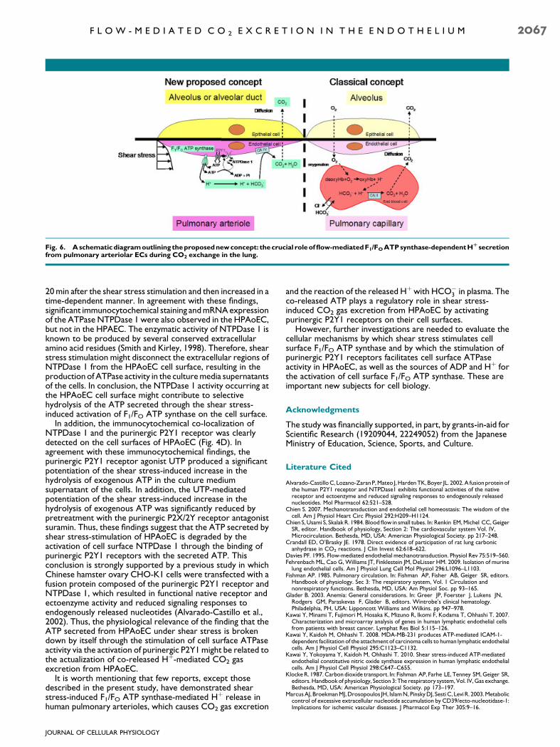
The present study demonstrates another important aspectof CO2 exchange in the human lung; i.e., that ectoenzymaticcarbonic anhydrase type IV is located on the EC surfaces ofhuman pulmonary arterioles, which might facilitate theexcretionofCO2 fromarteriolar ECs in combinationwith flow-mediated Hþ excretion through the activation of F1/FO ATPsynthase on their cell surfaces. The presence of CA IV enzymeson pulmonary arterioles raises the possibility that plasmaHCO�
3 participates directly in the dehydration reactionwithout entering erythrocytes in exchange for Cl�. If thisoccurs in smaller arterioles of less than 100mm in diameter, thiswould radically alter the classical concepts of CO2 exchange(Klocke, 1987). In agreement with this conclusion, markedimmunocytochemical staining of cell surface CAIV wasdetected on the non-permeabilized HPAoEC (Fig. 5B). Incontrast, little or no immunocytochemical expression of CAIVwas observed on the non-permeabilized HPAEC (Fig. 5B).Similar findings were confirmed for CAIV mRNA expression inthe HPAoEC and HPAEC (Fig. 5C).
Proposed new concept of shear stress-induced CO2 gasexchange in HPAoEC
Therefore, taking all the findings obtained in the present studyinto consideration, we would like to propose a new concept ofCO2 gas exchange in human pulmonary arterioles involving avery short-period of shear stress stimulation-induced F1/FOATP synthase-dependent Hþ secretion and the subsequentreaction of Hþwith HCO�
3 in plasma, resulting in the excretionof CO2 from HPAoEC (Fig. 6). In addition, the carbonicanhydrase type IV located on the EC surfaces of humanpulmonary arterioles also plays a critical role in CO2 gasexcretion from human pulmonary arterioles (Fig. 6). Flow(shear stress)-induced CO2 excretion might occur in vivo aftera relatively short capillary transient time ranging from 0.1 to1.0 sec in the human lung (Roughton, 1964) because the shearstress-induced CO2 gas excretion response has beenconfirmed to occur after a similarly short period of shear stressstimulation (�1.0 sec). In addition, if CO2 gas exchange in thehuman lung occurs in the pulmonary arterioles, the shortcapillary transient time in the human lung might offer sufficienttime for O2 diffusion and hence allow O2 transport from thealveolar space to blood capillaries.
The proposed concept of pulmonary flow-mediatedCO2 gasexchange might explain the previous finding that thephysiological conversion of HCO�
3 to CO2 was observedin isolated rat lungs perfused with a red blood cell-freeKrebs-Ringer bicarbonate solution (Crandall and O’Brasky,1978); i.e., CO2 gas exchange could bemaintained in the humanlung if the pulmonary flow of Krebs-Ringer solution througharterioles subjects the ECswithin them to a shear stress rangingfrom 2.0 to 5.0 dyn/cm2. In fact, arteriolar ECs arephysiologically exposed to shear stress ranging from 2.0 to5.0 dyn/cm2 (Chien et al., 1984).
However, further in vivo and/or in situ experiments areneeded to evaluate the proposed new concept of CO2
exchange in the pulmonary arterioles.
Regulatory roles of ATP co-released by shear stressduring flow-induced CO2 gas excretion from HPAoEC
To evaluate the physiological role of the ATP co-released byshear stress stimulation in HPAoEC, we first investigated theeffects of shear stress stimulation on the hydrolysis of ATP intheHPAoEC supernatant.Our studies showed that in the shearstress (5.0 dyn/cm2, 60-sec stimulation)-loaded culture mediasupernatant, ATPase activity was significantly increased at
JOURNAL OF CELLULAR PHYSIOLOGY
2066 K A W A I E T A L .
20min after the shear stress stimulation and then increased in atime-dependent manner. In agreement with these findings,significant immunocytochemical staining andmRNAexpressionof the ATPase NTPDase 1 were also observed in the HPAoEC,but not in the HPAEC. The enzymatic activity of NTPDase 1 isknown to be produced by several conserved extracellularamino acid residues (Smith and Kirley, 1998). Therefore, shearstress stimulation might disconnect the extracellular regions ofNTPDase 1 from the HPAoEC cell surface, resulting in theproduction of ATPase activity in the culturemedia supernatantsof the cells. In conclusion, the NTPDase 1 activity occurring atthe HPAoEC cell surface might contribute to selectivehydrolysis of the ATP secreted through the shear stress-induced activation of F1/FO ATP synthase on the cell surface.
In addition, the immunocytochemical co-localization ofNTPDase 1 and the purinergic P2Y1 receptor was clearlydetected on the cell surfaces of HPAoEC (Fig. 4D). Inagreement with these immunocytochemical findings, thepurinergic P2Y1 receptor agonist UTP produced a significantpotentiation of the shear stress-induced increase in thehydrolysis of exogenous ATP in the culture mediumsupernatant of the cells. In addition, the UTP-mediatedpotentiation of the shear stress-induced increase in thehydrolysis of exogenous ATP was significantly reduced bypretreatment with the purinergic P2X/2Y receptor antagonistsuramin. Thus, these findings suggest that the ATP secreted byshear stress-stimulation of HPAoEC is degraded by theactivation of cell surface NTPDase 1 through the binding ofpurinergic P2Y1 receptors with the secreted ATP. Thisconclusion is strongly supported by a previous study in whichChinese hamster ovary CHO-K1 cells were transfected with afusion protein composed of the purinergic P2Y1 receptor andNTPDase 1, which resulted in functional native receptor andectoenzyme activity and reduced signaling responses toendogenously released nucleotides (Alvarado-Castillo et al.,2002). Thus, the physiological relevance of the finding that theATP secreted from HPAoEC under shear stress is brokendown by itself through the stimulation of cell surface ATPaseactivity via the activation of purinergic P2Y1might be related tothe actualization of co-released Hþ-mediated CO2 gasexcretion from HPAoEC.
It is worth mentioning that few reports, except thosedescribed in the present study, have demonstrated shearstress-induced F1/FO ATP synthase-mediated Hþ release inhuman pulmonary arterioles, which causes CO2 gas excretion
and the reaction of the released Hþwith HCO�3 in plasma. The
co-released ATP plays a regulatory role in shear stress-induced CO2 gas excretion from HPAoEC by activatingpurinergic P2Y1 receptors on their cell surfaces.
However, further investigations are needed to evaluate thecellular mechanisms by which shear stress stimulates cellsurface F1/FO ATP synthase and by which the stimulation ofpurinergic P2Y1 receptors facilitates cell surface ATPaseactivity in HPAoEC, as well as the sources of ADP and Hþ forthe activation of cell surface F1/FO ATP synthase. These areimportant new subjects for cell biology.
Acknowledgments
The study was financially supported, in part, by grants-in-aid forScientific Research (19209044, 22249052) from the JapaneseMinistry of Education, Science, Sports, and Culture.
Literature Cited
Alvarado-CastilloC, Lozano-ZaranP,Mateo J,HardenTK, Boyer JL. 2002.A fusion protein ofthe human P2Y1 receptor and NTPDase1 exhibits functional activities of the nativereceptor and ectoenzyme and reduced signaling responses to endogenously releasednucleotides. Mol Pharmacol 62:521–528.
Chien S. 2007. Mechanotransduction and endothelial cell homeostasis: The wisdom of thecell. Am J Physiol Heart Circ Physiol 292:H209–H1124.
Chien S, Usami S, Skalak R. 1984. Blood flow in small tubes. In: Renkin EM,Michel CC,GeigerSR, editor. Handbook of physiology, Section 2: The cardiovascular system Vol. IV,Microcirculation. Bethesda, MD, USA: American Physiological Society. pp 217–248.
Crandall ED, O’Brasky JE. 1978. Direct evidence of participation of rat lung carbonicanhydrase in CO2 reactions. J Clin Invest 62:618–622.
Davies PF. 1995. Flow-mediated endothelial mechanotransduction. Physiol Rev 75:519–560.Fehrenbach ML, Cao G, Williams JT, Finklestein JM, DeLisser HM. 2009. Isolation of murinelung endothelial cells. Am J Physiol Lung Cell Mol Physiol 296:L1096–L1103.
Fishman AP. 1985. Pulmonary circulation. In: Fishman AP, Fisher AB, Geiger SR, editors.Handbook of physiology. Sec 3: The respiratory system, Vol. 1 Circulation andnonrespiratory functions. Bethesda, MD, USA: Am Physiol Soc. pp 93–165.
Glader B. 2003. Anemia: General considerations. In: Greer JP, Foerster J, Lukens JN,Rodgers GM, Paraskevas F, Glader B, editors. Wintrobe’s clinical hematology.Philadelphia, PH, USA: Lipponcott Williams and Wilkins. pp 947–978.
Kawai Y, Minami T, Fujimori M, Hosaka K, Mizuno R, Ikomi F, Kodama T, Ohhashi T. 2007.Characterization and microarray analysis of genes in human lymphatic endothelial cellsfrom patients with breast cancer. Lymphat Res Biol 5:115–126.
Kawai Y, Kaidoh M, Ohhashi T. 2008. MDA-MB-231 produces ATP-mediated ICAM-1-dependent facilitation of the attachment of carcinoma cells to human lymphatic endothelialcells. Am J Physiol Cell Physiol 295:C1123–C1132.
Kawai Y, Yokoyama Y, Kaidoh M, Ohhashi T. 2010. Shear stress-induced ATP-mediatedendothelial constitutive nitric oxide synthase expression in human lymphatic endothelialcells. Am J Physiol Cell Physiol 298:C647–C655.
Klocke R. 1987. Carbon dioxide transport. In: Fishman AP, Farhe LE, Tenney SM,Geiger SR,editors. Handbookof physiology, Section 3: The respiratory system, Vol. IV, Gas exchange.Bethesda, MD, USA: American Physiological Society. pp 173–197.
MarcusAJ, BroekmanMJ,Drosopoulos JH, IslamN, PinskyDJ, SestiC, Levi R. 2003.Metaboliccontrol of excessive extracellular nucleotide accumulation by CD39/ecto-nucleotidase-1:Implications for ischemic vascular diseases. J Pharmacol Exp Ther 305:9–16.
Fig. 6. Aschematicdiagramoutlining theproposednewconcept: thecrucial roleofflow-mediatedF1/FOATPsynthase-dependentHR secretionfrom pulmonary arteriolar ECs during CO2 exchange in the lung.
JOURNAL OF CELLULAR PHYSIOLOGY
F L O W - M E D I A T E D C O 2 E X C R E T I O N I N T H E E N D O T H E L I U M 2067

MilovanovaT, Chatterjee S,Manevich Y, Kotelnikova I, Debolt K,MadeshM,Moore JS, FisherAB. 2006. Lung endothelial cell proliferation with decreased shear stress in mediated byreactive oxygen species. Am J Physiol Cell Physiol 290:C66–C76.
Moser TL, KenanDJ, Ashley TA, Roy JA, GoodmanMD,Misra UK, CheekDJ, Pizzo SV. 2001.Endothelial cell surface F1-FO ATP synthase is active in ATP synthesis and is inhibited byangiostatin. Proc Natl Acad Sci USA 98:6656–6661.
Murata T, Lin MI, Stan RV, Bauer PM, Yu J, Sessa WC. 2007. Genetic evidence supportingcaveolae microdomain regulation of calcium entry in endothelial cells. J Biol Chem282:16631–16643.
Roughton FJW. 1964. Transport of oxygen and carbon dioxide. In: Fenn WO, Rahn H,editors. Handbook of Physiology: Section 3, Vol. I, Respiration. Washington DC, USA:American Physiological Society. pp 767–825.
Smith TM, Kirley TL. 1998. Cloning, sequencing, and expression of a human brain ecto-apyrase related to both the ecto-ATPases and CD39 ecto-apyrases1. Biochim BiophysActa 1386:65–78.
Yamamoto K, Shimizu N, Obi S, Kumagaya S, Taketani Y, Kamiya A, Ando J. 2007.Involvement of cell surface ATP synthase in flow-induced ATP release by vascularendothelial cells. Am J Physiol Heart Circ Physiol 293:H1646–H1653.
JOURNAL OF CELLULAR PHYSIOLOGY
2068 K A W A I E T A L .