Embed Size (px)
Citation preview

3/28/12 VIRAL PATHOGENESIS
Sergei Nekhai, Ph.D.
Objectives:
• Courses of viral infections
•Prinicipes of viral pathogenesis
•Role of different factors in pathogenesis
•Examples

Viral Pathogenesis
• Process by which a viral infection leads to disease
• Viral pathogenesis is an abnormal situation of no value to the virus.
• The majority of viral infections are subclinical. It is not in the interest of the virus to severely harm or kill the host.
• The consequences of viral infections depend on the interplay between a number of viral and host factors.


Outcome of Viral Infection
• Acute Infection
– Recovery with no residue effects
– Recovery with residue effects e.g. acute viral encephalitis leading to neurological sequelae.
– Death
– Proceed to chronic infection
• Chronic Infection
– Silent subclinical infection for life e.g. CMV, EBV
– A long silent period before disease e.g. HIV, SSPE, PML
– Reactivation to cause acute disease e.g. herpes and shingles.
– Chronic disease with relapses and excerbations e.g. HBV, HCV.
– Cancers e.g. EBV, HTLV-1, HPV, HBV, HCV, HHV-8

`
Clinical latency


Factors in Viral Pathogenesis
• Effects of viral infection on cells (Cellular
Pathogenesis)
• Entry into the Host
• Course of Infection (Primary Replication, Systemic
Spread, Secondary Replication)
• Cell/Tissue Tropism
• Cell/Tissue Damage
• Host Immune Response
• Virus Clearance or Persistence

Cellular Pathogenesis
• Cells can respond to viral infections in 3 ways: (1) No apparent change, (2) Death, and (3) Transformation
• Direct cell damage and death from viral infection may result from
– diversion of the cell's energy
– shutoff of cell macromolecular synthesis
– competition of viral mRNA for cellular ribosomes
– competition of viral promoters and transcriptional enhancers for cellular transcriptional factors such as RNA polymerases, and inhibition of the interferon defense mechanisms.
• Indirect cell damage can result from
– integration of the viral genome
– induction of mutations in the host genome
– inflammation
– host immune response.

Cytopathic Effect (CPE) of Herpes Simplex Virus Type 1 In
Vero Cells
A, Tube culture of uninfected monolayer of vero cells. Phase
contrast microscopy, × 40. B, Tube culture inoculated with a
clinical specimen of HSV-1 showing the presence of CPE after 5
days of incubation. Phase contrast microscopy, × 200.
A B

Photomicrographs of HSV-1-infected primary rabbit kidney cells collected
from human vesicle fluid. Primary rabbit kidney cells are shown at 48 h post
infection. Following methanol fixation, cells were visualized by the Giemsa
staining technique. A, uninfected cells, 100x. B, HSV-1 infected cells, 100x,
shows viral cytopathic effect in which multinucleated giant cells have formed
due to fusion of infected cells. Since respiratory syncytial virus, measles virus,
and herpes zoster virus do not grow in rabbit kidney cells, presence of this type
CPE is presumptive evidence of HSV.
© American Society for Microbiology, Washington DC.
Cytopathic Effect (CPE) of Herpes Simplex Virus Type 1 In
Primary Rabbit Kidney Cells
A B

A, uninfected Vero cells form a continuous monolayer of spindle-
shaped cells. B, a strong CPE was observed after 24 hours of
incubation of Vero cells with the patient sputum sample (primary
isolate).
© Elisa Vicenzi, et al. Coronaviridae and SARS-associated Coronavirus Strain HSR1. Emerging Infectious diseases. 2004.
Vp;10 N3.
Cytopathic Effect (CPE) of Primary Severe Acute Respiratory
Syndrome–Associated Coronavirus (SARS)

Use of Indicator Cell Lines to Detect Viruses
(primarily HSV and HIV-1)
• Culture an indicator cell line to determine the presence of a the
particular virus.
• Inoculate the specimen, incubate for 24 hours or longer
• Detect the expression of a reporter gene (b-galactosidase or Luciferase)
A, Monolayer of uninfected HeLa-MAGI cells. B, HeLa-MAGI cells
infected with adenovirus expressing HIV-1 transcriptional activator (Tat). C,
HeLa-MAGI cells infected with T-tropic HIV-1. All cells were fixed and
exposed to b-galactosidase sunstrate, X-Gal, which shows as blue color when
processed by b-galactosidase. Magnification 100x.
A. Control C. HIV-1 B. Adeno-Tat
Ammosova et al., J. Biol. Chem. 2003

Viral Entry • Skin - Most viruses which infect via the skin require a breach in the
physical integrity of this effective barrier, e.g. cuts or abrasions. Many viruses employ vectors, e.g. ticks, mosquitos or vampire bats to breach the barrier.
• Conjunctiva and other mucous membranes - rather exposed site and relatively unprotected
• Respiratory tract - In contrast to skin, the respiratory tract and all other mucosal surfaces possess sophisticated immune defence mechanisms, as well as non-specific inhibitory mechanisms (cilliated epithelium, mucus secretion, lower temperature) which viruses must overcome.
• Gastrointestinal tract - a hostile environment; gastric acid, bile salts, etc. Viruses that spread by the GI tract must be adapted to this hostile environment.
• Genitourinary tract - relatively less hostile than the above, but less frequently exposed to extraneous viruses (?)


Course of Viral Infection
• Primary Replication
– The place of primary replication is where the virus replicates after
gaining initial entry into the host.
– This frequently determines whether the infection will be localized
at the site of entry or spread to become a systemic infection.
• Systemic Spread
– Apart from direct cell-to-cell contact, the virus may
spread via the blood stream and the CNS.
• Secondary Replication
– Secondary replication takes place at susceptible organs/tissues
following systemic spread.


Cell Tropism
Viral affinity for specific body tissues (tropism) is determined by
– Cell receptors for virus.
– Cell transcription factors that recognize viral promoters and enhancer sequences.
– Ability of the cell to support virus replication.
– Physical barriers.
– Local temperature, pH, and oxygen tension enzymes and non-specific factors in body secretions.
– Digestive enzymes and bile in the gastrointestinal tract that may inactivate some viruses.

Cell Damage
• Viruses may replicate widely throughout the body without
any disease symptoms if they do not cause significant cell
damage or death.
• Retroviruses do not generally cause cell death, being
released from the cell by budding rather than by cell lysis,
and cause persistent infections.
• Conversely, Picornaviruses cause lysis and death of the cells
in which they replicate, leading to fever and increased mucus
secretion in the case of Rhinoviruses, paralysis or death
(usually due to respiratory failure) for Poliovirus.

Immune Response
• The immune response to the virus probably has the greatest impact on
the outcome of infection.
• In the most cases, the virus is cleared completely from the body and
results in complete recovery.
• In other infections, the immune response is unable to clear the virus
completely and the virus persists.
• In a number of infections, the immune response plays a major
pathological role in the disease.
• In general, cellular immunity plays the major role in clearing virus
infection whereas humoral immunity protects against reinfection.

Immune Pathological Response
• Enhanced viral injury could be due to one or a mixture of the
following mechanisms;-
– Increased secondary response to Tc cells (HBV)
– Complement mediated cell lysis
– Binding of un-neutralized virus-Ab complexes to cell surface Fc
receptors, and thus increasing the number of cells infected (Dengue
haemorrhagic fever, HIV)
– Immune complex deposition in organs such as the skin, brain or
kidney (rubella, measles)

Viral Clearance or Persistence
• The majority of viral infections are cleared but certain
viruses may cause persistent infections. There are 2
types of chronic persistent infections.
• True Latency - the virus remains completely latent
following primary infection e.g. HSV, VZV. Its genome
may be integrated into the cellular genome or exists as
episomes.
• Persistence - the virus replicates continuously in the
body at a very low level e.g. HIV, HBV, CMV, EBV.

Mechanisms of Viral Persistence
– antigenic variation
– immune tolerance, causing a reduced response to an antigen, may be due to genetic factors, pre-natal infection, molecular mimicry
– restricted gene expression
– down-regulation of MHC class I expression, resulting in lack of recognition of infected cells e.g. Adenoviruses
– down-regulation of accessory molecules involved in immune recognition e.g. LFA-3 and ICAM-1 by EBV.
– infection of immunopriviliged sites within the body e.g. HSV in sensory ganglia in the CNS
– direct infection of the cells of the immune system itself e.g. Herpes viruses, Retroviruses (HIV) - often resulting in immunosuppression.


Viral Virulence
• The ability of a virus to cause disease in an infected host
• A virulent strain causes significant disease
• An avirulent or attenuated strain causes no or reduced disease
• Virulence depends on
– Dose
– Virus strain (genetics)
– Inoculation route - portal of entry
– Host factors - eg. Age SV in adult neurons goes persistent but is lytic in young


Examples of Viral Pathogenesis

Herpes Simplex Viruse
• Herpesvirus family
•ds DNA enveloped viruse
•DNA codes for up 100 proteins (?)
•HSV genome consists long and short segments(4 isomers)

A. Primary Infection
• humans are the only natural host to HSV
• spread by contact, the usual site for the implantation is skin or
mucous membrane
• HSV spreads to craniospinal ganglia.
B. Latency
• HSV escapes the immune response
• persists indefinitely in a latent state in trigeminal ganglia and, to a
lesser extent, in cervical, sacral and vagal ganglia..
C. Reactivation
• By stress - physical or psychological
• pneumococcal infection
• meningococcal infection
• Fever
• irradiation, including sunlight
• menstruation
Herpes Simplex Viruse Pathogenesis

HSV Clinical Manifestations
• Acute gingivostomatitis
• Herpes Labialis (cold sore)
• Ocular Herpes
• Herpes Genitalis
• Other forms of cutaneous herpes
• Meningitis
• Encephalitis
• Neonatal herpes

CYTOMEGALOVIRUS
•Named after the appearance of its cytopathic effect in cell
culture
•CMV produces typical owl's eye intranuclear inclusion bodies
in infected cells
Properties
• A member of the herpesvirus family
•ds DNA enveloped virus
• CMV is physically associated with host ß2-microglobulin,
which might have a protective effect against host
immunoglobulins
•Fibroblasts are the only cells fully permissive for CMV
infection in vitro

CYTOMEGALOVIRUS PATHOGENESIS
Routes_of_transmission
•Intrauterine, follows maternal viraemia and placental infection
•Perinatal - (i) Infected genital secretions, or (ii) Breast milk
•Postnatal-(1) Saliva; (2) Sexual transmission (?); (3) Blood
transfusion - 3 - 5% of blood taken from seropositive donors leads to
infection (4) Organ transplantation
Mechanism_of_Virus_Recurrence
•In immunosuppression
• Recurrent CMV infection - very low after the age of 30 years
Immune_Responses
•Humoral Response - CMV specific IgM antibodies are produced
during the primary infection and persists for 3 or 4 months
•immunocompromised individuals may fail to produce IgM2.
•Cell mediated immunity (CMI) - plays a key role in the suppression of
CMI infection

Influenza Viruses
•Orthomyxovirus
•ssRNA enveloped viruses with a helical symmetry
•4 antigens present, the haemagglutinin (HA),
neuraminidase (NA), nucleocapsid (NA), the matrix
(M) and the nucleocapsid proteins

Influenza Viruses Pathogenesis •Infection is spread via respiratory droplets
•The virus infects respiratory epithelium cells
•Neuraminidase releases virus particles bound by the mucous
Clinical Features
•Incubation period of 48 hours
•The onset is characterized by marked fever, headache, photophobia,
shivering, a dry cough, malaise, myalgia, and a dry tickling throat
•The fever is continuous and lasts around 3 days
•Influenza B infection is similar to influenza A, but infection with
influenza C is usually subclinical or very mild in nature.
Complications: tracheobronchitis and bronchiolitis (eldery more often),
pneumonia ( primary viral or a secondary bacterial, more often S.
aureus); myositis and myoglobinuria; Reye's syndrome (encephalopathy
and fatty liver degeneration).

Rubella
Transmitted by the respiratory route and replicates upper/lower
respiratory tract and then local lymphoid tissues.
Following an incubation period of 2 weeks, a viremia occurs and
the virus spreads throughout the body.
Clinical Features:-
maculopapular rash due to immune complex deposition
lymphadenopathy
fever
arthropathy (joints inflammation, up to 60% of cases)

Rubella Infection During Pregnancy
• Rubella virus enters the fetus during the maternal viraemic phase through the placenta.
• The damage to the fetus seems to involve all germ layers and results from rapid death of some cells and persistent viral infection in others.
Preconception Risks
0-12 weeks 100% risk of fetus being congenitally infected
resulting in major congenital abnormalities.
Spontaneous abortion occurs in 20% of cases.
13-16 weeks deafness and retinopathy 15%
after 16 weeks normal development, slight risk of deafness and retinopathy

Dengue
• Dengue is the biggest arbovirus problem in the world
today with over 2 million cases per year. Dengue is found
in SE Asia, Africa and the Caribbean and S America.
• transmitted by Aedes mosquitoes which reside in water-
filled containers.
• Human infections arise from a human-mosquitoe-human
cycle
• Classically, dengue presents with a high fever,
lymphadenopathy, myalgia, bone and joint pains, headache,
and a maculopapular rash.
•Flavivirus, 4 serotypes
•Enveloped ssRNA viruses
•7-8 nm spikes protrude from the envelope
Pathogenesis .

Distribution of Dengue

Hepatitis B Virus
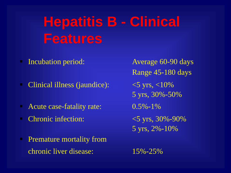
Incubation period: Average 60-90 days
Range 45-180 days
Clinical illness (jaundice): <5 yrs, <10%
5 yrs, 30%-50%
Acute case-fatality rate: 0.5%-1%
Chronic infection: <5 yrs, 30%-90%
5 yrs, 2%-10%
Premature mortality from
chronic liver disease: 15%-25%
Hepatitis B - Clinical
Features

Spectrum of Chronic Hepatitis B Diseases
• Chronic Persistent Hepatitis - asymptomatic
• Chronic Active Hepatitis - symptomatic
exacerbations of hepatitis
• Cirrhosis of Liver
• Hepatocellular Carcinoma

Viral Infections of Skin and Mucosa
Symptoms: vesicular or ulcerative lesions
Specimen: scrapes from the base of a lesion
Cause: usually HSV or VZV
Respiratory Infections
Specimen: nasospharyngeal aspirates, washings, swabs,
bronchial washington or bornchoalveola fluids
Cause: HSV, VZV or enteroviruses

Infections of CNS
• Acute meningitis in immunonologically
normal hosts
• Acute encephalitis in immunologically
normal hosts
• Opportunistic infections in
immunocompromised individuals

Sporadic encephalitus in immunonologically normal
hosts
Cause: usually HSV-1, 70% M&M in untreated cases
Virus
HSV-1 PCR analysis of CSF fluid, earlier – brain biopsies
and synthesis of HSV antibodies
Arboviruses including California (LaCross), St.Louis, eastern
equine, western equine, Venezuelan equine, and West
Nile, detected by serology of serum and SCF
Coltivirus (Colorado tick fever) infects erythrocytes, detected by
culture, FA and PCR from blood clot

CNS infection in immunocompromised individuals
Cause: CMV, EBV, VZV and JC usually HSV-1, 70% M&M
in untreated cases
Encephalitus of unknown ethiology
Virus Rabies
Acute meningitis in immunonologically normal hosts
Cause: usually enteroviruses and HSV-2 (and possibly VZV)

Gastrointestinal Tract Infections
Cause: rotaviruses, enteric adenoviruses,
caliciviruses and astroviruses. Originally
discovered by electron microscopy of feces
Epstein-Barr Virus
•Causes infectious mononucleosis (clinical findings –
atypical lymphocytes, heterophile antibodies (react
across species lines)
•Primary CNS lymphoma in AIDS patients
•Posttransplantational lymphoproliferative syndrome
in transplant recipients

Viral Hepatitis Virus Detection:
Hepatitis A IgM (acute infection), total antibody (past, current
infection or immunization)
Hepatitis B surface antigen (acute or chronic infection), total
core antibody (current or past infection), core
antibody (positive in current infection), e antigen
(current infection or correlation with infectivity),
antibody to e antigen (less current infection with
lower infectivity)
Hepatitis C ELISA (current or past infection); RNA (current
infection); genotype (affects response to interferon)
Hepatitis D antibody (current or recent infection)
Hepatitis E IgM (current infection), IgG (past or curent infection
-immunization status

Summary
• Viral Pathogenesis depends on the complex interplay of a large number of viral and host factors.
• Viral factors include cell tropism and cellular
pathogenesis.
• The immune response is the most important host
factor, as it determines whether the virus is cleared
or not.
• Sometimes, the immune response itself is
responsible for the damage.


![On Barriers and the Gap between Active and Passive Replication · or Megastore [1] use primary replicas to produce state updates or state mutations. Passive replication uses two types](https://img.dokumen.tips/doc/110x75/6009166fd8449c33127af31a/on-barriers-and-the-gap-between-active-and-passive-replication-or-megastore-1.jpg)


