Embed Size (px)
Citation preview
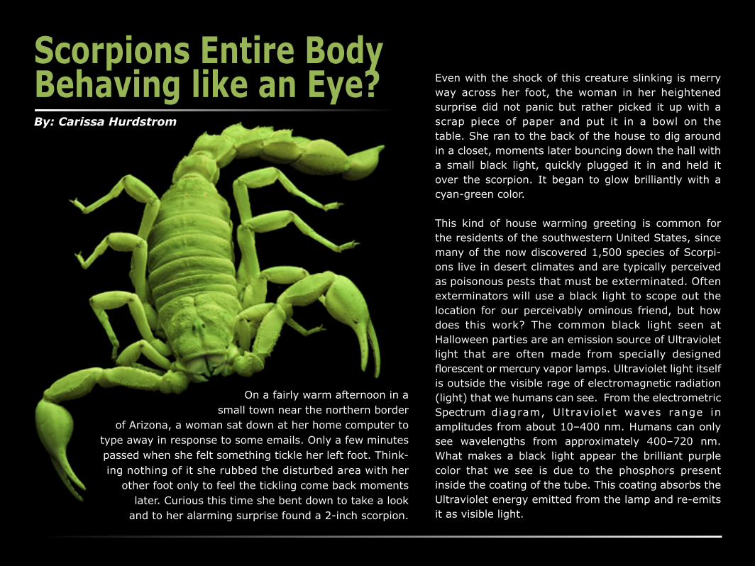
Scorpions Entire Body Behaving like an Eye?By: Carissa Hurdstrom
On a fairly warm afternoon in a small town near the northern border
of Arizona, a woman sat down at her home computer to type away in response to some emails. Only a few minutes passed when she felt something tickle her left foot. Think-ing nothing of it she rubbed the disturbed area with her
other foot only to feel the tickling come back moments later. Curious this time she bent down to take a look
and to her alarming surprise found a 2-inch scorpion.
Even with the shock of this creature slinking is merry way across her foot, the woman in her heightened surprise did not panic but rather picked it up with a scrap piece of paper and put it in a bowl on the table. She ran to the back of the house to dig around in a closet, moments later bouncing down the hall with a small black light, quickly plugged it in and held it over the scorpion. It began to glow brilliantly with a cyan-green color.
This kind of house warming greeting is common for the residents of the southwestern United States, since many of the now discovered 1,500 species of Scorpi-ons live in desert climates and are typically perceived as poisonous pests that must be exterminated. Often exterminators will use a black light to scope out the location for our perceivably ominous friend, but how does this work? The common black light seen at Halloween parties are an emission source of Ultraviolet light that are often made from specially designed florescent or mercury vapor lamps. Ultraviolet light itself is outside the visible rage of electromagnetic radiation (light) that we humans can see. From the electrometric Spectrum diagram, Ultraviolet waves range in amplitudes from about 10–400 nm. Humans can only see wavelengths from approximately 400–720 nm. What makes a black light appear the brilliant purple color that we see is due to the phosphors present inside the coating of the tube. This coating absorbs the Ultraviolet energy emitted from the lamp and re-emits it as visible light.

So how do scorpions glow in the presence of black light? Well, scientists found that fluorescent chemicals in the cuticle, or exoskeleton, are responsible for the Cyan-green glow. The chemicals found in species like Hadrurus arizonensis, is Beta Carboline and 7-hydroxy-4-methylcoumarin and behave like the phosphor coating in that they absorb Ultraviolet light and re-emit it as visible light but differ in that they re-emit light in the cyan-green range of the electromagnetic spectrum. What’s also interesting is that these chemicals are created during sclerotization, which is the process of the cuticle becoming hard.
Is there a purpose for the scorpion’s ability to fluoresce? There have been many theories from metabolic byproducts all the way to prey lure and mate finding but researcher Douglas Gaffin and his team say that it function is used more like an additional eye.
Griffin and his team tested the species Paruroctonus utahensis by exposing them to Green light at 505nm and Ultraviolet light at 395nm. Their test was to examine the scorpion’s reactions to the different sources of light with their eyes covered. When under
green light with their eyes covered the scorpions moved less then when their eyes were uncovered. But their light responsive behavior remained the same with both covered and uncovered eyes when exposed to Ultraviolet light. The new theory Griffin brought to the table incorporates the idea that the UV light is transduced to cyan-green and then passed to the brain like an advanced photoreceptor. This would be useful to
the scorpion in finding shelter because anything blocking the source would stop the transduction and indicate that the scorpion is now covered or blocked.
Scorpions have anywhere from 2 to 12 eyes. There are 2 poorly sensitive, simple eyes located medially on the dorsal side of the cephalothorax used to detect light and contrast. Then there are 2-5 more sensitive
clusters of eyes located laterally on the cephalothorax used to detect movement and light direction. This particular specimen had 3 lateral eyes on each side. Pictured above are
the comparisons of the eyes exposed to white light (bottom left) Ultraviolet light with barrier filter in place (bottom right & center) and Ultraviolet light without the barrier filter and captured in monochrome (bottom middel).

DevilScorpionAnatomy of the
ClassificationKingdom: Animalia (Animals)Phylum: Arthropoda (Arthropods)Class: Arachnida (Arachnids)
Met
asoma Segments (Rings of the Tial)
1st
2nd3rd 4th
5th
Telso
n
Proma
zoma
Mesos
oma
Metas
oma
Ped
ip
alps “pincer”
Fem
ur
Tibia
Tarsus
trochanter
femurpatell
a
4 Pairs of le
gs
“Mouth”Chelicerae
EYES
Seve
n Te
rgite
s (Pl
ates
)
Mantu
starsas
Ti bia
Bod
yH
ead
Tail
Opitho
soma
Order: Scorpiones (Scorpions)Family: Vaejovidae Genus: VaejovisSpecies: spinigerus

Fluorescence of a Scorpion Imaged
I imaged the H. Spinigerus scorpion species is also known as “Devil Scorpion” and “Arizona Striped Tailed Scorpion”. It was preserved inside a small jar filled with Vodka and mailed to me to be imaged. My main curiosity was the intensity of the excitation florescence that the scorpion would give off and how it visibly looked for imaging. I was surprised that it appeared much brighter then the many phosphorescent (glow in the dark) toys I grew up with.
It’s important to detail that scorpions have anywhere from 2 to 12 eyes. There are 2 poorly sensitive, simple eyes located medially on the dorsal side of the cephalothorax used to detect light and contrast. Then there are 2-5 more sensitive clusters of eyes located laterally on the cephalothorax used to detect movement and light direction. This particular specimen had 3 lateral eyes on each side.
Chelicerae or mouth parts compairison (Top Left)Tip of the Telson compairison (bottom Left)Ungues of the Tarsus compairison (above, right)
I also wanted to image some key points of the scorpion in fluoresces in comparison to the scorpion exposed to normal white light. It is interesting to note that the chelicerae, the tip of the Telson, and the Ungues of the Tarsus do not fluoresce which indicated that they do not contain the same chemical components as the cuticle.
Lastly I wanted to include images of the ventral side of the metasoma (Tail) showing the defining characteristics, which is also a major species identification feature.

Image Set Up
DevilScorpionIn order to image the entire body there are a few necessary items to be used: a black light(s) and a deep yellow barrier filter placed on the lens of the camera. The Barrier filter emits UV to violet blue light and eliminates any extra excitation energy from the view of the camera.
For the full specimen view of the fluorescent image of the scorpion I placed the dead preserved specimen on a rock with an inclined edge that angled approximately 45 degrees from the table. I then positioned its pincer arms and 4 pairs of legs to give it the appearance that the scorpion is crawling down the rock. I set up black lights around the species in order to achieve the most detail and revel the best excitation. One was placed above and slightly behind the other was placed above and angle towards the specimen from above the lens. From here I set my camera on a tripod and leveled it vertically perpendicular with the table then adjusted the angle to achieve the presented view. Using a Nikon 105mm Macro Lens I filled the specimen in the frame, which was about a 2:1 ratio.

About the PhotographerCarissa has a love for color and science that has driven her to a variety of imaging but is best know for her commercial images in event and food photography as well as her Macrographs. Carissa began in the industry with a science degree in commercial photography from the College of Southern Nevada. She will be graduating form Rochester Institute of Technology with a Bachelors of Science degree in Biomedical Photographic communication. Her major concentrations are Ophthalmic and High Magnification Imaging. If you would like to see more of her work please visit her website at: http://cxh4496.cias.rit.edu/361/index.html
For questions or comments please email her at:[email protected] [email protected]
Carissa Hurdstrom Sourceshttp://www.physik.unibas.ch/Praktikum/VPII/Fluoreszenz/Fluorescence_and_
Phosphorescence.pdfhttp://www.sciencedirect.com/science/article/pii/S0003347211005069
http://www.physics.org/article-questions.asp?id=66http://chemistry.about.com/od/glowinthedarkprojects/a/What-Is-A-Black-Light.
htmhttp://www.sciencedirect.com/science/article/pii/S1074552199800854
http://www.ncbi.nlm.nih.gov/pubmed/18470601http://scorpion-files.blogspot.com/2012/01/scorpions-walking-eyes.html
http://en.wikipedia.org/wiki/Hoffmannius_spinigerushttp://www.care-sheet.com/index/Hoffmannius_spinigerus
http://www.arizona-leisure.com/gallery/desert-wild/scorpion-anatomy.gifhttp://lanwebs.lander.edu/faculty/rsfox/invertebrates/vaejovis.html
http://www.ntnu.no/ub/scorpion-files/scorpion_anatomy.jpghttp://scorpionevolution.blogspot.com/2007/08/eyes.html
http://scorpion-files.blogspot.com/2012/01/scorpions-walking-eyes.htmlhttp://en.wikipedia.org/wiki/Scorpion
http://en.wikipedia.org/wiki/Sclerite#Arthropods