Embed Size (px)
Citation preview
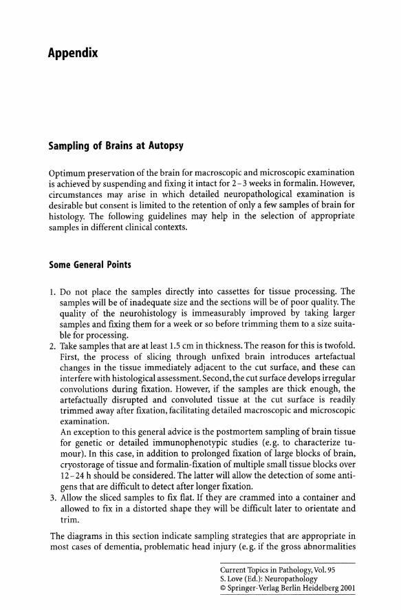
Appendix
Sampling of Brains at Autopsy
Optimum preservation of the brain for macroscopic and microscopic examination is achieved by suspending and fixing it intact for 2 - 3 weeks in formalin. However, circumstances may arise in which detailed neuropathological examination is desirable but consent is limited to the retention of only a few samples of brain for histology. The following guidelines may help in the selection of appropriate samples in different clinical contexts.
Some General Points
1. Do not place the samples directly into cassettes for tissue processing. The samples will be of inadequate size and the sections will be of poor quality. The quality of the neurohistology is immeasurably improved by taking larger samples and fixing them for a week or so before trimming them to a size suitable for processing.
2. Take samples that are at least 1.5 cm in thickness. The reason for this is twofold. First, the process of slicing through unfixed brain introduces artefactual changes in the tissue immediately adjacent to the cut surface, and these can interfere with histological assessment. Second, the cut surface develops irregular convolutions during fixation. However, if the samples are thick enough, the artefactually disrupted and convoluted tissue at the cut surface is readily trimmed away after fixation, facilitating detailed macroscopic and microscopic examination. An exception to this general advice is the postmortem sampling of brain tissue for genetic or detailed immunophenotypic studies (e. g. to characterize tumour). In this case, in addition to prolonged fixation of large blocks of brain, cryostorage of tissue and formalin-fixation of multiple small tissue blocks over 12-24 h should be considered. The latter will allow the detection of some antigens that are difficult to detect after longer fixation.
3. Allow the sliced samples to fix flat. If they are crammed into a container and allowed to fix in a distorted shape they will be difficult later to orientate and trim.
The diagrams in this section indicate sampling strategies that are appropriate in most cases of dementia, problematic head injury (e.g. if the gross abnormalities
Current Topics in Pathology, Vol. 95 S. Love (Ed.): Neuropathology © Springer-Verlag Berlin Heidelberg 2001

268 Appendix
appear too mild to account for the clinical findings) and miscellaneous neurodegenerative disorders. Once fixed, the tissue can be sliced and subdivided into multiple blocks. The suggested samples should be supplemented by blocks of macroscopically visible lesions noted when the brain is sliced at autopsy.
The appendix concludes with brief guidelines for taking samples of cerebrospinal fluid, brain tissue and other material for microbiological studies in suspected eNS infection.
I DEMENTIA I
1. Frontal pole of one hemisphere
Fig. I
Appearance of cut sulfaces
3. Brain stem 'rom upper midbrain to upper medulla
2. A 3cm coronal slice (or 2 adjacent 1.Scm slices) through one hemisphere, from just In front of the midbrain to lust behind It.

Appendix
HEAD INJURY, SUDDEN UNEXPLAINED DEATH IN ADULTS
1. A 3cm coronal slice (or 2 adjacent 1.5cm slices) the anterior face of which Is Just behind the temporal pole.
Fig. 2
3. Midbrain and upper half of pons
4. Middle of medulla to upper cervical cord
2. A 1.5cm coronal slice that Includes the back (splenium) of the corpus callosum.
Appearance of cut surfaces
5. Generous wedge of cerebellum that Includes dentate nucleus.
269

270
NEURODEGENERATIVE DISORDER OF UNCERTAIN NATURE
1. Frontal pole 01 one hemisphere
Fig. 3
Appearance o( cuI surfaces
3. Brain stem from upper midbrain to low medulla
2. A 3cm coronal slice (or 2 adjacent 1.5cm slices) through one hemisphere, from Just In Iront of the midbrain to just behind It.
4. Generous wedge of cerebellum that spans midline and includes dentate nucleus.
Appendix
If there is clinical evidence of spinal or neuromuscular involvement, take cord and samples of peripheral nerve and muscle, as well as the blocks shown in Fig. 3.
If the clinical history raises the possibility of Creutzfeldt-Jakob disease (and permission to keep the whole brain is not given), the following steps should be added to the above protocol:
• Sample the occipital pole of one cerebral hemisphere for paraffin histology.

Appendix 271
• Freeze one frontal pole (i. e. contralateral to that sampled for paraffin histology) and, if possible, one cerebellar hemisphere at - 70°C (for Western blotting, transmission studies, DNA sequencing, etc).
• Ideally, also take samples of tonsil, spleen, appendix and lymph nodes, pituitary, trigeminal ganglia, spinal cord and dorsal root ganglia for paraffin histology, and freeze one tonsil at - 70 °C for Western blot analysis.
Specimens for Microbiological Investigation
Cerebrospinal Fluid
Reflect the dura with the brain in situ, gently part the cerebral hemispheres and insert the needle of a sterile syringe about 30° to the vertical through the corpus callosum and into the lateral ventricle. Slight negative pressure should then be applied to the plunger of the syringe whilst slowly withdrawing the needle. When the tip of the needle enters the lateral ventricle, the fluid will be drawn into the syringe. In addition, swabs should be taken from the subarachnoid space, particularly in the region of any exudates - to do so use fine forceps to elevate the leptomeninges, make a small cut into the meninges with a sterile blade and insert the swab into the subarachnoid space.
If viral infection is suspected, samples of cerebrospinal fluid, brain (see below) and serum or clotted blood should be frozen, ideally at - 70°C. The frozen samples can later be used for viral culture, polymerase chain reaction (PCR) studies and serology, as needed.
Abscess or Empyema
In most cases, the nature of the infection will not be in doubt. However, if identification of the responsible micro-organisms is needed, the purulent material should be cultured, in which case it should be aspirated into a sterile syringe, expelled into a sterile universal container and taken to a microbiology laboratory with as little delay as possible. The likelihood of recovering the responsible micro-organisms is greater if the contents of the abscess are aspirated than if only swabs are taken.
Brain or Spinal Tissue
Samples of abnormal tissue for bacterial or fungal culture should be removed with a sterile scalpel, placed in a sterile universal container and taken to a microbiology laboratory as soon as possible.
In suspected viral infection, several small blocks of tissue (1- 2 g is ample) should be taken from regions showing macroscopic abnormalities and, together with samples of CSF and serum or clotted blood (see above),frozen at -70°C for later culture or PCR studies. The regions sampled in suspected herpes encephalitis should include the anterior part of the temporal lobes and the inferior part of the frontal lobes.

Subject Index
Abscess, retropharyngeal 13 Acanthamoeba 9, 17,39 Acetylcholine 156 - receptor 215 Acid maltase deficiency 222 Acquired immune deficiency syndrome
(AIDS) 249 Acrylamide 234 Actinomyces israelii 11 Adenovirus 37 Adult T-cell lymphocytic leukaemia (ATLL)
252 African trypanosomiasis 30 Agammaglobulinaemia, X-linked 7 AIDS (acquired immune deficiency
syndrome) 249 Air embolism 68 Alcohol abuse 128 Alcoholism 2 All-trans retinoic acid (ATRA) 254 Alpha synuclein 160 American trypanosomiasis 18 Amoebae,free-living 9 Amoebic encephalitis 17,39 Amyloid angiopathy 195 Amyloid f3-protein (Af3 protein) 62,154 Amyloid plaques - in Alzheimer's disease 154 - in Creutzfeldt-Jakob disease 198 f3-amyloid precursor protein 62,154,156 - immunoreactivity, interpreting III - traumatic axonal injury 110 Amyloidosis, multiple myeloma 258 Aneurysm 83 Angiitis 56 Angiopathy, cerebral amyloid (congophilic)
56,62,83,154 Anterior horn cell disease 214 Anticipation, in myotonic dystrophy 221 Anticoagulants 83,89, l38 Anticonvulsant drug 127 Antiphospholipid antibodies 53 Apolipoprotein E 156 Arbovirus 24 Arthritis, rheumatoid 235
Arrhythmias 66 Arterial dissection 56, 104 Arterial rupture 104 Arteriolosclerosis 60,164 Arteriosclerosis 56, 60, 84, 164 Arteriovenous malformation 83,87 Arteritis 56 Aspergillus 15,16 ATRA (all-trans retinoic acid) 254 Ataxia-telangiectasia, primary CNS
lymphoma 246 Atherosclerosis 56, 57 ATLL (adult T-cell lymphocytic leukaemia)
252 Atrial fibrillation 66 Atrophy - gonadal 221 - granular cortical 164 - grouped 213 Auto-immune disorders, primary CNS
lymphoma 246 Autoregulation 54, 141 Axonal degeneration 231 Axon reaction 231 Axonal injury, assessing 107 Azidothymidine 226
B virus 21 Balamuthia 9, 17 Balamuthia mandrillaris 40 Balding in myotonic dystrophy 221 B-cell chronic lymphatic leukaemia
253 B-celilymphoma - large 248 - mucosa-associated of the gastro-intestinal
tract 240 - polymorphous high-grade 249 - small 249 BCNU 256 Becker muscular dystrophy 209,219 Beh<;et disease 56 Berryaneurysm l34 Binswanger's disease 164

274
Binswanger's subcortical (arteriosclerotic) leukoencephalopathy 56,73
Blastomyces dermatitidis 5, 14 Bleeding into paraspinal muscles in
non-accidental injury 116 Blood dyscrasia 134, 138 Borrelia burgdorferi 29 Bovine spongiform encephalopathy (BSE)
166,180,195 Braak stages 154 Brain abscess - amoebic 11 - sudden death 139 Brain infarct, embolic 65 Bronchiectasis 11 Bruising 102 BSE (bovine spongiform encephalopathy)
166,180,195 Blingner, bands 231 Burkitt's lymphoma 253
CADASIL (cerebral autosomal dominant arteriopathy with subcortical infarcts and leukoencephalopathy) 56,165
Calcific aortic stenosis 68 Calcification of the mitral annulus 68 Calcium channel gene mutations 226 Candida 15,39 Carbon monoxide 54 Cardiac arrest 54 Cardiac arrhythmia 130 Cardiac conduction defect, in myotonic
dystrophy 221 Cardiac valves, prosthetic 68 Carnitine deficiency 222 Carnitine palmitoyltransferase deficiency
222 Cataract in myotonic dystrophy 221 Cavernous hemangioma 89 Cavernous sinus thrombosis, septic 75 Central core disease 226 Cerebral amyloid (congophilic) angiopathy
56,62,83,154 Cerebral autosomal dominant arteriopathy
with subcortical infarcts and leukoencephalopathy (CADASIL) 56,165
Cerebral infarct 83 Cerebral leukoencephalopathy 35 Chagas disease 18,30 Charcot-Bouchard microaneurysm 74,84 Charcot -Marie-Tooth disease/peroneal
muscular atrophy (HMSN I) 233 Chicken pox 23 Chloroquine 226 Chorioretinitis, macular 27 Chromatolysis 231
Subject Index
Chronic enteroviral meningoencephalitis 39
Chronic inflammatory demyelinating neuropathy (CIDP) 214,231,234
Chronic subdural membrane 108 CIDP (Chronic inflammatory demyelinating
polyradiculopathy) 214, 231, 234 Circle of Willis 58,135, 136 Citrobacter 9,18 Class I MHC antigen expression 209,217 CMV, peripheral neuropathy 231 CNS lymphoma - computerised tomography (CT) scan
findings in primary 247 - staging of suspected primary 247 Coagulation, inherited disorder 53 Coagulopathy 89 Coarctation of the aorta 79 Cocaine 89 Coccidioidomycosis 5,14 Coccidoides immitis 5,14 Codon 129 - of the prion protein gene 180 Cognition, fluctuating and dementia with
Lewy bodies 157 Colloid cysts of the third ventricle, sudden
death 138 Complement protein deficiencies 7 Contusion 105 Contusional tear 114 Core disease, central 226 Coxsackie virus 32,215 Craniocervical junction damage in non-
accidental injury 116 Creutzfeldt-Jakob disease 166,181,183 - clinical diagnostic criteria for sporadic
CJD 181 - clinical diagnostic criteria for variant CJD
181 - familial 166, 180 - focal deposits 198 - granular deposits 198 - MRI 184 - routes of transmission of iatrogenic CJD
183 - strain typing 199 Critical illness neuromyopathy 209 Cryostat sections of skeletal muscle
209 Cryptococcoma 14 Cryptococcus neoformans 5,7,256 Cyanide 54 Cystic medial necrosis 56 Cysticercosis 18, 219 Cytomegalovirus 36,235 Cytopathy, mitochondrial 222 Cytosine arabinoside 256

Subject Index
DAI (diffuse axonal injury) 109 Delusion and dementia with Lewy bodies
157 Dementia - definition 150 - focal neurobehavioural disorder 151 - frontotemporal 151,161 - multi-infarct 163 - temporoparietal 151 Demyelination, segmental 233 Denervated fibres 213 Denervation 212 Dermatomyositis 218 Desmin 209 - storage disorder 209 Diabetes mellitus, peripheral neuropathy
231 Diabetes, in myotonic dystrophy 221 Diffuse axonal injury (DAr, see also
traumatic axonal injury) 109, l33 Diffuse brain damage in non-accidental
injury 117 Diffuse large B-celilymphoma (DLBC)
240 - CNS 240 - primary mediastinal 253 - primary testicular 253 Diphtheria 235 - peripheral neuropathy 231 Disorder of coagulation, inherited 53 Disorder of the neuromuscular junction
212 Disseminated intravascular coagulation 89 DLBC (diffuse large B-celilymphoma) 240 Drowning 54 Drugs, peripheral neuropathy 231 Duchenne muscular dystrophy 209,219 Dural fistula 2 Dysferlin 209 Dysphagia, in myotonic dystrophy 221 Dystrophin 209 - complex 220
Ear, discharging, sudden death l39 Eastern equine encephalitis virus 24 Eaton-Lambert disease 214,215 Echinococcus granulosus 18 Echinococcus multilocularis 18 Echovirus 39 EEG, periodic triphasic complexes 184 Ehlers-Danlos syndrome 79 Electromyography 230 Emboli of intervertebral disc material 69 Embolic brain infarct 65 Embolization, iatrogenic 69 Emerin 209
Emery-Dreifuss dystrophy 222 Encephalitis associated with lymphoma
255 Encephalitis virus - Eastern equine 24 - Murray Valley 24 - Powassan 24 - Russian spring-summer 24 - St. Louis 24 - Venezuelan equine 24 - Western equine 24 Endarterectomy 59 Endocarditis 11
275
- infectious and non-bacterial thrombotic (marantic) 66
- marantic 67 Endoneurium 230 Endothelial adhesion molecules 58 Entamoeba histolytica 11 Enterobacter 9 Enterovirus 32,33,40 - infection 214, 215 - non-polio 2 Enterovirus 71 21 Eosinophilic myositis 219 Epilepsy 127 Epineurium 229 Escherichia coli 9 Extradural haemorrhage and non-accidental
injury 115,116 Extrafusal muscle 211 Fabry's disease 233 Factor V Leiden mutation 54 Familial Creutzfeldt -Jakob disease 180 Fat emboli 68 Fatal familial insomnia 180 Fibromuscular dysplasia (FMD) 56,79,83 Finger-print myopathy 225 Floppy-baby syndrome 225 Florid plaque 194 FMD (fibromuscular dysplasia) 56,79,83 Focal deposits in Creutzfeldt-Jakob disease
198 Fracture 103 Frontotemporal degeneration and
parkinsonism linked to chromosome 17 161
Gammopathy, monoclonal 236 Ganglioneuropathy, sensory 256 Gerstmann-Straussler-Scheinker syndrome
166,180 Giant cell arteritis 56,65 Giant cell myositis 219 Glioblastoma, sudden death l38 Global brain ischemia 54
276
Glucose-6-phosphate dehydrogenase deficiency 7
Gonadal atrophy 221 Gottron's sign 218 Gower's sign 219 Gram-negative bacilli causing meningitis 3 Granular deposits in Creutzfeldt-Jakob
disease 198 Granulocytopenia 7 Granulovacuolar degeneration 154 Grouped atrophy 213 Guillain-Barre syndrome 214,231,234,
258 Gummas 13
lfachinskiscore 163 lfaemophilia 89 lfaemophilus influenza 3 lfaemorrhage 132 - acute subdural 76 - ageing of 106 - - of subdural 106 - chronic subdural 76 - intracranial 140 - intraventricular 103 - lobar 85 - perinatal subdural 115 - subdural and non-accidental injury 115 lfallucinations and dementia with Lewy
bodies 157 lfead injury - non-accidental 77,107 - residua of old 109 lfeart disease - congenital 11 - rheumatic 68 lfeparin 83 lfereditary cerebral haemorrhage with
amyloidosis, Dutch type 62 lfereditary cerebral haemorrhage with
amyloidosis, Icelandic type 62 lfereditary cystatin C amyloid angiopathy
62 lfereditary motor and sensory neuropathies
(lfMSN) 233 lfereditary neuropathy with liability to
pressure palsies (lfNPP) 233 lfereditary sensorimotor neuropathy
(lfSMN) 231 lfereditary sensory neuropathy (lfSN) 231 lfernia - cingulate gyrus 142 - reversed tentorial 143 - supracallosal 142 - tentorial 142 - tonsillar 143
lferniation, internal 142 lfERNS 56
Subject Index
lferpes encephalitis, atypical forms of 35 lferpes simplex virus type 1 21,40 lferpes simplex virus type 2 2,40 lferpesvirus simiae 21 lfirano bodies 154 lfistoplasma capsulatum 7 lfistotoxic hypoxia 54 mv - associated vasculopathy 56 - encephalitis 25 - infection, primary CNS lymphoma 246 - peripheral neuropathy 231 - neuropathy 235 lfMSN (hereditary motor and sensory
neuropathies) 233 lfMSN I (Charcot -Marie-Tooth
disease/peroneal muscular atrophy) 233 lfMSN II 233 lfNPP (hereditary neuropathy with liability
to pressure palsies) 233 lfodgkin's lymphoma 246,254 lfSMN (hereditary sensorimotor neuro
pathy) 231 lfSN (hereditary sensory neuropathy) 231 lfTLV-l (human T-celllymphotropic
virus-I) 25,252 lfTLV-I-associated myelopathy 28 lfuman immunodeficiency virus (see also
mV) 2,25,219 lfuman T-celilymphotropic virus type I
(lfTLV-I) 25,252 lfydatid disease 18,20 lfydrocephalus 143 - acute obstructive 140 lfypereosinophilia syndrome 219 lfypersensitivity vasculitis 56 lfypertension 83, 138, 134 lfyperthermia, malignant 224,226 lfypotension 54 lfypoxia 128 - hypoxemic 54
Iatrogenic Creutzfeldt-Jakob disease 180 Iatrogenic embolization 69 Immunodeficiency syndrome, primary, and
primary CNS lymphoma 246 Immunodeficiency - combined variable 39 - severe combined, primary CNS lymphoma
246 Immunoglobulin, rearrangement 255 Inclusion body myositis 218 Infarction, ageing of 106 Infective endocarditis 66

Subject Index
Influenza A 219 Insomnia, fatal familial 166 Internal carotid artery, examination of
105 Internal nuclei 216 Intracranial pressure 103, 140 - raised 108 Intrafusal muscle fibres 212 Intraocular bleeding 116 Intraventricular haemorrhage 103 Isolated granulomatous angiitis of the CNS
5,56,64 Isoniazid 234
Jamestown virus 24 Japanese encephalitis 24,35 JC virus 35
Kearns-Sayre syndrome 223 Kernohan lesion 142 Klebsiella 9 Kugelberg-Welander disease 215 Kuru 180,195 - type plaque 193
La Crosse virus 24 Lambert-Eaton myastenic syndrome 256 Laminar necrosis 55 a2-laminin (merosin) 220,221 L-asparaginase 256 Lead toxicity 234 Left atrial myxoma 66 Leprosy 235 - peripheral neuropathy 231 Leukaemia 7,89 - acute lymphoblastic 253 - acute myeloid 253 - acute promyelotic 254 Leuko-araiosis 163 Leukocyte adhesion molecules 58 Leukodystrophy, metachromatic 233 Leukoencephalopathy - Binswanger's subcortical (arteriosclerotic)
56,73 - necrotising, due to HIV 35 - progressive multifocal 35,37,255 Limbic encephalitis 256 Lipohyalinosis 56,60,84 Listeria monocytogenes 9,14,256 Localised myositis 219 Long-chain fatty acids 222 Loss of consciousness in dementia with
Lewy bodies 157 Lung abscess 11
277
Lupus erythematosus, systemic, and primary CNS lymphoma 246
Lyme disease 29,235 - peripheral neuropathy 231 Lymphoma (see also specific types) - extranodal 240 - human immunodeficiency virus-asso-
ciated 241 - small cell 253 - WHO classification 240 Lymphomatoid granulomatosis 56
Malaria, cerebral 24,30 MALT (mucosa-associated lymphoid tissue)
240 Mantle-celllymphoma 253 Marantic endocarditis 67 Marfan syndrome 79 McArdle's disease 222 Measles 40 - inclusion-body encephalitis 37 - subacute sclerosing panencephalitis 26 - virus 25 MELAS (mitochondrial encephalopathy with
lactic acidosis and stroke-like episodes) 56,223
Meningioma, confusion with lymphoma 248
Meningitis - confusion with lymphoma 248 - neonatal 9 - purulent 7 - tuberculous 4,7 Meningococcus 3 Meningoencephalitis, amoebic 23 Merosin (a2-laminin) 209,221,222 MERRF (myoclonic epilepsy with ragged red
fibres) 223 Methotrexate 256 Metronidazole 234 Meynert, nucleus basalis 156 Microaneurysm, miliary 74 Microglia, traumatic axonal injury 110 Microvacuolation, frontotemporal dementia
161 Middle ear infection 11 Miliary microaneurysm 74 Mitochondrial encephalopathy with lactic
acidosis and stroke-like episodes (MELAS) 56,223
Mitral annulus calcification 66 Mitral valve prolapse or stenosis 66,68 Morula-type deposits in Creutzfeldt-Jakob
disease 198 Moth-eaten fibres 216 Motor neuron disease 161,214

278
Moyamoya disease 56 MRI in Creutzfeldt-Jakob disease 184 Mucoraceae 15 Mucormycosis - disseminated cerebral 15 - rhino cerebral 15,16 Mucosa-associated lymphoid tissue (MALT)
240 Multicentric plaque 194 Multinucleated giant cell encephalitis due to
HIV 35 Mumps - aseptic meningitis 2 - acute disseminated encephalomyelitis
40 Murray Valley encephalitis virus 24 Muscle fibre - atrophy 216 - atrophy, type IIb 226 - denervated 213 - hypertrophy, prominent 216 - internal nuclei 221 - lobulated 216 - necrosis 216 - paracrystalline inclusions 223 - splitting 216 - type grouping 213 Muscle spindle 212 Muscular dystrophy 209 - distal 222 - oculopharyngeal 222 - scapuloperoneal 222 Myasthenia gravis 214 Mycobacterium tuberculosis 4,13,14 Mycoplasma pneumonia 40 Myeloma 258 Myocardial abnormalities 66 Myocardial fibrosis 221 Myocardial infarct 66 Myoclonic epilepsy with ragged red fibres
(MERRF) 223 Myopathy 212 - mitochondrial 223 Myopathy - alcoholic 222 - corticosteroid 226 - mitochondrial 223 - myotubular 225 - steroid 222,226 - tubular aggregate 225 - vacuolar 226 - zebra-body 225 Myophosphorylase deficiency 222 Myosins, antibodies to slow and fast 209 Myositis, eosinophilic 219 Myotonia in myotonic dystrophy 221 Myxoma, left atrial 66
Subject Index
Naegleria fowleri 23 NARP (neuropathy, ataxia and retinitis
pigmentosa) 223 Neisseria meningitidis 3,7 Neoplasm - and brain haemorrhage 83, 134 - and sudden death 138 Nerve root disease 214 Neurites, dystrophic 154 Neurofibrillary tangle 221 Neurogenic muscle disease 212 Neuroleptic malignant syndrome 226 Neuroleptic sensitivity and dementia with
Lewy bodies 157 Neurological syndrome, paraneoplastic
256 Neuromuscular junction 214 Neuromyopathy - amyloid 231,233 - autonomic 230 - critical illness 209,226 - demyelinating 233 - diabetic 234 - hereditary sensory 233 - hypertrophic 233 - paraneoplastic 228,231 - subacute sensory 236 Neuropathy, ataxia and retinitis pigmentosa
(NARP) 223 Neurosarcoidosis 5 Nipah virus 23 Nocardia asteroides 14 Non-accidental head injury 77, 107 Non-bacterial thrombotic endocarditis
67 Non-Hodgkin's lymphoma 236 North American blastomycosis 5 Nucleus basalis of Meynert 156
Onion-bulb 233 Organ transplantation, therapy, primary
CNS lymphoma 246 Organophosphate 234 Osteomyelitis 13 Otitis media and sudden death 139
Pachymeningitis, idiopathic hypertrophic 5
Panencephalopathic variant of CJD 195 Papovavirus 35,37 Paraparesis, hereditary spastic 166 Paraneoplastic encephalitis 256 Paraproteinaemia 258 - peripheral neuropathy 236 Paresis, general 28

Subject Index
Parkinson's disease 157 Patent foramen ovale 66 Penicillamine 226 Penumbra, peri-infarct 55 Perifascicular atrophy 218 Perineurium 230 Periodic paralysis - hyperkalaemic 225 - hypokalaemic 225 Peripheral myelin protein (PMP-22) gene
233 Perivascular infiltration, confusion with
lymphoma 248 Pick bodies 161 Pick cells 161 Plaques - in Alzheimer's disease 154 - in Creutzfeldt -Jakob disease and other
prion diseases 193 Plasmatic differentiation, in primary CNS
lymphoma 250 Plasmodium falciparum 30 PNET (primitive neuro-ectodermal
tumours), confusion with lymphoma 248
Pneumococcus 2 Poliomyelitis 214,215 Poliovirus 21,32 Polyarteritis nodosa 56,235 Polycystic kidney disease 79 Polymyositis 217 Pompe's disease 222 Pork tape worm 18 Porphyria 231,233 Post-polio syndrome 215 Powassan encephalitis virus 24 Presenilins 156 Primary angiitis of the CNS 5,56,64 Primary immundeficiency syndromes,
primary CNS lymphoma 246 Primitive neuro-ectodermal tumours
(PNET), confusion with lymphoma 248 Prion - disorders 166, 178 - glycoforms 199 PRNP codon 129 - polymorphism 182 Progressive multifocalleukoencephalopathy
35,37,255 Properdin deficiency 7 14.3.3 protein 181 Protein C deficiency 53 Protein S deficiency 54 Proteus mirabilis 18 Prpm, detection of 190 Pseudoaneurysm 104 Pseudomonas aeruginosa 9
Pseudoxanthoma elasticum 79 Ptosis in myotonic dystrophy 221 Pulmonary arteriovenous malformation
11
Ragged-red fibres 223 REAL (revised European-American
lymphoma) classification 240 Rearrangement of immunoglobulin
255 Recreational drug use 83,89 Reed-Sternberg cells 252 Refsum's disease 231,233 Respiratory arrest 54 Retinal haemorrhage 116 Revised European-American lymphoma
(REAL) classification 240 Rhabdomyolysis 226 - acute renal failure 227 Rhabdovirus 33 Rheumatic heart disease 68 Rheumatoid arthritis 235 Rhizopus 16 Rich's foci 7 Rickettsial infections 30 Ring fibre 216,221 Rubella 25
279
Russian spring-summer encephalitis virus 24
Ryanodine receptor gene 226
Saccular aneurysm (see also berry aneurysm) 134
Sarcoglycan 209,220,221 - deficiency 221 Scalp bruising and non-accidental injury
114 Schistosomiasis 14 Schwann cells 230 Scrapie-associated fibrils 195 Secondary immunodeficiency syndromes,
primary CNS lymphoma 246 Seizure 127 Selective vulnerability 54 Shaken-baby syndrome 120 Shunt infection, ventricular 4 Sick sinus syndrome 66 Sickle cell disease 79, 134, 138 Sjogren's syndrome, primary CNS
lymphoma 246 Skull fracture 102 - and non-accidental injury 114 SLE (systemic lupus) 56 Slime-producing strains of Staphylococcus
epidermidis 4

280
Sodium channel gene mutation 226 Spinal muscular atrophy 214 Splenectomy 7 Spongiform change 190 - and dementia with Lewy bodies 160 Sporadic Creutzfeldt-Jakob disease 180 Sporadic fatal insomnia 180 Sprouting, collateral, of nerves 213 St. Louis encephalitis virus 24 Staging of suspected primary CNS
lymphoma 247 Stagnant hypoxia 54 Staphylococcus aureus 4,9, 13 Staphylococcus epidermidis, slime-
producing strains 4 Status spongiosus 154,192 Stavudine 234 Streptococcus - anaerobic 11 - group B 9 - microaerophilic 11 Streptococcus milleri 11 Streptococcus pneumoniae (pneumococcus)
2,7 Streptococcus viridans 11 Subacute sclerosing panencephalitis 26 Subarachnoid haemorrhage 103 Subdural haemorrhage - acute 76 - ageing of 106 - and non-accidental injury 115 - chronic 76 - perinatal 115 Sudden death 138, 139 Sural nerve 228 Survival motor neuron 214 Synaptic labelling in Creutzfeldt-Jakob
disease 198 Syncope or transient loss of consciousness
and dementia with Lewy bodies 157 Syphilis 13 - meningovascular 5 Syringomyelia 214,215 Systemic lupus (SLE) 56 - and primary CNS lymphoma 246
Tabes dorsalis 29 Taenia solium 18 Tahna virus 24 Takayasu arteritis 56 Tangier disease 233 Target fibres 213 Tau protein 154,161 T -cell leukaemia 28 T-ceillymphoma 28 - peripheral 254
Subject Index
T-cell receptor gene, rearrangement 255 Teased nerve fibres 229 Threads, neuropil 154 Thrombocytopoenia 89 Thrombotic thrombocytopenic purpura 56 Thymoma 219 Thyrotoxicosis 226 Tick-borne encephalitis 3S Tomacula 233 Tonsillar herniation 103 Toxins, peripheral neuropathy 231 Toxoplasma gondii 14 Toxoplasmosis 39 Trauma to the neck 82 Traumatic axonal injury (see also diffuse
axonal injury) 133 Treponema pallidum 13,28 Trichinella spiralis 219 Trinucleotide repeat sequences 221 Tropheryma whippelii 13,14 Tropical spastic paraparesis (TSP) 28 Tropomyosin 224 Trypanosoma brucei 30 Trypanosoma brucei rhodesiense 30 Trypanosoma cruzi 18,30,39 TSP (tropical spastic paraparesis) 28 Tuberculosis 13 Tubularin 225 Tumour - metastatic, and sudden death 138 Type I fibres 211 Type II fibres 211
Ubiquitin 160 Ubiquitin-immunoreactive inclusions 161 Unicentric plaque 193 Uraemia 234 - peripheral neuropathy 231
Vacuolar deposits in Creutzfeldt -Jakob disease 198
Vacuolar myelopathy due to HIV 35 Valvular abnormalities 66 Variant Creutzfeldt-Jakob disease 180 Varicella-zoster virus 23,36, 6S, 2S5 Vascular disease, peripheral neuropathy
231 Vascular malformation 134 Vasculitis 56,235 - hypersensitivity 56 Vasculopathy - cerebroretinal S6 - HIV-associated 56 Vasospasm, post-subarachnoid haemor
rhage 79
Subject Index
Venezuelan equine encephalitis virus 24 Venous angioma 89 Venous sinus thrombosis 74 Vertebral artery - examination of 104, l32 - exposure and removal 53 - tears l31 Vincristine 234 Vitamin B deficiency 234 Vitamin Bl2 deficiency 234 Vitamin deficiency, peripheral neuropathy
231 Vitamin E deficiency 234
Waldenstrom's macroglobulinaemia 258 Waterhouse-Friderichsen syndrome 4 Wegener's granulomatosis 5, 56,235
Welander disease 215 Werdnig-Hoffmann disease 214 Western equine encephalitis virus 24 Whipple's disease l3,14 WHO classification of lymphoma 240 Whorled fibres 216 Wiskott -Aldrich syndrome, primary CNS
lymphoma 246
X-linked agammaglobulinaemia 7,39 X-linked lymphoproliferative syndrome,
primary CNS lymphoma 246
Za!citabine 234 Zebra-body myopathy 225 Zoster 23
281

Index of Volumes 92 - 94 Current Topics in Pathology
Volume 92: Transplantation Pathology. Edited by e. L. BERRY
F. VARTDAL and E. THORSBY, Transplantation Immunology - The Role of Human Leucocyte Antigen in Allorecognition
e. e. KIBBLER, Infections in Solid Organ Transplant Recipients S.M. DODD, Chronic Allograft Nephropathy: The Inevitable Outcome of Renal
Transplantation? B. PORTMANN and G. KOUKOULIS, Pathology of the Liver Allograft K. ATKINSON, Bone-Marrow and Blood Stem-Cell Transplantation J. P. MINDAN and A. PANIZO, Pathology of Heart Transplant E. ALVAREZ-FERNANDEZ, Pathology of Pulmonary Transplatation S. LOVE and D.A. HILTON, Transplantation in the Central Nervous System
Volume 93: Tissue Repair and Fibrosis. Edited by A. DESMOULIERE and B. TUCHWEBER
G. GABBIANI, Some Historical and Philosophical Reflections on the Myofibroblast Concept
M. MERICSKAY, Z. LI, D. PAULIN, Transcriptional Regulation of the Desmin and SM22 Genes in Vascular Smooth Muscle Cells
R.B. Low, Modulation of Myofibroblast and Smooth-Muscle Phenotypes in the Lung
P. LINDAHL, H. BOSTROM, L. KARLSSON, M. HELLSTROM, M. KALEN, e. BETSHOLTZ, Role of Platelet -Derived Growth Factors in Angiogenesis and Alveogenesis
J. GAULDIE, P.J. SIME, Z. XING, B. MARR, G.M. TREMBLAY, Transforming Growth Factor-fJ Gene Transfer to the Lung Induces Myofibroblast Presence and Pulmonary Fibrosis
N. NARANI, P. D. ARORA, A. LEW, L. Luo, M. GLOGAUER, B. GANSS, C. A. G. MCCULLOCH, Transforming Growth Factor-fJ Induction of a-Smooth Muscle Actin is Dependent on the Deformability of the Collagen Matrix
F. GRINNELL, Signal Transduction Pathways Activated During Fibroblast Contraction of Collagen Matrices
D. DOGIc, B. ECKES, M. AUMAILLEY, Extracellular Matrix, Integrins and Focal Adhesions
F. A. AUGER, F. BERTHOD, F. GOULET, L. GERMAIN, What Is New in Mechanical Properties of Tissue-Engineered Organs
A. SIMEON, F. MONIER, H. EMONARD, Y. WEGROWSKI, G. BELLON, J.e. MONBOISSE, P. GILLERY, W. HORNEBECK, F. X. MAQUART, Fibroblast - Cytokine - Extracellular Matrix Interactions in Wound Repair
B. TUCHWEBER, A. DESMOULIERE, A.M.A. COSTA, I.M. YOUSEF, G. GABBIANI, Myofibroblastic Differentiation and Extracellular Matrix Deposition in Early Stages of Cholestatic Fibrosis in Rat Liver

284 Index of Volumes 92 - 94
J. M. DAVIDSON, J.S. WHITSITT, B. PENNINGTON, C.B. BALLAs, S. EMING, S.1. BENN, Gene Therapy of Wounds with Growth Factors
V. MOULIN, D. GARREL, EA. AUGER, M. O'CONNOR-McCOURT, G. CASTILLOUX, L. GERMAIN, What's New in Human Wound-Healing Myofibroblasts?
W. SCHURCH, The Myofibroblast in Neoplasia . J.E CORDIER, The Concept of Organizing Pneumonia A. I. GOTLIEB, T. Y. J. LEE, Endothelial Repair in Atherogenesis M. EL NAHAs, E.C. MUCHANETA-KuBARA, N. TAMIMI, D. GOUMENOS, Glomer
ulosclerosis: The Role of Interstitial Myofibroblasts in its Progression S. H. PHAN, K. ZHANG, H. Y. ZHANG, M. GHARAEE-KERMANI, The Myofibroblast as
an Inflammatory Cell in Pulmonary Fibrosis A. NOEL, E KEBERS, E. MAQUOI, J.M. FOIDART, Cell-Cell and Cell-Matrix Inter
actions During Breast Cancer Progression V. NEAuD, S. FAOUZI, J. GUIROUILH, A. MONVOISIN, J. ROSENBAUM, Hepatocyte
Growth Factor Secreted by Human Liver Myofibroblasts Increases Invasiveness of Hepatocellular Carcinoma Cells
D. SCHUPPAN, J. J. CHO, J. D. JIA, E. G. HAHN, Interplay of Matrix and Myofibroblasts During Hepatic Fibrogenesis
Volume 94: Dermatopathology. Edited by R. CERIO
D. INNOCENZI, Skin Diseases Associated with HIV Infection W. KEMPF, G. BURG, Applications of Molecular Virology to Modern Dermatopatho
logy P. PAQUET, J.E. ARRESE, Y. BEGUIN, G.E. PIERARD, Clinicopathological Differential
Diagnosis of Drug-Induced Toxic Epidermal Necrolysis (Lyell's Syndrome) and Acute Graft -Versus-Host Reaction
W. J. MOOI, Histopathology of Spitz Naevi and "Spitzoid" Melanomas L. CERRONI, H. KERL, New Concepts in Cutaneous B-Cell Lymphomas S. WHITTAKER, Clinical and Prognostic Significance of Molecular Studies in
Cutaneous T-Cell Lymphoma B.G. ZELGER, B. ZELGER, Skin Lesions of Fibrocytic and Fibrohistiocytic Dif
ferentiation: A New Concept and Classification