Embed Size (px)
Citation preview
Safety and Role of Repeated Administrations of indium-111-Labeled Anti-carcinoembryonic Antigen Monoclonal Antibody ZCE 025 in the Postoperative Follow-up of Colorectal Carcinoma Patients Hani H. Abdel-Nabi, Ralph J. Doerr, Him-Wing Chan, Elongia Farrell, Nancy H. Evans, Monica B. Spaulding, Sally Schweighardt, and E. Bruce Merchant
Departments of Nuclear Medicine, Surgery, Radiology and Medical Oncology, Veterans Administration Medical Center and State University of New York at Buffalo, Buffalo, New York and Hybritech Incorporated, San Diego, California
The safety and clinical utility of repeated administrations of 1111n-ZCE 025 were evaluated in 25 patients who have under- gone colorectal carcinoma resection. Fifteen patients were clinically and radiotogically free of recurrences and asympto- matic while 10 had rising CEA and/or symptoms. We repeat- edly imaged the patients following intravenous administra- tions of 40 mg ZCE 025, every 4 to 6 mo. Side effects occurred in 16% of patients who received two or more infusions. Sixteen lesions were detected by immunoscintig- raphy in 11 patients who were free of disease by CT scans or other imaging modalities. Ten recurrences were surgically confirmed in seven patients. Radiographic and clinical follow- up confirmed the remaining 6 Mab-positive lesions. Elevated human anti-mouse antibody (HAMA) titers were detectable in the sera of 30% and 64% of patients following the 1st and 2nd Mab injection respectively, but did not interfere with successful immunoscintigraphy nor correlated with the occur- rence of side effects. This study suggests that a negative Mab scan indicates that a patient will remain free of recur- rence; conversely, a positive scan was associated with recur- rences of disease.
J Nucl Med 1992; 33:14-22
F o l l o w i n g curative resection of colorectal carcinoma, approximately 60% of patients will develop regional re- currences and/or distant metastases; the majority occur- ring within the first 2 yr after surgery (1). A rise in the serum CEA concentration in a patient after successful curative resection has been shown to portend tumor re- currence (2), and usually initiates a vigorous diagnostic
Received May 23, 1991; revision accepted July 26, 1991. For reprints contact: For reprints contact: Hani AbdeI-Nabi, MD, PhD,
Department of Nuclear Medicine, 105 Parker Hall, 3435 Main St., Buffalo, NY 14214.
work-up to elicit the site of recurrence. The difficulty in differentiating local recurrent colorectal carcinoma from postoperative or post-radiation changes by computed to- mography has been described by a number of authors (3-5).
Using a murine monoclonal antibody (Mab) against carcinoembryonic antigen (ZCE 025, Hybritech Incorpo- rated, San Diego, CA) radiolabeled with t' l In, we as well as others have demonstrated the feasibility of imaging primary colorectal carcinoma tumors as well as previously known sites of recurrences or metastases (6-10). In pa- tients with rising serum CEA and negative radiologic and endoscopic work-up, we were able to identify tumor de- posits in intraabdominal lymph nodes, local recurrences and metastases with ,i 'In-ZCE 025 weeks to months before other x-ray modalities, including computerized tomogra- phy became positive (11,12), confirming similar findings by other investigators using this Mab (13-15) or different Mabs against colorectal cancers (16-18). Independently, Beatty et al. (15) and Patt et al. (13) have demonstrated benefits to their patients studied with 1~ qn-anti-CEA Mab scanning, which allowed early resection of recurrent dis- ease or initiation of definitive therapy.
Using ~ qn-labeled CYT-103 (monoclonal antibody B72.3 directed against TAG-72, a high molecular weight glycoprotein expressed on the majority of adenocarcino- mas), we demonstrated beneficial patient management effects in approximately 20% of patients with primary and recurrent disease. In addition, antibody imaging detected occult disease in 12% of patients who subsequently under- went curative surgical resection (19).
In addition to endoscopy, laboratory tests and diagnostic x-ray modalities routinely used in the follow-up of colo- rectal carcinoma patients in the postoperative period, im- munoscintigraphy is becoming a valuable tool for the diagnosis of recurrences and may be useful in conjunction
14 The Journal of Nuclear Medicine • Vol. 33 ° No. 1 ° January 1992
by on May 18, 2018. For personal use only. jnm.snmjournals.org Downloaded from

with those tests. To fulfill that role, radiolabeled Mabs must be capable of re-adminis t rat ion without significant loss of safety and diagnostic efficacy as compared to the initial administrat ion. Because the major i ty of Mabs used for r ad io immunode tec t ion clinically are derived from mouse tissues, the format ion of ant i -mouse antibodies by the pat ient might l imit the repeated use of Mab and thus reduce their efficacy.
The objectives of this prospective trial were to evaluate the safety of repeated intravenous adminis t ra t ion of mu- rine monoc lona l ant ibody ZCE 025 radiolabeled with l~In, and whether t umor recurrence and distant metas- tases could be detected by rad io immunosc in t ig raphy when ant i -mouse antibodies are present in patients ' sera. The role of mult iple infusions of ~] qn-ZCE 025 in the follow- up of patients at high risk of developing recurrences also was evaluated.
MATERIALS AND METHODS
ZCE 025 is a murine monoclonal antibody of the IgG~ subclass directed against CEA with an affinity of 6 × l09 mol/liter. The antibody is derived from the same clone as MAB 35 (20) and is produced and licensed by Hybritech, Incorporated, San Diego, CA.
In recently conducted multi-institutional clinical trials, ~] qn- ZCE 025 was shown to detect primary and recurrent colorectal carcinoma lesions with a sensitivity of 80%, and a positive predictive value (percent of positive scans confirmed histopath- ologically) of 94% (21,22). These studies also demonstrated that anti-mouse antibodies developed in approximately 40% of pa- tients following single administration of jl ]In-ZCE 025 (23).
Antibody labeling and quality control is accomplished by the addition of 5.5 mCi "qn-citrate to the vial containing 2.0 mg ZCE 025, already conjugated with DTPA (diethylenetriamine- pentaacetic acid) (24), followed by the addition of neutralizing buffer solution. Ninety percent or more of the radioactivity is bound to the antibody as demonstrated by instant thin-layer chromatography (Gelman Sciences, Ann Arbor, MI). The relative immunoreactive fraction of I~'In-labeled ZCE 025 was assayed by a radioimmunoassay kit (RhoMed, Albuquerque, NM) (25). This test employs solid-phase antigen to colorectal tumors and is conducted under conditions of antigen excess (25). Using this assay kit, mean binding of ]' qn-ZCE 025 was 59.7% _+ 6.1% and significantly exceeded the percent binding of positive control antigen (30%).
Patients Twenty-one male patients and four female patients partici-
pated in this prospective uncontrolled and non-randomized study which was conducted under a U.S. Food and Drug Administra- tion Investigation New Drug Exemption filed by Hybritech, Inc (Table l). Patients were enrolled in the study based on the following criteria: (1) previous curative resection of primary, recurrent or metastatic colorectal carcinoma; and no radiographic or endoscopic evidence of disease recurrence or distant metastasis (Group A); or (2) strong clinical suggestion of disease recurrence based on symptoms and/or elevated serum CEA level (Group B). In addition, all patients must have had: a performance status of 70% or more on Karnofsky's scale, no prior exposure to murine
Mabs, no current chemotherapy or radiation therapy for at least 3 wk prior to and 1 wk after ~E,In.ZCE 025 administration and no participation in other Phase I/II diagnostic or therapeutic clinical trials.
Fifteen patients were clinically and radiographically free of recurrences following potentially curative resections of colon/ rectal adenocarcinomas (Group A). Postoperative serum CEA levels measured by a radioimmunoassay kit (Abbott Laboratories, North Chicago, IL) within 1 wk prior to the first Mab infusion ranged from 2.0 ng/ml to 6.4 ng/ml (mean _ s.d. 3.23 _ 1.58, normal up to 3,5 ng/ml in nonsmokers and up to 5.0 ng/ml in smokers). Four patients (3, 10, 11 and 14) had elevated serum CEA levels upon entering the study. Their postoperative CEA values were not significantly different from their preoperative CEA levels and were considered not indicative for tumor recur- rences. Thirteen patients were enrolled in this study 2-9 mo following resection of their tumors. Patient 10 was 36 mo post- operative resection of rectal adenocarcinoma and was enrolled because of an elevated CEA (6.4 ng/ml). Patient 23 was enrolled 24 mo following successful resection of right lung metastases that developed 2 yr post-resection of sigmoid adenocarcinoma.
Ten patients (Group B) with negative CT scans or other imaging modalities presented with symptoms suggestive of recur- rent disease. Nine of these patients (nos. 1, 5, 12, 13, 15, 19, 20, 21 and 25) had mild (4. I ng/ml) to significantly elevated (72 ng/ ml) postoperative serum CEA levels, while Patient 24 was en- rolled in the study because of progressive pelvic pain, but a normal CEA level (2.0 ng/ml). All patients enrolled in this study had a Karnofsky Scale greater than 70 and had not received murine Mabs previously. Pre-study evaluation included medical history and physical examination, chest radiographs and colon- oscopy. Computerized tomography of the liver, abdomen and pelvis after oral administration of dilute barium with and without intravenous contrast was obtained on an 8800 scanner (GE Medical Systems, Milwaukee, WI) at l-cm cross sections through- out the liver and 1.5-era cross sections in the lower abdomen and pelvis. These evaluations were usually performed within 1-6 wk before each Mab infusion and were repeated every 6 mo during the first 2 yr of patient folIow-up. Within 1 wk after each Mab infusion, complete blood counts with differential, chemistry panel, liver and kidney function tests and urinalysis were per- formed. These tests were repeated 3-7 days following each Mab infusion.
After signing an informed consent as required by our Institu- tional Human Subjects Review Committee, each patient received approximately 5.5 mCi (203 MBq) of ]~]In-labeled to 2.0 mg of ZCE 025 in addition to 38.0 mg unlabeled ZCE 025 in 50 ml, co-infused intravenously over 1 hr. This procedure was repeated every 4-6 too, except that during the second and subsequent Mab infusions, 1.0 mg unlabeled ZCE 025 was administered intrave- nously as a test dose first, followed l hr later (if no adverse reactions were elicited) by the labeled and unlabeled Mab. As a safety measure, diphenylhydramine (25 mg in 50 ml saline) was co-infused in the contralateral arm during the third and subse- quent Mab infusion cycles. Vital signs were monitored every 15 rain for the first hour, and then each 30 min for the following 3 hr.
Radioimmunoscintigraphy Planar and emission computed tomography were obtained 2-
3 days and 5-7 days following t~ qn-ZCE 025 infusions. Anterior and posterior images of the head, chest, abdomen, and pelvis
Repeated ~In-ZCE 025 Mat) Infusions • Abdel-Nabi et al 15
by on May 18, 2018. For personal use only. jnm.snmjournals.org Downloaded from

were acquired on a large field of view gamma camera Siemens ZLC LFOV (Siemens Medical Systems, Iselin, N J) equipped with a medium-energy parallel-hole collimator, and peaked at both the 174 and 247 keV photon energies of ~Jln, with 20% window. Acquisition time was 7.5 min for each view. Data were stored in a 128 x 128 byte matrix on a Picker PCS Plus Computer System (Picker International, Highland Heights, OH).
Digital images of the liver were processed and displayed in nine windows varying from lightest to darkest intensity in order to enhance image contrast between "hot liver lesions" and normal liver background (26). Indium 111-ZCE 025 SPECT of the liver was obtained 2-3 days postinfusion and SPECT images of the lower abdomen and pelvis were obtained 5-7 days later using a single-head large field of view camera (General Electric 400T, GE Medical Systems, Iselin, N J) fitted with a medium-energy colli- mator. Sixty-four projections were acquired over 360 degrees using a circular orbit. Average acquisition time was 20 sec per stop for liver SPECT and 40 see/stop for pelvic SPECT. Data was acquired on a 64 x 64 word matrix. Images were recon- structed using a modified Shepp Logan filter (number 4) with a slice thickness of I pixel each.
Follow-up 99"~Tc-sulfur colloid liver spleen SPECT scans (8.0 mCi) were obtained because of suspicious findings on the Mab scans in eight patients. Also, 99roTe bone scans were obtained in four patients. Indium-11 I-ZCE 025 scans were blindly inter- preted by an experienced nuclear medicine physician. Radio-
graphs and CT scans were interpreted by a radiologist without the benefit of pertinent clinical information, except that patients were being evaluated for possible recurrent colorectal carcinomas.
Human Anti-Mouse Antibody Formation The presence of human anti-mouse (HAMA) was tested in
patient's sera obtained at 1, 3, and 6-8 wk following each Mab infusion. Preinfusion samples were available for comparison, with each patient serving as his/her own control. HAMAs were de- tected by a solid-phase enzyme-linked immunosorbant assay (ELISA) described previously (2 7). Wells of microtiter plates were coated with mouse IgGj (ZCE 025). Serial dilutions of human sera were added to the wells, washed and incubated with horse- radish peroxidase-conjugated goat anti-human IgG. A color suly- strate solution was added to each well and the plate read at 405 nm on an automatic ELISA Reader (Dynatech Instruments, Inc., Santa Monica, CA). Patients were determined "HAMA-positive" if there was an increase in the optical density of the post-treatment values compared to the pre-treatment values by at least a two- fold dilution at each time period. For example, a patient with baseline HAMA titers of 1 : 160 would not be considered positive unless post-infusion HAMA titers measured 1:640 or higher.
RESULTS
To date, seven pat ients have received four infusions of
I~IIn-ZCE 025; five have received three infusions, and
TABLE 1 Characteristics of Patients Undergoing Repeated 1111n-ZCE 025 Administrations and Imaging
Serum CEA (ng/ml)
Patient Site of init ial Duke's at Mab first no. Sex Age carcinoma staging Pre-op imaging
III IIII I I
Group A 2 M 69 Cecum B1 3.3 2.0 3 M 65 Hepatic flexure B2 4.5 5.8 4 M 68 Rectum C1 4.8 2.0 6 M 62 Cecum B2 2.0 2.0 7 M 53 Transverse colon C1 3.6 2.0 8 M 70 Sigmoid colon C2 5.4 2.0 9 M 37 Rectum C2 9.7 2.8
10 M 67 Rectum B2 5.4 6.4 11 M 72 Sigmoid colon B1 4.3 6.0 14 M 62 Sigmoid colon A1 3.9 4.3 16 F 70 Rectum B3 8.6 2.2 17 M 62 Cecum A1 3.1 3.3 18 M 74 Descending colon B2 18.2 2.0 22 F 69 Ascending colon B1 3.1 3.4 23 M 69 Sigmoid colon B2 2.8 2.0
Group B 1 M 71 Sigmoid colon C1 2.0 3.6 5 M 56 Descending colon C2 14.5 37.0
12 M 51 Rectum C2 3.1 9.7 13 M 55 Sigmoid colon C2 3.5 72.0 15 M 76 Ascending colon B2 5.5 13.3 19 F 50 Sigmoid colon C1 2.8 9.6 20 M 64 Ascending colon C2 2.9 16.0 21 M 65 Splenic flexure B2 2.2 4.6 24 F 35 Sigmoid colon B2 2.0 2.0 25 M 68 Sigmoid colon B2 2.1 4.1
Interval (in months) Duration of
between in i t ia l follow-up (in surgery and first months) since No. of Mab
infusion first infusion infusions
6 30 4 6 30 2 1.0 30 2 6.5 27 3 8 31 4 2 30 4 5 23 3
36 24 2 7 28 3 2 26 4 9 24 3 4 24 2 9 24 3 7 25 2
24 18 2
29 30 4 11 5 2 31 29 4 26 27 4 72 18 2 36 27 2 14 14 2 5 15 2 8 17 2
11 19 2
16 The Joumal of Nuclear Medicine • Vol. 33 ° No. 1 • January 1992
by on May 18, 2018. For personal use only. jnm.snmjournals.org Downloaded from

thirteen have received two infusions. Adverse reactions were encountered in 4 of 25 patients (16%) who received two or more Mab infusions. These reactions, which usually developed within 12 hr from the second infusion, were reported by phone to the physician. In three patients, side effects consisted of skin rashes and pruritis and resolved spontaneously. By contrast, a more severe form of reaction was witnessed in one patient following the fourth infusion. It consisted of bronchospasm, flushing, and generalized skin rash developing within 5 min of the full (38.0 mg) Mab dose. These reactions responded promptly to intra- muscular Benadryl ® with no long-term sequelae. In all patients, postinfusion laboratory tests (CBC, liver, kidney and bone marrow functions) were unchanged from prein- fusion values.
Indium-ZCE 025 Imaging Results Positive Mab scans suggestive of tumor recurrence and/
or metastases to distant organs (liver, extra-abdominal lymph nodes and bones) were detected in 11 patients, 10 from Group B, and only 1 from Group A (Patient 9). This patient's first and second scans, performed 3 mo apart, were negative and he was clinically disease-free. The third scan, performed after serum CEA climbed to 9.0 ng/ml, was positive for local recurrence in the perineum, con- firmed surgically and also mediastinal lymphadenopathy which was confirmed radiologically.
Sixteen separate lesions could be identified by planar
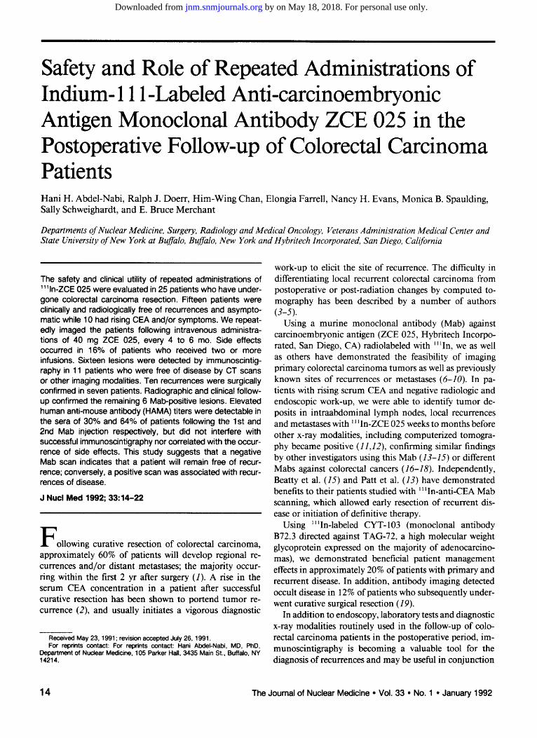
and/SPECT images. The location of these lesions are summarized in Table 2. "Hot" spots suggestive of liver metastasis were identified in two patients after the first Mab infusion and in another two patients after the second infusion. Figure 1 shows the anterior projection images of Patient 5 who presented with elevated CEA level and negative radiographic work-up. The first Mab scan ob- tained 3 mo prior to this study was negative. The subse- quent scans clearly show a lesion in the right lobe of the liver and suggests the presence of another lesion in the left lobe. Initially, all four patients had negative CT and 99roTe- sulfur colloid scans of the liver at the time of the Mab infusion, but these studies became positive 4 mo later in two patients who did not have surgery. One of these patients is represented in Figures 2 and 3, which demon- strate a lesion in the right lobe of the liver on the Mab scan (Fig. 2) preceding positive findings on CT scans obtained 4 mo later (Fig. 3).
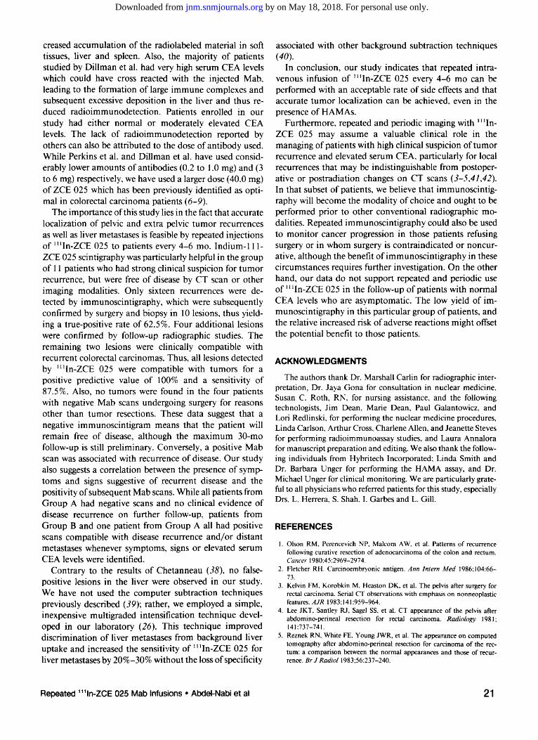
Seven of I 1 patients with positive ~t tln-ZCE 025 find- ings had exploratory laparotomy. Three of these patients had surgical exploration twice (Patients 13 and 24), while Patient 1 had three exploratory surgeries. This patient's first Mab scans (Fig. 4A-B) suggested the presence of a lesion in the anterior abdominal wall on the left side which was not evident on physical examination and was clearly delineated on a concurrent abdominal CT scan (Fig. 4C) where this mass was thought to be a portion of herniated bowel through the lateral rectus muscle. In retrospect, the
TABLE 2 Characteristics of Patients with Positive 1111n.ZCE 025 Scans and Confirmation
First Mab Infusion Second Mab Infusion Third Mab Infusion Fourth Mab Infusion
Patient CEA CEA CEA CEA no. {ng/ml) Scan findings Confirmation (ng/ml) Scan findings Confirmation (ng/ml) Scan findings Confirmation (ng/mt) Scan findings Confirmation
IIII III II III I I I I I
1 36.3 Anterior abdominal S, H 2.2 Negative (2560) E, R 38.1 Negative C, A, S 906 Metastasis to the ant. S & H wall recurrence abdominal wall (160)
5 37.0 Negative (40) E, R 184.0 Two liver metas- S, H . . . . . . tases (160)
9 2.8 Negative (640)(1) E, R 3.9 Negative (640) E, R 11.8 Perineal recurrence S, H, R - - - - - - mediastinal LN mets (640)
12 9.7 Negative(160) E,R 30.3 Possible pelvic re- C,A 68.0 Progression of pelvic C,A 247 Further progression of C,A currance (160) recurrence (640) pelvic recurrence
(160) 13 72 Anterior abdominal S, H 9.2 Negative (2560) C, E, R 29.5 Bilateral inguinal C 41.0 Negative (81920) E, R
wall recurrence lymphaOer~pathy (160) 0 024o)
15 13.3 Bone metastases to B, R 16.9 Bone metastases to B, R L4 vertebra (40) L4 vertebra (2560)
19 3.6 Mediastinal LN mets R, P, H 3.1 Mediastinal LN mets R, C (i 6o) (2o48o)
20 16.6 Abdominal LN mets S, H 39.0 Multiple bone rnetas- B, R (160) tases (1280)
21 4.6 IntTa~Jmin~ recur- E, S, H 11.5 Multiple liver mets R refce (160) (160)
24 2.0 ~ metastases S, H 2.0 Negative E, R S,H
25 4.1 Liver metastases R, L/S Progression of liver R, L/S metastases
Numbers ~7 parentheses indicate the amount of HAMA tRers in sera obtained 1-4 wk before each Mab infusion. E = endoscopy; R = radiography, including chest x-ray and/or CT scans of the ~ and pelvis; S = surgery; H = histopathotogy; C -- clinical follow-up; A -- serum CEA levets; P = percutaneous biopsy; B = radionuclide bone scan; LN -- lymph node;
and L/S = K v o r / s p ~ scan.
Repeated 11'In-ZCE 025 Mab Infusions • AbdeI-Nabi et al 17
by on May 18, 2018. For personal use only. jnm.snmjournals.org Downloaded from

~ , ~ .... gt'
B
J t l | ' i
FIGURE 1. Patient 5. (A) Anterior projection of the liver obtained 3 days after the second infusion of I~In-ZCE 025. (B) Multigrade intensification image showing a lesion in the right lobe of the liver (arrow) and possibly another lesion in the left lobe (arrow head), was neither seen on the first Mab scan nor the regular scan in (A). lntraoperatively, this lesion measured 2.5 cm in diameter and histologically was a metastatic well-differentiated adenocarcinoma.
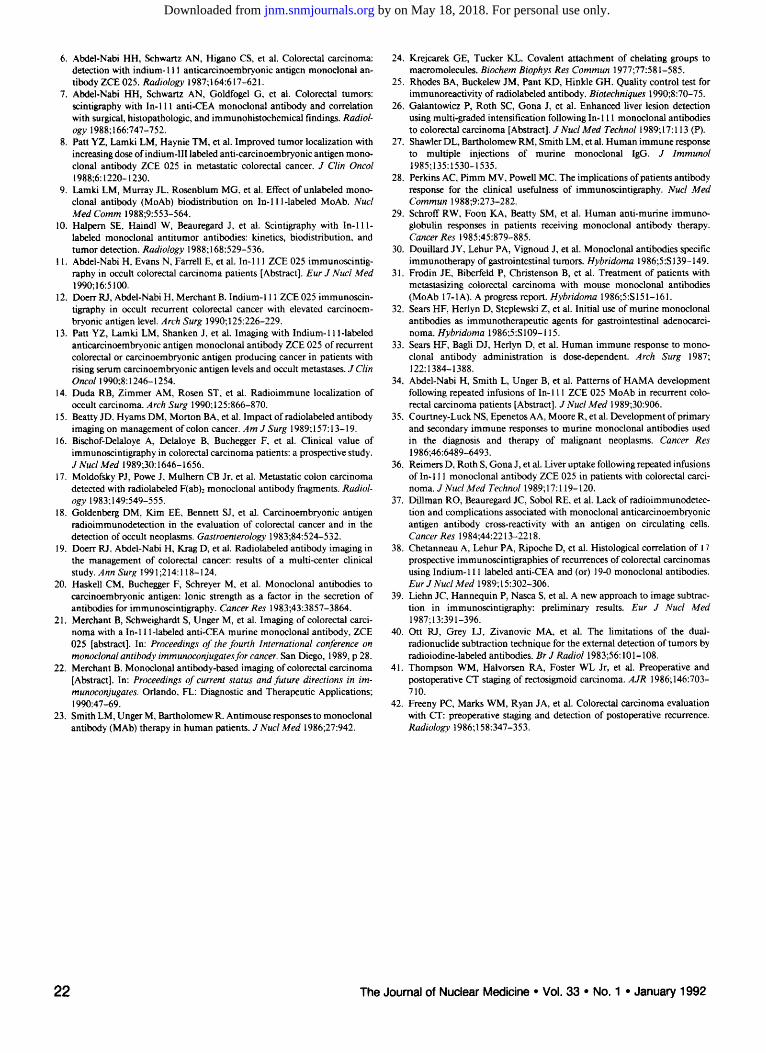
CT scans were interpreted positive for a mass in that region, since it could be separated from underlying bowel by a thin facial layer. A repeat fourth Mab scan (Fig. 5) was performed approximately 27 mo after the first one, because of very high CEA levels (906 ng/ml) and again showed metastatic involvement in the left anterior abdom- inal wall. Interim Mab scans 2 and 3 were negative.
Surgery was suggested by a progressive rise in serum CEA levels in six patients or for symptoms suggestive of local recurrence in one patient (no. 24). In this patient, '~ ~In-ZCE 025 Mab scan could not identify pelvic tumor recurrence, but detected a solitary lesion in the right lobe of the liver not previously suspected because of a normal serum CEA and a normal CT scan of the liver. This lesion was removed during surgery. The remaining four patients with positive Mab findings refused surgery; three were treated with chemotherapy and one patient (no. 15) re- fused all forms of treatment.
Ten of the 16 (62.5%) positive sites by Mab were con- firmed as cancerous by surgery (eight lesions) or biopsy (two lesions). Longitudinal radiographic follow-up studies confirmed the presence of liver metastasis in two patients
FIGURE 2, A multigrade intensification image of the liver 3 days after the second Mab infusion to Patient 21, showing a focal area of increased 1111n-Mab in the right lobe of the liver (arrow).
and bone metastases in the remaining two. In one patient with positive Mab uptake in the inguinal lymph nodes, physical examination revealed enlarged, non-tender, and freely mobile lymph nodes. A repeat physical examination performed 6 wk later was entirely negative, and a repeat Mab scan performed 4 mo later was also negative. In another patient (no. 12) with a positive lesion in the pelvis detected on the second Mab scan, follow-up CT scans did not confirm the Mab findings. However, increased size and radioactivity accumulation in that lesion was dem- onstrated on two subsequent Mab scans performed 6 mo apart. This patient refused explarotory laporatomy because of rapid progression of CEA from a value of 9.7 ng/ml immediately preceding the first negative Mab scan to values of 30.3, 68.0 and 343 ng/ml prior to the second, third, and fourth Mab scans, respectively, and is currently being treated with weekly 5-fluorourcil administrations.
As shown in Table 2, eight positive sites were detectable following the first Mab infusion, four were detected follow- ing the second, three following the third and one following the fourth infusion.
In the remaining 14 patients from Group A, no recur- rences were detected by repeated ~lJln-ZCE 025 scans, endoscopy, CT scans, periodic clinical evaluations and serum CEA determinations up to 36 mo from the initial scans. Two of the 14 patients with negative scans under- went exploratory laparotomy within 2.5 mo from the
FIGURE 3. Comput- erized tomograms of the liver with contrast obtained 3 mo fol- lowing the second Mab scan now show- ing multiple lesions in the liver.
1 8 The Journal of Nuclear Medicine • Vol. 33 • No. 1 ° January 1992
by on May 18, 2018. For personal use only. jnm.snmjournals.org Downloaded from

~ L S£¢YlOH$
FIGURE 4. Patient 1. (A) Anterior planar projection of the abdomen and pelvis obtained 5 days after the first Mab infusions showing an area of increased Mab accumulation in the left lower quadrant (arrow) suggestive of metastatic involvement of the anterior abdominal wall. (B) Transaxial SPECT image showing the anterior location of this lesion (arrowhead). (C) Abdominal CT with contrast showing as a 2-cm rounded soft-tissue mass lo- cated between the left lateral rectus muscle (curved arrow). A thin facial layer appears to be separating this mass from under- lying bowel (short arrow).
second Mab infusion. Indications for surgery were bowel obstruction secondary to adhesions (Patient 2) and chole- cystectomy for acute cholecystitis (Patient 10). No tumors were found by palpation and inspection during these op- erations. Also, two patients from the second group under- went surgery for relief of bowel obstruction. Patient 13 had surgery approximately 6 mo following the fourth Mab scans (no tumor found) and Patient 24 had surgery 2 mo following the second Mab scan, which was also negative. In both patients, no tumor was found intraoperatively.
FIGURE 5. Anterior planar projection of the abdomen and pelvis 5 days following the fourth infusion of ~I~In-ZCE 025 Mab to same patient as in Figure 4. Positive Mab localization is seen in the left hypochondrial area, suggestive of recurrent tumor (arrows). Intraoperatively, metastatic moderately well-differen- tiated adenocarcinoma measuring 7 x 5 x 3.5 cm was found in the jejunum. Transmural and peri-intestinal adipose tissue in- volvement and direct extension into the anterior abdominal wall were also found.
HAMA Development Serum samples were available from 24 patients for
HAMA evaluation. Pre-Mab infusion samples were nega- tive (_< l:160 titer) in 20 of 24 patients and positive in four patients. Those four patients remained positive during successive Mab infusions (Table 3). Of the 20 patients with baseline negative HAMAs, 6 (30%) became positive 2--4 wk following the first infusion, while 9 of 14 (64%) patients became positive after the second infusion. Two patients had modest HAMA titers ( 1:40 - _< 1 : 160) after the third infusion, and three patients had increased HAMA titers after the fourth infusion. All HAMA titers reported were obtained approximately 3-4 wk following each Mab in- fusion. Increase in HAMA titers from baseline levels fol- lowing repeated Mab infusion did not interfere with suc- cessful detection of metastasis in three patients. Patient 1 (Fig. 5) is a representative example, whereas an increase in HAMA titers from 1:160 baseline to 1:2560 immedi- ately preceding the fourth Mab administration was incon- sequential in the detection of this patient's metastases by immunoscintigraphy.
Patient Status and Fol low-up To date, 13 of the 14 patients from Group A who had
negative Mab scans are alive; 12 have no clinical or radiographic evidence of disease recurrence 18 to 31 mo following the first Mab scan. Four patients have received adjuvant chemotherapy with 5-Fluorouracil and leucovo- fin under a separate adjuvant chemotherapy trial at our institution. One patient (no. 10) died from cardiac arrest 8 mo after the second Mab scan, while another patient (no. 2) developed transitional-cell carcinoma of the urinary bladder. Local exploration during surgical resection of this patient's bladder cancer showed no evidence of abdominal recurrence. Of the 11 patients with positive Mab scans (one from Group A and I0 from Group B), 6 patients have died, 4 due to progression of metastatic disease (Patients 9, 20, 21, and 25) and 2 from complications
Repeated 1111n-ZCE 025 Mab Infusions • AbdeI-Nabi et al 19
by on May 18, 2018. For personal use only. jnm.snmjournals.org Downloaded from

TABLE 3 Human AntiMouse Antibody Titers
Following Repeated Infusions of in Pat ients ' Sera 1111N-ZCE 025
Patient First Second Third Fou~h no. Baseline* infus~n t infusion infusion infusion
II II I IIIIII III I lllllllIll IIII lUlll
1 160 160 2560 2560 NA* 2 160 640 160 2560 160 3 40 640 160 4 160 160 40,960 5 40 40 160 6 160 160 655,360 40,960 7 160 160 2560 160 81,920 8 640 2560 10,240 1280 NA 9 640 640 640 640
10 160 160 10,240 81,920 11 160 160 2560 640 12 160 160 160 640 160 13 160 160 2560 10,240 81,920 14 2560 2560 640 163,840 NA 15 40 40 2560 16 160 40 160 2560 17 640 640 163,840 18 160 640 163,840 655,360 19 160 160 20,480 20 160 640 40,960 21 160 160 10,240 10,240 22 160 160 160 23 160 320 1280
* Baseline values obtained 1-3 days before first Mab injection. * HAMAs present in serum samples obtained 3-4 wk following
each Mab infusion. * NA = not available. Grading System: HAMA titers 1:40-1:160 ( - - ) , >1:160-1:2560
(+), >1:2560-1:40,960 (++), >1:40,960-1:655,360 (+++).
following surgery to resect metastases from the anterior abdominal wall (Patient 1) or the liver (Patient 5). Five patients are still active; two without evidence of disease recurrence following four Mab infusions (Patient 13) or two Mab infusions (Patient 24), while the remaining three patients have recurrent and/or metastatic disease and con- tinue to receive chemotherapy.
DISCUSSION
The results of this study indicate that the administration of four separate doses of 40 mg of ZCE 025 at 4-6-mo intervals were associated with an increased incidence of side effects, although none of these reactions were serious or life-threatening. This study also demonstrates that re- current colorectal carcinomas can be detected by immu- noscintigraphy following repeated administration of 11 qn- ZCE 025 despite positive HAMA titers.
The relative safety of multiple infusions ofmurine Mabs in diagnostic amounts has been reported previously (28) and is redemonstrated in this larger study. Several thera- peutic trials with unlabeled routine monoclonal antibodies in patients with a variety of malignancies also have dem-
onstrated the relative safety of multiple infusions of m urine Mabs. Schroff et al. reported no adverse reactions in their clinical trials of patients with chronic lymphocytic leuke- mia, cutaneous T-cell lymphomas, or melanoma treated with mouse Mab at doses ranging from 1 to 200 mg bi- weekly (29). The lack of adverse reactions was also re- ported by Douillard and associates in patients with gas- trointestinal carcinoma receiving Mab 17-1A at doses of 100 to 200 mg, even though HAMAs were detectable in the sera of 40% of their patients (30). Other reports by Frodin et al. (31), or Sears et al. (32,33), also have con- firmed the safety of multiple infusions of murine Mab.
In this study minor adverse reactions (skin rash and pruritis) were reported by three patients following the second administration of murine Mab. These patients did not exhibit adverse signs during or following the 1.0 mg pre-test tracer dose given intravenously 1 hr before the full dose of antibody. Pretreatment with diphenylhydramine instituted prior to the third and subsequent Mab infusions tended to decrease the incidence of adverse reactions after the third (0/13 patients) or fourth (1/7 patients) infusion. However when compared with the rate of adverse reactions (4.7%) following a single intravenous infusion of I~In- ZCE 025 (21,22), administration of four doses of 40 mg of ZCE 025 had a cumulative incidence (16%) of side effects. In our opinion, these side effects represented acute allergic reactions, which could neither be predicted nor avoided by prescreening by injecting 1 mg of antibody intravenously to patients. However the development of adverse reactions did not correlate with the presence of HAMAs which were detected in the sera of 30% and 64% of patients following the first and second infusions respec- tively. As previously described, human anti-murine im- munoglobulin responses developed within 1-3 wk follow- ing murine Mab administration and either returned to baseline levels, increased in titer or remained unchanged (34). In general, positive HAMA titers were detectable up to 12 mo following the last infusion in some patients. The solid-phase ELISA assay used in this study is specific only for human IgG anti-mouse IgG antibodies and does not detect human IgM response. Also, we have not attempted to identify anti-idiotypic, anti-isotopic, or anti-species an- tibodies, which have been shown to develop following single or multiple infusions of murine Mabs (27,29,35).
The development of immune response to the murine Mab in this study did not interfere with successful tumor detection and did not compromise the quality of the image obtained following each infusion, nor did we observe noticeable alteration in organ distribution or increased deposition of 1~ qn-ZCE 025 in the liver (36). These find- ings are in disagreement with previously reported studies, particularly those by Perkins et al. (28) and Dillman et aI. (37). The differences can be attributed to the choice and/ or dose of antibody used in those studies and ours. The Mabs used in Dillman's study were shown to be reactive with granulocytes and erythrocytes with subsequent in-
20 The Journal of Nuclear Medicine • Vol. 33 ° No. 1 • January 1992
by on May 18, 2018. For personal use only. jnm.snmjournals.org Downloaded from
creased accumulation of the radiolabeled material in soft tissues, liver and spleen. Also, the majority of patients studied by DiIlman et al. had very, high serum CEA levels which could have cross reacted with the injected Mab, leading to the formation of large immune complexes and subsequent excessive deposition in the liver and thus re- duced radioimmunodetection. Patients enrolled in our study had either normal or moderately elevated CEA levels. The lack of radioimmunodetection reported by others can also be attributed to the dose of antibody used. While Perkins et al. and Dillman et al. have used consid- erably lower amounts of antibodies (0.2 to 1.0 mg) and (3 to 6 mg) respectively, we have used a larger dose (40.0 mg) of ZCE 025 which has been previously identified as opti- mal in colorectal carcinoma patients (6-9).
The importance of this study lies in the fact that accurate localization of pelvic and extra pelvic tumor recurrences as well as liver metastases is feasible by repeated injections of t~qn-ZCE 025 to patients every 4-6 mo. Indium-I 11- ZCE 025 scintigraphy was particularly helpful in the group of 11 patients who had strong clinical suspicion for tumor recurrence, but were free of disease by CT scan or other imaging modalities. Only sixteen recurrences were de- tected by immunoscintigraphy, which were subsequently confirmed by surgery and biopsy in 10 lesions, thus yield- ing a true-positive rate of 62.5%. Four additional lesions were confirmed by follow-up radiographic studies. The remaining two lesions were clinically compatible with recurrent colorectat carcinomas. Thus, all lesions detected by ~In-ZCE 025 were compatible with tumors for a positive predictive value of 100% and a sensitivity of 87.5%. Also, no tumors were found in the four patients with negative Mab scans undergoing surgery for reasons other than tumor resections. These data suggest that a negative immunoscintigram means that the patient will remain free of disease, although the maximum 30-mo follow-up is still preliminary. Conversely, a positive Mab scan was associated with recurrence of disease. Our study also suggests a correlation between the presence of symp- toms and signs suggestive of recurrent disease and the positivity of subsequent Mab scans. While all patients from Group A had negative scans and no clinical evidence of disease recurrence on further follow-up, patients from Group B and one patient from Group A all had positive scans compatible with disease recurrence and/or distant metastases whenever symptoms, signs or elevated serum CEA levels were identified.
Contrary to the results of Chetanneau (38), no false- positive lesions in the liver were observed in our study. We have not used the computer subtraction techniques previously described (39); rather, we employed a simple, inexpensive multigraded intensification technique devel- oped in our laboratory (26). This technique improved discrimination of liver metastases from background liver uptake and increased the sensitivity of t t~In-ZCE 025 for liver metastases by 20%-30% without the loss of specificity
associated with other background subtraction techniques (40).
In conclusion, our study indicates that repeated intra- venous infusion of ~ IIn-ZCE 025 every 4-6 mo can be performed with an acceptable rate of side effects and that accurate tumor localization can be achieved, even in the presence of HAMAs.
Furthermore, repeated and periodic imaging with ~ ~ In- ZCE 025 may assume a valuable clinical role in the managing of patients with high clinical suspicion of tumor recurrence and elevated serum CEA, particularly for local recurrences that may be indistinguishable from postoper- ative or postradiation changes on CT scans (3-5,41,42). In that subset of patients, we believe that immunoscintig- raphy will become the modality of choice and ought to be performed prior to other conventional radiographic mo- dalities. Repeated immunoscintigraphy could also be used to monitor cancer progression in those patients refusing surgery or in whom surgery is contraindicated or noncur- ative, although the benefit of immunoscintigraphy in these circumstances requires further investigation. On the other hand, our data do not support repeated and periodic use of ~In-ZCE 025 in the follow-up of patients with normal CEA levels who are asymptomatic. The low yield of im- munoscintigraphy in this particular group of patients, and the relative increased risk of adverse reactions might offset the potential benefit to those patients.
ACKNOWLEDGMENTS
The authors thank Dr. Marshall Carlin for radiographic inter- pretation, Dr. Jaya Gona for consultation in nuclear medicine, Susan C. Roth, RN, for nursing assistance, and the following technologists, Jim Dean, Marie Dean, Paul Galantowicz, and Lori Redlinski, for performing the nuclear medicine procedures, Linda Carlson, Arthur Cross, Charlene Allen, and Jeanette Steves for performing radioimmunoassay studies, and Laura Annalora for manuscript preparation and editing. We also thank the follow- ing individuals from Hybritech Incorporated; Linda Smith and Dr. Barbara Unger for performing the HAMA assay, and Dr. Michael Unger for clinical monitoring. We are particularly grate- ful to all physicians who referred patients for this study, especially Drs. L. Herrera, S. Shah, I. Garbes and L. Gill.
REFERENCES
1. Olson RM, Perencevich NP, Malcom AW, et al. Patterns of recurrence following curative resection of adenocarcinoma of the colon and rectum. Cancer 1980:45:2969-2974.
2. Fletcher RH. Carcinoembryonic antigen. Ann Intern Med t986;104:66- 73.
3. Kelvin FM, Korobkin M, Heaston DK, et al. The pelvis after surgery for rectal carcinoma. Serial CT observations with emphasis on nonneoplastic features. A JR 1983:141:959-964.
4. Lee JKT, Santley R J, Sagel SS, et at. CT appearance of the pelvis after abdomino-pefineal resection for rectal carcinoma. Radiology 1981: t41:737-741.
5. Reznek RN, White FE, Young JWR, et al. The appearance on computed tomography after abdomino-perineal resection for carcinoma of the rec- tum: a comparison between the normal appearances and those of recur- fence. Br J Radiol 1983;56:237-240.
Repeated 1111n-ZCE 025 Mab Infusions • AbdeI-Nabi et al 21
by on May 18, 2018. For personal use only. jnm.snmjournals.org Downloaded from
6. AbdeI-Nabi HH, Schwartz AN, Higano CS, et al. Colorectal carcinoma: detection with indium-111 anticarcinoembryonic antigen monoclonal an- tibody ZCE 025. Radiology 1987;164:617-621.
7. Abdel-Nabi HH, Schwartz AN, Goldfogel G, et al. Colorectal tumors: scintigraphy with in-I 11 anti-CEA monoclonal antibody and correlation with surgical, histopathologic, and immunohistochemical findings. Radiol- ogy 1988;166:747-752.
8. Patt YZ, Lamki LM, Haynie TM, et al. Improved tumor localization with increasing dose ofindium-llI labeled anti-carcinoembryonic antigen mono- clonal antibody ZCE 025 in metastatic colorectal cancer. J Clin Oncol 1988;6:1220-1230.
9. Lamki LM, Murray JL, Rosenblum MG, et al. Effect of unlabeled mono- clonal antibody (MoAb) biodistribution on In-11 l-labeled MoAb. Nucl Med Comm 1988;9:553-564.
10. Halpern SE, Haindl W, Beauregard J, et al. Scintigraphy with In-11 l- labeled monoclonal antitumor antibodies: kinetics, biodistribution, and tumor detection. Radiology 1988; 168:529-536.
l 1. Abdel-Nabi H, Evans N, Farrell E, et al. In-111 ZCE 025 immunoscintig- raphy in occult cotorectal carcinoma patients [Abstract]. Eur J Nucl Med 1990;16:5t00.
12. Doerr R J, Abdel-Nabi H, Merchant B. Indium-I 11 ZCE 025 immunoscin- tigraphy in occult recurrent colorectal cancer with elevated carcinoem- bryonic antigen level. Arch Surg 1990;125:226-229.
13. Patt YZ, Lamki LM, Shanken J, et at. Imaging with Indium-11 l-labeled anticarcinoembryonic antigen monoclonal antibody ZCE 025 of recurrent colorectal or carcinoembryonic antigen producing cancer in patients with rising serum carcinoembryonic antigen levels and occult metastases. J Clin Oncol 1990;8:1246-1254.
14. Duda RB, Zimmer AM, Rosen ST, et al. Radioimmune localization of occult carcinoma. Arch Surg 1990;125:866-870.
15. Beatty JD. Hyams DM, Morton BA, et al. Impact of radiolabeted antibody imaging on management of colon cancer. A m J Surg 1989; 157:13-19.
16. Bischof-Delaloye A, Delaloye B, Buchegger F, et at. Clinical value of immunoscintigraphy in colorectal carcinoma patients: a prospective study. J Nucl Med 1989;30:1646-1656.
17. Motdofsky PJ, Powe J, Mulhern CB Jr, et al. Metastatic colon carcinoma detected with radiolabeted F(ab)2 monoclonal antibody fragments. Radiol- ogy 1983:149:549-555.
18. Goldenberg DM, Kim EE, Bennett S J, et at. Carcinoembryonic antigen radioimmunodetection in the evaluation of colorectal cancer and in the detection of occult neoplasms. Gastroenterology 1983;84:524-532.
19. Doerr RJ, Abdet-Nabi H, Krag D, et al. Radiotabeled antibody imaging in the management of colorectal cancer, results of a multi-center clinical study. Ann Surg 1991;214:118-124.
20. Haskell CM, Buchegger F, Schreyer M, et al. Monoclonal antibodies to carcinoembryonic antigen: Ionic strength as a factor in the secretion of antibodies for immunoscintigraphy. Cancer Res 1983;43:3857-3864.
21. Merchant B, Schweighardt S, Unger M, et al. Imaging of colorectal carci- noma with a In-11 l-labeled anti-CEA murine monoclonal antibody, ZCE 025 [abstract]. In: Proceedings of the fourth International conference on monoclonal antibody immunoconjugatesfor cancer. San Diego, 1989, p 28.
22. Merchant B. Monoclonal antibody-based imaging of colorectal carcinoma [Abstract]. In: Proceedings of current status and future directions in im- rnunoconjugates. Orlando, FL: Diagnostic and Therapeutic Applications; ! 990:47-69.
23. Smith LM, Unger M, Bartholomew R. Antimouse responses to monoclonal antibody (MAb) therapy in human patients. J Nucl Med t986;27:942.
24. Krejcarek GE, Tucker KL. Covalent attachment of chelating groups to macromotecules. Biochem Biophys Res Commun 1977;77:581-585.
25. Rhodes BA, Buckelew JM, Pant KD, Hinkle GH. Quality control test for immunoreactivity of radiolabeled antibody. Biotechniques t 990;8:70-75.
26. Galantowicz P, Roth SC, Gona J, et al. Enhanced liver lesion detection using multi-graded intensification following In- 111 monoclonat antibodies to colorectal carcinoma [Abstract]. J Nud Med Technol 1989;17:113 (P).
27. Shawler DL, Bartholomew RM, Smith LM, et at. Human immune response to multiple injections of murine monoctonal IgG. J Immunol 1985;135: i 530-1535.
28. Perkins AC, Pimm MV, Powell MC. The implications of patients antibody response for the clinical usefulness of immunoscintigraphy. Nucl Med Commun 1988;9:273-282.
29. Schroff RW, Foon KA, Beatty SM, et al. Human anti-murine immuno- globulin responses in patients receiving monoclonal antibody therapy. Cancer Res 1985;45:879-885.
30. Douillard J Y, Lehur PA, Vignoud J, et al. Monoclonal antibodies specific immunotherapy of gastrointestinal tumors. Hybridoma 1986;5:S 139-149.
31. Frodin JE, Biberfeld P, Christenson B, et al. Treatment of patients with metastasizing colorectal carcinoma with mouse monoclonal antibodies (MoAb 17-1A). A progress report. Hybridoma 1986;5:S151-161.
32. Sears HF, Herlyn D, Steplewski Z, et al. Initial use of murine monoclonal antibodies as immunotherapeutic agents for gastrointestinal adenocarci- noma. Hybridoma 1986;5:S109-115.
33. Sears HF, Bagli D J, Herlyn D, et al. Human immune response to mono- clonal antibody administration is dose-dependent. Arch Surg 1987; 122:1384-1388.
34. Abdet-Nabi H, Smith L, Unger B, et al. Patterns of HAMA development following repeated infusions of In- 111 ZCE 025 MoAb in recurrent colo- rectal carcinoma patients [Abstract]. J Nucl Med 1989;30:906.
35. Courtney-Luck NS, Epenetos AA, Moore R, et al. Development of primary and secondary immune responses to murine monoclonal antibodies used in the diagnosis and therapy of malignant neoplasms. Cancer Res 1986;46:6489-6493.
36. Reimers D, Roth S, Gona J, et at. Liver uptake following repeated infusions of In-I 11 monoclonal antibody ZCE 025 in patients with colorectal carci- noma. J Nucl Med Technol 1989;17:119-120.
37. Dillman RO, Beauregard JC, Sobol RE, et al. Lack of radioimmunodetec- tion and complications associated with monoclonal anticarcinoembryonic antigen antibody cross-reactivity with an antigen on circulating cells. Cancer Res 1984;44:2213-2218.
38. Chetanneau A, Lehur PA, Ripoche D, et al. Histological correlation of 1 ] prospective immunoscintigraphies of recurrences of colorectal carcinomas using Indium- 111 labeled anti-CEA and (or) 19-0 monoclonal antibodies. Eur J Nucl Med 1989;15:302-306.
39. Liehn JC, Hannequin P, Nasca S, et al. A new approach to image subtrac- tion in immunoscintigraphy: preliminary, results. Eur J Nucl Med 1987;13:391-396.
40. Ott RJ, Grey LJ, Zivanovic MA, el al. The limitations of the duaJ- radionuclide subtraction technique for the external detection of tumors by radioiodine-labeted antibodies. Br J Radiol 1983;56: l 01-108.
41. Thompson WM, Haivorsen RA, Foster WL Jr, et al. Preoperative and postoperative CT staging of rectosigmoid carcinoma. A JR 1986;146:703- 710.
42. Freeny PC, Marks WM, Ryan JA, et al. Colorectal carcinoma evaluation with CT: preoperative staging and detection of postoperative recurrence. Radiology t986;158:347-353.
22 The Joumal of Nuclear Medicine ° Vol. 33 ° No. 1 ° January 1992
by on May 18, 2018. For personal use only. jnm.snmjournals.org Downloaded from
1992;33:14-22.J Nucl Med. Schweighardt and E. Bruce MerchantHani H. Abdel-Nabi, Ralph J. Doerr, Him-Wing Chan, Elongia Farrell, Nancy H. Evans, Monica B. Spaulding, Sally Postoperative Follow-up of Colorectal Carcinoma PatientsAnti-carcinoembryonic Antigen Monoclonal Antibody ZCE 025 in the Safety and Role of Repeated Administrations of Indium-111-Labeled
http://jnm.snmjournals.org/content/33/1/14This article and updated information are available at:
http://jnm.snmjournals.org/site/subscriptions/online.xhtml
Information about subscriptions to JNM can be found at:
http://jnm.snmjournals.org/site/misc/permission.xhtmlInformation about reproducing figures, tables, or other portions of this article can be found online at:
(Print ISSN: 0161-5505, Online ISSN: 2159-662X)1850 Samuel Morse Drive, Reston, VA 20190.SNMMI | Society of Nuclear Medicine and Molecular Imaging
is published monthly.The Journal of Nuclear Medicine
© Copyright 1992 SNMMI; all rights reserved.
by on May 18, 2018. For personal use only. jnm.snmjournals.org Downloaded from