Embed Size (px)
Citation preview

1
“Role of Locking Compression Plate For
Distal Radius Fractures in
postmenopausal women ”
Dissertation submitted in
Partial fulfillment of the regulations required for the award of
M.S. DEGREE
In
Orthopaedic Surgery Branch - II
THE TAMILNADU
DR. M.G.R. MEDICAL UNIVERSITY
CHENNAI
APRIL 2014

2
Certificate Certificate Certificate Certificate

3
CERTIFICATE
This is to certify that this dissertation titled “Role of Locking
Compression Plate For Distal Radius Fractures in postmenopausal
women” submitted to the Tamil Nadu Dr. M.G.R. Medical University,
Chennai in partial fulfillment of the requirement for the award of M.S
Degree Branch - II (Orthopaedic Surgery) is a bonafide work done by
DR.KARTHIKEYAN.S, under my direct guidance and supervision in the
Department of Orthopaedic Surgery, Coimbatore Medical College
Hospital, Coimbatore during his period of study from May 2011-April
2014.
Dr.S.VetrivelChezian,MSOrtho,FRCS, D(Ortho)
Associate Professor,
Department Of Orthopaedics,
Coimbatore Medical College Hospital,
Coimbatore.
Prof. S. Dhandapani, M.S. Ortho, MCh Ortho.
Professor and Head of the Department,
Department of Orthopaedics,
Coimbatore Medical College Hospital,
Coimbatore.
Dr. Vimala, M.D.
Dean,
Coimbatore Medical College Hospital
Coimbatore

4

5

6

7
DeclarationDeclarationDeclarationDeclaration

8
DECLARATION
I, Dr.KARTHIKEYAN.S declare that the Dissertation titled “Role of
Locking Compression Plate For Distal Radius Fractures in
postmenopausal women” submitted to the Dr. MGR medical university,
Guindy, Chennai is an original work done by me during the academic period
from May 2011-April 2014 at the Department of Orthopaedics, Coimbatore
Medical College Hospital, Coimbatore, under the guidance and direct
supervision of Dr.S.VetrivelChezian,MS Ortho,FRCS, D(ortho) in partial
fulfillment of the rules & regulations of the Dr. MGR Medical university for MS
Orthopaedics post graduate degree.
All the details of the patients, the materials and methods used are true to
the best of my knowledge.
I assure that this dissertation has not been submitted to or evaluated by
any other Medical University.
Dr. KARTHIKEYAN .S

9
Acknowledgement Acknowledgement Acknowledgement Acknowledgement

10
ACKNOWLEDGEMENT
My sincere thanks and gratitude to Dr.Vimala, MD, Dean,
Coimbatore Medical College, for permitting me to utilize the clinical
materials of this hospital.
I have great pleasure in thanking my teacher and guide
Dr.S.VetrivelChezian, MS Ortho,FRCS,D(Ortho) Associate Professor,
Department of Orthopaedic Surgery, Coimbatore Medical College for
permitting me to use the clinical materials and for his valuable advice and
encouragement in preparing this dissertation.
I am very much grateful to Prof.S.Dhandapani D Ortho, MS
Ortho, MCh Ortho, Professor and Head Of the Department,
Department of Orthopaedic Surgery, Coimbatore Medical College Hospital
for his valuable support and guidance that he has provided me throughout
this study.
I have great pleasure in acknowledging the help rendered by
Prof.S.Elangovan D Ortho., MS Ortho., for his valuable advice and
guidance.
My sincere thanks to Dr.P.Balamurugan, Dr.Major.K.Kamalanathan,
Dr.K.S.Maheswaran, Dr.M.S.Mugundhan, Dr.R.Prabhakar Singh, for
their suggestion and help during my study.
I am also thankful to all my colleagues and staff members of the
Department of Orthopaedics who helped me in all possible ways.

11
Contents Contents Contents Contents

12
CONTENTS
S.No Title Page
No
1. INTRODUCTION 1
2. AIM AND OBJECTIVES 4
3. HISTORICAL ASPECTS 5
4. REVIEW OF LITERATURE 7
5. ANATOMY 14
6. MECHANISM OF INJURY 25
7. CLASSIFICATION 28
8. PRINCIPLES OF MANAGEMENT 42
9. OSTEOPOROSIS 45
10. IMPLANT FEATURES 47
11. SPECIFIC SURGICAL CONSIDERATIONS 51
12. COMPARISION BETWEEN CONVENTIONAL
PLATE AND LOCKING COMPRESSION PLATE 54
13. COMPLICATIONS 56
14. ADVANTAGES OF LOCKING COMPRESSION
PLATING 57
15. MATERIALS AND METHODS 59
16. RESULTS 65
17. DISCUSSION 75
18. CONCLUSION 80
19. ANNEXURES

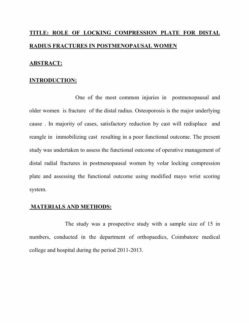
TITLE: ROLE OF LOCKING COMPRESSION PLATE FOR DISTAL
RADIUS FRACTURES IN POSTMENOPAUSAL WOMEN
ABSTRACT:
INTRODUCTION:
One of the most common injuries in postmenopausal and
older women is fracture of the distal radius. Osteoporosis is the major underlying
cause . In majority of cases, satisfactory reduction by cast will redisplace and
reangle in immobilizing cast resulting in a poor functional outcome. The present
study was undertaken to assess the functional outcome of operative management of
distal radial fractures in postmenopausal women by volar locking compression
plate and assessing the functional outcome using modified mayo wrist scoring
system.
MATERIALS AND METHODS:
The study was a prospective study with a sample size of 15 in
numbers, conducted in the department of orthopaedics, Coimbatore medical
college and hospital during the period 2011-2013.

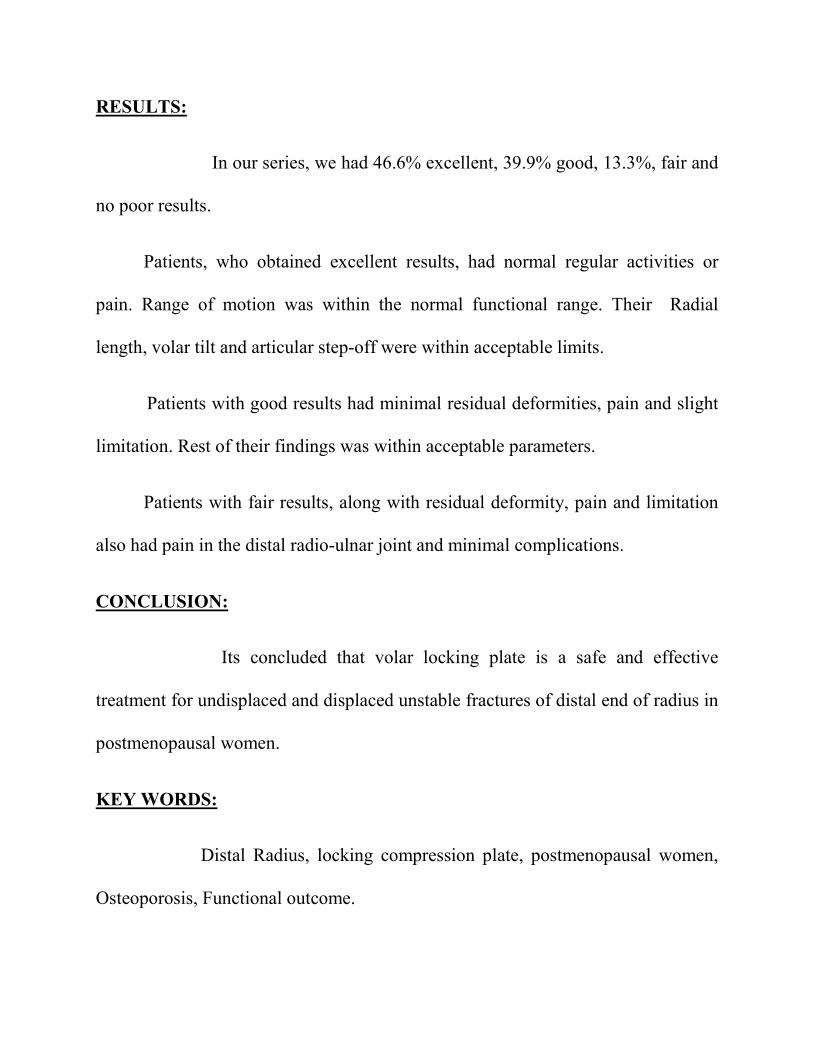
RESULTS:
In our series, we had 46.6% excellent, 39.9% good, 13.3%, fair and
no poor results.
Patients, who obtained excellent results, had normal regular activities or
pain. Range of motion was within the normal functional range. Their Radial
length, volar tilt and articular step-off were within acceptable limits.
Patients with good results had minimal residual deformities, pain and slight
limitation. Rest of their findings was within acceptable parameters.
Patients with fair results, along with residual deformity, pain and limitation
also had pain in the distal radio-ulnar joint and minimal complications.
CONCLUSION:
Its concluded that volar locking plate is a safe and effective
treatment for undisplaced and displaced unstable fractures of distal end of radius in
postmenopausal women.
KEY WORDS:
Distal Radius, locking compression plate, postmenopausal women,
Osteoporosis, Functional outcome.

TITLE: ROLE OF LOCKING COMPRESSION PLATE FOR DISTAL
RADIUS FRACTURES IN POSTMENOPAUSAL WOMEN
ABSTRACT:
INTRODUCTION:
One of the most common injuries in postmenopausal and
older women is fracture of the distal radius. Osteoporosis is the major underlying
cause . In majority of cases, satisfactory reduction by cast will redisplace and
reangle in immobilizing cast resulting in a poor functional outcome. The present
study was undertaken to assess the functional outcome of operative management of
distal radial fractures in postmenopausal women by volar locking compression
plate and assessing the functional outcome using modified mayo wrist scoring
system.
MATERIALS AND METHODS:
The study was a prospective study with a sample size of 15 in
numbers, conducted in the department of orthopaedics, Coimbatore medical
college and hospital during the period 2011-2013.

RESULTS:
In our series, we had 46.6% excellent, 39.9% good, 13.3%, fair and
no poor results.
Patients, who obtained excellent results, had normal regular activities or
pain. Range of motion was within the normal functional range. Their Radial
length, volar tilt and articular step-off were within acceptable limits.
Patients with good results had minimal residual deformities, pain and slight
limitation. Rest of their findings was within acceptable parameters.
Patients with fair results, along with residual deformity, pain and limitation
also had pain in the distal radio-ulnar joint and minimal complications.
CONCLUSION:
Its concluded that volar locking plate is a safe and effective
treatment for undisplaced and displaced unstable fractures of distal end of radius in
postmenopausal women.
KEY WORDS:
Distal Radius, locking compression plate, postmenopausal women,
Osteoporosis, Functional outcome.
TITLE: ROLE OF LOCKING COMPRESSION PLATE FOR DISTAL
RADIUS FRACTURES IN POSTMENOPAUSAL WOMEN
ABSTRACT:
INTRODUCTION:
One of the most common injuries in postmenopausal and
older women is fracture of the distal radius. Osteoporosis is the major underlying
cause . In majority of cases, satisfactory reduction by cast will redisplace and
reangle in immobilizing cast resulting in a poor functional outcome. The present
study was undertaken to assess the functional outcome of operative management of
distal radial fractures in postmenopausal women by volar locking compression
plate and assessing the functional outcome using modified mayo wrist scoring
system.
MATERIALS AND METHODS:
The study was a prospective study with a sample size of 15 in
numbers, conducted in the department of orthopaedics, Coimbatore medical
college and hospital during the period 2011-2013.
RESULTS:
In our series, we had 46.6% excellent, 39.9% good, 13.3%, fair and
no poor results.
Patients, who obtained excellent results, had normal regular activities or
pain. Range of motion was within the normal functional range. Their Radial
length, volar tilt and articular step-off were within acceptable limits.
Patients with good results had minimal residual deformities, pain and slight
limitation. Rest of their findings was within acceptable parameters.
Patients with fair results, along with residual deformity, pain and limitation
also had pain in the distal radio-ulnar joint and minimal complications.
CONCLUSION:
Its concluded that volar locking plate is a safe and effective
treatment for undisplaced and displaced unstable fractures of distal end of radius in
postmenopausal women.
KEY WORDS:
Distal Radius, locking compression plate, postmenopausal women,
Osteoporosis, Functional outcome.

TITLE: ROLE OF LOCKING COMPRESSION PLATE FOR DISTAL
RADIUS FRACTURES IN POSTMENOPAUSAL WOMEN
ABSTRACT:
INTRODUCTION:
One of the most common injuries in postmenopausal and
older women is fracture of the distal radius. Osteoporosis is the major underlying
cause . In majority of cases, satisfactory reduction by cast will redisplace and
reangle in immobilizing cast resulting in a poor functional outcome. The present
study was undertaken to assess the functional outcome of operative management of
distal radial fractures in postmenopausal women by volar locking compression
plate and assessing the functional outcome using modified mayo wrist scoring
system.
MATERIALS AND METHODS:
The study was a prospective study with a sample size of 15 in
numbers, conducted in the department of orthopaedics, Coimbatore medical
college and hospital during the period 2011-2013.

RESULTS:
In our series, we had 46.6% excellent, 39.9% good, 13.3%, fair and
no poor results.
Patients, who obtained excellent results, had normal regular activities or
pain. Range of motion was within the normal functional range. Their Radial
length, volar tilt and articular step-off were within acceptable limits.
Patients with good results had minimal residual deformities, pain and slight
limitation. Rest of their findings was within acceptable parameters.
Patients with fair results, along with residual deformity, pain and limitation
also had pain in the distal radio-ulnar joint and minimal complications.
CONCLUSION:
Its concluded that volar locking plate is a safe and effective
treatment for undisplaced and displaced unstable fractures of distal end of radius in
postmenopausal women.
KEY WORDS:
Distal Radius, locking compression plate, postmenopausal women,
Osteoporosis, Functional outcome.

13
Introduction Introduction Introduction Introduction

14
INTRODUCTION
Distal radius fractures are common and produce a major orthopaedic
injuries because of the advancing population age and increase in physical
activity. Distal radius fractures constitute up to 15% of all extremity
fractures. In females the incidence rises sharply. In the age of 40 it is
approximately 36.8/10,000. It is estimated at the age 70 years to be
115/10,000. Distal radius is the most common osteoporotic fracture in
elderly females it has been linked to estrogen withdrawal. These injuries
are sustained overwhelmingly from low energy falls in an increasingly
osteoporotic population.
This group of patients expect increased functional demands since
they are independent and active. Treating the growing number of these
difficult injuries presents a particular challenge for orthopaedic surgeons.
Locking compression plate (LCP) is a new generation plate and screw
system for internal fixation of fractures(1,2). The LCP with combi holes
have additional dynamic compression holes providing options for axial
compression in addition to locking mechanism. The LCP can be used as a
compression plate, a locked internal fixator, or a combination of both,
depending on the situation(3,4).

15
The use of locked volar plates for distal radius fractures is
increasingly popular. Proposed advantages of locked volar plating include
improved pull out strength even in osteoporotic bone and a volar surgical
approach that avoids the need for an extensive dorsal dissection. The plate
is positioned in a well padded area beneath pronator quadratus to avoid
flexor tendon irritation and it is thought that patients tolerate volar wrist
scars better than dorsal ones.
Internal fixation has the advantage of allowing early mobilisation but its
application is limited by the degree of comminution and osteoporosis. Loss
of reduction and fixation is common due to poor purchase of screws on
osteoporotic bone with the conventional plates, delay in postoperative
mobilization results in stiffness of the joint which is an indicator of poor
outcome.
Screws used in distal radial fractures (3.5mm cortical screws) can also be
used in addition to locking screws. Locking plates have advantages such as
a decreased incidence of loss of reduction secondary to screw toggling and
there is improved bone healing. A locking plate decreases the screw-plate
toggle and motion at bone-screw interface and provides more rigid
fixation. Rigid fixation is felt to be one key to the successful treatment of
these fractures(5,6).

16
But fixation in osteoporotic and comminuted fractures is difficult to obtain
anatomical reduction and adequate purchase. So now with the evolution of
locking compression plating for osteoporotic and peri-articular fractures
especially for the comminuted intra–articular fractures restoring the
anatomical congruity and providing stable fixation with resulting increased
stability allowing for early mobilization(7-9).

17
Aim Of The StudyAim Of The StudyAim Of The StudyAim Of The Study

18
AIM AND OBJECTIVES
The aim of the study is to analyze the functional outcome of locking
compression plate in distal radial fractures in postmenopausal women.

19
HISTORIAL ASPECTS
In the year 1814, Sir Abraham Colles, a surgeon from Ireland
described the fracture pattern affecting the distal radius before the
invention of X-rays. Ponteau, a French surgeon is said to have described
the same fracture earlier.
Other surgeons notably Smith and Barton also described fractures
of distal radius in the nineteenth century.
After the introduction of radiography, Hutchinson described radial
styloid fracture and named it as Chauffeur’s fracture. Initially surgeons
treated distal radius fractures with casts and splints.
Anderson and O’Neil described external fixator for distal radius
fractures in 1944. They were the pioneers in using external fixators for
management of distal radius fractures. They produced excellent results in
most of their patients.
In 1951, Gartland and Werley published their Demerit Point System
of functional evaluation of outcome of distal radius fracture.
In 1959, Lindstrom published his study on the end results of the
fractures of distal radius in the Journal of Acta Orthopaedica Scandinavia.
In 1967, Frykman introduced his classification. Cole and Obletz
described alternative method utilizing pins and plaster.

20
In 1965, Ellis described volar buttress plate for Barton’s fractures.
In 1985, Diego L. Fernandez introduced his system of distal radius
fracture classification.
In 1980s and 1990s, articles about open fixation with or without
external neutralization were published.

21
Review of LiteratureReview of LiteratureReview of LiteratureReview of Literature
22
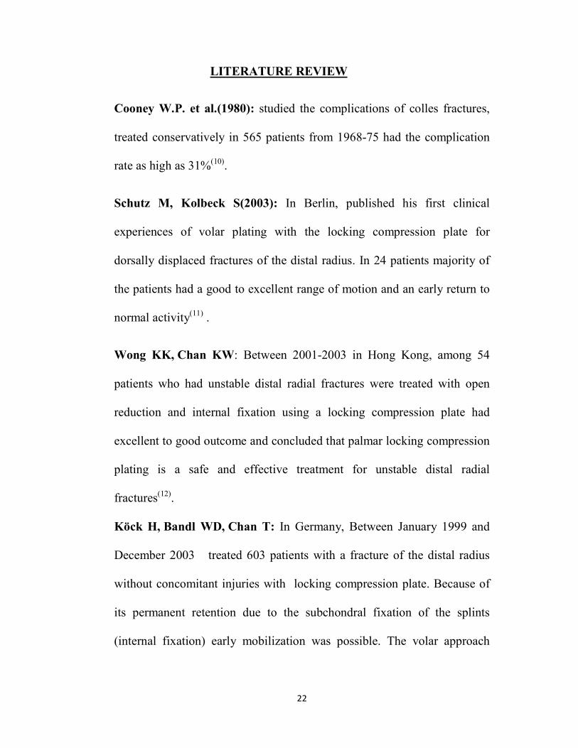
LITERATURE REVIEW
Cooney W.P. et al.(1980): studied the complications of colles fractures,
treated conservatively in 565 patients from 1968-75 had the complication
rate as high as 31%(10).
Schutz M, Kolbeck S(2003): In Berlin, published his first clinical
experiences of volar plating with the locking compression plate for
dorsally displaced fractures of the distal radius. In 24 patients majority of
the patients had a good to excellent range of motion and an early return to
normal activity(11) .
Wong KK, Chan KW: Between 2001-2003 in Hong Kong, among 54
patients who had unstable distal radial fractures were treated with open
reduction and internal fixation using a locking compression plate had
excellent to good outcome and concluded that palmar locking compression
plating is a safe and effective treatment for unstable distal radial
fractures(12).
Köck H, Bandl WD, Chan T: In Germany, Between January 1999 and
December 2003 treated 603 patients with a fracture of the distal radius
without concomitant injuries with locking compression plate. Because of
its permanent retention due to the subchondral fixation of the splints
(internal fixation) early mobilization was possible. The volar approach

23
reduced the risk of infections and offered the possibility of not having to
remove the plate(13).
Musgrave DS, Idler RS (2005): In Washington, determined whether
locking compression plates could be used to treat unstable distal radius
fractures. They proved that volar fixed-angle plate fixation with or without
radial styloid fixed-angle plate fixation would provide sufficient rigidity to
allow early active range of motion without compromising fracture
reduction and improve initiation of early functional outcomes(14).
Cognet JM(2006): In US, he analysed plate fixation with locking screw
for distal fractures of the radius in 67 patients. He concluded that
appropriate fixation method for distal fracture of radius remains a
controversial issue, but primary stability achieved with a locking screw in
a plate enables early mobilization associated with more rapid recovery of
function(15).
Scott M.Levin (2007): In Mineola, USA, stated that fixed angle constructs
withstand cyclical loading representing normal physiologic forces
encountered during postoperative rehabilitation. They concluded that
volar fixed-angle locking plates are an effective treatment for unstable
extra-articular distal radius fractures, allowing early postoperative
rehabilitation to safely be initiated(16).

24
Strohm PC(2007): In German, analysed whether locking, 3.5mm volar T-
Plate is the implant of choice for displaced distal radius fractures. He
concluded that 3.5mm T-LCP is a good implant for the stabilisation of
displaced distal radius fractures if the fragments are not too small for the
3.5mm screws(17).
Rohit Arora, et al.(2007): In Austria, 114 patients who had displaced,
unstable fractures involving the distal radius were treated with ORIF using
palmar LCP of 2.4mm size and followed up over a minimum time frame
of 12 months. All the data based on the clinical and radiological findings
were analyzed at the end of the research period and it was found that
fixation with fixed angled plates allowed only a minimal loss of reduction
and thereby provided stability to formerly unstable dorsally displaced
fractures involving the distal radius(18).
RE Anakwe(2010): In UK, Conducted a study on locked volar plating for
complex distal radius fractures. Over a 12 month period 21 patients with
type C distal radius fractures were treated using locked volar plating and
stated that Locked volar plating for complex distal radius fractures
produces good results when assessed using patient locked volar plating
offers superior outcomes and patient satisfaction compared to external
fixation(19).

25
Claudio Roberto Martins Xavier(2011): In Brazil, 64 subjects with distal
radius fractures were studied and followed up over a period of six months.
Their clinical findings and radiological findings were compiled after they
underwent surgery with fixed angled volar locked plates. They concluded
Use of volar plates is a treatment method with a low complication rate.
However, caution is needed in indicating this for elderly patients, and the
patient's activity level and the risks and benefits need to be taken into
consideration(20) .
Hanae Minegishi (may 2011): In Japan, a retrospective study of patients
with unstable intra and extra articular fractures involving the distal end of
radius who were treated with volar locking plates at Tohoku Kosai
Hospital was done. Treatment of unstable distal radius fractures with a
volar locking plate lead to satisfactory results, provided the operative
technique carefully performed to prevent complications(21).
Chris Dillingham et al.(june 2011): In USA, 27 patients with unstable
displaced fractures of distal radius were studied and followed up from
August 2002 to October 2008.All the subjects were treated with open
reduction and fixation using fixed angled volar locking plates. They
recommended the use of fixed angled volar locking plates when a rapid
recovery and early return to functional activity is desired by the patient(22).

26
Joideep Phadnis in 2012: In UK, 183 fractures amongst 180 patients of a
mean age of 62.4years were retrospectively studied and functional
assessment using modified MAYO wrist scores and it was found that
results were good to excellent in most of the patients treated with fixed
angled volar locking plates and complication rates comparable to
alternative lines of treatment be it operative or non-operative(23).
27
Indian studies in distal radius locking compression plating
Sawalha(2007): In Warrington retrospectively analysed radiological and
clinical outcomes of 52 patients treated with locking compression plate.At
14 months follow up,15 out of 52 patients had complications like median
nerve compression, hardware related complications, malunion and failure
of fixation. They concluded volar locking compression plate for distal
radius is associated with high complication rate and they recommended
cautious use of distal radius locking compression plate(24).
Shetty.M.S (Nov 2011): In Andhra, he conducted a prospective study
from March 2008 to September 2009, 23 cases of intra-articular distal
radius fractures were included in the study to assess the ability of volar
locking plates to maintain fracture reduction when used to treat dorsally
displaced intra-articular distal radial fractures and to assess the patient-
related outcome. All these fractures underwent open reduction and internal
fixation with 2.4 volar locking distal radius plates. He concluded Volar
locking plate is a viable option for treating intra-articular distal radius
fractures(25).
Agarwala(2012): In Mumbai, he retrospectively analysed 25 cases of
distal radius fracture treated with locking compression plate and obtained
88% of excellent to good result as assessed by Mayo wrist score. They

28
concluded locking compression plate allows early functional mobility with
minimal complications(26).
Nagesh Naik and Deshpande Shrikant Balakrishna(2012): In Sangli,
they studied 25 cases of dorsally displaced distal radius fracture treated
with volar locking compression plate. They obtained 80% excellent to
good results as assessed by Lidstrom’s anatomical criteria. They concluded
that volar locking plate is an effective and safe method of treatment for
unstable distal radius fracture(27).

29
AnatomyAnatomyAnatomyAnatomy

30
ANATOMY
The distal radius consists of
(1) metaphysis,
(2) scaphoid facet,
(3) lunate facet, and
(4) sigmoid notch.
The anatomy of the radius is unique. The bone has a cylindrical contour
proximally turning flatter distally with a concave contour on the dorsal and
ventral aspect .The distal articular end is triangular in shape and is lined
with hyaline cartilage. A ridge divides the distal articular surface into a
triangular facet for the scaphoid laterally. Medially it articulates with the
lunate over an articular surface that is shaped like a quadrilateral(28).
The distal end articulates with the ulna medially. The medial articular
surface is semilunar and is lined with hyaline cartilage.
Laterally the distal end of radius provides attachment to the brachioradialis
muscle via an elongated process called the radial styloid.

31
ANTERIOR DISTAL END OF RADIUS AND ULNA.
The metaphysis is flared distally in both the AP and the lateral
planes with thinner cortical bone lying dorsally and radially. The
significance of the thinness of these cortices is that the fractures typically
collapse dorsoradially. In addition, the bone with the greatest trabecular
density lies in the palmar ulnar cortex(29). The fact that this bone is thicker
even in osteoporotic cadaver specimens may explain the success of internal
fixation techniques, which take advantage of this superior bone. Distally
the radius has a somewhat trapezoidal shape. The radial styloid rotates
palmarly 15 degrees off the axis of the radius, which makes capture
difficult from a dorsal approach.
In the anteroposterior plane the strongest bone is found under the
lunate facet of the radius. The line of force passes down the long finger
axis through the capitolunate articulation and contacts the radius at this
location. The “palmar ulnar corner” is often referred to as the keystone of
32
the radius. It serves as the attachment for the palmar distal radioulnar
ligaments and also for the stout radiolunate ligament. Displacement of this
fragment is associated with palmar displacement of the carpus and also
with loss of forearm rotation(30).The result is loss of rotation as well as a
step-off in the articular surface.
Ligamentous Anatomy:The extrinsic ligaments of the wrist play a major
role in the use of indirect reduction techniques. The palmar extrinsic
ligaments are attached to the distal radius, and it is these ligaments that are
relied on to reduce the components of a fracture using closed methods.
There are two factors about these ligaments that make them
significant for reduction: First the orientation of the extrinsic ligaments
from the radial styloid is oblique relative to the more vertical orientation of
the ligaments attached to the lunate facet.
The second significance of the ligamentous anatomy is because of
the relative strengths of the thicker palmar ligaments when compared to
the thinner dorsal ligaments. In addition, the dorsal ligaments are oriented
in a relative “z” orientation, which allows them to lengthen with less force
than the more vertically oriented palmar ligaments. The significance is that
distraction will result in the palmar ligaments becoming taut before the
dorsal ligaments.

33
Thus the palmar cortex is brought out to length before the dorsal
cortex. It is for this reason that it is difficult to achieve reduction of the
normal 12 degrees of palmar tilt using distraction alone .
Applied Anatomy
The three column concept
The forearm consists of 3 columns:
a) Radial column-It comprises the radial styloid and the scaphoid fossa.
b) Intermediate column-It consists of the lunate fossa and the sigmoid
notch.

34
c) Ulnar column-It is formed of the distal radio-ulnar distal ulna
(DRUJ) with the triangular fibrocartilaginous complex (TFCC).
a. Radial column: Because of the radial inclination of 22 degreees,
impaction of the scaphoid on the articular surface results in a shear
moment on the radial styloid causing failure laterally at the radial cortex.
The radial column, therefore, is best stabilized by buttressing the lateral
cortex.
b. Intermediate column:
The intermediate column consists of the lunate fossa and the
sigmoid notch of the radius. The intermediate column may be considered
the cornerstone of the radius because it is critical for both articular
congruity and distal radioulnar function. Failure of the intermediate
column occurs as a result of impaction of the lunate on the articular surface
with dorsal comminution. The column is stabilized by a direct buttress of
the dorsal ulnar aspect of the radius.
c. Ulnar column: The ulnar column consists of the ulna styloid but also
should include the TFCC and the ulnocarpal ligaments.
Triangular Fibrocartilage Complex: The complex is attached by its
base to the medial margin of the distal radius and by its apex to the lateral
side of the base of the ulnar styloid. It is biconcave and articulates with the
distal ulna proximally and with the proximal carpal row, primarily the
35
triquetrum, on its distal surface. The triangular fibrocartilage is reinforced
anteriorly and posteriorly by fibrous bands that extend into the anterior and
posterior capsule of the distal radioulnar joint. This capsule is only
minimally reinforced and provides only minor support to the joint. The
TFCC is at maximum tension in approximately mid-position between
pronation and supination.
IMAGING TECHNIQUES FOR DISTAL RADIUS
FRACTURES
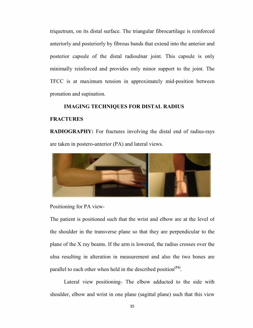
RADIOGRAPHY: For fractures involving the distal end of radius-rays
are taken in postero-anterior (PA) and lateral views.
Positioning for PA view-
The patient is positioned such that the wrist and elbow are at the level of
the shoulder in the transverse plane so that they are perpendicular to the
plane of the X ray beams. If the arm is lowered, the radius crosses over the
ulna resulting in alteration in measurement and also the two bones are
parallel to each other when held in the described position(31).
Lateral view positioning- The elbow adducted to the side with
shoulder, elbow and wrist in one plane (sagittal plane) such that this view

36
is perpendicular to PA view.
In the postero anterior view, for an extra articular fracture distal radius, the
following are noted.
1) Radial shortening
2) Ulnar variance
3) Radial angulation
4) Comminution
5) Ulnar styloid fracture location
In the lateral view, for an extra articular fracture distal radius, following
are noted.
1) Palmar tilt
2) Extent of metaphyseal comminution
3) Displacement of volar cortex
4) Position of distal radio ulnar joint.
A 5º rotational change produces 1.6º change in palmar tilt in conventional
lateral view. An oblique view may be useful to assess comminution in an
extra articular fracture. Postero anterior and lateral views are taken also for
contralateral wrist to assess the patient’s normal radiological parameters.

37
NORMAL RADIOLOGICAL PARAMETERS
Measurement of normal average radial angulation, radial length, and
palmar angulation.

38
Dorsal/Palmar Tilt
Volar tilt=11°.On a true lateral view a line is drawn connecting the
most distal points of the volar and dorsal lips of the radius. The dorsal or
palmar tilt is the angle created with a line drawn along the longitudinal
axis of the radius.
Radial Length
Radial length is measured on the PA radiograph. It is the measured
in millimeters between two lines,one line drawn perpendicular to the long
axis of the radius and tangential to the most distal point of the ulnar head
and the other line drawn perpendicular to the long axis of the radius and at
the level of the tip of the radial styloid . Average radial length from the tip
of the radial styloid to the ulna head is 12 mm, although the variance can
be considerable .
Ulnar Variance
Ulnar variance -0.6mm. This is a measure of radial shortening and is
different from measurement of radial length. It is the vertical distance
between a line parallel to the medial corner of the articular surface of the
radius and another line drawn parallel to the most distal point of the
articular surface of the ulnar head, both of which are perpendicular to the
long axis of the radius.

39
Radial Inclination
On the PA view the radius inclines towards the ulna. This is
measured by the angle between a line drawn from the tip of the radial
styloid to the medial corner of the articular surface of the radius and a line
drawn perpendicular to the long axis of the radius. There is normally an
average of 23° of radial angulation in the anteroposterior plane.
Carpal Malalignment
Two types of carpal malalignment are associated with fracture of the
distal radius. The most common is malalignment that compensates for the
tilt of the distal radius which is extrinsic to the carpus(32). On a lateral view
one line is drawn along the long axis of the capitate and one down the long
axis of the radius. If the carpus is aligned, the lines will intersect within the
carpus. If not, they will intersect outwith the carpus. Carpal malalignment
can also be caused by associated carpal ligament disruption.
KINEMATICS
The motors of the wrist are attached to the metacarpals. Capitate is
the centre of rotation of wrist joint.
Wrist flexion – extension occur equally through radio carpal and
midcarpal joints.

40
Radial – ulnar deviations occur 60% through midcarpal joint and
40% through radio carpal joint.
Normal range of Movements
Movements Normal range
Flexion 0 to 70-90º
Extension 0 to 70-90º
Radial deviation 0 to 15-25º
Ulnar deviation 0 to 25-35º
Supination 0 to 70-90º
Pronation 0 to 70-90º
Normally, 82% of the axial load at the wrist is borne by Radius and
18% by Ulna.

41
MECHANISM OF INJURY
Distal radius fractures are usually as a result of fall on an outstretched hand
is. There are multiple factors that determine the pattern of the fracture that
include-
1) Velocity
2) Position of hand and wrist at impact
3) Degree of rotation of forearm
4) Bone quality and density
When an individual falls forward fall on pronated forearm with the
hand and wrist in extension, bending of the metaphyseal bone because the
weight of the body is transmitted along the long axis of the radius. Also the
hard diaphyseal bone causes impaction of cancellous metaphyseal bone
that result in metaphyseal collapse. During the fall, compressive forces
over the dorsal cortex and tensile stress acting over the volar cortex result
in volar and dorsal cortical bone disruption. When there is a supination of
distal end of radius with respect of the radial diaphysis, a dorsal
displacement fracture occurs. In about fifty to sixty percent of distal radius
fractures, associated ulnar styloid fractures. Also ulnar styloid fractures
may be associated with triangular fibrocartilage disruption which may
sometimes be an isolated finding.
Three main theories have been developed;

42
• The theory of compression impaction
• The avulsion theory
• The incurvation theory.
The Theory of Compression Impaction:
When the wrist is in extension the carpal bones are in contact with
the surface of the impact. At the same time, the radial head is in
compression against the humerus. This force is then automatically
transmitted to the distal end of the radius. It is at this moment that the
fracture occurs.
It is therefore a mechanism of compression impaction and crush; the
wrist is an anvil on which the radius is crushed. This theory is based on
the very important fact that all distal radial fractures are compression
fractures and the fall occurs on a wrist in extension-pronation. Tensile
forces act on the anterior part and compression forces on the posterior part.
The posterior constraint forces are very high.
The Avulsion theory: The indirect forces presented by the body
weight are transmitted through the humerus, the ulna, the interosseous
membrane, the distal radius and then the volar wrist ligaments to the point
of impact of the hand. The distal radial fracture is then caused by an
avulsion mechanism applied by the tensile forces transmitted by the volar
wrist ligaments.

43
The Incurvation theory: This theory stated that fractures are
produced by bending forces. The fracture line is affected by three factors.
• The position of the hand;
• The extent of the area of impact;
• The magnitude of the applied force.
If tension increases at the level of the ulnar collateral ligament when
the radial fracture occurs, an ulnar styloid process fracture will occur at the
same time.The skin is usually not lacerated at the palm, implying that the
hand has not slipped but was blocked on the floor. The body continues to
go forward, moved by kinetic energy or inertia, and the volar wrist
ligaments become tense because the wrist is placed in a hyperextended
position(33).
If these ligaments resist, the forces are transmitted to the radiocarpal
joint and the radius is in compression against the articular facets of the
bones of the first carpal row. If the scaphoid and lunate are not crushed, the
forces end at the level of the radius to produce a fracture at the weakest
part of this bone. The dorsomedial fragment that separates due to this
impact is called the die punch fragment(34).

44
CLASSIFICATION
Various classification systems are available for distal radius
fractures.
These classifications have been based on; 1) Radiographic
appearance of fracture displacement direction.
a) The AO classification
b) The Sarmiento classification
c) The Lidstorm classification.
2) The mechanism of injury.
a) The Casting classification
b) The Fernandez classification
c) The Lincheid classification.
3) Articular joint surface involvement.
a) The Mayo classification
b) The McMurty and Jupiter classification
c) The Melone classification.
4) The degree of comminution:
a) The Gartland and Werley classification.
b) The Jenkins classification
c) The Older classification.

45
The OTA/AO classification emphasizes the increasing severity of
the bony injury. It is based on the location of the fracture line(s), the
displacement of the distal fragment, the extent of articular involvement,
and the presence of an ulnar styloid fracture(35).
Type A - Extraarticular fracture. Subgroups are based on direction of
displacement and comminution.
TypeA1, - .1 Styloid process Extraarticular fracture of ulna, radius intact
.2 Metaphyseal simple
.3 Metaphyseal multifragmentary
Type A2, - Extraarticular fracture of radius, simple & impacted
.1 Without any tilt
.2 With dorsal tilt(pouteau-colles)
.3 With volar tilt(Goyrand-smith)

46
Type A3, - Extraarticular fracture of radius, multifragmentary
.1 - Impacted with axial shortening
.2 - With a wedge
.3 - complex
Type B - Partial articular fracture. Subgroups are based on lateral
(radial styloid) palmar or dorsal fragments.
Type B1 - Partial articular fracture of radius, saggital
.1 - Lateral simple
.2 - Lateral multifragmentary
.3 - medial

47
Type B2 - Partial articular fracture of radius, dorsal rim
.1 - simple
.2 - With lateral saggital fracture
.3 - With dorsal dislocation of the carpus
Type B3 - Partial articular fracture of radius, volar rim
.1 - Simple, with a small fragment
.2 - Simple, with a large fragment
.3 - multifragmentary

48
Type C - Complete articular. Subgroups are based on the degree of
comminution of the articular surface and the metaphysis.
Type C1 - Complete articular fracture of radius, articular and metaphyseal
simple
.1 - Posteromedial articular fragment
.2 - Saggital articular fracture line
.3 - Frontal articular fracture line
TypeC2 - Complete articular of radius, articular simple and metaphyseal
multifragmentary
.1 - Saggital articular fracture line
.2 - Frontal articular fracture line
.3 - Extending in to diaphysis

49
Type C3 - Complete articular of radius, multifragmentary
.1 Metaphyseal simple
.2 metaphyseal multifragmentary
.3 Extending in to diaphysis
Frykman established in 1967, a classification that incorporated
individual involvement of the radiocarpal and radioulnar joints as well as
the presence or absence of a fracture of the ulnar styloid.
Frykman’s classification is the most popular but does not provide
treatment options or prognosis. Classification of unstable fractures became
more necessary as the importance of fracture displacement, instability;
excess dorsal angulation and shortening were recognized. These
radiographic parameters were major factors in predicting unsatisfactory
results. Although the Frykman classification could be considered
beneficial because a majority of practitioners were familiar with it, it also

50
had weaknesses because it did not make a distinction between displaced
and nondisplaced intra-articular fractures for which treatment modalities
vary widely. The previously ignored ulnar styloid fracture also became
important, and fractures entering the distal radioulnar joint were observed
to have a high incidence of complications.
Type I: Extra-articular fracture
Type II: Extra-articular fracture with ulnar styloid fracture
Type III: Radiocarpal articular involvement
Type IV: Radiocarpal involvement with ulnar styloid fracture
Type V: Radioulnar involvement
Type VI: Radioulnar involvement with ulnar styloid fracture
Type VII: Radioulnar and radiocarpal involvement
Type VIII: Radioulnar and radiocarpal involvement with ulnar styloid
fracture.
Gartland and Werley proposed in 1951 a classification system that
assessed the three basic components of these injuries:
(1) metaphyseal comminution,
(2) intra-articular extension, and

51
(3) displacement of the fragments.
Their classification system (which follows) has been accompanied
by one of the first clinically useful outcomes scores.
Group II - Comminuted Colles' fractures with intra-articular extension
without displacement
Group III - Comminuted Colles' fractures with intra-articular extension
with displacement
Group I - Simple Colles' fracture with no involvement of the radial
articular
52
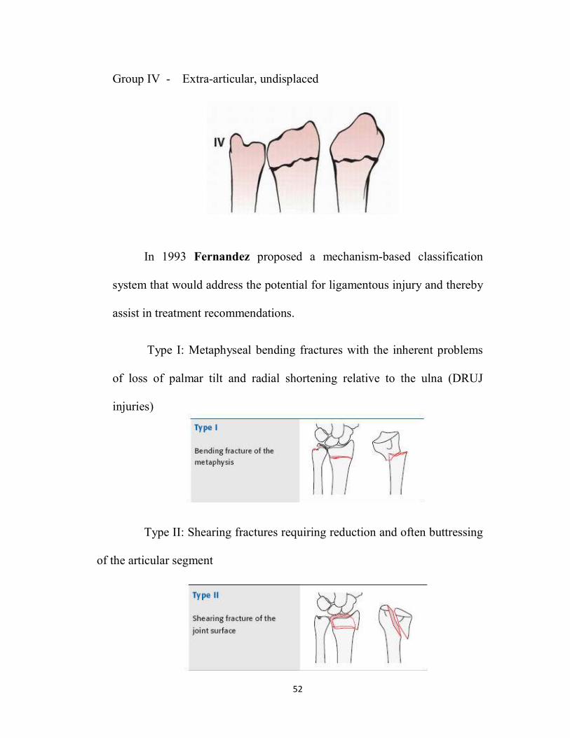
Group IV - Extra-articular, undisplaced
In 1993 Fernandez proposed a mechanism-based classification
system that would address the potential for ligamentous injury and thereby
assist in treatment recommendations.
Type I: Metaphyseal bending fractures with the inherent problems
of loss of palmar tilt and radial shortening relative to the ulna (DRUJ
injuries)
Type II: Shearing fractures requiring reduction and often buttressing
of the articular segment

53
Type III: Compression of the articular surface without the
characteristic fragmentation; also the potential for significant interosseous
ligament injury
Type IV: Avulsion fractures or radiocarpal fracture Dislocations
Type V: Combined injuries with significant soft tissue involvement
because of the high energy nature of these fractures

54
McMURTRY AND JUPITER CLASSIFICATION (1991):
They defined intra-articular fractures on the basis of the number of
their parts.
Two parts - The opposite portion of the radiocarpal joint
remains intact. (Dorsal or palmar Barton,
Chauffeur and “die punch” fractures).
Three parts - The lunate and scaphoid facets separate from
each other and the proximal portion of radius.
Four parts - Same as three parts except the lunate facet is
further fractured into dorsal and volar
fragments
Five parts or more - Including wide variety of comminuted
fragments
Melone emphasized the effect of the impaction of the lunate on the
radial articular surface to create four characteristic fracture fragments.
Further refinement of the classification for intra-articular fractures
was proposed by Melone, who recognized that many intra-articular
fractures not only had instability but specific patterns of displacement. He
identified that most intra-articular distal radius fractures have three or four
part fracture components (1: shaft; 2: radial styloid; 3: dorsal medial; 4:

55
Palmar medial) and that certain displacements (types 3 and 4) are not
easily reduced and have a poor prognosis.
Type I - Stable fracture without displacement. This pattern has
characteristic fragments of the radial styloid and a palmar and
dorsal lunate facet
Type II - Unstable “die punch” with displacement of the characteristic
fragments and comminution of the anterior and posterior cortices
TypeIIA Reducible
TypeIIB - Irreducible (central impaction fracture)

56
Type III - “Spike” fracture. Unstable. Displacement of the articular
surface and also of the proximal spike of the radius
Type IV - “Split” fracture. Unstable medial complex that is severely
comminuted with separation and or rotation of the distal
and palmar fragments
Type V - Explosion injury

57
TREATMENT MODALITIES OF DISTAL RADIUS FRACTURES
I) CONSERVARTIVE:
Different types and positions of immobilization of the wrist and
hand have been described; straight, in flexion-ulnar deviation, in
extension; with or without permanent cast, with a below or over the elbow
cast; with the forearm pronated or supinated. It seems that redisplacement
during healing is not correlated with the type of immobilization but only
with the dorsal comminution and the initial displacement.
II) SURGICAL :
- percutaneous direct pinning
- elastic intra focal or extra focal pinning
- external fixation
- external fixation and direct pinning
- orif with buttress plate
- orif with locking compression plate.
- bone grafting
- wrist arthroscopy

58
PRINCIPLES OF MANAGEMENT OF DISTAL RADIAL
FRACTURES
Goals of Treatment
The goals of treatment are to restore maximum function, maintain
strength, limit the development of post-traumatic arthritis, and avoid
complications. Achievement and maintainance of a satisfactory reduction
until healing occurs, followed by rehabilitation of the wrist to restore
motion and strength is the ultimate goals of treatment of radial fractures.
Signs of DRUJ injury:
• fracture at the base of the ulnar styloid,
• widening of the DRUJ space seen on the P/A xray,
• >20° of dorsal radial angulation, and
• >5 mm of proximal displacement of the distal part of the radius.
1mm-2mm sagital CT - best to view articular depression fracture.
MRI if TFCC or scapholunate ligment tears suspected.
Acceptable Reduction:
• <150 dorsal and <20
0 palmar tilt
• >150 radial inclination
• <5mm radial shortening

59
• Ulnar variance negative or neutral
• Articular gap<2mm
• Articular step<1mm
Evaluation of operation results On radiographic examination (PA view),
the normal radius shows the following.
1. The bistyloid line (BSL) is tilted at +10-15 on the horizontal line;
2. The radial angulation (RA) is tilted at +21-24 on the horizontal line;
3. The ulnar variance (UV) is normally equal to 0 to -2mm
An imperfect reduction is characterized radiographically by the
following;
1. The bistyloid line (BSL) becomes horizontal or inverted to negative
values (-10°);
2. Radial angulation (RA) also tends to be horizontal or even inverted,
to a measurement of -10°;
3. The ulnar variance (UV) becomes positive (+4), indicating a
compression of the distal part of the radius onto the proximal radius;
4. A radio-ulnar diastasis may be noted eventually, indicating a rupture
or a tearing of the triangular fibrocartilage(36).
On lateral radiographs the normal criterion is;
• Palmar tilt (PT) is +15°. The articular surface of the radius is
oriented downwards and slightly forwards.

60
An imperfect reduction is characterized by ;
• Tilting to neutral or with dorsal angulation. The measurement of
the palmar tilt (PT) is -10°, which indicates a dorsal or posterior tilt. Even
a zero value for the palmar tilt indicates a posterior tilt; and when the UV
becomes positive, it allows measurement of the degree of compression of
the distal fragment.

61
OSTEOPOROSIS
Bone strength is determined by the mass and quality of the bone.
Osteoporosis is a condition that is characterized by a marked reduction is
bone strength and subsequently results in an increased frequency of
fractures due to microarchitectural disorientation. The oldest
documentation dates back to 990 BC from Egypt.
A major failure of fractures that occur amongst postmenopausal
women are attributed to osteoporosis. Hip, spine and wrist fractures occur
at a higher frequency although any bone may be affected. However unlike
fractures that occur in otherwise healthy bones, fractures involving
osteoporotic bones result in higher morbidity.
The patients are usually asymptomatic until fractures occur as a
result of trivial falls or in some cases as a result of bumps or stress. It is
now believed that it is a preventable condition that occurs as a result of
suboptimal bone development during childhood and adolescence that is
later followed by bone loss during adulthood. Also osteoporosis if already
set in can be controlled by slowing its progression.
WHO defines osteoporosis by gender matched comparison of bone
mineral density. A T score less than -2.5 is the gold standard of diagnosis.
T scoring is done at the following sites:

62
• Neck of femur
• Lumbar spine
• Distal end of radius
Although T score is the gold standard method, BMD using Dual energy
X-ray absorptiometry at hip and lumbar spine is widely used.
63
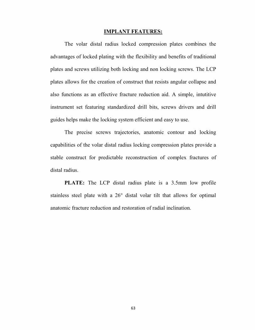
IMPLANT FEATURES:
The volar distal radius locked compression plates combines the
advantages of locked plating with the flexibility and benefits of traditional
plates and screws utilizing both locking and non locking screws. The LCP
plates allows for the creation of construct that resists angular collapse and
also functions as an effective fracture reduction aid. A simple, intutitive
instrument set featuring standardized drill bits, screws drivers and drill
guides helps make the locking system efficient and easy to use.
The precise screws trajectories, anatomic contour and locking
capabilities of the volar distal radius locking compression plates provide a
stable construct for predictable reconstruction of complex fractures of
distal radius.
PLATE: The LCP distal radius plate is a 3.5mm low profile
stainless steel plate with a 26° distal volar tilt that allows for optimal
anatomic fracture reduction and restoration of radial inclination.

64

65
The distal articular end of the plate consists of 3 locking holes for 3.5mm
locking screws angled at 15°.
• The shaft of the plate consists of combiholes for insertion of
3.5mm locking or cortical screws.
• The plates are available in 3, 4, 5, 6, 7, 8 hole shaft length.
SCREWS :
• The screws are 3.5mm stainless steel self tapping and locking screws.
Threaded conical head locks securely into the threaded holes in the plate to
provide angular stability.
• Locked screws allow unicortical screw fixation and load transfer to
near cortex.
• Available in 6mm to 30mm lengths (2mm increments).

66
DRILL BIT :
• A 2.8mm regular drill bit is used for all the screws.
THREADED LCP DRILL GUIDE :
• The 3.5mm threaded LCP drill guide centers the 2.8mm drill bit to
ensure the engagement of the locking screw in the threaded hole in the
plate.

67
SPECIFIC SURGICAL CONSIDERATIONS FOR TREATING
FRACTURES IN AN OSTEOPOROTIC BONE
Multiple factors must be taken into consideration when deciding on
a line of management for fractures in older patients as the functional
demands of older individuals vary from that of younger patients. In the
elderly it is important to achieve a stable fracture fixation thereby reducing
pain and facilitating mobilization.
A common obstacle while dealing with osteoporotic patients is
fixation of the device to the bone which occurs as a result of a higher
frequency of bone failure which is attributed to reduced bone mass
resulting in brittle bones and altered bone structure which may even
include medullary expansion. The above characteristics must hence be
taken into consideration while planning lines of management. It must also
be duly noted that patients with osteoporotic bones generally have
proportionately lower physical demands. Also there is a documented
reduction in the life expectancy amongst individuals with osteoporosis.
A more conservative line of management such as immobilization
using casts may present with a different set of hurdles such as:
a) Joint stiffness
b) No control over bone shortening
c) Poor bone fixation especially when the skin is loose

68
Hence internal fixation is required to provide stability and maintain
reduction. Immobilization in casts has the disadvantage of immobilizing
the joints adjacent to the fracture often leading to joint stiffness.
Furthermore, a cast does not control fracture shortening which is often
seen in osteoporotic bone; and if the subcutaneous tissue is very mobile, as
it often is in the elderly, cast fixation will not provide adequate fracture
fixation. Metaphyseal fractures in osteoporotic bone are associated
with specific fixation problems as the metaphyseal fragment is often
very small. The major problem in osteoporotic fracture treatment is
fixation of the device to the bone as bone failure is much more common.
Problems in osteoporotic fractures are,
1. Purchase will be poor.
2. Unstable fractures.
3. Needs splinting with plaster.
4. Early mobility is compromised.
Because the distal radius is important in the kinematics of the
radiocarpal and radioulnar joints, open reduction of the articular surface
and restoration of the radial length, volar angulation, and radial inclination
are the prerequisites for good clinical outcome. To improve fixation and
resist bending forces a screw and plate construct with a locked angle
between the plate and metaphyseal screw is often used. Recently locked

69
plates have been introduced threaded screw holes in the plates, which
create angular stability between the screws and the plates.
Volar application of a locking compression plate for dorsally
displaced distal radial fracture in postmenopausal women is a safe
alternative. It provides stable fixation to dorsally displaced fractures of the
distal radius with excellent radiographic and functional results and
minimal complications. Locking screws provides fixed angle construct and
improved fixation in osteoporotic bones.
Locked plates are a good option as they have threads in the holes
that provide angular stability between the screw and plate hence allowing
good maintenance of reduction and better quality fixation. These are good
option in case of metaphyseal fractures seen in osteoporotic bones where
the metaphyseal fragments are usually small making fixation with locked
plates advantageous.

70
COMPARISION BETWEEN CONVENTIONAL PLATE AND
LOCKING COMPRESSION PLATE
Conventional plates must be perfectly contoured prior to application.
Fracture reduction can be lost from axial loads causing excessive
shear forces on the construct that are greater than the frictional loads
between the bone-plate-screw construct. Screw loosening and loss of
plate-bone fixation occurs as a result of toggling of cortical screws . Each
screw works independently; the construct depends on a single screw's
stiffness or pullout strength(37).
The biomechanical goals of the LCPs are to increase the stiffness
of the construct in a biological environment. The LCP is a fixed angle
construct that does not rely on screw purchase in bone. The fixed-angle
converts shear stress into compressive stress at the screw-bone interface
once the screw is locked into the plate. The load is now perpendicular to
the screw axis. In order for the construct to fail under an axial load, the
bone must collapse in compression. Therefore, the strength in the LCP is
the sum of all the screw and plate interfaces(38,39)
.
Locking screws does not generate compression between the plate
and the bone since they are designed with smaller threads. They have a
larger core diameter that ensures greater bending and shear strength and
dissipate the load over a larger area of bone. They have self- retaining

71
head(stays on the screw driver without a holding device) and allows
65% greater insertion torque than conventional hexagonal drivers. The
locked screw has a conical, double-lead thread design that facilitates
alignment with the threaded plate hole.
In vitro studies in bone models do show that locked screw
constructs fail at higher loads than cortical screws and their advantage is
magnified in osteoporotic bone.
The primary indications for LCPs :
1. Poor quality bone as in osteoporosis
2. Complex periarticular fractures where contouring is difficult in the
metaphyseal area
3. Inability to get minimal number of conventional cortical screw
purchase
4. Periprosthetic fractures
5. Nonunions due to screw stripping or back out
6. Polytrauma cases (complex fractures that cannot be anatomically
reconstructed).

72
COMPLICATION AFTER LOCKING COMPRESSION PLATING
• Median nerve irritation is common. Generally, symptoms are
mild and disappear with elevation. Persistent paresthesia may be
due to median neuropraxia at the site of fracture, or acute carpal
tunnel syndrome.
• Hard-ware related complications
• Skin irritation at the wrist as a result of a prominent plate.
• Loss of alignment and fracture malunion may occur. In the
elderly, small degrees of malunion are tolerated well and result in
minimal disability. Severe malunion may result in reduced grip
strength, cosmetic deformity, stiffness, restriction of forearm
rotation, and pain.
• Complex regional pain syndrome
• Infection
• Failure of fixation, due to separation of the distal radius bony
fragment from the plate may result.

73
ADVANTAGES OF LOCKING COMPRESSION PLATING
LCP volar column plates have the following attributes:
1) It enables the surgeon to address individual fragments separately
2) Anatomical reduction depending on the pattern of fracture may be
done and stabilization may be provided by using k-wires temporarily
as the plate is applied.
3) Plates are provided with elongated holes that make it convenient to
make adjustments in the position of the plates.
4) Universal anatomical shape eliminates need for anatomical
contouring of plate based on variations in bone anatomy.
5) Stable Fixation : As the system is versatile, it allows stabilization of
even complex fractures. In complex fractures treated based on the
tree column theory, radius and ulnar fragments may be separately
dealt with. Also there are a variety of locking options which is
beneficial in fractures near distal radio-ulnar joint where additional
screws may be used to support the styloid process.
6) Preservation of Blood Supply: The plates are specifically designed
with a low profile cross sectional design with undercuts and rounded
edges reducing chances of soft tissue irritation and ensuring good
periosteal blood supply.

74
7) Early Mobilization : A combination of AO technique during surgery
and the plate when used allow faster healing and early
mobilization(40).

75
Materials and MethodsMaterials and MethodsMaterials and MethodsMaterials and Methods

76
MATERIALS AND METHODS
The study was conducted in the department of orthopaedics,
Coimbatore medical college and hospital, Coimbatore. The number of
patients in the study group was fifteen. The period of study was between
July 2011- May 2013. All the postmenopausal women with distal fractures
were investigated for our study. The age group was between 47 to 75
years. Minimal period of study was 6 months. Functional outcome was
evaluated using modified mayo wrist score.
MODIFIED MAYO WRIST SCORE
I. PAIN
No Pain -25
Mild Occasional Pain -20
Moderate, Tolerable Pain -15
Severe to intolerable Pain -0
II. ACTIVITY
Returned to Regular Activities -25
Regular Activities with Some Restrictions -20
Low Demand Activities -15
Unable to work / play /engage in sports -0
III. RANGE OF MOTION (Supination and Pronation)
>1450 -25
1300-144
0 - 20
1100-129
0 -15

77
800-109
0 -10
400-79
0 - 5
00-39
0 - 0
IV GRIP STRENGTH (Percentage of Normal)
90-100% -25
75-89% -15
50-74% -10
25-49% -5
0-24% -0
FINAL RATING (From Total Point Scored)
Excellent - 90-100 Points
Good - 80-89 Points
Fair - 65-79 Points
Poor - < 65 Points
Inclusion criteria:
1. Fracture distal radius in the Postmenopausal women.
2. All the women in the study group was independent to take care of their
activities of daily living.
3. Fractures occurring at or within 2cms of distal radius.
Exclusion criteria:
1. women in menstrual age group
2. Undisplaced distal radial fractures
3. Patients not willing for internal fixation.

78
Patients admitted with distal radius fractures will be classified under
AO and Gartland and Werley classfication.
Detailed informed consent was obtained from all the persons in the
study group.
The procedure performed under supraclavicular block/axillary
block. Our standard practice, preoperative prophylactic intravenous
cefotaxime and bipolar diathermy for haemostasis.
INSTRUMENTS AND IMPLANTS USED:
• Locking compression plates of varying length
• 3.5mm LCP drill bit and sleeve system
• Hand drill / power drill
• Tap for 3.5mm cortical screws and 3.5mm depth gauge
• Hexagonal screw driver for 3.5mm cortical screws and
locking screw driver
• General instruments like retractors, periosteal elevators,
reduction clamps, bone levers etc.
• Pneumatic tourniquet.
OPERATIVE PROCEDURE
Description:
By volar Henry approach: The radial styloid fragment was approached
initially using an incision centered longitudinally over the flexor carpi

79
radialis tendon and then dissected between the flexor carpi radialis tendon
and radial artery.
The parona’s space underneath the flexor tendons developed and the
distal and radial borders of pronator quadratus were lifted and retracted
ulnarly. Image intensifier used in theatre to assist the evaluation of fracture
reduction and fixation. Patients with unstable fractures, the wrist
immobilised in a below elbow splint for 2 weeks.
Volar Henry approach
1. Skin incision
Fig. 1
2. Flexorcarpi radialis retracted medially and radial artery laterally to
expose pronator quadrates
Fig. 2

80
3. Pronator quadratus erased, elevated medially and distal radius
exposed
Fig. 3
4. Sleeve fixed in the threads of locking compression plate after
temporary stabilizing of plate with K-wires
Fig. 4 Fig. 5
5. Fracture reduced and LCP fixed
Fig. 6

81
PER OPERATIVE PROTOCOL:
Unstable fractures were immobilized with plaster of paris support
for a period of 2 weeks.
POST OPERATIVE PROTOCOL:
Post operative data will include time to full wrist movements, post
operative complications such as median nerve compression symptoms,
malunion, failure of fixation, wound infections and complex regional pain
syndrome (CRPS).
Patients will be allowed to start wrist movements at an average of three
weeks post operatively.
After the discharge, patient will be followed at regular intervals
6weeks , 6 months and 1year.
Subjective and objective functional results were graded using
modified mayo scoring system.

82
ResultsResultsResultsResults

83
RESULTS
Fifteen cases of postmenopausal women with distal radius fractures
were treated surgically by locking compression plate in Coimbatore
medical college Hospital treated between July 2011 to may 2013. All
cases were followed up periodically during the period July 2011 to may
2013. Average age in our study was 59.9 years.
We evaluated our results and compared the functional outcome with
various other studies.
Involved side :
The right side (dominant wrist) was involved in 8 of the cases in our study
series and 7 involved in left side.
Side No of cases Percentage
Right 8 53.3
Left 7 46.6
Side Distribution
Right
Left

84
Mode of injury :
In our study 26.6% of the patients had road traffic accident and
73.3% had a house hold fall.
Mechanism of injury No. of cases percentage
Road traffic accident 4 26.6
House hold fall 11 73.3
CHART 2:MODE OF INJURY
ROAD TRAFFIC ACCIDENT-4
HOUSE HOLD FALL-11

85
Type of fracture:
Based on AO and Gartland and Werley classification
AO Type Number of cases
A2 9
A3 1
B2 2
B3 1
C1 1
C2 1

86
Groups as per Gartland and Werley classification
Type of Groups Number of
cases
Group I 8
Group II 1
Group III 4
Group IV 2
0
1
2
3
4
5
6
7
8
GROUP I GROUP II GROUP III GROUP IV
CHART-4:GARTLAND AND WERLEY
GROUP
GARTLAND AND WERLEY
GROUP

87
Extra articular and intra articular fracture:
Type No. of cases Percentage
Extraarticular fractures 10 66.66
Intra articular fractures 5 33.3
Out of 15 cases, 10 of the fracture were of extra articular type and 5
were intra articular fractures.
Extra
articular
Intra
articular
Type of fracture 66.6 33.3
0
10
20
30
40
50
60
70
80
90
100
Pe
rce
nta
ge
of
case
s
CHART 5:TYPE OF FRACTURE

88
Associated injuries in our study group:
Associated injuries No. of cases percentage
Ipsilateral fracture both bone leg 2 13.3
Head injury 1 6.6
Ipsilateral distal ulna fractures 5 33.3
Total 8 53.3
Out of 15 cases 8 (53.28%) patients had associated injuries.
0
2
4
6
8
10
12
14
Ipsilateral fracture both
bones leg
Head injury Ipsilateral fracture ulna
CHART 6: ASSOCIATED INJURIES

89
Surgical waiting period:
Duration No. of cases Percentage
1-5 days 8 53.3
6-15 days 7 46.6
Surgery was done between 1-5 days in 8 (53.3%) patients as an
elective procedure. Surgery was delayed upto the 14th day in 7 (46.6%)
because those patients had history of ischaemic heart disease, diabetes
mellitus, associated head injury and surgery was done after clearance from
respective specialities.
Fracture union data:
Time of Union No.of Cases Percentage
2-3 months 12 79.9
3-4 months 3 19.9
The present study 12 (79.9%) patients had union within 2-3
months and 03(19.9%) patients had union in 3-4 months. There was no
case of delayed union.
90
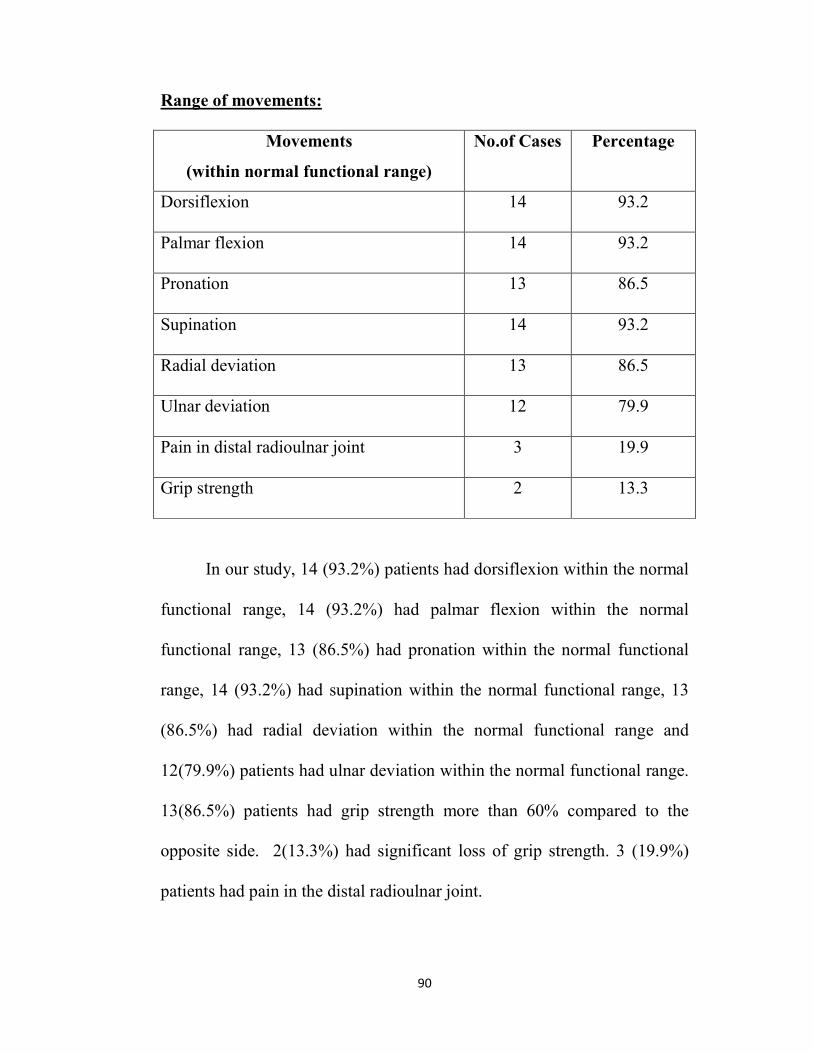
Range of movements:
Movements
(within normal functional range)
No.of Cases Percentage
Dorsiflexion 14 93.2
Palmar flexion 14 93.2
Pronation 13 86.5
Supination 14 93.2
Radial deviation 13 86.5
Ulnar deviation 12 79.9
Pain in distal radioulnar joint 3 19.9
Grip strength 2 13.3
In our study, 14 (93.2%) patients had dorsiflexion within the normal
functional range, 14 (93.2%) had palmar flexion within the normal
functional range, 13 (86.5%) had pronation within the normal functional
range, 14 (93.2%) had supination within the normal functional range, 13
(86.5%) had radial deviation within the normal functional range and
12(79.9%) patients had ulnar deviation within the normal functional range.
13(86.5%) patients had grip strength more than 60% compared to the
opposite side. 2(13.3%) had significant loss of grip strength. 3 (19.9%)
patients had pain in the distal radioulnar joint.

91
Complications in our study:
Complications No.of Cases Percentage
Infection (stich abscess) 1 6.66
ComplexRegionalpain
Syndrome(CRPS)
1 6.66
Reduced ROM 1 6.66
Total 3 20
Infection (stich
abscess)CRPS Reduced ROM
Complications 6.6 6.6 6.6
0
2
4
6
8
10
% o
f ca
ses
CHART 7:COMPLICATIONS
We encountered a complication rate of 20%, out of which 1 (6.6%)
was due to infection (stich abscess) and another 1(6.6%) developed
reduced range of movements and another 1(6.6%) developed complex
regional pain syndrome (CRPS).

92
Evaluation of results:
The assessment of results were made using the modified mayo wrist
score based on pain, activity, range of motion (supination & pronation)
and hand grip strength.
Results No. of cases Percentage
Excellent 7 46.6
Good 6 39.9
Fair 2 13.3
Poor 0 0
In our series, we had 46.6% excellent, 39.9% good, 13.3%, fair and
no poor results.
DISCUSSION
0%10%20%30%40%50%60%70%80%90%
100%
Excellent Good Fair poor
RESULTS
RESULTS

93
DiscussionDiscussionDiscussionDiscussion

94
DISCUSSION
A combination of an improved understanding of distal radial
anatomy, patient demands and the new fixation devices have changed the
management of distal radial fractures. Locking plates are preferred in
osteoporotic and in multiple complex fractures. During the recent years,
volar approach has become more popular. Use of locking compression
plate was first published by Chan KW in HongKong, in 2003 for distal
radius fractures. In India, the locking compression plate for distal radius
fractures was published in 2007.
Our results are compared with various other studies, in various
parameters like involved side, mode of injury, type of fractures etc.,
1) Involved side:
In our study the right side (dominant wrist) was involved in 8 of the
cases and left side involvement was 7. In Arora Rohit et al., (2007) right
side involvement was 70 and left was 44, Ayhan Kilic et al., (2009) right
side involvement 14 and left side was 13, R.E. Anakwe et al.,(2010) right
side involvement was 15 and left side was 6, Sanjay Agarwala(2012) right
side involvement was 11 and left side was 14. All the above series had
increased involvement of the right wrist in their series which was also the
case in our series.

95
2) Mode of injury:
In our study 26.6% (4 cases) of the patients had road traffic accident
and 73.3%(11 cases) had a house hold fall.
In Ayhan Kilic et al (2009) road traffic accident was 13 and house
hold fall was 14 , Arora Rohit et al (2007) road traffic accident was 40 and
house hold fall was 60, R.E. Anakwe et al.,(2010) road traffic accident was
14 and house hold fall was 7and Sanjay Agarwala(2012) Road traffic
accident reported was 7 and house hold fall was 17.
All the above series had increased house hold fall than road traffic
accidents. In our series also house hold fall is the more common mode of
injury.
3) Type of fracture:
Based on AO classification, we had 9(59.4%) cases were
A2(Extraarticular fracture of radius, simple & impacted) type
fractures,1(6.6%) was A3(Extraarticular fracture of radius,
multifragmentary), 2(13.2%) cases were B2(Partial articular fracture of
radius, dorsal rim), 1(6.6%) case was B3(, Partial articular fracture of
radius, volar rim), 1(6.6%) case was C1(Complete articular fracture of
radius, articular and metaphyseal simple), and 1(6.6%)case was C2
(Complete articular of radius, articular simple and metaphyseal
multifragmentary) fractures.

96
Based on AO classification Arora Rohit et al., (2007) reported 39
cases by A2, 16 cases by A3, 24 cases by C1, 30 cases and by C2, 5 cases
by C3 .
Based on AO classification Ayhn Kilic et al., (2009) reported 3
cases by B2, 2 cases by B3, 2 cases by C1,14 cases by C2 and 6 cases by
C3.
Based on AO classification R.E. Anakwe et al., (2010) reported 4
cases by C1, 8 cases by C2 and 9 cases by C3.
Based on AO classification Sanjay Agarwala(2012) reported 1 case
by A1, 5 cases by A2, 7 cases by A3, 7 cases by B2, 3 cases by B3, 1 case
each by C2 and C3.
In our series the majority of the cases were of A2(extraarticular)
type.
4) Complications:
We encounterd three complications (20%) in our study. One being
stich abscess, another developed reduced range of movements and another
developed CRPS.
Arora Rohit et al., (2007) reported a complication rate of 57%.
Ayhn Kilic et al., (2009) reported a complication rate of 11.1%.
R.E. Anakwe et al., (2010) reported a complication rate of 4.8% and
Sanjay Agarwala(2012) reported a complication rate of 4%.

97
5) Results compared with other studies:
In our series, we had 46.6% excellent, 39.9% good, 13.3%, fair and
no poor results.
Patients, who obtained excellent results, had normal regular
activities or no pain. Range of motion was within the normal functional
range. Radial length, volar tilt and articular step-off were within acceptable
limits. They underwent earlier physiotherapy.
Patients with good results had minimal residual deformities, pain and
slight limitation.
Patients with fair results, along with residual deformity, pain and
limitation also had pain in the distal radio-ulnar joint and minimal
complications. Few of their movements were less than that required for
normal function.
Rohit Arora et al., (2007) had 31 excellent, 54 good , 23 fair and 6
poor results based on functional outcome.
R.E. Anakwe et al., (2010) system outcome was assessed using
clinical examination grip strength measures, radiographs and PRWE
(patient related wrist evaluation) scoring. In his series 95% patient very
high level of satisfaction, good functional outcome and increased grip
strength.

98
Sanjay Agarwala(2012) used mayo modified wrist score for
interpreting results, which showed 100% of excellent results after one year
followup.
Our series is comparable to that of Ayhan Kilic et al., (2009) who had
44.4% excellent, 44.4% good, 11.2% fair.

99
ConclusionConclusionConclusionConclusion

100
CONCLUSION
In India, osteoporosis is an epidemic problem. Hence the women in
the postmenopausal age will have fractures due to trivial injury. A fall on a
outstretched hand is the common mode of injury causing distal radius
fractures among postmenopausal women. Distal radial fractures which
occur due to road traffic acidents (high energy trauma) are mostly intra-
articular, displaced and unstable Gartland and Werley group II and III and
AO type B2, B3, C1 and C3.
The results are evidence that locked plates are good implant in the
treatment of intra-articular unstable fractures of distal radius. It allows
effective anatomic realignment and early wrist mobilization. It is
biomechanically superior due to closer joint interface placement and better
screwing capability in different orders. A successful anatomic alignment
was made possible regardless of the direction of fracture angulation with
volar locking plate. 90% the patients went back to their daily activities
with good recovery.
Use of locked compression plates in distal radius fractures provide
good to excellent results and are effective in the correction and
maintenance of distal radius anatomy. By using these plates, joint motions
and daily functioning is recovered in a shorter time. Hence locking
compression plate is a useful implant in stabilizing in osteoporotic distal
radius fractures in postmenopausal women.

101
Our study is the first study of distal radius among postmenopausal
women in India. Our study is a smaller one in terms of number of cases. A
large multicentric study is needed to confirm our results.

102
Illustrative CasesIllustrative CasesIllustrative CasesIllustrative Cases

103
Annexures
ANNEXURE-I
ILLUSTRATIVE CASES:
CASE : 1
Parvathy,52yrs, IP.No:49264, H/O House hold Fall and injured her left
wrist.
Pre-op Immediate post-op
1 year follow-up

104
Range of Movements – 1 Year follow up
Palmar flexion Dorsiflexion
Supination Pronation
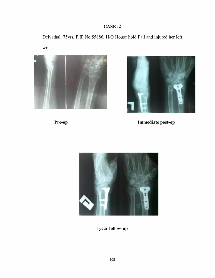
105
CASE :2
Deivathal, 75yrs, F,IP.No:55886, H/O House hold Fall and injured her left
wrist.
Pre-op Immediate post-op
1year follow-up

106
Range of Movements – 1 Year follow up
Palmar flexion Dorsiflexion
Pronation Supination

107
CASE : 3
Ruckmani, 47yrs, F, IP.No:50582, H/O House hold Fall and injured her
left wrist.
Pre-op
Immediate post-op
1 year follow-up

108
Range of Movements – 1 Year follow up
Palmar flexion
Dorsiflexion

109
CASE : 4
Pachainayagi, 55yrs, F, IP.No:7576, H/O House hold Fall and injured her
left wrist.
Pre-op Immediate post-op
1 year follow-up

110
Range of Movements – 1 Year follow up
Palmar flexion
Dorsiflexion

111
CASE : 5
Janaki, 56yrs, F,IP.NO. 5916 H/O House hold Fall and injured her right
wrist.
Pre op Immediate post op
6 months follow up
1 year follow up

112
Range of Movements – 1 Year follow up
Palmar flexion
Dorsiflexion

113
BibliographyBibliographyBibliographyBibliography

114
ANNEXURE-II
BIBLIOGRAPHY
(1). Bagby GW. Compression bone-plating:historical considerations. J
Bone Joint surg Am.1977;59:625-631.
(2). Helfet DL, Haas NP, Schatzker J, et al. AO philosophy and
principles of fracture management-its evolution and evaluation. J
Bone Joint surg Am. 2003;85:1156-1160.
(3). FRIGG, R.: Development of the Locking Compression Plate.
Injury, 34 Suppl. 2: B6-10, 2003
(4). PERREN, S. M.: Backgrounds of the technology of internal fixators.
Injury, 34 Suppl. 2: B1-3, 2003.
(5). Egol KA, Kubiak EN, Fulkerson E, et al. Biomechanics of locked
plates and screws.J Orthop Trauma. 2004;:488–93.
(6). Haidukewych GJ. Innovations in locking plate technology.J Am
AcadOrthop Surg. 2004;12:205–212.
(7). Chan KW, Kwok TK, Mak KH. Early experience with locking
compression plate in the treatment of distal radius fracture. Hong
Kong J Orthop Surg. 2003;7:88–93.

115
(8). Leung F, Zhu L, Ho H, Lu WW, Chow SP. Palmar plate fixation of
AO type C2 fracture of distal radius using a locking compression
plate: a biomechanical study in a cadaveric model. J Hand Surg.
2003;28B:263–6.
(9). Orbay JL, Fernandez DL. Volarfixed-angle platefixation for
unstable distal radius fractures in the elderly patient. J Hand Surg.
2004;29A:96–102.
(10) Cooney WP III, Dobyns JH, Linscheid RL. Complications of
colles’ fractures. J Bone Joint Surg 1980; 62-A: 613.
(11). Schütz M, Kolbeck S, Spranger A, Arndt-Kolbeck M, Haas NP.
Palmar plating with the locking compression plate for dorsally
displaced fractures of the distal radius--first clinical experiences
Zentralbl Chir. 2003 Dec;128(12):997-1002.
(12). Wong KK, Chan KW, Kwok TK, Mak KH, Volar fixation of
dorsally displaced distal radial fracture using locking compression
plate. J Orthop Surg (Hong Kong). 2005 Aug;13(2):153-7
(13). Köck H, Bandl WD, Chan T.,Experiences and results with the
locked compression plate for 603 fractures of the distal radius.
Handchir Mikrochir plast chir. 2005 oct;37(5):303-8.

116
(14). Musgrave DS, Idler RS., Volar fixation of dorsally displaced distal
radius fractures using the 2.4-mm locking compression plates. J
Hand Surg Am. 2005 Jul;30(4):743-9.
(15). Cognet JM, Geanah A, Marsal C, Kadoch V, Gouzou S, Simon P.
Plate fixation with locking screw for distal fractures of the radius
,Rev Chir Orthop Reparatrice Appar Mot. 2006 Nov;92(7):663-72.
(16). Scott M. Levin,Cory O. Nelson,Jonathan D. Botts, Glenn A. Teplitz,
Yong Kwon, and Fred Serra-Hsu Biomechanical Evaluation of
Volar Locking Plates for Distal Radius Fractures J Orthop Surg
Received April 2, 2007; Accepted June 20, 2007.
(17). Strohm PC, Müller CA, Helwig P, Mohr B, Südkamp NP. Is the
locking, 3.5 mm Palmar T-Plate the implant of choice for displaced
distal radius fractures. Orthop Unfall. 2007 May-Jun;145(3):331-7.
(18). Arora R, Lutz M, Hennerbichler A, Krappinger D, Espen D, Gabl
M. Complications following internal fixation of unstable distal
radius fracture with a palmar locking-plate. J Orthop Trauma. 2007
May;21(5):316-22.
(19). RE Anakwe, LAK Khan,RE Cook, and JE McEachan Locked volar
plating for complex distal radius fractures: Patient reported
outcomes and satisfaction J Orthop Surg Res. 2010; 5: 51.
(20). Claudio Roberto Martins Xavier, Danilo Canesin Dal Molin, Rafael
Mota Marins dos Santos, Surgical treatment of distal radius fractures

117
with a volar locked plate: correlation of clinical and radiographic
results, Rev Bras Ortop. 2011;46(5):505-13.
(21). Hanae minegishi, Osamudohi, Soukanan & Hidetsugu sato,
Treatment of unstable distal radius fractures with the volar locking
plate. Upsala Journal of Medical Sciences. 2011; 116: 280–284.
(22). Chris Dillingham, MaryBeth Horodyski, Aimee M. Struk, and
Thomas Wright, Rate of Improvement following Volar Plate Open
Reduction and Internal Fixation of Distal Radius Fractures. SAGE-
Hindawi Access to Research Advances in Orthopedics Volume
2011, Article ID 565642,4pages doi:10.4061/2011/565642.
(23). Joideep Phadnis, Alex Trompeter, Kieran Gallagher, Lucy
Bradshaw, David S Elliott and Kevin J Newman, Mid-term
functional outcome after the internal fixation of distal radius
fractures Phadniset al. Journal of Orthopaedic Surgery and
Research2012,7:4.
(24). S Sawalha, P Kumar.Complications of Volar Locking Plates for
Distal Radius Fractures: Experience of a District General Hospital.
The Internet Journal of Orthopedic Surgery. 2007 Volume 9 No 1.
(25). Shetty MS, Kumar MA, Kiran K, Kini AR. Locking distal radius
plate-early results from India. J Trauma. 2011 Nov;71(5):1359-63.

118
(26). Sanjay Agarwala, Ganesh S Mohrir, Shreyans D. Functional
Outcome in Distal Radius Fractures Treated with Locking
Compression Plate. Gadiya Bombay Hospital Journal, Vol. 54, No.
2, 2012 .
(27). Nagesh Prabhakar Naik, Deshpande Shrikant Balkrishna.
Management of dorsally displaced distal radius fracture by volar
fixation using locking compression plate. Indian Journal of Basic &
Applied Medical Research; December 2012: Issue-5, Vol.-2, P. 419-
425.
(28). Brunelli F, Pagliei A, Smussi C. Anatomy of the distal radius. In
Saffar P, Cooney WP III Edts. Fractures of the distal radius :
London: Martin Dunitz Ltd, 1995. p1-11.
(29). Gausepohl T, Worner S, Pennig D, et al. Extraarticular external
fixation in distal radius fractures Pinplacement in osteoporotic bone.
Injury 2001;32(Suppl 4):SD79-SD85
(30). Apergis E, Darmanis S, Theodoratos G, Maris J. Beware of the
ulno-palmar distal radial fragment. J Hand Surg Br 2002;27:139-
145.
(31). Metz M, Gilula LA. Imaging techniques for distal radius fractures
and related injuries - Distal radius fractures. Orthop Clin of N-Am
1993; 24(2): 217-228.

119
(32). Constantine KJ, Clawson MC, Stern PJ. Volar neutralization plate
fixation of dorsally displaced distal radius fractures. Orthopedics
2002;25:125-128.
(33). Semaan I, Saffar P. Pathomechanism of intra-articular distal radial
fractures. In Saffar P, Cooney WP III Edts. Fractures of the distal
radius: London: Martin Dunitz Ltd, 1995. P19-27.
(34). Szabo RM. Comminuted distal radius fractures.Orthop Clin of
NAm1992;23: 1-5.
(35). Saffar P. Current trends in treatment and classification of distal
radial fractures. In Saffar P, Cooney WP III Edts. Fractures of the
distal radius: London: Martin Dunitz Ltd, 1995. P12-18.
(36). Kapandji AI. Treatment of non-articular distal radial fractures by
intrafocal pinning with arum pins. In Saffar P, Cooney WP III Edts.
Fractures of the distal radius : London: Martin Dunitz Ltd, 1995.
p71-83.
(37). Stoffel K, Dieter U, Stachowiak G et al: Biomechanical testing of
the LCP-how can stability in locked internal fixators be
controlled? Injury 34: S-B-11- S-B19
(38). Wagner M. General principles for the clinical use of the LCP.
Injury. 2003; 34(suppl 2):B31-42.
(39). Ricci W, Zheng, Z, Jones, B, Cartner, J. Does Locked Plating
Provide Improved Fatigue Properties over Nonlocked Plating and

120
Does Bone Quality Matter? OTA Annual Meeting Poster
Presentation Boston, MA; 2007.
(40). M.E. Müller, M. Allgöwer, R. Schneider, and H. Willenegger
(1991) AO Manual of Internal Fixation, 3rd Edition. Berlin:
Springer.

121
ProformaProformaProformaProforma

122
ANNEXURE-III
PROFORMA
Case No. : I.P.No. :
Name : Hospital :
Age/Sex : Date of admission :
Occupation : Date of surgery :
Address : Date of discharge :
HISTORY
Presenting complaints:
Side affected:
Pain in the wrist:
* Onset ,Duration ,Type /Severity and Aggravating/Relieving
factors
Swelling:
* Onset ,Duration and Progress
Restriction of movements:
Deformity:
Mechanism of injury:
* Fall on outstretched hand
* Road traffic accident
* Assault
* Miscellaneous injury
Associated soft tissue and skeletal injury:
History of presenting illness:
* Immediate treatment of injured hand
* Duration between the injury and first visit

123
FAMILY HISTORY:
PERSONAL HISTORY:
EXAMINATION
1. General physical examination:
Vitals -Pulse_____ Beats/min Temp______ oC
B.P ______ mm of Hg R.R. ______ Cycles/min
2. Systemic examination:
CVS PA
RS CNS
3. Local Examination:
Inspection:
Attitude:
Deformity:
* Closed dinner fork deformity
* Garden spade deformity
* Radial deviation of hand
* Ulnar deviation
Swelling, Scars / sinuses and Other findings:
Palpation:
* Local rise of temperature
* Tenderness
* Swelling
* Crepitus
* Abnormal mobility
* Step sign
* Deformity and Other findings
Range of movements:
* Palmar flexion (0-90)
* Dorsiflexion (0-90)

124
* Radial deviation (0-25)
* Ulnar deviation (0-35)
* Supination (0-80)
* Pronation (0-75)
* Circumduction
Measurements: Rt. Lt.
* Linear measurement
* Circumferential measurement
Associated complaints:
* Neurological - Ulnar nerve
Median nerve
Radial nerve
* Vascular -
* Other bone injuries - In the injured hand In the body
INVESTIGATIONS:
Blood : Hb% TC: DC:
ESR:
Urine : Albumin: Sugar: Micro :
RBS: Blood urea: Serum creatinine:
HIV: HBsAg:
ECG:
X-ray wrist - PA Type of fracture
- Lateral (Gartland and werley /AO classification)
- Oblique
Special investigations:
TREATMENT:
1. Pre-operative:
* Below elbow POP slab with sling
* Antibiotics
* Analgesics

125
2. Surgical procedure:
* Type of anaesthesia - GA/brachial block
* Type of fixation
* Duration of surgery
* Tourniquet time
* Operative findings
* Operative Complications - Excessive bleeding
Difficult reduction
Stable/Unstable
3. Post-operative:
* Immobilisation
* Antibiotics
* Suture removal
* Complications
4. Duration of hospital stay:
5. Follow up:
Follow
up
Radio
graph
Movements
Functional score
Remarks
PF DF RD UD S P
1ST Month
6th Month
1 year
6. Radiological follow-up:
* Radial shortening
* Dorsal angulation
* Radial inclination
* Ulnar variance
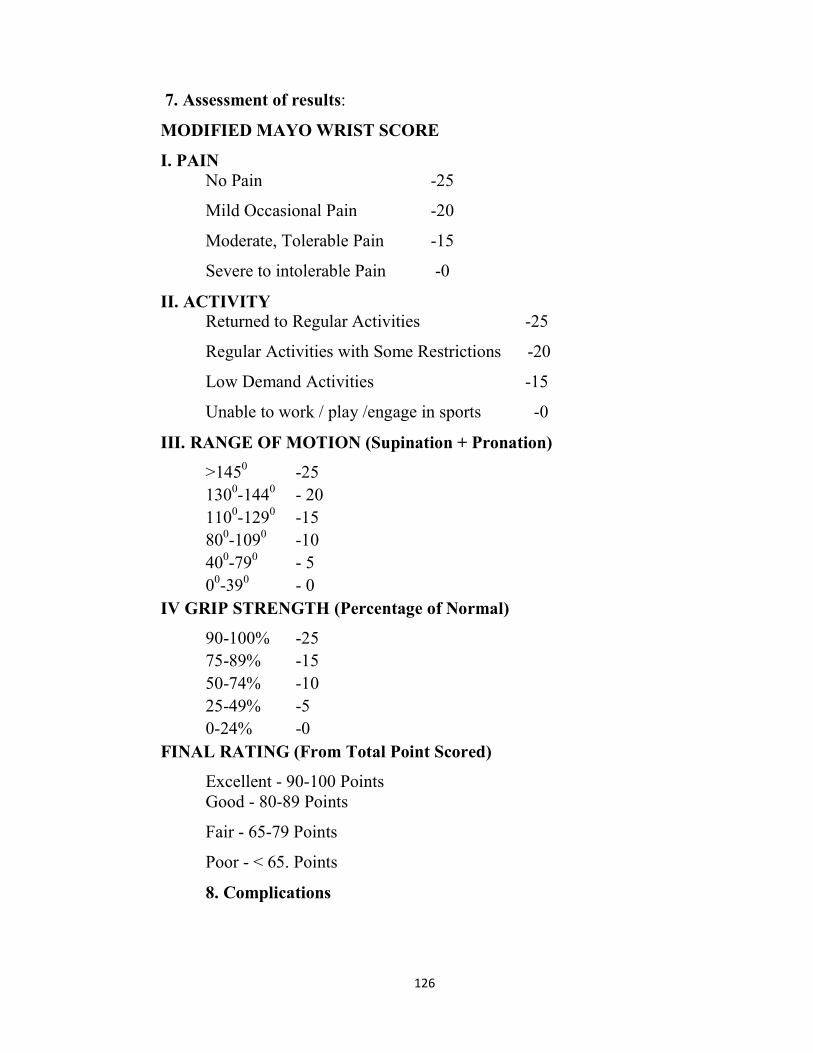
126
7. Assessment of results:
MODIFIED MAYO WRIST SCORE
I. PAIN
No Pain -25
Mild Occasional Pain -20
Moderate, Tolerable Pain -15
Severe to intolerable Pain -0
II. ACTIVITY
Returned to Regular Activities -25
Regular Activities with Some Restrictions -20
Low Demand Activities -15
Unable to work / play /engage in sports -0
III. RANGE OF MOTION (Supination + Pronation)
>1450 -25
1300-144
0 - 20
1100-129
0 -15
800-109
0 -10
400-79
0 - 5
00-39
0 - 0
IV GRIP STRENGTH (Percentage of Normal)
90-100% -25
75-89% -15
50-74% -10
25-49% -5
0-24% -0
FINAL RATING (From Total Point Scored)
Excellent - 90-100 Points
Good - 80-89 Points
Fair - 65-79 Points
Poor - < 65. Points
8. Complications

127
Consent FormConsent FormConsent FormConsent Form

128
ANNEXURE - IV
CONSENT FORM
FOR OPERATION/ANAESTHESIA
I ___________________ Hosp. No.____________ in my full
senses hereby give my complete consent for ________________ or any
other procedure deemed fit which is a diagnostic procedure / biopsy /
transfusion / operation to be performed on me / my son / my daughter / my
ward __________ aged __________ under any anaesthesia deemed fit.
The nature and risks involved in the procedure have been explained to me
to my satisfaction. For academic and scientific purpose the
operation/procedure may be televised or photographed.
Date: signature/Thumb impression
of patient/Guardian
Name:
Designation:
Guardian
Relationship
Full address

129
ANNEXURE - V
ABBREVIATIONS:
BMD - Bone mineral density
BSL - Bistyloid line
CRPS - Complex regional pain syndrome
DRUJ - Distal radioulnar joint
DF - Dorsiflexion
LCP - Locking compression plate
MRI - Magnetic resonance and imaging
ORIF - Open reduction internal fixation
OTA - Orthopaedic trauma association
P - Pronation
PA - Palmar angulation
PF - Palmar flexion
POP - Plaster of paris
PT - Palmar tilt
RA - Radial angulation
RD - Radial deviation
S - Supination
TFCC - Triangular fibro cartilaginous complex
UD - Ulnar deviation
UV - Ulnar variance

130
Master ChartMaster ChartMaster ChartMaster Chart

131
ANNEXURE – VI
MASTER CHART
Case
No Name
Age
(yrs)
Side
Mode
of
Injury
Fracture
type
Gartland
/AO
Associated
Injuries
Surgical
waiting
period
(days)
Surgical
procedure
Duration
Of
Follow up
months
Radiological Findings
Deformity
Range of motion
Compli
cations
Modified
Mayo
score
Results Radial
Length
(mm)
Palmar
tilt (O)
Articular
step-off
(mm)
PF DF RD UD S P
1 Parvathy 52 L Fall lll/C1 # distal left
Ulna 8 ORIF+LCP 24 9 8 0 - 80 85 20 30 80 90 - 90 Excellant
2 Ruckmani 47 L Fall I/A2 - 12 ORIF+LCP 24 9 8 0 - 75 80 20 25 85 80 - 95 Excellant
3 Deivathal 75 L Fall l/A2 # distal left
Ulna 4 ORIF+LCP 24 9 7 0 - 75 85 20 25 85 90 - 90 Excellant
4 chinnakutty 75 R RTA II/B2 #Both bone
right leg 5 ORIF+LCP 12 10 8 0 - 65 70 20 20 75 75 - 85 Good
5 Janaki 56 R Fall l/A2 2 ORIF+LCP 18 10 9 0 - 75 80 15 20 70 75 - 90 Excellant
6 Saraswathi 48 R Fall l/A2 #distal right
ulna 4 ORIF+LCP 12 8 10 0 - 70 60 15 20 70 80 - 80 Good
7 Pongiammal 55 R Fall lV/A2 - 13 ORIF+LCP 18 8 9 0 - 60 65 20 25 70 80 - 85 Good
8 sarasammal 65 R RTA lll/C2 - 8 ORIF+LCP 6 10 -5 3 - 25 45 5 10 40 45 Reduced
ROM 70 Fair
9 Ruckmani 65 R RTA lV/A2 Head injury 2 ORIF+LCP 12 9 7 0 - 70 75 20 25 80 75 - 95 Excellant
10 Manimegalai 55 L Fall l/A3 - 9 ORIF+LCP 12 9 8 0 - 80 85 15 30 90 80 - 80 Good
11 Abirami 50 L Fall l/A2 #distal left
ulna 4 ORIF+LCP 18 9 5 1 - 70 75 15 25 75 80 - 95 Excellant
12 Pankajavalli 55 R Fall l/A2 - 11 ORIF+LCP 12 7 6 1 - 75 80 15 10 65 70 - 85 Good
13 Pachainayaki 55 L Fall l/A2 # distal left
Ulna 5 ORIF+LCP 10 9 8 0 - 60 65 15 25 75 70
Stich
abscess 90 Excellant
14 kuppamal 64 L RTA lll/B3 #Both bone
left leg 14 ORIF+LCP 12 7 6 2 - 50 55 10 10 60 45 CRPS 65 Fair
15 vanathai 52 R Fall lll/B2 - 9 ORIF+LCP 12 13 10 0 - 70 60 15 20 70 80 - 85 Good