Embed Size (px)
Citation preview
7/27/2019 Role of Heme Oxygenase in Inflammation, Insulin-Signalling,
http://slidepdf.com/reader/full/role-of-heme-oxygenase-in-inflammation-insulin-signalling 1/18
Hindawi Publishing CorporationMediators of InflammationVolume 2010, Article ID 359732, 18 pagesdoi:10.1155/2010/359732
Review ArticleRole of Heme Oxygenase in Inflammation, Insulin-Signalling,Diabetes and Obesity
Joseph Fomusi Ndisang
Department of Physiology, College of Medicine, University of Saskatchewan, 107 Wiggins Road, Saskatoon, SK, Canada S7N 5E5
Correspondence should be addressed to Joseph Fomusi Ndisang, [email protected]
Received 12 December 2009; Revised 15 February 2010; Accepted 24 February 2010
Academic Editor: Giuseppe Matarese
Copyright © 2010 Joseph Fomusi Ndisang. This is an open access article distributed under the Creative Commons AttributionLicense, which permits unrestricted use, distribution, and reproduction in any medium, provided the original work is properly cited.
Diabetes and obesity are chronic conditions associated with elevated oxidative/inflammatory activities with a continuum of tissue insults leading to more severe cardiometabolic and renal complications including myocardial infarction and end-stage-renal damage. A common denominator of these chronic conditions is the enhanced the levels of cytokines like tumour necrosisfactor-alpha (TNF-α), interleukin (IL-6), IL-1 β and resistin, which in turn activates the c-Jun-N-terminal kinase (JNK) and NF-κBpathways, creating a vicious cycle that exacerbates insulin resistance, ty pe-2 diabetes and related complications. Emerging evidenceindicates that heme oxygenase (HO) inducers are endowed with potent anti-diabetic and insulin sensitizing eff ects besides theirability to suppress immune/inflammatory response. Importantly, the HO system abates inflammation through several mechanismsincluding the suppression of macrophage-infiltration and abrogation of oxidative/inflammatory transcription factors like NF-κB,JNK and activating protein-1. This review highlights the mechanisms by which the HO system potentiates insulin signalling, withparticular emphasis on HO-mediated suppression of oxidative and inflammatory insults. The HO system could be explored in thesearch for novel remedies against cardiometabolic diseases and their complications.
1. Background
There has been a dramatic rise in the number of patientswith the metabolic syndrome, a comorbid condition of hypertension, obesity, and diabetes. Diabetes mellitus is achronic syndrome of impaired carbohydrate, protein, andfat metabolism caused by insufficient secretion of insulin
and/or defects in insulin action in tissues due to insulinresistance. The incidence of diabetes is increasing globally [1] and type-2 diabetes (TD2) accounts for almost 90% of the cases diagnosed [2–4]. It is projected that the prevalenceof T2D may reach 366 million in 2030 [1]. Similarly, thecondition of obesity has escalated as more than 300 millionadults, the majority of whom live in the developed world,are aff ected [5]. Obesity is amongst the main risk factor forinsulin resistant T2D, hypertension, and other cardiovascularand renal complications [6]. Although inadequate insulinproduction is traditionally linked to type-1 diabetes (T1D),emerging evidence suggests that pancreatic beta-cell massis reduced during the early stages of T2D and declines
further with the progression of disease, eventually leadingto loss of beta cells and reduced insulin production [7,8]. This is consistent with previous observation indicatingthat T2D is not solely due to insulin resistance but alsodue to a failure of the insulin producing beta-cells tosecrete an adequate amount of insulin [9]. On the otherhand, in T1D it is a well-established concept that genetic
defects trigger autoimmunity leading to the destruction of pancreatic beta cells and insulin insufficiency [10], andthese events are further accentuated by apoptosis [11–13].Similarly, in T2D, intense inflammatory activities character-ized by the presence of cytokines, apoptotic cells, immunecell infiltration, amyloid deposits, and fibrosis may causereduction of pancreatic beta-cell mass [14]. In both T1Dand T2D, elevated inflammatory events play a major patho-physiological role in the disruption of islet architecture [10,14–20]. Several factors are responsible for inflammation inT1D and T2D. These include dyslipidemia, hyperglycaemia,elevated nuclear-factor kappaB (NF-κB) activity, increasedlevels of adipokines such as tumour necrosis factor-alpha
7/27/2019 Role of Heme Oxygenase in Inflammation, Insulin-Signalling,
http://slidepdf.com/reader/full/role-of-heme-oxygenase-in-inflammation-insulin-signalling 2/18
2 Mediators of Inflammation
(TNFα), interleukins (ILs), resistin, leptin and free fatty acids[14, 21]. Seen in this light, the suppression of apoptosis,necrosis, and intraislet inflammatory/immune events may be important for the preservation of islet architecture andbeta-cell morphology. Therefore, the regulation of beta-cellnumber through the processes of proliferation, neogenesis,
and apoptosis is important to safeguard islet function [22,23] and the maintenance of adequate insulin productionin T1D and T2D. Taken together, these studies suggestthat impaired insulin secretion is not only an importantetiological factor in the pathogenesis of T1D and T2D,but also an important pathophysiological driving forcethat is capable of dictating the dynamics and progressionof the disease. Thus novel therapeutic modalities capableof suppressing inflammatory/immune responses, apoptosis,and necrosis would be beneficial in the conditions of T1Dand T2D.
Generally, insulin resistance and T2D frequently occurin obesity [24–35]. Amongst the contributing factors, areovernutrition and inactivity. As an adaptive response toinsulin resistance, pancreatic islets enhance their secretory activity. In most individuals, such an adaptation does occurduring early stages of overnutrition and metabolism wouldappear normal at this stage. However, at later stages, thisadaptation eventually fails in some individuals, dependingon the genetic ability of the beta-cell to adapt and theseverity of the resistance to insulin [36]. The reasons forthis failure to maintain sufficient insulin secretion area combined decrease in beta-cell mass and insufficientsecretion of insulin. This reduction of insulin levels may be due to elevate inflammation, oxidative stress, amyloiddeposition, lipotoxicity, and glucotoxicity [36]. Obesity and insulin resistance are associated with a state of low-grade inflammation due to chronic activation of innateimmune system [37]. Although epidemiological studieshave linked inflammation with obesity for decades, theunderlying mechanisms remained obscured until the lastdecade. It is now widely accepted that the activation of inflammatory mediators such as NF-κB, TNFα, and c-Jun-N-terminal kinase (JNK) is amongst the common causesof insulin resistant T2D in obsessed conditions [24–35].Thus, novel strategies that can preserve beta-cell integrity improve insulin sensitivity, and counteract inflammatory mediators like NF-κB, TNF-α, and JNK would be useful inthe prevention and management of insulin resistant T2D andrelated cardiometabolic complications. Recent evidence has
highlighted the important role of the heme oxygenase (HO)in insulin release and glucose metabolism [38–52]. Beside itsemerging antidiabetic eff ects, the HO system is also knownto abate oxidative stress and immune/inflammatory response[53–57]. This review will highlight the mechanisms by whichthe HO system potentiates insulin signalling, with particularemphasis on HO-mediated suppression of inflammation.
2. The HO System and Insulin Signaling
HO is a microsomal enzyme that cleaves the α-methenebridge of heme moiety to produce equimolar amounts of
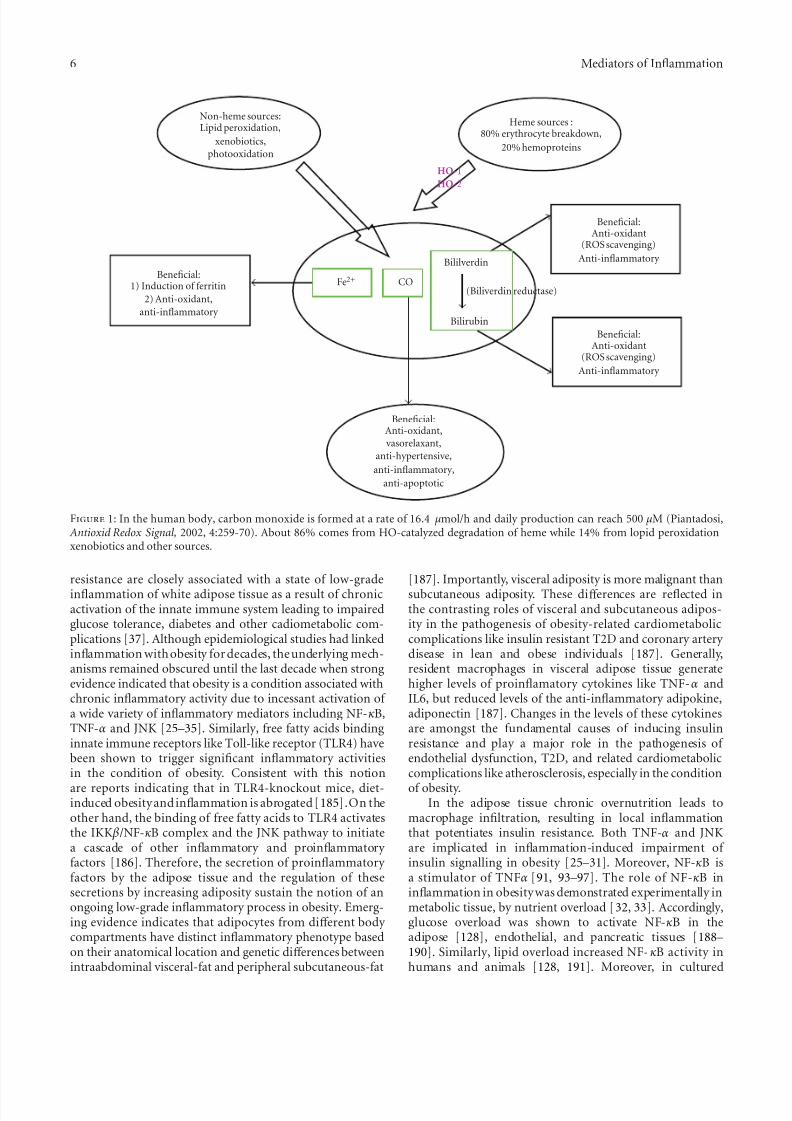
carbon monoxide (CO), bilirubin, and iron [58, 59](Figure 1). CO and bilirubin are known to suppress apop-tosis, necrosis, inflammation, and oxidative stress [56, 60–69], while the iron formed enhances the synthesis of theantioxidant, ferritin [70, 71].
The main isoforms of HO include HO-1 (inducible)
and HO-2 (constitutive) isoforms [58, 59, 72, 73]. HO-1and HO-2 are largely responsible for HO enzymatic activity [58, 72, 73], while the third isoform, HO-3, has no functionalgenes in rat and is considered a pseudotranscripts of HO-2[74, 75]. The basal HO activity is maintained by HO-2 [58,59, 72, 73, 76], while HO-1 is stimulated by a wide variety of diff erent physical, chemical, and pathophysiological stimuliincluding oxidative and inflammatory insults [58, 59, 77–80], as well as metabolic and hemodynamic factors suchas high glucose [80], elevated blood pressure [64], andlipids [81]. Therefore, HO-1 may be considered a sensitiveindex that is triggered in the onset of pathophysiologicalchanges. However, in most cases the pathophysiologicalactivation of HO-1 results only to a transient or marginalincrease of HO-1 that falls below the threshold necessary to activate the downstream signalling components of theHO system [59, 63, 82]. For example, the pathophysio-logical activation of HO-1 by the hemodynamic stress of elevated blood pressure is not accompanied by changes of important component of HO-signalling like cyclic guanosinemonophosphate (cGMP) [59, 63, 82–85]. Therefore thetransient upregulation of HO-1 that normally accompaniesmany pathophysiological conditions may represent the firstline of defense mounted against tissue injury to counteractadverse changes that would destabilize the homeostatic con-ditions in physiological milieu. Since the pathophysiologicalactivation of HO-1 may fall below the threshold necessary toactivate important signalling components through which theHO system elicits its eff ects of restoring tissue homeostasis[63, 82], a more robust enhancement of HO-1 would beneeded to surmount the threshold [63, 82–85]. This canbe achieved by pharmacological agents capable of inducingHO like some metalloprotoporphyrin such as hemin (ferricprotoporphyrin IX chloride), stannous mesoporphyrin, cop-per protoporphyrin, and cobalt protoporphyrin. Given thatmany of the adverse factors which stimulate HO-1 such aselevated blood pressure [64] and high glucose and lipid [80,81] concentrations are implicated in the pathophysiology of metabolic syndrome, the HO system may constitute a novelapproach that could be explored against metabolic syndrome
and related cardiometabolic complications (Figure 2).The emerging role of the HO system in insulin release
and glucose metabolism is becoming increasingly clear [38–52]. HO-mediated stimulation of insulin release has beenreported in diff erent rats strains [38, 46, 49–52] and mice[86, 87]. These studies suggest a central role of CO in glucosemetabolism. In the human body, CO is formed at a rate of 16.4 μmol/h and daily production of may reach 500μmole[88]. Interestingly, under normal physiological conditions,islets of Langerhans produce CO and nitric oxide (NO)to regulate insulin release [45, 46]. While NO negatively modulates glucose-stimulated insulin release, CO stimulatesinsulin secretion [45, 46]. Moreover, glucose stimulates
7/27/2019 Role of Heme Oxygenase in Inflammation, Insulin-Signalling,
http://slidepdf.com/reader/full/role-of-heme-oxygenase-in-inflammation-insulin-signalling 3/18
Mediators of Inflammation 3
pancreatic beta-cells to produce CO, which in turn triggersinsulin release [45, 46]. The critical role of the HO systemin insulin release and glucose metabolism was reported inGoto-Kakizaki (GK) rats, a model with defective pancreaticbeta-cell HO-2 [38]. Since HO-2 is largely responsible forbasal HO activity [58, 59, 72, 73, 76] and thus the production
CO, the impairment of the HO system in GK rats resultedin reduced CO and insulin insufficiency [38]. Interestingly,treatment with the HO-inducer, hemin, or CO correctedthe defective HO system and enhanced insulin release withimprovement of glucose metabolism [38]. Collectively, thesestudies suggest that reduced beta-cell CO and/or impairedHO system may lead to dysfunctional glucose metabolism.
3. The Role of HO System in Inflammation andInsulin Resistance
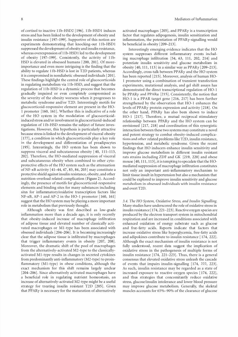
The inflammatory and metabolic systems are among themost fundamental for survival, and these systems havebeen evolutionarily well-conserved in species [37]. However,the conditions of nutrient-overload or obesity may off setthese systems leading to inflammation in metabolic siteslike the adipose tissue, liver, and skeletal muscles. Oneconsequence of such imbalance is the increased produc-tion of proinflammatory cytokines, adipokines, and otherinflammatory/oxidative transcription factors including NF-κB activating protein (AP-1) and JNK. Although both JNKand NF-κB play important roles in inflammation-inducedinsulin resistance, accumulated evidence suggests that they do so through diff erent mechanisms. The principal mech-anism by which JNK causes insulin resistance is throughthe phosphorylation of serine residues in insulin receptorsubstrate-1 (IRS-1) [89–91]. However, since JNK is a stresskinase that also phosphorylates the c-Jun component of theAP-1 [92], the activation of AP-1 by JNK may contribute toaggravate inflammatory insults and hence insulin resistance.NF-κB causes insulin resistance by stimulating proinflamma-tory cytokines like TNF-α, IL-6, IL-1 β, and resistin, which inturn activates JNK and NF-κB pathways to create a viciouscycle that will exacerbate tissue damage [89, 91, 93–97].
An important trigger of NF-κB, AP-1, and JNK is therenin-angiotensin-aldosterone system (RAS). Like angiot-ensin-II, aldosterone stimulates inflammation and fibrosisby activating transcription factors such as NF-κB, AP-1, andJNK [98, 99]. Moreover, oxidative stress will further enhancethe activation of JNK [100]. On the other hand, JNK blocks
insulin biosynthesis [100] and regulates AP-1 [101]. Thesetranscription factors modify insulin signaling and thus areinvolved in the development of insulin resistance. There-fore, the reduction of oxidative/inflammatory transcriptionfactors in T2D would not only limit tissue insults butalso decrease the oxidative destruction of a wide variety of important metabolic regulators including adiponectin andinsulin [100, 102]. Therefore, novel therapeutic strategiesthat concomitantly ablate inflammation and insulin resis-tanc, but enhance adiponectin are needed. Interestingly, theHO system has been shown to modulate both the metabolicand inflammatory systems suppressing insulin resistance andinflammation while enhancing adiponectin levels [40–44, 47,
48, 51, 55, 56, 82, 103–113]. Therefore the inflammatory and metabolic eff ects of HO may be highly integrated andthe proper function of each may depend on the other[37]. Given that insulin resistance may trigger inflammatory events [114], it remains to be clarified whether insulinresistance precedes the development of inflammation or vice
versa. Further investigation in this regard will advance ourknowledge in the development of more specific therapeuticmodalities.
Adiponectin is a cytoprotective protein produced by theadipose tissue. It is composed of several multimeric speciesor isoforms with low-, middle-, or high-molecular weights[115]. The high-molecular-weight isoform is thought to bethe most clinically relevant. Generally adiponectin elicitsits eff ects through its receptors (adiponectin receptor-1and -2) which, besides activating adenosine monophos-phate protein kinase (AMPK), also activates peroxisomeproliferator-activated receptor alpha (PPAR α) in the liver toincrease insulin sensitivity and decrease inflammation [116–118]. Generally, the high-molecular weight adiponectinplays a crucial role in obesity-linked insulin resistanceand metabolic syndrome. Interestingly, PPAR γ upregulateshigh-molecular weight adiponectin to enhance insulin sen-sitivity and glucose metabolism [117, 119, 120]. Besidesits insulin-sensitizing eff ect, adionectin has also protectiveeff ects against atherosclerosis [121] and inflammation [122].Moreover, clinical evidence indicates that adiponectin levelsare low in patients with obesity, atherosclerosis, and insulinresistance [119]. Furthermore, knocking-out adiponectinleads to insulin-resistant T2D [120]. Collectively, thesestudies underscore the important role of adiponectin incytoprotection, insulin sensitivity, and glucose metabolism.Insulin insensitivity is a hallmark of T2D [123, 124] thecauses include excessive NF-κB activity [125–129], elevatedJNK activation [100] and increased production of adipokinesincluding free fatty acids, TNFα, ILs, resistin, leptin by theadipose tissue [130–133]. In T2D diabetic patients, insulinresistance may lead to metabolic syndrome, a pathologi-cal condition with hyperinsulinemia, hypertension, glucoseintolerance, and dyslipidemia [122, 134, 135].
We recently showed that the HO inducer hemin isendowed with potent antihypertensive and antidiabeticeff ects. Interestingly hemin therapy is eff ective againstT1D and T2D. Our findings showed that upregulating theHO system with hemin reduced fasting and postprandialhyperglycaemia in diff erent insulin-resistant T2D models,
including nonobese Goto-Kakizaki rats (GK) [42, 44] andZucker diabetic fatty rats (ZDF) [43], a genetically obeseleptin receptor-deficient (fa/fa) model [136, 137]. Interest-ingly, after termination of therapy, the antidiabetic eff ectsprevailed for 3 and 4 months, respectively, in GK andZDF [42–44]. Further revelations from our findings indicatethat hemin therapy is also eff ective against streptozotocin-(STZ-) induced diabetes [41] and improves insulin sen-sitivity/glucose metabolism in spontaneously hypertensiverats (SHRs) [47], a model of essential hypertension andwith features of metabolic syndrome like insulin resistanceand impaired glucose metabolism [138, 139]. Furthermorewe showed that hemin improved insulin-signaling/glucose
7/27/2019 Role of Heme Oxygenase in Inflammation, Insulin-Signalling,
http://slidepdf.com/reader/full/role-of-heme-oxygenase-in-inflammation-insulin-signalling 4/18
4 Mediators of Inflammation
metabolism in deoxycorticosterone-acetate (DOCA) hyper-tension, a model of primary aldosteronism [48], suggestinga role of the HO system against dysfunctional glucosemetabolism in aldosteronism. Interestingly, the antidiabeticeff ect of hemin was accompanied by a paradoxical increaseof plasma insulin and enhanced insulin-sensitivity [41–
44], alongside the potentiation of agents that promoteinsulin-signalling, including adiponectin [40–44, 47, 48,108–113] cGMP [45, 140], cyclic adenosine monophosphate(cAMP) [45], adenosine monophosphate-activated protein-kinase (AMPK) [141, 142], aldolase-B [143], and glucose-transporter-4 (GLUT4) expression [142, 144]. Correspond-ingly, hemin improved intraperitoneal glucose-tolerance(IPGTT), reduced insulin-tolerance (IPITT), and loweredinsulin resistance (HOMA index), and the inability of insulinto enhance GLUT4 was overturned [41–44]. Furthermore,hemin therapy potentiated the antioxidant status in ZDF,GK, and STZ-diabetic rats with the suppression of oxida-tive/inflammatory mediators including 8-isoprostane, NF-κB, AP-1, AP-2, and JNK [41–44].
Given that diabetes is characterized by elevated oxidativeand inflammatory insults, the HO system would suppressthese insults by generating CO, bilirubin/biliverdin andferritin against apoptosis, inflammation and oxidative stress[66–68, 71, 145–147]. Thus, the insulin-sensitizing eff ectsof hemin, when combined to its antihypertensive eff ects[58, 59, 63–65, 83–85, 148–154], underscores the importantrole of the HO system that could be explored againstimpaired glucose metabolism and hypertension given therising incidence of comorbidities of essential hypertension,glucose intolerance, and insulin resistance [155, 156] as wellas pathophysiological conditions like primary aldosteronism,glucose intolerance, and insulin resistance [157–159].
3.1. The HO System, NF-κB, and Inflammation. The HO-1 promoter harbours consensus binding sites for many substances including inflammatory/oxidative transcriptionfactors like NF-κB, AP-1, and AP-2 as well as motifs forglucocorticoid-responsive elements [160, 161]. As such, theHO system may regulate inflammation and insulin release[41–44, 47, 48, 162]. Given that HO-1 is induced by diff erentstimuli including high glucose levels [77, 80], the diversity of HO inducers may be indicative of multiple regulatory elements for the HO-1 gene with binding sites for diff erenttranscription factors or genes. These arrays of genes may
account for the diverse and pleitropic eff ects of the HOsystem in many cellular events including defence and glucosemetabolism [40–44, 47, 48, 65, 163–166]. By modulatinga wide variety of transcription factors, cellular metabolismmay be regulated. Thus, the HO system may be crucial for thecoordination and proper functioning of basic physiologicalunits in animals. More importantly, the regulation of NF-κB by HO-1 may be important for cellular homeostasisgiven the pleitropic eff ects of NF-κB-signalling in many pathophysiological conditions including inflammation andinsulin resistance [125–129] (Figure 2).
Transcription factors are proteins that act as a sensor tomonitor cellular change and convert the signals into gene
expression. Generally, a specific cellular signal pathway canactivate multiple transcription factors and the expression of a specific gene may be controlled by multiple transcriptionfactors. Importantly, transcription factors mediate signaltransduction by binding to specific DNA sequence in genepromoters to regulate transcription activity. Although the
exact characterization of the series of events and themechanisms that integrate the inflammatory response withmetabolic homeostasis at the cellular and systemic levelare not fully understood, emerging data indicates that NF-κB plays a key role [125, 127, 128, 167–169]. NF-κB is afamily of transcription factors that generally function asheterodimers to regulate specific gene expression. In thequiescent state, NF-κB is trapped in the cytoplasm by itsinteraction with the inhibitory protein, “inhibitor of NF-κBkinase subunit beta” (IKK β). The IKK β/NF-κB complex isan essential mediator of inflammatory cascades. Importantly,the IKK β/NF-κB complex plays a critical and fundamentalrole for immunity and survival [125, 167]. The proteosomaldegradation of the IKK β/NF-κB complex is triggered by diff erent stimuli or pathophysiological conditions. Uponactivation by stimuli like oxidative stress, lipopolysaccharideendotoxin (LPS), mitogens, or cytokines, the phosphoryla-tion of Ser177 and Ser181 activates the complex, triggeringa cascade of reactions that leads to proteolysis of IKK β-specific protein kinase and the release of the NF-κB. Uponrelease, NF-κB translocates into the cell nucleus where itmediates the transcriptional activity of a wide variety of target genes [170–172]. The transcriptional products of NF-κB in immune cells include diff erent cytokines and theirreceptors, adhesion molecules, immune modulators, andapoptotic factors, all of which are implicated at various stagesduring the inflammatory cascade.
Besides its traditional role in the immune/inflammationsystem, emerging evidence suggests that NF-κB also mediateschronic low-grade metabolic inflammation in a variety of diff erent tissues including adipose [128], liver [168], andskeletal muscle [127, 169]. Therefore NF-κB can interferewith several molecular programs to cause the diff erentaspects of metabolic dysfunction, especially under chronicconditions like hypertension, diabetes, and obesity or nutri-tional excess. For example, the NF-κB has been linked toinsulin resistance and numerous physiological deregulationsthat underlie overnutrition [125–129]. Generally, insulinresistant T2D is associated with the chronic activation of NF-κB pathway and elevated inflammation [126, 173, 174].
A commonly used strategy to alleviate tissue insultsand restore cellular metabolism in conditions of elevatedinflammation and insulin resistance is PPAR γ agonists [175].PPAR γ agonists are a class of drugs used against insulinresistance and T2D [175]. PPAR γ is a genetic sensor of fatty acids and a member of the nuclear receptor superfamily of ligand-dependent transcription factors. PPAR γ is requiredfor fat cell development and is the molecular target of thiazolidines, a class of insulin-sensitizing drugs that exerttheir eff ects in adipose tissue and skeletal muscle [175].Although a variety of PPAR γ agonists are available [175],novel pharmacological agents would be needed in thetherapeutic armament giving the recent escalation of insulin

7/27/2019 Role of Heme Oxygenase in Inflammation, Insulin-Signalling,
http://slidepdf.com/reader/full/role-of-heme-oxygenase-in-inflammation-insulin-signalling 5/18
Mediators of Inflammation 5
resistant T2D, metabolic syndrome, and cardiometaboliccomplications.
We recently showed that upregulating the HO systemwith hemin suppressed NF-κB in diff erent models of T2Dincluding ZDF and GK rats [42–44], as well as diff erenthypertensive models including SHR [47, 84] and DOCA-
hypertensive rats [48, 65, 84, 150, 151]. Similarly, otherHO inducers has been shown to be eff ective against insulinresistant T2D [39, 40, 46, 49, 111, 113, 176]. Therefore,HO inducers may be explored in the design of novelstrategies against insulin resistant diabetes. Incidentally,PPAR γ have been shown to upregulate high-molecularweight adiponectin [117, 119, 120], an insulin-sensitizingagent. Similarly, adiponectin is upregulated by the HOsystem [40–44, 47, 48, 108–113]. Therefore the synergisticeff ects of PPAR γ and the HO system in improving insulinsensitivity and glucose metabolism may be a novel approachto combat insulin resistance and related cardiometaboliccomplications.
3.2. The HO System, cJNK, and Inflammation. JNK proteinsbelong to the mitogen activated protein kinase family andcontrol transcriptional activity of AP-1 via phosphorylationof c-Jun [92]. Three closely related JNK isoforms includingJNK1, JNK2, and JNK3 have been described. Generally,JNK-signalling is activated by inflammatory cytokines andenvironmental stressors [177]. Reports indicate that thediff erent JNK isoforms are implicated in a wide variety of pathophysiological conditions caused by inflammatory insults. These include insulin resistance, T2D, infectiousdiseases, stroke, Parkinson’s disease, and cardiovasculardisorders [92]. The tissue distribution and activities of
JNK1, JNK2 and JNK3 isoforms are diff erent. JNK1 andJNK2, are widely expressed in tissues and are involvedin diff erent activities including T-cell activation and braindevelopment [92]. On the contrary, JNK3 is less-diff used andis predominantly expressed in neurons in the hippocampusand mediates neuronal apoptosis.
In obesity, JNK activity is increased in the liver, muscle,and fat tissues probably due to the increase of free fatty acidsand TNF-α [92, 177]. Interestingly, JNKs are key signallingmolecules that link inflammation and insulin resistance(Figure 2). The role of JNK in insulin resistance is highlightedin studies showing that the abrogation of JNK preventsinsulin resistance in obese and diabetic mice [178–180].
In contrast, overexpression of a dominant-negative proteinsfor JNKs or knocking down JNK1 by RNA interferenceassay resulted in the inhibition of JNK with improvedinsulin sensitivity [178–180]. Similarly, genetic disruption of JNK1 gene reportedly prevented the development of insulinresistance in obese and diabetic mice [181]. Moreover, underdiabetic conditions, oxidative stress activates JNK, which inturn suppresses insulin biosynthesis [100] causing impairedinsulin-signalling and glucose metabolism. Conversely, thesuppression of JNK resulted in reduced insulin resistance andimproved glucose tolerance in diabetic mice [100].
The role of JNK in insulin resistance has been furtherhighlighted by its interaction with IRS-1. An important
step during the insulin-signal transduction cascade is theactivation of insulin receptor tyrosine kinase and theresulting phosphorylation of IRS-1. Subsequently, throughthe activation of phosphatidylinositol 3-phosphate kinase(PI3K), insulin regulates diff erent metabolic pathways. Theseinclude the activation of glucose uptake in muscle and fat,
downregulation of gluconeogenesis in liver, upregulationof glycogen synthesis, and induction of protein synthesis.However, these important insulin-mediated signalling eventscould be halted if serine of the IRS-1 is phosphorylatedinstead of tyrosine. Several stress-related kinases, includingJNK, induce the serine phosphorylation of IRS-1 and thusinhibit the insulin-signal transduction cascade. Interestingly,JNK-mediated phosphorylation of serine is a commonpathophysiological event in obesity [90, 91]. In a relatedstudy, obesity-induced stress was shown to cause insulinresistance via JNK-mediated phosphorylation of inhibitory serine residues IRS-1 [90, 91]. Collectively, these studiesunderscore the important role of JNK in insulin resistanceand suggest that inhibitors of JNK-signalling may be used asinsulin sensitizing agents. Thus, the genetic ablation of oneor more JNK isoforms may be a novel strategy against insulinresistant T2D and related obesity-induced cardiometaboliccomplications.
A number of diff erent pharmacological agents capa-ble of inhibiting JNK are presently under investiga-tions. These include diff erent classes of inhibitors: small-molecule JNK inhibitors which may be derivatives of an-thrapyrazolone, imidazoles, anilinoindazole, pyrazoloquino-linones, aminopyridines, or pyridine carboxamide [182,183]. Other classes of compounds under studies areATP-competitive JNK inhibitors and peptide substrate-competitive ATP-noncompetitive JNK inhibitors [182, 183].These include diaryl-imidazoles, anilinoindazoles, pya-zoloquinolinones, aminopyridines, pyridine carboxamides,anilino-bipyridines, and anilino-pyrimidines and compoundSP600125 [182, 183]. Although these compounds arepromising as they are endowed with good potency andgreater selectivity, their practical application in clinics is along way ahead; so other alternative modalities to block JNK-signalling would be useful. Interestingly, we recently showed that upregulation of the HO system with heminsuppressed JNK and improved insulin sensitivity and glucosemetabolism in STZ-induced diabetes, insulin resistant T2Dmodels like ZDF and GK; as well as in hypertensivemodels like SHR and uinnephrectomised DOCA-salt rats
[41–44, 47, 48]. The attenuation of JNK by hemin wasconsistent with previous reports in which an upregulated HOsystem reportedly abrogated JNK [184]. Although significantcontributions have been made in delineating the role of JNK and its isoforms in cardiometabolic complications,further studies are needed to identify more specific inhibitorsand/or novel compounds with improved pharmacokineticsand pharmacodynamics.
3.3. The HO System and Obesity and Inflammation. Obesity and insulin resistance are pathophysiological cardinal fea-tures of metabolic syndrome. Generally, obesity and insulin
7/27/2019 Role of Heme Oxygenase in Inflammation, Insulin-Signalling,
http://slidepdf.com/reader/full/role-of-heme-oxygenase-in-inflammation-insulin-signalling 6/18
6 Mediators of Inflammation
Non-heme sources:Lipid peroxidation,
xenobiotics,photooxidation
Heme sources :80% erythrocyte breakdown,
20% hemoproteins
Beneficial:1) Induction of ferritin
2) Anti-oxidant,
anti-inflammatory
Beneficial:Anti-oxidant,vasorelaxant,
anti-hypertensive,
anti-inflammatory,
anti-apoptotic
Beneficial:Anti-oxidant
(ROS scavenging)
Anti-inflammatory
Beneficial:Anti-oxidant
(ROS scavenging)
Anti-inflammatory
HO-1
HO-2
Fe2+ CO
Bililverdin
(Biliverdin reductase)
Bilirubin
Figure 1: In the human body, carbon monoxide is formed at a rate of 16.4 μmol/h and daily production can reach 500 μM (Piantadosi, Antioxid Redox Signal, 2002, 4:259-70). About 86% comes from HO-catalyzed degradation of heme while 14% from lopid peroxidationxenobiotics and other sources.
resistance are closely associated with a state of low-gradeinflammation of white adipose tissue as a result of chronicactivation of the innate immune system leading to impairedglucose tolerance, diabetes and other cadiometabolic com-plications [37]. Although epidemiological studies had linkedinflammation with obesity for decades, the underlying mech-anisms remained obscured until the last decade when strongevidence indicated that obesity is a condition associated withchronic inflammatory activity due to incessant activation of a wide variety of inflammatory mediators including NF-κB,TNF-α and JNK [25–35]. Similarly, free fatty acids bindinginnate immune receptors like Toll-like receptor (TLR4) havebeen shown to trigger significant inflammatory activitiesin the condition of obesity. Consistent with this notion
are reports indicating that in TLR4-knockout mice, diet-induced obesity and inflammation is abrogated [185].On theother hand, the binding of free fatty acids to TLR4 activatesthe IKK β/NF-κB complex and the JNK pathway to initiatea cascade of other inflammatory and proinflammatory factors [186]. Therefore, the secretion of proinflammatory factors by the adipose tissue and the regulation of thesesecretions by increasing adiposity sustain the notion of anongoing low-grade inflammatory process in obesity. Emerg-ing evidence indicates that adipocytes from diff erent body compartments have distinct inflammatory phenotype basedon their anatomical location and genetic diff erences betweenintraabdominal visceral-fat and peripheral subcutaneous-fat
[187]. Importantly, visceral adiposity is more malignant thansubcutaneous adiposity. These diff erences are reflected inthe contrasting roles of visceral and subcutaneous adipos-ity in the pathogenesis of obesity-related cardiometaboliccomplications like insulin resistant T2D and coronary artery disease in lean and obese individuals [187]. Generally,resident macrophages in visceral adipose tissue generatehigher levels of proinflamatory cytokines like TNF-α andIL6, but reduced levels of the anti-inflammatory adipokine,adiponectin [187]. Changes in the levels of these cytokinesare amongst the fundamental causes of inducing insulinresistance and play a major role in the pathogenesis of endothelial dysfunction, T2D, and related cardiometaboliccomplications like atherosclerosis, especially in the condition
of obesity.In the adipose tissue chronic overnutrition leads to
macrophage infiltration, resulting in local inflammationthat potentiates insulin resistance. Both TNF-α and JNKare implicated in inflammation-induced impairment of insulin signalling in obesity [25–31]. Moreover, NF-κB isa stimulator of TNFα [91, 93–97]. The role of NF-κB ininflammation in obesity was demonstrated experimentally inmetabolic tissue, by nutrient overload [32, 33]. Accordingly,glucose overload was shown to activate NF-κB in theadipose [128], endothelial, and pancreatic tissues [188–190]. Similarly, lipid overload increased NF-κB activity inhumans and animals [128, 191]. Moreover, in cultured

7/27/2019 Role of Heme Oxygenase in Inflammation, Insulin-Signalling,
http://slidepdf.com/reader/full/role-of-heme-oxygenase-in-inflammation-insulin-signalling 7/18
Mediators of Inflammation 7
Inhibits insulinsignalling and
cause insulinresistance via
activation of TNF-α,
IL-1 β and IL6
Inhibits insulinsignalling causing
insulin resistance,
diabetes andcardiometabolic
complications
Inhibits insulinsignalling causing
insulin resistance,
diabetes andcardiometabolic
complications
Inhibits insulin signalling
and cause insulinresistance by
phosphorylating serine
residue of IRS-1. Inaddition, JNK blocks
insulin biosynthesis
NF-κB
Oxidative stress The HO system Inflammation
JNK
Figure 2: Schematic representation illustrating the protective role of the HO system in glucose metabolism. Inflammatory andoxidative mediators like NF-κB, JNK, TGF-α, IL1 β and IL-6 are amongst the pathophysiological factors that impair insulin signalling.Generally these substances stimulate oxidative/inflammatory events destroying tissue. Conversely, other factors including cytokines andinflammatory/oxidative transcription factors like NF-κB, JNK stimulate a variety of diff erent pathophysiological pathways to furtheraggravate oxidative/inflammatory insult, creating a vicious cycle of intense inflammation that would severely damage tissue and compromisemany physiological functions including glucose metabolism. However, the HO system suppresses these inflammatory/oxidative mediatorsand pro-inflammatory cytokines to enhance insulin signalling and improve glucose metabolism.
cells, tissues and whole animals, NF-κB has been shownto activate TNFα, IL6, IL-1 β, and plasminogen activatorinhibitor 1 (PAI-1) inducing insulin resistance [91, 93–97]. Collectively, these studies strongly suggest a role of the NF-κB pathway in nutrition-overload induced insulinresistance and its involvement in aggravating inflammationand exacerbating insulin resistance. Moreover, the presenceof NF-κB in diff erent tissues may trigger distinct signalsto mediate the complex manifestations of overnutrition-induced diseases. Therefore the activation of the NF-κB may be considered not only a key mechanism for the developmentof insulin resistance but also an important contributor for
metabolic dysfunction and the development of nutrition-overload induced complications. Seen in this light, block-ade of NF-κB activity would be imperative to maintaincellular homeostasis and adequate physiological function inobesity (Figure 2). Moreover, dysfunctional metabolism dueto excessive inflammation may lead to premature aging inobesity.
Although obesity is escalating in all population groups,a causal relationship between obesity and premature aginghas been postulated for years. The molecular mechanismsinvolved in obesity-induced aging are only beginning tobe unraveled now. Recent evidence suggests that obesity accelerates the aging of adipose tissue due to increased
formation of reactive oxygen species in fat cells and short-ened telomeres which ultimately results in activation of thep53 tumor suppressor, inflammation, and the promotionof insulin resistance and hypertension [192, 193]. Thereforeobesity may be considered a chronic stress factor that createsa pathphysiological milieu that may ultimately compromisethe metabolic system. Overnutrition-induced chronic stressoff sets the balance between metabolic and immune functionsand contributes to the development of visceral obesity, T2D;and the metabolic syndrome. Moreover, obesity-inducedproinflammatory cytokines from the adipose tissue may act as an additional chronic stimulus for stimulation of
other stress-related pathways including the hypothalamic-pituitary-adrenal axis [194], creating a vicious cycle betweenmetabolic and immune responses during nutrient overload.Accordingly, obesity-induced stress has been reported toimpair the systemic metabolic homeostasis [37]. Conversely,stress has been linked to the development of visceral obesity [177]. Generally, stress is characterized by elevated levels of glucocorticoid, a hormone implicated in the developmentand diff erentiation of preadipocytes [195]. Reports indicatethat glucocorticoids regulate the expression of the stress-related enzyme 11b-hydroxysteroid dehydrogenase (11b-HSD). This enzyme has dual function as it converts inactivecortisone to active 11b-HSD1 or, alternatively, the conversion
7/27/2019 Role of Heme Oxygenase in Inflammation, Insulin-Signalling,
http://slidepdf.com/reader/full/role-of-heme-oxygenase-in-inflammation-insulin-signalling 8/18
8 Mediators of Inflammation
of cortisol to inactive 11b-HSD2 [196]. 11b-HSD1 inducesstress and has been linked to the development of obesity andinsulin resistance [197–199]. Supportive of this notion areexperiments demonstrating that knocking-out 11b-HSD1suppressed the development of obesity and insulin resistance,whereas overexpression of 11b-HSD1 led to the development
of obesity [197–199]. Consistently, the activity of 11b-HSD is elevated in obsessed humans [200, 201]. Of more-importance and even more intriguing is the finding that theability to regulate 11b-HSD is lost in T2D patients, whereasit is compromised in nondiabetic obsessed individuals [201].These findings highlight the central role of glucocorticoidsin regulating metabolism via 11b-HSD, and suggest that theregulation of 11b-HSD is a dynamic process that becomesgradually impaired or even completely compromised asthe severity of the obesity worsens when it progresses tometabolic syndrome and/or T2D. Interestingly motifs forglucocorticoid-responsive element are present in the HO-1 promoter [160, 161]. Whether this is indicative of a roleof the HO system in the modulation of glucocorticoid-induced stress and/or involvement in glucocorticoid-inducedregulation of 11b-HSD remains the subject of future inves-tigations. However, this hypothesis is particularly attractivebecause stress is linked to the development of visceral obesity [177], a condition in which glucocorticoids play a key rolein the development and diff erentiation of preadipocytes[195]. Interestingly, the HO system has been shown tosuppress visceral and subcutaneous obesity [40, 111–113,202]. Therefore, the HO-mediated suppression of visceraland subcutaneous obesity when combined to other cyto-protective eff ects of the HO system such as the attenuationof NF-κB activity [41–44, 47, 83, 84, 203] may constitute aprotective shield against insulin resistance, obesity, and othernutrition-overload related complication (Figure 2). Accord-ingly, the presence of motifs for glucocorticoid-responsiveelements and binding sites for many substances includingsites for inflammatory/oxidative transcription factors likeNF-κB, AP-1 and AP-2 in the HO-1 promoter [160, 161]suggest that the HO system may be playing a more importantrole in metabolism that previously thought.
Although obesity was first described as low-gradeinflammation more than a decade ago, it is only recently that obesity-induced increase of macrophage infiltrationof adipose tissue and elevated number of classically acti-vated macrophages or M1-type has been associated withobsessed individuals [204–206]. It is becoming increasingly
clear that the adipose tissue is infiltrated by macrophagesthat trigger inflammatory events in obesity [207, 208].Moreover, the dramatic shift of the pool of macrophagesfrom the alternatively-activated M2-type to the classically-activated M1-type results in changes in secreted cytokinesfrom predominantly anti-inflammatory (M2-type) to proin-flammatory (M1-type) in obese conditions, although theexact mechanism for this shift remains largely unclear[204–206]. Since alternatively activated macrophages havea beneficial role in regulating nutrient homeostasis, anincrease of alternatively-activated M2-type might be a usefulstrategy for treating insulin resistant T2D [205]. Giventhat PPAR γ is necessary for the maturation of alternatively
activated macrophages [205], and PPAR γ is a transcriptionfactor that regulates adipogenesis, insulin sensitization andinflammation, the potentiation of PPAR γ-signalling wouldbe beneficial in obesity [209–213].
Interestingly emerging evidence indicates that the HOsystem suppresses diff erent inflammatory events includ-
ing macrophage infiltration [54, 63, 111, 202, 214] andpotentiate insulin sensitivity and glucose metabolism inobesity [40, 111, 113] in a similar way as PPAR γ [209–213].Accordingly, cross-talk between PPAR γ and the HO systemhas been reported [215]. Moreover, analysis of human HO-1 promoter using a combination of transient transfectionexperiments, mutational analysis, and gel shift assays hasdemonstrated the direct transcriptional regulation of HO-1by PPAR γ and PPAR α [215]. Consistently, the notion thatHO-1 is a PPAR target gene [216, 217] has been furtherstrengthened by the observation that HO-1 enhances thelevels of PPAR γ protein expression and activity [218]. Onthe other hand, PPAR γ has also been shown to induce
HO-1 [217]. Therefore, a mutual reciprocal stimulatory relationship between PPAR γ and the HO system can beenvisioned [217, 218] and coordination of this synergisticinteraction between these two systems may constitute a noveland potent strategy to combat obesity-induced complica-tions and other related problems like T2D, insulin resistance,hypertension, and metabolic syndrome. Given the recentfindings that HO inducers enhance insulin sensitivity andimprove glucose metabolism in diff erent insulin resistantrats strains including ZDF and GK [219, 220] and obesemouse [40, 111, 113], it is tempting to speculate that the HO-mediated suppression of macrophage infiltration constitutesnot only an important anti-inflammatory mechanism tolimit tissue insult in hypertension but also a mechanism thatcould be explored to improve insulin sensitivity and glucosemetabolism in obsessed individuals with insulin resistanceand overt T2D.
3.4. The HO System, Oxidative Stress, and Insulin Signalling.Many studies have underscored the role of oxidative stress ininsulin resistance [174, 221–223]. Reactive oxygen species areproduced by the electron transport system in mitochondrialrespiration and are increased in conditions associated withenhanced oxidation of energy substrate such as glucoseand free-fatty acids. Reports indicate that factors thatincrease oxidative stress like hyperglycemia, free-fatty acids
and adipokines contribute to insulin resistance [174, 222].Although the exact mechanism of insulin resistance is notfully understood, recent data suggest the implication of oxidative stress in the pathogenesis of multiple forms of insulin resistance [174, 221–223]. Thus, there is a generalconsensus that elevated oxidative stress unleash the cascadeof events that impairs insulin-signalling [174, 222, 223].As such, insulin resistance may be regarded as a state of increased exposure to reactive oxygen species [174, 222],and thus strategies that concomitantly reduce oxidativestress, glucose/insulin intolerance and lower blood pressuremay improve glucose metabolism. Generally, the skeletalmuscles accounts for 65%–90% of the clearance of glucose
7/27/2019 Role of Heme Oxygenase in Inflammation, Insulin-Signalling,
http://slidepdf.com/reader/full/role-of-heme-oxygenase-in-inflammation-insulin-signalling 9/18
Mediators of Inflammation 9
clearance [140]. Under healthy conditions, the vascularactions of insulin stimulate the production of NO from theendothelium leading to vasodilation and increased bloodflow to skeletal muscles that enhance glucose-uptake [224].However, in hypertensive conditions, elevated levels of superoxide quenche NO by forming peroxynitrite [225],
that subsequently oxidizes arachidonic acid to generate 8-isoprostane, a potent vasoconstrictor which may decreaseskeletal muscle blood flow, and thus reduce glucose-uptake.
Although many studies support the link between hyper-tension and insulin resistance, the underlying mechanismsare not completely understood. However, CO from theHO system and NO may be implicated because thesevasoactive gases are important not only as a vasodilators,but also in the regulation of insulin signaling [45, 46, 226–230]. Recent evidence indicates that insulin stimulates theproduction of NO [45, 46, 226], and thus insulin may regulate blood pressure via the NO pathway. The bindingand subsequent activation of IRS-1 and IRS-2 by insulintriggers a cascade of events that ultimately lead to activationof PI3K and protein kinase (PKB) or Akt. In healthy subjects,both P13K and Akt activate endothelial NO synthase togenerate NO [231, 232] and thus promote vasodilation.However, in insulin-resistant conditions, oxidative stressimpairs the activation of P13K/Akt-signaling resulting inimpaired vasorelaxation [232–234]. Similarly, TNFα impairsvasorelaxation by inhibiting the P13K/Akt-signaling [233,235]. The P13K/Akt-signaling is important for glucosetransport and is involved in the translocation of GLUT4 tothe cell membrane [232]. However, in hypertensive subjects,these cascades of events may be impaired, and so insulin-stimulated NO may be insufficient [232] leading to reducedvasorelaxation, decreased blood to skeletal muscles, andimpaired translocation of GLUT4. Thus, hypertension andinsulin resistance may compromise endothelial function andcause overt T2D.
Since GLUT4 and eff ective dilation of skeletal muscleand are largely responsible for glucose disposal, reducedGLUT4 translocation and impaired skeletal muscle dilationwould result in reduced removal of glucose, leading to hyper-glycemia, hyperinsulinemia, and eventually insulin resistance[232, 236]. Alternatively, diminished action of insulin andthe resultant hyperglycemia may result in the accumulationof advanced glycation end-products (AGE) and this wouldincrease oxidative/inflammatory events [237–239], which inturn would further increase the production of AGE, and
thus creating a vicious cycle that potentiates the oxidativedestruction of beta-cells in both T1D and T2D [237, 240–242]. Moreover, increased oxidative stress and AGE may leadto DNA damage, the activation of NF-κB, and derangedtranscription [235], all of which will accentuate cell damage.Therefore the progressive loss of beta-cell function andthe corresponding decline of insulin production reportedin TD1 and TD2 could be attributed, at least in part tooxidative stress [243, 244]. Accordingly, the maintenance of specialized islet architecture and the regulation of beta-cellnumber by antioxidants and antiapoptotic agents may beimportant for the preservation of intact pancreatic structureto safeguard the insulin-producing capability of beta-cells.
Interestingly, our recent studies indicate that upregulatingthe HO system enhances GLUT4 expression and improvesglucose metabolism [41–44, 47, 48]. On the other hand, theP13K/Akt-signaling may also regulate vascular contractility and blood pressure homeostasis by modulating calciumion transport [232, 234, 245]. Moreover, insulin triggers
vasodilatation by inhibiting voltage-gated calcium influx [232, 234]. Similarly, glucose transport and glucose-6-phosphate synthesis have been reported to reduce smoothmuscle vascular resistance by enhancing calcium efflux [232, 234]. The P13K/Akt-signaling and glucose transportmay be blunted in the pathophysiological conditions likeinsulin resistance and hypertension [232, 234]. The dys-functional P13K/Akt-signaling coupled to reduced calciumefflux may result in elevated vascular resistance in insulinresistant diabetes and hypertensive conditions [232, 234].Therefore oxidative stress, impaired glucose transport andutilization, and reduced NO production are amongst thecontributing factors of hypertension and these factors may also lead to the development of insulin resistance [232, 233,246].
From the above mentioned studies, it could be envisagedthat elevated vascular resistance may constitute a commondenominator in hypertension and insulin resistant diabetes,and strategies like HO inducers that enhance vascularrelaxation [228, 229] and improves glucose metabolism [38–52] may constitute an alternative approach to simultaneously combat hypertension and insulin resistance in patientssymptomatic with these comorbid conditions. However,given that many insulin resistant patients are normoten-sive, further studies are needed to fully characterize theP13K/Akt-signaling and calcium efflux in hypertensionand insulin resistance. Given the close association betweenthe P13K/Akt-signaling and the HO system [247–251],further exploration of these pathways may lead to betterunderstanding of the multifaceted interaction between theHO system and the P13K/Akt-signalling and the develop-ment of novel strategies against hypertension and insulinresistance.
4. Concluding Remarks
Obesity, insulin resistant T2D, and many related car-diometabolic complications share a metabolic milieu char-acterized by elevated inflammatory/oxidative insults. Whileinflammation-induced insulin resistance is increasing in par-
allel with the epidemic of obesity and metabolic syndrome,there are additional unrelated mechanisms associated withthe polygenic conditions of insulin resistance, T2D, andcardiometabolic complications that create a great challengefor future therapeutic modalities. With the polygenic natureof these conditions, treatment strategies should not be lim-ited to monogenic targets. Interestingly, emerging data haveunderscored the role of the HO system in insulin sensitivity and cellular metabolism. The HO system has been shownto suppress visceral and subcutaneous obesity [40, 111–113,202], potentiating the antioxidant status in cells and abatingoxidative/inflammatory mediators including 8-isoprostaneJNK AP-1 and AP-2 [41–44, 47, 83, 84, 203]. These qualities,
7/27/2019 Role of Heme Oxygenase in Inflammation, Insulin-Signalling,
http://slidepdf.com/reader/full/role-of-heme-oxygenase-in-inflammation-insulin-signalling 10/18
10 Mediators of Inflammation
in combination to the HO-mediated attenuation of NF-κBactivity [41–44, 47, 83, 84, 203] may constitute a protectiveshield against insulin resistance, obesity, and other nutrition-overload-related complications. Moreover, the presence of motifs for glucocorticoid-responsive elements and bindingsites for many substances including sites for inflamma-
tory/oxidative transcription factors like NF-κB, AP-1 andAP-2 in the HO-1 promoter [160, 161], suggest that theHO system may be playing a more important role in theregulation of cellular metabolism.
Finally, the mutual reciprocal stimulatory relationshipbetween PPAR γ and the HO system may be explored in thedesign of novel remedies. The coordination of this synergisticinteraction may constitute a novel approach that could beexplored in the search of more-eff ective and potent strategiesagainst the polygenic conditions of insulin resistance, T2D,and cardiometabolic complications.
Abbreviations
(AP-1): Activating protein-1(AP-2): Activating protein-2(AMPK): Adenosine monophosphate-activated
protein kinase(AGE): Advanced glycation end-products(CO): Carbon monoxide(cAMP): Cyclic adenosine monophosphate(cGMP): Cyclic guanosine monophosphate(JNK): c-Jun-N-terminal kinase(DNA): Deoxyribonucleic acid(DOCA): Deoxycorticosterone-acetate(GK): Goto-Kakizaki rats(GLUT4): Glucose-transporter-4(IKK β): Inhibitor of nuclear factor kappa B
kinase subunit beta(IRS-1): Insulin receptor substrate-1(IL-6): Interleukin(IL-1 β): Interleukin-1 beta(IPGTT): Intraperitoneal glucose-tolerance(IPITT): Intraperitoneal insulin-tolerance(HO): Heme oxygenase(HOMA): Homeostasis model of insulin
resistance(11b-HSD): 11b-hydroxysteroid dehydrogenase(LPS): Lipopolysaccharide endotoxin(NO): Nitric oxide
(NF-κB): Nuclear-factor kappaB(PPAR γ): Peroxisome proliferator-activated
receptors gamma(PKB or Akt): Protein kinase(PI3K): Phosphatidylinositol 3-phosphate
kinase(RAS): Renin-angiotensin-aldosterone
system(RNA): Ribonucleic acid(STZ): Streptozotocin(TLR4): Toll-like receptor(T1D): Type-1 diabetes(T2D): Type-2 diabetes
(TNF-α): Tumour-necrosis factor-alpha(ZDF): Zucker diabetic fatty rats.
Acknowledgment
This work was supported in part by the Heart & Stroke
Foundation of Saskatchewan, Canada, and the CanadianInstitutes of Health Research/University of SaskatchewanBridge funding.
References
[1] S. Wild, G. Roglic, A. Green, R. Sicree, and H. King, “Globalprevalence of diabetes: estimates for the year 2000 andprojections for 2030,” Diabetes Care, vol. 27, no. 5, pp. 1047–1053, 2004.
[2] G. Roglic, N. Unwin, P. H. Bennett, et al., “The burden of mortality attributable to diabetes: realistic estimates for the
year 2000,” Diabetes Care, vol. 28, no. 9, pp. 2130–2135, 2005.
[3] M. McCredie, “Geographic, ethnic, age-related and temporalvariation in the incidence of end-stage renal disease inEurope, Canada and the Asia-Pacific region, 1998–2002,”
Nephrology Dialysis Transplantation, vol. 21, no. 8, pp. 2178–2183, 2006.
[4] W. J. Millar and T. K. Young, “Tracking diabetes: prevalence,incidence and risk factors,” Health Reports, vol. 14, no. 3, pp.35–47, 2003.
[5] S. Bleich, D. Cutler, C. Murray, and A. Adams, “Why is thedeveloped world obese?” Annual Review of Public Health, vol.29, pp. 273–295, 2008.
[6] K. Nagao and T. Yanagita, “Medium-chain fatty acids:functional lipids for the prevention and treatment of themetabolic syndrome,” Pharmacological Research, vol. 61, no.
3, pp. 208–212, 2010.[7] A. E. Butler, J. Janson, S. Bonner-Weir, R. Ritzel, R. A. Rizza,and P. C. Butler, “ β-cell deficit and increased β-cell apoptosisin humans with type 2 diabetes,” Diabetes, vol. 52, no. 1, pp.102–110, 2003.
[8] Z. H. Israili, “Advances in the treatment of type 2 diabetesmellitus,” American Journal of Therapeutics. In press.
[9] E. Cerasi and R. Luft, “Insulin response to glucose infusion indiabetic and non-diabetic monozygotic twin pairs. Geneticcontrol of insulin response?” Acta Endocrinologica, vol. 55,no. 2, pp. 330–345, 1967.
[10] P. Dantonio, N. Meredith, M. Earley, et al., “A screeningsystem for detecting genetic risk markers of type 1 diabetesin dried blood spots,” Diabetes Technology and Therapeutics,vol. 8, no. 4, pp. 433–443, 2006.
[11] J. J. Meier, A. Bhushan, A. E. Butler, R. A. Rizza, and P.C. Butler, “Sustained beta cell apoptosis in patients withlong-standing type 1 diabetes: indirect evidence for isletregeneration?” Diabetologia, vol. 48, no. 11, pp. 2221–2228,2005.
[12] D. Liuwantara, M. Elliot, M. W. Smith, et al., “Nuclear factor-κB regulates β-cell death: a critical role for A20 in β-cellprotection,” Diabetes, vol. 55, no. 9, pp. 2491–2501, 2006.
[13] M. D. McKenzie, E. M. Carrington, T. Kaufmann, et al.,“Proapoptotic BH3-only protein bid is essential for deathreceptor-induced apoptosis of pancreatic β-cells,” Diabetes,vol. 57, no. 5, pp. 1284–1292, 2008.
[14] M. Y. Donath, D. M. Schumann, M. Faulenbach, H. Ellings-gaard, A. Perren, and J. A. Ehses, “Islet inflammation in type
7/27/2019 Role of Heme Oxygenase in Inflammation, Insulin-Signalling,
http://slidepdf.com/reader/full/role-of-heme-oxygenase-in-inflammation-insulin-signalling 11/18
Mediators of Inflammation 11
2 diabetes: from metabolic stress to therapy,” Diabetes Care,vol. 31, supplement 2, pp. S161–S164, 2008.
[15] S. S. Vukkadapu, J. M. Belli, K. Ishii, et al., “Dynamicinteraction between T cell-mediated β-cell damage and β-cellrepair in the run up to autoimmune diabetes of the NODmouse,” Physiological Genomics, vol. 21, pp. 201–211, 2005.
[16] R. N. Bergman, D. T. Finegood, and S. E. Kahn, “The
evolution of beta-cell dysfunction and insulin resistance intype 2 diabetes,” European Journal of Clinical Investigation,vol. 32, supplement 3, pp. 35–45, 2002.
[17] G. S. Dave and K. Kalia, “Hyperglycemia induced oxidativestress in type-1 andtype-2 diabetic patients with andwithoutnephropathy,” Cellular and Molecular Biology , vol. 53, no. 5,pp. 68–78, 2007.
[18] G. L. King and M. R. Loeken, “Hyperglycemia-inducedoxidative stress in diabetic complications,” Histochemistry and Cell Biology , vol. 122, no. 4, pp. 333–338, 2004.
[19] B. Calderon, A. Suri, and E. R. Unanue, “In CD4+ T-cell-induced diabetes, macrophages are the final eff ector cells thatmediate islet β-cell killing: studies from an acute model,”
American Journal of Pathology , vol. 169, no. 6, pp. 2137–2147,
2006.[20] S. Winer, H. Tsui, A. Lau, et al., “Autoimmune islet destruc-
tion in spontaneous type 1 diabetes is not β-cell exclusive,” Nature Medicine, vol. 9, no. 2, pp. 198–205, 2003.
[21] M. Cnop, N. Welsh, J.-C. Jonas, A. Jorns, S. Lenzen, and D. L.Eizirik, “Mechanisms of pancreatic β-cell death in type 1 andtype 2 diabetes: many diff erences, few similarities,” Diabetes,vol. 54, supplement 2, pp. S97–S107, 2005.
[22] J. D. Johnson, N. T. Ahmed, D. S. Luciani, et al., “Increasedislet apoptosis in Pdx 1+ / − mice,” Journal of Clinical Investiga-tion, vol. 111, no. 8, pp. 1147–1160, 2003.
[23] S. Bonner-Weir, “ β-cell turnover: its assessment and implica-tions,” Diabetes, vol. 50, supplement 1, pp. S20–S24, 2001.
[24] M. Ridderstrale and L. Groop, “Genetic dissection of type 2
diabetes,” Molecular and Cellular Endocrinology , vol. 297, no.1-2, pp. 10–17, 2009.
[25] R. Feinstein, H. Kanety, M. Z. Papa, B. Lunenfeld, andA. Karasik, “Tumor necrosis factor-α suppresses insulin-induced tyrosine phosphorylation of insulin receptor and itssubstrates,” The Journal of Biological Chemistry , vol. 268, no.35, pp. 26055–26058, 1993.
[26] G. S. Hotamisligil, N. S. Shargill, and B. M. Spiegelman,“Adipose expression of tumor necrosis factor-α: direct role inobesity-linked insulin resistance,” Science, vol. 259, no. 5091,pp. 87–91, 1993.
[27] G. S. Hotamisligil and B. M. Spiegelman, “Tumor necrosisfactor α: a key component of the obesity-diabetes link,”Diabetes, vol. 43, no. 11, pp. 1271–1278, 1994.
[28] K. T. Uysal, S. M. Wiesbrock, M. W. Marino, and G.S. Hotamisligil, “Protection from obesity-induced insulinresistance in mice lacking TNF-α function,” Nature, vol. 389,no. 6651, pp. 610–614, 1997.
[29] K. P. Karalis, P. Giannogonas, E. Kodela, Y. Koutmani, M.Zoumakis, and T. Teli, “Mechanisms of obesity and relatedpathology: linking immune responses to metabolic stress,”FEBS Journal , vol. 276, no. 20, pp. 5747–5754, 2009.
[30] S. Fernandez-Veledo, R. Vila-Bedmar, I. Nieto-Vazquez, andM. Lorenzo, “c-Jun N-terminal kinase 1/2 activation by tumor necrosis factor-α induces insulin resistance in humanvisceral but not subcutaneous adipocytes: reversal by liverX receptor agonists,” Journal of Clinical Endocrinology and
Metabolism, vol. 94, no. 9, pp. 3583–3593, 2009.
[31] G. Tuncman, J. Hirosumi, G. Solinas, L. Chang, M. Karin,and G. S. Hotamisligil, “Functional in vivo interactionsbetween JNK1 and JNK2 isoforms in obesity and insulinresistance,” Proceedings of the National Academy of Sciencesof the United States of America, vol. 103, no. 28, pp. 10741–10746, 2006.
[32] H. Tilg and A. R. Moschen, “Inflammatory mechanisms in
the regulation of insulin resistance,” Molecular Medicine, vol.14, no. 3-4, pp. 222–231, 2008.
[33] P. A. Permana, C. Menge, and P. D. Reaven, “Macrophage-secreted factors induce adipocyte inflammation and insulinresistance,” Biochemical and Biophysical Research Communi-cations, vol. 341, no. 2, pp. 507–514, 2006.
[34] G. Sabio, M. Das, A. Mora, et al., “A stress signaling pathway in adipose tissue regulates hepatic insulin resistance,” Science,vol. 322, no. 5907, pp. 1539–1543, 2008.
[35] B. Scazzocchio, R. Varı, M. D’Archivio, et al., “OxidizedLDL impair adipocyte response to insulin by activatingserine/threonine kinases,” Journal of Lipid Research, vol. 50,no. 5, pp. 832–845, 2009.
[36] M. Y. Donath, M. Boni-Schnetzler, H. Ellingsgaard, and J. A.
Ehses, “Islet inflammation impairs the pancreatic B-ceII intype 2 diabetes,” Physiology , vol. 24, no. 6, pp. 325–331, 2009.
[37] G. S. Hotamisligil, “Inflammation and metabolic disorders,” Nature, vol. 444, no. 7121, pp. 860–867, 2006.
[38] H. Mosen, A. Salehi, P. Alm, et al., “Defective glucose-stimulated insulin release in the diabetic Goto-Kakizaki(GK) rat coincides with reduced activity of the islet carbonmonoxide signaling pathway,” Endocrinology , vol. 146, no. 3,pp. 1553–1558, 2005.
[39] C. R. Bruce, A. L. Carey, J. A. Hawley, and M. A. Febbraio,“Intramuscular heat shock protein 72 and heme oxygenase-1 mRNA are reduced in patients with type 2 diabetes:evidence that insulin resistance is associated with a disturbedantioxidant defense mechanism,” Diabetes, vol. 52, no. 9, pp.
2338–2345, 2003.[40] M. Li, D. H. Kim, P. L. Tsenovoy, et al., “Treatment of obese
diabetic mice with a heme oxygenase inducer reduces visceraland subcutaneous adiposity, increases adiponectin levels, andimproves insulin sensitivity and glucose tolerance,” Diabetes,vol. 57, no. 6, pp. 1526–1535, 2008.
[41] J. F. Ndisang and A. Jadhav, “Heme oxygenase systemenhances insulin sensitivity and glucose metabolism instreptozotocin-induced diabetes,” American Journal of Physi-ology , vol. 296, no. 4, pp. E829–E841, 2009.
[42] J. F. Ndisang and A. Jadhav, “Up-regulating the hemeoxy-genase system enhances insulin sensitivity and improvesglucose metabolism in insulin-resistant diabetes in Goto-Kakizaki rats,” Endocrinology , vol. 150, no. 6, pp. 2627–2636,
2009.[43] J. F. Ndisang, N. Lane, and A. Jadhav, “The heme oxygenase
system abates hyperglycemia in Zucker diabetic fatty ratsby potentiating insulin-sensitizing pathways,” Endocrinology ,vol. 150, no. 5, pp. 2098–2108, 2009.
[44] J. F. Ndisang, N. Lane, and A. Jadhav, “Upregulation of the heme oxygenase system ameliorates postprandial andfasting hyperglycemia in type 2 diabetes,” American Journal of Physiology , vol. 296, no. 5, pp. E1029–E1041, 2009.
[45] H. Mosen, A. Salehi, R. Henningsson, and I. Lundquist,“Nitric oxide inhibits, and carbon monoxide activates, isletsacid α-glucoside hydrolase activities in parallel with glucose-stimulated insulin secretion,” Journal of Endocrinology , vol.190, no. 3, pp. 681–693, 2006.

7/27/2019 Role of Heme Oxygenase in Inflammation, Insulin-Signalling,
http://slidepdf.com/reader/full/role-of-heme-oxygenase-in-inflammation-insulin-signalling 12/18
12 Mediators of Inflammation
[46] R. Henningsson, P. Alm, P. Ekstrom, and I. Lundquist,“Heme oxygenase and carbon monoxide: regulatory roles inislet hormone release: a biochemical, immunohistochemical,and confocal microscopic study,” Diabetes, vol. 48, no. 1, pp.66–76, 1999.
[47] J. F. Ndisang, N. Lane, N. Syed, andA. Jadhav,“Up-regulatingthe heme oxygenase system with hemin improves insulin
sensitivity and glucose metabolism in adult spontaneously hypertensive rats,” Endocrinology , vol. 151, no. 2, pp. 549–560, 2010.
[48] J. F. Ndisang and A. Jadhav, “The heme oxygenase systemattenuates pancreatic lesions and improves insulin sensitiv-ity and glucose metabolism in deoxycorticosterone acetatehypertension,” American Journal of Physiology , vol. 298, no.1, pp. R211–R223, 2010.
[49] I. Lundquist, P. Alm, A. Salehi, R. Henningsson, E. Grapengi-esser, and B. Hellman, “Carbon monoxide stimulates insulinrelease and propagates Ca2+ signals between pancreatic β-cells,” American Journal of Physiology , vol. 285, no. 5, pp.E1055–E1063, 2003.
[50] N. Welsh and S. Sandler, “Protective action by hemin against
interleukin-1 β induced inhibition of rat pancreatic isletfunction,” Molecular and Cellular Endocrinology , vol. 103, no.1-2, pp. 109–114, 1994.
[51] R. Henningsson, P. Alm, and I. Lundquist, “Occurrenceand putative hormone regulatory function of a constitutiveheme oxygenase in rat pancreatic islets,” American Journal of Physiology , vol. 273, no. 2, pp. C703–C709, 1997.
[52] J. Ye and S. G. Laychock, “A protective role for heme oxyge-nase expression in pancreatic islets exposed to interleukin-1 β,” Endocrinology , vol. 139, no. 10, pp. 4155–4163, 1998.
[53] J. D. Dimitrov, S. Dasgupta, A. M. Navarrete, et al.,“Induction of heme oxygenase-1 in factor VIII-deficient micereduces the immune response to therapeutic factor VIII,”Blood , vol. 115, no. 13, pp. 2682–2685, 2010.
[54] S. Tzima, P. Victoratos, K. Kranidioti, M. Alexiou, and G.Kollias, “Myeloid heme oxygenase-1 regulates innate immu-nity and autoimmunity by modulating IFN- β production,”
Journal of Experimental Medicine, vol. 206, no. 5, pp. 1167–1179, 2009.
[55] C. Mirabella, R. Baronti, L. A. Berni, et al., “Hemin andcarbon monoxide modulate the immunological responseof human basophils,” International Archives of Allergy and Immunology , vol. 118, no. 2–4, pp. 259–260, 1999.
[56] J. F. Ndisang, R. Wang, A. Vannacci, et al., “Haeme oxygenas-e-1 and cardiac anaphylaxis,” British Journal of Pharmacology ,vol. 134, no. 8, pp. 1689–1696, 2001.
[57] L. Bellner, L. Martinelli, A. Halilovic, et al., “Heme oxygen-ase-2 deletion causes endothelial cell activation marked by oxidative stress, inflammation, and angiogenesis,” Journal of
Pharmacology and Experimental Therapeutics, vol. 331, no. 3,pp. 925–932, 2009.
[58] N. G. Abraham andA. Kappas, “Pharmacological andclinicalaspects of heme oxygenase,” Pharmacological Reviews, vol. 60,no. 1, pp. 79–127, 2008.
[59] J. F. Ndisang, H. E. N. Tabien, and R. Wang, “Carbonmonoxide and hypertension,” Journal of Hypertension, vol.22, no. 6, pp. 1057–1074, 2004.
[60] J. F. Ndisang, P. Gai, L. Berni, et al., “Modulation of theimmunological response of guinea pig mast cells by carbonmonoxide,” Immunopharmacology , vol. 43, no. 1, pp. 65–73,1999.
[61] J. F. Ndisang and A. Jadhav, “Upregulating the hemeoxygenase system suppresses left ventricular hypertrophy in
adult spontaneously hypertensive rats for 3 months,” Journal of Cardiac Failure, vol. 15, no. 7, pp. 616–628, 2009.
[62] J. F. Ndisang, M. Moncini, P. Gai, et al., “Induction of hemeoxygenaase provides protection against cardiac anaphylaxis,”Inflammation Research, vol. 49, supplement 1, pp. S76–S77,2000.
[63] J. F. Ndisang, L. Wu, W. Zhao, and R. Wang, “Induction of
heme oxygenase-1 and stimulation of cGMP production by hemin in aortic tissues from hypertensive rats,” Blood , vol.101, no. 10, pp. 3893–3900, 2003.
[64] J. F. Ndisang, W. Zhao, and R. Wang, “Selective regulationof blood pressure by heme oxygenase-1 in hypertension,”Hypertension, vol. 40, no. 3, pp. 315–321, 2002.
[65] A. Jadhav, E. Torlakovic, and J. F. Ndisang, “Interactionamong heme oxygenase, nuclear factor-κB, and transcriptionactivating factors in cardiac hypertrophy in hypertension,”Hypertension, vol. 52, no. 5, pp. 910–917, 2008.
[66] D. E. Baranano, M. Rao, C. D. Ferris, and S. H. Snyder,“Biliverdin reductase: a major physiologic cytoprotectant,”Proceedings of the National Academy of Sciences of the United States of America, vol. 99, no. 25, pp. 16093–16098, 2002.
[67] R. Stocker, A. N. Glazer, and B. N. Ames, “Antioxidantactivity of albumin-bound bilirubin,” Proceedings of the
National Academy of Sciences of the United States of America,vol. 84, no. 16, pp. 5918–5922, 1987.
[68] R. Stocker, Y. Yamamoto, A. F. McDonagh, A. N. Glazer,and B. N. Ames, “Bilirubin is an antioxidant of possiblephysiological importance,” Science, vol. 235, no. 4792, pp.1043–1046, 1987.
[69] S. A. Bainbridge, L. Belkacemi, M. Dickinson, C. H.Graham, and G. N. Smith, “Carbon monoxide inhibitshypoxia/reoxygenation-induced apoptosis and secondary necrosis in syncytiotrophoblast,” American Journal of Pathol-ogy , vol. 169, no. 3, pp. 774–783, 2006.
[70] G. Balla, H. S. Jacob, J. Balla, et al., “Ferritin: a cytoprotective
antioxidant strategem of endothelium,” The Journal of Bio-logical Chemistry , vol. 267, no. 25, pp. 18148–18153, 1992.
[71] K. J. Hintze and E. C. Theil, “DNA and mRNA elements withcomplementary responses to hemin, antioxidant inducers,and iron control ferritin-L expression,” Proceedings of the
National Academy of Sciences of the United States of America,vol. 102, no. 42, pp. 15048–15052, 2005.
[72] H. Zhuang, Y.-S. Kim, K. Namiranian, and S. Dore,“Prostaglandins of J series control heme oxygenase expres-sion: potential significance in modulating neuroinflamma-tion,” Annals of the New York Academy of Sciences, vol. 993,pp. 208–216, 2003.
[73] Y.-S. Kim, H. Zhuang, R. C. Koehler, and S. Dore, “Dis-tinct protective mechanisms of HO-1 and HO-2 against
hydroperoxide-induced cytotoxicity,” Free Radical Biology and Medicine, vol. 38, no. 1, pp. 85–92, 2005.
[74] W. K. McCoubrey Jr., J. F. Ewing, and M. D. Maines, “Humanheme oxygenase-2: characterization and expression of a full-length cDNA and evidence suggesting that the two HO-2transcripts may diff er by choice of polyadenylation signal,”
Archives of Biochemistry and Biophysics, vol. 295, no. 1, pp.13–20, 1992.
[75] S. Hayashi, Y. Omata, H. Sakamoto, et al., “Characterizationof rat heme oxygenase-3 gene. Implication of processedpseudogenes derived from heme oxygenase-2 gene,” Gene,vol. 336, no. 2, pp. 241–250, 2004.
[76] N. G. Abraham, H. Jiang, M. Balazy, and A. I. Goodman,“Methods for measurements of heme oxygenase (HO)
7/27/2019 Role of Heme Oxygenase in Inflammation, Insulin-Signalling,
http://slidepdf.com/reader/full/role-of-heme-oxygenase-in-inflammation-insulin-signalling 13/18
Mediators of Inflammation 13
isoforms-mediated synthesis of carbon monoxide and HO-1 and HO-2 proteins,” Methods in Molecular Medicine, vol.86, pp. 399–411, 2003.
[77] S. M. Keyse and R. M. Tyrrell, “Heme oxygenase is the major32-kDa stress protein induced in human skin fibroblasts by UVA radiation, hydrogen peroxide, and sodium arsenite,”Proceedings of the National Academy of Sciences of the United
States of America, vol. 86, no. 1, pp. 99–103, 1989.[78] Y. Wei, X.-M. Liu, K. J. Peyton, et al., “Hypochlorous acid-
induced heme oxygenase-1 gene expression promotes humanendothelial cell survival,” American Journal of Physiology , vol.297, no. 4, pp. C907–C915, 2009.
[79] T. Mohri, H. Ogura, T. Koh, et al., “Enhanced expressionof intracellular heme oxygenase-1 in deactivated monocytesfrom patients with severe systemic inflammatory responsesyndrome,” Journal of Trauma: Injury, Infection and Critical Care, vol. 61, no. 3, pp. 616–623, 2006.
[80] J. C. Jonas, Y. Guiot, J. Rahier, and J. C. Henquin, “Haeme-oxygenase 1 expression in rat pancreatic beta cells is stim-ulated by supraphysiological glucose concentrations and by cyclic AMP,” Diabetologia, vol. 46,no. 9, pp. 1234–1244, 2003.
[81] A. Landar, J. W. Zmijewski, D. A. Dickinson, et al.,“Interaction of electrophilic lipid oxidation products withmitochondria in endothelial cells and formation of reactiveoxygen species,” American Journal of Physiology , vol. 290, no.5, pp. H1777–H1787, 2006.
[82] J. F. Ndisang, P. F. Mannaioni, and R. Wang, “Carbonmonoxide and cardiovascular inflammation,” in Carbon
Monoxide and Cardiovascular Functions, R. Wang, Ed., pp.165–180, CPC Press, Boca Raton, Fla, USA, 2002.
[83] J. F. Ndisang and A. Jadhav, “Upregulating the hemeoxygenase system suppresses left ventricular hypertrophy inadult spontaneously hypertensive rats for 3 months,” Journal of Cardiac Failure, vol. 15, no. 7, pp. 616–628, 2009.
[84] J. F. Ndisang, N. Lane, and A. Jadhav, “Crosstalk between the
heme oxygenase system, aldosterone, and phospholipase Cin hypertension,” Journal of Hypertension, vol. 26, no. 6, pp.1188–1199, 2008.
[85] J. F. Ndisang and R. Wang, “Alterations in heme oxy-genase/carbon monoxide system in pulmonary arteries inhypertension,” Experimental Biology and Medicine, vol. 228,no. 5, pp. 557–563, 2003.
[86] B. R.-S. Hsu, S.-T. Chen, and S.-H. Fu, “A single-dose of cobalt-protoporphyrin protects islet beta cells from glucocor-ticoid suppression,” Transplantation Proceedings, vol. 37, no.4, pp. 1826–1827, 2005.
[87] S.-H. Fu, B. R.-S. Hsu, J.-H. Juang, S.-T. Chen, T.-Y. Yang, andS. Hsu, “Cobalt-protoporphyrin treatment enhances murineisoislets engraftment,” Transplantation Proceedings, vol. 36,no. 7, pp. 2205–2206, 2004.
[88] C. A. Piantadosi, “Biological chemistry of carbon monoxide,” Antioxidants and Redox Signaling , vol. 4, no. 2, pp. 259–270,2002.
[89] S. E. Shoelson, J. Lee, and A. B. Goldfine, “Inflammation andinsulin resistance,” Journal of Clinical Investigation, vol. 116,no. 7, pp. 1793–1801, 2006.
[90] U. Ozcan, Q. Cao, E. Yilmaz, et al., “Endoplasmic reticulumstress links obesity, insulin action, and type 2 diabetes,”Science, vol. 306, no. 5695, pp. 457–461, 2004.
[91] G. S. Hotamisligil, P. Peraldi, A. Budavari, R. Ellis, M. F.White, and B. M. Spiegelman, “IRS-1-mediated inhibitionof insulin receptor tyrosine kinase activity in TNF-α- andobesity-induced insulin resistance,” Science, vol. 271, no.5249, pp. 665–668, 1996.
[92] R. J. Davis, “Signal transduction by the JNK group of MAPkinases,” Cell , vol. 103, no. 2, pp. 239–252, 2000.
[93] G. S. Hotamisligil, A. Budavari, D. Murray, and B. M.Spiegelman, “Reduced tyrosine kinase activity of the insulinreceptor in obesity- diabetes. Central role of tumor necrosisfactor-α,” Journal of Clinical Investigation, vol. 94, no. 4, pp.1543–1549, 1994.
[94] P. J. Klover, A. H. Clementi, and R. A. Mooney, “Interleukin-6 depletion selectively improves hepatic insulin action inobesity,” Endocrinology , vol. 146, no. 8, pp. 3417–3427, 2005.
[95] P. J. Klover, T. A. Zimmers, L. G. Koniaris, and R. A. Mooney,“Chronic exposure to interleukin-6 causes hepatic insulinresistance in mice,” Diabetes, vol. 52, no. 11, pp. 2784–2789,2003.
[96] T. Kanemaki, H. Kitade, M. Kaibori, et al., “Interleukin 1 βand interleukin 6, but not tumor necrosis factor α, inhibitinsulin-stimulated glycogen synthesis in rat hepatocytes,”Hepatology , vol. 27, no. 5, pp. 1296–1303, 1998.
[97] L.-J. Ma, S.-L. Mao, K. L. Taylor, et al., “Prevention of obesity and insulin resistance in mice lacking plasminogen activatorinhibitor 1,” Diabetes, vol. 53, no. 2, pp. 336–346, 2004.
[98] X. Li, Y. Meng, P. Wu, Z. Zhang, and X. Yang, “AngiotensinII and Aldosterone stimulating NF-κB and AP-1 activation inhepatic fibrosis of rat,” Regulatory Peptides, vol. 138, no. 1, pp.15–25, 2007.
[99] H. Otani, F. Otsuka, K. Inagaki, et al., “Antagonistic eff ectsof bone morphogenetic protein-4 and -7 on renal mesangialcell proliferation induced by aldosterone through MAPKactivation,” American Journal of Physiology , vol. 292, no. 5,pp. F1513–F1525, 2007.
[100] H. Kaneto, Y. Nakatani, D. Kawamori, et al., “Role of oxidative stress, endoplasmic reticulum stress, and c-Jun N-terminal kinase in pancreatic β-cell dysfunction and insulinresistance,” International Journal of Biochemistry and Cell Biology , vol. 38, no. 5-6, pp. 782–793, 2006.
[101] B. L. Bennett, Y. Satoh, and A. J. Lewis, “JNK: a new thera-peutic target for diabetes,” Current Opinion in Pharmacology ,vol. 3, no. 4, pp. 420–425, 2003.
[102] M. Kamigaki, S. Sakaue, I. Tsujino, et al., “Oxidative stressprovokes atherogenic changes in adipokine gene expressionin 3T3-L1 adipocytes,” Biochemical and Biophysical ResearchCommunications, vol. 339, no. 2, pp. 624–632, 2006.
[103] U. M. Florczyk, A. Jozkowicz, and J. Dulak, “Biliverdinreductase: new features of an old enzyme and its potentialtherapeutic significance,” Pharmacological Reports, vol. 60,no. 1, pp. 38–48, 2008.
[104] C. Mirabella, J. F. Ndisang, L. A. Berni, P. Gai, E. Masini, andP. F. Mannaioni, “Modulation of the immunological activa-tion of human basophils by carbonmonoxide,” Inflammation
Research, vol. 48, supplement 1, pp. S11–S12, 1999.[105] J. F. Ndisang, P. Gai, L. Berni, et al., “Modulation of the
immunological response of guinea pig mast cells by carbonmonoxide,” Immunopharmacology , vol. 43, no. 1, pp. 65–73,1999.
[106] J. McDaid, K. Yamashita, A. Chora, et al., “Heme oxygenase-1 modulates the allo-immune response by promotingactivation-induced cell death of T cells,” FASEB Journal , vol.19, no. 3, pp. 458–460, 2005.
[107] M. P. Soares and F. H. Bach, “Heme oxygenase-1 in organtransplantation,” Frontiers in Bioscience, vol. 12, pp. 4932–4945, 2007.
[108] L. F. Rodella, L. Vanella, S. J. Peterson, et al., “Hemeoxygenase-derived carbon monoxide restores vascular
7/27/2019 Role of Heme Oxygenase in Inflammation, Insulin-Signalling,
http://slidepdf.com/reader/full/role-of-heme-oxygenase-in-inflammation-insulin-signalling 14/18
14 Mediators of Inflammation
function in type 1 diabetes,” Drug Metabolism Letters, vol. 2,no. 4, pp. 290–300, 2008.
[109] J. Cao, G. Drummond, K. Inoue, K. Sodhi, X. Y. Li, andS. Omura, “Upregulation of heme oxygenase-1 combinedwith increased adiponectin lowers blood pressure in diabeticspontaneously hypertensive rats through a reduction inendothelial cell dysfunction, apoptosis and oxidative stress,”
International Journal of Molecular Sciences, vol. 9, no. 12, pp.2388–2406, 2008.
[110] K. Sodhi, K. Inoue, K. H. Gotlinger, et al., “Epoxye-icosatrienoic acid agonist rescues the metabolic syndromephenotype of HO-2-null mice,” Journal of Pharmacology and Experimental Therapeutics, vol. 331, no. 3, pp. 906–916, 2009.
[111] A. Nicolai, M. Li, D. H. Kim, et al., “Heme oxygenase-1 induction remodels adipose tissue and improves insulinsensitivity in obesity-induced diabetic rats,” Hypertension,vol. 53, no. 3, pp. 508–515, 2009.
[112] H. K. Dong, A. P. Burgess, M. Li, et al., “Heme oxygenase-mediated increases in adiponectin decrease fat contentand inflammatory cytokines tumor necrosis factor-α andinterleukin-6 in Zucker rats and reduce adipogenesis in
human mesenchymal stem cells,” Journal of Pharmacology and Experimental Therapeutics, vol. 325, no. 3, pp. 833–840,2008.
[113] S. J. Peterson, G. Drummond, D. H. Kim, et al., “L-4Ftreatment reduces adiposity, increases adiponectin levels, andimproves insulin sensitivity in obese mice,” Journal of Lipid Research, vol. 49, no. 8, pp. 1658–1669, 2008.
[114] S. E. Shoelson, L. Herrero, and A. Naaz, “Obesity, inflamma-tion, and insulin resistance,” Gastroenterology , vol. 132, no. 6,pp. 2169–2180, 2007.
[115] F. Magkos and L. S. Sidossis, “Recent advances in themeasurement of adiponectin isoform distribution,” Current Opinion in Clinical Nutrition and Metabolic Care, vol. 10, no.5, pp. 571–575, 2007.
[116] G. R. Steinberg, M. J. Watt, and M. A. Febbraio, “CytokineRegulation of AMPK signalling,” Frontiers in Bioscience, vol.14, pp. 1902–1916, 2009.
[117] T. Yamauchi and T. Kadowaki, “Physiological and pathophys-iological roles of adiponectin and adiponectin receptors inthe integrated regulation of metabolic and cardiovasculardiseases,” International Journal of Obesity , vol. 32, supple-ment 7, pp. S13–S18, 2008.
[118] T. Yamauchi, J. Kamon, H. Waki, et al., “Globularadiponectin protected ob/ob mice from diabetes and ApoE-deficient mice from atherosclerosis,” The Journal of Biological Chemistry , vol. 278, no. 4, pp. 2461–2468, 2003.
[119] M. Haluzik, “Adiponectin and its potential in the treatmentof obesity, diabetes and insulin resistance,” Current Opinionin Investigational Drugs, vol. 6, no. 10, pp. 988–993, 2005.
[120] N. Kubota, Y. Terauchi, T. Yamauchi, et al., “Disruptionof adiponectin causes insulin resistance and neointimalformation,” The Journal of Biological Chemistry , vol. 277, no.29, pp. 25863–25866, 2002.
[121] C. J. Lyon, R. E. Law, and W. A. Hsueh, “Minireview:adiposity, inflammation, and atherogenesis,” Endocrinology ,vol. 144, no. 6, pp. 2195–2200, 2003.
[122] B. J. Goldstein and R. Scalia, “Adiponectin: a novel adipokinelinking adipocytes and vascular function,” Journal of Clinical Endocrinology and Metabolism, vol. 89, no. 6, pp. 2563–2568,2004.
[123] D. Yavuz, M. Koc, A. Toprak, et al., “Eff ects of ACE inhibitionand AT1-receptor antagonism on endothelial function andinsulin sensitivity in essential hypertensive patients,” Journal
of the Renin-Angiotensin-Aldosterone System, vol. 4, no. 3, pp.197–203, 2003.
[124] M. A. G. Essers, L. M. M. de Vries-Smits, N. Barker, P.E. Polderman, B. M. T. Burgering, and H. C. Korswagen,“Functional interaction between β-catenin and FOXO inoxidative stress signaling,” Science, vol. 308, no. 5725, pp.1181–1184, 2005.
[125] M. Yuan, N. Konstantopoulos, J. Lee, et al., “Reversal of obesity- and diet-induced insulin resistance with salicylatesor targeted disruption of Ikk β,” Science, vol. 293, no. 5535,pp. 1673–1677, 2001.
[126] D. Cai, M. Yuan, D. F. Frantz, et al., “Local and systemicinsulin resistance resulting from hepatic activation of IKK- β and NF-κB,” Nature Medicine, vol. 11, no. 2, pp. 183–190,2005.
[127] D. Cai, J. D. Frantz, N. E. Tawa Jr., et al., “IKK β/NF-κBactivation causes severe muscle wasting in mice,” Cell , vol.119, no. 2, pp. 285–298, 2004.
[128] J. K. Kim, Y.-J. Kim, J. J. Fillmore, et al., “Prevention of fat-induced insulin resistance by salicylate,” Journal of Clinical Investigation, vol. 108, no. 3, pp. 437–446, 2001.
[129] B. A. Bhatt, J. J. Dube, N. Dedousis, J. A. Reider, and R. M.O’Doherty, “Diet-induced obesity and acute hyperlipidemiareduce IκBα levels in rat skeletal muscle in a fiber-typedependent manner,” American Journal of Physiology , vol. 290,no. 1, pp. R233–R240, 2006.
[130] G. E. Sonnenberg, G. R. Krakower, and A. H. Kissebah, “Anovel pathway to the manifestations of metabolic syndrome,”Obesity Research, vol. 12, no. 2, pp. 180–186, 2004.
[131] A. G. Pittas, N. A. Joseph, and A. S. Greenberg, “Adipocy-tokines and insulin resistance,” Journal of Clinical Endocrinol-ogy and Metabolism, vol. 89, no. 2, pp. 447–452, 2004.
[132] W. A. Hsueh and M. J. Quinones, “Role of endothelialdysfunction in insulin resistance,” The American Journal of Cardiology , vol. 92, no. 4, pp. 10J–17J, 2003.
[133] N. Ouchi, S. Kihara, T. Funahashi, Y. Matsuzawa, and K.Walsh, “Obesity, adiponectin and vascular inflammatory disease,” Current Opinion in Lipidology , vol. 14, no. 6, pp.561–566, 2003.
[134] J. R. Sowers, “Obesity as a cardiovascular risk factor,” American Journal of Medicine, vol. 115, no. 8, supplement 1,pp. S37–S41, 2003.
[135] G. Reaven, F. Abbasi, and T. McLaughlin, “Obesity, insulinresistance, and cardiovascular disease,” Recent Progress inHormone Research, vol. 59, pp. 207–223, 2004.
[136] D. T. Finegood, M. D. McArthur, D. Kojwang, et al., “ β-cellmass dynamics in Zucker diabetic fatty rats: rosiglitazoneprevents the rise in net cell death,” Diabetes, vol. 50, no. 5,pp. 1021–1029, 2001.
[137] J. Kuhlmann, C. Neumann-Haefelin, U. Belz, et al., “Intramy-
ocellular lipid and insulin resistance: a longitudinal invivo 1H-spectroscopic study in Zucker diabetic fatty rats,”Diabetes, vol. 52, no. 1, pp. 138–144, 2003.
[138] M. A. Potenza, F. L. Marasciulo, D. M. Chieppa, et al.,“Insulin resistance in spontaneously hypertensive rats isassociated with endothelial dysfunction characterized by imbalance between NO and ET-1 production,” American
Journal of Physiology , vol. 289, no. 2, pp. H813–H822, 2005.[139] T. J. Aitman, A. M. Glazier, C. A. Wallace, et al., “Identifi-
cation of Cd36 (Fat) as an insulin-resistance gene causingdefective fatty acid and glucose metabolism in hypertensiverats,” Nature Genetics, vol. 21, no. 1, pp. 76–83, 1999.
[140] V. A. Lira, Q. A. Soltow, J. H. D. Long, J. L. Betters, J. E.Sellman, and D. S. Criswell, “Nitric oxide increases GLUT4
7/27/2019 Role of Heme Oxygenase in Inflammation, Insulin-Signalling,
http://slidepdf.com/reader/full/role-of-heme-oxygenase-in-inflammation-insulin-signalling 15/18
Mediators of Inflammation 15
expression and regulates AMPK signaling in skeletal muscle,” American Journal of Physiology , vol. 293, no. 4, pp. E1062–E1068, 2007.
[141] T. Yamauchi, J. Kamon, Y. Minokoshi, et al., “Adiponectinstimulates glucose utilization and fatty-acid oxidation by activating AMP-activated protein kinase,” Nature Medicine,vol. 8, no. 11, pp. 1288–1295, 2002.
[142] Z. Guo, Z. Xia, V. G. Yuen, and J. H. McNeill, “Cardiacexpression of adiponectin and its receptors in streptozotocin-induced diabetic rats,” Metabolism, vol. 56, no. 10, pp. 1363–1371, 2007.
[143] D. Q. Shih, M. Heimesaat, S. Kuwajima, R. Stein, C. V. E.Wright, and M. Stoff el, “Profound defects in pancreatic β-cell function in mice with combined heterozygous mutationsin Pdx-1, Hnf-1α, and Hnf-3 β,” Proceedings of the National
Academy of Sciences of the United States of America, vol. 99,no. 6, pp. 3818–3823, 2002.
[144] B. F. Holmes, D. P. Sparling, A. L. Olson, W. W. Winder, andG. L. Dohm, “Regulation of muscle GLUT4 enhancer factorand myocyte enhancer factor 2 by AMP-activated proteinkinase,” American Journal of Physiology , vol. 289, no. 6, pp.
E1071–E1076, 2005.[145] R. Song, M. Kubo, D. Morse, et al., “Carbon monoxide
induces cytoprotection in rat orthotopic lung transplantationvia anti-inflammatory and anti-apoptotic eff ects,” American
Journal of Pathology , vol. 163, no. 1, pp. 231–242, 2003.
[146] J. Janke, S. Engeli, K. Gorzelniak, F. C. Luft, and A. M.Sharma, “Mature adipocytes inhibit in vitro diff erentiationof human preadipocytes via angiotensin type 1 receptors,”Diabetes, vol. 51, no. 6, pp. 1699–1707, 2002.
[147] Y. Hattori, K. Akimoto, S. S. Gross, S. Hattori, andK. Kasai, “Angiotensin-II-induced oxidative stress elicitshypoadiponectinaemia in rats,” Diabetologia, vol. 48, no. 6,pp. 1066–1074, 2005.
[148] J. F. Ndisang and R. Wang, “Age-related alterations in
soluble guanylyl cyclase and cGMP pathway in spontaneously hypertensive rats,” Journal of Hypertension, vol. 21, no. 6, pp.1117–1124, 2003.
[149] J. F. W. L. Ndisang, X. Wang, and R. Wang, “A nine-month antihypertensive eff ect of hemin opens a new horizonin the fight against hypertension,” in Proceedings of theExperimental Biology Meeting , p. A498, San Diego, Calif,USA, 2005.
[150] A. Jadhav and J. F. Ndisang, “Heme arginate suppresses car-diac lesions and hypertrophy in deoxycorticosterone acetate-salt hypertension,” Experimental Biology and Medicine, vol.234, no. 7, pp. 764–778, 2009.
[151] A. Jadhav, E. Torlakovic, and J. F. Ndisang, “Hemin therapy attenuates kidney injury in deoxycorticosterone acetate-salt
hypertensive rats,” American Journal of Physiology , vol. 296,no. 3, pp. F521–F534, 2009.
[152] N. G. Abraham and A. Kappas, “Heme oxygenase andthe cardiovascular-renal system,” Free Radical Biology and
Medicine, vol. 39, no. 1, pp. 1–25, 2005.
[153] T. Aizawa, N. Ishizaka, J.-I. Taguchi, et al., “Heme oxygenase-1 is upregulated in the kidney of angiotensin II-inducedhypertensive rats: possible role in renoprotection,” Hyperten-sion, vol. 35, no. 3, pp. 800–806, 2000.
[154] F. K. Johnson, W. Durante, K. J. Peyton, and R. A. John-son, “Heme oxygenase-mediated endothelial dysfunctionin DOCA-salt, but not in spontaneously hypertensive, ratarterioles,” American Journal of Physiology , vol. 286, no. 5, pp.H1681–H1687, 2004.
[155] N. K. C. Lima, F. Abbasi, C. Lamendola, and G. M. Reaven,“Prevalence of insulin resistance and related risk factors forcardiovascular disease in patients with essential hyperten-sion,” American Journal of Hypertension, vol. 22, no. 1, pp.106–111, 2009.
[156] J. Garcıa-Puig, L. M. Ruilope, M. Luque, J. Fernandez, R.Ortega, and R. Dal-R e, “Glucose metabolism in patients with
essential hypertension,” American Journal of Medicine, vol.119, no. 4, pp. 318–326, 2006.
[157] C. Catena, R. Lapenna, S. Baroselli, et al., “Insulin sensitivity in patients with primary aldosteronism: a follow-up study,”
Journal of Clinical Endocrinology and Metabolism, vol. 91, no.9, pp. 3457–3463, 2006.
[158] G. Giacchetti, V. Ronconi, F. Turchi, et al., “Aldosteroneas a key mediator of the cardiometabolic syndrome inprimary aldosteronism: an observational study,” Journal of Hypertension, vol. 25, no. 1, pp. 177–186, 2007.
[159] F. Fallo, F. Veglio, C. Bertello, et al., “Prevalence andcharacteristics of the metabolic syndrome in primary aldos-teronism,” Journal of Clinical Endocrinology and Metabolism,vol. 91, no. 2, pp. 454–459, 2006.
[160] Y. Lavrovsky, M. L. Schwartzman, R. D. Levere, A. Kappas,and N. G. Abraham, “Identification of binding sites fortranscription factors NF-κB and AP-2 in the promoter regionof the human heme oxygenase 1 gene,” Proceedings of the
National Academy of Sciences of the United States of America,vol. 91, no. 13, pp. 5987–5991, 1994.
[161] Y. Lavrovsky, C. S. Song, B. Chatterjee, and A. K. Roy, “Age-dependent increase of heme oxygenase-1 gene expressionin the liver mediated by NFκB,” Mechanisms of Ageing and Development , vol. 114, no. 1, pp. 49–60, 2000.
[162] T. Sasaki, T. Takahashi, H. Shimizu, et al., “Heme arginatepretreatment attenuates pulmonary NF-κB and AP-1 activa-tion induced by hemorrhagic shock via heme oxygenase-1induction,” Medicinal Chemistry , vol. 2, no. 3, pp. 271–274,
2006.[163] S. Shibahara, M. Nakayama, T. Kitamuro, R. Udono-
Fujimori, and K. Takahashi, “Repression of heme oxygenase-1 expression as a defense strategy in humans,” Experimental Biology and Medicine, vol. 228, no. 5, pp. 472–473, 2003.
[164] S. W. Chung, X. Liu, A. A. Macias, R. M. Baron, and M.A. Perrella, “Heme oxygenase-1-derived carbon monoxideenhances the host defense response to microbial sepsis inmice,” Journal of Clinical Investigation, vol. 118, no. 1, pp.239–247, 2008.
[165] M. D. Maines and N. Panahian, “The heme oxygenase systemand cellular defense mechanisms: do HO-1 and HO-2 havediff erent functions?” Advances in Experimental Medicine and Biology , vol. 502, pp. 249–272, 2001.
[166] L. E. Otterbein and A. M. K. Choi, “Heme oxygenase:colors of defense against cellular stress,” American Journal of Physiology , vol. 279, no. 6, pp. L1029–L1037, 2000.
[167] M. Delhase, M. Hayakawa, Y. Chen, and M. Karin, “Positiveand negative regulation of IκB kinase activity through IKK βsubunit phosphorylation,” Science, vol. 284, no. 5412, pp.309–313, 1999.
[168] J. Tsai, R. Zhang, W. Qiu, Q. Su, M. Naples, and K.Adeli, “Inflammatory NF-κB activation promotes hepaticapolipoprotein B100 secretion: evidence for a link betweenhepatic inflammation and lipoprotein production,” American
Journal of Physiology , vol. 296, no. 6, pp. G1287–G1298, 2009.
[169] K. J. Ladner, M. A. Caligiuri, and D. C. Guttridge, “Tumornecrosis factor-regulated biphasic activation of NF-κB is
7/27/2019 Role of Heme Oxygenase in Inflammation, Insulin-Signalling,
http://slidepdf.com/reader/full/role-of-heme-oxygenase-in-inflammation-insulin-signalling 16/18
16 Mediators of Inflammation
required for cytokine-induced loss of skeletal muscle geneproducts,” The Journal of Biological Chemistry , vol. 278, no.4, pp. 2294–2303, 2003.
[170] H. Buss, A. Dorrie, M. L. Schmitz, E. Hoff mann, K. Resch,and M. Kracht, “Constitutive and interleukin-1-induciblephosphorylation of p65 NF-κB at serine 536 is mediatedby multiple protein kinases including IκB kinase (IKK)-
α, IKK β, IKKε, TRAF family member-associated (TANK)-binding kinase 1 (TBK1), and an unknown kinase andcouples p65 to TATA-binding protein-associated factor II31-mediated interleukin-8 transcription,” The Journal of Biolog-ical Chemistry , vol. 279, no. 53, pp. 55633–55643, 2004.
[171] P. A. Baeuerle and D. Baltimore, “Nf-κB: ten years after,” Cell ,vol. 87, no. 1, pp. 13–20, 1996.
[172] D. M. Rothwarf and M. Karin, “The NF-kappa B activationpathway: a paradigm in information transfer from mem-brane to nucleus,” Science’s STKE , vol. 1999, no. 5, p. RE1,1999.
[173] M. C. Arkan, A. L. Hevener, F. R. Greten, et al., “IKK- β linksinflammation to obesity-induced insulin resistance,” Nature
Medicine, vol. 11, no. 2, pp. 191–198, 2005.
[174] J. L. Evans, I. D. Goldfine, B. A. Maddux, and G. M. Grodsky,“Oxidative stress and stress-activated signaling pathways: aunifying hypothesis of type 2 diabetes,” Endocrine Reviews,vol. 23, no. 5, pp. 599–622, 2002.
[175] C. A. de la Lastra, S. Sanchez-Fidalgo, I. Villegas, and V.Motilva, “New pharmacological perspectives and therapeu-tic potential of PPAR-γ agonists,” Current Pharmaceutical Design, vol. 10, no. 28, pp. 3505–3524, 2004.
[176] R. Henningsson, P. Alm, andI. Lundquist, “Evaluationof isletheme oxygenase-CO and nitric oxide synthase-NO pathwaysduring acute endotoxemia,” American Journal of Physiology ,vol. 280, no. 5, pp. C1242–C1254, 2001.
[177] K. E. Wellen and G. S. Hotamisligil, “Inflammation, stress,and diabetes,” Journal of Clinical Investigation, vol. 115, no. 5,
pp. 1111–1119, 2005.[178] J. Hirosumi, G. Tuncman, L. Chang, et al., “A central, role forJNK in obesity and insulin resistance,” Nature, vol. 420, no.6913, pp. 333–336, 2002.
[179] Y. Nakatani, H. Kaneto, D. Kawamori, et al., “Modulation of the JNK pathway in liver aff ects insulin resistance status,” The
Journal of Biological Chemistry , vol. 279, no. 44, pp. 45803–45809, 2004.
[180] R. Yang, D. M. Wilcox, D. L. Haasch, et al., “Liver-specificknockdown of JNK1 up-regulates proliferator-activatedreceptor γ coactivator 1 β and increases plasma triglyceridedespite reduced glucose and insulin levels in diet-inducedobese mice,” The Journal of Biological Chemistry , vol. 282, no.31, pp. 22765–22774, 2007.
[181] H. Kaneto, Y. Nakatani, T. Miyatsuka, et al., “Possible novel
therapy for diabetes with cell-permeable JNK-inhibitory peptide,” Nature Medicine, vol. 10, no. 10, pp. 1128–1132,2004.
[182] G. Liu and C. M. Rondinone, “JNK: bridging the insulinsignaling and inflammatory pathway,” Current Opinion inInvestigational Drugs, vol. 6, no. 10, pp. 979–987, 2005.
[183] M. A. Bogoyevitch and P. G. Arthur, “Inhibitors of c-Jun N-terminal kinases-JuNK no more?” Biochimica et Biophysica
Acta, vol. 1784, no. 1, pp. 76–93, 2008.[184] L. C. de la Rosa, T. E. Vrenken, R. A. Hannivoort, et al.,
“Carbon monoxide blocks oxidative stress-induced hepato-cyte apoptosis via inhibition of the p54 JNK isoform,” FreeRadical Biology and Medicine, vol. 44, no. 7, pp. 1323–1333,2008.
[185] D. M. L. Tsukumo, M. A. Carvalho-Filho, J. B. C. Carvalheira,et al., “Loss-of-function mutation in toll-like receptor 4 pre-vents diet-induced obesity and insulin resistance,” Diabetes,vol. 56, no. 8, pp. 1986–1998, 2007.
[186] M. J. Song, K. H. Kim, J. M. Yoon, and J. B. Kim, “Activationof Toll-like receptor 4 is associated with insulin resistance inadipocytes,” Biochemical and Biophysical Research Communi-
cations, vol. 346, no. 3, pp. 739–745, 2006.[187] O. Hamdy, S. Porramatikul, and E. Al-Ozairi, “Metabolic
obesity: the paradox between visceral and subcutaneous fat,”Current Diabetes Reviews, vol. 2, no. 4, pp. 367–373, 2006.
[188] F. Garcia Soriano, L. Virag, P. Jagtap, et al., “Diabeticendothelial dysfunction: the role of poly(ADP-ribose) poly-merase activation,” Nature Medicine, vol. 7, no. 1, pp. 108–113, 2001.
[189] M. Morigi, S. Angioletti, B. Imberti, et al., “Leukocyte-endothelial interaction is augmentedby high glucose concen-trations and hyperglycemia in a NF-kB-dependent fashion,”
Journal of Clinical Investigation, vol. 101, no. 9, pp. 1905–1915, 1998.
[190] A. S. Dias, M. Porawski, M. Alonso, N. Marroni, P. S. Collado,
and J. Gonzalez-Gallego, “Quercetin decreases oxidativestress, NF-κB activation, and iNOS overexpression in liverof streptozotocin-induced diabetic rats,” Journal of Nutrition,vol. 135, no. 10, pp. 2299–2304, 2005.
[191] S. I. Itani, N. B. Ruderman, F. Schmieder, and G. Boden,“Lipid-induced insulin resistance in human muscle is asso-ciated with changes in diacylglycerol, protein kinase C, andIκB-α,” Diabetes, vol. 51, no. 7, pp. 2005–2011, 2002.
[192] R. S. Ahima, “Connecting obesity, aging and diabetes,” Nature Medicine, vol. 15, no. 9, pp. 996–997, 2009.
[193] T. Minamino, M. Orimo, I. Shimizu, et al., “A crucial rolefor adipose tissue p53 in the regulation of insulin resistance,”
Nature Medicine, vol. 15, no. 9, pp. 1082–1087, 2009.[194] G. P. Chrousos and P. W. Gold, “ The concepts of stress and
stress system disorders: overview of physical and behavioralhomeostasis,” Journal of the American Medical Association,vol. 267, no. 9, pp. 1244–1252, 1992.
[195] J. Q. Purnell, S. E. Kahn, M. H. Samuels, D. Brandon, D. L.Loriaux, and J. D. Brunzell, “Enhanced cortisol productionrates, free cortisol, and 11 β-HSD-1 expression correlate withvisceral fat and insulin resistance in men: eff ect of weightloss,” American Journal of Physiology , vol. 296, no. 2, pp.E351–E357, 2009.
[196] J. R. Seckl andB. R. Walker, “Minireview: 11 β-hydroxysteroiddehydrogenase type 1—a tissue-specific amplifier of gluco-corticoid action,” Endocrinology , vol. 142, no. 4, pp. 1371–1376, 2001.
[197] Y. Kotelevtsev, M. C. Holmes, A. Burchell, et al., “11 β-hydroxysteroid dehydrogenase type 1 knockout mice show
attenuated glucocorticoid-inducible responses and resisthyperglycemia on obesity or stress,” Proceedings of the
National Academy of Sciences of the United States of America,vol. 94, no. 26, pp. 14924–14929, 1997.
[198] N. M. Morton, M. C. Holmes, C. Fievet, et al., “Improvedlipid and lipoprotein profile, hepatic insulin sensitivity, andglucose tolerance in 11 β-hydroxysteroid dehydrogenase type1 null mice,” The Journal of Biological Chemistry , vol. 276, no.44, pp. 41293–41300, 2001.
[199] H. Masuzaki, J. Paterson, H. Shinyama, et al., “A transgenicmodel of visceral obesity and the metabolic syndrome,”Science, vol. 294, no. 5549, pp. 2166–2170, 2001.
[200] S. Engeli, J. Bohnke, M. Feldpausch, et al., “Regulation of 11 β-HSD genes in human adipose tissue: influence of central
7/27/2019 Role of Heme Oxygenase in Inflammation, Insulin-Signalling,
http://slidepdf.com/reader/full/role-of-heme-oxygenase-in-inflammation-insulin-signalling 17/18
Mediators of Inflammation 17
obesity and weight loss,” Obesity Research, vol. 12, pp. 9–17,2004.
[201] G. Valsamakis, A. Anwar, J. W. Tomlinson, et al., “11 β-hydroxysteroid dehydrogenase type 1 activity in lean andobese males with type 2 diabetes mellitus,” Journal of Clinical Endocrinology and Metabolism, vol. 89, no. 9, pp. 4755–4761,2004.
[202] S. Shakeri-Manesch, M. Zeyda, J. Huber, B. Ludvik, G. Prager,and T. M. Stulnig, “Diminished upregulation of visceraladipose heme oxygenase-1 correlates with waist-to-hip ratioand insulin resistance,” International Journal of Obesity , vol.33, no. 11, pp. 1257–1264, 2009.
[203] J. F. Ndisang, A. Jadhav, and N. Lane, “ Interaction betweenthe heme oxygenase system and aldosterone in hyperten-sion,” International Journal of Angiology , vol. 16, pp. 92–97,2007.
[204] C. N. Lumeng, J. B. Delproposto, D. J. Westcott, and A. R.Saltiel, “Phenotypic switching of adipose tissue macrophageswith obesity is generated by spatiotemporal diff erences inmacrophage subtypes,” Diabetes, vol. 57, no. 12, pp. 3239–3246, 2008.
[205] J. I. Odegaard, R. R. Ricardo-Gonzalez, M. H. Goforth, et al.,“Macrophage-specific PPAR γ controls alternative activationand improves insulin resistance,” Nature, vol. 447, no. 7148,pp. 1116–1120, 2007.
[206] S. Gordon, “Alternative activation of macrophages,” NatureReviews Immunology , vol. 3, no. 1, pp. 23–35, 2003.
[207] S. P. Weisberg, D. McCann, M. Desai, M. Rosenbaum, R.L. Leibel, and A. W. Ferrante Jr., “Obesity is associatedwith macrophage accumulation in adipose tissue,” Journal of Clinical Investigation, vol. 112, no. 12, pp. 1796–1808, 2003.
[208] H. Xu, G. T. Barnes, Q. Yang, et al., “Chronic inflammation infat plays a crucial role in the development of obesity-relatedinsulin resistance,” Journal of Clinical Investigation, vol. 112,no. 12, pp. 1821–1830, 2003.
[209] H. Tao, S. Aakula, N. N. Abumrad, and T. Hajri, “Peroxisomeproliferator-activated receptor-γ regulates the expression andfunction of very-low-density lipoprotein receptor,” American
Journal of Physiology , vol. 298, no. 1, pp. E68–E79, 2010.
[210] T. Kawai, T. Masaki, S. Doi, et al., “PPAR-γ agonist attenuatesrenal interstitial fibrosis and inflammation through reduc-tion of TGF- β,” Laboratory Investigation, vol. 89, no. 1, pp.47–58, 2009.
[211] R. Pakala, P. Kuchulakanti, S.-W. Rha, E. Cheneau, R. Baf-four, and R. Waksman, “Peroxisome proliferator-activatedreceptor γ: its role in metabolic syndrome,” Cardiovascular Radiation Medicine, vol. 5, no. 2, pp. 97–103, 2004.
[212] D. E. Moller and J. P. Berger, “Role of PPARs in the regulationof obesity-related insulin sensitivity and inflammation,”
International Journal of Obesity , vol. 27, supplement 3, pp.S17–S21, 2003.
[213] L. Fajas, M.-B. Debril, and J. Auwerx, “PPAR γ: an essentialrole in metabolic control,” Nutrition, Metabolism and Car-diovascular Diseases, vol. 11, no. 1, pp. 64–69, 2001.
[214] J. Ma, C. K. Lau, A. Obed, et al., “A cell penetrat-ing heme oxygenase protein protects heart graft againstischemia/reperfusion injury,” Gene Therapy , vol.16,no. 3,pp.320–328, 2009.
[215] G. Kronke, A. Kadl, E. Ikonomu, et al., “Expression of heme oxygenase-1 in human vascular cells is regulated by peroxisome proliferator-activated receptors,” Arteriosclerosis,Thrombosis, and Vascular Biology , vol. 27, no. 6, pp. 1276–1282, 2007.
[216] A. V. Finn, M. John, G. Nakazawa, et al., “Diff erentialhealing after sirolimus, paclitaxel, and bare metal stentplacement in combination with peroxisome proliferator-activator receptor γ agonists: requirement for mTOR/Akt2 inPPAR γ activation,” Circulation Research, vol. 105, no. 10, pp.1003–1012, 2009.
[217] H. E. Ferguson, T. H. Thatcher, K. C. Olsen, et al., “Peroxi-
some proliferator-activated receptor-γ ligands induce hemeoxygenase-1 in lung fibroblasts by a PPAR γ-independent,glutathione-dependent mechanism,” American Journal of Physiology , vol. 297, no. 5, pp. L912–L919, 2009.
[218] M. Bilban, P. Haslinger, J. Prast, et al., “Identification of novel trophoblast invasion-related genes: heme oxygenase-1 controls motility via peroxisome proliferator-activatedreceptor γ,” Endocrinology , vol. 150, no. 2, pp. 1000–1013,2009.
[219] S. C. Griff en, J. Wang, and M. S. German, “A genetic defectin β-cell gene expression segregates independently from thefa locus in the ZDF rat,” Diabetes, vol. 50, no. 1, pp. 63–68,2001.
[220] J. F. Ndisang, N. Lane, and A. Jadhav, “The heme oxygenase
system abates hyperglycaemia in Zucker diabetic fatty ratsby potentiating insulin-sensitizing pathways,” Endocrinology ,vol. 151, no. 2, pp. 549–560, 2010.
[221] N. Houstis, E. D. Rosen, and E. S. Lander, “Reactive oxygenspecies have a causal role in multiple forms of insulinresistance,” Nature, vol. 440, no. 7086, pp. 944–948, 2006.
[222] J. L. Evans, I. D. Goldfine, B. A. Maddux, and G. M.Grodsky, “Are oxidative stress—activated signaling pathwaysmediators of insulin resistance and β-cell dysfunction?”Diabetes, vol. 52, no. 1, pp. 1–8, 2003.
[223] R. Vinayagamoorthi, Z. Bobby, and M. G. Sridhar, “Antiox-idants preserve redox balance and inhibit c-Jun-N-terminalkinase pathway while improving insulin signaling in fat-fed rats: evidence for the role of oxidative stress on IRS-
1 serine phosphorylation and insulin resistance,” Journal of Endocrinology , vol. 197, no. 2, pp. 287–296, 2008.
[224] A. D. Baron and M. G. Clark, “Role of blood flow inthe regulation of muscle glucose uptake,” Annual Review of
Nutrition, vol. 17, pp. 487–499, 1997.
[225] W. A. Pryor and G. L. Squadrito, “The chemistry of per-oxynitrite: a product from the reaction of nitric oxide withsuperoxide,” American Journal of Physiology , vol. 268, no. 5,pp. L699–L722, 1995.
[226] S. A. Ritchie, C. F. Kohlhaas, A. R. Boyd, et al., “Insulin-stimulated phosphorylation of endothelial nitric oxide syn-thase at serine-615 contributes to nitric oxide synthesis,”Biochemical Journal , vol. 426, no. 1, pp. 85–90, 2010.
[227] H. P. Kim, S. W. Ryter, and A. M. K. Choi, “CO as a cellular
signaling molecule,” Annual Review of Pharmacology and Toxicology , vol. 46, pp. 411–449, 2006.
[228] A. Loboda, A. Jazwa, A. Grochot-Przeczek, et al., “Hemeoxygenase-1 and the vascular bed: from molecular mecha-nisms to therapeutic opportunities,” Antioxidants and Redox Signaling , vol. 10, no. 10, pp. 1767–1812, 2008.
[229] D. E. Stec, T. Vera, G. R. McLemore Jr., et al., “Hemeoxygenase-1 induction does not improve vascular relaxationin angiotensin II hypertensive mice,” American Journal of Hypertension, vol. 21, no. 2, pp. 189–193, 2008.
[230] J. F. Ndisang and R. Wang, “Novel therapeutic strategiesfor impaired endothelium-dependent vascular relaxation,”Expert Opinion on Therapeutic Patents, vol. 12, no. 8, pp.1237–1247, 2002.

7/27/2019 Role of Heme Oxygenase in Inflammation, Insulin-Signalling,
http://slidepdf.com/reader/full/role-of-heme-oxygenase-in-inflammation-insulin-signalling 18/18
18 Mediators of Inflammation
[231] N. Kobayashi, S.-I. Mita, K. Yoshida, et al., “Celiprololactivates eNOS through the PI3K-Akt pathway and inhibitsVCAM-1 Via NF-κB induced by oxidative stress,” Hyperten-sion, vol. 42, no. 5, pp. 1004–1013, 2003.
[232] J. R. Sowers, “Insulin resistance and hypertension,” American Journal of Physiology , vol. 286, no. 5, pp. H1597–H1602,2004.
[233] R. Nistala andC. S. Stump, “Skeletal muscle insulin resistanceis fundamental to the cardiometabolic syndrome,” Journal of the cardiometabolic syndrome, vol. 1, no. 1, pp. 47–52, 2006.
[234] F. El-Atat, A. Aneja, S. Mcfarlane, andJ. Sowers, “Obesity andhypertension,” Endocrinology and Metabolism Clinics of North
America, vol. 32, no. 4, pp. 823–854, 2003.
[235] M. Diamant and M. E. Tushuizen, “The metabolic syndromeand endothelial dysfunction: common highway to type 2diabetes and CVD?” Current Diabetes Reports, vol. 6, no. 4,pp. 279–286, 2006.
[236] S. Addison, S. Stas, M. R. Hayden, and J. R. Sowers, “Insulinresistance and blood pressure,” Current Hypertension Reports,vol. 10, no. 4, pp. 319–325, 2008.
[237] M. S. Rendell and L. Jovanovic, “Targeting postprandial
hyperglycemia,” Metabolism, vol. 55, no. 9, pp. 1263–1281,2006.
[238] P. Song, Y. Wu, J. Xu, et al., “Reactive nitrogen speciesinduced by hyperglycemia suppresses Akt signaling andtriggers apoptosis by upregulating phosphatase PTEN (phos-phatase and tensin homologue deleted on chromosome 10)in an LKB1-dependent manner,” Circulation, vol. 116, no. 14,pp. 1585–1595, 2007.
[239] C. Tang, P. Han, A. I. Oprescu, et al., “Evidence for a role of superoxide generation in glucose-induced β-cell dysfunctionin vivo,” Diabetes, vol. 56, no. 11, pp. 2722–2731, 2007.
[240] H. Kaneto, T.-A. Matsuoka, N. Katakami, et al., “Oxidativestress and the JNK pathway are involved in the developmentof type 1 and type 2 diabetes,” Current Molecular Medicine,
vol. 7, no. 7, pp. 674–686, 2007.[241] S. Lenzen, “Oxidative stress: the vulnerable β-cell,” Biochem-
ical Society Transactions, vol. 36, no. 3, pp. 343–347, 2008.
[242] E. Devangelio, F. Santilli, G. Formoso, et al., “Soluble RAGEin type 2 diabetes: association with oxidative stress,” FreeRadical Biology and Medicine, vol. 43, no. 4, pp. 511–518,2007.
[243] R. P. Robertson, J. Harmon, P. O. T. Tran, and V. Poitout, “ β-cell glucose toxicity, lipotoxicity, and chronic oxidative stressin type 2 diabetes,” Diabetes, vol. 53, supplement 1, pp. S119–S124, 2004.
[244] J. G. Mabley, G. J. Southan, A. L. Salzman, and C. Szabo,“The combined inducible nitric oxide synthase inhibitorand free radical scavenger guanidinoethyldisulfide prevents
multiple low-dose streptozotocin-induced diabetes in vivoand interleukin-1beta-induced suppression of islet insulinsecretion in vitro,” Pancreas, vol. 28, no. 2, pp. E39–E44, 2004.
[245] J. R. Sowers and E. D. Frohlich, “Insulin and insulinresistance: impact on blood pressure and cardiovasculardisease,” Medical Clinics of North America, vol. 88, no. 1, pp.63–82, 2004.
[246] M. H. Schutta, “Diabetes and hypertension: epidemiol-ogy of the relationship and pathophysiology of factorsassociated with these comorbid conditions,” Journal of theCardiometabolic Syndrome, vol. 2, no. 2, pp. 124–130, 2007.
[247] P. Geraldes, K. Yagi, Y. Ohshiro, et al., “Selective regulationof heme oxygenase-1 expression and function by insulinthrough IRS1/phosphoinositide 3-kinase/Akt-2 pathway,”
The Journal of Biological Chemistry , vol. 283, no. 49, pp.34327–34336, 2008.
[248] J.-T. Hsu, W.-H. Kan, C.-H. Hsieh, M. A. Choudhry, K. I.Bland, and I. H. Chaudry, “Mechanism of salutary eff ects of estrogen on cardiac function following trauma-hemorrhage:Akt-dependent HO-1 up-regulation,” Critical Care Medicine,vol. 37, no. 8, pp. 2338–2344, 2009.
[249] Y. P. Hwang and H. G. Jeong, “Ginsenoside Rb1 pro-tects against 6-hydroxydopamine-induced oxidative stressby increasing heme oxygenase-1 expression through anestrogen receptor-related PI3K/Akt/Nrf2-dependent path-way in human dopaminergic cells,” Toxicology and Applied Pharmacology , vol. 242, no. 1, pp. 18–28, 2010.
[250] K. C. Kim, K. A. Kang, R. Zhang, et al., “Up-regulationof Nrf2-mediated heme oxygenase-1 expression by eckol,a phlorotannin compound, through activation of Erk andPI3K/Akt,” International Journal of Biochemistry and Cell Biology , vol. 42, no. 2, pp. 297–305, 2010.
[251] Y. P. Hwang and H. G. Jeong, “The coff ee diterpene kahweolinduces heme oxygenase-1 via the PI3K and p38/Nrf2pathway to protect human dopaminergic neurons from 6-
hydroxydopamine-derived oxidative stress,” FEBS Letters,vol. 582, no. 17, pp. 2655–2662, 2008.