Embed Size (px)
Citation preview
Hindawi Publishing CorporationInternational Journal of HypertensionVolume 2012, Article ID 842632, 13 pagesdoi:10.1155/2012/842632
Review Article
Heme Oxygenase-1 Induction and Organic Nitrate Therapy:Beneficial Effects on Endothelial Dysfunction, Nitrate Tolerance,and Vascular Oxidative Stress
Andreas Daiber,1 Matthias Oelze,1 Philip Wenzel,1, 2 Franziska Bollmann,3
Andrea Pautz,3 and Hartmut Kleinert3
1 2nd Medical Clinic, Department of Cardiology, University Medical Center of the Johannes Gutenberg University,55131 Mainz, Germany
2 The Center for Thrombosis and Hemostasis, University Medical Center of the Johannes Gutenberg University, 55131 Mainz, Germany3 Department of Pharmacology, University Medical Center of the Johannes Gutenberg University, 55131 Mainz, Germany
Correspondence should be addressed to Andreas Daiber, [email protected]
Received 27 October 2011; Accepted 21 November 2011
Academic Editor: David E. Stec
Copyright © 2012 Andreas Daiber et al. This is an open access article distributed under the Creative Commons AttributionLicense, which permits unrestricted use, distribution, and reproduction in any medium, provided the original work is properlycited.
Organic nitrates are a group of very effective anti-ischemic drugs. They are used for the treatment of patients with stable angina,acute myocardial infarction, and chronic congestive heart failure. A major therapeutic limitation inherent to organic nitrates isthe development of tolerance, which occurs during chronic treatment with these agents, and this phenomenon is largely based oninduction of oxidative stress with subsequent endothelial dysfunction. We therefore speculated that induction of heme oxygenase-1 (HO-1) could be an efficient strategy to overcome nitrate tolerance and the associated side effects. Indeed, we found that hemincotreatment prevented the development of nitrate tolerance and vascular oxidative stress in response to chronic nitroglycerintherapy. Vice versa, pentaerithrityl tetranitrate (PETN), a nitrate that was previously reported to be devoid of adverse side effects,displayed tolerance and oxidative stress when the HO-1 pathway was blocked pharmacologically or genetically by using HO-1+/−
mice. Recently, we identified activation of Nrf2 and HuR as a principle mechanism of HO-1 induction by PETN. With the presentpaper, we present and discuss our recent and previous findings on the role of HO-1 for the prevention of nitroglycerin-inducednitrate tolerance and for the beneficial effects of PETN therapy.
1. Organic Nitrate Therapy and Side Effects
Nitroglycerin (GTN) has been one of the most widely usedanti-ischemic drugs for more than a century. Given acutely,organic nitrates are excellent agents for the treatment ofstable effort angina, acute myocardial infarction, chroniccongestive heart failure, pulmonary edema, and severe arte-rial hypertension (for review see [1, 2]). The chronic efficacyof nitrates, however, is blunted due to the development ofnitrate tolerance and endothelial dysfunction, phenomenathat are largely associated with increased vascular oxidativestress (for review see [1–5]). Oxidative stress was demon-strated to be a hallmark of most cardiovascular diseases [6].The term oxidative stress defines a state with either increasedformation of reactive oxygen and nitrogen species (RONS)
and/or impaired cellular antioxidant defense system (e.g.,downregulation of important antioxidant proteins) withsubsequent depletion of low-molecular-weight antioxidantsand a shift in the cellular redox balance. The central role ofthe endothelium for the regulation of vascular tone makesit a vulnerable target for RONS which can interfere at manypositions with the NO/cGMP signaling cascade [7].
It is well established that most organic nitrates causenitrate tolerance and/or cross-tolerance to endothelium-dependent vasodilators (e.g., acetylcholine) [8–11]. The firstreport on a role for oxidative stress in the developmentof nitrate tolerance was published in 1995 by Munzel andcoworkers for nitroglycerin therapy [12]. These authorsfound that superoxide levels were twofold higher in aortic
2 International Journal of Hypertension
segments from nitrate tolerant vessels with intact endothe-lium. Based on these findings, they suspected that theenhanced levels of superoxide in nitroglycerin tolerant vesselsmight contribute not only to nitroglycerin tolerance, but alsoto cross-tolerance to 3-morpholinosydnonimine (Sin-1) andendogenous NO production stimulated by acetylcholine. Totest this hypothesis, they examined the effects of bovine Cu,Zn-superoxide dismutase (SOD) entrapped in pH sensitiveliposomes. In nitroglycerin-tolerant aortic segments withendothelium, liposomal SOD markedly enhanced the relax-ations evoked by nitroglycerin, Sin-1, and acetylcholine. Thesource of RONS formation in the setting of nitrate tolerancewas first found to be NADH oxidase. This finding wasmainly based on the observation that the superoxide signalwas most pronounced in the presence of NADH and thatit was located in the particulate and not cytosolic fraction[13]. More compelling data came from the observationthat the protein kinase C inhibition effectively suppressednitroglycerin-induced vascular RONS formation and vaso-constrictor supersensitivity in tolerant vessels, keeping inmind that protein kinase C activates NADPH oxidase [14,15].
Since nitroglycerin is thought to release NO and inducesuperoxide formation simultaneously, the formation ofperoxynitrite from the reaction of NO and superoxide couldbe expected. Indeed, some studies have reported on increasedlevels of tyrosine-nitrated proteins, which is a marker forincreased peroxynitrite formation in tissue from nitrate-tolerant animals [16]. We could also identify higher con-centrations of nitrated prostacyclin synthase and decreasedprostacyclin levels in these animals [17]. Indirect prooffor a role of peroxynitrite for nitrate tolerance came fromthe observation that hydralazine, which efficiently improvesnitrate tolerance, is a powerful peroxynitrite scavengerand inhibitor of protein tyrosine nitration [18]. Moreover,authentic or in situ generated (Sin-1-derived) peroxynitritewas most efficient in inhibiting the bioactivating enzyme ofnitroglycerin [19]. In addition, three independent reportsprovided data that peroxynitrite plays a central role in thedevelopment and pathogenesis of nitrate tolerance [20–22].
The concept of NAD(P)H oxidase-driven RONS for-mation as the most important source of oxidative stressin nitrate tolerance was accepted for almost 10 years. In2004, we reported for the first time on mitochondrial ROSformation in nitroglycerin induced tolerance [23], althoughbioactivation of nitroglycerin by mitochondrial aldehydedehydrogenase (ALDH-2) was already reported 2 years ear-lier [24]. Despite the fact that the harmful effects of organicnitrates on mitochondria have already been described in the1960s by Needleman and coworkers (mitochondrial swelling,thiol depletion, and impaired respiration) [25, 26], it tookmore than 40 years to reveal the pivotal role of mitochondriain nitroglycerin toxicity [23, 24, 27]. To test this hypothesis,we used mice with heterozygous Mn-SOD deficiency (Mn-SOD+/−), which is the mitochondrial isoform of superox-ide dismutases [28, 29]. Nitroglycerin-driven vascular andmitochondrial ROS formation was increased in Mn-SOD+/−
mice, and, vice versa, the ALDH-2 activity in these sampleswas decreased by nitroglycerin in a more pronounced
manner. Moreover, nitroglycerin potency was significantlyimpaired in response to low-dose nitroglycerin in vivotreatment indicating the development of nitrate toleranceby this low dose in Mn-SOD+/− mice but not in wild-type controls. The detrimental role of mitochondrial RONSformation for the development of GTN-induced nitratetolerance was further supported by a subsequent report onthe prevention of GTN side effects by the mitochondria-targeted antioxidant mitoquinone (mitoQ) [30].
2. Effects of HO-1 Induction and Suppression onNitrate Tolerance and Oxidative Stress
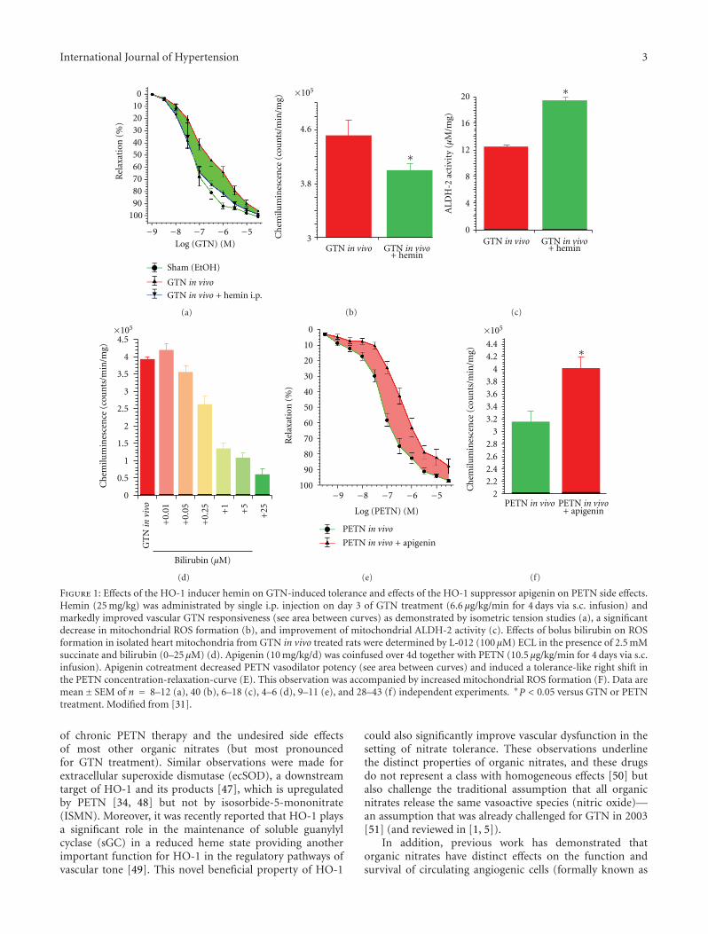
With a previous study, we demonstrated that chronic nitro-glycerin (GTN) therapy results in impaired vasodilatorypotency of GTN (nitrate tolerance) and of the endothelium-dependent vasodilator acetylcholine (endothelial dysfunc-tion) as well as increased vascular and mitochondrial RONSformation [31]. Since another organic nitrate (PETN) waspreviously described to be devoid of nitrate tolerance andinduction of oxidative stress due to induction of the hemeoxygenase-1 (HO-1) system [35, 36], we hypothesized thatpharmacological activation of the HO-1 system may be suit-able to prevent GTN-dependent side effects. In deed, cotreat-ment of GTN-infused rats with the potent HO-1 inducerhemin completely prevented nitrate tolerance (restored theGTN-dependent relaxation), restored NO/cGMP signaling,increased the activity of the GTN bioactivating enzymeALDH-2, and suppressed the mitochondrial RONS forma-tion (Figures 1(a)–1(c)) [31]. The HO-1 product bilirubinsuppressed GTN-induced RONS formation in isolated heartmitochondria (Figure 1(d)). To test the essential role ofHO-1 for the tolerance devoid action of PETN, we cotreatedPETN-infused rats with the HO-1 suppressor apigenin andobserved a tolerance-like phenomenon displaying impairedPETN-dependent relaxation, disturbed NO/cGMP signaling,and increased mitochondrial RONS formation (Figures 1(e)-1(f)) [31].
These observations are in good accordance with pre-vious reports on HO-1 induction by statins [37, 38] andprevention of nitrate tolerance in GTN-infused experimentalanimals [39] as well as human individuals [40, 41]. Therole of HO-1 for prevention of organic nitrate inducedtolerance, endothelial dysfunction, and oxidative stress isfurther supported by observations that the HO-1 productbilirubin efficiently scavenged GTN-induced RONS (mostprobably peroxynitrite) formation in isolated mitochondria[29, 31]. Likewise, the HO-1 products bilirubin and carbonmonoxide as well as PETN increased the expression ofthe GTP-cyclohydrolase-1 (GCH-1), the most importantenzyme for de novo synthesis of tetrahydrobiopterin (BH4),an essential cofactor for endothelial NO synthase (eNOS)function [34, 42]. In contrast, GTN in vivo therapy decreasedthe expression of the GCH-1 [43]. Since BH4 levels aredirectly linked to eNOS activity and endothelial function [44,45] and GCH-1 is oxidatively degraded by activation of theproteasome26S [46], activation of antioxidant pathways byinduction of HO-1 may represent an attractive explanationfor the tolerance and endothelial dysfunction devoid profile
International Journal of Hypertension 3
GTN in vivo
GTN in vivo + hemin i.p.
0102030405060708090
100
Rel
axat
ion
(%
)
Log (GTN) (M)
Sham (EtOH)
−9 −8 −7 −6 −5
(a)
GTN in vivo GTN in vivo+ hemin
∗
4.6
3.8
3Ch
emilu
min
esce
nce
(co
un
ts/m
in/m
g)
×105
(b)
GTN in vivo GTN in vivo+ hemin
20
16
12
8
4
0
AL
DH
-2 a
ctiv
ity
(μM
/mg)
∗
(c)
GT
Nin
viv
o
4.5
4
3.5
3
2.5
2
1.5
1
0.5
0
+0.
01
+0.
05
+0.
25 +5
+1
+25
Bilirubin (μM)
Ch
emilu
min
esce
nce
(co
un
ts/m
in/m
g)
×105
(d)
PETN in vivo
PETN in vivo + apigenin
0
10
20
30
40
50
60
70
80
90
100
Rel
axat
ion
(%
)
Log (PETN) (M)
−9 −8 −7 −6 −5
(e)
PETN in vivo PETN in vivo+ apigenin
4.44.2
43.83.63.43.2
32.82.62.42.2
2
∗
Ch
emilu
min
esce
nce
(co
un
ts/m
in/m
g)
×105
(f)
Figure 1: Effects of the HO-1 inducer hemin on GTN-induced tolerance and effects of the HO-1 suppressor apigenin on PETN side effects.Hemin (25 mg/kg) was administrated by single i.p. injection on day 3 of GTN treatment (6.6 μg/kg/min for 4 days via s.c. infusion) andmarkedly improved vascular GTN responsiveness (see area between curves) as demonstrated by isometric tension studies (a), a significantdecrease in mitochondrial ROS formation (b), and improvement of mitochondrial ALDH-2 activity (c). Effects of bolus bilirubin on ROSformation in isolated heart mitochondria from GTN in vivo treated rats were determined by L-012 (100 μM) ECL in the presence of 2.5 mMsuccinate and bilirubin (0–25 μM) (d). Apigenin (10 mg/kg/d) was coinfused over 4d together with PETN (10.5 μg/kg/min for 4 days via s.c.infusion). Apigenin cotreatment decreased PETN vasodilator potency (see area between curves) and induced a tolerance-like right shift inthe PETN concentration-relaxation-curve (E). This observation was accompanied by increased mitochondrial ROS formation (F). Data aremean± SEM of n = 8–12 (a), 40 (b), 6–18 (c), 4–6 (d), 9–11 (e), and 28–43 (f) independent experiments. ∗P < 0.05 versus GTN or PETNtreatment. Modified from [31].
of chronic PETN therapy and the undesired side effectsof most other organic nitrates (but most pronouncedfor GTN treatment). Similar observations were made forextracellular superoxide dismutase (ecSOD), a downstreamtarget of HO-1 and its products [47], which is upregulatedby PETN [34, 48] but not by isosorbide-5-mononitrate(ISMN). Moreover, it was recently reported that HO-1 playsa significant role in the maintenance of soluble guanylylcyclase (sGC) in a reduced heme state providing anotherimportant function for HO-1 in the regulatory pathways ofvascular tone [49]. This novel beneficial property of HO-1
could also significantly improve vascular dysfunction in thesetting of nitrate tolerance. These observations underlinethe distinct properties of organic nitrates, and these drugsdo not represent a class with homogeneous effects [50] butalso challenge the traditional assumption that all organicnitrates release the same vasoactive species (nitric oxide)—an assumption that was already challenged for GTN in 2003[51] (and reviewed in [1, 5]).
In addition, previous work has demonstrated thatorganic nitrates have distinct effects on the function andsurvival of circulating angiogenic cells (formally known as
4 International Journal of Hypertension
endothelial progenitor cells) [52, 53]. These studies showedthat isosorbide dinitrate in contrast to PETN impair themigration and incorporation activities of these circulatingangiogenic cells in an experimental model of myocardialinfarction, whereas GTN in vitro exposure increased apop-tosis while decreasing phenotypic differentiation, migration,and mitochondrial dehydrogenase activity in these cells. In asubsequent study, Lin et al. investigated the involvement ofheme oxygenase-1 for the related neovascularization processby hematopoietic stem cells and endothelial progenitor cellsin the infarcted area [54]. Thum et al. have shown thatthe impaired function of these circulating angiogenic cellsis based on oxidative stress as envisaged in the setting ofdiabetes, leading to eNOS uncoupling, which was improvedby antioxidants (e.g., superoxide dismutase) [55]. Thesefindings underline the importance of maintaining the BH4
levels to prevent eNOS uncoupling and the role of HO-1 forthis antioxidant mechanism via increase in GCH-1 expres-sion by carbon monoxide and bilirubin as outlined above.This concept is further supported by protective effects of folicacid (a precursor of BH4) on impaired endothelial functionin GTN-treated healthy volunteers [56], and decreased BH4
levels in GTN-treated rabbits were restored by cotherapywith pioglitazone [57]. It should be noted that another groupfound no association between aortic BH4 content and eNOSfunction in response to GTN ex vivo and in vivo treatment[58].
Finally, it should be mentioned that oxidative stressin response to organic nitrates may also be protectiveby a process called ischemic preconditioning (IPC) [59–61]. Recently, the involvement of HO-1 in organic nitrate-mediated IPC was proposed [59, 62] as an explanation for thesustained IPC protective effect under chronic PETN therapybut loss of this beneficial effect under chronic GTN therapy.This is in accordance with the accepted view that HO-1 playsa role in IPC [63, 64].
3. Molecular Proof of a Role ofHO-1 for the Tolerance-Devoid Profile ofPETN by Using HO-1+/− Mice
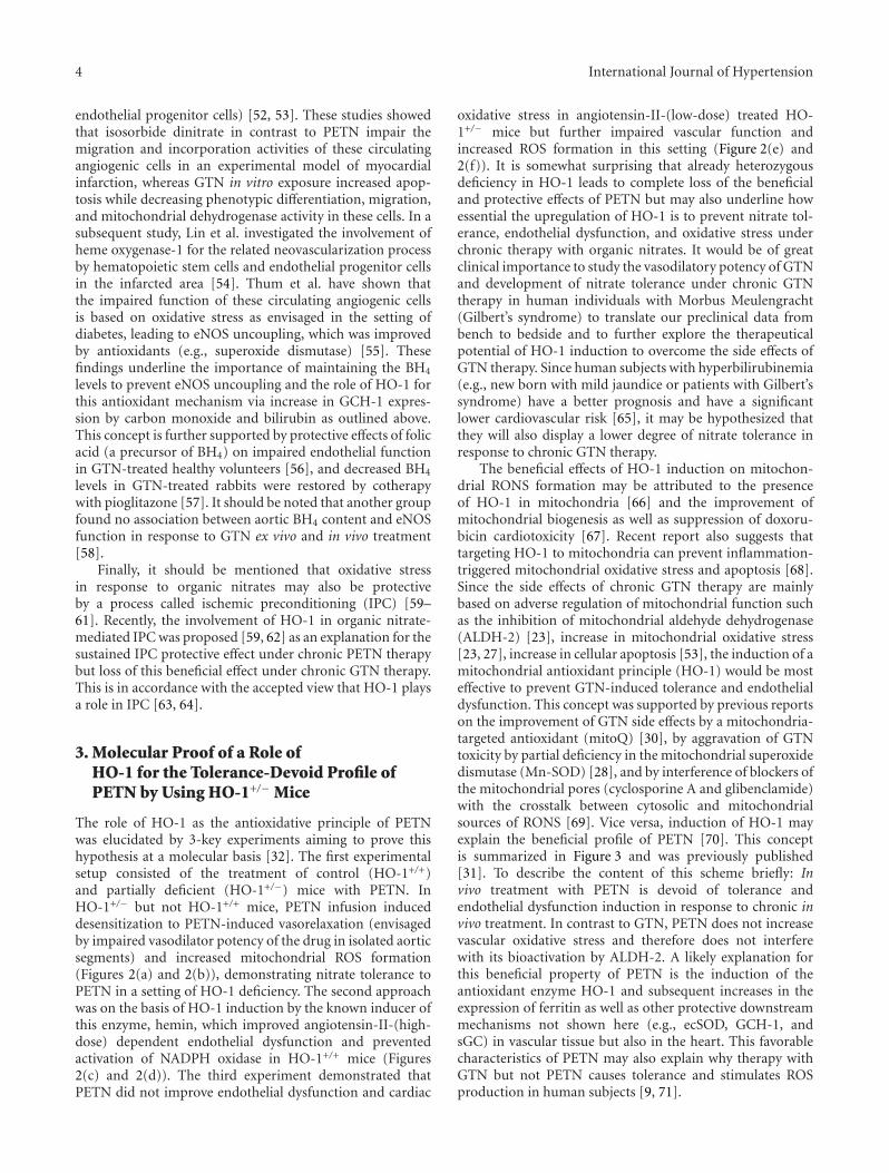
The role of HO-1 as the antioxidative principle of PETNwas elucidated by 3-key experiments aiming to prove thishypothesis at a molecular basis [32]. The first experimentalsetup consisted of the treatment of control (HO-1+/+)and partially deficient (HO-1+/−) mice with PETN. InHO-1+/− but not HO-1+/+ mice, PETN infusion induceddesensitization to PETN-induced vasorelaxation (envisagedby impaired vasodilator potency of the drug in isolated aorticsegments) and increased mitochondrial ROS formation(Figures 2(a) and 2(b)), demonstrating nitrate tolerance toPETN in a setting of HO-1 deficiency. The second approachwas on the basis of HO-1 induction by the known inducer ofthis enzyme, hemin, which improved angiotensin-II-(high-dose) dependent endothelial dysfunction and preventedactivation of NADPH oxidase in HO-1+/+ mice (Figures2(c) and 2(d)). The third experiment demonstrated thatPETN did not improve endothelial dysfunction and cardiac
oxidative stress in angiotensin-II-(low-dose) treated HO-1+/− mice but further impaired vascular function andincreased ROS formation in this setting (Figure 2(e) and2(f)). It is somewhat surprising that already heterozygousdeficiency in HO-1 leads to complete loss of the beneficialand protective effects of PETN but may also underline howessential the upregulation of HO-1 is to prevent nitrate tol-erance, endothelial dysfunction, and oxidative stress underchronic therapy with organic nitrates. It would be of greatclinical importance to study the vasodilatory potency of GTNand development of nitrate tolerance under chronic GTNtherapy in human individuals with Morbus Meulengracht(Gilbert’s syndrome) to translate our preclinical data frombench to bedside and to further explore the therapeuticalpotential of HO-1 induction to overcome the side effects ofGTN therapy. Since human subjects with hyperbilirubinemia(e.g., new born with mild jaundice or patients with Gilbert’ssyndrome) have a better prognosis and have a significantlower cardiovascular risk [65], it may be hypothesized thatthey will also display a lower degree of nitrate tolerance inresponse to chronic GTN therapy.
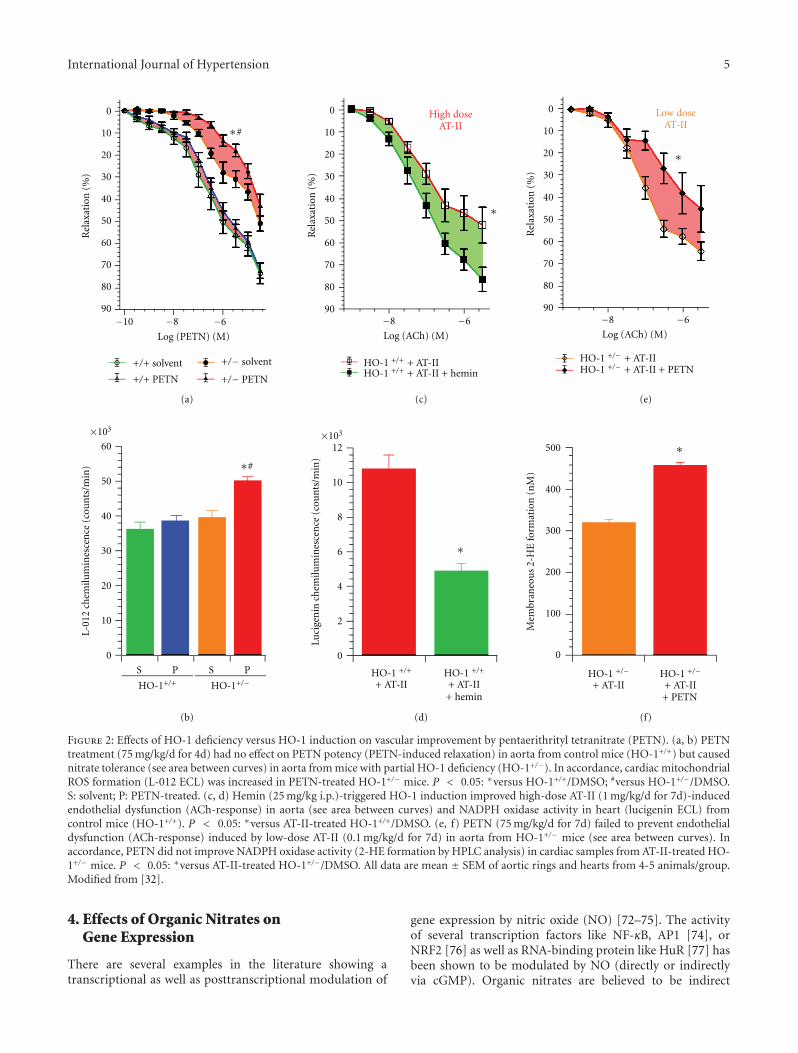
The beneficial effects of HO-1 induction on mitochon-drial RONS formation may be attributed to the presenceof HO-1 in mitochondria [66] and the improvement ofmitochondrial biogenesis as well as suppression of doxoru-bicin cardiotoxicity [67]. Recent report also suggests thattargeting HO-1 to mitochondria can prevent inflammation-triggered mitochondrial oxidative stress and apoptosis [68].Since the side effects of chronic GTN therapy are mainlybased on adverse regulation of mitochondrial function suchas the inhibition of mitochondrial aldehyde dehydrogenase(ALDH-2) [23], increase in mitochondrial oxidative stress[23, 27], increase in cellular apoptosis [53], the induction of amitochondrial antioxidant principle (HO-1) would be mosteffective to prevent GTN-induced tolerance and endothelialdysfunction. This concept was supported by previous reportson the improvement of GTN side effects by a mitochondria-targeted antioxidant (mitoQ) [30], by aggravation of GTNtoxicity by partial deficiency in the mitochondrial superoxidedismutase (Mn-SOD) [28], and by interference of blockers ofthe mitochondrial pores (cyclosporine A and glibenclamide)with the crosstalk between cytosolic and mitochondrialsources of RONS [69]. Vice versa, induction of HO-1 mayexplain the beneficial profile of PETN [70]. This conceptis summarized in Figure 3 and was previously published[31]. To describe the content of this scheme briefly: Invivo treatment with PETN is devoid of tolerance andendothelial dysfunction induction in response to chronic invivo treatment. In contrast to GTN, PETN does not increasevascular oxidative stress and therefore does not interferewith its bioactivation by ALDH-2. A likely explanation forthis beneficial property of PETN is the induction of theantioxidant enzyme HO-1 and subsequent increases in theexpression of ferritin as well as other protective downstreammechanisms not shown here (e.g., ecSOD, GCH-1, andsGC) in vascular tissue but also in the heart. This favorablecharacteristics of PETN may also explain why therapy withGTN but not PETN causes tolerance and stimulates ROSproduction in human subjects [9, 71].
International Journal of Hypertension 5
0
10
20
30
40
50
60
70
80
90
Rel
axat
ion
(%
)
−10 −8 −6
Log (PETN) (M)
+/+ solvent
+/+ PETN
+/− solvent
+/− PETN
∗#∗#
0
10
20
30
40
50
60
70
80
90R
elax
atio
n (
%)
−8 −6
Log (ACh) (M)
High doseAT-II
∗
HO-1 +/+ + AT-IIHO-1 +/+ + AT-II + hemin
0
10
20
30
40
50
60
70
80
90
Rel
axat
ion
(%
)
−8 −6
Log (ACh) (M)
Low doseAT-II
∗
HO-1 +/− + AT-IIHO-1 +/− + AT-II + PETN
∗#
60
50
40
30
20
10
0
×103
L-01
2 ch
emilu
min
esce
nce
(co
un
ts/m
in)
S P S P
HO-1+/+ HO-1+/−
∗
×103
Luci
gen
in c
hem
ilum
ines
cen
ce (
cou
nts
/min
)
12
10
8
6
4
2
0
HO-1 +/+
+ AT-IIHO-1 +/+
+ AT-II+ hemin
∗
HO-1 +/−+ AT-II
HO-1 +/−+ AT-II+ PETN
500
400
300
200
100
0
Mem
bran
eou
s 2-
HE
form
atio
n (
nM
)
(a)
(b)
(c)
(d)
(e)
(f)
Figure 2: Effects of HO-1 deficiency versus HO-1 induction on vascular improvement by pentaerithrityl tetranitrate (PETN). (a, b) PETNtreatment (75 mg/kg/d for 4d) had no effect on PETN potency (PETN-induced relaxation) in aorta from control mice (HO-1+/+) but causednitrate tolerance (see area between curves) in aorta from mice with partial HO-1 deficiency (HO-1+/−). In accordance, cardiac mitochondrialROS formation (L-012 ECL) was increased in PETN-treated HO-1+/− mice. P < 0.05: ∗versus HO-1+/+/DMSO; #versus HO-1+/−/DMSO.S: solvent; P: PETN-treated. (c, d) Hemin (25 mg/kg i.p.)-triggered HO-1 induction improved high-dose AT-II (1 mg/kg/d for 7d)-inducedendothelial dysfunction (ACh-response) in aorta (see area between curves) and NADPH oxidase activity in heart (lucigenin ECL) fromcontrol mice (HO-1+/+). P < 0.05: ∗versus AT-II-treated HO-1+/+/DMSO. (e, f) PETN (75 mg/kg/d for 7d) failed to prevent endothelialdysfunction (ACh-response) induced by low-dose AT-II (0.1 mg/kg/d for 7d) in aorta from HO-1+/− mice (see area between curves). Inaccordance, PETN did not improve NADPH oxidase activity (2-HE formation by HPLC analysis) in cardiac samples from AT-II-treated HO-1+/− mice. P < 0.05: ∗versus AT-II-treated HO-1+/−/DMSO. All data are mean ± SEM of aortic rings and hearts from 4-5 animals/group.Modified from [32].
4. Effects of Organic Nitrates onGene Expression
There are several examples in the literature showing atranscriptional as well as posttranscriptional modulation of
gene expression by nitric oxide (NO) [72–75]. The activityof several transcription factors like NF-κB, AP1 [74], orNRF2 [76] as well as RNA-binding protein like HuR [77] hasbeen shown to be modulated by NO (directly or indirectlyvia cGMP). Organic nitrates are believed to be indirect
6 International Journal of Hypertension
Oxidative stress
ROS/RNS
Vasodilation
Lipoic
LAR
Trx/TrxR
GSH/GR
PETNchronic
treatment
Metabolites
mtALDH
mtALDH
S S
HS SH
+ +
− −+
−
Ferritin↑Free iron↓
HO-1bilirubin
CO
acidred
Lipoicacidox
GTNchronic
treatment
GTN orPETN/PETriN
NOx
Figure 3: Scheme illustrating the mechanisms underlying the oxidative stress concept of nitrate tolerance in response to GTN treatment andthe mechanisms underlying the beneficial vascular effects in response to PETN. PETN and GTN are bioactivated by mitochondrial ALDH(ALDH-2) yielding 1,2-glyceryl dinitrate and PETriN, respectively, as well as a yet undefined nitrogen species (NOx, probably nitrite) thatundergoes further reduction by the mitochondrial respiratory chain or acidic disproportionation to form an activator of sGC (probablynitrico xide). GTN treatment induces mitochondrial reactive oxygen and nitrogen species formation (ROS/RNS). These ROS/RNS in turninhibit the GTN bioactivation process by inactivation of ALDH-2 or by inhibiting the repair system of the ALDH-2, which includes lipoicacid, as well as a reductase system depending on the NADH or NADPH (lipoicacid reductase (LAR), thioredoxin/thioredoxin reductase(Trx/TrxR) or glutathione/glutathione reductase (GSH/GR). In contrast to GTN, PETN provides potent antioxidative effects by inducingHO-1 and ferritin, which in turn decrease ROS levels and therefore protect the ALDH-2 from ROS mediated inactivation. Adapted from[31].
NO-donors. Therefore, it seems very likely that treatmentwith organic nitrates may have implications on the expres-sion of multiple genes. GTN has been described to enhancethe expression of c-fos, COX-2, Bcl2, and nNOS in brainnuclei [78–81], to reduce beta-catenin expression in coloncancer cells [82], and to reduce NOX1, NOX2, NOX4 andALDH2-expression in rat aorta and rat smooth musclecells [83]. Using microarray analysis, Wang et al. describedchanges of the expression of 290 genes in the aortas of ratstreated with GTN for 8 h [84]. Analyzing the gene expressionin the hearts treated for 4 days with GTN, the authorsdescribed expressional changes of more than 500 genes [85].
There are also some reports about the expressional effectsof PETN [29, 31, 35, 36, 62]. PETN (but not GTN) has beenshown to enhance the expression of the antioxidant genesHO-1 and ferritin heavy chain (FeHc) in human endothelialcells [29, 35, 36, 62] and rat aorta [31]. In microarrayexperiments, the authors showed that PETN modulated theexpression of more than 1200 genes in the hearts of ratstreated with PETN for 4 days [85].
5. Molecular Mechanisms Involved in the Regu-lation of Gene Expression by Organic Nitrates
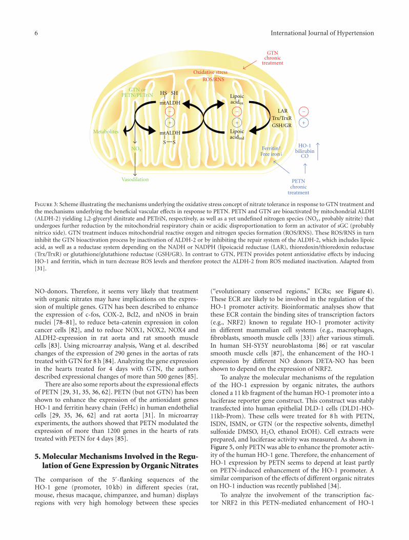
The comparison of the 5′-flanking sequences of theHO-1 gene (promoter, 10 kb) in different species (rat,mouse, rhesus macaque, chimpanzee, and human) displaysregions with very high homology between these species
(“evolutionary conserved regions,” ECRs; see Figure 4).These ECR are likely to be involved in the regulation of theHO-1 promoter activity. Bioinformatic analyses show thatthese ECR contain the binding sites of transcription factors(e.g., NRF2) known to regulate HO-1 promoter activityin different mammalian cell systems (e.g., macrophages,fibroblasts, smooth muscle cells [33]) after various stimuli.In human SH-SY5Y neuroblastoma [86] or rat vascularsmooth muscle cells [87], the enhancement of the HO-1expression by different NO donors DETA-NO has beenshown to depend on the expression of NRF2.
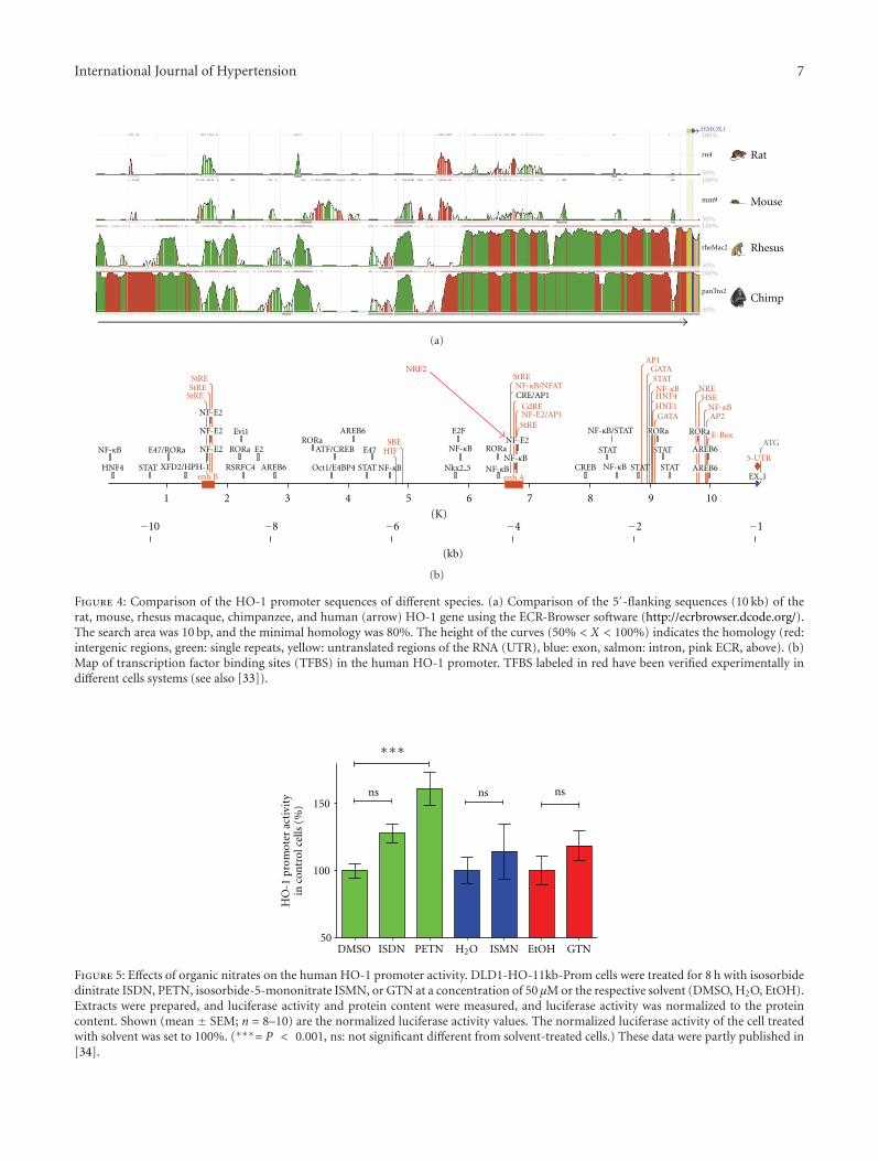
To analyze the molecular mechanisms of the regulationof the HO-1 expression by organic nitrates, the authorscloned a 11 kb fragment of the human HO-1 promoter into aluciferase reporter gene construct. This construct was stablytransfected into human epithelial DLD-1 cells (DLD1-HO-11kb-Prom). These cells were treated for 8 h with PETN,ISDN, ISMN, or GTN (or the respective solvents, dimethylsulfoxide DMSO, H2O, ethanol EtOH). Cell extracts wereprepared, and luciferase activity was measured. As shown inFigure 5, only PETN was able to enhance the promoter activ-ity of the human HO-1 gene. Therefore, the enhancement ofHO-1 expression by PETN seems to depend at least partlyon PETN-induced enhancement of the HO-1 promoter. Asimilar comparison of the effects of different organic nitrateson HO-1 induction was recently published [34].
To analyze the involvement of the transcription fac-tor NRF2 in this PETN-mediated enhancement of HO-1
International Journal of Hypertension 7
100%
rn4
50%100%
50%100%
50%
50%
100%
mm9
rheMac2
panTro2
Rat
Mouse
Rhesus
Chimp
HMOX1
(a)
HNF4
E47/RORa
STAT XFD2/HPH-1
StREStRE
StRE
NF-E2
NF-E2
NF-E2
enh B
Evi1
RORa E2
RSRFC4 AREB6
RORaATF/CREB
Oct1/E4BP4
E47
STAT
SBE
NRF2
HIF
AREB6 E2F
StRENF-κB/NFATCRE/AP1
CdRENF-E2/AP1StRE
NF-E2RORa
enh A
STAT
CREB
NF-κB/STAT
AP1GATASTAT
HNF4HNF1GATA
RORa RORa
STAT
STAT STAT
NREHSE
AP2
E-Box
AREB6
AREB6
ATG
5-UTRNkx2 5
EX 1
1 2 3 4 5 6 7 8 9 10
(K)−10 −8 −6 −4 −2 −1
(kb)
NF-κBNF-κBNF-κB
NF-κB NF-κBNF-κB
NF-κB
NF-κB
(b)
Figure 4: Comparison of the HO-1 promoter sequences of different species. (a) Comparison of the 5′-flanking sequences (10 kb) of therat, mouse, rhesus macaque, chimpanzee, and human (arrow) HO-1 gene using the ECR-Browser software (http://ecrbrowser.dcode.org/).The search area was 10 bp, and the minimal homology was 80%. The height of the curves (50% < X < 100%) indicates the homology (red:intergenic regions, green: single repeats, yellow: untranslated regions of the RNA (UTR), blue: exon, salmon: intron, pink ECR, above). (b)Map of transcription factor binding sites (TFBS) in the human HO-1 promoter. TFBS labeled in red have been verified experimentally indifferent cells systems (see also [33]).
50
100
150nsnsns
DMSO
HO
-1 p
rom
oter
act
ivit
yin
con
trol
cel
ls (
%)
ISDN PETN ISMN EtOH GTNH2O
∗∗∗
Figure 5: Effects of organic nitrates on the human HO-1 promoter activity. DLD1-HO-11kb-Prom cells were treated for 8 h with isosorbidedinitrate ISDN, PETN, isosorbide-5-mononitrate ISMN, or GTN at a concentration of 50 μM or the respective solvent (DMSO, H2O, EtOH).Extracts were prepared, and luciferase activity and protein content were measured, and luciferase activity was normalized to the proteincontent. Shown (mean ± SEM; n = 8–10) are the normalized luciferase activity values. The normalized luciferase activity of the cell treatedwith solvent was set to 100%. (∗∗∗= P < 0.001, ns: not significant different from solvent-treated cells.) These data were partly published in[34].
8 International Journal of Hypertension
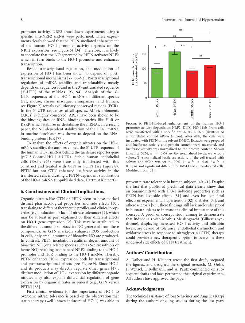
promoter activity, NRF2-knockdown experiments using aspecific anti-NRF2 siRNA were performed. These experi-ments clearly showed that the PETN-mediated enhancementof the human HO-1 promoter activity depends on theNRF2 expression (see Figure 6) [34]. Therefore, it is likelyto speculate that the NO generated by PETN activates NRF2which in turn binds to the HO-1 promoter and enhancestranscription.
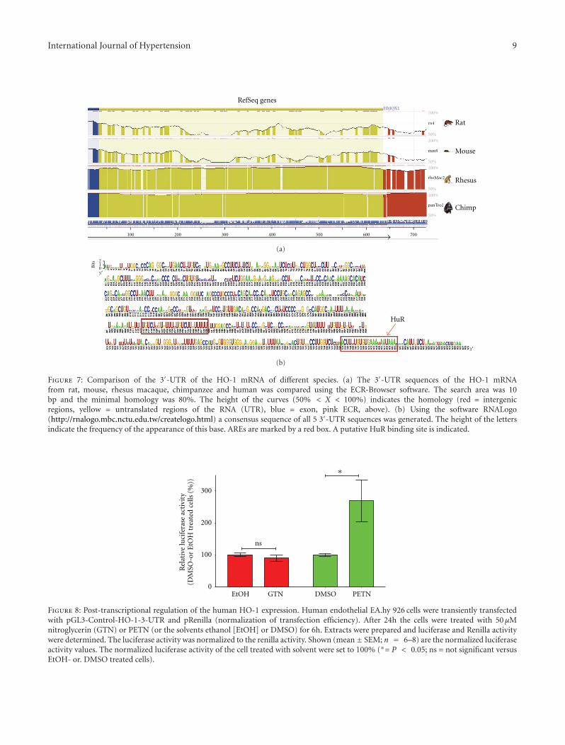
Beside transcriptional regulation, the modulation ofexpression of HO-1 has been shown to depend on post-transcriptional mechanisms [77, 88–92]. Posttranscriptionalregulation of mRNA stability and translatability mostlydepends on sequences found in the 3′-untranslated sequence(3′-UTR) of the mRNAs [93, 94]. Analysis of the 3′-UTR sequences of the HO-1 mRNA of different species(rat, mouse, rhesus macaque, chimpanzee, and human,see Figure 7) reveals evolutionary conserved regions (ECR).In the 3′-UTR sequences of all species, AU-rich elements(AREs) is highly conserved. AREs have been shown to bethe binding sites of RNA, binding proteins like HuR orKSRP, which stabilize or destabilize the mRNAs. In a recentpaper, the NO-dependent stabilization of the HO-1 mRNAin murine fibroblasts was shown to depend on the RNA-binding protein HuR [77].
To analyze the effects of organic nitrates on the HO-1mRNA stability, the authors cloned the 3′-UTR sequence ofthe human HO-1 mRNA behind the luciferase reporter gene(pGL3-Control-HO-1-3-UTR). Stable human endothelialcells (EA.hy 926) were transiently transfected with thisconstruct and treated with GTN or PETN (see Figure 8).PETN but not GTN enhanced luciferase activity in thetransfected cells indicating a PETN-dependent stabilizationof the HO-1 mRNA (unpublished data, Hartmut Kleinert).
6. Conclusions and Clinical Implications
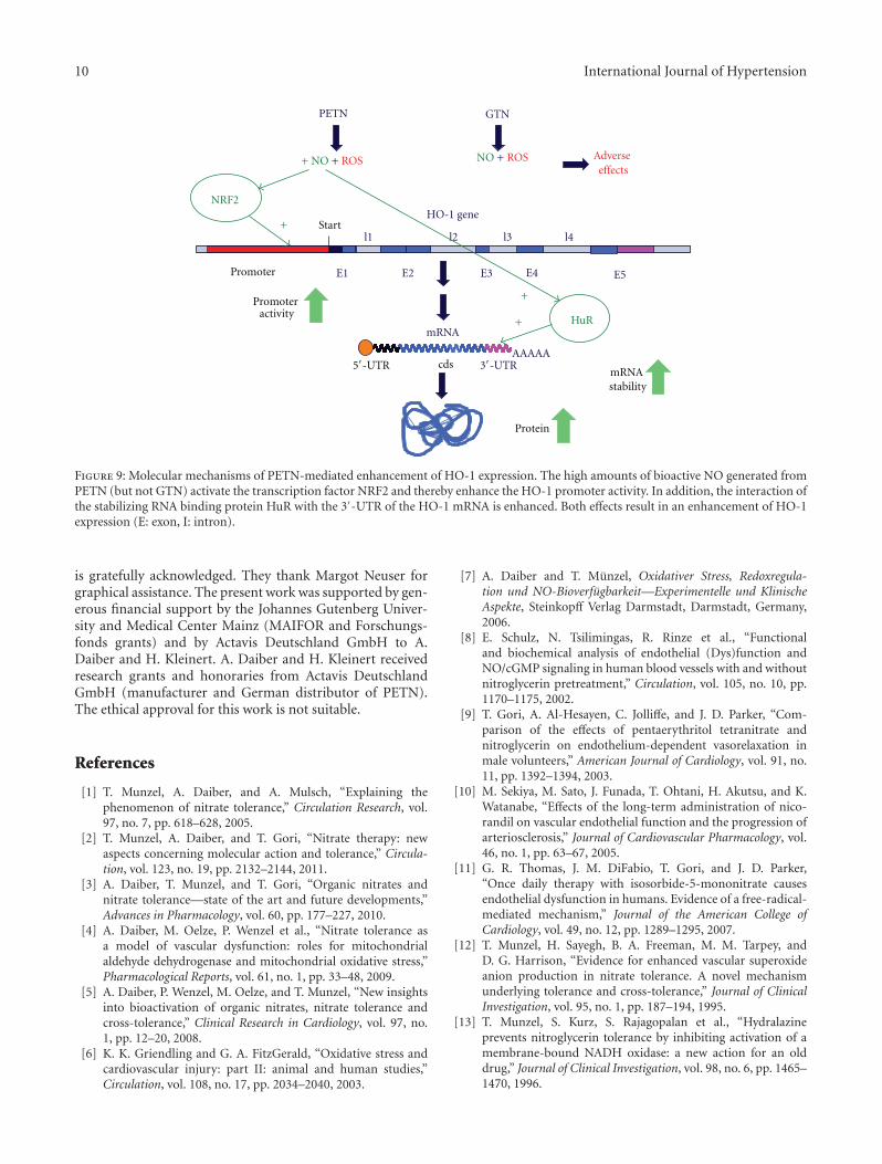
Organic nitrates like GTN or PETN seem to have markeddistinct pharmacological properties and side effects [50],translating to different therapeutic profiles and clinical prop-erties (e.g., induction or lack of nitrate tolerance) [9], whichmay be at least in part explained by their different effectson HO-1 gene expression [2]. This may be attributed tothe different amounts of bioactive NO generated from thesecompounds. As GTN markedly enhances ROS productionin cells, only small amounts of bioactive NO are produced.In contrast, PETN incubation results in decent amount ofbioactive NO (or a related species such as S-nitrosothiols orheme-NO) resulting in enhanced NRF2 binding to the HO-1promoter and HuR binding to the HO-1 mRNA. Thereby,PETN enhances HO-1 expression both by transcriptionaland posttranscriptional effects (see Figure 9). Since HO-1and its products may directly regulate other genes [47],distinct modulation of HO-1 expression by different organicnitrates may also explain differential regulation of geneexpression by organic nitrates in general (e.g., GTN versusPETN) [85].
First clinical evidence for the importance of HO-1 toovercome nitrate tolerance is based on the observation thatstatin therapy (well-known inducers of HO-1) was able to
0
50
100
150
ns
ns
ns
HO
-1 p
rom
oter
act
ivit
yin
siC
on-t
reat
ed c
ells
(%
)
∗∗
∗∗
siC
onP
ET
N
siC
onD
MSO
siN
RF2
DM
SO
siN
RF2
PE
TN
Figure 6: PETN-induced enhancement of the human HO-1promoter activity depends on NRF2. DLD1-HO-11kb-Prom cellswere transfected with a specific anti-NRF2 siRNA (siNRF2) ora nonrelated control siRNA (siCon). After 48 h, the cells wereincubated with PETN or the solvent DMSO. Extracts were preparedand luciferase activity and protein content were measured, andluciferase activity was normalized to the protein content. Shown(mean ± SEM; n = 5-6) are the normalized luciferase activityvalues. The normalized luciferase activity of the cell treated withsolvent and siCon was set to 100%. (∗∗= P < 0.01, ∗= P <0.05, ns: not significant different to DMSO and siCon-treated cells.Modified from [34].
prevent nitrate tolerance in human subjects [40, 41]. Despitethe fact that published preclinical data clearly show thatan organic nitrate with HO-1 inducing properties such asPETN has less side effects [31] and even has beneficialeffects on experimental hypertension [32], diabetes [34], andatherosclerosis [95], these findings still lack molecular proofin human subjects to increase the clinical importance of thisconcept. A proof of concept study aiming to demonstratethat individuals with Morbus Meulengracht (Gilbert’s syn-drome), displaying increased HO-1 activity and bilirubinlevels, are devoid of tolerance, endothelial dysfunction andoxidative stress in response to nitroglycerin (GTN) therapycould provide a new therapeutic option to overcome theseundesired side effects of GTN treatment.
Authors’ Contribution
A. Daiber and H. Kleinert wrote the first draft, preparedthe figures, and designed the original research. M. Oelze,P. Wenzel, F. Bollmann, and A. Pautz commented on sub-sequent drafts and have performed the original experiments.All authors have approved the paper.
Acknowledgments
The technical assistance of Jorg Schreiner and Angelica Karpiduring the authors ongoing studies during the last years
International Journal of Hypertension 9
100%
rn4
50%
100%
50%
100%
50%
50%
100%
mm9
rheMac2
panTro2
Rat
Mouse
Rhesus
Chimp
100 200 300 400 500 600 700
RefSeq genes
100%
rn4
50%
100%
50%
100%
50%
50%
100%
mm9
rheMac2
panTro2
HMOX1
(a)
210B
its
HuR
5
3
(b)
Figure 7: Comparison of the 3′-UTR of the HO-1 mRNA of different species. (a) The 3′-UTR sequences of the HO-1 mRNAfrom rat, mouse, rhesus macaque, chimpanzee and human was compared using the ECR-Browser software. The search area was 10bp and the minimal homology was 80%. The height of the curves (50% < X < 100%) indicates the homology (red = intergenicregions, yellow = untranslated regions of the RNA (UTR), blue = exon, pink ECR, above). (b) Using the software RNALogo(http://rnalogo.mbc.nctu.edu.tw/createlogo.html) a consensus sequence of all 5 3′-UTR sequences was generated. The height of the lettersindicate the frequency of the appearance of this base. AREs are marked by a red box. A putative HuR binding site is indicated.
0
100
200
300
EtOH GTN DMSO PETN
Rel
ativ
e lu
cife
rase
act
ivit
y(D
MSO
-or
EtO
H t
reat
ed c
ells
(%
))
ns
∗
Figure 8: Post-transcriptional regulation of the human HO-1 expression. Human endothelial EA.hy 926 cells were transiently transfectedwith pGL3-Control-HO-1-3-UTR and pRenilla (normalization of transfection efficiency). After 24h the cells were treated with 50 μMnitroglycerin (GTN) or PETN (or the solvents ethanol [EtOH] or DMSO) for 6h. Extracts were prepared and luciferase and Renilla activitywere determined. The luciferase activity was normalized to the renilla activity. Shown (mean± SEM; n = 6–8) are the normalized luciferaseactivity values. The normalized luciferase activity of the cell treated with solvent were set to 100% (∗= P < 0.05; ns = not significant versusEtOH- or. DMSO treated cells).
10 International Journal of Hypertension
AAAAA
AAAAA
E1 E2 E3 E4 E5
l1 l2 l3 l4
mRNAHuR
cds
Promoter
Start
Protein
PETN GTN
Adverse effects
+
+
+
NRF2
Promoter activity
mRNA stability
NO + ROS+ NO + ROS
HO-1 gene
3-UTR5-UTR
Figure 9: Molecular mechanisms of PETN-mediated enhancement of HO-1 expression. The high amounts of bioactive NO generated fromPETN (but not GTN) activate the transcription factor NRF2 and thereby enhance the HO-1 promoter activity. In addition, the interaction ofthe stabilizing RNA binding protein HuR with the 3′-UTR of the HO-1 mRNA is enhanced. Both effects result in an enhancement of HO-1expression (E: exon, I: intron).
is gratefully acknowledged. They thank Margot Neuser forgraphical assistance. The present work was supported by gen-erous financial support by the Johannes Gutenberg Univer-sity and Medical Center Mainz (MAIFOR and Forschungs-fonds grants) and by Actavis Deutschland GmbH to A.Daiber and H. Kleinert. A. Daiber and H. Kleinert receivedresearch grants and honoraries from Actavis DeutschlandGmbH (manufacturer and German distributor of PETN).The ethical approval for this work is not suitable.
References
[1] T. Munzel, A. Daiber, and A. Mulsch, “Explaining thephenomenon of nitrate tolerance,” Circulation Research, vol.97, no. 7, pp. 618–628, 2005.
[2] T. Munzel, A. Daiber, and T. Gori, “Nitrate therapy: newaspects concerning molecular action and tolerance,” Circula-tion, vol. 123, no. 19, pp. 2132–2144, 2011.
[3] A. Daiber, T. Munzel, and T. Gori, “Organic nitrates andnitrate tolerance—state of the art and future developments,”Advances in Pharmacology, vol. 60, pp. 177–227, 2010.
[4] A. Daiber, M. Oelze, P. Wenzel et al., “Nitrate tolerance asa model of vascular dysfunction: roles for mitochondrialaldehyde dehydrogenase and mitochondrial oxidative stress,”Pharmacological Reports, vol. 61, no. 1, pp. 33–48, 2009.
[5] A. Daiber, P. Wenzel, M. Oelze, and T. Munzel, “New insightsinto bioactivation of organic nitrates, nitrate tolerance andcross-tolerance,” Clinical Research in Cardiology, vol. 97, no.1, pp. 12–20, 2008.
[6] K. K. Griendling and G. A. FitzGerald, “Oxidative stress andcardiovascular injury: part II: animal and human studies,”Circulation, vol. 108, no. 17, pp. 2034–2040, 2003.
[7] A. Daiber and T. Munzel, Oxidativer Stress, Redoxregula-tion und NO-Bioverfugbarkeit—Experimentelle und KlinischeAspekte, Steinkopff Verlag Darmstadt, Darmstadt, Germany,2006.
[8] E. Schulz, N. Tsilimingas, R. Rinze et al., “Functionaland biochemical analysis of endothelial (Dys)function andNO/cGMP signaling in human blood vessels with and withoutnitroglycerin pretreatment,” Circulation, vol. 105, no. 10, pp.1170–1175, 2002.
[9] T. Gori, A. Al-Hesayen, C. Jolliffe, and J. D. Parker, “Com-parison of the effects of pentaerythritol tetranitrate andnitroglycerin on endothelium-dependent vasorelaxation inmale volunteers,” American Journal of Cardiology, vol. 91, no.11, pp. 1392–1394, 2003.
[10] M. Sekiya, M. Sato, J. Funada, T. Ohtani, H. Akutsu, and K.Watanabe, “Effects of the long-term administration of nico-randil on vascular endothelial function and the progression ofarteriosclerosis,” Journal of Cardiovascular Pharmacology, vol.46, no. 1, pp. 63–67, 2005.
[11] G. R. Thomas, J. M. DiFabio, T. Gori, and J. D. Parker,“Once daily therapy with isosorbide-5-mononitrate causesendothelial dysfunction in humans. Evidence of a free-radical-mediated mechanism,” Journal of the American College ofCardiology, vol. 49, no. 12, pp. 1289–1295, 2007.
[12] T. Munzel, H. Sayegh, B. A. Freeman, M. M. Tarpey, andD. G. Harrison, “Evidence for enhanced vascular superoxideanion production in nitrate tolerance. A novel mechanismunderlying tolerance and cross-tolerance,” Journal of ClinicalInvestigation, vol. 95, no. 1, pp. 187–194, 1995.
[13] T. Munzel, S. Kurz, S. Rajagopalan et al., “Hydralazineprevents nitroglycerin tolerance by inhibiting activation of amembrane-bound NADH oxidase: a new action for an olddrug,” Journal of Clinical Investigation, vol. 98, no. 6, pp. 1465–1470, 1996.
International Journal of Hypertension 11
[14] T. Munzel and D. G. Harrison, “Evidence for a role of oxygenderived free radicals and protein kinase C in nitrate tolerance,”Journal of Molecular Medicine, vol. 75, no. 11-12, pp. 891–900,1997.
[15] T. Munzel, A. Giaid, S. Kurz, D. J. Stewart, and D. G. Harrison,“Evidence for a role of endothelin 1 and protein kinase C innitroglycerin tolerance,” Proceedings of the National Academyof Sciences of the United States of America, vol. 92, no. 11, pp.5244–5248, 1995.
[16] A. Warnholtz, H. Mollnau, T. Heitzer et al., “Adverse effectsof nitroglycerin treatment on endothelial function, vascularnitrotyrosine levels and cGMP-dependent protein kinaseactivity in hyperlipidemic Watanabe rabbits,” Journal of theAmerican College of Cardiology, vol. 40, no. 7, pp. 1356–1363,2002.
[17] U. Hink, M. Oelze, P. Kolb et al., “Role for peroxynitrite inthe inhibition of prostacyclin synthase in nitrate tolerance,”Journal of the American College of Cardiology, vol. 42, no. 10,pp. 1826–1834, 2003.
[18] A. Daiber, M. Oelze, M. Coldewey et al., “Hydralazine is apowerful inhibitor of peroxynitrite formation as a possibleexplanation for its beneficial effects on prognosis in patientswith congestive heart failure,” Biochemical and BiophysicalResearch Communications, vol. 338, no. 4, pp. 1865–1874,2005.
[19] P. Wenzel, U. Hink, M. Oelze et al., “Role of reduced lipoicacid in the redox regulation of mitochondrial aldehyde dehy-drogenase (ALDH-2) activity: implications for mitochondrialoxidative stress and nitrate tolerance,” Journal of BiologicalChemistry, vol. 282, no. 1, pp. 792–799, 2007.
[20] Q. Fan, F. Gao, L. Zhang, T. A. Christopher, B. L. Lopez,and X. L. Ma, “Nitrate tolerance aggravates postischemicmyocardial apoptosis and impairs cardiac functional recoveryafter ischemia,” Apoptosis, vol. 10, no. 6, pp. 1235–1242, 2005.
[21] G. Abou-Mohamed, J. A. Johnson, L. Jin et al., “Roles of super-oxide, peroxynitrite, and protein kinase C in the developmentof tolerance to nitroglycerin,” Journal of Pharmacology andExperimental Therapeutics, vol. 308, no. 1, pp. 289–299, 2004.
[22] M. J. Mihm, C. M. Coyle, L. Jing, and J. A. Bauer, “Vascularperoxynitrite formation during organic nitrate tolerance,”Journal of Pharmacology and Experimental Therapeutics, vol.291, no. 1, pp. 194–198, 1999.
[23] K. Sydow, A. Daiber, M. Oelze et al., “Central role ofmitochondrial aldehyde dehydrogenase and reactive oxygenspecies in nitroglycerin tolerance and cross-tolerance,” Journalof Clinical Investigation, vol. 113, no. 3, pp. 482–489, 2004.
[24] Z. Chen, J. Zhang, and J. S. Stamler, “Identification ofthe enzymatic mechanism of nitroglycerin bioactivation,”Proceedings of the National Academy of Sciences of the UnitedStates of America, vol. 99, no. 12, pp. 8306–8311, 2002.
[25] P. Needleman and F. E. Hunter Jr., “Effects of organicnitrates on mitochondrial respiration and swelling: possiblecorrelations with the mechanism of pharmacologic action,”Molecular Pharmacology, vol. 2, no. 2, pp. 134–143, 1966.
[26] B. Jakschik and P. Needleman, “Sulfhydryl reactivity of organicnitrates: biochemical basis for inhibition of glyceraldehydep dehydrogenase and monoamine oxidase,” Biochemical andBiophysical Research Communications, vol. 53, no. 2, pp. 539–544, 1973.
[27] A. Daiber, M. Oelze, M. Coldewey et al., “Oxidative stress andmitochondrial aldehyde dehydrogenase activity: a comparisonof pentaerythritol tetranitrate with other organic nitrates,”Molecular Pharmacology, vol. 66, no. 6, pp. 1372–1382, 2004.
[28] A. Daiber, M. Oelze, S. Sulyok et al., “Heterozygous deficiencyof manganese superoxide dismutase in mice (Mn-SOD+/-):a novel approach to assess the role of oxidative stress for thedevelopment of nitrate tolerance,” Molecular Pharmacology,vol. 68, no. 3, pp. 579–588, 2005.
[29] H. Mollnau, P. Wenzel, M. Oelze et al., “Mitochondrial oxida-tive stress and nitrate tolerance—comparison of nitroglycerinand pentaerithrityl tetranitrate in Mn-SOD+/- mice,” BMCCardiovascular Disorders, vol. 6, article 44, 2006.
[30] J. V. Esplugues, M. Rocha, C. Nunez et al., “ComplexI dysfunction and tolerance to nitroglycerin: an approachbased on mitochondrial-targeted antioxidants,” CirculationResearch, vol. 99, no. 10, pp. 1067–1075, 2006.
[31] P. Wenzel, M. Oelze, M. Coldewey et al., “Heme oxygenase-1:a novel key player in the development of tolerance in responseto organic nitrates,” Arteriosclerosis, Thrombosis, and VascularBiology, vol. 27, no. 8, pp. 1729–1735, 2007.
[32] S. Schuhmacher, P. Wenzel, E. Schulz et al., “Pentaerythritoltetranitrate improves angiotensin II-induced vascular dys-function via induction of heme oxygenase-1,” Hypertension,vol. 55, no. 4, pp. 897–904, 2010.
[33] J. Alam and J. L. Cook, “How many transcription factors doesit take to turn on the heme oxygenase-1 gene?” AmericanJournal of Respiratory Cell and Molecular Biology, vol. 36, no.2, pp. 166–174, 2007.
[34] S. Schuhmacher, M. Oelze, F. Bollmann et al., “Vascu-lar dysfunction in experimental diabetes is improved bypentaerithrityl tetranitrate but not isosorbide-5-mononitratetherapy,” Diabetes, vol. 60, no. 10, pp. 2608–2616, 2011.
[35] S. Oberle, A. Abate, N. Grosser et al., “Heme oxygenase-1 induction may explain the antioxidant profile of pen-taerythrityl trinitrate,” Biochemical and Biophysical ResearchCommunications, vol. 290, no. 5, pp. 1539–1544, 2002.
[36] S. Oberle, P. Schwartz, A. Abate, and H. Schroder, “Theantioxidant defense protein ferritin is a novel and specifictarget for pentaerithrityl tetranitrate in endothelial cells,”Biochemical and Biophysical Research Communications, vol.261, no. 1, pp. 28–34, 1999.
[37] N. Grosser, A. Hemmerle, G. Berndt et al., “The antioxidantdefense protein heme oxygenase 1 is a novel target for statinsin endothelial cells,” Free Radical Biology and Medicine, vol. 37,no. 12, pp. 2064–2071, 2004.
[38] F. Gueler, J. K. Park, S. Rong et al., “Statins attenuateischemia-reperfusion injury by inducing heme oxygenase-1 ininfiltrating macrophages,” American Journal of Pathology, vol.170, no. 4, pp. 1192–1199, 2007.
[39] D. Fontaine, A. Otto, J. Fontaine, and G. Berkenboom,“Prevention of nitrate tolerance by long-term treatment withstatins,” Cardiovascular Drugs and Therapy, vol. 17, no. 2, pp.123–128, 2003.
[40] T. Inoue, K. Takayanagi, T. Hayashi, and S. Morooka,“Fluvastatin attenuates nitrate tolerance in patients withischemic heart disease complicating hypercholesterolemia,”International Journal of Cardiology, vol. 90, no. 2-3, pp. 181–188, 2003.
[41] A. Liuni, M. C. Luca, G. Di Stolfo et al., “Coadministrationof atorvastatin prevents nitroglycerin-induced endothelialdysfunction and nitrate tolerance in healthy humans,” Journalof the American College of Cardiology, vol. 57, no. 1, pp. 93–98,2011.
[42] T. Jansen, M. Hortmann, M. Oelze et al., “Conversion ofbiliverdin to bilirubin by biliverdin reductase contributes toendothelial cell protection by heme oxygenase-1-evidence fordirect and indirect antioxidant actions of bilirubin,” Journal of
12 International Journal of Hypertension
Molecular and Cellular Cardiology, vol. 49, no. 2, pp. 186–195,2010.
[43] M. Knorr, M. Hausding, S. Kroller-Schuhmacher et al.,“Nitroglycerin-induced endothelial dysfunction and toleranceinvolve adverse phosphorylation and S-glutathionylation ofendothelial nitric oxide synthase: beneficial effects of therapywith the AT1 receptor blocker telmisartan,” Arteriosclerosis,Thrombosis, and Vascular Biology, vol. 31, no. 10, pp. 2223–2231, 2011.
[44] N. J. Alp, S. Mussa, J. Khoo et al., “Tetrahydrobiopterin-dependent preservation of nitric oxide-mediated endothe-lial function in diabetes by targeted transgenic GTP-cyclohydrolase I overexpression,” Journal of Clinical Investiga-tion, vol. 112, no. 5, pp. 725–735, 2003.
[45] C. Antoniades, C. Cunnington, A. Antonopoulos et al.,“Induction of vascular GTP-cyclohydrolase i and endogenoustetrahydrobiopterin synthesis protect against inflammation-induced endothelial dysfunction in human atherosclerosis,”Circulation, vol. 124, no. 17, pp. 1860–1870, 2011.
[46] J. Xu, Y. Wu, P. Song, M. Zhang, S. Wang, and M. H.Zou, “Proteasome-dependent degradation of guanosine 5′-triphosphate cyclohydrolase I causes tetrahydrobiopterin defi-ciency in diabetes mellitus,” Circulation, vol. 116, no. 8, pp.944–953, 2007.
[47] N. G. Abraham and A. Kappas, “Pharmacological and clinicalaspects of heme oxygenase,” Pharmacological Reviews, vol. 60,no. 1, pp. 79–127, 2008.
[48] M. Oppermann, V. Balz, V. Adams et al., “Pharmacologi-cal induction of vascular extracellular superoxide dismutaseexpression in vivo,” Journal of Cellular and Molecular Medicine,vol. 13, no. 7, pp. 1271–1278, 2009.
[49] A. W. Jones, W. Durante, and R. J. Korthuis, “Hemeoxygenase-1 deficiency leads to alteration of soluble guanylatecyclase redox regulation,” Journal of Pharmacology and Exper-imental Therapeutics, vol. 335, no. 1, pp. 85–91, 2010.
[50] T. Gori and A. Daiber, “Non-hemodynamic effects of organicnitrates and the distinctive characteristics of pentaerithrityltetranitrate,” American Journal of Cardiovascular Drugs, vol. 9,no. 1, pp. 7–15, 2009.
[51] A. L. Kleschyov, M. Oelze, A. Daiber et al., “Does nitric oxidemediate the vasodilator activity of nitroglycerin?” CirculationResearch, vol. 93, no. 9, pp. e104–112, 2003.
[52] T. Thum, D. Fraccarollo, S. Thum et al., “Differential effects oforganic nitrates on endothelial progenitor cells are determinedby oxidative stress,” Arteriosclerosis, Thrombosis, and VascularBiology, vol. 27, no. 4, pp. 748–754, 2007.
[53] J. M. DiFabio, G. R. Thomas, L. Zucco et al., “Nitroglycerinattenuates human endothelial progenitor cell differentiation,function, and survival,” Journal of Pharmacology and Experi-mental Therapeutics, vol. 318, no. 1, pp. 117–123, 2006.
[54] H. H. Lin, Y. H. Chen, P. F. Chang, Y. T. Lee, S. F. Yet, and L.Y. Chau, “Heme oxygenase-1 promotes neovascularization inischemic heart by coinduction of VEGF and SDF-1,” Journalof Molecular and Cellular Cardiology, vol. 45, no. 1, pp. 44–55,2008.
[55] T. Thum, D. Fraccarollo, M. Schultheiss et al., “Endothelialnitric oxide synthase uncoupling impairs endothelial progen-itor cell mobilization and function in diabetes,” Diabetes, vol.56, no. 3, pp. 666–674, 2007.
[56] T. Gori, J. M. Burstein, S. Ahmed et al., “Folic acid preventsnitroglycerin-induced nitric oxide synthase dysfunction andnitrate tolerance: a human in vivo study,” Circulation, vol. 104,no. 10, pp. 1119–1123, 2001.
[57] H. Ikejima, T. Imanishi, H. Tsujioka et al., “Effect ofpioglitazone on nitroglycerin-induced impairment of nitricoxide bioavailability by a catheter-type nitric oxide sensor,”Circulation Journal, vol. 72, no. 6, pp. 998–1002, 2008.
[58] K. Schmidt, M. Rehn, H. Stessel, G. Wolkart, and B. Mayer,“Evidence against tetrahydrobiopterin depletion of vasculartissue exposed to nitric oxide/superoxide or nitroglycerin,”Free Radical Biology and Medicine, vol. 48, no. 1, pp. 145–152,2010.
[59] T. Gori, S. Dragoni, G. Di Stolfo et al., “Tolerance tonitroglycerin-induced preconditioning of the endothelium: ahuman in vivo study,” American Journal of Physiology, vol. 298,no. 2, pp. H340–H345, 2010.
[60] T. Gori, M. Lisi, and S. Forconi, “Ischemia and reperfusion: theendothelial perspective. A radical view,” Clinical Hemorheologyand Microcirculation, vol. 35, no. 1-2, pp. 31–34, 2006.
[61] T. Gori and J. D. Parker, “Nitrate-induced toxicity andpreconditioning. A rationale for reconsidering the use of thesedrugs,” Journal of the American College of Cardiology, vol. 52,no. 4, pp. 251–254, 2008.
[62] S. Dragoni, T. Gori, M. Lisi et al., “Pentaerythrityl tetranitrateand nitroglycerin, but not isosorbide mononitrate, preventendothelial dysfunction induced by ischemia and reperfu-sion,” Arteriosclerosis, Thrombosis, and Vascular Biology, vol.27, no. 9, pp. 1955–1959, 2007.
[63] D. K. Das and N. Maulik, “Cardiac genomic response follow-ing preconditioning stimulus,” Cardiovascular Research, vol.70, no. 2, pp. 254–263, 2006.
[64] G. Jancso, B. Cserepes, B. Gasz et al., “Expression andprotective role of heme oxygenase-1 in delayed myocardialpreconditioning,” Annals of the New York Academy of Sciences,vol. 1095, pp. 251–261, 2007.
[65] L. Vitek and H. A. Schwertner, “The heme catabolic pathwayand its protective effects on oxidative stress-mediated dis-eases,” Advances in Clinical Chemistry, vol. 43, pp. 1–57, 2007.
[66] D. P. Converso, C. Taille, M. C. Carreras, A. Jaitovich, J.J. Poderoso, and J. Boczkowski, “HO-1 is located in livermitochondria and modulates mitochondrial heme contentand metabolism,” The FASEB Journal, vol. 20, no. 8, pp. 1236–1238, 2006.
[67] H. B. Suliman, M. S. Carraway, A. S. Ali, C. M. Reynolds,K. E. Welty-Wolf, and C. A. Piantadosi, “The CO/HO systemreverses inhibition of mitochondrial biogenesis and preventsmurine doxorubicin cardiomyopathy,” Journal of ClinicalInvestigation, vol. 117, no. 12, pp. 3730–3741, 2007.
[68] S. Bindu, C. Pal, S. Dey et al., “Translocation of hemeoxygenase-1 to mitochondria is a novel cytoprotective mech-anism against non-steroidal anti-inflammatory drug-inducedmitochondrial oxidative stress, apoptosis, and gastric mucosalinjury,” Journal of Biological Chemistry, vol. 286, no. 45, pp.39387–39402, 2011.
[69] P. Wenzel, H. Mollnau, M. Oelze et al., “First evidence fora crosstalk between mitochondrial and NADPH oxidase-derived reactive oxygen species in nitroglycerin-triggeredvascular dysfunction,” Antioxidants and Redox Signaling, vol.10, no. 8, pp. 1435–1447, 2008.
[70] A. Daiber and T. Munzel, “Characterization of the antioxidantproperties of pentaerithrityl tetranitrate (PETN)-induction ofthe intrinsic antioxidative system heme oxygenase-1 (HO-1),”Methods in Molecular Biology, vol. 594, pp. 311–326, 2010.
[71] U. Jurt, T. Gori, A. Ravandi, S. Babaei, P. Zeman, and J. D.Parker, “Differential effects of pentaerythritol tetranitrate andnitroglycerin on the development of tolerance and evidenceof lipid peroxidation: a human in vivo study,” Journal of the
International Journal of Hypertension 13
American College of Cardiology, vol. 38, no. 3, pp. 854–859,2001.
[72] J. Pfeilschifter and K. F. Beck, “Nitric oxide and gene expres-sion,” in Nitric Oxide, Cell Signaling, and Gene Expression, E.Cadenas and S. Lamas, Eds., pp. 327–348, Taylor & Francis,Boca Raton, Fla, USA, 2005.
[73] T. Thum and J. Bauersachs, “Microarray-based gene expres-sion profiling to elucidate cellular responses to nitric oxide-a review from an analytical and biomedical point of view,”Journal of Chromatography B, vol. 851, no. 1-2, pp. 3–11, 2007.
[74] C. Bogdan, “Nitric oxide and the regulation of gene expres-sion,” Trends in Cell Biology, vol. 11, no. 2, pp. 66–75, 2001.
[75] R. B. Pilz and D. E. Casteel, “Regulation of gene expression bycyclic GMP,” Circulation Research, vol. 93, no. 11, pp. 1034–1046, 2003.
[76] B. J. Buckley, Z. M. Marshall, and A. R. Whorton, “Nitric oxidestimulates Nrf2 nuclear translocation in vascular endothe-lium,” Biochemical and Biophysical Research Communications,vol. 307, no. 4, pp. 973–979, 2003.
[77] Y. Kuwano, A. Rabinovic, S. Srikantan, M. Gorospe, andB. Demple, “Analysis of nitric oxide-stabilized mRNAs inhuman fibroblasts reveals HuR-dependent heme oxygenase 1upregulation,” Molecular and Cellular Biology, vol. 29, no. 10,pp. 2622–2635, 2009.
[78] C. Tassorelli and S. A. Joseph, “NADPH-diaphorase activityand Fos expression in brain nuclei following nitroglycerinadministration,” Brain Research, vol. 695, no. 1, pp. 37–44,1995.
[79] C. Tassorelli, R. Greco, M. T. Armentero, F. Blandini, G.Sandrini, and G. Nappi, “A role for brain cyclooxygenase-2and prostaglandin-E2 in migraine: effects of nitroglycerin,”International Review of Neurobiology, vol. 82, pp. 373–382,2007.
[80] R. Greco, D. Amantea, F. Blandini et al., “Neuroprotectiveeffect of nitroglycerin in a rodent model of ischemic stroke:evaluation of Bcl-2 expression,” International Review of Neu-robiology, vol. 82, pp. 423–435, 2007.
[81] C. Suwattanasophon, P. Phansuwan-Pujito, and A. Sriki-atkhachorn, “5-HT1B/1D serotonin receptor agonist attenu-ates nitroglycerin-evoked nitric oxide synthase expression intrigeminal pathway,” Cephalalgia, vol. 23, no. 8, pp. 825–832,2003.
[82] L. Prevotat, R. Filomenko, E. Solary, J. F. Jeannin, and A.Bettaieb, “Nitric oxide-induced down-regulation of β-cateninin colon cancer cells by a proteasome-independent specificpathway,” Gastroenterology, vol. 131, no. 4, pp. 1142–1152,2006.
[83] K. Szocs, B. Lassegue, P. Wenzel et al., “Increased superoxideproduction in nitrate tolerance is associated with NAD(P)Hoxidase and aldehyde dehydrogenase 2 downregulation,” Jour-nal of Molecular and Cellular Cardiology, vol. 42, no. 6, pp.1111–1118, 2007.
[84] E. Q. Wang, W. I. Lee, D. Brazeau, and H. L. Fung,“cDNA microarray analysis of vascular gene expression afternitric oxide donor infusions in rats: implications for nitratetolerance mechanisms,” AAPS PharmSci, vol. 4, no. 2, articleE10, 2002.
[85] A. Pautz, P. Rauschkolb, N. Schmidt et al., “Effects ofnitroglycerin or pentaerithrityl tetranitrate treatment on thegene expression in rat hearts: evidence for cardiotoxic andcardioprotective effects,” Physiological Genomics, vol. 38, no.2, pp. 176–185, 2009.
[86] S. Dhakshinamoorthy and A. G. Porter, “Nitric oxide-inducedtranscriptional up-regulation of protective genes by Nrf2 via
the antioxidant response element counteracts apoptosis ofneuroblastoma cells,” Journal of Biological Chemistry, vol. 279,no. 19, pp. 20096–20107, 2004.
[87] X. M. Liu, K. J. Peyton, D. Ensenat et al., “Nitric oxide stimu-lates heme oxygenase-1 gene transcription via the Nrf2/AREcomplex to promote vascular smooth muscle cell survival,”Cardiovascular Research, vol. 75, no. 2, pp. 381–389, 2007.
[88] C. L. Hartsfield, J. Alam, J. L. Cook, and A. M. Choi,“Regulation of heme oxygenase-1 gene expression in vascularsmooth muscle cells by nitric oxide,” American Journal ofPhysiology, vol. 273, no. 5, part 1, pp. L980–L988, 1997.
[89] C. Bouton and B. Demple, “Nitric oxide-inducible expressionof heme oxygenase-1 in human cells. Translation-independentstabilization of the mRNA and evidence for direct action ofnitric oxide,” Journal of Biological Chemistry, vol. 275, no. 42,pp. 32688–32693, 2000.
[90] V. Leautaud and B. Demple, “Regulation of heme oxygenase-1 mRNA deadenylation and turnover in NIH3T3 cells bynitrosative or alkylation stress,” BMC Molecular Biology, vol.8, article 116, 2007.
[91] U. Hinkelmann, N. Grosser, K. Erdmann, H. Schroder, and S.Immenschuh, “Simvastatin-dependent up-regulation of hemeoxygenase-1 via mRNA stabilization in human endothelialcells,” European Journal of Pharmaceutical Sciences, vol. 41, no.1, pp. 118–124, 2010.
[92] J. D. Beckman, C. Chen, J. Nguyen et al., “Regulation of hemeoxygenase-1 protein expression by miR-377 in combinationwith miR-217,” Journal of Biological Chemistry, vol. 286, no.5, pp. 3194–3202, 2011.
[93] C. M. Misquitta, V. R. Iyer, E. S. Werstiuk, and A. K. Grover,“The role of 3′-untranslated region (3′-UTR) mediatedmRNA stability in cardiovascular pathophysiology,” Molecularand Cellular Biochemistry, vol. 224, no. 1-2, pp. 53–67, 2001.
[94] K. S. Khabar, “The AU-rich transcriptome: more than interfer-ons and cytokines, and its role in disease,” Journal of Interferonand Cytokine Research, vol. 25, no. 1, pp. 1–10, 2005.
[95] A. Hacker, S. Muller, W. Meyer, and G. Kojda, “The nitric oxidedonor pentaerythritol tetranitrate can preserve endothelialfunction in established atherosclerosis,” British Journal ofPharmacology, vol. 132, no. 8, pp. 1707–1714, 2001.

Submit your manuscripts athttp://www.hindawi.com
Hindawi Publishing Corporationhttp://www.hindawi.com Volume 2013
Oxidative Medicine and Cellular Longevity
Hindawi Publishing Corporation http://www.hindawi.com Volume 2013Hindawi Publishing Corporation http://www.hindawi.com Volume 2013
The Scientific World Journal
International Journal of
EndocrinologyHindawi Publishing Corporationhttp://www.hindawi.com
Volume 2013
ISRN Anesthesiology
Hindawi Publishing Corporationhttp://www.hindawi.com Volume 2013
OncologyJournal of
Hindawi Publishing Corporationhttp://www.hindawi.com Volume 2013
PPARRe sea rch
Hindawi Publishing Corporationhttp://www.hindawi.com Volume 2013
OphthalmologyJournal of
Hindawi Publishing Corporationhttp://www.hindawi.com Volume 2013
ISRN Allergy
Hindawi Publishing Corporationhttp://www.hindawi.com Volume 2013
BioMed Research International
Hindawi Publishing Corporationhttp://www.hindawi.com Volume 2013
ObesityJournal of
Hindawi Publishing Corporationhttp://www.hindawi.com Volume 2013
ISRN Addiction
Hindawi Publishing Corporationhttp://www.hindawi.com Volume 2013
Hindawi Publishing Corporationhttp://www.hindawi.com Volume 2013
Computational and Mathematical Methods in Medicine
ISRN AIDS
Hindawi Publishing Corporationhttp://www.hindawi.com Volume 2013
Clinical &DevelopmentalImmunology
Hindawi Publishing Corporationhttp://www.hindawi.com
Volume 2013
Diabetes ResearchJournal of
Hindawi Publishing Corporationhttp://www.hindawi.com Volume 2013
Evidence-Based Complementary and Alternative Medicine
Volume 2013Hindawi Publishing Corporationhttp://www.hindawi.com
Hindawi Publishing Corporationhttp://www.hindawi.com Volume 2013
Gastroenterology Research and Practice
Hindawi Publishing Corporationhttp://www.hindawi.com Volume 2013
ISRN Biomarkers
Hindawi Publishing Corporationhttp://www.hindawi.com Volume 2013
MEDIATORSINFLAMMATION
of