Embed Size (px)
Citation preview
RIKEN RESEARCH VOLUME 7 NUMBER 4
Breaking the ice with beetle antifreeze
Rare-earth half-sandwiches prove rewardingDouble chemical action yields double successThe origin of organic magnetsMaking sharper x-raysMismatch repair protein meets its matchBuilding a beetle antifreezeMoving a mandatory mineralAnother piece of the ion pump puzzleGreat leap forward for regenerative medicineLong-term benefits from a ‘moment of silence’
Physics: Fingerprinting a new class of materials
APRIL 2012
HIGHLIGHT OF THE MONTH
Proving the protein-only hypothesis in neurodegenerationFRONTLINE
Chiharu Yamada, Research Promotion DivisionRIKEN PEOPLE
RIKEN-Karolinska Institutet Joint International Doctoral Course held in SwedenThe 2nd RIKEN Advanced Institute for Computational Science symposiumThe 4th LIAI-RCAI Joint Workshop
NEWS ROUNDUP
RESEARCH HIGHLIGHTS
RIKEN RESEARCH | VOL. 7 | NUMBER 4 | APRIL 2012 | www.rikenresearch.riken.jp2
HIGHLIGHT OF THE MONTH
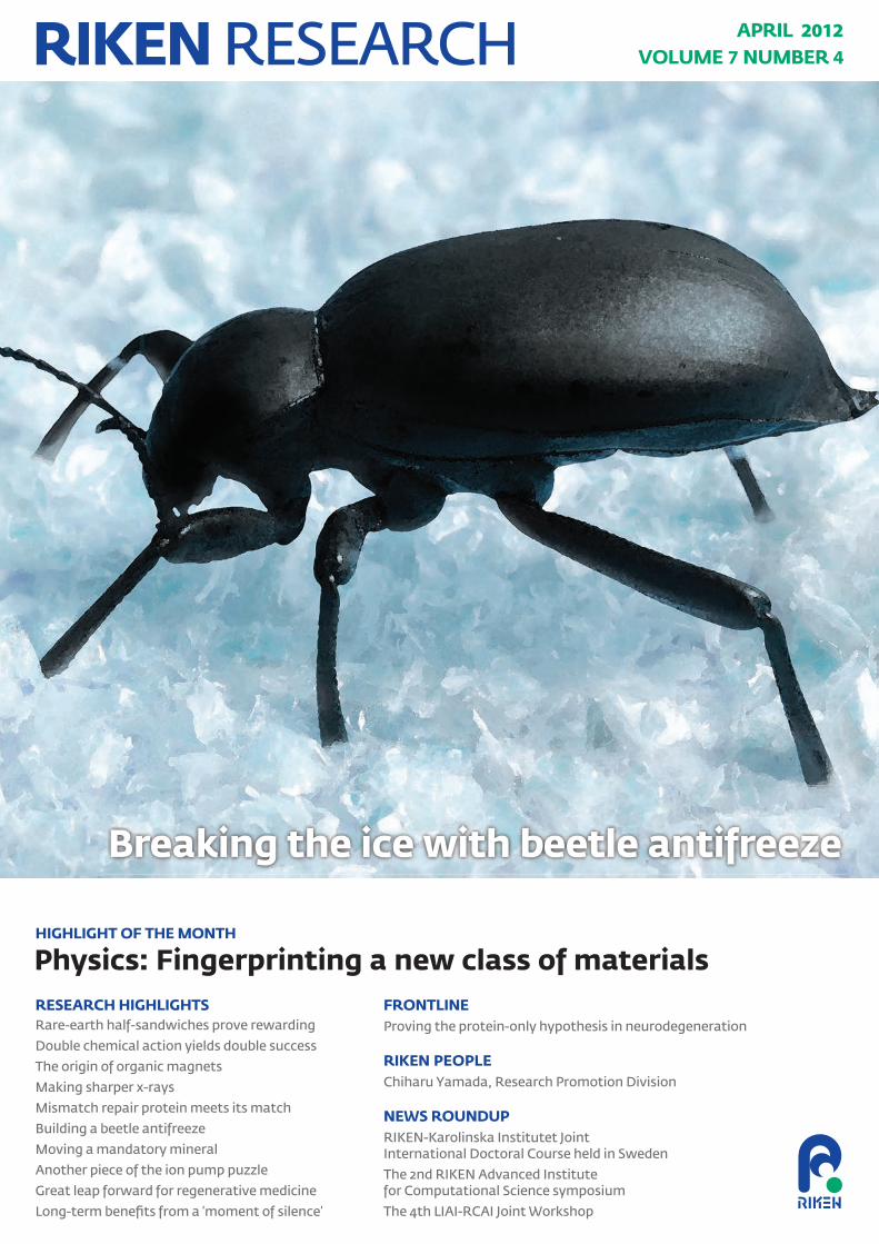
The discovery in 2010 of a new class of materials known as topological superconductors promised exotic new physics and the potential for an unexplored realm of applications, ranging from transistors to power lines and beyond (Fig. 1). Topological superconductors combine the properties of two existing classes of materials: superconductors, which carry current at zero resistance; and topological insulators, whose centers, or ‘bulks’, are insulating but their surfaces are highly conductive. However, experiments with topological superconductors are complicated by the difficulty in distinguishing them from regular superconductors. Since the bulk of a topological superconductor is very conductive, the highly conductive surface states that distinguish it from a regular superconductor are hard to measure directly.
Now, a research team from Japan and the USA has predicted the existence of a unique and unexpected signature of topological superconductors1: the speed of their rotation affects their ability to transport heat and vice versa. Naoto Nagaosa from the RIKEN Advanced Science Institute (ASI), Wako, led the team, which included Kentaro Nomura and Akira Furusaki, also from RIKEN ASI, and Shinsei Ryu from the University of Illinois.
Topological superconductors also intrigue physicists because they are expected to host exotic particles, known as Majorana fermions. Although predicted to exist in 1937, these particles have never been observed directly. Because they differ fundamentally from
any other particle, their observation would be an important development for fundamental physics. e particles are also expected to have applications in quantum computation. According to Nagaosa, detecting a Majorana fermion is the greatest challenge in the field at the present time, and topological superconductors may prove instrumental in meeting this challenge. is new method of detecting topological superconductors is expected to simplify experiments on the new materials, as well as opening new lines of investigation.
When insulators switch rolesNagaosa notes that topological insulators—which were observed experimentally only a few years before topological superconductors—also have a telltale signature. Each signature relates to how electrons move in the materials. Electrons are forced to move at the surface of a topological insulator and not in its bulk, which produces peculiar properties. For example, the spins of electrons traveling with a particular momentum along a surface of a topological insulator all point in the same direction.
Physics
Fingerprinting a new class of materialsCalculations have revealed the unique signature of an exotic new material with potential applications to electronics and beyond
Figure 1: Although potentially a long way off, the unique properties of electrons at the surface of topological superconductors may find practical applications in electronics.
© 20
12 iS
tock
phot
o/ kr
ystia
nnaw
rock
i
3RIKEN RESEARCH | VOL. 7 | NUMBER 4 | APRIL 2012 | www.rikenresearch.riken.jp
HIGHLIGHT OF THE MONTH
This means that physicists can use electric fields to control the magnetic properties of a topological insulator, and they can use magnetic fields to control its electrical properties. is kind of cross-coupling, called the ‘magneto-electric’ effect, is not only a useful signature of a topological insulator, but has a variety of potential applications. In some cases, the conducting properties of topological insulators are as good as graphene, a two-dimensional sheet of carbon atoms in which electrons can move so freely that they effectively have no mass.
To turn topological insulators into topological superconductors, physicists found that they simply needed to add, or ‘dope in’, a small quantity of copper atoms. The resulting transformation is important not only because it introduces a new type of material, but also because of how this new material differs from regular superconductors. Of particular note is the fact that topological superconductors have far fewer charge carriers, and that their properties are very sensitive to the position of the dopant atoms.
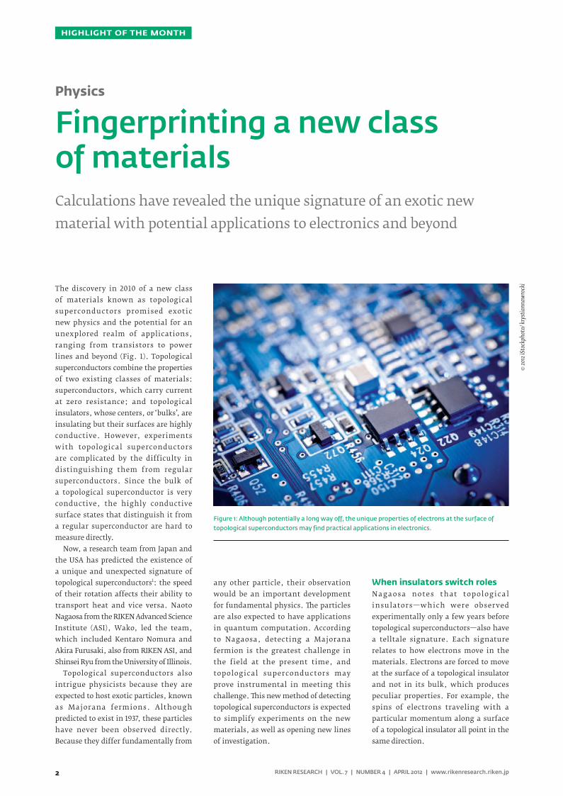
Joining forcesJust as topological insulators feature a cross-coupling between magnetic and electric properties, Nagaosa and colleagues calculated that topological superconductors would also exhibit a cross-coupling: the thermal conductivity of a topological superconductor, which is a measure of how easily heat flows across it, affects its physical rotation about an axis and vice versa (Fig. 2).
The magneto-electric coupling in topological insulators results from the
fact that a charge moving along the surface of a sample under the influence of an electric field will experience a force due to a relativistic spin-orbit interaction. Similarly, a particle in a topological superconductor moving under the influence of a thermal gradient, from hot to cold, will encounter a force if it is in a rotating frame, so that increasing the speed of rotation of the material increases the difference in temperature across its surface. As a result, the thermal conductivity of the topological superconductor can assume only one of a series of discrete values. is restriction is known as quantization, and is also a feature of the electrical conductance of topological insulators in the presence of a magnetic field.
Such a direct connection between mechanical rotation and thermal properties has not been predicted or observed previously, says Nagaosa, and may hold promise for new applications. In the near term, however, its scientific consequences may be of most importance. By directing experimentalists to examine the thermal response of topological superconductors, it eases further characterization and development of these new materials—and, in particular, it should aid the search for Majorana fermions. e change in temperature for a given change in rotational frequency should be experimentally feasible to measure in topological superconductors because their heat capacity is small, Nagaosa says.
The results of Nagaosa and his team also provide a set of equations that
can be applied to other systems, such as topological superfluids. In these materials, particles flow past each other without friction. Scientists have debated how much angular momentum is present in these materials when they are in their lowest-energy state, or ground state. e theory developed by Nagaosa and his colleagues provides an equation that can be used to calculate this momentum, as well as how the momentum will change with the superfluid’s temperature.
1. Nomura, K., Ryu, S., Furusaki, A. & Nagaosa, N. Cross-correlated responses of topological superconductors and superfluids. Physical Review Letters 108, 026802 (2012).
Repr
oduc
ed, w
ith pe
rmiss
ion,
from
Ref.
1© 20
12 A
mer
ican
Phys
ical S
ociet
y
Figure 2: Schematic diagram of charges (black dots) moving in a topological insulator (left) and a topological superconductor (right). In the topological insulator, the circular motion is caused by the
magnetic field pointing in the vertical direction (Bz). In the topological superconductor, the physical rotation of the sample (Ωz) induces the change in the temperature.
Bz Ωz
Naoto Nagaosa was born in Amagasaki,
Hyogo, Japan, in 1958. He graduated
from the Faculty of Engineering at the
University of Tokyo in 1980, and obtained
his PhD in 1986 from the same university.
Nagaosa then held a postdoctoral
position for just under two years at the
Department of Physics, Massachusetts
Institute of Technology in Cambridge,
USA, before returning to Japan where he
was appointed lecturer at the Department
of Applied Physics at the University of
Tokyo. He was promoted to professor in
1998 at the same department. From 2007,
he also became a team leader in RIKEN,
and is now the leader of the Theoretical
Design Team of Cross-Correlated
Materials Research Group (CMRG), and
the Strong-Correlation Theory Research
Team of Correlated Electron Research
Group (CERG) at the Emergent Materials
Department of the RIKEN Advanced
Science Institute. His main research
interests are the theories of strongly
correlated electronic systems, spintronics,
and the gauge theories of topological
aspects of electrons in solids.
ABOUT THE RESEARCHER
4
RESEARCH HIGHLIGHTS
RIKEN RESEARCH | VOL. 7 | NUMBER 4 | APRIL 2012 | www.rikenresearch.riken.jp
The chemical frameworks of ‘natural p r o d u c t s ’ — m o l e c u l e s g e n e r at e d b y b i o l o g i c a l o r g a n i s m s — h av e inspired many of today’s most potent pharmaceuticals. But the complexity of these compounds makes time-consuming tricks necessary to produce them at large scales. Bing-Tao Guan and Zhaomin Hou from the RIKEN Advanced Science Institute in Wako, however, have developed a rare-earth catalyst system that promises to make natural product synthesis significantly easier by enabling direct modification of aromatic pyridine compounds1.
Pyridine, a benzene-like ring that contains nitrogen and five carbon–hydrogen (C–H) atoms, is a chemical structure found in many natural products. Ideally, chemists would insert double-bonded olefins into pyridine’s C–H groups to synthesize new medicinal compounds. But this approach is rarely viable owing to a lack of efficient and selective catalysts.
The researchers envisaged that their ‘half-sandwich’ rare-earth catalysts, which they have previously used for olefin polymerization2, might offer unprecedented control over this transformation. These molecules are named after their shape, in which elements such as scandium (Sc) center above a flat pentagonal ring. They can both dehydrogenate pyridine’s C–H bonds and promote olefin insertion—two critical features in making pyridine modification a success, Hou notes.
When the researchers mixed ethylene gas with a pyridine derivative and an Sc half-sandwich catalyst, they discovered
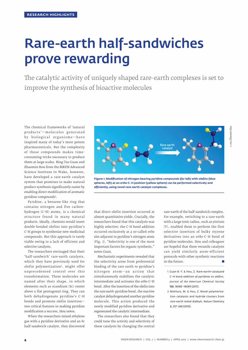
that direct olefin insertion occurred at almost quantitative yields. Crucially, the researchers found that this catalysis was highly selective: the C–H bond addition occurred exclusively at a so-called ortho site adjacent to pyridine’s nitrogen atom (Fig. 1). “Selectivity is one of the most important factors for organic synthesis,” notes Guan.
Mechanistic experiments revealed that the selectivity arose from preferential binding of the rare earth to pyridine’s n i t r o g e n at o m — a n a c t i o n t h at simultaneously stabilizes the catalytic intermediate and activates the ortho-C–H bond. After the insertion of the olefin into the rare earth–pyridine bond, the reactive catalyst dehydrogenated another pyridine molecule. This action produced the newly modified pyridine derivative and regenerated the catalytic intermediate.
The researchers also found that they could tune the activity and selectivity of these catalysts by changing the central
rare-earth of the half-sandwich complex. For example, switching to a rare-earth with a large ionic radius, such as yttrium (Y), enabled them to perform the first selective insertion of bulky styrene derivatives into an ortho-C–H bond of pyridine molecules. Hou and colleagues are hopeful that these versatile catalysts can yield similarly atom-efficient protocols with other synthetic reactions in the future.
1. Guan B.-T. & Hou, Z. Rare-earth-catalyzed
C–H bond addition of pyridines to olefins.
Journal of the American Chemical Society
133, 18086–18089 (2011).
2. Nishiura, M. & Hou, Z. Novel polymeriza-
tion catalysts and hydride clusters from
rare-earth metal dialkyls. Nature Chemistry
2, 257–268 (2010).
Rare-earth half-sandwiches prove rewardinge catalytic activity of uniquely shaped rare-earth complexes is set to improve the synthesis of bioactive molecules
© 20
12 Z
haom
in H
ou
Figure 1: Modification of nitrogen-bearing pyridine compounds (far left) with olefins (blue spheres, left) at an ortho-C–H position (yellow sphere) can be performed selectively and efficiently, using novel rare-earth catalyst complexes.
Rare earthcatalyst
N N
5RIKEN RESEARCH | VOL. 7 | NUMBER 4 | APRIL 2012 | www.rikenresearch.riken.jp
RESEARCH HIGHLIGHTS
Molecules containing silicon double bonds, or disilenes, can be nearly twice as responsive to light as double-bonded hydrocarbons—a feature that makes them irresistible to researchers developing novel devices such as organic light-emitting diodes. But because disilenes are difficult to isolate and tend to polymerize, chemists struggle to control them with their usual synthetic tricks. Now, Kohei Tamao and colleagues from the RIKEN Advanced Science Institute in Wako have discovered a unique halogen-substituted disilene complex that makes assembling advanced conjugated materials easier than ever before1.
Halogen elements such as chlorine or bromine can boost the synthetic capabil it ies of many molecules once attached to their frameworks. Techniques known as substitution reactions can then switch the halogens for other groups, such as aromatic species. However, chemists have scarcely studied halogenated disilenes because theoretical calculations indicate that they are inherently volatile.
Recently however, Tamao and colleagues developed compounds that are extraordinarily adept at stabilizing disilenes2. Known as ‘Rind’ ligands, these molecules have a unique fused-ring structure that locks silicon double bonds into place. They also have chemically tunable side chains that optimize compatibility with a variety of substrates and solvents. Based on these capabilities, Tamao and team postulated that their technique could capture the halogenated targets.
Experiments proved that their instincts were correct: combining a Rind-protected bromine–silicon precursor with a reducing agent successfully produced the sought-after dibromo-disilene crystals. But closer examination of the new product’s reactivity revealed a surprise. Simply mixing it with an acetylene derivative caused the disilene to cleave in half and join to both sides of the carbon triple bond, producing a triangle-shaped unsaturated ring.
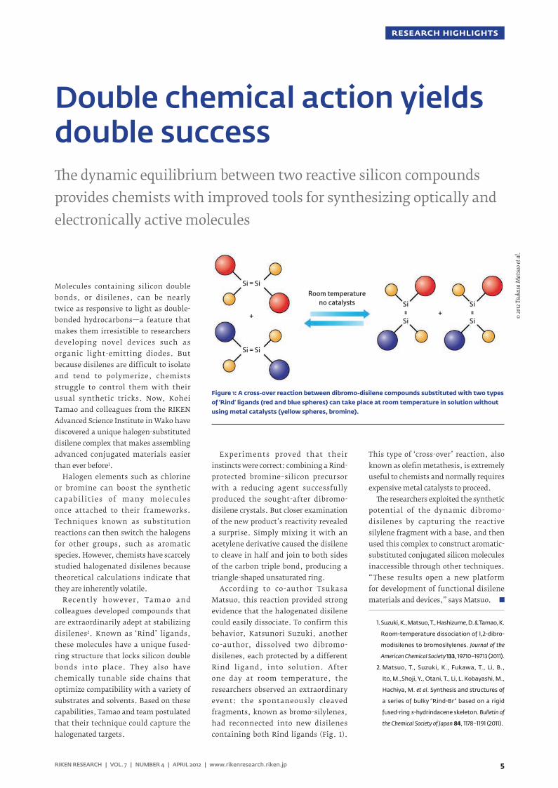
According to co-author Tsukasa Matsuo, this reaction provided strong evidence that the halogenated disilene could easily dissociate. To confirm this behavior, Katsunori Suzuki, another co-author, dissolved two dibromo-disilenes, each protected by a different Rind ligand, into solution. After one day at room temperature, the researchers observed an extraordinary event: the spontaneously cleaved fragments, known as bromo-silylenes, had reconnected into new disilenes containing both Rind ligands (Fig. 1).
This type of ‘cross-over’ reaction, also known as olefin metathesis, is extremely useful to chemists and normally requires expensive metal catalysts to proceed.
e researchers exploited the synthetic potential of the dynamic dibromo-disilenes by capturing the reactive silylene fragment with a base, and then used this complex to construct aromatic-substituted conjugated silicon molecules inaccessible through other techniques. “These results open a new platform for development of functional disilene materials and devices,” says Matsuo.
1. Suzuki, K., Matsuo, T., Hashizume, D. & Tamao, K.
Room-temperature dissociation of 1,2-dibro-
modisilenes to bromosilylenes. Journal of the
American Chemical Society 133, 19710–19713 (2011).
2. Matsuo, T., Suzuki, K., Fukawa, T., Li, B.,
Ito, M.,Shoji, Y., Otani, T., Li, L. Kobayashi, M.,
Hachiya, M. et al. Synthesis and structures of
a series of bulky “Rind-Br” based on a rigid
fused-ring s-hydrindacene skeleton. Bulletin of
the Chemical Society of Japan 84, 1178–1191 (2011).
Double chemical action yields double successe dynamic equilibrium between two reactive silicon compounds provides chemists with improved tools for synthesizing optically and electronically active molecules
© 20
12 T
suka
sa M
atsu
o et a
l.
Figure 1: A cross-over reaction between dibromo-disilene compounds substituted with two types of ‘Rind’ ligands (red and blue spheres) can take place at room temperature in solution without using metal catalysts (yellow spheres, bromine).
Room temperatureno catalysts
Si = Si
Si
Si+ +
Si = Si
=
Si
Si
=
6
RESEARCH HIGHLIGHTS
RIKEN RESEARCH | VOL. 7 | NUMBER 4 | APRIL 2012 | www.rikenresearch.riken.jp
Electrical engineers are starting to consider materials made from organic molecules—including those made from carbon atoms—as an intriguing alternative to the silicon and metals used currently in electronic devices, since they are easier and cheaper to produce. A RIKEN-led research team has now demonstrated the origin of magnetism in organic molecules1, a property that is rarely found in this class of material, but is vital if a full range of organic electronic devices is to be created.
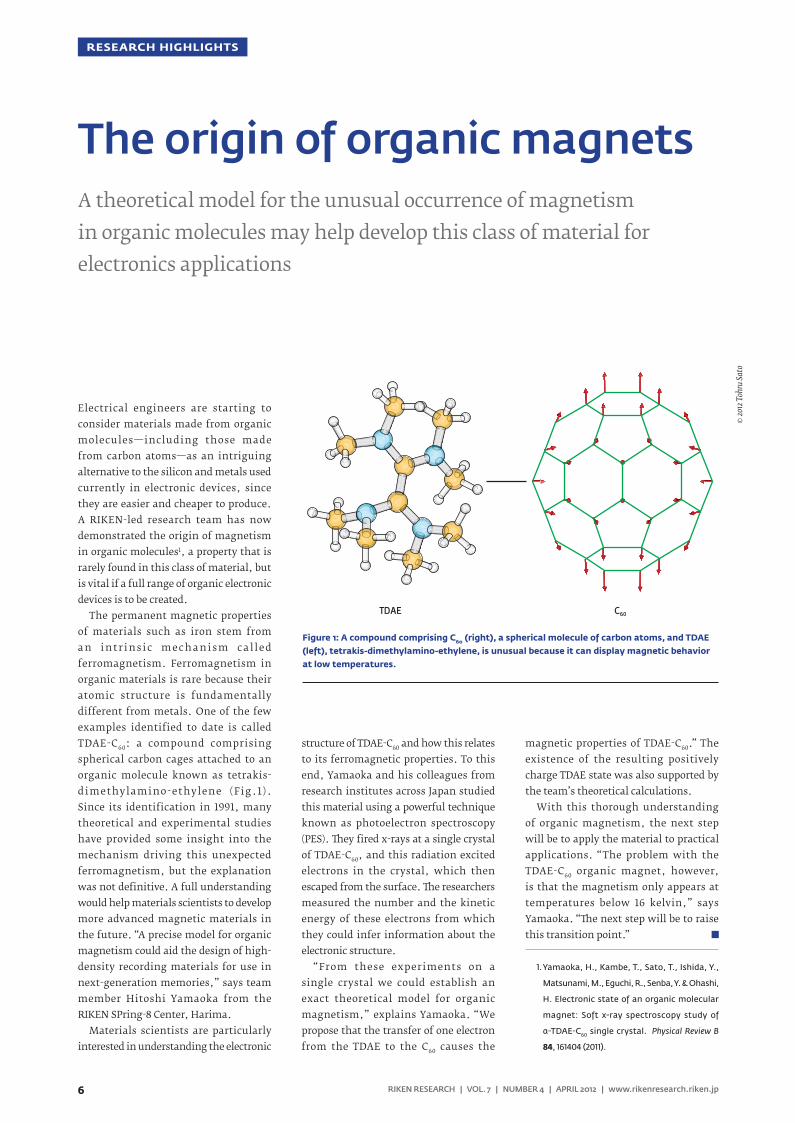
The permanent magnetic properties of materials such as iron stem from a n i n t r i n s i c m e c h a n i s m c a l l e d ferromagnetism. Ferromagnetism in organic materials is rare because their atomic structure is fundamentally different from metals. One of the few examples identified to date is called TDAE-C60: a compound comprising spherical carbon cages attached to an organic molecule known as tetrakis-dimethylamino-ethylene (Fig.1). Since its identification in 1991, many theoretical and experimental studies have provided some insight into the mechanism driving this unexpected ferromagnetism, but the explanation was not definitive. A full understanding would help materials scientists to develop more advanced magnetic materials in the future. “A precise model for organic magnetism could aid the design of high-density recording materials for use in next-generation memories,” says team member Hitoshi Yamaoka from the RIKEN SPring-8 Center, Harima.
Materials scientists are particularly interested in understanding the electronic
structure of TDAE-C60 and how this relates to its ferromagnetic properties. To this end, Yamaoka and his colleagues from research institutes across Japan studied this material using a powerful technique known as photoelectron spectroscopy (PES). ey fired x-rays at a single crystal of TDAE-C60, and this radiation excited electrons in the crystal, which then escaped from the surface. e researchers measured the number and the kinetic energy of these electrons from which they could infer information about the electronic structure.
“From these experiments on a single crystal we could establish an exact theoretical model for organic magnetism,” explains Yamaoka. “We propose that the transfer of one electron from the TDAE to the C60 causes the
magnetic properties of TDAE-C60.” The existence of the resulting positively charge TDAE state was also supported by the team’s theoretical calculations.
With this thorough understanding of organic magnetism, the next step will be to apply the material to practical applications. “The problem with the TDAE-C60 organic magnet, however, is that the magnetism only appears at temperatures below 16 kelvin,” says Yamaoka. “e next step will be to raise this transition point.”
1. Yamaoka, H., Kambe, T., Sato, T., Ishida, Y.,
Matsunami, M., Eguchi, R., Senba, Y. & Ohashi,
H. Electronic state of an organic molecular
magnet: Soft x-ray spectroscopy study of
α-TDAE-C60 single crystal. Physical Review B
84, 161404 (2011).
C60TDAE
The origin of organic magnetsA theoretical model for the unusual occurrence of magnetism in organic molecules may help develop this class of material for electronics applications
© 20
12 T
ohru
Sato
Figure 1: A compound comprising C60 (right), a spherical molecule of carbon atoms, and TDAE (left), tetrakis-dimethylamino-ethylene, is unusual because it can display magnetic behavior at low temperatures.
7RIKEN RESEARCH | VOL. 7 | NUMBER 4 | APRIL 2012 | www.rikenresearch.riken.jp
RESEARCH HIGHLIGHTS
A variety of imaging technologies rely on light with short wavelengths because it allows very small structures to be resolved. However, light sources which produce short, extreme ultraviolet or x-ray wavelengths often have unstable emission wavelength and timing. Now, by illuminating a gas with a powerful laser, a research team in Japan has demonstrated a light source that may solve many of these problems. The research was published by Mitsuru Nagasono, from the RIKEN SPring-8 Center in Harima, and his colleagues from RIKEN and three other institutes1.
The team’s approach relies on superfluorescence, which occurs when the distance between two or more excited atoms is less than the wavelength of light they emit when they relax. This short distance causes the atoms to relax collectively, so that the peaks and troughs in the light waves produced by each atom occur simultaneously in the same location. The resulting ‘coherent’ superfluorescent emission is of a high quality, with high peak intensity and short duration.
Nagasono and colleagues observed superfluorescence when they illuminated helium gas with an energetic laser pulse from the RIKEN extreme ultraviolet light (EUV) free-electron laser. Previous work by other researchers with free-electron lasers characterized materials by monitoring the charged particles that were emitted on illumination by the laser. Rather than monitoring charged particle emission, Nagasono and his team monitored fluorescence. Also, in distinction from previous work, they
kept their sample at relatively high densities to ensure that they satisfied the conditions for superfluorescence.
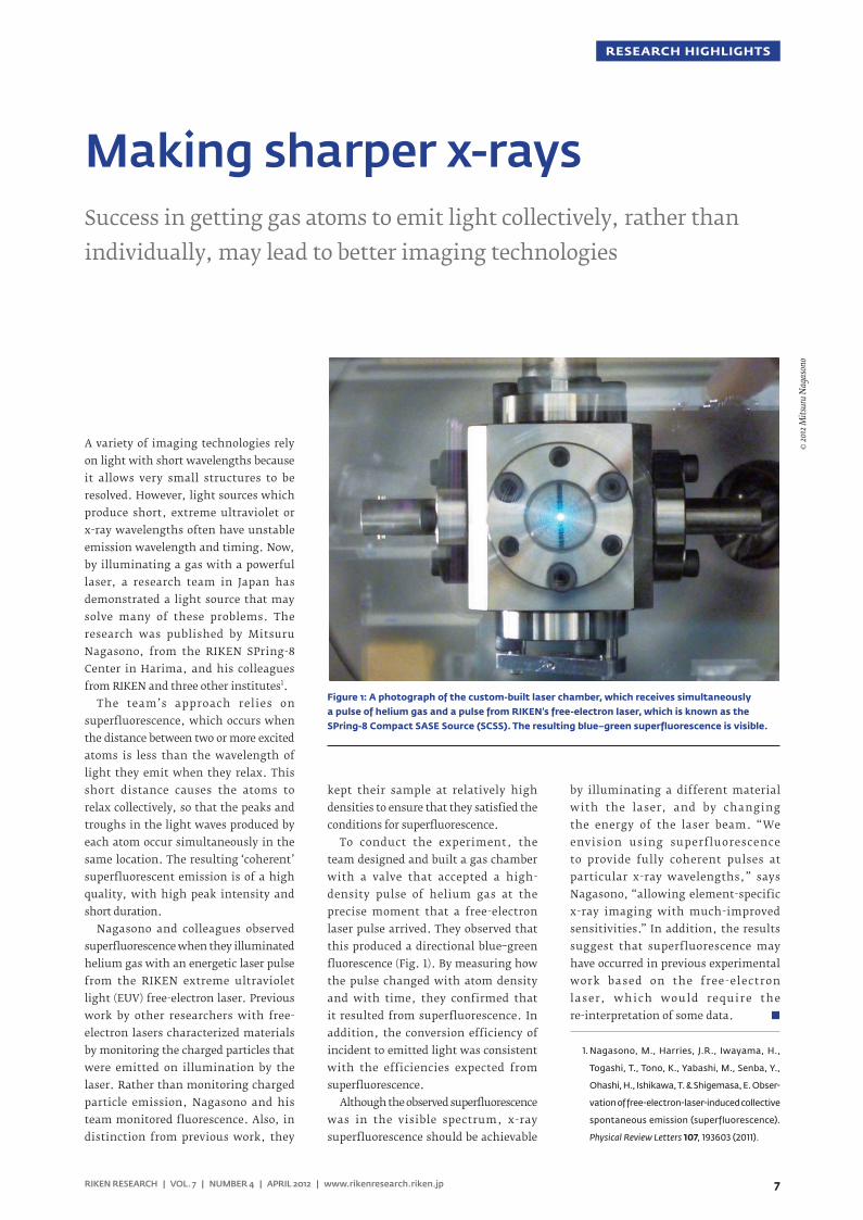
To conduct the experiment, the team designed and built a gas chamber with a valve that accepted a high-density pulse of helium gas at the precise moment that a free-electron laser pulse arrived. They observed that this produced a directional blue–green fluorescence (Fig. 1). By measuring how the pulse changed with atom density and with time, they confirmed that it resulted from superfluorescence. In addition, the conversion efficiency of incident to emitted light was consistent with the efficiencies expected from superfluorescence.
Although the observed superfluorescence was in the visible spectrum, x-ray superfluorescence should be achievable
by illuminating a different material with the laser, and by changing the energy of the laser beam. “We envision using superfluorescence to provide fully coherent pulses at particular x-ray wavelengths,” says Nagasono, “allowing element-specific x-ray imaging with much-improved sensitivities.” In addition, the results suggest that superfluorescence may have occurred in previous experimental work based on the free-electron laser, which would require the re-interpretation of some data.
1. Nagasono, M., Harries, J.R., Iwayama, H.,
Togashi, T., Tono, K., Yabashi, M., Senba, Y.,
Ohashi, H., Ishikawa, T. & Shigemasa, E. Obser-
vation of free-electron-laser-induced collective
spontaneous emission (superfluorescence).
Physical Review Letters 107, 193603 (2011).
Making sharper x-raysSuccess in getting gas atoms to emit light collectively, rather than individually, may lead to better imaging technologies
© 20
12 M
itsur
u N
agas
ono
Figure 1: A photograph of the custom-built laser chamber, which receives simultaneously a pulse of helium gas and a pulse from RIKEN’s free-electron laser, which is known as the SPring-8 Compact SASE Source (SCSS). The resulting blue–green superfluorescence is visible.
8
RESEARCH HIGHLIGHTS
RIKEN RESEARCH | VOL. 7 | NUMBER 4 | APRIL 2012 | www.rikenresearch.riken.jp
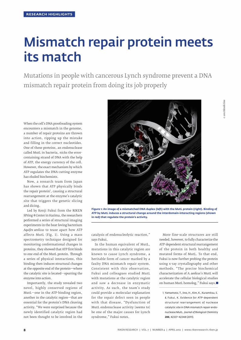
When the cell’s DNA proofreading system encounters a mismatch in the genome, a number of repair proteins are thrown into action, ripping up the mistake and filling in the correct nucleotides. One of these proteins, an endonuclease called MutL in bacteria, nicks the error-containing strand of DNA with the help of ATP, the energy currency of the cell. However, the exact mechanism by which ATP regulates the DNA-cutting enzyme has eluded biochemists.
Now, a research team from Japan has shown that ATP physically binds the repair protein1, causing a structural rearrangement at the enzyme’s catalytic site that triggers the genetic slicing and dicing.
Led by Kenji Fukui from the RIKEN SPring-8 Center in Harima, the researchers performed a series of structural imaging experiments in the heat-loving bacterium Aquifex aeolicus to tease apart how ATP affects MutL (Fig. 1). Using a mass spectrometry technique designed for monitoring conformational changes in proteins, they showed that ATP first binds to one end of the MutL protein. rough a series of physical interactions, this binding then induces structural changes at the opposite end of the protein—where the catalytic site is located—spurring the enzyme into action.
Importantly, the study revealed two novel, highly conserved regions of MutL—one in the ATP-binding region, another in the catalytic region—that are essential for the protein’s DNA cleaving activity. “We were surprised because the newly identified catalytic region had not been thought to be involved in the
catalysis of endonucleolytic reaction,” says Fukui.
In the human equivalent of MutL, mutations in this catalytic region are known to cause Lynch syndrome, a heritable form of cancer marked by a faulty DNA mismatch repair system. Consistent with this observation, Fukui and colleagues studied MutL with mutations at the catalytic region and saw a decrease in enzymatic activity. As such, the team’s study could provide a molecular explanation for the repair defect seen in people with that disease. “Dysfunction of MutL endonuclease activity [seems to] be one of the major causes for Lynch syndrome,” Fukui notes.
More fine-scale structures are still needed, however, to fully characterize the ATP-dependent structural rearrangement of the protein in both healthy and mutated forms of MutL. To that end, Fukui is now further probing the protein using x-ray crystallography and other methods. “The precise biochemical characterization of A. aeolicus’s MutL will accelerate the cellular biological studies on human MutL homolog,” Fukui says.
1. Yamamoto, T., Iino, H., Kim, K., Kuramitsu, S.
& Fukui, K. Evidence for ATP-dependent
structural rearrangement of nuclease
catalytic site in DNA mismatch repair endo-
nuclease MutL. Journal of Biological Chemistry
286, 42337–42348 (2011).
Mismatch repair protein meets its matchMutations in people with cancerous Lynch syndrome prevent a DNA mismatch repair protein from doing its job properly
© 20
12 H
itosh
i Iin
o
Figure 1: An image of a mismatched DNA duplex (left) with the MutL protein (right). Binding of ATP by MutL induces a structural change around the interdomain-interacting regions (shown in red) that regulate the protein’s activity.
9RIKEN RESEARCH | VOL. 7 | NUMBER 4 | APRIL 2012 | www.rikenresearch.riken.jp
RESEARCH HIGHLIGHTS
Animals and plants have evolved all sorts of chemical tricks that allow them to colonize extreme environments. For species that call Antarctica or the Arctic home, surviving sub-zero temperatures is an essential ability, and chemists have isolated many natural antifreeze compounds from these organisms. The antifreeze called xylomannan, which is produced by the freeze-tolerant Alaskan beetle Upis ceramboides, is being studied by Akihiro Ishiwata and Yukishige Ito at the RIKEN Advanced Science Institute at Wako and their colleagues. Their findings to date show that xylomannan is a particularly unusual antifreeze1.
Xylomannan was first reported in 2009, and has been shown to be amongst the most active insect antifreezes found to date2. Antifreeze compounds, which are also known as thermal hysteresis factors (THFs), protect the insects’ cells from damage as temperatures fall and ice crystals begin to form. THFs seem to work by sticking to the surface of nascent ice crystals and somehow stopping them from growing, protecting nearby cell membranes from being punctured by needles of ice.
e unusual thing about xylomannan is its constituents. Every natural THF isolated to date is protein based, but xylomannan is a glycan, a long-chain sugar-based compound. “Xylomannan is the first example of a THF biomolecule with little or no protein component,” says Ishiwata. “Its mode of action is not entirely clear, but it should be different to those of common THFs such as antifreeze proteins and glycoproteins.”
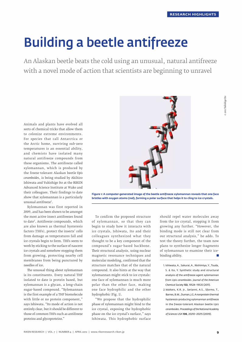
To confirm the proposed structure of xylomannan, so that they can begin to study how it interacts with ice crystals, Ishiwata, Ito and their colleagues synthesized what they thought to be a key component of the compound’s sugar-based backbone. eir structural analysis, using nuclear magnetic resonance techniques and molecular modeling, confirmed that the structure matches that of the natural compound. It also hints at the way that xylomannan might stick to ice crystals: one face of xylomannan is much more polar than the other face, making one face hydrophilic and the other hydrophobic (Fig. 1).
“We propose that the hydrophilic phase of xylomannan might bind to the ice crystal, exposing the hydrophobic phase on the ice crystal’s surface,” says Ishiwata. This hydrophobic surface
should repel water molecules away from the ice crystal, stopping it from growing any further. “However, the binding mode is still not clear from our structural analysis,” he adds. To test the theory further, the team now plans to synthesize longer fragments of xylomannan to examine their ice-binding ability.
1. Ishiwata, A., Sakurai, A., Nishimiya, Y., Tsuda,
S. & Ito, Y. Synthetic study and structural
analysis of the antifreeze agent xylomannan
from Upis ceramboides. Journal of the American
Chemical Society 133, 19524–19535 (2011).
2. Walters, K.R. Jr., Serianni, A.S., Sformo, T.,
Barnes, B.M., Duman, J.G. A nonprotein thermal
hysteresis-producing xylomannan antifreeze
in the freeze-tolerant Alaskan beetle Upis
ceramboides. Proceedings of the National Academy
of Sciences USA 106, 20210–20215 (2009).
Building a beetle antifreezeAn Alaskan beetle beats the cold using an unusual, natural antifreeze with a novel mode of action that scientists are beginning to unravel
© 20
12 Y
ukish
ige I
to
Figure 1: A computer-generated image of the beetle antifreeze xylomannan reveals that one face bristles with oxygen atoms (red), forming a polar surface that helps it to cling to ice crystals.
10
RESEARCH HIGHLIGHTS
RIKEN RESEARCH | VOL. 7 | NUMBER 4 | APRIL 2012 | www.rikenresearch.riken.jp
Up to one-tenth of the proteins encoded in the human genome incorporate zinc, making this element indispensable to biological function. Indeed, most organisms employ a host of specialized transporter proteins to maintain appropriate zinc levels, and failures in these pathways can cause serious health issues.
Recent research from Toshiyuki Fukada and colleagues at the RIKEN Research Center for Allergy and Immunology in Yokohama has characterized the structure and function of one of these proteins, ZIP131. From previous research on ZIP13, they knew that mice lacking this protein suffer diverse problems2. “Zinc-rel ated events mo dul ated bone, tooth and connective tissue development,” says Fukada, “and there was a link between ZIP13 and human disease.” For example, patients with a new type of inherited disease, currently called spondylocheiro dysplastic Ehlers-Danlos syndrome (SCD-EDS) that mirrors defects seen in these mice, exhibit a crippling mutation in this gene2.
ZIP13 belongs to a subset of proteins that facilitate zinc entry into the cell, known as the LIV-1 subfamily of ZIP zinc transporters (LZT)3. From their characterization work, Fukada and colleagues confirmed that increased ZIP13 expression leads to more efficient zinc influx.
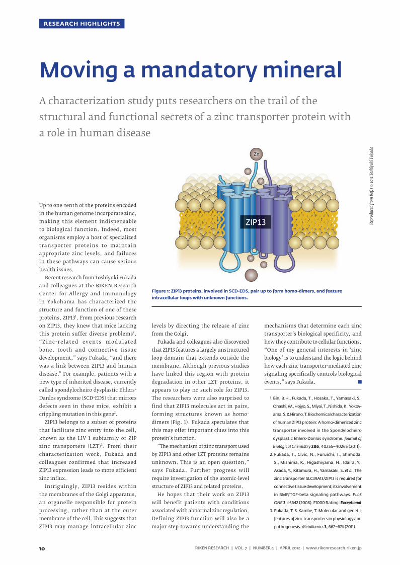
Intriguingly, ZIP13 resides within the membranes of the Golgi apparatus, an organelle responsible for protein processing, rather than at the outer membrane of the cell. is suggests that ZIP13 may manage intracellular zinc
levels by directing the release of zinc from the Golgi.
Fukada and colleagues also discovered that ZIP13 features a largely unstructured loop domain that extends outside the membrane. Although previous studies have linked this region with protein degradation in other LZT proteins, it appears to play no such role for ZIP13. The researchers were also surprised to find that ZIP13 molecules act in pairs, forming structures known as homo-dimers (Fig. 1). Fukada speculates that this may offer important clues into this protein’s function.
“e mechanism of zinc transport used by ZIP13 and other LZT proteins remains unknown. This is an open question,” says Fukada. Further progress will require investigation of the atomic-level structure of ZIP13 and related proteins.
He hopes that their work on ZIP13 will benefit patients with conditions associated with abnormal zinc regulation. Defining ZIP13 function will also be a major step towards understanding the
mechanisms that determine each zinc transporter’s biological specificity, and how they contribute to cellular functions. “One of my general interests in ‘zinc biology’ is to understand the logic behind how each zinc transporter-mediated zinc signaling specifically controls biological events,” says Fukada.
1. Bin, B.H., Fukada, T., Hosaka, T., Yamasaki, S.,
Ohashi, W., Hojyo, S., Miyai, T., Nishida, K., Yokoy-
ama, S. & Hirano, T. Biochemical characterization
of human ZIP13 protein: A homo-dimerized zinc
transporter involved in the Spondylocheiro
dysplastic Ehlers-Danlos syndrome. Journal of
Biological Chemistry 286, 40255–40265 (2011).
2. Fukada, T., Civic, N., Furuichi, T., Shimoda,
S., Mishima, K., Higashiyama, H., Idaira, Y.,
Asada, Y., Kitamura, H., Yamasaki, S. et al. The
zinc transporter SLC39A13/ZIP13 is required for
connective tissue development; its involvement
in BMP/TGF-beta signaling pathways. PLoS
ONE 3, e3642 (2008). F1000 Rating: Exceptional
3. Fukada, T. & Kambe, T. Molecular and genetic
features of zinc transporters in physiology and
pathogenesis. Metallomics 3, 662–674 (2011).
Moving a mandatory mineralA characterization study puts researchers on the trail of the structural and functional secrets of a zinc transporter protein with a role in human disease
Repr
oduc
ed fr
om R
ef. 1 ©
2012
Tos
hiyu
ki F
ukad
a
Figure 1: ZIP13 proteins, involved in SCD-EDS, pair up to form homo-dimers, and feature intracellular loops with unknown functions.
11RIKEN RESEARCH | VOL. 7 | NUMBER 4 | APRIL 2012 | www.rikenresearch.riken.jp
RESEARCH HIGHLIGHTS
From bacteria to humans, all cells use molecules of adenosine triphosphate (ATP) as fuel to power a broad range of biochemical reactions. For example, massive multi-subunit enzymes known as V-ATPases convert ATP molecules into energy that helps drive the transport of ions across cellular membranes.
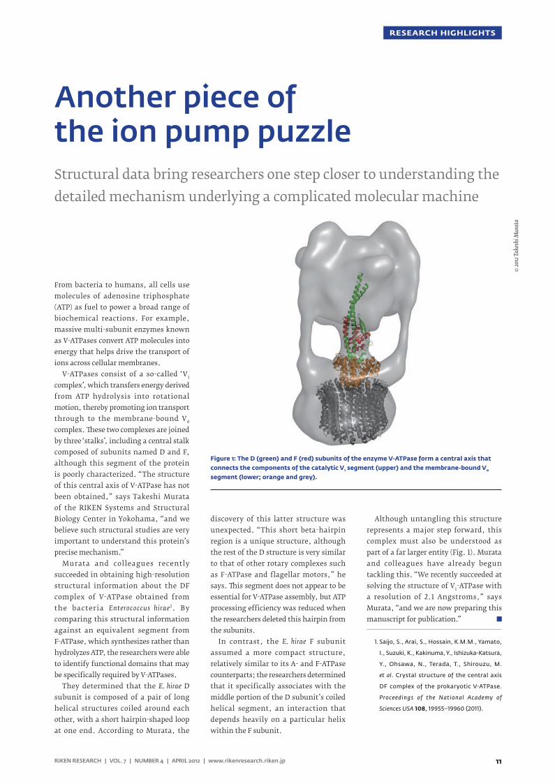
V-ATPases consist of a so-called ‘V1 complex’, which transfers energy derived from ATP hydrolysis into rotational motion, thereby promoting ion transport through to the membrane-bound V0 complex. ese two complexes are joined by three ‘stalks’, including a central stalk composed of subunits named D and F, although this segment of the protein is poorly characterized. “The structure of this central axis of V-ATPase has not been obtained,” says Takeshi Murata of the RIKEN Systems and Structural Biology Center in Yokohama, “and we believe such structural studies are very important to understand this protein’s precise mechanism.”
Murata and colleagues recently succeeded in obtaining high-resolution structural information about the DF complex of V-ATPase obtained from the bacteria Enterococcus hirae 1. By comparing this structural information against an equivalent segment from F-ATPase, which synthesizes rather than hydrolyzes ATP, the researchers were able to identify functional domains that may be specifically required by V-ATPases.
They determined that the E. hirae D subunit is composed of a pair of long helical structures coiled around each other, with a short hairpin-shaped loop at one end. According to Murata, the
discovery of this latter structure was unexpected. “This short beta-hairpin region is a unique structure, although the rest of the D structure is very similar to that of other rotary complexes such as F-ATPase and flagellar motors,” he says. is segment does not appear to be essential for V-ATPase assembly, but ATP processing efficiency was reduced when the researchers deleted this hairpin from the subunits.
In contrast, the E. hirae F subunit assumed a more compact structure, relatively similar to its A- and F-ATPase counterparts; the researchers determined that it specifically associates with the middle portion of the D subunit’s coiled helical segment, an interaction that depends heavily on a particular helix within the F subunit.
Although untangling this structure represents a major step forward, this complex must also be understood as part of a far larger entity (Fig. 1). Murata and colleagues have already begun tackling this. “We recently succeeded at solving the structure of V1-ATPase with a resolution of 2.1 Angstroms,” says Murata, “and we are now preparing this manuscript for publication.”
1. Saijo, S., Arai, S., Hossain, K.M.M., Yamato,
I., Suzuki, K., Kakinuma, Y., Ishizuka-Katsura,
Y., Ohsawa, N., Terada, T., Shirouzu, M.
et al. Crystal structure of the central axis
DF complex of the prokaryotic V-ATPase.
Proceedings of the National Academy of
Sciences USA 108, 19955–19960 (2011).
Another piece of the ion pump puzzleStructural data bring researchers one step closer to understanding the detailed mechanism underlying a complicated molecular machine
© 20
12 T
akes
hi M
urat
a
Figure 1: The D (green) and F (red) subunits of the enzyme V-ATPase form a central axis that connects the components of the catalytic V1 segment (upper) and the membrane-bound V0 segment (lower; orange and grey).
12
RESEARCH HIGHLIGHTS
RIKEN RESEARCH | VOL. 7 | NUMBER 4 | APRIL 2012 | www.rikenresearch.riken.jp
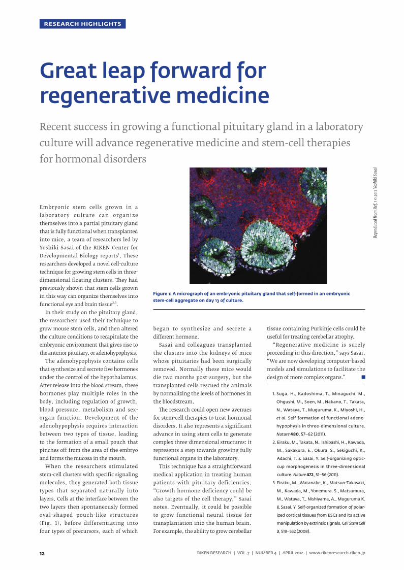
Embryonic stem cells grown in a laborator y culture can organize themselves into a partial pituitary gland that is fully functional when transplanted into mice, a team of researchers led by Yoshiki Sasai of the RIKEN Center for Developmental Biology reports1. These researchers developed a novel cell-culture technique for growing stem cells in three-dimensional floating clusters. ey had previously shown that stem cells grown in this way can organize themselves into functional eye and brain tissue2,3.
In their study on the pituitary gland, the researchers used their technique to grow mouse stem cells, and then altered the culture conditions to recapitulate the embryonic environment that gives rise to the anterior pituitary, or adenohypophysis.
The adenohypophysis contains cells that synthesize and secrete five hormones under the control of the hypothalamus. After release into the blood stream, these hormones play multiple roles in the body, including regulation of growth, blood pressure, metabolism and sex-organ function. Development of the adenohypophysis requires interaction between two types of tissue, leading to the formation of a small pouch that pinches off from the area of the embryo and forms the mucosa in the mouth.
When the researchers stimulated stem-cell clusters with specific signaling molecules, they generated both tissue types that separated naturally into layers. Cells at the interface between the two layers then spontaneously formed oval-shaped pouch-like structures (Fig. 1), before differentiating into four types of precursors, each of which
began to synthesize and secrete a different hormone.
Sasai and colleagues transplanted the clusters into the kidneys of mice whose pituitaries had been surgically removed. Normally these mice would die two months post-surgery, but the transplanted cells rescued the animals by normalizing the levels of hormones in the bloodstream.
e research could open new avenues for stem-cell therapies to treat hormonal disorders. It also represents a significant advance in using stem cells to generate complex three-dimensional structures: it represents a step towards growing fully functional organs in the laboratory.
This technique has a straightforward medical application in treating human patients with pituitary deficiencies. “Growth hormone deficiency could be also targets of the cell therapy,” Sasai notes. Eventually, it could be possible to grow functional neural tissue for transplantation into the human brain. For example, the ability to grow cerebellar
tissue containing Purkinje cells could be useful for treating cerebellar atrophy.
“Regenerative medicine is surely proceeding in this direction,” says Sasai. “We are now developing computer-based models and simulations to facilitate the design of more complex organs.”
1. Suga, H., Kadoshima, T., Minaguchi, M.,
Ohgushi, M., Soen, M., Nakano, T., Takata,
N., Wataya, T., Muguruma, K., Miyoshi, H.,
et al. Self-formation of functional adeno-
hypophysis in three-dimensional culture.
Nature 480, 57–62 (2011).
2. Eiraku, M., Takata, N., Ishibashi, H., Kawada,
M., Sakakura, E., Okura, S., Sekiguchi, K.,
Adachi, T. & Sasai, Y. Self-organizing optic-
cup morphogenesis in three-dimensional
culture. Nature 472, 51–56 (2011).
3. Eiraku, M., Watanabe, K., Matsuo-Takasaki,
M., Kawada, M., Yonemura. S., Matsumura,
M., Wataya, T., Nishiyama, A., Muguruma K.
& Sasai, Y. Self-organized formation of polar-
ized cortical tissues from ESCs and its active
manipulation by extrinsic signals. Cell Stem Cell
3, 519–532 (2008).
Great leap forward for regenerative medicineRecent success in growing a functional pituitary gland in a laboratory culture will advance regenerative medicine and stem-cell therapies for hormonal disorders
Repr
oduc
ed fr
om R
ef. 1 ©
2012
Yos
hiki
Sasa
i
Figure 1: A micrograph of an embryonic pituitary gland that self-formed in an embryonic stem-cell aggregate on day 13 of culture.
13RIKEN RESEARCH | VOL. 7 | NUMBER 4 | APRIL 2012 | www.rikenresearch.riken.jp
RESEARCH HIGHLIGHTS
In principle, somatic cell nuclear transfer (SCNT) is a potent tool for scientists looking to produce exact genetic replicas of a particular animal. By injecting a nucleus from an adult cell into an oocyte from which the nucleus has been removed, one can initiate the embryonic development process and derive a clone of the ‘donor’ animal.
Unfortunately, this technique is terribly inefficient, with a success rate of 1–2% in mice. “This must be due to some errors in the reprogramming of the donor genome into the ‘totipotent’ state, which is equivalent to the state observed in conventionally fertilized embryos,” explains Atsuo Ogura of the RIKEN BioResource Center in Tsukuba. However, Ogura and colleagues have now made significant progress in clearing a major roadblock thwarting SCNT success1.
During development of female mammalian embryos, one of the two X chromosomes is targeted for inactivation, thereby ensuring that both males and females achieve equivalent expression of X-linked genes. This inactivation depends on RNA produced by the Xist gene, which blankets the selected chromosome and sets the inactivation process in motion.
Ogura and his team previously determined that Xist is inappropriately activated in SCNT embryos2, impairing expression of essential genes, and have now set about correcting this defect. Irreversibly inactivating this gene is not an option, so the researchers injected molecules called ‘short interfering RNAs’ (siRNAs) that directly inhibited
Xist activity in early stage male SCNT embryos, which must maintain their single X chromosome in order to survive.
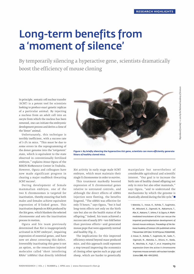
This treatment markedly boosted expression of X chromosomal genes relative to untreated controls, and although the direct effects of siRNA injection were fleeting, the benefits lingered. “The siRNA was effective for only 72 hours,” says Ogura, “but it had long-term effects not only on the birth rate but also on the health status of the offspring.” Indeed, his team achieved a success rate of nearly 20%—ten-fold better than previous efforts—and generated mouse pups that were apparently normal and healthy (Fig. 1).
The implications for this improved efficiency extend beyond mass-produced mice, and this approach could represent a step toward improving the economics of cloning other species such as pigs and sheep, which are harder to genetically
manipulate but nevertheless of considerable agricultural and scientific interest. “Our goal is to increase the birth rate of healthy cloned offspring not only in mice but also other mammals,” says Ogura, “and to understand the mechanisms by which the genome is drastically altered during the life cycle.”
1. Matoba, S., Inoue, K., Kohda, T., Sugimoto,
M., Mizutani, E., Ogonuki, N., Nakamura, T.,
Abe, K., Nakano, T., Ishino, F. & Ogura, A. RNAi-
mediated knockdown of Xist can rescue the
impaired postimplantation development of
cloned mouse embryos. Proceedings of the Na-
tional Academy of Sciences USA published online
7 November 2011 (doi: 10.1073/pnas.1112664108).
2. Inoue, K., Kohda, T., Sugimoto, M., Sado, T.,
Ogonuki, N., Matoba, S., Shiura, H., Ikeda,
R., Mochida, K., Fujii, T., et al. Impeding Xist
expression from the active X chromosome
improves mouse somatic cell nuclear transfer.
Science 330, 496–499 (2010).
Long-term benefits from a ‘moment of silence’By temporarily silencing a hyperactive gene, scientists dramatically boost the efficiency of mouse cloning
Repr
oduc
ed fr
om R
ef. 1 ©
2012
by th
e Nat
iona
l Aca
dem
y of S
cienc
es
Figure 1: By briefly silencing the hyperactive Xist gene, scientists can more efficiently generate litters of healthy cloned mice.
14 RIKEN RESEARCH | VOL. 7 | NUMBER 4 | APRIL 2012 | www.rikenresearch.riken.jp
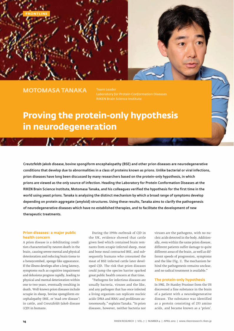
MOTOMASA TANAKA Team LeaderLaboratory for Protein Conformation DiseasesRIKEN Brain Science Institute
Proving the protein-only hypothesis in neurodegeneration
FRONTLINE
Prion diseases: a major public health concernA prion disease is a debilitating condi-tion characterized by neuron death in the brain, causing severe mental and physical deterioration and reducing brain tissue to a honeycombed, sponge-like appearance. If the illness develops after a long latency, symptoms such as cognitive impairment and delusions progress rapidly, leading to physical and mental deterioration within one to two years, eventually resulting in death. Well-known prion diseases include scrapie in sheep, bovine spongiform en-cephalopathy (BSE, or ‘mad cow disease’) in cattle, and Creutzfeldt-Jakob disease (CJD) in humans.
During the 1990s outbreak of CJD in the UK, evidence showed that cattle given feed which contained brain rem-nants from scrapie-infected sheep, meat and bone meal contracted BSE, and sub-sequently humans who consumed the meat of BSE-infected cattle later devel-oped CJD. The risk that prion diseases could jump the species barrier sparked great public health concern at that time.
“Pathogens for infectious diseases are usually bacteria, viruses and the like, and any pathogen that has once infected a living organism can replicate nucleic acids (DNA and RNA) and proliferate au-tonomously,” explains Tanaka. “In prion diseases, however, neither bacteria nor
viruses are the pathogens, with no nu-cleic acids detected in the body. Addition-ally, even within the same prion disease, different patients suffer damage to quite different areas of the brain, as well as dif-ferent speeds of progression, symptoms and the like (Fig. 1). e mechanism be-hind the pathogenesis remains unclear, and no radical treatment is available.”
The protein-only hypothesisIn 1982, Dr Stanley Prusiner from the US discovered a fine substance in the brain of a patient with a neurodegenerative disease. The substance was identified as a protein consisting of 253 amino acids, and became known as a ‘prion’.
Creutzfeldt-Jakob disease, bovine spongiform encephalopathy (BSE) and other prion diseases are neurodegenerative
conditions that develop due to abnormalities in a class of proteins known as prions. Unlike bacterial or viral infections,
prion diseases have long been discussed by many researchers based on the protein-only hypothesis, in which
prions are viewed as the only source of infection. Heading the Laboratory for Protein Conformation Diseases at the
RIKEN Brain Science Institute, Motomasa Tanaka, and his colleagues verified the hypothesis for the first time in the
world using yeast prions. Tanaka is analyzing the distinct mechanism by which a broad range of symptoms develop
depending on protein aggregate (amyloid) structures. Using these results, Tanaka aims to clarify the pathogenesis
of neurodegenerative diseases which have no established therapies, and to facilitate the development of new
therapeutic treatments.
15
FRONTLINE
RIKEN RESEARCH | VOL. 7 | NUMBER 4 | APRIL 2012 | www.rikenresearch.riken.jp
Dr Prusiner was later awarded the 1997 Nobel Prize in Physiology or Medicine for his discovery of prions. “Dr Prusiner pro-posed the protein-only hypothesis that prion diseases are caused by infectious proteins called prions, rather than by bac-teria and viruses. He hypothesized that in addition to normal prions, there are ab-normal prions having exactly the same amino acid sequences as those of the nor-mal type, and the etiology is attributed to abnormal prions (Fig. 1),” says Tanaka.
“Any protein can work normally pro-vided its sequence of amino acids folds properly. This process is called folding. Abnormal prions are the result of mis-folded amino acids, and have many band-like structures known as β -sheets. In the protein-only hypothesis, the structure of an abnormal prion is thought to act as a template for a normal prion, which is converted to the structure of the abnor-mal type, and in which fibrous amyloids form gradually.”
Although currently available experi-mental data support the protein-only hy-pothesis, it is controversial because the concept that infections can occur with proteins alone contradicts established medical dogma. “To verify this hypoth-esis, it must be demonstrated that prions are the only source of infection for prion diseases, with no other factors involved.
It is also necessary to solve the riddle of why distinct phenotypes such as sever-ity and diseased-site specificity emerge from prions having the same amino acid sequence,” says Tanaka.
Verification of the hypothesisTanaka and Jonathan Weissman (Univer-sity of California, San Francisco) first at-tempted to show that prions are the sole source of infection for prion diseases in 2004, when they cultivated an amyloid for a yeast prion. “Yeast, which is a sin-gle-cell organism, has proteins that be-have like mammalian prion protein, and these are called yeast prions. We created an amyloid for the Sup35 yeast prion and developed a technique for introducing it directly into uninfected yeast. e infec-tion with Sup35 amyloid converted nearly 100% of the non-prion yeast to the prion state. e infection was established with the aggregated form of Sup35 alone.”
Tanaka next tried to unravel the puz-zle of why distinct phenotypes emerge from prions having the same amino acid sequence by examining amyloid struc-tures (Fig. 1). “We developed a method of generating amyloids with different ‘conformations’ (steric configurations of various atoms in a molecule) from Sup35 proteins which had the same amino acid sequence. e key point is that the Sup35
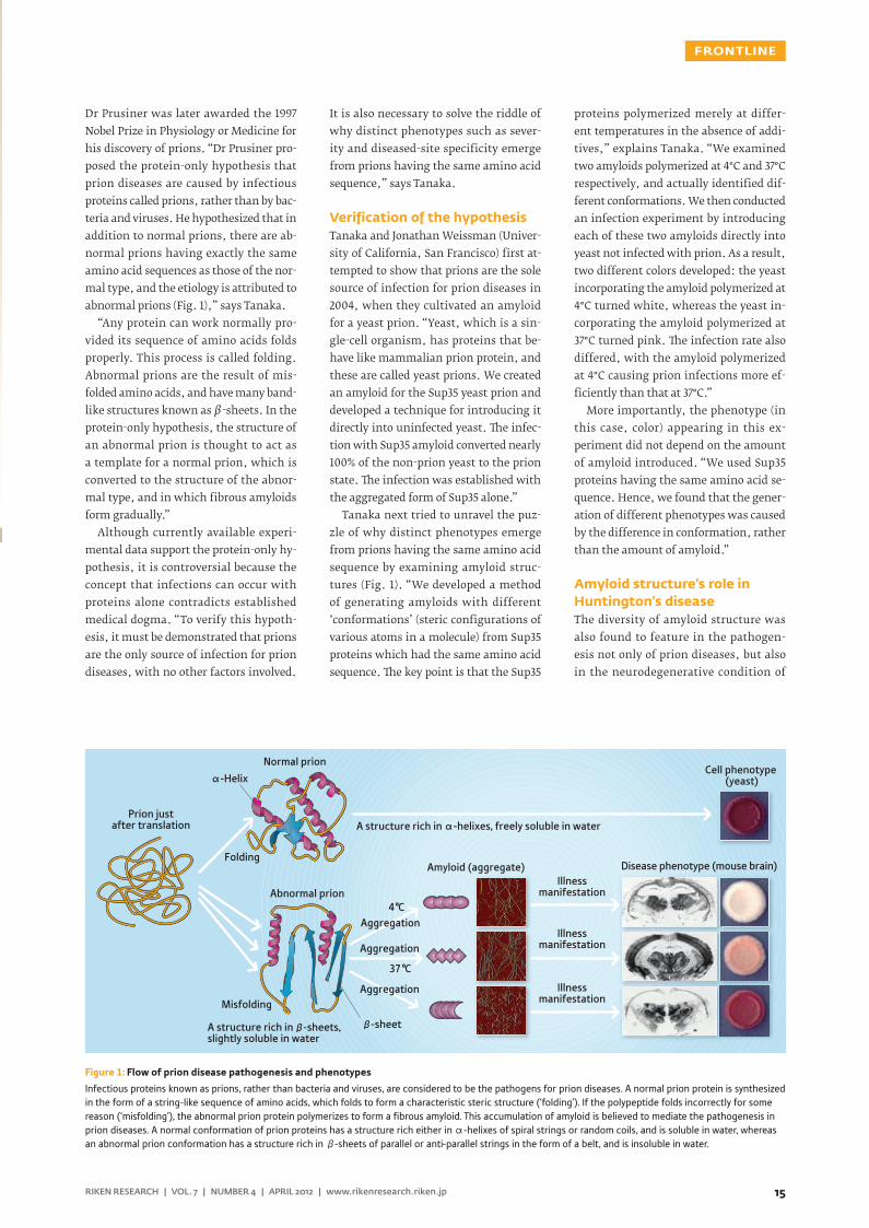
proteins polymerized merely at differ-ent temperatures in the absence of addi-tives,” explains Tanaka. “We examined two amyloids polymerized at 4°C and 37°C respectively, and actually identified dif-ferent conformations. We then conducted an infection experiment by introducing each of these two amyloids directly into yeast not infected with prion. As a result, two different colors developed: the yeast incorporating the amyloid polymerized at 4°C turned white, whereas the yeast in-corporating the amyloid polymerized at 37°C turned pink. e infection rate also differed, with the amyloid polymerized at 4°C causing prion infections more ef-ficiently than that at 37°C.”
More importantly, the phenotype (in this case, color) appearing in this ex-periment did not depend on the amount of amyloid introduced. “We used Sup35 proteins having the same amino acid se-quence. Hence, we found that the gener-ation of different phenotypes was caused by the difference in conformation, rather than the amount of amyloid.”
Amyloid structure’s role in Huntington’s diseaseThe diversity of amyloid structure was also found to feature in the pathogen-esis not only of prion diseases, but also in the neurodegenerative condition of
Figure 1: Flow of prion disease pathogenesis and phenotypes
Infectious proteins known as prions, rather than bacteria and viruses, are considered to be the pathogens for prion diseases. A normal prion protein is synthesized in the form of a string-like sequence of amino acids, which folds to form a characteristic steric structure (‘folding’). If the polypeptide folds incorrectly for some reason (‘misfolding’), the abnormal prion protein polymerizes to form a fibrous amyloid. This accumulation of amyloid is believed to mediate the pathogenesis in prion diseases. A normal conformation of prion proteins has a structure rich either in α-helixes of spiral strings or random coils, and is soluble in water, whereas an abnormal prion conformation has a structure rich in β-sheets of parallel or anti-parallel strings in the form of a belt, and is insoluble in water.
Disease phenotype (mouse brain)
Cell phenotype (yeast)
A structure rich in α-helixes, freely soluble in water
Aggregation
Aggregation
4°C
Misfolding
Folding
α-Helix
β-sheet
Aggregation
Amyloid (aggregate)
Normal prion
Prion justafter translation
Abnormal prion
37°C
Illnessmanifestation
Illnessmanifestation
Illnessmanifestation
A structure rich in -sheets, slightly soluble in water
β
16 RIKEN RESEARCH | VOL. 7 | NUMBER 4 | APRIL 2012 | www.rikenresearch.riken.jp
FRONTLINE
Huntington’s disease. An intractable dis-ease characterized by involuntary move-ment, cognitive decline and psychiatric problems, Huntington’s disease has no known cure.
In the process of protein synthesis, bases are arranged in the gene domain of the DNA (base sequence) to represent ciphers for the arrangement of amino acids. A combination of only three of the bases adenine (A), thymine (T), guanine (G) and cytosine (C) defines one amino acid, and these amino acids join together to form a protein.
“In exon 1, which is one of the gene do-mains of huntingtin — the protein that causes Huntington’s disease — the CAG repeat is present and consists of a repeat sequence of the C, A and G combination,” explains Tanaka. “is repeat sequence defines the amino acid glutamine, and a long chain of glutamine units form polyglutamines. In patients with Hun-tington’s disease, more than 36 CAG re-peats are found — which is more than the normal length of 7–35 — and they produce an abnormally elongated polyglutamine. The huntingtin containing this abnor-mal polyglutamine tends to form amy-loid and plays a role in the pathogenesis of Huntington’s disease. However, the detailed pathogenetic mechanism re-mains unclear.”
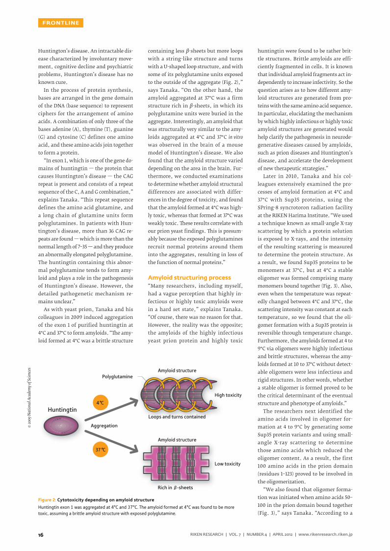
As with yeast prion, Tanaka and his colleagues in 2009 induced aggregation of the exon 1 of purified huntingtin at 4°C and 37°C to form amyloids. “e amy-loid formed at 4°C was a brittle structure
containing less β-sheets but more loops with a string-like structure and turns with a U-shaped loop structure, and with some of its polyglutamine units exposed to the outside of the aggregate (Fig. 2),” says Tanaka. “On the other hand, the amyloid aggregated at 37°C was a firm structure rich in β-sheets, in which its polyglutamine units were buried in the aggregate. Interestingly, an amyloid that was structurally very similar to the amy-loids aggregated at 4°C and 37°C in vitro was observed in the brain of a mouse model of Huntington’s disease. We also found that the amyloid structure varied depending on the area in the brain. Fur-thermore, we conducted examinations to determine whether amyloid structural differences are associated with differ-ences in the degree of toxicity, and found that the amyloid formed at 4°C was high-ly toxic, whereas that formed at 37°C was weakly toxic. ese results correlate with our prion yeast findings. is is presum-ably because the exposed polyglutamines recruit normal proteins around them into the aggregates, resulting in loss of the function of normal proteins.”
Amyloid structuring process“Many researchers, including myself, had a vague perception that highly in-fectious or highly toxic amyloids were in a hard set state,” explains Tanaka. “Of course, there was no reason for that. However, the reality was the opposite; the amyloids of the highly infectious yeast prion protein and highly toxic
huntingtin were found to be rather brit-tle structures. Brittle amyloids are effi-ciently fragmented in cells. It is known that individual amyloid fragments act in-dependently to increase infectivity. So the question arises as to how different amy-loid structures are generated from pro-teins with the same amino acid sequence. In particular, elucidating the mechanism by which highly infectious or highly toxic amyloid structures are generated would help clarify the pathogenesis in neurode-generative diseases caused by amyloids, such as prion diseases and Huntington’s disease, and accelerate the development of new therapeutic strategies.”
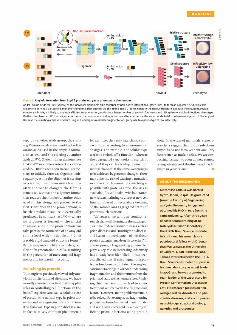
Later in 2010, Tanaka and his col-leagues extensively examined the pro-cesses of amyloid formation at 4°C and 37°C with Sup35 proteins, using the SPring-8 syncrotoron radiation facility at the RIKEN Harima Institute. “We used a technique known as small-angle X-ray scattering by which a protein solution is exposed to X-rays, and the intensity of the resulting scattering is measured to determine the protein structure. As a result, we found Sup35 proteins to be monomers at 37°C, but at 4°C a stable oligomer was formed comprising many monomers bound together (Fig. 3). Also, even when the temperature was repeat-edly changed between 4°C and 37°C, the scattering intensity was constant at each temperature, so we found that the oli-gomer formation with a Sup35 protein is reversible through temperature change. Furthermore, the amyloids formed at 4 to 9°C via oligomers were highly infectious and brittle structures, whereas the amy-loids formed at 10 to 37°C without detect-able oligomers were less infectious and rigid structures. In other words, whether a stable oligomer is formed proved to be the critical determinant of the eventual structure and phenotype of amyloids.”
The researchers next identified the amino acids involved in oligomer for-mation at 4 to 9°C by generating some Sup35 protein variants and using small-angle X-ray scattering to determine those amino acids which reduced the oligomer content. As a result, the first 100 amino acids in the prion domain (residues 1–123) proved to be involved in the oligomerization.
“We also found that oligomer forma-tion was initiated when amino acids 50–100 in the prion domain bound together (Fig. 3),” says Tanaka. “According to a
Figure 2: Cytotoxicity depending on amyloid structure
Huntingtin exon 1 was aggregated at 4°C and 37°C. The amyloid formed at 4°C was found to be more toxic, assuming a brittle amyloid structure with exposed polyglutamine.
Huntingtin
Amyloid structurePolyglutamine
Amyloid structure
Loops and turns contained
High toxicity
Low toxicity
Aggregation
Rich in
4°C
37°C
β-sheets
© 20
09 N
atio
nal A
cade
my o
f Scie
nces
17
FRONTLINE
RIKEN RESEARCH | VOL. 7 | NUMBER 4 | APRIL 2012 | www.rikenresearch.riken.jp
report by another study group, the start-ing 35 amino acids were identified as the amino acids used in the amyloid forma-tion at 4°C, and the starting 70 amino acids at 37°C. ese findings demonstrate that at 4°C monomers interact via amino acids 50–100 in each (non-native interac-tion) to initially form an oligomer. Sub-sequently, while the oligomer is serving as a scaffold, monomer units bind one after another to elongate the fibrous structure. Because the oligomer forma-tion reduces the number of amino acids used in this elongation process to the first 35 residues in the prion domain, a brittle amyloid structure is eventually produced. By contrast, at 37°C — where no oligomer is formed — the initial 70 amino acids in the prion domain can take part in the formation of an amyloid core, a level which is double at 4°C, so a stable rigid amyloid structure forms.” Brittle amyloids are likely to undergo ef-ficient fragmentation in cells, resulting in the generation of more amyloid frag-ments and increased infectivity.
Switching by protein“Although we previously viewed only am-yloids as the cause of diseases, we have recently come to think that they may play roles in controlling cell functions in the body,” explains Tanaka. “A soluble state of protein (the normal type in prion dis-eases) and an aggregated state of protein (the abnormal type in prion diseases) are in fact relatively common phenomena;
for example, they may interchange with each other according to environmental changes. For example, the soluble type works to switch off a function, whereas the aggregated type works to switch it on, and they can both adapt to environ-mental changes. If the same switching is to be achieved by genomic changes, there may arise the risk of causing a mutation in some site; however, if switching is possible with proteins alone, the risk is avoidable,” says Tanaka, who has started new research aiming to discover new cell functions based on reversible switching of the soluble and aggregated states of proteins such as prions.
“Of course, we will also conduct re-search that will illuminate the pathogen-esis in neurodegenerative diseases such as prion diseases and Huntington’s disease, and lead to the development of new thera-peutic strategies and drug discoveries.” In a yeast prion, a fragmenting protein that plays a key role in increasing infectivity has already been identified. It has been established that, if this fragmenting pro-tein is functionally inhibited, the amyloid continues to elongate without undergoing fragmentation and then returns from the prionized state to the normal state. Apply-ing this mechanism may lead to a new treatment which blocks the fragmenting factor. “However, many problems remain to be solved. For example, no fragmenting protein has been discovered in mammals, and none have succeeded in achieving ef-ficient prion infections using protein
alone. In the case of mammals, some re-searchers suggest that highly infectious amyloids do not form without auxiliary factors such as nucleic acids. We are con-ducting research to open up new routes, taking advantage of the discovered mech-anism in yeast prions.”
Motomasa Tanaka was born in
Kyoto, Japan, in 1971. He graduated
from the Faculty of Engineering
at Kyoto University in 1994 and
obtained his PhD in 1999 from the
same university. After three years
of postdoctoral training at Dr
Nobuyuki Nukina’s laboratory in
the RIKEN Brain Science Institute,
he continued his research as a
postdoctoral fellow with Dr Jona-
than Weissman at the University
of California, San Francisco (UCSF).
Tanaka later returned to the RIKEN
Brain Science Institute to supervise
his own laboratory as a unit leader
in 2006, and he was promoted to
team leader of the Laboratory for
Protein Conformation Diseases in
2011. His research focuses on neu-
rodegenerative disorders and psy-
chiatric diseases, and encompasses
neurobiology, structural biology,
genetics and proteomics.
ABOUT THE RESEARCHER
Figure 3: Amyloid formation from Sup35 protein and yeast prion strain phenotypes
At 4°C, amino acids 50–100 (yellow) of the individual monomers bind together by non-native interactions (green lines) to form an oligomer. Next, while the oligomer is serving as a scaffold, monomers bind one after another via the amino acids 1–35 to elongate the fibrous structure. Because the resulting amyloid structure is brittle, it is likely to undergo efficient fragmentation, producing a larger number of amyloid fragments and giving rise to a highly infectious phenotype. On the other hand, at 37°C, no oligomer is formed, but monomers bind together one after another via the amino acids 1–70 to achieve elongation of the amyloid. Because the resulting amyloid structure is rigid, it undergoes moderate fragmentation, giving rise to a phenotype of low infectivity.
Amino acids 50–100
Amino acids 1–35
Amino acids 1–70
Non-natural interaction
Oligomer
Amyloid Phenotype
Infectivity: highColor : white
Infectivity: lowColor : pink
Monomer
Brittle structure
Solid structure
4°C
37°C
© 20
10 N
PG
18 RIKEN RESEARCH | VOL. 7 | NUMBER 4 | APRIL 2012 | www.rikenresearch.riken.jp
CONTACT INFORMATION
For details about working at RIKEN, please contact the RIKEN Global Relations Office:Tel: +81-(0)48-462-1225 E-mail: [email protected]
What do you do at RIKEN?
I work as a PR Officer in the planning sec-tion of the Research Promotion Division at the RIKEN Yokohama Institute, which is located about 20 km southwest of Tokyo.
How and when did you join RIKEN?
I studied landscape design at university and then majored in information design. Following graduation, I worked in a range of information technology and PR-related positions. Although I found these posi-tions interesting and fulfilling, I wanted a new challenge that would allow me to apply the skills I had learned as a student and the skills and experience that I had acquired in the workplace to a completely fresh field. This coincided with an in-creasing awareness of the importance of the public communication of science. On the strength of this I applied for a position in the research promotion office at RIKEN and joined the team at Yokohama in 2011.
How was the transition to life at RIKEN?
My introduction to work at RIKEN was actually something of a baptism of fire. After only three days, I was put in charge of producing a special pamphlet for the tenth anniversary of the RIKEN Yokohama Institute. While it was a challenge to learn a great deal of new information in a short space of time, my colleagues were very supportive and answered all of my many questions about how to get things done.
Please tell us about your work at RIKEN.
My main work involves promoting the activities of the RIKEN Yokohama Institute, which is actually a collection of seven separate centers. is requires working closely with my counterparts in each center to ensure that the overall individual character of each section is preserved, whilst communicating what the institute is doing in an overall corporate sense. My department coordi-nates a range of events and activities for researchers and local residents and pro-motes them externally through websites and brochures, as well as liaising with news organizations. We also disseminate the information internally through bul-letin boards and advertising campaigns.
What have been the highlights of your
time at RIKEN so far?
Although I have only been working at RIKEN for a year, I have already gained a lot of valuable experience. I have particularly enjoyed working on an ini-tiative called the “Science Café” aimed at increasing public understanding of science, which RIKEN carries out with libraries in the Yokohama area. Based on focus group information, we designed promotional materials for the project and then revised them to create an even more appealing and effective campaign. e event was a success and we received many requests to hold more of them.
e RIKEN researchers who joined in the event responded enthusiastically to ques-tions from the public, and this inspires me to create an even better and more ef-fective arena for their activities.
What is the best thing about working
at RIKEN?
For me, the best thing about being at RIKEN is the chance to work alongside people with a diverse range of back-grounds and specialist skills. In addition to the research staff at RIKEN, I often need to collaborate with PR staff in other sec-tions. It is very stimulating to work with talented and knowledgeable colleagues who have an in-depth understanding of the work carried out by their own centers.
What would you say to other people
considering joining RIKEN?
I would highly recommend RIKEN as a place to work as it welcomes people with a wide range of skills and experience. is creates an atmosphere that is very accept-ing of people with different backgrounds and allows them to observe the work of RIKEN from a number of different view-points, which I think is very valuable.
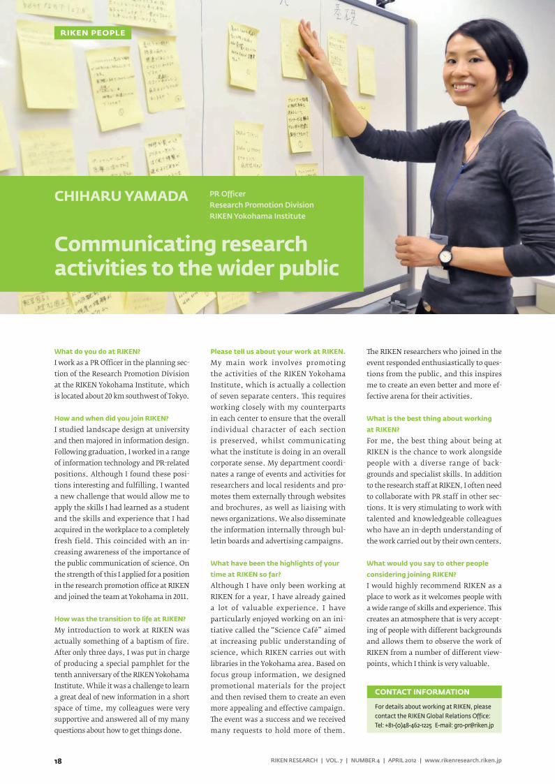
CHIHARU YAMADA
Communicating research activities to the wider public
PR OfficerResearch Promotion DivisionRIKEN Yokohama Institute
RIKEN PEOPLE
NEWS ROUNDUP
19RIKEN RESEARCH | VOL. 7 | NUMBER 4 | APRIL 2012 | www.rikenresearch.riken.jp
The 2nd RIKEN Advanced Institute for Computational Science symposiumOn 1 and 2 March 2012, the second interna-tional symposium for the RIKEN Advanced Institute for Computational Science (AICS) was held in Kobe, Japan. The symposium was organized and sponsored by RIKEN AICS, renowned for developing the K com-puter, which took first place in the 37th and 38th TOP500 list in June and November, 2011, respectively. The aim of the sympo-sium, through the AICS, is to promote and explore cross-disciplinary collaboration using the K computer and to create a world-class computational science hub.
The symposium was co-sponsored by the Strategic Programs for Innovative Research (SPIRE), supported by the Japanese Minis-try of Education, Culture, Sports, Science and Technology (MEXT). The program com-prised a series of lectures presented by eight
distinguished researchers from overseas, seven domestic invited speakers, and one speaker from RIKEN AICS. In addition, eight oral sessions were held which covered AICS’ eight research fields: computational sci-ences (molecular science, life science, solid-state physics, climate science, and particle physics) and computer sciences (design of processors, system software, and program-ming language). At the poster session held on 1 March, 58 posters were presented, in-cluding presentations that introduced some of the latest work conducted by AICS re-search teams. During the symposium, more than 120 participants engaged in discussions on computer simulations and future super-computing technologies. The success of this symposium further advances the progress of simulation studies using the K computer, as well as the development of next-generation supercomputers in Japan.
The 4th LIAI-RCAI Joint Workshop The RIKEN Research Center for Allergy & Immunology (RCAI) and the La Jolla In-stitute for Allergy & Immunology (LIAI) have run a collaborative program for sev-eral years, and held the 4th Joint LIAI-RCAI workshop at LIAI on 7–8 February 2012. The event drew nearly 100 participants, includ-ing RCAI Director Masaru Taniguchi and LIAI President and Chief Scientific Officer Mitchell Kronenberg.
The workshop addressed recent progress in immunological studies and included six sessions that focused on the themes of “Th & Treg cells,” “Signaling in the immune sys-tem,” “Disease regulation,” “Antigen recogni-tion by T cells and antibodies,” and “T cell development and memory.” Eighteen lead-ing researchers in these fields from the two institutes presented some of their most recent experimental data. The sessions enabled lively discussions on these topics, providing stimulus for further collaboration between RCAI and LIAI.
With both research institutes founded on the common goals of achieving break-throughs in the understanding of the immune system and improving human health through the development of thera-pies for immune system disorders, future collaborations between the RCAI and LIAI hold great promise for accelerating novel findings in immunology.
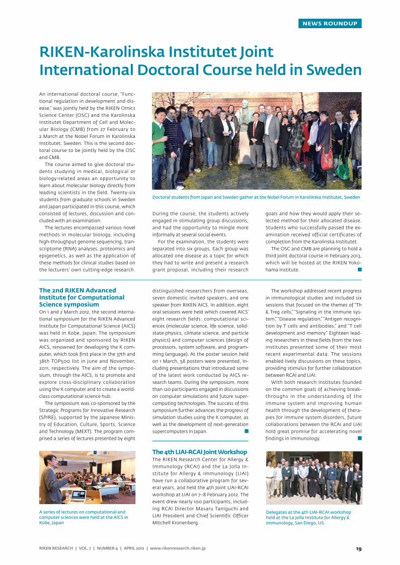
RIKEN-Karolinska Institutet Joint International Doctoral Course held in SwedenAn international doctoral course, “Func-tional regulation in development and dis-ease,” was jointly held by the RIKEN Omics Science Center (OSC) and the Karolinska Institutet Department of Cell and Molec-ular Biology (CMB) from 27 February to 2 March at the Nobel Forum in Karolinska Institutet, Sweden. This is the second doc-toral course to be jointly held by the OSC and CMB.
The course aimed to give doctoral stu-dents studying in medical, biological or biology-related areas an opportunity to learn about molecular biology directly from leading scientists in the field. Twenty-six students from graduate schools in Sweden and Japan participated in this course, which consisted of lectures, discussion and con-cluded with an examination.
The lectures encompassed various novel methods in molecular biology, including high-throughput genome sequencing, tran-scriptome (RNA) analyses, proteomics and epigenetics, as well as the application of these methods for clinical studies based on the lecturers’ own cutting-edge research.
During the course, the students actively engaged in stimulating group discussions, and had the opportunity to mingle more informally at several social events.
For the examination, the students were separated into six groups. Each group was allocated one disease as a topic for which they had to write and present a research grant proposal, including their research
goals and how they would apply their se-lected method for their allocated disease. Students who successfully passed the ex-amination received official certificates of completion from the Karolinska Institutet.
The OSC and CMB are planning to hold a third joint doctoral course in February 2013, which will be hosted at the RIKEN Yoko-hama Institute.
Doctoral students from Japan and Sweden gather at the Nobel Forum in Karolinska Institutet, Sweden
A series of lectures on computational and computer sciences were held at the AICS in Kobe, Japan
Delegates at the 4th LIAI-RCAI workshop held at the La Jolla Institute for Allergy & Immunology, San Diego, US
RIKEN Harima Institute
RIKEN Kobe Institute
Nagoya Facility
RIKEN Wako Institute
RIKEN Yokohama Institute
RIKEN Tsukuba Institute
Sendai Facility
www.riken.jp
RIKEN, Japan’s flagship research institute, conducts basic and applied experimental research in a
wide range of science and technology fields including physics, chemistry, medical science, biology
and engineering. Initially established as a private research foundation in Tokyo in 1917, RIKEN
became an independent administrative institution in 2003.
RIKEN RESEARCH is a website (www.rikenresearch.riken.jp) and print publication intended to
highlight the best research being published by RIKEN (www.riken.jp). It is written for a broad
scientific audience and policy makers interested in science and aims to raise global awareness of
RIKEN and its research.
For further information on the research presented in this publication or to arrange an interview
with a researcher, please contact
RIKEN Global Relations Office
2-1, Hirosawa, Wako, Saitama, 351-0198, Japan
TEL: +81 48 462 1225
FAX: +81 48 46
E-Mail: [email protected]
www.rikenresearch.riken.jp
RIKEN RESEARCH
HIGHLIGHT OF THE MONTH
Biology: Embrasing our dierences
FEBRUARY 2011 VOLUME 6 NUMBER 2
FRONTLINETowards a radical treatment for leukemia
NEWS ROUNDUPNew cherry blossom tree blooms all seasons
LIFE AT RIKENPIERO CARNINCI
THE BUTTERFLY EFFECT
RESEARCH HIGHLIGHTSMouse models: Almost humanHigh-energy physics: Too hot to handlePhysics: Gluons don’t explain the spin surpriseSuperconductivity: back to basicsPhysics: Forced to insulatePhysics: An all-in-one chipBiology: Learning by osmosisBiology: The need for new neurons
3 3687
RIKEN 2012-013