Embed Size (px)
Citation preview
RIKEN RESEARCH NOVEMBER 2012VOLUME 7 NUMBER 11
HIGHLIGHT OF THE MONTH
Lucky number 113
Eyeing up embryonic stem cells
RESEARCH HIGHLIGHTSMetal oxides get heavy
Powerful x-rays for less
The sweet sights of glycoproteins
Head-mounted device manipulates reality
Learning to simulate other people’s decisions
Re-tuning responses in the visual cortex
Uncovering the gene for a rare skeletal disease
Managing cellular security systems
A blueprint for the gut’s antimicrobial defenses
First mouse, now human, lab-grown eye tissue
FRONTLINECreating new products through ‘bio-fabrication’
RIKEN PEOPLEMasamichi Kawasaki, RIKEN Canteen
NEWS ROUNDUPThe First Global Summit of Research
Institute Leaders
K computer opens for shared use
HIGHLIGHT OF THE MONTH
2 3RIKEN RESEARCH | VOL. 7 | NUMBER 11 | NOVEMBER 2012 | www.rikenresearch.riken.jp RIKEN RESEARCH | VOL. 7 | NUMBER 11 | NOVEMBER 2012 | www.rikenresearch.riken.jp
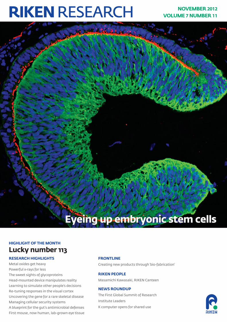
The heaviest element that occurs natu-rally on earth is uranium. More massive atoms have, however, been created in the laboratory. These so-called super-heavy elements have more than 103 protons in their nucleus, but the com-plicated nuclear interactions between these subatomic particles makes such nuclei highly unstable. Further, these particles live for only a fraction of a second. A team of scientists across Japan and China led by RIKEN researcher Kosuke Morita has now seen an indirect signature of element 113 by measur-ing the particles generated when superheavy elements disintegrate1. This new addition to the periodic table will improve our understanding of the building blocks of the Universe.
The scientists created element 113 by fusing together zinc and bismuth atoms. First, they generated a beam of zinc atoms travelling at 10% of the speed of light using the RIKEN Linear Accelerator. They then fired these high-energy particles at a thin bismuth film. To ensure that the beam did not damage the target, the scientists mounted 16 such foils on a 30-centimeter-diameter wheel and spun it at more than 3,000 revolutions per minute. At such high velocity, the zinc nucleus, made up of 30 protons and 40 neutrons, fused with the 83 protons and 126 neutrons in the nucleus of the bismuth atoms to form a nucleus with 113 protons and 165 neutrons, plus one free neutron. This seems simple enough, but this nuclear interaction is rare. The experi-ment, which began in September 2003, has been running for 553 days and yet
only three atoms of element 113 have been identified in that time. “This has been a painstakingly long experiment,” says Morita. “First, we had to create the 108th, 110th, 111th and 112th elements. This gave us confidence that we had the right experimental conditions to produce element 113.”
Timing is crucialThe difficulty faced by the team was how to prove that a superheavy element had been created. Element 113 cannot be seen directly because it only exists for
a short time before decaying through a process known as alpha decay. Instead, the team searched for the particles created when element 113 disintegrates. In alpha decay, a large nucleus breaks into a lighter one and a particle made of two protons and two neutrons, known as an alpha particle. Element 113, for example, should decay into an isotope of element 111 with 163 neutrons, which is known as roentgenium-274, plus an alpha particle. Roentgenium, in turn, also undergoes alpha decay to produce meitnerium-270 and another alpha
Chemistry
Lucky number 113The creation of an atom with 113 protons adds a new element to the periodic table
© 20
12 R
IKEN
Nish
ina
Cent
er fo
r Acc
eler
ator
-Bas
ed Sc
ience
Meitnerium
Roentgenium
Bohrium
Dubnium
Mendelevium
Lawrencium
Alphaparticle #1#2
#3
#4
#5
#6
Figure 1: Element 113 was identified by the alpha particles that are created when a heavy nucleus decays to a lighter one. Six alpha particles were measured at times corresponding to the half-life of the six decays matching element 113 decaying sequentially to mendelevium.

HIGHLIGHT OF THE MONTH
2 3RIKEN RESEARCH | VOL. 7 | NUMBER 11 | NOVEMBER 2012 | www.rikenresearch.riken.jp RIKEN RESEARCH | VOL. 7 | NUMBER 11 | NOVEMBER 2012 | www.rikenresearch.riken.jp
particle (Fig. 1). This process continues until an atom with a stable nucleus is created.
Once created, the nucleus of element 113 was separated from any unwanted ions in a piece of equipment known as GARIS—the gas-filled recoil ion sepa-rator (Fig. 2)—and directed into an arrangement of detectors that could measure the chain of alpha particles. Morita and his co-workers measured a sequence of six alpha particles in August 2012. The timing of these detec-tion events was crucial in showing that they started with the creation of an atom of the element 113 nucleus. Each decay occurs on a characteristic time scale known as the half-life. The sixth decay from lawrencium-258 to mendele-vium-254 has a half-life of 3.9 seconds. This corresponded to the observation of the sixth alpha particle 3.8 seconds after the fifth. Two further alpha par-ticles were detected about 7 hours after the sixth decay that could have cor-responded to a decay from fermium to californium (mendelevium decays to fermium via a process that does not create an alpha particle). However, the scientists could not discount the possi-bility that this was just a coincidental background alpha particle. The next
decay, from californium to curium has a half-life of more than 13 years.
Studying the nuclei of superheavy elements gives important information on ‘stability’. It is known from the elements lower down in the periodic table that nuclei with a certain number of neutrons or protons are particularly stable. The first six of these so-called magic numbers are known but the next has not yet been shown experimentally. “It is important that the created superheavy atom has 113 protons because it closely approaches one of the predicted proton magic numbers of 114,” explains Morita.
What’s in a name?The next big question is what to call this new element. It is currently known by the provisional name ununtrium (one-one-three), and has also been referred to as eka-thallium. But the discoverer of an element is traditionally awarded the honor of giving it a permanent name. First, however, the International Union of Pure and Applied Chemistry (IUPAC) must confirm the ‘sighting’, and they have some very tough criteria. Morita and his colleagues also saw a signature of element 113 in 2004 and 2005. On these two previous occasions only four alpha decays were detected. This was
not enough to convince IUPAC, nor were experiments conducted in a Russian laboratory. “I do not want to speak about the possible name until the naming-rights are confirmed,” says Morita. “However, in general, I can say I would like element 113 to be named after a country or a famous scientist.”
While evidence of the 118th element has already been demonstrated, Morita believes that with the high levels of motivation shown so far by his col-leagues at RIKEN and the Nishina Center for Accelerator-Based Science, the team can go even further: “I would like to challenge the totally unreached parts of the periodic table and produce the 119th and 120th elements.”
1. Morita, K., Morimoto, K., Kaji, D., Haba, H.,
Ozeki, K., Kudou, Y., Sumita, T., Wakabayashi,
Y., Yoneda, A., Tanaka, K., et al. New result in
the production and decay of an isotope, 278113,
of the 113th element. Journal of the Physical
Society of Japan 81, 103201 (2012).
© 20
12 K
osuk
e Mor
ita, R
IKEN
Nish
ina
Cent
er fo
r Acc
eler
ator
-Bas
ed Sc
ience
ABOUT THE RESEARCHER
Kosuke Morita was born in Kitakyushu,
Fukuoka, Japan, in 1957. He graduated
from the Faculty of Science, Kyushu
University in 1979 and obtained his PhD
from the same university in 1993. He
joined the Cyclotron Laboratory at the
RIKEN Nishina Center for Accelerator-
Based Science as a research scientist
in 1984. In a world-first in September
2004, Morita succeeded in observing a
signature of the 113th element. In 2006,
he became associate chief scientist of the
Superheavy Element Laboratory at the
RIKEN Nishina Center.Figure 2: The Gas-filled Recoil Ion Separator (GARIS) separates the nucleus of element 113 from any unwanted ions and directs the nucleus into a detector chamber.

4 5RIKEN RESEARCH | VOL. 7 | NUMBER 11 | NOVEMBER 2012 | www.rikenresearch.riken.jp RIKEN RESEARCH | VOL. 7 | NUMBER 11 | NOVEMBER 2012 | www.rikenresearch.riken.jp
RESEARCH HIGHLIGHTS RESEARCH HIGHLIGHTS
Metal oxides get heavyX-ray resonance scattering can reveal the magnetic properties of transition metal oxides made out of heavy elements
Transition metal oxides are known for their interesting properties, includ-ing high-temperature superconductiv-ity and resistance that can be tuned with a magnetic field. Researchers have mainly focused on oxides made from ‘3d’ transition metals—the elements from scandium to zinc—but they are starting to uncover new material properties in oxides containing the much heavier ‘5d’ transition metal elements found between hafnium and mercury.
Unfortunately, scientists have not been able to rely on their usual tool, neutron scattering, to study magnetic structure in these materials because the samples are often too small and 5d elements strongly absorb neutrons. Now, Shigeki Fujiyama at the RIKEN Advanced Science Institute and his colleagues have shown they can use x-rays to study magnetism in the 5d transition metal oxide Sr2IrO4 over a wide temperature range1. “The sample size needed is three orders of magnitude smaller than what is needed for conven-tional neutron scattering experiments,” explains Fujiyama, who says the tech-nique will be important for studying other 5d transition metal oxides.
Atoms in a solid are magnetic if their valence electrons have a net (non-zero) angular momentum. The valence elec-trons’ total angular momentum is a com-bination of their orbital motion about the nucleus and their ‘spin’. In heavy elements, like iridium (Ir), a relativistic effect called the spin-orbit interaction causes the electrons’ orbital momentum to drag their spin momentum with it. In materials where the spin-orbit effect is large, the electronic motion can be
controlled to affect the magnetic proper-ties (and vice versa).
In Sr2IrO4, a large spin-orbit interac-tion makes the material a ‘Mott insu-lator’, similar to La2CuO4, a 3d metal oxide that can be chemically modified to become a superconductor. In both materials, the magnetic ions (iridium and copper) also form the same magnetic structure—an antiferromagnet—at low temperature. It has not been clear if both magnetic structures can be described by the same models.
Fujiyama and his team therefore used resonance x-ray scattering, where the x-ray wavelength matches an absorption edge of the iridium ion so it is sensitive to the ion’s magnetic state (Fig. 1), to study
the onset of magnetic order in Sr2IrO4. Their measurements, performed at the RIKEN SPring-8 synchrotron, show that magnetic order in Sr2IrO4 develops in two-dimensional planes first and only becomes three-dimensional near the anti-ferromagnetic transition, similar to La2CuO4. Similar oxides containing iridium may exhibit superconductivity or a ‘quantum spin liquid’ and Fujiyama’s group is examining these possibilities.
1. Fujiyama, S., Ohsumi, H. Komesu, T., Matsuno,
J., Kim, B.J., Takata, M., Arima, T. & Takagi, H.
Two-dimensional Heisenberg behavior of Jeff = ½
isospins in the paramagnetic state of the spin-
orbital Mott insulator Sr2IrO4. Physical Review
Letters 108, 24721 (2012).
© 20
12 Th
e Am
erica
n Ph
ysica
l Soc
iety
Figure 1: The scattering of x-rays with a wavelength 'in resonance' with an ion’s absorption edge depends on the direction of the ion’s magnetic moment (red and blue arrows). The researchers used x-rays (pink) with a wavelength of 0.11 nanometers—close to two absorption edges in iridium—to study the onset of magnetic order in Sr2IrO4. X-rays scattered at the blue arrows are out of phase with those scattered at the red arrows.

4 5RIKEN RESEARCH | VOL. 7 | NUMBER 11 | NOVEMBER 2012 | www.rikenresearch.riken.jp RIKEN RESEARCH | VOL. 7 | NUMBER 11 | NOVEMBER 2012 | www.rikenresearch.riken.jp
RESEARCH HIGHLIGHTS RESEARCH HIGHLIGHTS
Studying small objects typically requires big machines. For example, the study of single atoms with a laser requires x-ray radiation of such high energy that it is only produced by accelerating electrons in large facilities. Researchers at the RIKEN SPring-8 Center in Harima have developed a more affordable electron laser design, the SPring-8 Angstrom Compact free-electron Laser (SACLA), which is not only compact and therefore economic to build but also delivers x-rays with unprecedented short wavelengths1.
User operation of SACLA began in March 2012. Makina Yabashi from the research team describes typical research as non-linear interactions of light and matter, biological imaging and ultrafast phase-transition in materials.
Construction of a high-energy laser is based on the concept that electrons accel-erated by going very fast around a curve also emit radiation. The energy of this radiation, and therefore its wavelength,
depends on the acceleration. The tighter the curved path, the shorter the wave-length of the light emitted. This is the operating principle of free electron lasers (Fig. 1).
At SPring-8 the aim was to push free electron lasers to new limits by pro-ducing ever shorter wavelengths. This means sending electrons on a very tight twisting path in a section of the laser known as the undulator. Normally, the period of the curved electron beam is about several centimeters. The SACLA team have realized a period of only 1.8 centimeters by directly placing the magnets that deflect the electron beams into the vacuum chamber of the beam. This has enabled a reduc-tion of laser wavelength down to 0.6 ångström, which is about the radius of a hydrogen atom.
The benefit of SACLA is that, in com-parison to other free-electron lasers, the device is also smaller. “Our x-ray free
electron laser facility has been designed to achieve a much more compact scale compared to those in the US and Europe," explains Yabashi. "The major reduction in construction and operating costs enables many research institutes or universities to build such a machine, and to utilize powerful laser light in a broad range of applications from biology, chemistry to physics,” he says.
The team plans to increase the energy density of the laser beam, which would, for example, make biological imaging easier. Already there is strong interest from scientists to use the laser and other institutions are planning similar machines. In the meantime, SACLA is open for business.
1. Ishikawa, T., Aoyagi, H., Asaka, T., Asano, Y.,
Azumi, N., Bizen, T., Ego, H., Fukami, K., Fukui,
T., Furukawa, Y., et al. A compact X-ray free-
electron laser emitting in the sub-ångström
region. Nature Photonics 6, 540–544 (2012).
C-TWA C-TWA
UND
UND
© 20
12 N
atur
e Pub
lishi
ng G
roup
Figure 1: A conceptual diagram of SACLA. The laser consists of various electron acceleration stages (C-TWA) and focusing elements. Key to achieving short wavelength operation is, however, the design of the undulator (UND).
Powerful x-rays for lessDesign improvements enable construction of compact x-ray lasers at ultra-short wavelengths, which can measure individual atoms

6 7RIKEN RESEARCH | VOL. 7 | NUMBER 11 | NOVEMBER 2012 | www.rikenresearch.riken.jp RIKEN RESEARCH | VOL. 7 | NUMBER 11 | NOVEMBER 2012 | www.rikenresearch.riken.jp
RESEARCH HIGHLIGHTS RESEARCH HIGHLIGHTS
The sweet sights of glycoproteinsAn imaging technique helps scientists profile the sugar structures on protein molecules
The body’s cell machinery often attaches sugar chains onto proteins to create molecules that play a crucial role in everything from protective immunity to structural stability. However, distin-guishing between the different sugar tags applied to these so-called ‘glyco-proteins’ can be difficult. Now, inves-tigators in Japan have developed a way to visualize the profile of sugar mol-ecules (known as ‘glycans’) on specific protein targets1.
The new method could help scientists investigate the expression and traffick-ing of glycoprotein targets with thera-peutic potential. For example, recent studies have revealed that changes in glycan structures accompany the progress of disease. “Our technique may help us to understand why this is happening,” says Tadashi Suzuki of the RIKEN Advanced Science Institute in Wako.
To determine the nature of attached sugars, Suzuki and his postdoctoral fellow Yoshimi Haga turned to an imaging technique known as ‘fluores-cence resonance energy transfer’, or FRET. The researchers labeled sugar mol-ecules with a fluorescent probe that is activated only when signals are emitted from a nearby protein tagged with green fluorescent protein (GFP). These signals are picked up by the FRET assay, which can reveal the complex spatial and geo-metrical characteristics of the glycopro-tein structures, even those that span the thick cell membrane.
As a proof of principle, Suzuki and Haga first applied the method to two well-studied glycoproteins—the glucose
transporter GLUT4 and the epidermal growth factor receptor—each tagged with a specific type of sugar tag, sialic acid (Fig. 1). The researchers then set up an experiment in human cell culture where they adjusted the levels of insulin. This hormone triggers the transport of GLUT4 to the cell surface, where the glycoprotein begins pumping glucose into the cell. The scientists found that lowering insulin levels led to GLUT4 proteins with sugars containing sialic acid re-entering the cell more slowly than GLUT4 proteins without this kind of sugar adornment. “We do not know why it happens,” Suzuki says. “This finding
may shed some light on the cell traffick-ing of closely related glycoproteins.”
The researchers now want to adapt the technique so they can visualize sugars on more complex glycoproteins. “GLUT4 happens to be fairly easy as it only contains a single glycan,” Suzuki says, “but for proteins bearing multiple glycans, it is not going to be so easy, as the structural diversity is much greater.”
1. Haga, Y., Ishii, K., Hibino, K., Sako, Y., Ito, Y.,
Taniguchi, N. & Suzuki, T. Visualizing specific
protein glycoforms by transmembrane
fluorescence resonance energy transfer. Nature
Communications 3, 907 (2012).
GLUT4 SiaNAz FRET
N57
QW
T
© 20
12 N
atur
e Pub
lishi
ng G
roup
Figure 1: The azide-tagged sialic acid (SiaNAz) sugar molecules on GLUT4 can be seen through FRET imaging in the wild-type glycoprotein (top) but not in the N57Q mutant lacking this glycosylation site (bottom). Scale bar equals 20 micrometers.

6 7RIKEN RESEARCH | VOL. 7 | NUMBER 11 | NOVEMBER 2012 | www.rikenresearch.riken.jp RIKEN RESEARCH | VOL. 7 | NUMBER 11 | NOVEMBER 2012 | www.rikenresearch.riken.jp
RESEARCH HIGHLIGHTS RESEARCH HIGHLIGHTS
A research team led by Naotaka Fujii of the RIKEN Brain Science Institute in Japan has developed a cheap virtual real-ity-like system that can be used to manip-ulate people’s perceptions of reality1. The ‘substitutional reality’ system consists of a video camera, a computer for storing recorded footage and a head-mounted device that displays, and switches between, recorded footage and a live feed captured by an attached camera and microphone.
Fujii and his colleagues recorded par-ticipants while giving them instruc-tions about the experiment. Each participant was then asked to wear the head-mounted device, which displayed a sequence of recorded and live scenes designed to surreptitiously substitute the live scenes with recorded ones.
The first scene was a recording of one of the researchers appearing at the door and asking if the participant felt comfortable wearing the device and
to test it by looking around. This was followed by a ‘doppelgänger’ scene, in which participants saw the recording of themselves receiving instructions from the researcher and a fake live scene in which the experimenter re-entered the room and explained how the experiment was designed. Finally, the device played a live feed of the researcher returning to reveal that the previous scene was actually a recording (Fig. 1).
The participants realized that the doppelgänger scene could not be real but failed to distinguish between the live and recorded scenes during the rest of the experiment, showing that the device can successfully substitute reality with recorded scenes and that the par-ticipants subjectively experienced the recorded scenes as real. The research-ers determined that head movements and motion parallax—how objects change shape and depth with changes in head position—did not influence the
performance of the system and that par-ticipants were less likely to notice the switch between live and recorded scenes if it was done while they looked around the room. They also established that some of the participants had noticed a difference in the audio quality of the live and recorded scenes and used these differences to establish when the switch between the two was made.
“We can replicate the delusions of psychiatric patients but the system is not directly usable for diagnosis or treat-ments,” says Fujii. “We are expanding the quality of the technology and trying to extend the system for people to use as an experience platform. It can be useful not only for scientific experiments but also for entertainment and art.”
1. Suzuki, K., Wakisaka, S. & Fujii, N. Substitu-
tional reality system: a novel experimental
platform for experiencing alternative reality.
Scientific Reports 2, 459 (2012).
© 20
12 N
atur
e Pub
lishi
ng G
roup
Head-mounted device manipulates realityA virtual reality-like device mixes real life with make believe
Figure 1: The experimental procedure for substituting a live feed with pre-recorded footage.
Live scene
Recorded scene
Live scene
8 9RIKEN RESEARCH | VOL. 7 | NUMBER 11 | NOVEMBER 2012 | www.rikenresearch.riken.jp RIKEN RESEARCH | VOL. 7 | NUMBER 11 | NOVEMBER 2012 | www.rikenresearch.riken.jp
RESEARCH HIGHLIGHTS RESEARCH HIGHLIGHTS
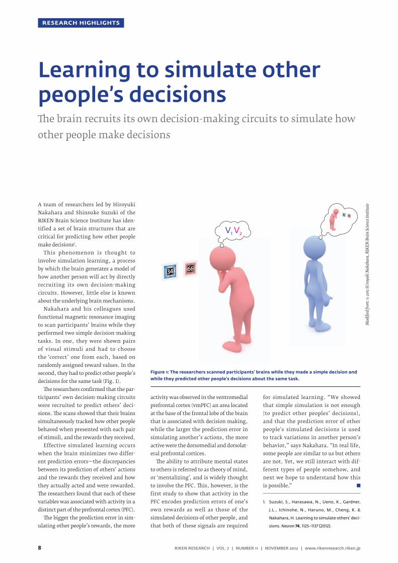
A team of researchers led by Hiroyuki Nakahara and Shinsuke Suzuki of the RIKEN Brain Science Institute has iden-tified a set of brain structures that are critical for predicting how other people make decisions1.
This phenomenon is thought to involve simulation learning, a process by which the brain generates a model of how another person will act by directly recruiting its own decision-making circuits. However, little else is known about the underlying brain mechanisms.
Nakahara and his colleagues used functional magnetic resonance imaging to scan participants’ brains while they performed two simple decision-making tasks. In one, they were shown pairs of visual stimuli and had to choose the ‘correct’ one from each, based on randomly assigned reward values. In the second, they had to predict other people’s decisions for the same task (Fig. 1).
The researchers confirmed that the par-ticipants’ own decision-making circuits were recruited to predict others’ deci-sions. The scans showed that their brains simultaneously tracked how other people behaved when presented with each pair of stimuli, and the rewards they received.
Effective simulated learning occurs when the brain minimizes two differ-ent prediction errors—the discrepancies between its prediction of others’ actions and the rewards they received and how they actually acted and were rewarded. The researchers found that each of these variables was associated with activity in a distinct part of the prefrontal cortex (PFC).
The bigger the prediction error in sim-ulating other people’s rewards, the more
activity was observed in the ventromedial prefrontal cortex (vmPFC) an area located at the base of the frontal lobe of the brain that is associated with decision making, while the larger the prediction error in simulating another’s actions, the more active were the dorsomedial and dorsolat-eral prefrontal cortices.
The ability to attribute mental states to others is referred to as theory of mind, or ‘mentalizing’, and is widely thought to involve the PFC. This, however, is the first study to show that activity in the PFC encodes prediction errors of one’s own rewards as well as those of the simulated decisions of other people, and that both of these signals are required
for simulated learning. “We showed that simple simulation is not enough [to predict other peoples’ decisions], and that the prediction error of other people's simulated decisions is used to track variations in another person’s behavior,” says Nakahara. “In real life, some people are similar to us but others are not. Yet, we still interact with dif-ferent types of people somehow, and next we hope to understand how this is possible.”
1. Suzuki, S., Harasawa, N., Ueno, K., Gardner,
J.L., Ichinohe, N., Haruno, M., Cheng, K. &
Nakahara, H. Learning to simulate others’ deci-
sions. Neuron 74, 1125–1137 (2012).
Learning to simulate other people’s decisionsThe brain recruits its own decision-making circuits to simulate how other people make decisions
V1 V2
Mod
ified
from
: © 20
12 H
iroy
uki N
akah
ara,
RIK
EN B
rain
Scien
ce In
stitu
teFigure 1: The researchers scanned participants' brains while they made a simple decision and while they predicted other people's decisions about the same task.

8 9RIKEN RESEARCH | VOL. 7 | NUMBER 11 | NOVEMBER 2012 | www.rikenresearch.riken.jp RIKEN RESEARCH | VOL. 7 | NUMBER 11 | NOVEMBER 2012 | www.rikenresearch.riken.jp
RESEARCH HIGHLIGHTS RESEARCH HIGHLIGHTS
New research led by Shigeru Tanaka of the University of Electro-Communications and visiting scientist at the RIKEN Brain Science Institute shows that the responses of cells in the visual cortex can be ‘re-tuned’ by experience1.
Experiments on kittens in the 1960s showed that the primary visual cortex contains neurons that fire selectively to straight lines of specific orientations. These cells are organized into alternating columns that receive inputs from the left or right eye. The kitten experiments also showed that proper brain development is highly dependent on sensory informa-tion. Closing one eye altered the organi-zation of the columns, so that those that should have received inputs from the closed eye were reduced in width, whereas those that received inputs from the open eye were much wider than normal.
The normal columnar organization can be restored if the closed eye is re-opened within a critical period of brain development. The effect of sensory expe-rience on the orientation selectivity of neurons in the primary visual cortex is, however, unknown.
To investigate, Tanaka and his col-leagues reared mice and fitted them with specially designed goggles (Fig. 1) through which they can only perceive vertically oriented visual stimuli, for a one-week period, between 3 and 15 weeks of age. Immediately after removing the goggles, they created a ‘window’ in the skull bone lying over the visual cortex to examine the cell response under the microscope.
Rearing the mice in this way had a significant effect on the properties of neurons in the primary visual cortex.
The researchers found that the number of cells responding to vertical orientation increased, while the number responding to other orientation decreased. They also found that the extent of these changes depended on the age at which they fitted the animals with the goggles. Mice fitted with the goggles between 4 and 7 weeks of age had more cells that were sensitive to the experienced (vertical) orientation than those fitted later.
These findings show that there is a critical period of plasticity between 4 and 7 weeks, during which cells in the primary visual cortex are particularly sensitive to sensory experience and that
plasticity persists in older animals, albeit to a lesser extent. They also suggest that plasticity in younger and older animals involves different mechanisms.
“When we put similar goggles on kittens, the age at which we started goggle rearing determined the revers-ibility of orientation selectivity,” says Tanaka. “We would now like to clarify the differences and commonalities of the mechanisms in cats and mice.”
1. Yoshida, T., Ozawa, K. & Tanaka, S. Sensitiv-
ity profile for orientation selectivity in the
visual cortex of goggle-reared mice. PLoS ONE
7, e40630.
Imag
e rep
rodu
ced u
nder
the t
erm
s of t
he C
CAL,
with
copy
right
shar
ed by
Tak
amas
a et
al
Figure 1: Specially designed goggles modify visual input so that only vertically oriented stimuli are perceived.
Re-tuning responses in the visual cortexMice wearing goggles show how early sensory experience alters the properties of visual cortical neurons

10 11RIKEN RESEARCH | VOL. 7 | NUMBER 11 | NOVEMBER 2012 | www.rikenresearch.riken.jp RIKEN RESEARCH | VOL. 7 | NUMBER 11 | NOVEMBER 2012 | www.rikenresearch.riken.jp
RESEARCH HIGHLIGHTS RESEARCH HIGHLIGHTS
The hereditary disease known as brachy-olmia is characterized by a short stature and various bone abnormalities that start becoming apparent in late child-hood. The vertebrae take on a flattened shape, with irregular spacing between the spinal discs (Fig. 1). This rare skeletal disease can be caused by dominantly inherited mutations in the gene TRPV4, but there are also more than a few reces-sive forms of brachyolmia for which the genetic cause has been a mystery— until now.
Reporting in the Journal of Medical Genetics, researchers in Japan have pin-pointed the gene responsible for recessive brachyolmia1. By sequencing the entire protein-coding regions of the genome of a young Turkish girl with the disease and two of her affected family members, the scientists found that an insertion in the PAPSS2 gene was the causative mutation.
The team, led by Shiro Ikegawa, an expert in the field of bone and joint diseases at the RIKEN Center for Genomic Medicine in Tokyo, also examined the DNA of three more children—two from Japan and one from Korea. All three had forms of brachyolmia that clinically mirrored those found in the Turkish family, and all three had loss-of-function mutations in both copies of their PAPSS2 gene, which encodes an enzyme involved in the formation of cartilage proteogly-can. “This finding indicates that brachyol-mia caused by the PAPSS2 mutation is not unique to the Turkish family but a univer-sal disease potentially common among different populations,” Ikegawa says.
Mutations in the PAPSS2 gene have been found occasionally in other bone growth
disorders with autosomal recessive inher-itance patterns. For example, researchers in the United States previously identified such mutations in a large, inbred Paki-stani family with a form of spondy-loepimetaphyseal dysplasia, another disease marked by short stature and other skeletal disorders. More recently, clini-cians from Western Europe have described a girl of Turkish origin with PAPSS2 sequence variants who suffered from precocious puberty and bone problems, although her skeletal defects were far milder than those found in other people with mutations in the same gene.
The finding that the six people with brachyolmia studied by Ikegawa’s
group had overlapping, but distinct, clinical features compared to the individuals with these other PAPSS2-associated diseases suggests that the loss of this key gene can lead to a gradient of disease states. “One gene mutation sometimes presents with a variety of phenotypes,” says Ikegawa. “The task now is to explain on a molec-ular scale why this happens with this particular gene.”
1. Miyake, N., Elcioglu, N.H., Iida, A., Isguven, P.,
Dai, J., Murakami, N., Takamura, K., Cho, T.-J.,
Kim, O.-H., Hasegawa, T. et al. PAPSS2 mutations
cause autosomal recessive brachyolmia. Journal
of Medical Genetics 49, 533–538 (2012).
Uncovering the gene for a rare skeletal disease Short stature and skeletal abnormalities arise from
mutations in the PAPSS2 gene
© 20
12 B
ritish
Med
ical J
ourn
al P
ublis
hing
Gro
up
Figure 1: X-ray image of the spine of a young girl with brachyolmia.

10 11RIKEN RESEARCH | VOL. 7 | NUMBER 11 | NOVEMBER 2012 | www.rikenresearch.riken.jp RIKEN RESEARCH | VOL. 7 | NUMBER 11 | NOVEMBER 2012 | www.rikenresearch.riken.jp
RESEARCH HIGHLIGHTS RESEARCH HIGHLIGHTS
Conventional dendritic cells (cDCs) are the immune system’s patrol (Fig. 1). They recognize foreign threats and trigger a defensive response, while restraining immune reactions against inappropriate targets like host proteins. They achieve the former via a mechanism called cross-presentation, which displays pieces of pathogens to cytotoxic T lymphocytes (CTLs)—the immune system’s ‘attack dogs’—while the latter function relies on cDC interactions with regulatory T (Treg) cells.
Katsuaki Sato’s group at the RIKEN Research Center for Allergy and Immu-nology in Yokohama recently identi-fied a subset of cDCs with an especially important role in fighting infection1. These cells can be classified based on the proteins they show on their surface and Sato’s team became especially inter-ested in cDCs featuring a protein called CD205. “CD205+ cDCs are more efficient in the cross-presentation of cell-bound or soluble antigens to CTLs than other dendritic cell subsets,” explains Sato. “However, their role in the immune system under physiological conditions was unclear.”
To clarify the function of these cDCs, Sato and colleagues genetically engi-neered mice in which CD205+ cDCs could be quickly and selectively killed off via injection with diphtheria toxin. This depletion lasted for several days, giving the researchers a powerful way to study the specific contribution of these cells to immune function. Initial experiments with the mice provided compelling evidence that CD205+ cDCs are required to marshal an effective CTL response.
Loss of these cells also resulted in abnormal Treg levels in various tissues, indicating that CD205+ cDCs are required to maintain appropriate levels of other T cell populations throughout the body.
Animals infected with high doses of the pathogenic bacterium Listerium mono-cytogenes normally perish quickly due to septic shock resulting from immune overreaction, but CD205+ cDC-deficient animals proved resistant to septic shock and tended to survive longer, reveal-ing a crippled inflammatory response. In the end, however, these animals were more vulnerable to bacterial infec-tion and proliferation, resulting from impaired cDC cross-presentation of bac-terial antigens to CTLs. The research-ers observed similar effects with viral infection.
These results position CD205+ cDCs at a critical juncture for regulating overall
immune system function as well as directed counterattacks against patho-gens and the researchers see clear poten-tial for exploiting these cells in clinical applications. “Further elucidation of CD205+ cDC function might provide insights into immune regulation and pathology and aid therapeutic interven-tions for infectious diseases as well as autoimmune and inflammatory disor-ders,” says Sato. “For example, we would like to develop vaccines that selectively target CD205+ cDCs with bacterial and viral antigens.”
1. Fukaya, T., Murakami, R., Takagi, H., Sato, K.,
Sato, Y., Otsuka, H., Ohno, M., Hijikata, A.,
Ohara, O., Hikida, M. et al. Conditional ablation
of CD205+ conventional dendritic cells impacts
the regulation of T-cell immunity and homeo-
stasis in vivo. Proceedings of the National Academy
Sciences USA 109, 11288–11293 (2012).
© 20
12 K
atsu
aki S
ato,
RIK
EN R
esea
rch
Cent
er fo
r Alle
rgy a
nd Im
mun
olog
y
Figure 1: Microscopic image of a mouse cDC.
Managing cellular security systemsCells that act as critical regulators of general immune function also play a key part in enabling prompt elimination of bacterial and viral threats

12 13RIKEN RESEARCH | VOL. 7 | NUMBER 11 | NOVEMBER 2012 | www.rikenresearch.riken.jp RIKEN RESEARCH | VOL. 7 | NUMBER 11 | NOVEMBER 2012 | www.rikenresearch.riken.jp
RESEARCH HIGHLIGHTS RESEARCH HIGHLIGHTS
Every bite of food or drink of water is an invitation for potentially harmful bacteria and viruses to set up shop in the body. In order to protect against such invaders, the mucous membrane that lines the intestine contains clusters of specialized microfold cells (M cells), which can absorb foreign proteins and particles from the digestive tract and deliver them to the immune system.
New work from Hiroshi Ohno’s group at the RIKEN Center for Allergy and Immunology in Yokohama, in collabora-tion with Ifor Williams and colleagues at Emory University in Atlanta, Georgia, has revealed valuable insights into how these M cells develop1. Previous research from Williams’ group showed that a signaling protein called RANKL switches on M cell development2 but virtually nothing was known about the subse-quent steps in this process. To find out, Ohno and Williams looked for genes that get switched on when intestinal cells undergo differentiation in response to RANKL exposure.
They discovered that treatment with RANKL causes immature intestinal epithelial cells to sharply increase the production of Spi-B, a protein that regulates the expression of other devel-opmental genes. To test the specific contribution of this protein to M cell maturation, the researchers collabo-rated with Tsuneyasu Kaisho’s group at Osaka University, which had engi-neered a genetically modified mouse strain lacking the gene encoding Spi-B. The resulting animals were devoid of mature M cells (Fig. 1). On the other hand, intestinal development as a
whole was unaffected by the absence of Spi-B, demonstrating that this protein’s impact is limited to this specific class of cells within the gut.
M cells normally localize to immune structures known as Peyer’s patches (PPs). Bacteria such as Salmonella enterica Typhimurium (S. Typhimurium) will normally accumulate within these PPs shortly after inoculation. This uptake was considerably reduced in Spi-B-deficient mice, indicating the absence of a functional M cell population. The mice showed a considerably weakened immune response following oral admin-istration of S. Typhimurium bacteria relative to wild-type animals, demon-strating the importance of M cell-medi-ated microbial uptake.
The identification of this critical ‘master switch’ for M cell development opens exciting new avenues of research into these mysterious cells. Ohno is eager
to investigate the details of how the cells perform their critical immunity-training function. “These questions could not be answered previously because of the lack of M cell-deficient mice,” he says. “But now, ‘knockout’ mice that spe-cifically lack Spi-B in their mucosal epi-thelium will provide the ideal tool for such studies.”
1. Kanaya, T., Hase, K., Takahashi, D., Fukuda, S.,
Hoshino, K., Sasaki, I., Hemmi, H., Knoop, K.A.,
Kumar, N., Sato M. et al. The Ets transcription
factor Spi-B is essential for the differentiation
of intestinal microfold cells. Nature Immunology
13, 729–736 (2012).
2. Knoop, K.A., Kumar, N., Butler, B.R., Sakthivel,
S.K., Taylor, R.T., Nochi, T., Akiba, H., Yagita,
H., Kiyono, H. & Williams, I.R. RANKL is neces-
sary and sufficient to initiate development
of antigen-sampling M cells in the intestinal
epithelium. The Journal of Immunology 183,
5738–5747 (2009).
A blueprint for the gut’s antimicrobial defensesThe identification of a developmental ‘master switch’ helps scientists explore the function of intestinal cells that help prevent infection
© 20
12 N
atur
e Pub
lishi
ng G
roup
Figure 1: Fluorescent labeling of GP2, a protein expressed in M cells (left) reveals that this cell population is virtually absent in mice lacking the gene encoding Spi-B (right).

12 13RIKEN RESEARCH | VOL. 7 | NUMBER 11 | NOVEMBER 2012 | www.rikenresearch.riken.jp RIKEN RESEARCH | VOL. 7 | NUMBER 11 | NOVEMBER 2012 | www.rikenresearch.riken.jp
RESEARCH HIGHLIGHTS RESEARCH HIGHLIGHTS
Producing retinal tissue from human embryonic stem cells is now possible thanks to a team of researchers led by Yoshiki Sasai of the RIKEN Center for Developmental Biology in Kobe1.
Sasai and his colleagues have devel-oped a novel cell culture method in which embryonic stem (ES) cells are grown in suspension instead of on a flat surface. ES cells grown under these conditions can organize themselves into complex three-dimensional structures when they are treated with the appropriate combi-nation of growth factors.
Last year, Sasai’s team reported that mouse ES cells cultured in this way reca-pitulate developmental mechanisms and self-organize into a cupped, layered structure that resembles the embry-onic eye and contains all the cell types found in the mature retina, including photoreceptor cells2.
In their latest study, the team repeated these experiments using human ES cells, and found major differences in how they form eye-like structures. The structures derived from human ES cells were sub-stantially larger and thicker than those formed by mouse cells, reflecting the dif-ferences in size between the two species (Fig. 1). And unlike the structures formed from mouse cells, the human-based structures also had a tendency to curve more at the edges.
Importantly, the human ES cells took significantly longer to form embryonic eyes—more than 100 days compared to just 20 days for mouse cells, presumably reflecting the differences in normal ges-tation times. This made the experiments technically challenging, because it is
difficult to maintain stable cell cultures for periods of longer several weeks.
Sasai and his colleagues noticed, however, that the cell cultures that grew well during the first month tended to generate well-formed retinal tissue. To keep the cultures stable at this critical stage, they developed a novel cryonic pres-ervation method for storing the tissue at this critical intermediate stage.
The cryopreservation method involves cutting the retinal tissue from the cupped structures after 18 days in culture and then leaving it to continue growing in suspen-sion for another 12 days. The tissue is then briefly cooled on ice before being sub-merged in liquid nitrogen. Crucially, the tissue can be stored in this state for long periods of time, but remains healthy and continues to grow when thawed later on.
“We now plan to test the functional-ity by grafting these tissues into animal eyes,” says Sasai. “The most straight-forward application would be for trans-plantation to patients suffering from retinitis pigmentosa, in which photo-receptors gradually degenerate, leading to blindness.”
1. Nakano, T., Ando, S., Takata, N., Kawada,
M., Muguruma, K., Sekiguchi, K., Saito, K.,
Yonemura, S., Eiraku, M. & Sasai, Y. Self-
formation of optic cups and storable stratified
neural retina from human ESCs. Cell Stem Cell
10, 771–785 (2012).
2. Eiraku, M., Takata, N., Ishibashi, H., Kawada,
M., Sakakura, E., Okuda, S., Sekiguchi, K.,
Adachi, T. & Sasai, Y. Self-organizing optic-cup
morphogenesis in three-dimensional culture.
Nature 472, 51–56 (2011).
© 20
12 E
lsevi
er
Figure 1: An embryonic eye derived from human embryonic stem cells.
First mouse, now human, lab-grown eye tissueHuman embryonic stem cells can self-organize into eye-like structures via a cell-culture technique developed at RIKEN

14 RIKEN RESEARCH | VOL. 7 | NUMBER 11 | NOVEMBER 2012 | www.rikenresearch.riken.jp
FRONTLINE
Chief ScientistNano Medical Engineering Laboratory RIKEN Advanced Science Institute
Creating new products through ‘bio-fabrication’
YOSHIHIRO ITO
iPS cells: great hope, big challengePluripotent cells, which are cells with the ability to develop into a variety of specific cell types, offer the poten-tial for major advances in regenerative medicine and the repair of tissues and organs that have been injured or have lost functionality. Human embryonic stem cells (ES cells) are pluripotent, but pose ethical problems because they are derived from cells taken from embryos in the early stages of development. Their use in therapies also carries increased risk of graft rejection because they origi-nate from a foreign donor. Thus, iPS cells—created by reprogramming dif-ferentiated somatic cells, such as skin cells, by transferring defined genes which confer pluripotency—are stimu-lating increasing interest.
When human iPS cells were first produced in 2007 by Shinya Yamanaka at Kyoto University, their applicability to medicine was obvious: iPS cells could be generated from a patient’s own cells, avoiding the ethical problems of ES cells and reducing the likelihood of graft rejec-tion. “However, iPS cells cannot actually be used in clinical settings until many problems are solved, including how to culture them,” points out Ito. Then, in March 2012, Ito announced that his team had developed a new cell culture material for an improved iPS cell culture substrate.
Fixing cells“My laboratory is promoting the concept of bio-fabrication,” says Ito. “‘Bio-fabrication’ refers to creating a new material using new technology estab-lished by integrating chemistry and
biotechnology. We are aiming to produce novel materials that will contribute greatly to medical practice. For example, we were the first in the world to develop a cell culture material that allows bio-molecules to be fixed in a micropattern by ultraviolet light stimulation (Fig. 1(A)). This material will be useful in regenerative medicine because it allows us to control cell growth, differentiation and migration”—and controlling cell growth, differentiation and movement is key to the effective therapeutic use of iPS cells.
It is common practice to use feeder cells when culturing iPS cells. Feeder cells supply nutrients to iPS cells in culture, allowing iPS cells to propagate and producing an environment suitable for the maintenance of iPS cell pluripo-tency. However, the use of live feeder
Yoshihiro Ito, chief scientist at the RIKEN Advanced Science Institute's Nano Medical Engineering Laboratory, is at the
forefront of ‘bio-fabrication’—a new approach to fabricating products through the integration of more traditional
chemical and biotechnological methods. Ito's team are working on establishing bio-fabrication techniques and
developing sophisticated new functional materials. Recently the team developed a groundbreaking cell culture
substrate that enables convenient cultivation of induced pluripotent stem cells (iPS cells). “We also employ molecular
evolution engineering with the aim of advancing bio-fabrication to better contribute to medicine and society,” says Ito. A
wide variety of new materials and technologies are expected to emerge from the laboratory.

15RIKEN RESEARCH | VOL. 7 | NUMBER 11 | NOVEMBER 2012 | www.rikenresearch.riken.jp
FRONTLINE
cells can also create complications.“It is painstaking to prepare confluent
feeder cells to synchronize the passage of stem cells,” explains Ito. “It is also necessary to prevent feeder cells from propagating by themselves. But, if the feeder cells become incorporated into the iPS graft and are still present when the graft is removed from culture and prepared for transplantation, the graft is useless. Many researchers are trying to pursue alternative safe and conveni-ent methods of culturing iPS cells.”
Ito conceived a bold method of cell culture: “I decided to fix the feeder cells by chemical treatment.”
A new approach to cell cultureFixing the feeder cells in the cell culture substrate would stop their growth and movement, allowing the iPS cells to grow separately and undisturbed. Ito chose two chemicals commonly used to fix cells and tissues, glutaraldehyde and formaldehyde.
“If feeder cells are fixed in glutaral-dehyde or formaldehyde, they will die. According to conventional knowledge, iPS cell culture using dead feeder cells is unlikely to go well,” says Ito. “However, feeder cell fixation is an attractive method of increasing the therapeutic usability of iPS cells by reducing the work involved in culturing them and preventing the co-presence of feeder cells in the iPS cell graft. I had the courage to try it at least once.”
Ito seeded glutaraldehyde- or formal-dehyde-fixed feeder cells over a Petri dish to obtain a culture substrate and attempted to culture iPS cells on the substrate. He succeeded in proliferat-ing iPS cells on the substrate while maintaining the cells’ pluripotency (Fig. 1(B)). Furthermore, he was able to stimulate the cultured iPS cells to differ-entiate into neurons, confirming that the cells were not only pluripotent but also able to complete the process of dif-ferentiation, which is important if the graft is intended for therapeutic use. “Feeder cells have been believed to be necessary for supplying nutrients in iPS cell culture, and therefore it was neces-sary that they were alive,” says Ito. “In fact, feeder cells may only be needed as a scaffold.”
Ito’s method offers some great advan-tages over conventional, live feeder cell culture techniques. For example, fixed feeder cells can be preserved in a freeze-dried state for a long time and can be used after thawing whenever needed. Fixed feeder cells are also reusable: even when glutaraldehyde-fixed feeder cells are used three times, approximately 95% of the cultured iPS cells maintain their pluripotency. “Because cost is impor-tant in clinical settings, this reusability is a major advantage. To obtain these results, we used mouse iPS cells. I am continuing to research whether chemi-cally fixed feeder cells can be used to cul-tivate human iPS cells.”
“Human iPS cells are more difficult to culture than mouse iPS cells because they are much more susceptible to ambient conditions. Very small envi-ronmental changes can make human iPS cells unable to propagate or maintain their pluripotency, so there is strong demand for a safe, convenient and inex-pensive method of iPS cell culture. I look forward to making a major contribu-tion to the development of regenerative medicine by creating a culture substrate of chemically fixed feeder cells that can be used to culture human iPS cells.”
Reprogramming of somatic cells by cell fusionIto has long been researching somatic cell reprogramming to confer pluripo-tency. “Professor Yamanaka successfully reprogrammed mouse somatic cells by transferring four defined genes into them in 2006. But even before then, somatic cells had been known to be reprogrammable by fusion with ES cells. I was aiming to reprogram somatic cells using this method.”
The cytoplasm of ES cells contains factors essential for cell reprogramming. It is hypothesized that when an ES cell and a somatic cell are fused together, the somatic cell becomes reprogrammed to acquire pluripotency. However, this method has a major drawback: the chromosome number becomes doubled when the cells fuse. To solve this problem, Ito first devised a method in
Figure 1: A new cell culture material developed via bio-fabrication
A: Using ultraviolet light to control biomolecule micropatterns and cells in cultureA biomolecular species such as a growth factor is applied over a cell culture substrate. After drying, the substrate is covered with an optical micropattern mask (upper images). Upon ultraviolet exposure, the biomolecules respond to the light and become fixed on the micropat-tern (middle images). When culturing cells on the substrate, the cells propagate on the micropattern of biomolecules (lower images).
B: Culturing iPS cells using chemically fixed feeder cellsMouse iPS cells were genetically modified to express labeled Nanog, a protein serving as an index of pluripotency, and cultured. Thus, the cells emitted green light in proportion to the amount of Nanog they expressed. The cells were visualized using two types of microscopes: a phase-contrast microscope (upper panels) and a fluorescence microscope (lower panels). The Nanog expression observed after cell culture using antibiotic-treated feeder cells (a method for reducing feeder cell propagation) demon-strates the maintenance of iPS cell pluripotency. Nanog was also expressed by iPS cells when the feeder cells had been chemically fixed in glutaraldehyde or formaldehyde; this finding further demonstrates the maintenance of the iPS cells’ pluripotency. Meanwhile, when the iPS cells were cultured on gelatin, they lost pluripotency. Scale bar is 100 μm.
Antibiotic treatment
Fluo
resc
ence
ph
otom
icro
grap
hsPh
ase-
cont
rast
ph
otom
icro
grap
hs
Glutaraldehyde fixation
Formaldehyde fixation
On gelatin
16 RIKEN RESEARCH | VOL. 7 | NUMBER 11 | NOVEMBER 2012 | www.rikenresearch.riken.jp
FRONTLINE
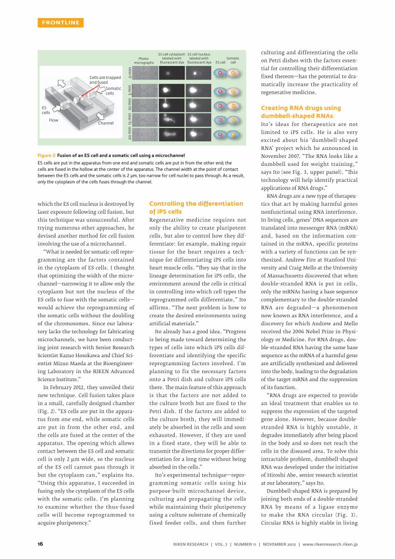
which the ES cell nucleus is destroyed by laser exposure following cell fusion, but this technique was unsuccessful. After trying numerous other approaches, he devised another method for cell fusion involving the use of a microchannel.
“What is needed for somatic cell repro-gramming are the factors contained in the cytoplasm of ES cells. I thought that optimizing the width of the micro-channel—narrowing it to allow only the cytoplasm but not the nucleus of the ES cells to fuse with the somatic cells—would achieve the reprogramming of the somatic cells without the doubling of the chromosomes. Since our labora-tory lacks the technology for fabricating microchannels, we have been conduct-ing joint research with Senior Research Scientist Kazuo Hosokawa and Chief Sci-entist Mizuo Maeda at the Bioengineer-ing Laboratory in the RIKEN Advanced Science Institute.”
In February 2012, they unveiled their new technique. Cell fusion takes place in a small, carefully designed chamber (Fig. 2). “ES cells are put in the appara-tus from one end, while somatic cells are put in from the other end, and the cells are fused at the center of the apparatus. The opening which allows contact between the ES cell and somatic cell is only 2 μm wide, so the nucleus of the ES cell cannot pass through it but the cytoplasm can,” explains Ito. “Using this apparatus, I succeeded in fusing only the cytoplasm of the ES cells with the somatic cells. I’m planning to examine whether the thus-fused cells will become reprogrammed to acquire pluripotency.”
Controlling the differentiation of iPS cellsRegenerative medicine requires not only the ability to create pluripotent cells, but also to control how they dif-ferentiate: for example, making repair tissue for the heart requires a tech-nique for differentiating iPS cells into heart muscle cells. “They say that in the lineage determination for iPS cells, the environment around the cells is critical in controlling into which cell types the reprogrammed cells differentiate,” Ito affirms. “The next problem is how to create the desired environments using artificial materials.”
Ito already has a good idea. “Progress is being made toward determining the types of cells into which iPS cells dif-ferentiate and identifying the specific reprogramming factors involved. I’m planning to fix the necessary factors onto a Petri dish and culture iPS cells there. The main feature of this approach is that the factors are not added to the culture broth but are fixed to the Petri dish. If the factors are added to the culture broth, they will immedi-ately be absorbed in the cells and soon exhausted. However, if they are used in a fixed state, they will be able to transmit the directions for proper differ-entiation for a long time without being absorbed in the cells.”
Ito’s experimental technique—repro-gramming somatic cells using his purpose-built microchannel device, culturing and propagating the cells while maintaining their pluripotency using a culture substrate of chemically fixed feeder cells, and then further
culturing and differentiating the cells on Petri dishes with the factors essen-tial for controlling their differentiation fixed thereon—has the potential to dra-matically increase the practicality of regenerative medicine.
Creating RNA drugs using dumbbell-shaped RNAsIto’s ideas for therapeutics are not limited to iPS cells. He is also very excited about his ‘dumbbell-shaped RNA’ project which he announced in November 2007. “The RNA looks like a dumbbell used for weight training,” says Ito (see Fig. 3, upper panel). “This technology will help identify practical applications of RNA drugs.”
RNA drugs are a new type of therapeu-tics that act by making harmful genes nonfunctional using RNA interference. In living cells, genes’ DNA sequences are translated into messenger RNA (mRNA) and, based on the information con-tained in the mRNA, specific proteins with a variety of functions can be syn-thesized. Andrew Fire at Stanford Uni-versity and Craig Mello at the University of Massachusetts discovered that when double-stranded RNA is put in cells, only the mRNAs having a base sequence complementary to the double-stranded RNA are degraded—a phenomenon now known as RNA interference, and a discovery for which Andrew and Mello received the 2006 Nobel Prize in Physi-ology or Medicine. For RNA drugs, dou-ble-stranded RNA having the same base sequence as the mRNA of a harmful gene are artificially synthesized and delivered into the body, leading to the degradation of the target mRNA and the suppression of its function.
“RNA drugs are expected to provide an ideal treatment that enables us to suppress the expression of the targeted gene alone. However, because double-stranded RNA is highly unstable, it degrades immediately after being placed in the body and so does not reach the cells in the diseased area. To solve this intractable problem, dumbbell-shaped RNA was developed under the initiative of Hiroshi Abe, senior research scientist at our laboratory,” says Ito.
Dumbbell-shaped RNA is prepared by joining both ends of a double-stranded RNA by means of a ligase enzyme to make the RNA circular (Fig. 3). Circular RNA is highly stable in living
Figure 2: Fusion of an ES cell and a somatic cell using a microchannel
ES cells are put in the apparatus from one end and somatic cells are put in from the other end; the cells are fused in the hollow at the center of the apparatus. The channel width at the point of contact between the ES cells and the somatic cells is 2 μm, too narrow for cell nuclei to pass through. As a result, only the cytoplasm of the cells fuses through the channel.
Cells are trapped and fused
Somatic cells
ES cells
FlowChannel
Photo-micrographs
ES cell cytoplasm labeled with
fluorescent dye
ES cell nucleus labeled with
fluorescent dye ES cellSomatic
cell
0 m
in5
min
10 m
in15
min
20 m
in

17RIKEN RESEARCH | VOL. 7 | NUMBER 11 | NOVEMBER 2012 | www.rikenresearch.riken.jp
FRONTLINE
organisms and can be delivered to the cells located in the diseased part of the body without being degraded. Upon entering the cells, an enzyme known as dicer cleaves the circular portion between the two ends, enabling the process of RNA interference. “It is a very chemistry-oriented idea to use an enzyme to increase stability. In addition to dumbbell-shaped ones, we suc-ceeded in synthesizing circular double-stranded RNA and the like, and have confirmed their respective functions. I hope that the nanostructured RNAs that have emerged using an integra-tion of chemical and biotechnological approaches will accelerate the develop-ment of RNA drugs.”
Adding molecular evolution engineering“In recent years, I have also been con-ducting research which is unrelated to medicine,” continues Ito. “In a joint
research project with the Advanced Device Laboratory and the Yu Initiative Research Unit in the Advanced Science Institute, we are working on binding peptides to carbon nanotubes to create new sensors and photodielectric materi-als. These studies are ongoing and use a new approach which we have developed by adding molecular evolution engineer-ing to bio-fabrication—a combination of chemistry and biotechnology.”
“The body of an organism consists of a wide variety of molecules. All of them are the result of the long process of evo-lution throughout the history of life, which spans more than three billion years. The essence of molecular evolu-tion engineering is to accelerate molec-ular evolution in vitro and create a wide variety of molecules that do not exist in nature. It is a way to discover molecules with novel functions and molecules that bind specifically and selectively to desired substances,” explains Ito.
There has been active research into molecular evolution engineering since the late twentieth century, but once again Ito is doing something unique. “In the conventional approach to molec-ular evolution engineering, new mol-ecules have been created by introducing selected substances of biological origin. I want to establish a new approach that will create unique molecules by intro-ducing artificial chemical substances and develop new functional materials.”
Ito’s group is currently screening a wide variety of molecules, created by introducing artificial chemical sub-stances in this new approach, for new molecules that bind specifically to carbon nanotubes, and is also develop-ing sensors and photodielectric materi-als with novel functions.
“I love the term ‘molecular evolution engineering’. It conjures up visions of something great, doesn’t it?” Ito looks happy. “I have long been troubled by the delay in yielding significant advance-ments due to operational complexity, but we are about to announce an achieve-ment. Please wait just a little longer!” ■
RNA interference by dumbbell-shaped RNADouble-stranded RNA is quickly degraded by nuclease in the body. However, circular and dumbbell-shaped RNA (RNA whose ends are joined using ligase, a joining enzyme) is protected from degradation outside of cells. Upon entering a cell, the circular portion is cleaved by the enzyme dicer, initiating RNA interference and resulting in the degradation of target mRNA.
Dumbbell-shaped RNA (circular single-stranded RNA)
Nuclease resistance
Intracellular space
Degradation by RNA interference
Base sequence complementary to target mRNA
Binding
Cleavage by the enzyme dicer
DisentanglingmRNA of target gene
Dumbbell-shaped RNA
Circular double-stranded RNA
Branched RNA with a three-way junction
Branched RNA witha four-way junction
Computer modeling of various nanostructured RNAs
Figure 3: Nanostructured RNA for RNA interference
ABOUT THE RESEARCHER
Yoshihiro Ito was born in Gifu,
Japan, in 1959. He received his
Bachelor’s (1981) and Master’s (1983)
degrees in polymer chemistry at
Kyoto University and was awarded
a Doctorate in Engineering from
the same university in 1987. Since
then he has held a number of posts
at various institutions including
research fellow of the Japan Society
for the Promotion of Science (1987),
assistant (1988) and associate
(1996) professor at Kyoto University,
research fellow at the University
of California, Irvine (1992–1993),
professor of the University of
Tokushima (1999), and Project
Leader at the Kanagawa Academy
of Science and Technology (2002).
Now he is chief scientist and director
of the Nano Medical Engineering
Laboratory at the RIKEN Institute
(from 2004). His research focuses on
biomaterial science, regenerative
medical engineering, combinatorial
bioengineering for the creation
of functional polymers, and
soft nanotechnology.

18 RIKEN RESEARCH | VOL. 7 | NUMBER 11 | NOVEMBER 2012 | www.rikenresearch.riken.jp
CONTACT INFORMATION
For details about working at RIKEN, please
contact the RIKEN Global Relations Office:
Tel: +81-(0)48-462-1225
E-mail: [email protected]
What do you do at RIKEN?
Commissioned by the RIKEN Welfare Section, I have worked at the canteen since October 1996 and currently work as manager. Approximately 350 people use our canteen daily, including the lunch delivery service. Our food service is avail-able throughout the day and our pub is open in the evenings three days a week. I contribute to RIKEN by ensuring that workers can relax during their breaks. My job is not just to provide meals, but also to maintain a high quality of food and a welcoming atmosphere.
Please tell us about how you manage
the canteen.
As manager, my most important respon-sibility is to create a relaxing atmosphere; therefore, I pay attention to details and have implemented effective methods and policies. With so many people using our canteen every day, it is crucial that they are able to access fresh food as quickly as possible. To help our customers, I work to locate accessible food stations within the canteen. When diners come into the can-teen I guide them to where they can find their food of choice and I carefully control the number of people in the hall so that there is no one waiting for seats, which could disturb the relaxing atmosphere.
How has the canteen changed in
recent years?
In June 2012, the Welfare Section refur-bished the canteen. This means that our canteen is more versatile, and we are now able to present food in more ef-fective ways. The new counter table is the best option for those who visit the canteen on their own, while our terrace offers a great chance to dine al fresco in a relaxing natural setting.
How do you support people with
different dietary requirements?
RIKEN has many workers with dif-ferent religions and different dietary preferences. We offer vegetarian meals and other options for people with spe-cific requirements. It may seem hard for non-Japanese people to understand what is available at the canteen due to the language barrier. However, once they visit us, they find our canteen has good options and start using our services regularly.
How does your service change during
the day?
The work patterns of the staff at RIKEN are quite varied and this has an impact on the profile of visitors to the canteen throughout the day. For example, there
tends to be more administrative staff using our facilities during lunch time, while more researchers, who often work until late, visit during the evening din-ner time. We strive to ensure that no matter what their schedules, all of our diners can enjoy their meals in a relaxed and sociable atmosphere.
What do you enjoy about working
at RIKEN?
I am proud that we have gained the trust of employees at RIKEN. I also enjoy getting feedback from visitors prais-ing our attention to detail, including the queue management system as I mentioned earlier.
Serving food is not only about feed-ing people, but also providing visitors a relaxing and enjoyable experience with both delicious and nutritious meals. We will continue to contribute to the development of RIKEN by maximizing our strength in terms of good food and quality service.
MASAMICHI KAWASAKI
Serving up a quality dining experience at RIKEN
Manager RIKEN CanteenTokyo Catering
RIKEN PEOPLE

19
NEWS ROUNDUP
RIKEN RESEARCH | VOL. 7 | NUMBER 11 | NOVEMBER 2012 | www.rikenresearch.riken.jp
Par ticipants from research institutes around the world gathered in Kyoto, Japan on 6 October 2012 for the First Global Sum-mit of Research Institute Leaders. The summit was held within the framework of the annual Science and Technology in Society (STS) Forum and was based on a proposal by STS Forum Chair, Koji Omi. Leaders from 16 organizations in 12 coun-tries—Australia, France, Germany, Indone-sia, Italy, Japan, Korea, Russia, Singapore, Thailand, Turkey, and the United States—joined the one-day event.
Co-chaired by RIKEN President Ryoji Noyori and Alain Fuchs, the president of the National Centre for Scientific Research (CNRS) in France, the summit began with presentations by participants who intro-duced their organizations and shared their ideas on how they could best contribute to the achievement of a sustainable society.
Following a brief congratulatory ad-dress by Koji Omi and a photo session, the participants discussed some of the common issues confronting research in-stitutes around the world. Major topics of the summit included a discussion of ‘brain drain’, where skilled labor moves to other countries, versus ‘brain circulation’,
a broader concept that looks at both the outflow and inflow of researchers from their home countries.
Other issues discussed at the summit included the proper balance between basic and applied research, how to quantify the return from investments into R&D in order to justify funding for basic research, how to integrate researchers from different cul-tures into a single team, and the need for collaboration between research centers and universities in order to nurture new generations of researchers.
In closing, the participants agreed to a joint statement that called for enhanced
international collaborations that tran-scend national and regional boundaries in order to address global concerns, such as changing population demographics, diminishing natural resources, and the spread of contagious diseases. The summit participants reaffirmed the role that basic science has played in history and called for the securing of scientific freedom, and concluded by agreeing to hold the summit on a yearly basis. The next sum-mit is scheduled to be held in Kyoto on 5 October 2013, with the agenda focusing on specific topics of interest common to research institutions.
The First Global Summit of Research Institute Leaders
K computer opens for shared useOn 28 September 2012, the K computer—RIKEN’s award-winning supercomputer based at the RIKEN Advanced Institute for Computational Science—was for the first time made available for shared use to mem-bers of academia and industry.
Jointly developed by RIKEN and Fujitsu since 2006, the K computer has collected top industry accolades in supercomput-ing performance including its No. 1 rank-ing both in June and November 2011 as the world’s fastest supercomputer in the TOP500, a renowned international ranking of the most powerful computer systems.
The K computer was also awarded top honors in the HPC Challenge and the Gor-don Bell Prize, proof of its distinguished performance in real-world applications.
Proposals for using the K computer by researchers and industry bodies were evaluated by the Research Organization for Information Science and Technology (RIST), a non-profit public-service orga-nization that promotes the development and utilization of computational science and technology to support a highly infor-mation-oriented society. On 3 September 2012, RIST announced the first selection of research proposals for 62 projects, in-cluding 29 general use projects, 8 young
researcher projects and 25 industry-related projects, as well as selected projects for HPCI strategic programs.
In the coming years RIKEN and RIST in cooperation with users of the K com-puter will work together to translate the K computer’s exceptional simulation precision and computational speed into world-class technological and research advancements. RIKEN will set about forg-ing links between computational science and computer science fields and strive to provide a user-friendly computational en-vironment for users of the K computer, while RIST will manage user support and program improvement.
RIKEN President Ryoji Noyori (1st row, 4th from left), CNRS President Alain Fuchs (1st row, 3rd from right), STS Forum Chair Koji Omi (1st row, 4th from right) and participants gathered for the First Global Summit of Research Institute Leaders in Kyoto, Japan.
Leaders from 16 research institutes came together to discuss current issues facing the international research community.

RIKEN Harima Institute
RIKEN Kobe Institute
Nagoya Facility
RIKEN Wako Institute
RIKEN Yokohama Institute
RIKEN Tsukuba Institute
Sendai Facility
www.riken.jp
RIKEN, Japan’s flagship research institute, conducts basic and applied experimental research in a
wide range of science and technology fields including physics, chemistry, medical science, biology
and engineering. Initially established as a private research foundation in Tokyo in 1917, RIKEN
became an independent administrative institution in 2003.
RIKEN RESEARCH is a website (www.rikenresearch.riken.jp) and print publication intended to
highlight the best research being published by RIKEN (www.riken.jp). It is written for a broad
scientific audience and policy makers interested in science and aims to raise global awareness of
RIKEN and its research.
For further information on the research presented in this publication or to arrange an interview
with a researcher, please contact
RIKEN Global Relations Office
2-1, Hirosawa, Wako, Saitama, 351-0198, Japan
TEL: +81 48 462 1225
FAX: +81 48 46
E-Mail: [email protected]
www.rikenresearch.riken.jp
RIKEN RESEARCH
HIGHLIGHT OF THE MONTH
Biology: Embrasing our di�erences
FEBRUARY 2011 VOLUME 6 NUMBER 2
FRONTLINETowards a radical treatment for leukemia
NEWS ROUNDUPNew cherry blossom tree blooms all seasons
LIFE AT RIKENPIERO CARNINCI
THE BUTTERFLY EFFECT
RESEARCH HIGHLIGHTSMouse models: Almost humanHigh-energy physics: Too hot to handlePhysics: Gluons don’t explain the spin surpriseSuperconductivity: back to basicsPhysics: Forced to insulatePhysics: An all-in-one chipBiology: Learning by osmosisBiology: The need for new neurons
3 3687
RIKEN 2012-020