Embed Size (px)
Citation preview

RIKEN RESEARCH MARCH 2014VOLUME 9 NUMBER 3
Synthetic nuclear physics
Accelerating toward the island of stability

Neural Circuit Genetics Research Building in Wako

Sendai
Tsukuba
Wako
Yokohama
Nagoya
Kobe
Harima
Osaka
RIKEN supports research at nine sites across Japan
Tokyo
PERSPECTIVES
4 The magic of nuclear physics
HIGHLIGHT OF THE MONTH
6 Twin x-ray pulses light up matter
RESEARCH HIGHLIGHTS
8 The benefits of a spotless mind
9 Self-healing hydrogels ease into production
10 Building blocks help silence genes
11 A critical theory in brain development
12 The magical stability of nuclei
13 The pauses that refresh the memory
14 Quarks and gluons go with the flow
15 Mapping objects in the brain
16 Keeping it clear
17 Insights into a cellular security system
FRONTLINE
18 Creating new medicines using non-natural DNA
RIKEN PEOPLE
22 Cracking the epigenetic code
NEWS ROUNDUP
23 RIKEN and Universiti Sains Malaysia nurture fruitful partnership
Katsuhiko Mikoshiba receives top French medal
RIKEN welcomes the New Year with Mochitsuki festivals
RIKEN, Japan’s flagship research institute,
conducts basic and applied experimental
research in a wide range of science and
technology fields including physics, chemistry,
medical science, biology and engineering.
Initially established as a private research
foundation in Tokyo in 1917, RIKEN became an
independent administrative institution in 2003.
RIKEN RESEARCH is a website and print
publication intended to highlight the best
research being published by RIKEN. It is written
for a broad scientific audience and policy
makers interested in science and aims to raise
global awareness of RIKEN and its research.
For further information on the research
presented in this publication or to arrange an
interview with a researcher, please contact:
RIKEN Global Relations and Research
Coordination Office
2-1, Hirosawa, Wako, Saitama, 351-0198, Japan
TEL: +81 48 462 1225
FAX: +81 48 463 3687
E-Mail: [email protected]
URL: www.riken research.riken.jp
www.riken.jp
RIKEN RESEARCH
Table of contents
23
18
9
6
TABLE OF CONTENTS
3RIKEN RESEARCH | VOL. 9 | NUMBER 3 | MARCH 2014 | www.rikenresearch.riken.jp

Except for hydrogen, all other chemical elements originated from violent nuclear processes in stars. Powerful machines known as accelerators allow physicists to study how these elements form and also to create new elements and atomic isotopes. As such, employing accelerators to study atoms not only improves our understanding of the Universe, but also produces synthetic isotopes useful in applications that include medical diagnostics.
Shortly after the Big Bang, the Universe consisted only of
hydrogen—the smallest of the chemical elements—and its
electrically uncharged partner, the neutron. The diversity
of chemical elements that have come into existence since then
resulted from nuclear processes taking place within stars. The
element helium soon followed hydrogen, forming from the fusion
of two hydrogen atoms, and then came carbon, which forms upon
the fusion of three helium atoms. Heavier elements, including
iron—the most energetically stable element—are created at the end
of the life of a star. And elements heavier than iron appear only as
a product of catastrophic stellar processes such as supernovae.
Nuclear physicists are using powerful accelerators to shed
light on the mechanisms underpinning the Universe’s violent
beginnings. Such accelerators can create new atoms with exotic,
unstable nuclei that had vital roles in the creation of heavier
elements during the explosion of stars.
The dawn of synthetic nuclear physics
Nuclear physics came of age as a research field in the 1920s, when
nuclear particles, such as the proton, were discovered. Initial
interest focused on understanding the properties of these particles
and their involvement in the fundamental nuclear processes that
were also discovered at the time: fission and fusion.
Yoshio Nishina was a pioneer of nuclear research in Japan.
Following his education at the University of Tokyo and research
stints in Europe in the 1920s, Nishina became a chief scientist
at RIKEN in 1931. His mission was to establish a nuclear
physics laboratory.
One of Nishina’s key research areas—and still the subject of
ongoing research—was the study of different nuclear isotopes.
Atomic cores consist of two types of particles: the proton, the
number of which determines the identity of a chemical element;
and the neutron, which is electrically neutral and determines an
element’s isotopes. Hydrogen, for example, has one proton and
three different isotopes; but heavier chemical elements, such as
uranium, can have many different isotopes.
The stability of an isotope strongly depends on its mix of
neutrons and protons, and this mix also influences how one
element transforms into another during a nuclear reaction. A
radioactive element, such as uranium, can follow many different
reaction pathways to become a more stable isotope. Even the radio-
active decay rates of an element can vary greatly between isotopes,
ranging from billions of years to a fraction of a second.
An ideal way to study isotope properties is by synthesizing
isotopes in the laboratory with the help of accelerators. These
machines accelerate and smash atoms together. At sufficiently
high energy, the collision of atoms creates new isotopes, or even
new elements. Using the first Japanese accelerator—a cyclotron—
Nishina discovered a new isotope of uranium, uranium-237, which
differed from the form typically found in nature, uranium-238
(containing 92 protons and 146 neutrons).
Nishina built two cyclotrons at RIKEN but both were destroyed
after the Second World War. New cyclotrons followed at RIKEN and
the latest, the ninth cyclotron, is the world’s most powerful super-
conducting ring cyclotron (SRC). The SRC can accelerate heavy
atomic nuclei to speeds of up to 70 per cent of the velocity of light
and has a beam intensity that is 100 times greater than any other
accelerator in the world.
The SRC is part of the nuclear research facility at RIKEN that
now bears Nishina’s name—the RIKEN Nishina Center for
© InvaderXan
Synthetic nuclear physics
The magic of nuclear physics
HIDETO EN’YODirectorRIKEN Nishina Center for Accelerator-Based Science
4 RIKEN RESEARCH | VOL. 9 | NUMBER 3 | MARCH 2014 | www.rikenresearch.riken.jp 5RIKEN RESEARCH | VOL. 9 | NUMBER 3 | MARCH 2014 | www.rikenresearch.riken.jp
PERSPECTIVES

Accelerator-Based Science (RNC). Inaugurated in 2006, the center
hosts some 200 full-time scientists and collaborates with many
international institutions.
Demystifying ‘magic numbers’
Experiments using the RNC’s latest generation of cyclotrons are
pushing frontiers in nuclear research, particularly in the search
for new isotopes with so-called ‘magic numbers’. These isotopes
have a set number of protons or neutrons in their nuclei and are
surprisingly stable in comparison to other isotopes. Calcium-54, for
example, consists of 20 protons and 34 neutrons. This isotope is very
unusual: typically, 34 neutrons do not constitute a ‘magic’ isotope;
and, its magic properties were theoretically proposed shortly before
their experimental discovery by researchers at RIKEN1.
Explaining how elements as heavy as uranium could have
resulted from stellar explosions hinges on understanding magic
numbers and the stability of their associated elements.
The intense beams of atomic isotopes generated by RIKEN’s cyclo-
trons are useful in the search for very heavy elements. As heavy
isotopes are accelerated and collide with each other, they re-assem-
ble into different atoms, allowing the discovery of new chemical
elements. Such research led to the discovery in 2012 of element 113
at the RNC2, after more than nine years of thorough searching.
The synthesis of new chemical elements could lead to a new
understanding of nuclear physics. Very heavy atoms are highly
unstable and have a very short life, making it difficult to prove
their existence following high-energy collisions. However, nuclear
physicists predict that even some of the heaviest atoms have magic
numbers and could survive for longer after a collision. They are
predicted to form a so-called ‘island of stability’ within the short-
lived heavy elements, which is typically the domain of elements
with approximately 120 protons (Fig. 1).
Since existing accelerators lack the power necessary to reveal
new elements within this island of stability, RIKEN plans to
replace its synchrotrons 5 and 6 with a new superconducting accel-
erator. This powerful accelerator would produce beam intensities
some 100 times greater than presently possible, securing RIKEN’s
leadership in the study of heavy isotopes.
Nuclear research for better living
Atomic accelerators also have application beyond fundamental
physics. In medicine, for example, certain radioactive isotopes are
used as markers in diagnostic experiments because their distri-
bution within the body can be precisely measured. Many of these
isotopes are a by-product of uranium fission in nuclear reactors. As
research reactors are being decommissioned, alternative methods
to produce radioactive isotopes, including accelerators, have
become increasingly important.
The use of high-energy ion beams further extends to agriculture.
Naturally occurring radioactivity is one of the causes of mutations
in plants. Radioactive rays can destroy DNA or cause small changes
to the genome. Over many generations, such changes can accu-
mulate and influence an organism’s evolution.
With ion beams, this process can be sped up by introducing
mutations at a faster rate. Careful selection of useful mutations in
each generation can produce better plants for cultivation. Develop-
ing rice that is tolerant to salty water, or even mixtures containing
up to 50 per cent seawater, is one example. Controlled mutations
could become increasingly important in ensuring a sufficient food
supply for an ever-increasing global population.
After more than 80 years of nuclear physics research at RIKEN,
much work is still required to better understand the properties of
magic isotopes, the formation of heavy elements, the interplay of
protons and neutrons in the nucleus and the internal structure of
protons and neutrons. Attaining this understanding will require
accelerators with even higher energies and intensities than are
presently available. In that quest, the RNC will continue to play an
important role. The new generation of accelerators being planned
will help us to understand how the elementary particles in an
atom interact with each other to form the world around us.
1. Steppenbeck, D., Takeuchi, S., Aoi, N., Doornenbal, P., Matsushita, M. et al.
Evidence for a new nuclear ‘magic number’ from the level structure of 54Ca.
Nature 502, 207–210 (2013).
2. Morita, K., Morimoto, K., Kaji, D., Haba, H., Ozeki, K. et al. New Result in the
production and decay of an isotope, 278113, of the 113th Element. Journal of the
Physical Society of Japan 81, 103201 (2012).
Figure 1: The island of stability
The white crosses indicate isotopes with ‘magic’ proton or neutron numbers, for example lead-208 (208Pb), which consists of two magic numbers—82 protons and 126 neutrons—making it especially stable. While very heavy atoms with proton numbers in the hundreds are highly unstable, nuclear physicists predict that even some of the heaviest atoms have magic numbers and form an ‘island of stability’. The as-yet-undiscovered ‘doubly magic’ nucleus with 114 protons and 184 neutrons is presumed to be located on this island.
Increasing stability
120
110
100
82
102
108
162Island ofstability
184
114
208Pb
152
126
100 110 120 130 140 150 160 170 180 190
90
80
70
Prot
on n
um
ber
Neutron number
4 RIKEN RESEARCH | VOL. 9 | NUMBER 3 | MARCH 2014 | www.rikenresearch.riken.jp 5RIKEN RESEARCH | VOL. 9 | NUMBER 3 | MARCH 2014 | www.rikenresearch.riken.jp
PERSPECTIVES
© Yuri Oganessian

X-ray beams are invaluable tools for scientific research. The patterns they create after diffracting through a crystal can reveal the atomic structure of the material, and blasting a sample with pulses of x-rays can help to identify the sample’s composition or capture snapshots of the atomic processes under-lying incredibly fast chemical reactions.
Toru Hara and colleagues from the RIKEN SPring-8 Center and the Japan Synchrotron Radiation Research Institute have now developed a technique that can produce pairs of very short x-ray pulses in which each pulse has a different wave-length1. The system promises to open up a new frontier in x-ray science, allowing researchers to probe the atomic world in unprecedented detail.
The x-ray pulses are produced at SACLA (SPring-8 Angstrom Compact Free Electron Laser), which forms part of the SPring-8 (Super Photon Ring-8 GeV) synchrotron radiation facility in Harima. The SACLA
x-ray free electron laser (XFEL) accelerates a stream of electrons to close to the speed of light and passes it through a series of five-meter-long undulator channels con-taining alternating magnetic fields. This path causes the electrons to ‘wiggle’ from side to side, forcing them to eject some of their energy by emitting x-rays (Fig. 1). These tightly bunched x-rays are all in phase, just like a coherent laser beam, and normally have the same wavelength, which is determined by the size of the gap between the undulator magnets. The energy of the x-ray pulses ranges from 5 to 15 kiloelectronvolts, and the pulses are typically less than 10 femtoseconds in length. The SACLA XFEL is one of only two facilities in the world where researchers can use such powerful, ultrashort x-ray pulses to study matter.
Generating a double pulseThe new system generates twin pulses of different wavelengths by introducing a
detour section between the eighth and ninth undulators. This magnetic chicane does not affect the x-rays generated in the first eight undulators, which continue on their course unimpeded. However, it forces the electrons to take a longer route, slightly delaying their arrival at the ninth and subse-quent undulators. This second leg has a different magnetic gap, causing the production of x-rays of a different wave-length. The second x-ray pulse trails the first by up to 40 femtoseconds, and the delay can be fine-tuned to a fraction of a femtosecond. The resultant paired x-ray pulse train is referred to as a two-color double-pulse (TCDP) XFEL.
The intensity of each pulse can be adjusted by changing the number of operational undulators, and the two pulses can be made to hit a sample from slightly different directions by reposi-tioning the second set of undulators. These features should make the x-rays
Electron gun Linear accelerator Undulator
ElectronElectron
Laser
Electron beam
Compressionand acceleration
Lightradiation
Wavelength
Magneticfield line
Permanentmagnet
Electronbeam
Concept of an undulator
Magneticpole direction
Experimentalhatch Cross-section of
‘in-vacuum’ undulators
Figure 1: When bunches of electrons moving close to the speed of light are forced to wiggle through undulators, they produce coherent x-ray pulses.
© 20
14 R
IKEN
Physics
Twin x-ray pulses light up matterThe SACLA free electron laser can now deliver pairs of high-intensity x-ray pulses with different wavelengths
HIGHLIGHT OF THE MONTH
6 RIKEN RESEARCH | VOL. 9 | NUMBER 3 | MARCH 2014 | www.rikenresearch.riken.jp 7RIKEN RESEARCH | VOL. 9 | NUMBER 3 | MARCH 2014 | www.rikenresearch.riken.jp

useful for answering many different types of scientific questions, says Hara.
A new light source with many usesThe TCDP system has many potential applications in research. In imaging experiments, for example, the pulses could be used to produce complementary diffraction patterns that help to refine crystal structures with greater accuracy. “Taking images of a three-dimensional structure from different angles provides more information,” explains Hara.
When low-intensity x-rays are used, samples can be rotated and images taken from several angles. “In the case of XFELs, however, the sample is destroyed by just one intense pulse in most instances. So, many identical samples are prepared and exchanged for each XFEL shot,” notes Hara. Being able to take two snapshots at once would ease the burden of sample preparation.
The TCDP system could also be used to capture extremely fast events, such as structural changes in nanoparti-cles. Each x-ray pulse would produce a snapshot of the structure, and reveal how it evolves over a femtosecond timescale. This sort of technique could uncover the physical properties of materials at the atomic scale or track the course of chemical changes—vital information for researchers designing new materials and
catalysts or trying to understand biologi-cal processes such as protein folding.
The two pulses could also serve different functions. The first could excite electrons in the target and the second could record the outcome of that process immediately after using a ‘pump–probe’ approach. “In this case, the two pulses would need to have different wave-lengths,” says Hara. “If the two pulses had the same wavelength, we would not be able to distinguish whether the observed signal was due to the pump pulse or the probe pulse.”
Greater separation makes the differenceAlthough other groups of researchers have also recently produced TCDP x-rays, the wavelengths of their two pulses only differ by a tiny percentage. “A separation of a few per cent is not enough to clearly separate the signals,” says Hara. “Also, it limits the range of target processes that can be observed, since some processes need a large gap between the two wavelengths.”
In comparison, the wavelengths of the two x-ray pulses at the SACLA facility (Fig. 2) can be separated by more than 30 per cent. The wavelengths are also much shorter than achieved by other TCDP systems, providing better spatial resolution and revealing finer atomic details. The pulses have enough energy
to rip electrons from the inner shells of atoms, triggering a cascade of other electrons that emit more x-rays in a char-acteristic spectrum that can be used to identify the atom—a technique known as energy-dispersive x-ray spectroscopy.
Researchers are now putting the system through its paces. “The two-color XFEL has already been used in experi-ments, but these are ongoing and the results have not yet been published,” says Hara. “I hope new ideas for experi-mental methods will emerge from these experiments in the future.”
1. Hara, T., Inubushi, Y., Katayama, T., Sato, T.,
Tanaka, H., Tanaka, T., Togashi, T., Togawa, K.,
Tono, K., Yabashi, M. & Ishikawa, T. Two-colour
hard x-ray free-electron laser with wide tun-
ability. Nature Communications 4, 2919 (2013).
ABOUT THE RESEARCHER
Toru Hara was born in Tokyo, Japan, in
1966. He graduated from the Faculty of
Engineering at the University of Tokyo
in 1989 and received his PhD from the
University of Paris XI in France in 1995.
He entered RIKEN as a research scientist
for the JAERI–RIKEN SPring-8 Project
Team, later joining the RIKEN SPring-8
Center. At the center, Hara contributed
to SACLA (SPring-8 Angstrom Compact
Free Electron Laser) and its precursor, the
SPring-8 Compact SASE Source (SCSS),
developing insertion devices and
accelerator optics design as well as
contributing to the operation of the free
electron lasers. In 2011, Hara became
team leader of the Beam Dynamics
Team at the RIKEN SPring-8 Center. His
current research focuses on the beam
dynamics of linear accelerators and free-
electron-laser physics.
Figure 2: SACLA (SPring-8 Angstrom Compact Free Electron Laser) can generate intense pulses of x-rays to probe the atomic structure of matter.
© 2014 RIKEN
© 20
14 R
IKEN
HIGHLIGHT OF THE MONTH
6 RIKEN RESEARCH | VOL. 9 | NUMBER 3 | MARCH 2014 | www.rikenresearch.riken.jp 7RIKEN RESEARCH | VOL. 9 | NUMBER 3 | MARCH 2014 | www.rikenresearch.riken.jp

Alzheimer’s disease is an age-related memory disorder characterized by the accumulation of clumps of the toxic amyloid-β (Aβ) protein fragment in the extracellular space around neurons in the brain. Drugs that help to ‘clean up’ cells by inducing autophagy—the degradation of unnecessary cellular components—are known to lower Aβ levels within cells and have been shown to rescue memory deficits in mice. A team of researchers including Per Nilsson and Takaomi Saido from the RIKEN Brain Science Institute have now found that autophagy also plays an important role in secreting Aβ from the cell into the extracellular space1.
The researchers set out to investigate what would happen to extracellular Aβ aggregates, called plaques, when genetic methods were used to eliminate the autophagy process. They started with transgenic mice commonly used as a model for Alzheimer’s disease. These mice have high levels of Aβ and Aβ plaque accumulation in their brains, and display learning and memory deficits.
Surprisingly, in genetically engineered variants of these mice lacking autophagy-related gene 7 (Atg7), which is required for normal autophagy, the researchers found fewer extracellular Aβ plaques in the brain; instead, the Aβ seemed to accumulate inside the neurons. Conversely, increasing the expression of the Atg7 protein in neurons grown in cell culture resulted in an increase in the release of Aβ from the cells into the tissue culture medium. The findings suggest that autophagy is required for the secretion of Aβ from neurons into the extracellular environment.
Mice with an elevated expression of Aβ but defective autophagy seemed to have degenerated brain structures (Fig. 1), as well as sicker neurons—as defined by their expression of markers of cell death—and poorer learning and memory functions than mice with high Aβ expression but normal autophagy. This result indicates that autophagy is important for maintaining normal neuronal function and cognition in
Alzheimer’s disease. Moreover, because autophagy lowers Aβ levels within the cell, the researchers deduced that intracellular Aβ may be more toxic than extracellular Aβ with respect to inducing neuronal dysfunction and memory impairment.
The findings suggest that the effec-tiveness of therapeutic strategies for Alzheimer’s disease may be improved by targeting the elimination of intra-cellular Aβ deposits rather than extra-cellular plaques. “Intraneuronal Aβ accumulation is seen in early Alzhei-mer’s disease in humans, similar to what we found upon autophagy deletion in mice,” explains Nilsson. “Targeting this pool of Aβ may therefore offer a potential treatment for Alzheimer’s disease,” he says.
1. Nilsson, P., Loganathan, K., Sekiguchi, M.,
Matsuba, Y., Hui, K., Tsubuki, S., Tanaka, M.,
Iwata, N., Saito, T. & Saido, T. C. Aβ secretion
and plaque formation depend on autophagy.
Cell Reports 5, 61–69 (2013).
Figure 1: Mice lacking autophagy and with high levels of Aβ (right) have degenerated brain structures compared with normal mice (left).
Repro
duce
d from
Ref.
1 an
d lice
nsed
unde
r CC
BY-N
C-ND
3.0 ©
2013
P. N
ilsson
et al
.
The benefits of a spotless mindA cell process that normally clears debris is also responsible for the secretion of a neurotoxic molecule that aggregates in the brain of individuals with Alzheimer’s disease
RESEARCH HIGHLIGHTS
8 RIKEN RESEARCH | VOL. 9 | NUMBER 3 | MARCH 2014 | www.rikenresearch.riken.jp 9RIKEN RESEARCH | VOL. 9 | NUMBER 3 | MARCH 2014 | www.rikenresearch.riken.jp

Hydrogels are semi-solid materials formed by polymer chains that trap water molecules in three-dimensional gels. They are used in a variety of applications, including soft contact lenses, but the fragile nature of the materials means that their utility has remained limited. Yasuhiro Ishida, Takuzo Aida and colleagues at the RIKEN Center for Emergent Matter Science are challenging this limitation by developing strong hydrogels, or ‘aqua materials’, that could outperform even conventional plastics. As part of an international collaboration, the team has now improved the production of a robust, moldable hydrogel that heals itself rapidly after being sliced open1.
The RIKEN team previously developed a hydrogel containing three primary ingredients: tiny flakes or ‘nanosheets’ of anionic c lay, a n exfo l i at i ng chemical that keeps the nanosheets from agglomerating, and a polymer binder containing positively charged guanidinium cations. Mixing small amounts of these substances into a beaker containing water causes a self-standing gel to form within seconds due to cross-linking interactions between the clay nanosheets and the polymer binder (Fig. 1). Since the hydrogel is held together by hydrogen bonding and electrostatic forces instead of permanent chemical bonds, if cut open it can be repaired by simply pressing the gel back together.
Developing practical applications for this hydrogel proved difficult, however, because the polymer binder contains
dendritic units—multiply branched, star-shaped molecular chains that can only be synthesized through time-consuming procedures. Unfortunately, binding agents made from more traditional acrylic polymers severely affect the performance of the self-healing aqua materials.
To devise a solution, the RIKEN team collaborated with Craig Hawker and colleagues from the University of California, Santa Barbara, in the United States to investigate the possibility of using advanced polymers known as ‘ABA triblock copolyethers’ that link adhesive ionic end-units (A blocks) and a flexible poly(ethylene oxide) core (B block) into a linear chain. This type of polymer mimics the essential attributes of dendritic binding agents but can also be easily synthesized.
Experiments demonstrated that the ABA triblock copolyethers cross-linked with the clay nanosheets as well as
the original dendritic polymer binder. After optimizing the chain lengths of each ABA triblock segment, their new polymer binder rapidly generated a hydrogel with comparable mechanical strength and self-mending capabilities. The hydrogel also displayed an intriguing ‘shape memory’ behavior that enabled it to retain its structure after drying and re-wetting with water and organic or ionic liquids. “With these advantageous features, the hydrogel could find applications in biomedical treatments and surgical operations, including use as anti-adhesive materials,” notes Ishida.
1. Tamesue, S., Ohtani, M., Yamada, K.,
Ishida, Y., Spruell, J. M., Lynd, N. A.,
Hawker, C. J. & Aida, T. Linear versus dendritic
molecular binders for hydrogel network
formation with clay nanosheets: Studies with
ABA triblock copolyethers carrying guani-
dinium ion pendants. Journal of the American
Chemical Society 135, 15650–15655 (2013).
Figure 1: Free-standing hydrogels formed by the interaction between clay nanosheets and polymer binding agents may find future use in biomedical applications.
Reproduced, with permission, from Ref. 1 © 2013 American Chemical Society
NH2+
NH2
NH2
H2N
+H2N
Cl–
Cl–
HNS
S
O
O O
O
OHOHm mn
( ) ( ) ( )
Hydrogel Clay nanosheet Binder
Self-healing hydrogels ease into productionMechanically tough hydrogels that repair themselves within seconds are now easier to manufacture thanks to novel polymer binding agents
8 RIKEN RESEARCH | VOL. 9 | NUMBER 3 | MARCH 2014 | www.rikenresearch.riken.jp 9RIKEN RESEARCH | VOL. 9 | NUMBER 3 | MARCH 2014 | www.rikenresearch.riken.jp
RESEARCH HIGHLIGHTS

Polycomb-group (PcG) proteins play an important role in controlling gene expres-sion. Complexes containing PcG proteins are thought to inhibit or ‘silence’ gene activity by localizing to specific targets in the genome and remodeling how DNA is wound up into chromosomes, but the exact mechanism by which these complexes repress gene activity remains poorly understood. Kyoichi Isono, Haruhiko Koseki and colleagues from the RIKEN Center for Integrative Medical Sciences have now pinpointed the part of a critical PcG protein complex that is essential for maintaining a robust, yet reversible, gene repression program during both mammalian development and cancer progression1.
Isono, Koseki and their colleagues set out to identify the formation mechanism of a cluster of PcG proteins known as Polycomb-group repressive complex-1 (PRC1). They focused their attention on a particular domain within one of the molecules in the PRC1 complex: the sterile alpha motif (SAM) of polyhomeo-tic-like protein 2 (Phc2). The SAM domain helps to keep Phc2 proteins in the same orientation, facilitating head-to-tail, building-block-like linking of repeated copies of the PRC1 complex (Fig. 1).
The team created human cells designed to express Phc2 carrying a mutation in the SAM domain. This mutation prevented PRC1 binding but did not affect the basic assembly of each complex. Nonetheless, the researchers observed a substantial reduction in PRC1 cluster or ‘body’ formation in the cell nucleus, indicating that the construc-tion of linked repeats of PRC1, driven by
SAM domain polymerization, may be necessary for the proper functioning of the complex. In embryonic mice geneti-cally engineered to possess a mutant SAM domain, Isono, Koseki and their team observed defects in the developing skeleton, further supporting the notion that the aberrant PRC1 bodies were not maintaining proper gene regulation in the absence of the SAM domain.
The findings illuminate a key aspect of mammalian embryo formation. According to Isono, however, the results could also have implications that go well beyond the understanding of basic developmental processes. “The repressive function of PRC1 has a strong impact on not only pluripotency and differentiation of stem cells but also tumorigenesis,” he
says. “The mechanism that underlies SAM polymerization and depolymeri-zation could be useful for regenerative medicine and cancer therapy.”
A particularly exciting potential therapeutic target might be compounds that destroy the interactions between SAM domains, which could halt tumor growth in cancer patients. “I believe we can develop such drugs by conduct-ing microscopic screenings for PcG body morphology,” Isono says.
1. Isono, K., Endo, T. A., Ku, M., Yamada, D.,
Suzuki, R., Sharif, J., Ishikura, T., Toyoda, T.,
Bernstein, B. E. & Koseki, H. SAM domain
polymerization links subnuclear clustering
of PRC1 to gene silencing. Developmental Cell
26, 565–577 (2013).
Figure 1: A functional SAM domain is needed to facilitate PRC1 clustering and PcG-mediated gene silencing.
© 2013 Elsevier
PRC1 clustering via Phc2-SAM polymerization
No SAM polymerizationNo PRC1 clustering
Phc2-SAM domain PRC1 Phc2-SAM mutant PRC1
Gene silenced Gene activated
Wild type Phc2-SAM mutation
Building blocks help silence genesHead-to-tail connections in gene-repressing complexes maintain proper DNA regulation during embryo and tumor formation
RESEARCH HIGHLIGHTS
10 RIKEN RESEARCH | VOL. 9 | NUMBER 3 | MARCH 2014 | www.rikenresearch.riken.jp 11RIKEN RESEARCH | VOL. 9 | NUMBER 3 | MARCH 2014 | www.rikenresearch.riken.jp

Experiments performed in the 1960s showed that rearing young animals with one eye closed dramatically altered brain development such that the parts of the visual cortex that would normally process information from the closed eye instead process information from the open eye. These effects can be induced only within a specific period of time—a ‘critical period’ during which the developing nervous system is particularly sensitive to its environment.
Subsequent work has shown that the onset of the critical period in the primary visual cortex requires the maturation of circuits containing neurons that synthesize and release an inhibitory neurotransmitter called gamma-aminobutyric acid (GABA). Now, Taro Toyoizumi and colleagues from the RIKEN Brain Science Institute have presented a theory that explains how this inhibition triggers the critical period1.
The theory is based on a computer model of the primary visual cortex con-taining neurons that receive and process information from the eyes. The model incorporates spontaneous and visually evoked neuronal activity as reported in earlier studies. The simulation also incorporates an activity-dependent form of synaptic plasticity—the process by which connections between neurons are strengthened or weakened in response to neuronal activity.
During early development, spontane-ous activity accounts for the majority of activity in the primary visual cortex. With time, however, spontaneous neuronal activity decreases whereas
activity evoked by visual experiences increases. The new theory states that the critical period is triggered by the maturation of inhibitory neuronal circuitry, which suppresses the spon-taneous activity in the primary visual cortex relative to the activity driven by incoming visual information.
The researchers turned to mice to find evidence to support the theory. Using electrodes to record primary visual cortex activity in freely moving mice, they showed as predicted by theory that the anti-anxiety drug diazepam, which enhances inhibitory activity, lowered the ratio of spontaneous to visual activity in mutant mice with we a k i n h i b i t i o n — t h o s e l a c k i n g the gene encoding glutamic acid decarboxylase-65, an enzyme for synthesizing GABA. Such mice are
known not to enter the critical period even in adulthood, but can be induced to do so by administration of diazepam.
Importantly, the theory explains distinct experience-dependent plasticity that takes place before the onset of the critical period, which has been observed in experiments but not explained by other theories. “In the future,” says Toyoizumi, “it would be useful to be able to control developmental plasticity stages by manipulating spontaneous activity in specific brain areas, which could have therapeutic applications.”
1. T o y o i z u m i , T . , M i y a m o t o , H . ,
Yazaki-Sugiyama, Y., Atapour, N., Hensch, T.
K. & Miller, K. D. A theory of the transition to
critical period plasticity: Inhibition selectively
suppresses spontaneous activity. Neuron 80,
51–63 (2013).
Figure 1: Closing one eye during the ‘critical period’ for ocular dominance plasticity can induce experience-dependent rewiring of the visual cortex.
© Dorling Kindersley RF/Thinkstock
A critical theory in brain developmentA new theory explains how ‘critical periods’ are triggered during development of the nervous system
10 RIKEN RESEARCH | VOL. 9 | NUMBER 3 | MARCH 2014 | www.rikenresearch.riken.jp 11RIKEN RESEARCH | VOL. 9 | NUMBER 3 | MARCH 2014 | www.rikenresearch.riken.jp
RESEARCH HIGHLIGHTS

Almost all the mass of an atom is con-centrated in its nucleus in the form of protons and neutrons. Some nuclei are intrinsically unstable, existing for only a short time before decaying into stable nuclei. Understanding why some nuclei are stable and others are not could help to explain the presence of matter in the Universe. Hiroshi Watanabe at the RIKEN Nishina Center for Accelerator-Based Science and an international team of scientists have now shown experimentally that nuclei with many neutrons can be more stable than might be expected, but only if the number of neutrons is just right1.
The number of protons in an atom is fixed for each element, but the number of neutrons can vary. Palladium nuclei have 46 protons, and in naturally occurring palladium nuclei on Earth, the number of neutrons can vary between 56 and 64, resulting in a series of ‘isotopes’ of palladium. In the lab, scientists can create short-lived nuclei with many more neutrons, known as ‘heavy’ isotopes.
Using the Radioactive Isotope Beam Factory (RIBF), Watanabe and his col-leagues created palladium nuclei with 80 and 82 neutrons—labeled palladium-126 (46 protons plus 80 neutrons) and palladium-128, respectively. “Only palladium isotopes with up to 74 neutrons have been studied to date,” explains Watanabe. “Our experiment reached 82 thanks to high-intensity uranium beams and a highly efficient gamma-ray detection system.”
The experiment involved accelerating a beam of uranium ions toward a beryllium target. The collision created a wide variety
of nuclei with different numbers of protons and neutrons, and the sophis-ticated ion selection system at the RIBF allowed specific heavy isotope nuclei to be isolated based on their physical and elec-tromagnetic properties to create a pure, heavy ion beam. The team then assessed the stability of these artificially generated nuclei by observing gamma rays emitted from nuclear excited states using an array of sensitive detectors known as EURICA (Euroball–RIKEN Cluster Array) (Fig. 1).
Nuclei with 2, 8, 20, 28, 50, 82 or 126 neutrons or protons are empirically known to be stable, representing the ‘magic numbers’ of nuclear particles. Yet some studies have suggested that these conventional magic numbers disappear
if the number of protons and neutrons is highly unbalanced. The results from the RIBF research, however, show that the magic number of 82 neutrons is fairly robust, making palladium-128 relatively stable. “Next, we want to see if 82-neutron nuclei sustain their magical stability when the proton and neutron ratio is even more extremely unbalanced,” says Watanabe.
1. Watanabe, H., Lorusso, G., Nishimura, S.,
Xu, Z. Y., Sumikama, T., Söderström, P.-A.,
Doornenbal, P., Browne, F., Gey, G., Jung, H.
S. et al. Isomers in 128Pd and 126Pd: Evidence for
a robust shell closure at the neutron magic
number 82 in exotic palladium isotopes.
Physical Review Letters 111, 152501 (2013).
Figure 1: Gamma rays emitted during the decay of neutron-rich palladium nuclei were detected using the EURICA array of germanium detectors.
© 2013 RIKEN Nishina Center for Accelerator-Based Science
The magical stability of nucleiExperiments on neutron-rich atomic nuclei could help scientists to understand nuclear reactions in exploding stars
RESEARCH HIGHLIGHTS
12 RIKEN RESEARCH | VOL. 9 | NUMBER 3 | MARCH 2014 | www.rikenresearch.riken.jp 13RIKEN RESEARCH | VOL. 9 | NUMBER 3 | MARCH 2014 | www.rikenresearch.riken.jp

Sufferers of schizophrenia experience a broad gamut of symptoms, including hallucinations and delusions, as well as disorientation and problems with learning and memory. This diversity of neurological deficits has made schizo-phrenia extremely difficult for scientists to understand, thwarting the develop-ment of effective treatments. A research team led by Susumu Tonegawa from the RIKEN–MIT Center for Neural Circuit Genetics has now revealed disruptions in the activity of particular clusters of neurons that might account for certain core symptoms of this disorder1.
Tonegawa’s laboratory previously found that mice lacking the protein calcineurin in certain regions of the brain exhibit many behavioral deficits that are char-acteristic of schizophrenia. In their most recent study, the researchers sought out physiological alterations at the single-cell or circuit level that could connect the absence of the calcineurin protein in the brain with these behavioral impairments.
Their study focused on the hippocam-pus, a region of the brain associated with memory and spatial learning. Within the hippocampus, specialized ‘place cells’ switch on and off as an animal explores its environment. During subse-quent periods of wakeful rest, these place cells continue to fire in patterns that essentially ‘replay’ recent wanderings, allowing the brain to build memories based on these experiences. The research-ers used precisely positioned electrodes to measure differences in brain activity in these cells for normal mice and the calcineurin-deficient mouse model of schizophrenia.
Remarkably, essentially identical place cell activity patterns were observed for both sets of mice during active explo-ration. Once the animals were at rest, however, the calcineurin-deficient mice displayed a dramatic increase in place cell activity. In the normal hippocampus, the resting replay process depended on sequential activity from place cells cor-responding to specific, real-world spatial coordinates. In contrast, this correlation was all but lost in the calcineurin-defi-cient mice (Fig. 1). Instead, these neurons often seemed to fire indiscriminately, creating high levels of ‘noise’ that over-whelmed actual location information and thwarted memory formation.
“Our study provides the first potential evidence of disorganized thinking
processes in a schizophrenia model at the single-cell and circuit level,” says Junghyup Suh, a member of Tonegawa’s research team. These findings fit with an emerging model that suggests that schizophrenic symptoms may arise from excess activation of brain regions within a ‘default mode network’—which includes the hippocampus—during wakeful rest. “Neurobiological approaches that can calm down the default mode network may therefore open up new avenues to allevi-ating symptoms or curing this mental disorder,” says Suh.
1. Suh, J., Foster, D. J., Davoudi, H., Wilson, M. A.
& Tonegawa, S. Impaired hippocampal ripple-
associated replay in a mouse model of schizo-
phrenia. Neuron 80, 484–493 (2013).
Figure 1: A map of neuronal activity during wakeful rest in calcineurin-deficient mice. In normal mice, the map reveals complex firing patterns, which are absent in this mouse model of schizophrenia.
© 2013 Susumu Tonegawa, RIKEN–MIT Center for Neural Circuit Genetics
The pauses that refresh the memoryCertain symptoms of schizophrenia may arise from uncontrolled activation of neurons that help to build memories during periods of rest
12 RIKEN RESEARCH | VOL. 9 | NUMBER 3 | MARCH 2014 | www.rikenresearch.riken.jp 13RIKEN RESEARCH | VOL. 9 | NUMBER 3 | MARCH 2014 | www.rikenresearch.riken.jp
RESEARCH HIGHLIGHTS
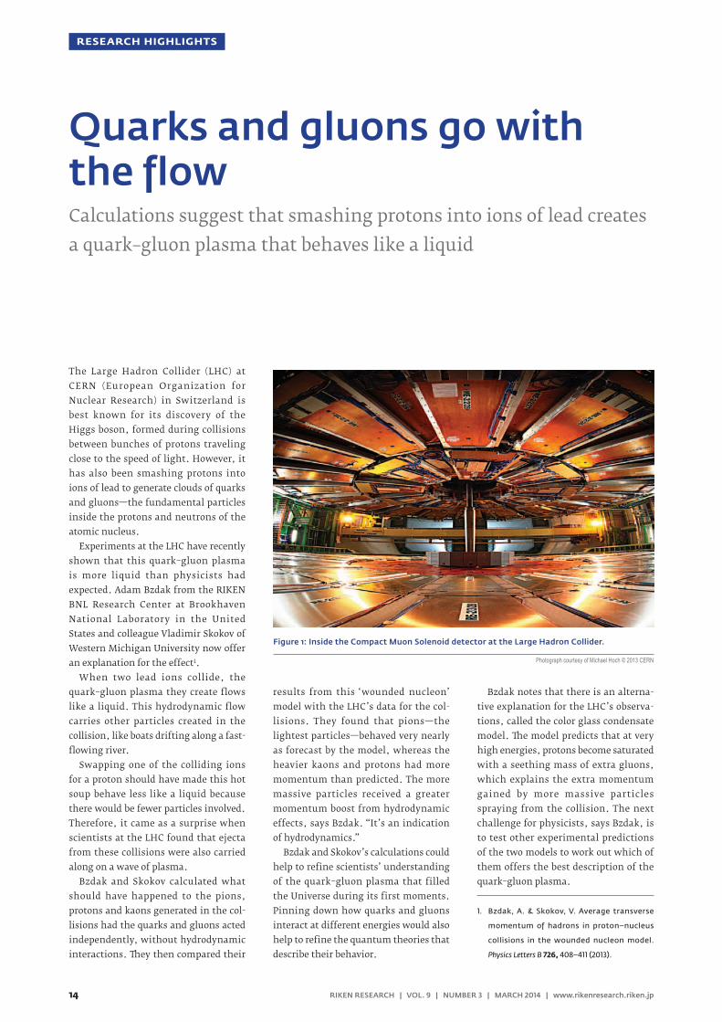
The Large Hadron Collider (LHC) at CERN (European Organization for Nuclear Research) in Switzerland is best known for its discovery of the Higgs boson, formed during collisions between bunches of protons traveling close to the speed of light. However, it has also been smashing protons into ions of lead to generate clouds of quarks and gluons—the fundamental particles inside the protons and neutrons of the atomic nucleus.
Experiments at the LHC have recently shown that this quark–gluon plasma is more liquid than physicists had expected. Adam Bzdak from the RIKEN BNL Research Center at Brookhaven National Laboratory in the United States and colleague Vladimir Skokov of Western Michigan University now offer an explanation for the effect1.
When two lead ions collide, the quark–gluon plasma they create flows like a liquid. This hydrodynamic flow carries other particles created in the collision, like boats drifting along a fast-flowing river.
Swapping one of the colliding ions for a proton should have made this hot soup behave less like a liquid because there would be fewer particles involved. Therefore, it came as a surprise when scientists at the LHC found that ejecta from these collisions were also carried along on a wave of plasma.
Bzdak and Skokov calculated what should have happened to the pions, protons and kaons generated in the col-lisions had the quarks and gluons acted independently, without hydrodynamic interactions. They then compared their
results from this ‘wounded nucleon’ model with the LHC’s data for the col-lisions. They found that pions—the lightest particles—behaved very nearly as forecast by the model, whereas the heavier kaons and protons had more momentum than predicted. The more massive particles received a greater momentum boost from hydrodynamic effects, says Bzdak. “It’s an indication of hydrodynamics.”
Bzdak and Skokov’s calculations could help to refine scientists’ understanding of the quark–gluon plasma that filled the Universe during its first moments. Pinning down how quarks and gluons interact at different energies would also help to refine the quantum theories that describe their behavior.
Bzdak notes that there is an alterna-tive explanation for the LHC’s observa-tions, called the color glass condensate model. The model predicts that at very high energies, protons become saturated with a seething mass of extra gluons, which explains the extra momentum gained by more massive particles spraying from the collision. The next challenge for physicists, says Bzdak, is to test other experimental predictions of the two models to work out which of them offers the best description of the quark–gluon plasma.
1. Bzdak, A. & Skokov, V. Average transverse
momentum of hadrons in proton–nucleus
collisions in the wounded nucleon model.
Physics Letters B 726, 408–411 (2013).
Figure 1: Inside the Compact Muon Solenoid detector at the Large Hadron Collider.
Photograph courtesy of Michael Hoch © 2013 CERN
Quarks and gluons go with the flowCalculations suggest that smashing protons into ions of lead creates a quark–gluon plasma that behaves like a liquid
RESEARCH HIGHLIGHTS
14 RIKEN RESEARCH | VOL. 9 | NUMBER 3 | MARCH 2014 | www.rikenresearch.riken.jp 15RIKEN RESEARCH | VOL. 9 | NUMBER 3 | MARCH 2014 | www.rikenresearch.riken.jp

The ability to recognize objects in the environment is mediated by the brain’s ability to integrate and process massive amounts of visual information. A research group led by Takayuki Sato and Manabu Tanifuji from the RIKEN Brain Science Institute has now discovered that in macaque monkeys, this remarkable ability is supported by mosaic-like struc-tures in the anterior inferior temporal (IT) cortex, where localized clusters of neurons encode different visual features in an organized hierarchy1.
Two competing models have been proposed to explain the functional organi-zation of brain regions that underlies object recognition in primates. One model states that discrete brain ‘modules’ process stimuli from particular categories, such as faces, with object recognition arising from communication among the modules. The other model postulates that the visual cortex extracts generic features, which are then composited to recognize specific objects. Since both models are based on measurements of functional signals produced by metabolic changes associated with neural activity rather than measurements of the neuronal activity itself, the precise underlying mechanism responsible for object recognition has remained unclear.
To resolve this debate, the research-ers undertook dense electrophysiological mapping of neural activity in anesthe-tized macaque monkeys exposed to a series of color images from different object categories: faces, hands, bodies, food and various other objects. Sato and his colleagues directly recorded neuronal activity from multiple locations within
the anterior IT cortex, which allowed them to track the location of neurons that responded to a particular object category.
The team found that some regions responded best to faces and others to monkey bodies (Fig. 1). While there were also regions that responded worst to faces, none appeared to respond preferentially to hands, food or manufactured items.
Interestingly, small neuron clusters within a region appeared to be selective to different facial features, responding differently to human and monkey faces and to scrambled and normal faces. This indicates that a region in the anterior IT cortex that is selective for an object category consists of smaller-scale neuron clusters that are selective for particular visual features.
“The cortical mosaics that encode visual information seem to be efficient func-tional structures where object-category information and information about con-stituent features are represented within the limited space of the brain,” explains Sato. “This could also be the way that the brain organizes information in other sensory modalities, such as hearing.” If the results are also found to extend to humans, they may offer insight into the visual recognition of objects and the development of language.
1. Sato, T., Uchida, G., Lescroart, M. D., Kitazono, J.,
Okada, M. & Tanifuji, M. Object representation
in inferior temporal cortex is organized hierar-
chically in a mosaic-like structure. The Journal of
Neuroscience 33, 16642–16656 (2013).
Figure 1: Neuronal activity during exposure to various images reveals distinct spatial groupings. The red region, for example, responds well to face stimuli.
© 2013 Takayuki Sato, RIKEN Brain Science Institute
mm
A
CW
D
E
X
G
Z
F
aa
ee
ll
H
O
kk
U
J K
V
S ii T l
�
hh
Q
L
cc ddbb
gg
P M N
R
jj
B
Y
Red region
Blue region
Green region
Best 5 Worst 5
1 mm
DA
VP
Mapping objects in the brainA brain region that responds to a particular category of objects is found to consist of small clusters of neurons encoding visual features of these objects
14 RIKEN RESEARCH | VOL. 9 | NUMBER 3 | MARCH 2014 | www.rikenresearch.riken.jp 15RIKEN RESEARCH | VOL. 9 | NUMBER 3 | MARCH 2014 | www.rikenresearch.riken.jp
RESEARCH HIGHLIGHTS

‘Smart glass’ can switch from transpar-ent to opaque at the flick of a switch and is increasingly used in cars, aircraft and homes to reduce the Sun’s glare and filter out infrared light and heat. Masaki Nakano and colleagues from the RIKEN Center for Emergent Matter Science have now used vanadium dioxide to make a transparent material that can be activated to block infrared light without affecting its transparency for visible light1.
Vanadium dioxide is a well-known thermochromic material that is trans-parent below about 30˚C and reflects infrared light above 60˚C. This transition is related to a change in crystal structure that also results in a shift from electri-cally insulating properties at lower tem-peratures to conductive properties at higher temperatures.
For the first time, Nakano and col-leagues have been able to trigger this change using a static voltage rather than heat. Previous attempts were unable to create a large enough electric field to completely switch the material from an insulator to a conductor.
The researchers developed a type of field-effect transistor in which a voltage at a ‘gate’ terminal controls the conduc-tivity of a vanadium dioxide channel running between ‘source’ and ‘drain’ terminals (Fig. 1). In their electric dou-ble-layer device, the gate terminal lies not on top of the channel but next to it, which ensures that it does not get in the way of light passing through the vanadium dioxide.
The channel and terminals are covered by an ionic liquid, which contains
positive and negative molecules. When a voltage is applied, positive molecules accumulate in a nanometer-thick layer on top of the channel, creating a very high electric field. This triggers a metal–insulator phase transition through-out the 50 nanometer-thick layer of vanadium dioxide, causing it to become a conductor and block infrared light.
Changing the gate voltage from 1 volt to 3 volts caused the same change in conductivity in vanadium dioxide film as an increase in temperature from 30˚C to 77˚C. While this roughly halved the transmittance of infrared light, visible light was not affected.
The device draws almost no dissipative current, which keeps its power consump-tion very low. “We think that this would be very promising for a future low-energy consumption society,” says Nakano. The researchers are now trying to improve the contrast in infrared transmittance between the device’s ‘ON’ and ‘OFF’ states. “For practical applications,” adds Nakano, “demonstrating similar performance in a large-area device is essential.”
1. Nakano, M., Shibuya, K., Ogawa, N.,
Hatano, T., Kawasaki, M., Iwasa, Y. & Tokura, Y.
Infrared-sensitive electrochromic device based
on VO2. Applied Physics Letters 103, 153503 (2013).
Figure 1: In its ‘OFF’ state (top), the vanadium dioxide (VO2) film is transparent to visible and infrared light. When the transistor is switched ‘ON’ (bottom), it draws positively charged molecules across the vanadium dioxide film, changing its electronic state and crystal structure so that the material reflects infrared light but allows visible light to pass through.
© 2013 AIP Publishing
TiO2
VO2Source Drain
Gate
OFF
++
+
+
−−
−
−
TiO2
VO2Source Drain
Gate
ON
+ ++ +
+ +
−− −
Keeping it clearVanadium dioxide ‘smart glass’ can be activated to block infrared light while remaining transparent to visible light
RESEARCH HIGHLIGHTS
16 RIKEN RESEARCH | VOL. 9 | NUMBER 3 | MARCH 2014 | www.rikenresearch.riken.jp 17RIKEN RESEARCH | VOL. 9 | NUMBER 3 | MARCH 2014 | www.rikenresearch.riken.jp

Bacteria may lack a true immune system, but this does not leave them defense-less against bacteriophage viruses and other pathogens. A system of genomic sequence elements called clustered regularly interspaced short palin-dromic repeats (CRISPR) and various CRISPR-associated proteins (Cas) help to recognize and destroy foreign genetic material delivered by such invaders.
An international research group led by Akeo Shinkai from the RIKEN SPring-8 Center and John van der Oost of Wage-ningen University in the Netherlands has now dissected one such CRISPR-Cas pathway, revealing functional insights that also highlight important differ-ences in how these systems operate across bacterial species1.
The researchers focused their attention on Thermus thermophilus, a bacterium that thrives at high tempera-tures and features a relatively simple and compact genome, making it amenable to experimental work. Of the bacterium’s multiple CRISPR-Cas pathways, the researchers explored the pathway known as subtype III-B, which targets foreign RNA rather than DNA.
Every CRISPR element contains multiple repeats of gene sequences separated by unique spacer sequences, each of which corresponds to a potential CRISPR-Cas target. Importantly, the CRISPR-Cas system also collects ‘trophies’ from novel invaders, incor-porating their sequence information into new CRISPR-spacer elements, thus enabling future recognition of the same pathogen. These CRISPR genes are transcribed to produce spacer-specific
CRISPR RNAs (crRNAs), which combine with a collection of Cas proteins known as the Cmr complex. Shinkai and van der Oost were able to isolate the Cmr complex and identified an elongated structure that they describe as resem-bling a ‘sea worm’, with a channel that could potentially accommodate the crRNA strand (Fig. 1).
The researchers were also able to isolate these associated crRNAs and determined that Cmr predominantly uses spacer sequences from just 4 of the 11 CRISPR loci in the T. thermophilus genome. They also identified a surprising mechanism for Cmr-induced cleavage, where the complex cuts at multiple sites at fixed distances along the target, as opposed to the sequence-specific or single-site cleavage mechanisms identified in other CRISPR-Cas pathways. “The T. thermophilus
Cmr complex may have evolved to kill bacteriophages as quickly as possible,” says Shinkai. “This demonstrates the diversity of the CRISPR-Cas system.”
Shinkai and his colleagues now hope to probe the three-dimensional structure of the complex in more depth and to embark on similar analyses for the other CRISPR-Cas pathways active in T. thermophilus. “This will hopefully lead to a more systematic understand-ing of these systems within the cell and of the diversity of these systems in the microbial world,” says Shinkai.
1. Staals, R. H. J., Agari, Y., Maki-Yonekura, S.,
Zhu, Y., Taylor, D. W., van Duijn, E., Barendregt,
A., Vlot, M., Koehorst, J. J., Sakamoto, K. et al.
Structure and activity of the RNA-targeting
type III-B CRISPR-Cas complex of Thermus ther-
mophilus. Molecular Cell 52, 135–145 (2013).
Figure 1: Structure of the Cmr complex, which contributes to bacterial antiviral defense. Each color represents a different protein component and the dashed line indicates a channel where the crRNA is likely to reside.
© 2013 Elsevier
~20 Å
Cmr2
Cmr5(1-3)
Cmr1/6
Cmr4(4)
Cmr4(2)
Cmr4(3)
Cmr4(1)
Cmr3
90˚ 180˚
Insights into a cellular security systemA bacterial defense mechanism against viral invasion shows a surprising degree of functional diversity
16 RIKEN RESEARCH | VOL. 9 | NUMBER 3 | MARCH 2014 | www.rikenresearch.riken.jp 17RIKEN RESEARCH | VOL. 9 | NUMBER 3 | MARCH 2014 | www.rikenresearch.riken.jp
RESEARCH HIGHLIGHTS

Expanding the function of DNAIn the science fiction film Evolution (2001), a
unicellular extraterrestrial life form trans-
ported to Earth on a meteor very quickly
evolves into a multicellular creature. The
film postulates that the presence of ten
different bases in the DNA of the extra-
terrestrial life form could facilitate such
rapid evolution.
“In real life, the American researcher
Alexander Rich hypothesized in 1962 that
the information carried by DNA and its
function can be increased by expanding
the variety of DNA bases,” says Hirao.
The DNA of all organisms on
Earth consists of four bases: adenine
(A), thymine (T), guanine (G) and
cytosine (C). Two strands of DNA are
joined by pairings between A–T and G–C
to form a double helix structure (Fig. 1).
The alignment of the bases, known as
the base sequence, is converted into RNA
during the process of transcription and
proteins are subsequently biosynthesized
during translation.
Nucleic acids, DNA and RNA, work
as ‘information molecules’ to produce
proteins, which in turn serve as the ‘func-
tional molecules’ that take responsibil-
ity for the chemical reactions required
to absorb nutrients from the external
environment to generate energy and
produce body-forming substances—the
basis of life. “Proteins are biosynthesized
based on information stored in DNA but
proteins are also required to produce
DNA. So the question arises: when life
emerged on primitive Earth, which was
produced first—DNA or protein?” notes
Hirao. “In the early 1980s, ribozyme was
discovered as a unique RNA that functions
like an enzyme—a protein that catalyzes
chemical reactions—leading to the
hypothesis that the first life form was an
RNA-based entity.”
Much research has been performed
to increase the functional variation of
RNA. Molecules that bind specifically to
disease-causing proteins, cancer cells and
other targets, such as the protein anti-
bodies that defend the body from foreign
entities, have the potential for use in
therapeutics and diagnostics. Research-
ers later discovered that DNA has similar
traits. Fragments of RNA or DNA that bind
to target molecules are called nucleic acid
aptamers (Fig. 2).
Classically, DNA molecules contain four different ‘bases’—chemical moieties that bind to each
other in a specific pairwise manner. However, Ichiro Hirao and his colleagues have succeeded in
developing a DNA molecule known as an ‘aptamer’ that can specifically bind to a target molecule,
such as a disease-related protein, using ‘non-natural’ DNA that contains a fifth synthetic base.
Aptamers are just one potential application of non-natural DNA, says Hirao, who is working to
establish a new technical basis for the life sciences using synthetic bases.
© 2014 RIKEN
ICHIRO HIRAO
Team Leader Synthetic Molecular Biology TeamRIKEN Center for Life Science Technologies
Creating new medicines using non-natural DNA
FRONTLINE
18 RIKEN RESEARCH | VOL. 9 | NUMBER 3 | MARCH 2014 | www.rikenresearch.riken.jp 19RIKEN RESEARCH | VOL. 9 | NUMBER 3 | MARCH 2014 | www.rikenresearch.riken.jp

Scientists usually require a long period
of time in order to prepare antibodies
because they must be produced in an
animal. In contrast, an aptamer can be
quickly prepared by using an in vitro evo-
lutionary engineering approach. Once
obtained, an aptamer can be mass-pro-
duced by chemical synthesis according
to its base sequence. Furthermore, while
the administration of an antibody often
stimulates the body’s immune system to
eliminate it as a foreign substance, this
rejection seldom occurs with a nucleic
acid aptamer. Therefore, nucleic acid
aptamers have attracted much attention
for their potential use as new medicines.
In order to bind, an aptamer must ‘fit’
the shape of its target just like a key fits
in a keyhole. To create an aptamer, bases
are randomly arranged to build a library of
nucleic acid molecules of various shapes.
When a target is placed in the library,
some of the nucleic acid molecules will
bind to it and can be ‘fished’ from the
pool. After amplification, the nucleic acid
molecules are exposed to increasingly
severe fishing conditions, allowing the
selection of nucleic acid molecules with
increasingly higher bindability. In evolu-
tionary engineering this process, which is
like natural selection, is repeated to select
desired aptamers.
However, the only pharmaceutical
aptamer to have thus far been developed
is pegaptanib, or ‘Macugen’, a therapeu-
tic drug developed to treat age-related
macular degeneration, a disease that
affects the retina. Macugen is a modified
RNA aptamer that binds to vascular
endothelial growth factor (VEGF) and
inhibits its function.
A non-natural DNA baseThe availability of just a single aptamer-
based drug indicates that drug discovery
using nucleic acid aptamers is a difficult
process. “An antibody-forming protein
can consist of up to 20 different amino
acids. This wide variety accounts for
the morphological and chemical diver-
sities of antibodies,” explains Hirao.
Antibodies are estimated to be present
in a vast number of shape variations,
which imparts them with the ability
to bind to all foreign substances in the
body, offering protection against these
foreign substances.
“On the other hand, because there
are only four variations of the bases that
constitute RNA or DNA, the variations in
their morphology and chemical properties
are limited, making it difficult to develop
aptamers that bind strongly to targets,”
continues Hirao. “By increasing the kinds
of bases available, it may be possible to
broaden the morphological variation
and hence to create strongly binding
aptamers. Accordingly, a project to create
DNA containing a non-natural base was
launched in Japan in 1996 and I returned
home to start it.”
To repeat selection using the evolution-
ary engineering approach, researchers
must be able to amplify DNA that contains
non-natural bases. In 2009, Hirao and his
colleagues were the first to successfully
develop the non-natural base pair ‘Ds–Px’,
which can be replicated with high accuracy
using the traditional PCR technique for
amplifying DNA in vitro (Fig. 3).
Subsequently, Hirao and his team
investigated whether the functional
variation of a non-natural DNA could be
increased by expanding the variation of
bases contained within. First, they built
a library containing between 100 trillion
and 1 quadrillion different non-natu-
ral DNA fragments, each consisting of
about 40–50 randomly arranged combi-
nations of five different bases: A, T, G, C
plus the non-natural base Ds. Next, they
performed experiments to create a DNA
aptamer that bound to VEGF and inter-
feron γ, both of which are implicated in a
wide variety of diseases.
“We performed seven rounds of target
fishing and PCR amplification to select
a non-natural DNA aptamer, which we
found to have a binding power more
than 100 times that of natural DNA
aptamers,” says Hirao. Through their
efforts, Hirao and his team were able
ATGCXGTTCAAG
TACGYCAAGTTC
------------
……
…
AUGCXGUUCAAG
…
…
…
……
Non-natural base pair
Replication
Codon
Transcription Translation
In RNA, T is replaced by U (uracil)
Methionine
Phenylalanine
Lysine
Non-natural amino acid
DNA RNA Protein
Figure 1: Expanding the DNA replication, transcription and translation system through the addition of a non-natural base pair
By adding a non-natural base pair (X–Y) to DNA and allowing it to be transcribed, non-natural bases can be incorporated into RNA to increase the variety of codons. Allocating the extra codons to non-natural amino acids and translating them into amino acids allows a protein comprising many non-natural amino acids to be created.
© 20
14 R
IKEN
18 RIKEN RESEARCH | VOL. 9 | NUMBER 3 | MARCH 2014 | www.rikenresearch.riken.jp 19RIKEN RESEARCH | VOL. 9 | NUMBER 3 | MARCH 2014 | www.rikenresearch.riken.jp
FRONTLINE

to demonstrate the hypothesis that
increasing the kinds of component bases
expands the functional variation of DNA
was correct.
Exploiting non-natural DNA in drug discoveryThe non-natural DNA aptamer created by
Hirao and his team possesses a greater
binding capacity than conventional
nucleic acid aptamers and protein anti-
bodies. With its strong ability to bind,
the aptamer could be used as a drug that
securely captures the target and is highly
effective, even at low doses.
Hirao was keen to discover why the
mere addition of the non-natural base Ds
increased the bindability of the aptamer to
such a great extent. “When we examined
the non-natural DNA aptamer, only 2 or
3 of the 40–50 aligned bases were found
to contain the non-natural base Ds,” he
says. “We conjecture that the aptamer’s
affinity for protein was increased because
without the addition of Px—to serve as the
partner base of Ds—structural diversity
was introduced due to the protruding
synthetic base, and also because Ds is
highly hydrophobic.”
Proteins are composed from 20 amino
acids, some of which are readily soluble
in water (hydrophilic) and others of which
are hardly soluble in water (hydropho-
bic). Hence, proteins contain hydropho-
bic regions. On the other hand, nucleic
acids containing only natural bases are
highly hydrophilic. Since hydrophilic
and hydrophobic substances are unlikely
to bind to each other, hydrophilic nucleic
acid aptamers composed of natural bases
are unsuitable for binding to the hydro-
phobic regions of proteins. However,
the addition of Ds, a hydrophobic base,
is thought to overcome this drawback
to increase the bindability of the non-
natural aptamer.
Of concern to scientists is the potential
for inducing adverse reactions in the body
if a therapeutic DNA aptamer binds to a
non-target protein or other substance.
Therefore, Hirao and his colleagues made
sure to select an aptamer that possessed
both strong bindability and extremely
high selectivity, and thus predominantly
binds to the target.
In addition, a nucleic acid aptamer can
only serve as a medicine if it reaches the
target without being degraded. While
DNA is chemically more stable than
RNA, DNA-degrading enzymes are also
present in the body. “Previously, I discov-
ered that the addition of a certain short
base sequence makes DNA unlikely to
be degraded by such enzymes,” explains
Hirao. “Currently, we are conducting
an experimental study to increase the
stability of a non-natural DNA aptamer by
attaching this short base sequence.”
The final stage of drug discovery
requires that the drug under develop-
ment is administered to human subjects,
allowing for the examination of the phar-
macological effects and adverse reactions.
“Fortunately, we received a proposal
from an overseas research institution
to use our non-natural DNA aptamer in
such a study.”
Hirao’s team also receives many
requests for preparing non-natural DNA
aptamers that bind to new targets.
“While it takes about half a year to make
an organism that produces an antibody
against influenza virus, in contrast, we
are able to prepare a new non-natural
DNA aptamer in around two weeks to
a month.” At the RIKEN Center for Life
Science Technologies (CLST) Division of
Bio-Function Dynamics Imaging, where
Hirao’s Synthetic Molecular Biology
Team is based, researchers in the field of
molecular imaging have been working
to create radioisotope-labelled probes
in order to assist in drug discovery and
disease diagnosis. “Radioisotopes can
be attached to our non-natural DNA
aptamer,” confirms Hirao. The radiola-
belled aptamer can then selectively bind
to its target biomolecule, allowing the
biomolecule to be monitored using a
Figure 2: A non-natural DNA aptamer that binds to a target protein
A single-stranded molecule of DNA, known as an aptamer, composed from the four natural bases plus the non-natural base Ds (yellow sphere) is able to bind to a protein target. In the absence of the non-natural base Px to serve as the pair partner of Ds, the protrusion of hydrophobic Ds increases structural and chemical variation, resulting in the formation of an aptamer that binds strongly to the target.
Target
Aptamer
Folded
Single-stranded DNA
Ds
© 20
14 R
IKEN
20 RIKEN RESEARCH | VOL. 9 | NUMBER 3 | MARCH 2014 | www.rikenresearch.riken.jp 21RIKEN RESEARCH | VOL. 9 | NUMBER 3 | MARCH 2014 | www.rikenresearch.riken.jp
FRONTLINE

technique such as positron emission
tomography (PET). “A PET probe is a
suitable first practical application of
a non-natural DNA aptamer. A joint
research project for making such a probe
is underway at RIKEN.”
Non-natural DNA aptamers are also
promising as a drug delivery system
to transport drug molecules to targets
with pinpoint accuracy. Hirao believes
that it will be possible to create a non-
natural DNA aptamer that binds selec-
tively to a particular type of cancer cell,
and could even enter the cell. “Through-
out the history of drug discovery, many
candidate molecules failed to be turned
into medicines due to a lack of target
bindability and selectivity,” he notes.
“Attaching a non-natural DNA aptamer
may allow such molecules to be developed
as medicines.”
Allowing non-natural bases to function in cellsInitially, all experiments with non-
natural bases were conducted in vitro.
However, Hirao and his colleagues are
aiming to both replicate and transcribe
non-natural bases in biological cells.
Techniques, such as fluorescently tagged
proteins, have already been developed
to allow the visualization of proteins
within cells. However, there is no such
technique for DNA and RNA. “We are
developing a technique that will allow
non-natural bases to emit light,” says
Hirao. “By achieving intracellular repli-
cation and transcription involving non-
natural bases, we will be able to visualize
the behavior of particular DNA and
RNA molecules.”
Intriguingly, researchers from the
Division of Genomic Technologies at the
CLST have obtained mounting evidence
that RNA not only serves as a mediator for
protein biosynthesis by transcribing infor-
mation stored in DNA but also controls
gene expression. “If we can realize non-
natural base transcription in living cells,
we will be able to make a particular RNA
emit light, and subsequently monitor
its behavior and explore its function,”
Hirao explains.
Meanwhile, Kensaku Sakamoto,
who heads the Nonnatural Amino Acid
Technology Team at the CLST, and his
colleagues are conducting research to
create non-natural proteins that function
better than their natural counterparts
through the addition of non-natural
amino acids. However, only one or two
non-natural amino acids can be inte-
grated into a single protein because the
number of ‘instructions’ that can be
written in the RNA is limited. At trans-
lation, one amino acid is specified for
each set of three RNA bases, known as a
codon. The number of codons available
when there are four different bases is
thus 4 × 4 × 4 = 64, to which the particu-
lar instructions for 20 different amino
acids plus the completion of translation
are allocated.
“If six different bases are available with
the addition of the Ds and Px synthetic
bases, the number of available codons will
be 6 × 6 × 6 = 216. By allocating non-natural
amino acids to the extra codons, non-nat-
ural proteins incorporating many different
non-natural amino acids may be created in
an organism,” says Hirao.
The ability to functionalize non-nat-
ural bases in living cells would create a
new technique that surpasses currently
available gene recombination technology.
“Cells incorporating a non-natural
base could not survive without being
continuously fed a food containing the
non-natural base,” notes Hirao. “These
cells would never proliferate spontane-
ously in nature and thus could be biologi-
cally contained.”
Exploring the origins of lifeHirao’s interests also extend to the
origins of life. “Before arriving at the
Ds–Px paired synthetic bases, we made a
wide variety of bases and selected those
that were suitable for use as informa-
tion-storing molecules. This process
was similar to the history of evolution,
during which life chose A, T, G and C.”
In 2014, the Japan Aerospace Explo-
ration Agency (JAXA) will launch the
asteroid explorer Hayabusa-2, which has
a mission to bring back samples contain-
ing organic matter from an asteroid. “The
primary interest is whether molecules
that could serve as a starting material for
life are present,” says Hirao. “However,
the researchers need not focus on
molecules being used by life on Earth, like
A, T, G and C and the 20 natural amino
acids. Instead, other molecules might be
involved in biological function and infor-
mation.” Indeed, Hirao has high hopes
that his non-natural base research will
lead to exploratory studies of the variabil-
ity of substances that can serve as genes
for organisms that could exist elsewhere
in the Universe.
G–C
Ds–Px
A–T
Figure 3: DNA consisting of 6 different bases, including the non-natural base pair Ds–Px
ABOUT THE RESEARCHER
Ichiro Hirao was born in Shizuoka,
Japan, in 1956. He graduated from
the Numazu National College of
Technology in 1976, and obtained
his PhD in 1983 from Tokyo Institute
of Technology. In 1984, he joined
Kin-ichiro Miura’s laboratory at
the University of Tokyo, where he
discovered unusual DNA mini-hairpin
structures. To expand his research
toward molecular biology and
evolutionary engineering, in 1995
he moved to Andrew D. Ellington’s
laboratory at Indiana University. In
1997, he returned to Japan to begin
work on the expansion of DNA’s
genetic alphabet. He now leads the
Synthetic Molecular Biology Team
at the RIKEN Center for Life Science
Technologies, which focuses on
non-natural base pair systems. He
also serves as a visiting professor
at Yokohama City University,
Tokyo Institute of Technology and
Hokkaido University.
© 20
14 R
IKEN
20 RIKEN RESEARCH | VOL. 9 | NUMBER 3 | MARCH 2014 | www.rikenresearch.riken.jp 21RIKEN RESEARCH | VOL. 9 | NUMBER 3 | MARCH 2014 | www.rikenresearch.riken.jp
FRONTLINE

What made you decide to become
a scientist?
When I was young, I suffered from asthma. As a result, I always wanted to have a job where I could help other children who were suffering from diseases but never wanted to become a doctor. Then in high school, I read a book about the Ebola virus, which capti-vated me and drew me toward becoming a researcher.
Please describe your current research
at RIKEN.
Our bodies are made up of several hundreds of different types of cells, for example hair cells and heart cells, but they all contain exactly the same DNA—the genome. How is this possible? It is achieved by a process called epi-genetics—‘epi-’ meaning ‘besides’ or ‘above’ in Latin.
Each cell type knows which sections of the genome to utilize owing to a complex epigenetic marking system on and around the DNA. I am trying to crack this code—the ‘epigenetic code’. Many diseases and cancers have an incorrectly written code; I hope to detect the incorrectly marked regions in order to develop biomarkers and therapeutic drugs.
How did you become interested
in epigenetics?
During my PhD research, I studied yeast, a very simple single-celled organism and a huge contrast to the estimated 30 trillion cells that make up the bodies of humans. Neverthe-less, even such simple organisms save energy by using an epigenetic code to read only the necessary parts of the genome. I found this very interesting and wanted to know more about how this is achieved.
What is the best thing about working
at RIKEN?
The best thing about working at RIKEN is how international the organization is. My division in particular—the Division of Genomic Technologies—is one of the most international divisions at RIKEN and I am extremely proud to be a part of it. Having lived abroad for a long time, I feel very comfortable here.
Please tell us about your professional
and personal goals.
I strive to continue to enjoy my research with as much passion as I have now, and for my work to have a meaningful impact on society and the individuals who are affected by various diseases.
Furthermore, as a female researcher in Japan, I would like to be a role model for young female scientists, especially given that the ratio of female to male scientists in Japan is a lot lower than in the West.
How do you balance family life with
your work at RIKEN?
My husband is American, and we moved together to Japan for my position at RIKEN. Consequently, he had to leave his job in the United States, which was a big decision for us to make. Fortu-nately, he is very understanding.
In general, I try to not work late during the week and to minimize working on the weekends. I would like others to know that it is possible to have a successful career as a female scientist and to also spend time with family on the weekends.
© 2014 RIKEN
AKI MINODA
Unit Leader Epigenome Technology Exploration UnitRIKEN Center for Life Science Technologies
Cracking the epigenetic code
RIKEN PEOPLE
CONTACT INFORMATION
For details about working at RIKEN, please
contact the RIKEN Global Relations and
Research Coordination Office:
Tel: +81 48 462 1225
E-mail: [email protected]
22 RIKEN RESEARCH | VOL. 9 | NUMBER 3 | MARCH 2014 | www.rikenresearch.riken.jp

RIKEN and Universiti Sains Malaysia nurture fruitful partnership
Katsuhiko Mikoshiba receives top French medalKatsuhiko Mikoshiba, head of the Labo-ratory for Developmental Neurobiol-ogy at the RIKEN Brain Science Institute, was presented with the French Legion of Honor—the highest decoration in France—in a special ceremony held at the Embassy of France in Tokyo in December 2013.
Mikoshiba received the distinction for his important contributions to the field of brain science and his service to cultural and scien-tific exchange between Japan and France. Among Mikoshiba’s many notable achieve-ments is the discovery in 1976 of the inositol 1,4,5-trisphosphate (IP3) receptor, which plays an important role in many biological functions, including body development and brain plasticity. Mikoshiba’s current research focuses on how the brain develops and the role of calcium in cellular communication.
The Legion of Honor was established in 1802 by the reigning French leader, Napoleon Bonaparte. Around 150 non-French citizens have since received the prestigious badge, 10 per cent of whom were Japanese—including two other individuals from RIKEN.
RIKEN welcomes the New Year with Mochitsuki festivalsOn 17 January 2014, the RIKEN Mutual Benefit Society, or Kyosaikai, organized rice-cake-making festivals at RIKEN institutes in Wako and Yokohama to celebrate the New Year.
Mochitsuki is a traditional event in Japan, where people gather to pound rice in a wooden mortar using a large wooden mallet. While one person pounds the rice, another person kneels to flip the rice cake, or mochi, taking care to avoid being hit by the mallet. The rice cakes are then served with sweet bean paste and other sweet or savory condiments.
The annual event at Wako and inau-gural celebration in Yokohama were very popular among RIKEN researchers and their families, who enjoyed sharing in both the preparation and tasting of the rice cakes.
On 16–20 December 2013, Todd Taylor from the RIKEN Center for Integrative Medical Sciences conducted a bioinformatics workshop at the Universiti Sains Malaysia (USM) School of Biological Sciences (SBS)—the first to result from the estab-lishment of a formal relationship between RIKEN and USM.
Participants at the five-day workshop received practical introductory training in a wide range of fields, including the use of web-based resources, next-generation gene sequencing methods, gene prediction and functional analysis, RNA sequencing and analysis and metabolic-pathway recon-struction. “We hope that this bioinformat-ics workshop can be made into an annual event,” remarked Amirul Al-Ashraf Abdullah, acting deputy dean of research and associ-ate professor at the SBS, who was encour-aged by the overwhelming response.
The workshop is the latest in a series of collaborations between RIKEN and USM. In 2011, the RIKEN–USM Joint Research Unit was created to isolate novel com-pounds from plants in southeast Asia for the development of novel drugs to treat tropical diseases. The team of Japanese and Malaysian researchers has since compiled a searchable and freely accessible online database of 200 plant species. The growing repository of chemical compounds will support the identification of biologically
active compounds and the determination of target molecules for these compounds.
Partnership between the two insti-tutes extends to other areas as well, with over ten students from USM selected to conduct their PhD research at RIKEN as International Program Associates to date. “It is really an honor for USM to have a
world-class research institute such as RIKEN as its partner,” said Sudesh Kumar, a professor at USM and the coordinator of the RIKEN–USM partnership. “USM would like to strengthen this partnership with more joint activities, such as workshops, research training, technology transfer and other educational events,” he added.
Katsuhiko Mikoshiba of the RIKEN Brain Science Institute received the French Legion of Honor—the highest decoration in France.
From left: Todd Taylor of the RIKEN Center for Integrative Medical Sciences, Sudesh Kumar, coordina-tor of the RIKEN–USM partnership and Amirul Al-Ashraf Abdullah, acting deputy dean of research of the USM School of Biological Sciences, at the bioinformatics workshop held in Malaysia.
RIKEN researchers and their families prepared rice cakes to welcome the New Year, following Japanese tradition.
© 20
13 P
ublic
Rela
tions
Offic
e, Un
iversi
ti Sain
s Mala
ysia
© 20
14 R
IKEN
© 20
14 R
IKEN
RIKEN RESEARCH | VOL. 9 | NUMBER 3 | MARCH 2014 | www.rikenresearch.riken.jp 23
NEWS ROUNDUP

www.rikenresearch.riken.jp
RIKEN 2014-015