Embed Size (px)
Citation preview
RĪGA STRADIŅŠ UNIVERSITY
Jana Žodžika
ABNORMAL VAGINAL MICROFLORA:
RISK FACTORS, BED-SIDE DIAGNOSTIC
METHODS IN PREGNANCY AND EFFICIENCY
OF AN ALTERNATIVE NON-ANTIBACTERIAL
TREATMENT MODALITY IN PREGNANT
AND NON-PREGNANT WOMEN
For obtaining the degree of a Doctor of Medicine
Speciality Obstetrics and Gynaecology
Riga, 2014

2
The Doctoral thesis was developed in the: Department of Obstetrics and
Gynaecology in Rīga Stradiņš University, Infectology Centre of Latvia,
Medical company “ARS”, Dzirciema Clinic, Gynaecological practice
“Quartus”, Riga Maternity Hospital.
Research supervisor:
MD, PhD, Professor Dace Rezeberga, Rīga Stradiņš University
Research scientific consultant:
Professor Gilbert Donders, University of Antwerp, Belgium
Approved reviewers:
MD, PhD, Professor Juta Kroiča, Rīga Stradiņš University, Latvia
MD, PhD, Professor Aija Žileviča, University of Latvia
MD, PhD, Professor Babill Stray Pedersen, University of Oslo (Norway)
Presentation of doctoral study will be held on 29 April 2014 at 15.00, at Rīga
Stradiņš University, during open meeting of Doctoral Council of Medicine in
the Lecture theatre Hippocrates, 16 Dzirciema Street, Rīga.
The Promotion work was supported by European Social Fund project “Support of
doctoral and postdoctoral investigations in Rīga Stradiņš University”
Secretary of Promotion Council:
MD, PhD, Professor Ingmārs Mikažāns

3
CONTENT
1. INTRODUCTION 6
1.1. Study topicality 6
1.2. Study aim 8
1.3. Study objectives 8
1.4. Study hypothesis statement 9
1.5. Novelty of the study 9
1.6. The structure and volume of the doctoral thesis 10
2. MATERIALS AND METHODS 11
3. RESULTS 19
3.1. Factors associated with abnormal vaginal microflora 19
3.2. Symptoms and microscopical findings related to
increased vaginal pH 21
3.3. Vaginal bacteriological study 25
3.4. Vaginal ascorbic acid (vitamin C) study 27
3.4.1. Results in pregnant women population 27
3.4.2. Results in total study population and non-pregnant
subgroup 28
3.5. Results of the vaginal clindamycin group 30
3.6. Pregnancy outcome 30
4. DISCUSSION 34
4.1. Risk factors of abnormal vaginal microflora in the first
trimester of pregnancy 34
4.2. Bed-side diagnostic methods of abnormal vaginal microflora 38
4.2.1. Value of vaginal pH test in abnormal vaginal microflora
diagnostic in the first trimester of pregnancy 40

4
4.2.2. Correlations of bed-side diagnostic tests with vaginal
bacteriological findings 41
4.3. Impact of vaginal ascorbic acid on abnormal
vaginal microflora 45
4.4. Pregnancy outcome 47
4.5. Limitations of the study 50
5. CONCLUSIONS 51
6. CLINICAL IMPLICATION AND FUTURE ASPECTS 52
7. REFERENCES 53
8. PUBLICATIONS AND CONFERENCE THESIS 64
9. ACKNOWLEDGMENTS 67

5
TEXT ABBREVIATIONS
AV aerobic vaginitis
AVF abnormal vaginal flora
BV bacterial vaginosis
CI confidence intervals
CFU colony forming unit
CN coagulase negative
E. coli Escherichia coli
HIV Human immunodeficiency virus
H2O2 hydrogen peroxide
hpf high power field
ITT intention to treat
KOH potassium hydroxide
LBG lactobacillar grade
Mixed AV-BV mixed aerobic vaginitis and bacterial vaginosis
M. hominis Mycoplasma hominis
OR odds ratio
PASW Predictive Analytics Software
PP per protocol
S. aureus Staphylococcus aureus
Str. agalactiae Streptococcus agalactiae
spp species
SPSS Statistical Package for the Social Sciences
STI sexually transmitted infections
U. urealyticum Ureaplasma urealyticum
vs. versus

6
1. INTRODUCTION
1.1. Study topicality
Normal vaginal microflora is an important women`s health factor,
maintained by high numbers of different Lactobacillus species. Decreased
amount of Lactobacillus is associated with alterations of vaginal flora
[Redondo-Lopez, et al., 1990]. Abnormal vaginal microflora is linked to such
adverse obstetric outcomes as early/late miscarriage, recurrent abortions,
premature rupture of membranes, preterm birth and low birth weight in most
cohort studies [Ralph, et al., 1999; Leitich, et al., 2007; Donders, et al., 2009].
Preterm birth continues to be one of the most challenging problems in
perinatology. In Latvia the rate of preterm deliveries increased from 4.8% in
2008 to 5.8% in 2011 [Statistical Yearbook of Health Care in Latvia, 2012] and
is similar to the rate in neighbouring countries such as Finland (5.5%) and
Estonia (5.7%) [Blencowe, et al., 2012]. Infection related morbidity in the
perinatal period was 39.2 per 1000 live births in 2011, compared to 28.8 in
2010 [Statistical Yearbook of Health Care in Latvia, 2012].
Since ascending vaginal infections are an important reason for
miscarriage, preterm delivery and neonatal infections, multiple investigators
have attempted to identify patients at risk for preterm labor, followed by
treatment in a low risk of genital infections population, but the results have not
met initial hopes [Brocklehurst, et al., 2013].
While antimicrobial agents provide cure of infections, the prevalence of
urogenital pathogen drug resistance is increasing. Furthermore they can cause
systemic and local side effects and disrupt protective vaginal microflora.
Because of the potentially adverse effects on the newborn, many pregnant
women are very cautious about taking antibiotics. During pregnancy, treatment

7
that restores normal vaginal flora and acidity without systemic effects would be
preferable to any other [Othman, et al., 2012]. Acidification of the vagina with
ascorbic acid (vitamin C) is one possible alternative option to reach this goal.
There are only few studies about the efficacy of vaginal vitamin C
[Petersen, et al., 2004; Petersen, et al., 2011]. The results of these studies
support the effective and safe use of vaginal vitamin C in a six day, mono-
therapy regimen in the management of bacterial vaginosis, but there are no
sufficient data about long term use of vaginal ascorbic acid in pregnancy and its
influence on different abnormal microflora types.
Since there is growing evidence that treatment of abnormal vaginal
microflora in early pregnancy can prevent some of the infection-related preterm
births [Ugwumadu, et al., 2004; Lamont, 2005], it can be important to
recognize (and if necessary, treat) pregnant women with flora abnormalities
early in pregnancy, preferably at the first antenatal visit using “bed-side”
diagnostic tests like vaginal pH measurement and wet mount microscopy,
which are not extensively used in Latvia. Many gynecologists start antibiotics
based solely on culture of vaginal micro-organisms, leading to overtreatment
and antibacterial drug resistance.
Proper identification of risk groups, use of rapid, reliable “bed-side”
diagnostic tests during the first antenatal visit in order to detect all types of
abnormal vaginal flora, and subsequent early treatment with non-antibacterial
drug like vaginal vitamin C could improve antenatal care and pregnancy
outcome.

8
1.2. Study aim
The aim of the present study was to investigate abnormal vaginal
microflora in pregnancy and the influence of vaginal application of ascorbic
acid (vitamin C) on abnormal vaginal microflora in pregnant and non-pregnant
women.
1.3. Study objectives
1. Primary objective ‒ to investigate the impact of vaginal ascorbic acid
(vitamin C) in a treatment plus maintenance regimen on abnormal
vaginal environment, which is characterized by increased vaginal pH
and abnormal microflora on wet mounts in pregnancy.
2. Secondary objectives:
1) to assess risk factors associated with abnormal vaginal
microflora (socio-demographic, medical, reproductive and
sexual) in the first trimester of pregnancy;
2) to identify symptoms and microscopy findings on wet mounts
associated with increased vaginal pH in pregnancy;
3) to evaluate the correlation of elevated vaginal pH and abnormal
vaginal microflora on wet mounts with vaginal bacteriologic
findings in the first trimester of pregnancy;
4) to compare pregnancy outcome between the groups of women
with normal and abnormal vaginal microflora (who were
treated and not treated with vaginal vitamin C).

9
1.4. Study hypothesis statement
1. Vaginal ascorbic acid (vitamin C) in a treatment and maintenance regimen
improves the abnormal vaginal environment in the population of pregnant
and non-pregnant women.
2. Abnormal vaginal microflora is associated with a range of socio-
demographic, medical, reproductive and sexual risk factors in the first
trimester of pregnancy.
3. Increased vaginal pH is related to a number of symptoms and abnormal
vaginal flora on wet mounts in the first trimester of pregnancy.
4. Elevated vaginal pH and abnormal vaginal microflora on wet mounts are
related to growth of aerobic, facultative anaerobic bacteria and genital
mycoplasmas on vaginal cultures in the first antenatal visit of pregnancy.
5. Application of vaginal vitamin C can decrease pregnancy complication
rate related to abnormal vaginal microflora.
1.5. Novelty of the study
1. The impact of a non-antibacterial acidifying agent ‒ vaginal ascorbic acid,
in the new treatment and maintenance regimen on different types of
abnormal vagina flora, focused on the population of pregnant and non-
pregnant women is evaluated.
2. Abnormal vaginal microflora is defined in cases with both vaginal pH ≥
4.5 and decreased numbers or absent Lactobacillus morphotypes on wet
mounts.
3. Different types, including BV, AV, intermediate and mixed flora were
assessed.
4. The study focuses on the application of simple and bed-side abnormal
vaginal flora diagnostic tests.

10
5. Correlations between vaginal pH, wet mounts, cultures and different types
of abnormal vaginal microflora are analysed.
1.6. The structure and volume of the doctoral thesis
The thesis for PhD degree is written in English. The promotion work
consists of 8 parts: introduction, review of literature, materials and methods,
results, discussion, conclusions, the list of references and the supplements. The
volume of the promotion work comprises 150 pages, including 30 tables, 8
figures. The list of references consists of 252 titles. There are 7 publications, 2
oral presentations, 9 poster presentations on the topic of the promotion work
thesis.

11
2. MATERIALS AND METHODS
This study was performed in four outpatient clinics in Riga: “ARS”
(private clinic), Dzirciema Clinic (public clinic), “Quartus” (private clinic),
Riga Maternity Hospital (municipal owned public hospital). Patients were
asked to participate, if they were at least 18 years old, 6 to 14 weeks pregnant,
agreed with the study and signed the informed consent. Pregnant woman with
vaginal pH ≥ 4.5 were enrolled in a prospective intervention trial. Following
enrolment of a woman with pH ≥ 4.5, the next two pregnant seen at the clinic
with vaginal pH < 4.5 were included in the study as controls. Enrolment of
women was planned to continue until the required number vitamin C study
population had been included.
Assuming Type I error to be 5% and with a standard difference 0.65 of
pH values between treatment and control group, and in order to achieve 80%
study power, it was calculated that 140 participants with abnormal microflora
on microscopy had to be recruited from the cohort with increased pH.
From March 2010 until May 2012 150 pregnant women with vaginal pH
≥ 4.5 and 300 with vaginal pH < 4.5 at the first prenatal visit were enrolled. Out
of 150 pregnant women cohort with elevated vaginal pH, 85 were eligible for
interventional ascorbic acid study. To reach the vaginal vitamin C study power
additional 55 non-pregnant women with the same inclusion criteria were
enrolled from September 2011 till May 2012.
Summary of inclusion criteria:
Vaginal pH/abnormal vaginal microflora and related factors/pregnancy
outcome study – 150 pregnant women with vaginal pH ≥ 4.5 and in those
with normal vaginal acidity (n=300) were included at the first prenatal
visit.

12
Vaginal culture studies –the first 50 pregnant with vaginal pH ≥4.5 were
included as study cases and 50 pregnant women with vaginal pH less than
4.5 were used as a control.
Vaginal ascorbic acid study – out of 150 pregnant women cohort with
elevated vaginal pH, 85 were eligible for interventional ascorbinic acid
study (had AVF on native microscopy, were asymptomatic, had no history
of miscarriage/preterm birth and agreed to participate in the interventional
part of the study). To reach the vitamin C study power additional 55 non-
pregnant women with the same inclusion criteria were included. Overall
vitamin C study population consisted of 70 women in the interventional
group (42 pregnant and 28 non-pregnant) and 70 women in the control
group (43 pregnant and 27 non-pregnant). Principles of randomization are
described below.
Summary of exclusion criteria: age less than 18 years, less than six
and more than 14 weeks of gestation, multiple pregnancy on the first trimester
ultrasound scan, systemic diseases, like diabetes, kidney failure, hypertension
requiring medication, all women were tested for Chlamydia, gonorrhoea,
syphilis and Human Immunodeficiency virus (HIV) infections according to the
basic antenatal care program and were excluded if positive for any of them, did
not agree to participate in the study and sign informed consent, additional
exclusion criteria for ascorbic acid study ‒ currently/during the previous 2
weeks treated with systemic/local antibiotics, antimycotics and/or Lactobacillus
preparations, symptomatic vaginal infections, history of late miscarriage and
preterm deliveries, in addition for non-pregnant women – postmenopausal
status.
Factors related to abnormal vaginal pH/microflora and vaginal
bacteriological finding studies were cross sectional, observational. The vaginal
vitamin C study was an interventional, randomized study. Pregnancy outcomes
were assessed in the prospective cohort.

13
The study was approved by the Ethics Committee of Riga Stradins
University. All participants were informed, asked to sign an informed consent
and had rights to withdraw from the study at any time for any reasons and agree
only to non-interventional part of the study. Pregnant women continued to
receive their antenatal care according to the Regulation of the Cabinet of
Ministers Nr 611 “Organization of delivery services.” The study was done
according to principles deriving from the Helsinki declaration.
There were three principle visits in the study: inclusion, follow-up and
post-delivery. There was completed a questionnaire, performed physical
examination and collected specimens for microscopic and bacteriologic
examinations at the first inclusion visit. Interviews, physical exams and
specimen collection were done by the obstetricians/gynecologists. Interviews
consisted of questions about demographic, social, medical, reproductive,
sexual, recent medication use, genital infection history and current genital tract
complaints. During gynecological examination physical findings were
documented, two cotton-tipped swabs for cultures and three vaginal smears
with cytobrush were taken from the upper vaginal wall: two for wet mount and
one for pH measurement, whiff test with 10% potassium hydroxide (KOH).
Vaginal pH was measured by pressing a Machery Nagel pH strips with a pH
range of 3.1‒7 into the fluid on the glass slide, allowing it to soak for 10
seconds. These strips were chosen because of their accuracy and ease of use
[Donders (a), et al., 2007]. Vaginal pH ≥ 4.5 was considered abnormal
(elevated) [Amsel, et al., 1983]. Then a droplet of 10% KOH was added to
evaluate “fishy” order of discharges [Amsel, et al., 1983]. Specimens for wet
mounts were spread on the glass slide, air-dried and then transported to the
investigator (Jana Zodzika) for later microscopy after rehydration of the smear
with a droplet of saline [Larsson, et al., 1990]. A Leica DM1000 microscope
(Warburg, Germany) was used, with phase contrast at 400 times magnification.
14
Results of interviews and physical exams were blinded to the person who
performed microscopic examination.
Systematic microscopic examination of wet mounts according to
Donders’ modification of Schröders’ classification [Donders, 1999] was done
according the method used at Femicare Research Centre, Belgium.
According to Donders’ modification of Schröders’ classification
[Donders, 1999]:
Lactobacillar grade (LBG) I consists of predominant presence of
Lactobacillus morphotypes, with very few coccoid bacteria presented,
LBG IIa (intermediate) of lactobacilli outnumbering other
microorganisms,
LBG IIb (intermediate) of other microorganisms outnumbering
lactobacillary morphotypes,
LBG III (completely disturbed flora) has no lactobacilli present. LBG
III is further divided in three subgroups: bacterial vaginosis (BV),
aerobic vaginitis (AV) and a mixed aerobic vaginitis and bacterial
vaginosis (mixed AV-BV) flora.
A predominant granular flora with uncountable bacteria throughout the
slide and more than 20% of epithelial cells covered with bacteria (clue cells)
were defined as full blown BV, while mixed areas with streaks of BV-like flora
or sporadic clue cells combined with other types of microflora were classified
as partial BV [Donders (b), et al, 2007, Donders, et al, 2009]. AV was
diagnosed if short bacilli or cocci, leucocytes and/or parabasal cells were found.
The severity of aerobic vaginitis was represented by a composite AV score:
slight, moderate and severe (taking into account LBG, number/appearance of
leucocytes, presence of parabasal cells and background flora) [Donders, et al.,
2002].
15
Patterns with considerably decreased or absent Lactobacillus
morphotypes (LBG IIb and LBG III) were considered as an abnormal vaginal
flora (AVF) [Donders, 1999].
From the study population, vaginal cultures were done in the first 50
pregnant women with vaginal pH ≥ 4.5 (study group) and in the first 50
participants with vaginal pH < 4.5 (control group). Specimens from the upper
vaginal wall were taken with wool cotton-tipped swabs and were immediately
placed in universal Amies medium and transported within 24 hours to the
laboratory of the Infectology Centre of Latvia. Then the samples were
inoculated to the following media: Shaedler blood agar, MacConkey agar, egg-
salt agar, chocolate and Chromagar Candida agar for the investigation of
microorganisms such as Streptococcus pyogenes (Str. pyogenes), Str.
agalactiae, Viridans group streptococci, enterococci, Staphylococcus aureus (S.
aureus), Candida species (spp.), pathogenic enteric bacteria, Acinetobacter
spp., Haemophilus spp., Pseudomonas aeruginosa and Stenotrophomonas
maltophilia. To distinguish between Str. pyogenes, Str. agalactiae, Viridans
group streptococci, and enterococci, several specific tests were done.
Streptococci were cultivated on blood agar to see their degree of hemolysis. If
there was β – hemolysis, then susceptibility to bacitracin was tested, if it was
positive then it was diagnosed as Str. pyogenes, if it was resistant, then CAMP
(Christie-Atkins, Munch-Petersen) test was performed and if that was positive
then it was diagnosed as Str. agalactiae. If on blood agar there was α –
hemolysis then a further test for optochin susceptibility was done; if it was
negative, then the culture was inoculated on Bile-esculin media [Mahon, et al.,
2000]. To distinguish between Haemophilus species, testing for X and V factor
requirements was performed using impregnated strips [Mahon, et al., 2000].
Urea-Arginine broth was used for the investigation of Ureaplasma
urealyticum (U. urealyticum) and Mycoplasma hominis (M. hominis) [Mahon,
et al., 2000]. More than 105
CFU/ml (colony forming unit) of U. urealyticum

16
and M. hominis was considered elevated concentration and analysed separately
[Rosenstein, et al., 1996].
In the ascorbic acid study asymptomatic, low risk pregnant and
asymptomatic, non-pregnant, premenopausal women with vaginal pH ≥ 4.5 and
AVF on wet mounts (LBG IIb and III) were randomized to the intervention
group (70 participants received 250 mg vitamin C tablets, Feminella Vagi C,
provided by Polichem S.A., Switzerland, for vaginal insertion at bedtime, for
six days, followed one tablet a week, for 12 weeks) and the control group (70
participants had no treatment). Randomization was done using statistical
package for the social sciences (SPSS) random number generator. Allocation
principles were concealed to patients, caregivers and to the person, who
performed wet mounts. The intervention group women had additional
randomization visit with their gynecologist, when they received the package
with study medication, instruction and diary. Intervention group participants
made records in the diaries about the use of tablets and complaints.
According to the guidelines of Latvian Association of Gynecologists and
Obstetricians [Latvian Association of Gynecologists and Obstetricians],
pregnant participants with BV and complaints/history of miscarriage, preterm
deliveries were treated with clindamycin 2% vaginal cream (Dalacin, provided
by Pfizer) applications for seven days.
Visit 2 (follow-up visit) was at 4 months after randomization (28–32
weeks of gestation), corresponding to 2 to 3 weeks after the last vitamin C
tablet insertion for study group. There was questionnaire fulfilled with
interviews about sexual, recent medication use, current genital tract complaints,
gynecological examination physical findings and vaginal smears with
cytobrush were taken from the upper vaginal wall for pH measurement, wet
mount and whiff test with 10% KOH, physical, microscopic results at the
follow-up visit.

17
Visit 3 was 6‒8 weeks after delivery. Questionnaires about pregnancy
outcomes were completed during that visit.
There were different outcomes assessed in the study:
Demographic, social, medical, reproductive, sexual, recent medication use,
genital infection history factors associated with AVF.
Current genital tract complaints, gynecological and wet mounts examination
results related to elevated vaginal pH.
Vaginal culture results were compared between abnormal vaginal
pH/microflora and selected normal vaginal acidity/microflora groups.
Eficacy endpoint, defined as a composite finding of vaginal pH < 4.5 and
normal microflora (LBG I or IIa) on wet mount, as well as mean vaginal pH
and microflora patterns were evaluated in women using vitamin C and
controls.
Pregnancy outcomes (rate of miscarriage/preterm births, term deliveries,
mean newborns` weight and Apgar score levels, newborn admission to
Intensive Care unit and transfer to Children`s hospital) compared between:
normal and total AVF groups; normal and non-intervention AVF group;
intervention AVF and non-intervention AVF groups; vitamin C and control
groups; vitamin C and all non-intervention AVF group.
Statistical analysis was performed using SPSS version 18.0 (predictive
analytics software – PASW). Distribution of socioeconomic factors was
obtained from two-way and multi-way frequency tables. The prevalence rates
for vaginal microflora and adverse reactions to treatment were also obtained by
two-way frequency tables. Statistical significance of the differences in
prevalence rates and distribution of risk factors between groups was assessed
using chi-square or Fisher's exact test. Statistical significance of the differences
in mean values between groups was tested using independent sample t test. The
level of statistical significance was chosen at 5% (p<0.05). Relations between

18
pathological vaginal microflora and various risk factors were assessed using
univariate and multivariate logistic regression. Variables that showed a
significant association at the level 10% (p<0.1) in univariate analysis were
included in the multiple logistic regression analysis. Risk pathological vaginal
microflora depending on presence of various risk factors was also calculated as
odds ratios. Vaginal pH sensitivity and specificity were calculated following
the appropriate formula [Riegelman, 2000]. Ascorbic acid study outcomes were
evaluated considering both the intention to treat population (ITT: all
randomized patients) and per protocol population (PP: patients, who
completed the study without major protocol deviations). Patients with no results
for any reasons were considered as failures.

19
3. RESULTS
3.1. Factors associated with abnormal vaginal microflora
AVF group in this study was defined, if participants had combination of
vaginal pH ≥4.5 and LBG IIb and LBG III on wet mounts.
The 135 of 150 women with increased vaginal pH had LBG IIb-III and
were compared to 256 of 300 participants with normal pH, who had LBG I-IIa
on wet mount.
Pregnant women with AVF compared to those with normal vaginal
microflora were more often younger than 25 years, less educated, more often
unmarried and single or not living with a partner, smoked before and during
pregnancy and had BV, but rarely U. urealyticum, candidas at least once during
the year before pregnancy (p<0.01), they also had a trend to be more
unemployed/housewives, had ≥ 2 sexual partners during last year and
intercourse 48 hours before sampling (p<0.1).
AVF was not related to miscarriage/preterm delivery history, number of
lifetime sexual partners, frequency of intercourses during last month, a new
partner.
Univariate analysis of significant abnormal vaginal pH factors did not
show age < 25 years, ≥ 2 sexual partners during last year and intercourse 48
hours before sampling to be significant risk factor for AVF, conversely other
factors still increased risks.
There was a trend for more smoking before and during current
pregnancy associated with lower education.
Most of participants in all age groups had a higher education, but
women with only primary education were more often younger than the age of
25 years (p=0.05).
20
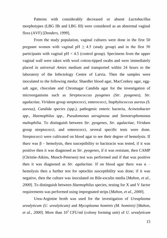
Multivariate logistic regression analysis showed that the highest risk of
AVF was associated independently with low level of education, smoking before
pregnancy and a history of BV a year before pregnancy, Table 3.1.
Table 3.1.
Multivariate analysis of significant AVF risk factors
Characteristic Odds ratio (OR) and
95% confidence interval
(CI)
P value
Age < 25 years (versus (vs.) ≥ 25 years)
Primary (≤ 9 classes) education (vs.
higher)
Secondary (12 classes) education
(vs. higher)
Not married, living with partner
(vs. married)
Single/not living with partner (vs.
married)
Housewife (vs. employed)
Unemployed (vs. employed)
≥ 2 sexual partners during last year
(vs. <2 partners)
Smoking before pregnancy (vs. not
smoking)
Smoking during pregnancy (vs. not
smoking)
BV in year before pregnancy (vs.
negative history)
U. urealyticum in year before pregnancy
(vs. negative history)
0.9 (0.5‒1.8)
3.2 (1.1‒9.4)
2.3 (1.4‒3.8)
1.4 (0.9‒2.2)
8.1 (0.8‒82.1)
1.8 (0.9‒3.7)
0.9 (0.4‒2.4)
1.6 (0.6‒4.2)
1.7 (1.0‒3.0)
1.6 (0.7‒3.8)
1.8 (0.8‒3.9)
0.2 (0.1–0.8)
0.991
0.033
0.001
0.193
0.076
0.122
0.935
0.355
0.046
0.297
0.044
0.027

21
Some interesting differences of risk factors between specific types of
abnormal vaginal flora, such as BV and AV microflora, were noted. Women
with a history of BV before pregnancy were more often less educated, smokers
and unmarried than women without previous BV, with similar, but less strong
associations for AV. Similarly, frequent intercourse and recent intercourse < 48
hours were more frequent in the BV versus the normal group, but now the
relation was even stronger in the AV group. Of note, compared to normal flora
women, Candida infection was found less often in BV cases, while its rate was
similar in the AV group, while U. urealyticum was more frequently found in
normal women and not or rarely in BV or AV women.
3.2. Symptoms and microscopical findings related to increased
vaginal pH
The 150 women with increased vaginal pH were compared to 300
participants with normal pH. Most complaints were similar between the two pH
groups, but 37% of women with increased pH complained of abundant vaginal
discharge, compared with 26% in controls (p=0.023) and 11% of participants
with elevated vaginal pH had experienced a bad smell (only 3% in normal
vaginal pH group, p=0.001). Upon examination, women with normal vaginal
pH more often had normal (74% vs. 24%, p<0.001), less often a thin,
homogeneous (5% vs. 48%, p<0.001) and yellow discharge (4% vs. 9%,
p=0.044). Positive whiff test was associated with elevated pH (p<0.001), but of
all women in the abnormal pH group, only 55% had positive amine test.
There was strong correlation between elevated vaginal pH and AVF on
wet mounts. AVF (LBG IIb-III) patterns on wet mounts were more often found
in women with elevated vaginal pH (135/150, 90%) than in participants with
vaginal pH < 4.5 (44/300, 15%), p<0.001.

22
The participants with vaginal pH ≥ 4.5 were more likely to have BV (36
women, 23%), AV (22 women, 14%), mixed AV-BV flora (52 women, 34%)
and LBG IIb (25 women, 16%). Of 300 participants with pH < 4.5, 44 had
abnormal flora patterns (29 had LBG IIb, 8 AV, 6 MF and 1had BV), Table
3.2.
Table 3.2.
Comparison of clinical and microscopic examinations results
between pH groups
Characteristic Total
(n=450)
N (%)
pH < 4.5
(n=300)
N (%)
pH≥4.5
(n=150)
N (%)
P value
Complaints:
increased discharge
burning
itching
bad smell
bloody discharge
low abdominal pain
others
134 (30)
15 (3)
31 (7)
26 (6)
6 (1)
54 (12)
4 (0.9)
78 (26)
12 (4)
22 (7)
9 (3)
4 (1)
36 (12)
3 (1)
56 (37)
3 (2)
9 (6)
17 (11)
2 (1)
18 (12)
1 (0.7)
0.023
0.284
0.546
0.001
1.000
0.913
1.000
Type of discharge:
normal
thin, homogeneous
“cheese” like
bloody
yellow
257 (57)
87 (19)
54 (12)
6 (1)
25 (5)
221 (74)
15 (5)
38 (13)
4 (1)
12 (4)
36 (24)
72 (48)
16 (11)
2 (1)
13 (9)
< 0.001
< 0.001
0.515
1.000
0.044
Positive whiff test 87 (19) 4 (1) 83 (55) < 0.001
Clue cells 90 (20) 6 (2) 84 (56) < 0.001

23
Continuation of the Table 3.2.
Characteristic Total
(n=450)
N (%)
pH < 4.5
(n=300)
N (%)
pH≥4.5
(n=150)
N (%)
P value
Lactobacillary grades:
I
IIa
IIb
III BV
III AV
III MF
177 (40)
94 (21)
54 (11)
37 (8)
30 (7)
58 (13)
169 (57)
87 (30)
29 (8)
1 (0.3)
8 (3)
6 (2)
8 (7)
7 (5)
25 (16)
36 (23)
22 (14)
52 (34)
< 0.001
Normal microflora patterns
(LBG I, Ia)
Abnormal microflora
patterns
(LBG IIb, LBG III)
271 (60)
179 (40)
256 (85)
44 (15)
15 (10)
135 (90)
< 0.001
BV type:
Partial
Full
57 (13)
37 (8)
4 (1)
1 (0.3)
53 (35)
36 (23)
< 0.001
AV score:
No/slight AV
Moderate
Severe
418 (92)
25 (6)
7 (2)
293 (98)
4 (1)
3 (1)
125 (83)
21 (14)
4 (3)
< 0.001
Lactobacillary
morphology:
Normal types
Leptosomic
Short, course
Absent
263 (58)
69 (15)
126 (28)
123 (27)
237 (79)
60 (20)
100 (34)
16 (5)
26 (17)
9 (6)
26 (17)
107 (70)
< 0.001
< 0.001
< 0.001
< 0.001
Sperm cells 34 (8) 15 (5) 19 (12) 0.005

24
Continuation of the Table 3.2.
Number of leucocytes:
<10 per high power field
(hpf)
>10 per hpf, <10 per
epithelial cell
>10 per epithelial cell
177 (39)
201 (45)
72 (16)
108 (36)
152 (51)
40 (13)
69 (46)
49 (33)
32 (21)
0.001
Candida:
none
spores
hyphae
both
379 (84)
33 (7)
8 (2)
30 (7)
254 (85)
23 (8)
5 (1)
18 (6)
125 (83)
10 (7)
3 (2)
12 (8)
0.866
Most cases of full BV were associated with increased pH. pH test
sensitivity for full BV was 97%, but less for severe aerobic vaginitis ‒ 60%.
Also for partial BV, pH test sensitivity (92%) was better than for moderate
aerobic vaginitis (70%). Specificity of pH for all BV cases was 83%, and for
total AV cases was 70%.
All lactobacillary morphotypes were found more often in the pH ≤ 4.5
group (p<0.001).
Sperm cells were more often detected in the vaginal smears of women
with abnormal than in those with normal pH (12% versus 5%, p=0.005), but
most of them (68% of participants with sperm cells) had abnormal vaginal
microflora on microscopy (p< 0.001). From the smears with sperm, two women
claimed no intercourse during the previous two days.
Elevated vaginal pH was associated with increased numbers of
leucocytes (> 10 per epithelial cell): 21% versus 13%, p=0.001.
25
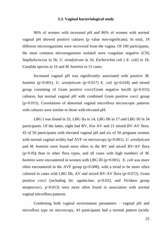
3.3. Vaginal bacteriological study
96% of women with increased pH and 86% of women with normal
vaginal pH showed positive cultures (p value non-significant). In total, 19
different microorganisms were recovered from the vagina. Of 100 participants,
the most common microorganisms isolated were coagulase negative (CN)
Staphylococcus in 56, U. urealyticum in 34, Escherichia coli ( E. coli) in 18,
Candida species in 16 and M. hominis in 15 cases.
Increased vaginal pH was significantly associated with positive M.
hominis (p<0.001), U. urealyticum (p=0.017) E. coli (p=0.018) and mixed
group consisting of Gram positive cocci/Gram negative bacilli (p=0.015)
cultures, but normal vaginal pH with combined Gram positive cocci group
(p=0.015). Correlations of abnormal vaginal microflora microscopic patterns
with cultures were similar to those with elevated pH.
LBG I was found in 35, LBG IIa in 14, LBG IIb in 17 and LBG III in 34
participants. Of the latter, eight had BV, five AV and 21 mixed BV-AV flora.
43 of 50 participants with elevated vaginal pH and six of 50 pregnant women
with normal vaginal acidity had AVF on microscopy (p<0.001). U. urealyticum
and M. hominis were found more often in the BV and mixed BV-AV flora
(p<0.05) than in other flora types, and all cases with high numbers of M.
hominis were encountered in women with LBG III (p=0.001). E. coli was more
often encountered in the AVF group (p=0.008), with a trend to be more often
cultured in cases with LBG IIb, AV and mixed BV-AV flora (p=0.072). Gram
positive cocci (including Str. agalactiae, p=0.032, and Viridans group
streptococci, p=0.013) were more often found in association with normal
vaginal microflora patterns.
Combining both vaginal environment parameters – vaginal pH and
microflora type on microscopy, 43 participants had a normal pattern (acidic
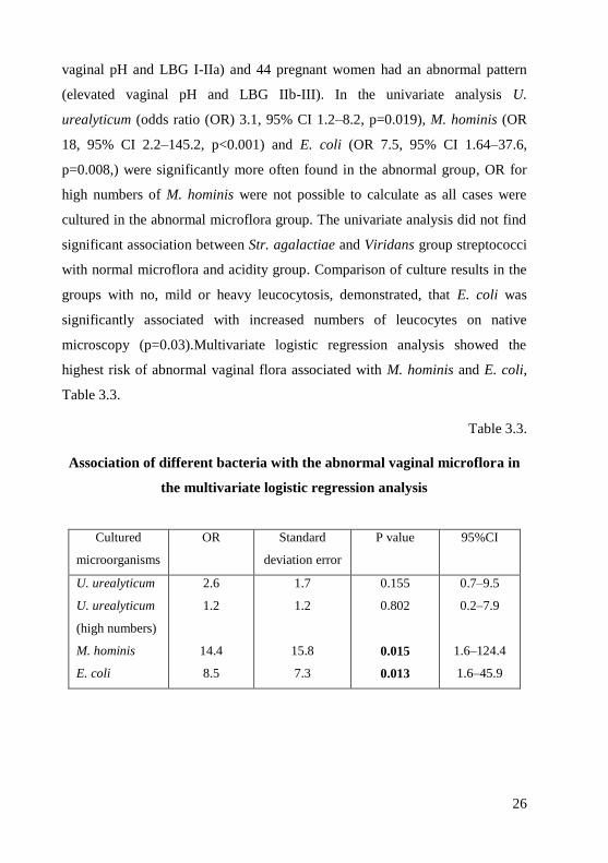
26
vaginal pH and LBG I-IIa) and 44 pregnant women had an abnormal pattern
(elevated vaginal pH and LBG IIb-III). In the univariate analysis U.
urealyticum (odds ratio (OR) 3.1, 95% CI 1.2‒8.2, p=0.019), M. hominis (OR
18, 95% CI 2.2‒145.2, p<0.001) and E. coli (OR 7.5, 95% CI 1.64‒37.6,
p=0.008,) were significantly more often found in the abnormal group, OR for
high numbers of M. hominis were not possible to calculate as all cases were
cultured in the abnormal microflora group. The univariate analysis did not find
significant association between Str. agalactiae and Viridans group streptococci
with normal microflora and acidity group. Comparison of culture results in the
groups with no, mild or heavy leucocytosis, demonstrated, that E. coli was
significantly associated with increased numbers of leucocytes on native
microscopy (p=0.03).Multivariate logistic regression analysis showed the
highest risk of abnormal vaginal flora associated with M. hominis and E. coli,
Table 3.3.
Table 3.3.
Association of different bacteria with the abnormal vaginal microflora in
the multivariate logistic regression analysis
Cultured
microorganisms
OR Standard
deviation error
P value 95%CI
U. urealyticum
U. urealyticum
(high numbers)
M. hominis
E. coli
2.6
1.2
14.4
8.5
1.7
1.2
15.8
7.3
0.155
0.802
0.015
0.013
0.7‒9.5
0.2‒7.9
1.6‒124.4
1.6‒45.9

27
3.4. Vaginal ascorbic acid (vitamin C) study
Eficacy endpoints were analysed in the pregnant, non-pregnant and total
study population.
3.4.1. Results in pregnant women population
There were 42 pregnant women in the vitamin C and 43 in the control
group. Correspondingly 36 and 37 of in each study arm completed the full
protocol and could be analysed as per protocol. Overview of study population is
described below in the section 4.2. The baseline characteristics (age, weight,
height, education, marital status, sexual and general history, mean vaginal pH,
results of native microscopy) of all cases of both groups were comparable.
In the ITT population 29 of 42 (70.7%) of the ascorbic acid and 12 of 43
(29.3%) participants in the control group demonstrated normalization of the
abnormal vaginal flora (difference 41.4%, 95% CI 21.8‒60.5, p<0.001). In the
PP population the flora normalized in 25 of 35 (71.4%) of study patients versus
10 of 37 (28.6%) of controls (difference 44.4%, 95% CI 23.7‒65.1, p<0.001).
Mean vaginal pH on follow-up visit decreased in both treatment and
control groups: from 5.05±0.37 to 4.3±0.4 in the intervention group (p<0.001)
and from 5.02±0.30 to 4.6±0.5 in the control group (p<0.001), but the decrease
was significantly more marked in the ascorbic acid group, than in the controls
(p<0.0003). On follow-up visit prevalence of normal vaginal pH and microflora
on wet mount was higher in ascorbic acid group (p<0,001).
Vaginal ascorbic acid did not reduce the prevalence of pure LBG III BV
flora (6/9 normalization in the vitamin C group vs. 6/11 in the control group
(p=0.622), nor on pure LBG III AV flora (6/10 in vitamin C group vs. 1/4 in
controls, p=0.25). However, in women with mixed LBG III AV-BV flora,
normalization of microflora was more evident in the vitamin C group (11/16)

28
than in the control group (4/18), p=0.005. Analysing the impact of vitamin C on
any kind of AV (combined group of LBG IIb associated with AV, pure LBG III
AV and mixed AV-BV) or any kind of BV flora (including LBG IIb associated
with BV, LBG III BV and mixed AV-BV), the result was significantly better in
the intervention group, Table 3.4.
Table 3.4.
Prevalence of normal vaginal flora in different abnormal vaginal flora
groups on follow-up visit
Microflora types on
screening visit
(number of cases)
Prevalence of LBG I-IIa on follow-up visit
Total
(n)
Vitamin C
group
(n = 42)
Control group
(n = 43)
P values
PureBV (20) 12 6 6 0.622
PureAV (14) 7 6 1 0.315
Any AV (59) 28 21 7 0.001
Mixed AV-BV (34) 15 11 4 0.005
3.4.2. Results in total study population and non-pregnant subgroup
There were 28 non-pregnant women in the vitamin C and 27 in the
control group. 58 of 70 participants in each study arm completed the full
protocol and could be analysed as per protocol. In the intervention group 7
(10%) women prematurely withdrew from the study because of side effects
(irritations), 1 had a spontaneous abortion, 3 were lost to follow up and 1 had
deviation from protocol because of systemic antibiotic use (due to urinary
infection) during the study period. In the control group 2 had spontaneous
abortions, 1 went into preterm labor at 29 gestational week, 4 were lost to

29
follow up and 5 had deviations from study protocol (4 used systemic
antibacterial medications due to urinary and respiratory infections and had
vaginal antibacterial treatment because of symptoms and 1 used per oral
probiotics).
In the ITT population, 36 of 70 (51.4%) of the ascorbic acid and 17 of
70 (24.3%) participants of the control group demonstrated normalization of the
abnormal vaginal flora (difference 27.1%, 95% CI 11.7‒42.6, p<0.05). In the
PP population the flora normalized in 31 of 58 (53.5%) of study patients versus
13 of 58 (22.4%) of controls (difference 31%, 95% CI 14.3‒47.8, p<0.05).
Results of ITT population non-pregnant subgroup analysis did not show
improvement of the microflora (7 with normal flora of 28 in vitamin C group
versus 5 of 27 participants in control group, p value nonsignificant).
On follow-up visit prevalence of normal vaginal pH and microflora on
wet mount was higher in ascorbic acid group (p<0.001), but not in the subgroup
of non-pregnant participants (p value nonsignificant).
Like in the pregnant women subgroup total study population results
showed that vaginal ascorbic acid did not reduce the prevalence of pure BV
flora (8/36 normalization in the vitamin C group versus 7/36 in the control
group (p=0.5), pure AV flora (9/26 in vitamin C group vs. 3/26 in controls,
p=0.25), but in women with mixed AV-BV flora and any kind of AV or BV
flora the normalization was more evident in the vitamin C group (p values
respectively 0.017, 0.001 and 0.031).
Most common side effects in the vitamin C group were itching and
irritation. Itching cause complaints in 11 cases (16%) after treatment and in 13
cases (19%) during the maintenance regimen. Irritation occurred in 16 patients
(23%) after the treatment regimen and 7 patients prematurely withdrew from
the study due to this reason. Irritation persisted in 6 cases (9%) during the
maintenance phase of vitamin C use. There were no significant associations

30
found between complains and vaginal pH, regimen of treatment, microflora
type, number of leucocytes or presence of Candida on wet mounts.
3.5. Results of the vaginal clindamycin group
17 pregnant women received treatment with vaginal clindamycin cream.
Mean vaginal pH on follow-up visit decreased from 5.02±0.05 to 4.21±0.14
and LBG I-IIa was restored in 80% (p<0.001). These results were comparable
to vitamin C data (p non-significant).
3.6. Pregnancy outcome
Pregnancy outcome data were available for 102 of 135 women with
pH≥4.5/LBG IIb-III and for 217 of 256 with normal and vaginal acidity and
microflora. The final data were missing, if participants did not arrive on visit 6‒
8 weeks after delivery. Drop outs baseline characteristics were similar to those
who completed the study.
There was no difference between both study groups regarding pregnancy
complications, use of antibiotics (systemic or vaginal) during pregnancy, type
or mode of deliveries. Most pregnant participants (90%) had term deliveries.
Analysis of pregnancy outcomes between normal and AVF (including
cases with as well as without any intervention), did not show major differences
between normal and AVF groups. However, both first and fifth minute Apgar
score levels were higher in newborns whose mothers` had both normal vaginal
microflora (p=0.027 and p=0.023).
Analysis of pregnancy outcome between normal and non-intervention
AVF group participants (cases without antibacterial or vitamin C treatment)
showed higher rates of miscarriage, preterm birth and Apgar score in the non-
treated abnormal microflora group, Table 3.5.

31
Table 3.5.
Pregnancy outcome analysis in the non-intervention population
Characteristic Total
(n=249)
N (%)
Vaginal pH
<4.5 and
LBG I-IIa
(n=191)
N (%)
Vaginal pH
≥4.5 and
LBG IIb-III
(n=58)
N (%)
P value
Pregnancy outcomes:
early miscarriage
late miscarriage
preterm delivery at
22‒26 weeks of
gestation
preterm delivery at
27‒36 weeks of
gestation term delivery
18 (7)
4 (2)
0
5 (2)
222 (89)
10 (5)
4 (2)
0
2 (1)
175 (92)
8 (14)
0
0
3 (5)
47 (81)
0.019
Newborn weight 3647±479 3515±553 0.099
Apgar score
1. minute
5. minute
7.7±0.8
8.8±0.8
7.2±1.4
8.3±1.7
0.001
0.003
Newborns of pregnant women with AVF, who had received antibacterial
treatment (Dalacin, other antibiotics) or vitamin C, had better fifth minute
Apgar score (p=0,003), compared to non-intervention participants, but
pregnancy outcomes were not significantly different.
Although mean newborn weight was not significantly different between
normal and all types abnormal vaginal microflora groups (p=0.1), the sub-
group analysis showed lower birth weights in the any AV (p=0.045) and mixed
BV-AV groups (p=0.02), Table 3.6.

32
Table 3.6.
Mean newborn weights in the different vaginal microflora groups
Mean newborn weight in
the normal vaginal
microflora group
Mean newborn weights in the
different abnormal vaginal microflora
groups
P value
LBG I-IIa (n=212):
3576±673 g
BV only (n=30): 3571±490 g 0.917
AV only (n=25): 3554±473 g 0.477
Any BV (n=76): 3513±450 g 0.091
Any AV (n=75): 3497±495 g 0.045
Mixed BV-AV (n=44): 3430±477 g 0.018
We also analysed the influence of different suspected risk factors
(smoking during pregnancy, AVF in the first trimester, hypertension, anemia,
fetal abnormalities, abnormal ultrasound scan findings) on newborns` weight in
the study. As expected ‒ current smoking had the most negative effect on birth
weight in the multivariate analysis (p=0.004).
Bacteriological findings of M. hominis, U. urealyticum or different types
of aerobic bacteria at the first antenatal visit were not associated with poor
pregnancy outcome when compared to culture negative pregnant women.
We didn’t see statistical difference in either pregnancy outcomes, or
newborn mean birth weight and the first minute Apgar score levels in the
vitamin C and control group in ITT population. Still, mean fifth minute Apgar
score was significantly better in the intervention group (p=0.031). Also, even
though numbers were too small for obtaining meaningful statistical differences,
there were 3 preterm deliveries in the control group (8%) and none in the
vitamin C group.
There was statistical difference found in pregnancy outcomes comparing
vitamin C with non-intervention AVF group participants (combined group of

33
controls and those participants who rejected any treatment): in non – treated
population more early miscarriage and preterm deliveries at 27-36 weeks of
gestation were observed (p=0.037), Table 3.7.
Table 3.7.
Comparison of pregnancy outcomes in vitamin C
and all non-intervention AVF group
Characteristic Vitamin C group
(n=41)
N(%)
Non-intervention
population (n=58)
N (%)
P
values
Pregnancy outcomes:
early miscarriage
late miscarriage
preterm delivery at 22‒26
weeks of gestation
preterm delivery at 27‒36
weeks of gestation
term delivery
1 (3)
0
0
0
40 (98)
8 (14)
0
0
3 (5)
47 (81)
0.037
Mean newborn weight 3528±422 3515±553 0.906
Apgar score
1. minute
5. minute
7.6±0.2
8.8±0.1
7.2±1.4
8.3±1.7
0.187
0.033
In PP population, all vitamin C had term deliveries, two of controls
delivered prematurely (p not significant), but Apgar score levels at the first and
fifth minutes of age were better in the vitamin C group (p= 0.032 and p=0.041).
We didn’t see statistical difference in either pregnancy outcomes, or
newborn mean birth weight and the first minute Apgar score levels compared
dalacin and vitamin C or control group.

34
4. DISCUSSION
4.1. Risk factors of abnormal vaginal microflora in the
first trimester of pregnancy
Since AVF is linked to many adverse obstetric outcomes (early/late
miscarriage, recurrent abortions, premature rupture of membranes, preterm
birth, low birth weight, neonatal infections) in most cohort studies [Hay, et al.,
1994; Ralph, et al., 1999; Leitich, et al., 2007; Donders, et al., 2009], it is
important to recognize who are at risk for AVF as early in pregnancy as
possible. This is especially might be important for women who have a history
of adverse obstetric outcomes making recommendations for appropriate further
management.
In this study AVF was defined in cases with both vaginal pH ≥ 4.5 and
LBG IIb/LBG III on wet mounts. We believed, that current approach could
better select the patients with potentially infections related risk for adverse
pregnancy outcomes, because elevated vaginal pH reflected alkaline vaginal
environment associated with lower numbers of lactobacilli [Rӧnnqvist, et al.,
2006] and women with different types of AVF (not only women with BV, but
also AV and mixed/intermediate AVF) had been included. Authors of recent
Cochrane Database Systematic review of antibiotics for treating BV in
pregnancy had concluded, that it did not reduce the risk of preterm birth before
37 weeks, but when screening criteria were broadened to include women with
AVF (intermediate and BV), there was 47% reduction in preterm birth
[Brocklehurst, et al., 2013].
In the present study, we found low educational level, smoking before
pregnancy and history of BV in the year before pregnancy to be the risk factors
most strongly associated with abnormal vaginal pH/microflora in the first

35
trimester of pregnancy, but history of U. urealyticum in the year before
pregnancy was associated with normal vaginal flora. Less significantly AVF
were related to age < 25 years, current smoking, being unmarried, not
employed, having ≥2 sexual partners in the year before pregnancy and
intercourse 48 hours before sampling, Table 3.2. Our findings are similar to
those of a large French population-based study, where low education level,
young age and tobacco use during pregnancy were recognized as BV risk
factors [Desseauve, et al., 2012]. Education promotes increasing awareness,
responsibility, knowledge of self-care, healthy lifestyles and behaviours [Koch,
et al., 2007]. Low education level usually is associated with low incomes,
social class and is related to risks such as unhealthy habits (smoking, alcohol,
and drug abuse), violence, weak families and others.
Following the publication of the Survey of Reproductive Health of
Inhabitants of Latvia in 2011, the impact of lack of education on reproductive
health issues in schools and families is an important subject for discussion in
society in Latvia. According to the survey, only 52% of women from 15‒19
years of age have discussed reproductive health issues in their families [Survey
of Reproductive Health of Inhabitants of Latvia, 2011]. Unfortunately, health
education is not a compulsory subject in schools in Latvia, and, even more, the
fraction of students, who have had this subject at schools has decreased from
26.2% in 2008/2009 to 18.4% in 2010/2011 [Survey of Reproductive Health of
Inhabitants of Latvia,2011]. The leading information sources on reproductive
health issues are media and internet, but the information provided is very often
incorrect and subjective. Therefore, health education in schools is important.
Sexual behaviour and smoking are modifiable (by education) risk factors
associated with abnormal vaginal microflora and adverse pregnancy outcomes.
Overlap of many risk factors is possible. In the present study it was found that
women with primary and secondary education were more likely to have

36
smoked before pregnancy, but denied (or possibly quit) smoking during
pregnancy, and they were more often unmarried.
Unlike other work, this study could not find a strong association
between abnormal vaginal microflora and sexual history (number of previous
sexual partners, new partner during last six month) and habits [Rezeberga, et
al., 2002; Beigi, et al., 2005; Schwebke, et al., 2005; Vogel (a), et al., 2006;
Larsson, et al., 2007; Fethers, et al., 2008]. Possibly this can be explained by
the selection of a slightly different study group: all abnormal vaginal flora
types, including intermediate, BV, AV and mixed AV-BV were analysed, but
the study excluded sexually transmitted infections (STI). Another possible
explanation is that pregnant women did not give honest answers. On the other
hand there were more associations observed between full BV cases and sexual
habits, such as frequent intercourse and unmarried status, also BV in past
history, but recent intercourse was more often associated with clear AV flora.
Smoking is known to be an important health risk factor. Smokers are at
increased risk for cancers, chronic lung, oral, cardiovascular and infectious
disease [Huttunen, et al., 2010; Lee, et al., 2012]. Like in the present study,
smoking was recognized by others as a risk factor for abnormal vaginal
microflora [Rezeberga, et al., 2002; Vogel (a), et al., 2006; Larsson, et al.,
2007; Desseauve, et al., 2012]. While in this study the numbers of cigarettes
per day was not analysed, it is known, that the risk of AVF and BV is directly
proportional to the number of cigarettes smoked [Smart, et al., 2004]. A variety
of chemicals from tobacco are present not only in airways, but also in cervical
mucus [McCann, et al., 1992]. They alter the innate immunity of mucosa and
adaptive immunity at the systemic level [Lee, et al., 2012] and so add the risk
of infections and related complications. Therefore, in young pregnant smokers,
vaginal flora has to be evaluated more carefully in order to avoid pregnancy
complications related to reproductive tract infections.
37
Similar to country data that has been reported, in the present study 9%
of all enrolled pregnant participants were current smokers, and almost one third
smoked before pregnancy. Most of those who had smoked also had abnormal
vaginal microflora. Larsson also has found the relations of ex-smokers to AVF:
three months before pregnancy 36.4% of the women with BV were smokers
compared to 19.4% among the women who had normal vaginal smears (OR
2.4) [Larsson, et al., 2007].
Because of the unsafe sexual behaviour of younger people, youth is
recognized as an important risk factor for STI [Larsson, et al., 2007]. In our
study age of <25 years was also associated with abnormal vaginal microflora
and younger women were less educated, but multivariate analysis did not show
as strong an association with younger age as with low educational level. These
results could be explained by the fact that women with STI were excluded from
the present study, AVF is not always associated with direct sexual transmission
of bacteria and the possibility that sperm can cause biochemical changes in the
vaginal environment, as, in our study, AV flora was more often found 48 hours
after intercourse.
Although we could not find any associations between AVF and sexual
history/habits, single and unmarried pregnant women more often had decreased
or absent Lactobacillus on wet mount, Table 3.1. According to European
Commission data, 43.5% of children in Latvia are born to unmarried women
[European Commission, 2011]. Our data are similar – only 51% of all
participants have registered marriage. Single and unmarried people might
indirectly present a risk group of unstable relationships, adverse sexual
behaviour, psychological and reproductive health problems and, subsequently,
adverse pregnancy outcomes.
Analysing the reproductive history, only previous BV during the year
preceding pregnancy was associated with AVF. A past history of BV is a
recognized risk factor for recurrent BV [Bradshaw (b), et al., 2006].

38
Conversely, Candida and U.urealyticum were more often found in the normal
vaginal microflora group. This is consistent with data that U. urealyticum and
Candida can exist in normal vaginal flora [Waits, et al., 2005; Donders (a),
2007].
It is more likely that data about sexual history and life style during
pregnancy is not as reliable as one would suppose. The presence of a current
partner, social constraints or the common belief that sexual intercourses during
pregnancy are not acceptable or even permitted may all play a role in response
accuracy during pregnancy. Probably in pregnant women, the indirect
indicators of abnormal vaginal microflora, like low education level, marital
status could be more accurate in recognizing the abnormal vaginal flora risk
group.
4.2. Bedside diagnostic methods of abnormal vaginal microflora
In many clinical settings, proper diagnostic workups of vulvovaginal
symptoms and/or risk assessment of the vaginal flora to prevent gestational
complications are inadequate or non-existent [Msuya, et al., 2009]. Some
obstetricians rely only on syndromic management, while others do vaginal
cultures on all pregnant women and (over)treat them with antibiotics.
In Latvia, all pregnant women have upper vaginal smears taken for
Gram staining in order to exclude gonorrhoea and evaluate vaginal
microflora, and also for cytological examinations in the first antenatal visit,
but the implications of these tests should be discussed. The aim of cytology is
detection of cervical precancerous/cancer lesions, not assessment of vaginal
flora. According to the European Cervical Cancer Screening guidelines,
cervical cytology tests should be taken in an organized screening programme
and postponed in pregnancy with negative screening histories unless the last
smear was more than 3‒5 years ago and, because of pregnancy associated

39
changes in the uterine cervix, if a woman has called for routine screening and
she is pregnant, the smear should be deferred [Arbyn, et al., 2008]. Neither
Nugent score, nor lactobacillary grades are detected in Gram stain smears in
Latvia. Gram stain recognizes clue cells as a part of BV diagnostic tests and
different type of microorganisms, though waiting for results takes additional
time and visits. According to the guidelines of the Latvian Association of
Gynecologists and Obstetricians, BV should be diagnosed if three out of four
Amsel criteria are present [Amsel, et al., 1983]. To evaluate them,
gynecologists are to use vaginal pH strips and take samples for microscopy,
bed – side wet mounts or send for Gram stain microscopy. Immediate
diagnostic of AVF during the first antenatal visit by using bed-side diagnostic
tests – vaginal pH test and wet mount microscopy, could accelerate this
process, but in-fact gynecologists in Latvia do not often use vaginal pH strips
and are not skilled at performing wet mounts. At the same time, BV diagnosis
is, not occasionally, based solely on positive clue cells or even only presence
of Gardnerella vaginalis on Gram stain laboratory reports, not on Amsel
criteria.
Another AVF diagnostic method widely used in Latvia is bacteriology.
Although there is strong evidence that microscopic findings indicating
abnormal vaginal microbial flora are associated with complications in
pregnancy such as preterm birth, chorioamnionitis and preterm rupture of the
membranes [Donders, et al., 2008], there is no consensus regarding use of
culture results as a substitute for these findings. This author strongly objects to
starting treatment based solely on culture of vaginal microorganisms, “as this
leads to overtreatment, exposes mother and fetus to unnecessary toxins,
increases the risk of bacterial antimicrobial resistance in both mother and
newborn, and enhances the risk of hard to treat, recurrent vulvovaginal
candidosis, along with other disturbances of the vaginal ecology”. All of the
diagnostic methods used should be based on indications and the validity of all

40
tests should be clear, because unnecessary analysis and controversial results
increase stress and anxiety in pregnant women. Pregnant women should receive
complete, evidence based information about the tests performed, their
specificity, sensitivity and possible influence of results on pregnancy care, risks
and benefits.
4.2.1. Value of vaginal pH test in abnormal vaginal microflora diagnostic
in the first trimester of pregnancy
We found that elevated vaginal pH in the first trimester of pregnancy is
associated with a bad smell, a thin, homogenous or yellowish discharge, and
the finding of abnormal vaginal flora, increased number of leucocytes (> 10
per epithelial cell), and presence of sperm cells on wet mount microscopy,
Table 3.2.
Our study findings are similar to those of Donders et al. [Donders (b), et
al., 2000], who confirmed a strong correlation between elevated vaginal pH and
abnormal vaginal flora on wet mount. We could not demonstrate a similar
association between vaginal acidity and lactobacillary morphology, Table 3.2.
According to Amsel [Amsel, et al., 1983], increased vaginal pH is one of
the four discriminative BV criteria. This study found not only BV, but also
aerobic vaginitis and mixed flora changes to be related to abnormal pH. Also,
13% of patients from the normal vaginal pH group had LBG IIb or LBG III
(most cases were aerobic or with mixed flora changes), thus making pH test
highly sensitive for BV and less sensitive and specific for AV flora diagnosis.
However, AV can also be associated with pregnancy complications [Donders,
et al., 2009]. Therefore, to improve diagnostic accuracy, additional microscopy
should be performed. Wet mounts can be done as a rapid “bed-side” test by
gynecologists and obstetricians to assess vaginal flora. Wet mount specimens

41
reflect vaginal lactobacillary flora at least as well as Gram stain and even air-
dried rehydrated samples are reliable [Donders, et al., 1996; Donders (a), et al.,
2000].
Although the finding of abnormal vaginal discharge was a poor
predictors of AVF in some studies [Schwiertz, et al., 2006], we found that thin,
homogenous or yellow discharge was more often associated to elevated
vaginal pH/abnormal vaginal microflora, while normal discharge was
associated with normal vaginal acidity, Table 3.2.
Donders has found that disturbed LBG and presence of increasing
vaginal leucocytosis were correlated with depressed lactate concentration,
elevated vaginal pH and increased concentrations of a variety of pro-
inflammatory cytokines in vaginal fluid [Donders (b), et al., 2000]. We
similarly observed that elevated vaginal pH was more often related to increased
numbers of leucocytes on wet mounts and also associated with yellow
appearance of the vaginal discharge (Table 3.2.). Since data confirms that
elevated vaginal pH and neutrophils in early pregnancy are strongly associated
with spontaneous preterm births and early third-trimester preterm rupture of
membranes, it reflects the importance of infection and/or inflammation in the
pathogenesis of this condition [Simhan, et al., 2003; Simhan, et al., 2005].
4.2.2. Correlations of bed-side diagnostic tests with vaginal
bacteriological findings
According to the data in the current study, it is clear, that the proportion
of positive vaginal cultures was above 85% in both normal and abnormal
vaginal microflora groups, including all types of aerobic and anaerobic
bacteria. Therefore, merely treating any positive culture obtained from the

42
vagina should never be an option in pregnant women. At the first trimester
pregnant women with elevated vaginal pH and abnormal vaginal flora on native
microscopy, a recognized risk factor for adverse pregnancy outcome, were
more likely to have high concentrations of M. hominis, U. urealyticum, and
positive cultures of E. coli, while Str. agalactiae and Viridans group
streptococci were more related to normal vaginal microflora. Vaginal
leucocytosis was significantly associated with E. coli colonization.
Although the association between U. urealyticum, M. hominis and
pregnancy complications, such as late miscarriages, preterm birth, low birth
weight and neonatal respiratory diseases is well established [Donders, (c), et
al., 2000; Taylor-Robinson, et al., 2007; Romero, et al., 2008; Donders, et al.,
2009], it is still unclear which pregnant women would benefit from cultures and
treatment of these bacteria. U. urealyticum commonly inhabits the lower
genital tract of sexually active women with colonization rates of up to 80%, and
M. hominis in 21 to 53% [Waits, et al., 2005]. E. coli vaginal colonization is
observed in 3 to 20%, and Str. agalactiae in 6.5 to 36% of pregnant women
[Watt, et al., 2003; Barcaite, et al., 2008]. Large numbers of M. hominis is
associated with BV and according to some investigations is an important risk
factor for development of preterm labor [Lamont, et al., 1987; Rosenstein, et
al., 1996].
Several studies have evaluated the role of antibiotics to prevent preterm
birth in BV cases. Clindamycin administered early in the second trimester to
women who test positive for BV seemed to be more effective than
metronidazole in reducing preterm birth rate [Carey, et al., 2000; Ugwumadu,
et al., 2004;Lamont, et al., 2005], probably because it has a larger scale
antibacterial activity –against anaerobic gram-negative, aerobic gram-positive
bacteria and also M. hominis as compared to metronidazole, which is only
active against anaerobic bacteria [Mylonas, 2010; Taylor-Robinson, et al.,
2011]. In our study, high numbers of M. hominis was found only in cases with

43
AVF and had the strongest association with presence of a pathological vaginal
environment (Table 3.3.). An association of U.urealyticum with decreased
lactobacilli and elevated vaginal pH was far weaker than that for M. hominis.
Hence, we postulate that the mere fact of vaginal colonization with U.
urealyticum and/or M. hominis per se is a poor predictor of an abnormal
pregnancy outcome, but that high density vaginal mycoplasma colonization and
its associated flora abnormalities should be considered a risk factor for late
miscarriage, chorioamnionitis and preterm birth. Other studies have confirmed,
that if any effect is present, only high loads of ureaplasmas are related to
adverse pregnancy outcomes [Kasper, et al., 2010].
In the present study, abnormal vaginal pH was associated not only with
BV, but also with AV flora, while M. hominis and U. urealyticum both were
more often found in women with BV and with mixed AV-BV flora. E. coli was
more typically found in LBG IIb and AV flora and, furthermore, associated
with increased vaginal leucocytosis. These results are in concordance with
another study, in which E. coli growth was inhibited by various Lactobacillus
strains [Juarez-Tomas, et al., 2003]. Our findings are also in line with those of
Donders et al. [Donders (b), et al., 2000], who demonstrated that aerobic
vaginitis has to be considered as an independent risk factor for preterm delivery
[Donders, et al., 2009].
Not only BV, but also aerobic vaginitis in early pregnancy is linked to
preterm delivery and chorioamnionitis [Rezeberga, et al., 2008; Donders, et al.,
2008; Donders, et al., 2009]. Since the extent of the inflammatory reaction has
a particularly important role in the pathogenesis of preterm delivery [Jacobson,
et al., 2003], the finding of a significant association of E. coli in the presence of
leucocytosis with AVF found in our study could be important. Increased
vaginal leucocytosis correlates with higher concentrations of pro-inflammatory
cytokines present in the vagina and with enzymatic activity leading to preterm
contractions and intrauterine infections [Donders, et al., 2002; Romero, et al.,

44
2002; Larsson, et al., 2006]. Carey and Klebanoff, in their metronidazole
treatment studies of abnormal vaginal microflora in pregnancy, could not show
any benefit – even worse – rates of preterm birth went up in the metronidazole
group [Klebanoff, et al., 2001; Carey, et al., 2005]. This was explained by
increased E. coli and Klebsiella pneumoniae concentrations in the vagina at
delivery [Carey, et al., 2005].Besides their association with prematurity, Str.
agalactiae and E. coli are also a major cause of early neonatal infection [Stoll,
et al., 2011]. Many authors have recognized the increasing importance of the
potential role of E. coli in the development of early neonatal disease and sepsis,
especially in preterm babies [Lin, et al., 2011]. Although, in the present study,
E. coli and M. hominis colonization was strongly associated with decreased or
absent Lactobacillus morphotypes on microscopy and increased vaginal pH, the
association could be weaker in a larger population, due to the wide confidence
intervals found.
Performing vaginal pH measurement and wet mounts during the first
antenatal visit, if not in all women, at least in women with signs or symptoms
of genital infections, history of miscarriages or preterm deliveries are
recommended for rapid, early diagnosis of abnormal vaginal microflora. We
hypothesize, that treatment should be based on antibiotics covering the
abnormal vaginal microflora type, taken into account the susceptibility to M.
hominis in BV and to E. coli in AV associated cases, or, alternatively, non-
antibacterial, broad spectrum antimicrobial medications or probiotics could be
used. Prospective studies to confirm the role of E. coli, M. hominis and U.
urealyticum to risk assessment of preterm birth and neonatal sepsis in women
with abnormal vaginal flora patterns and increased pH, are needed to confirm
the hypothesis that targeted treatment can reduce preterm birth.
As vaginal pH and microflora can be normal in Str. agalactiae
colonization cases, it has to be clear that such bed-side tests are not meant to

45
replace Str. agalactiae detection by cultures in the general population of
pregnant women.
4.3. Impact of vaginal ascorbic acid on abnormal vaginal microflora
The lak of solid evidence about the efficacy of antibiotics in preventing
infection related preterm deliveries, concerns about safety issues and risk of
increasing antibacterial resistance, has encouraged the study of new non-
antibacterial treatment options. A treatment that restores normal vaginal
microflora and acidity without causing systemic effects or bacterial resistance
problems could be preferable [Othman, et al., 2012]. Acidification of the
vagina with ascorbic acid (vitamin C) is one of these alternative approaches.
There are few studies about the efficacy of vaginal vitamin C [Petersen,
et al., 2004; Petersen, et al., 2011]. The results of these studies support an
effective and safe use of vaginal vitamin C in a six day mono-therapy regimen
in women with BV. Petersen et al. showed in 2011, those 8‒14 days after
treatment, the presence of all 4 Amsel criteria was significantly less frequent in
the vitamin C group. Since in the previous study results 14 days after insertion
of vaginal vitamin C had not been as significant as after 6 days, the six days
therapy regimen might be too short and had an insufficient long lasting effect.
There are no data about long term use of vaginal ascorbic acid in pregnancy
and its influence on different abnormal microflora types.
According to our data, long-term use of vaginal ascorbic acid improves
AVF, especially in pregnant women, but did not show a significant
improvement on pure AV or BV microflora. Our results are consistent with the
findings of Petersen`s double-blind, placebo-controlled study [Petersen, et al.,
2011]. However, the improvement of the microflora due to vitamin C in our

46
study was less significant than in earlier study, partly because of a larger
number of participants in Petersen’s trial, but maybe also due to slightly
different study design (included only BV patients), another treatment regimen
(6 days mono-therapy) and assessed different clinical outcomes. In addition,
our study had a vast proportion of pregnant women. Surprisingly, efficacy
endpoints in pregnant women showed better results than the total study
population with cure rates of 70.7% (vs. 29.3% in controls), which is
comparable to antibiotic treatment trials for AVF in pregnancy, using vaginal
clindamycin or oral metronidazole [Carey, et al., 2000; Lamont, et al., 2003],
but lower compared to oral clindamycin [Ugwumadu, et al., 2004]. In non-
pregnant patients the improvement of the microflora due to vitamin C could not
be demonstrated. The low number of non-pregnant women included in the
study can partly responsible for this unexpected finding. Another possible
explanation is the change of sexual behaviour during pregnancy and production
of high levels of estriol and other steroid hormones by the placenta. It is well
known that estrogens improve lactobacilli colonization by enhancing vaginal
epithelial cell production of glycogen, which breaks down into glucose and acts
as a substrate for the bacteria [Eckert, 2006]. High levels of estriol could give
additional benefit to acidic pH caused by ascorbic acid in re-establishing
normal vaginal environment. This may also explain the spontaneous, partial
improvement in vaginal pH in the control group, albeit less pronounced that in
the treatment group. This mechanism is also supported by results of vaginal
probiotic/estriol regimen studies, in which the microbiological cure of BV cases
was better in the study than in placebo group [Parent, et al., 1996].
In order to evaluate long-term effects of vaginal vitamin C use, a trial
with a treatment and a maintenance regimen was conducted. Previous studies
showed gradual increase of vaginal pH one and two weeks after 6 days mono-
therapy treatment with vaginal vitamin C [Petersen, et al., 2004], but in the
present study normal vaginal pH was maintained in a greater proportion of

47
participants using vitamin C in a maintenance regimen than in controls. As in
other studies [Ugwumadu, et al., 2004], the prevalence of abnormal vaginal
microflora and pH also somewhat decreased without treatment. Still, the results
were significantly better in the intervention group.
Although there were no significant associations between side effects and
vaginal pH, regimen of treatment, microflora type, number of leucocytes or
presence of Candida on wet mounts, those who prematurely withdrew from the
study generally had Candida, increased number of leucocytes and non-BV
flora. Probably such women with vaginal inflammation or candida colonisation
have greater tendency to develop side effects like irritation due to vitamin C.
On the other hand, the likelihood of having such irritation seems less
pronounced in BV cases, although larger numbers of participants are required
to demonstrate this unequivocally.
Vaginal acidification caused the largest improvement in the LBG III
mixed AV-BV group, but not in the pure BV or AV cases. As the effect on
normalization was not only significant in the combined AVF group, but also in
the groups with BV (with or without another mixed infection) and AV (with or
without another mixed infection), the failure to show a significant
normalization in the groups with AV and BV may have been due to low
numbers in these groups. Hence, we concluded that despite these negative
findings, lowering of vaginal pH with vitamin C can most likely be achieved
not only in BV, but as well as in AV and cases with intermediate or mixed
flora.
4.4. Pregnancy outcome
We found that elevated vaginal pH and abnormal vaginal microflora
during the first trimester of pregnancy were associated with early miscarriage
and preterm deliveries in the non-intervention population, compared to normal

48
vaginal microflora group, Table 3.5. Although early miscarriage was the most
common adverse pregnancy outcome in both normal and AVF study groups, it
was lower in pregnant women with normal vaginal microflora and acidity.
Hobel and Ralph also found BV associated with the first trimester miscarriage
[Hobel, et al., 1999; Ralph, et al., 1999]. These results might emphasize early
(< 20 weeks of gestation) treatment of AVF, suggested by clindamycin
treatment studies [Ugwumadu, et al., 2003], although in the present study
women with vaginal clindamycin treatment did not show any benefits,
compared to vitamin C or control group, which can be explained also by small
numbers of cases. Unlike findings of other studies [Leitich, et al., 2007;
Donders, et al., 2009], an association between AVF and late miscarriage was
not found, which also could be explained by the small number of participants.
Nevertheless, the present results are consistent with others’ data demonstrating
increased preterm birth risk in the pregnant women with AVF [Hauth, et al.,
2003; Leitich, et al., 2007; Donders, et al., 2009].
Although outcomes in all AVF intervention group (all participants with
AVF, who had received vaginal vitamin C or vaginal clindamycin or systemic
antibiotics) were not better compared to non-treated AVF group, pregnant
women, who received vitamin C, had statistically significant better results - less
miscarriage and no preterm deliveries, compared to non-intervention AVF
group, Table 3.7. Same results were not achieved, analysing solely vitamin C
and control group, probably because of few numbers.
The role of abnormal vaginal microflora on fetal growth is controversial,
because many factors can negatively influence it. There are studies that do not
support the impact of genital infections on fetal growth [Carey, et al., 1991],
but many other authors have shown the opposite [Svare, et al., 2006; Vogel (b),
et al., 2006; Donders, et al., 2008, Vedmedovska, et al., 2010]. In most studies,
BV was associated with fetal growth restriction [Svare, et al., 2008,
Vedmedovska, et al., 2010], but this study found the presence of aerobic

49
vaginitis, not pure BV, associated flora and mixed AV-BV flora related to
lower birth weight (Table 3.6.) A major reason for this discrepancy could be
that in others (and olders) studies AV was not addressed separately, and may
have been mixed up with the diagnosis of ‘BV’. U. urealyticum, Fusobacterium
spp. and M. hominis are the bacterial species most commonly isolated from the
amniotic cavity of women with preterm labor and intact membranes, such
aerobic bacteria as Str. agalactiae, S. aureus, Str. viridians, E. coli,
Enterococcus faecalis can be present as well [Zhou, et al., 2010]. Of
importance, aerobic bacteria and the finding of aerobic vaginitis at first prenatal
visit were associated with funisitis at birth [Rezeberga, et al., 2008], which may
lead to fetal inflammatory response syndrome, and most likely also impairment
of fetal growth. Although multivariate analysis in the present study showed
smoking during pregnancy to be the most relevant low newborn` weight risk
factor, the role of AVF, particularly associated with aerobic vaginitis, in the
fetal growth deserves further investigation.
The higher Apgar scores in the normal vaginal microflora compared to
AVF was also observed. In 1952, Virginia Apgar proposed the Apgar score as a
means of evaluating the physical condition of infants shortly after delivery
[Apgar, 1953]. The Apgar scoring system remains relevant for the prediction of
neonatal survival today [Casey, et al., 2001].
Others have found that low Apgar score is associated with BV positive
mothers [Laxmi, et al., 2012], while some authors have showed Gram-positive
cocci, Str. agalactiae, and Gram-negative bacilli [Wojkovska-Mach, et al.,
2012] related to low Apgar score as well as maternal chorioamnionitis and
early-onset septicemia and early-onset pneumonia in newborns. As vaginal
environment normalization may prevent abnormal vaginal microflora to ascend
in the uterus, by vaginal ascorbic acid application it may explain, why infant’s
Apgar score at five minutes improved in both ITT and PP populations.

50
However, small number of cases prevented us to find significant differences in
neonatal infectious diseases.
In order to decrease complication rates of AVF, treatment should most
likely be started early in pregnancy and be focused on the specific infections
related risk groups. There are data showing that risk assessment should be
based not only on history of miscarriage/preterm delivery, presence of
abnormal vaginal flora, but ideally also on pro- inflammatory cytokines and
genotype [Gomez, et al., 2010; Lamont (b), et al., 2011]. Despite the fact that
the present study was not designed to investigate the impact of AVF treatment
on pregnancy outcome, the findings probably indicate that the treatment of
AVF in early pregnancy with vaginal vitamin C in the treatment and
maintenance regimen can improve vaginal microflora and subsequently also
pregnancy outcome, but this should be verified in further trials.
4.5. Limitations of the study
Lack of placebo in the vitamin C study could increase the influence of
confounding factors, like pH levels measured by gynecologists, although
microscopic evaluation was blinded.
The small number of participants limited evaluation of AVF,
colonization of different bacteria and vaginal ascorbic acid treatment impact on
pregnancy outcome.
Additionally bacteria identification using standard culture techniques
can limit investigation of other microbes, which can be identified only by
molecular ‒ based methods. Nevertheless it had practical implications, as
advanced techniques are not widely available and suitable for everyday
practice, yet identification of abnormal flora might be crucial during the first
antenatal visit.

51
5. CONCLUSIONS
1. Vaginal ascorbic acid in long term ‒ treatment/maintenance regimen
improves abnormal vaginal microflora in pregnancy, in non-pregnant
women the effect is less pronounced.
2. Although overlap of different risk factors is possible, low level of
education, smoking and history of BV during the year before
pregnancy are the most important risk factors of abnormal vaginal
microflora in the first trimester of pregnancy.
3. Increased vaginal pH is strongly associated with abnormal vaginal
microflora (aerobic vaginitis, bacterial vaginosis and mixed flora),
with complaints of increased, bad smell and thin, homogenous, yellow
discharge on examination in the first trimester of pregnancy. Vaginal
pH measurement and wet mount microscopy are an ideal combination
for a reliable, rapid “bed-side” risk assessment of the vaginal
environment.
4. Abnormal vaginal flora and elevated vaginal pH in the first trimester
of pregnancy correlates with M. hominis and E. coli overgrowth in
cultures.
5. Abnormal vaginal microflora in the first trimester of pregnancy is
associated with early miscarriage, preterm birth and lower Apgar score
of newborns. Treatment with vaginal ascorbic acid in long term ‒
treatment/maintenance regimen is related to better Apgar scores in
newborns.

52
6. CLINICAL IMPLICATIONS AND FUTURE ASPECTS
As it is important to increase educational level and knowledge of
important women`s health issues in Latvia; health education in schools
should be compulsory
Vaginal pH measurements should be always used for routine
gynecology examination
For rapid, cheap AVF diagnostic bed side tests – pH and wet mounts
are recommended
Vaginal ascorbic acid in long term ‒ treatment/maintenance regimen
can be used to restore abnormal vaginal microflora and vaginal pH in
pregnancy, probably cases with symptomatic vaginal inflammation
should be excluded
Prospective studies to confirm E. coli, M. hominis and perhaps U.
urealyticum relation to risk assessment of preterm birth and neonatal
sepsis in women with abnormal vaginal flora patterns and increased
pH, and effect of abnormal vaginal flora type specific treatment on
preterm birth reduction are mandatory
Randomized, double blind, placebo-controlled studies of vaginal
vitamin C prevention of preterm delivery/late miscarriage and
neonatal infectious complications associated with abnormal vaginal
microflora could be extremely interesting

53
7. REFERENCES
1. Amsel R., Totten P.A., Spegiel C.A. Nonspecific vaginitis. Diagnostic
criteria and microbial and epidemiologic associations. Am J Med,
1983; 74: 14‒22.
2. Apgar V. Proposal for new method of evaluation of newborn infant.
Anesth Analg, 1953; 32: 260‒267.
3. Arbyn M., Antilla A., Jordan J., Ronco G., Schenck U., Segnan N., et
al. European guidelines for quality assurance in cervical cancer
screening; second edition. Belgium: European communities, 2008.
Available at: http://www. bookshop.europa.eu/guidelines.cervical-
cancer-screening/. Retrieved from the internet on 01.05.13.
4. Barcaite E., Bartusevicius A., Tameliene R., Kliucinskas M.,
Maleckiene I., Nadisauskiene R. Prevalence of maternal group B
streptococcal colonisation in Europian countries. Acta Obstet Gynecol
Scand, 2008; 87 (3): 260‒271.
5. Beigi R.H., Wiesenfeld H.C., Hillier S.L., Straw T., Krohn M.A.
Factors associated with absence of H2O2 – producing Lactobacillus
among women with bacterial vaginosis. J Infect Dis, 2005; 191: 924‒
929.
6. Blencowe H., Cousens S., Oestergaard M.Z., Chou D., Moller A.B.,
Narwal R., et all. National, regional, and worldwide estimates of
preterm birth rates in the year 2010 with time trends since 1990 for
selected countries: a systematic analysis and implications. Lancet,
2012; 379 (9832): 2162‒2172.
7. Brocklehurst P., Gordon A., Heatley E., Milan S.J. Antibiotics for
treating bacterial vaginosis in pregnancy. Cochrane Database of
Systematic Reviews, 2013; doi: 10.1002/14651858.

54
8. Carey J., Blackwelder W.C., Nugent R.P., Matteson M.A., Rao A.V.,
Eschenbach D.A., et al. Antepartum cultures of Ureaplasma
urealyticum are not useful in predicting pregnancy outcome. Am J
Obstet Gyn, 1991; 164: 728‒733.
9. Carey J., Klebanoff M.A., Hauth J.C., Hillier S.L., Thom E.A., Ernest
J.M., et al. Metronidazole to prevent preterm delivery in pregnant
women with asymptomatic bacterial vaginosis. N Engl J Med, 2000;
342: 534‒540.
10. Carey J., Christopher M.D., Klebanoff M.A. Is change in the vaginal
flora associated with an increased risk of preterm birth? Am J Obstet
Gyn, 2005; 192 (4): 1341‒1347.
11. Casey B.M., McIntire D.D., Leveno K.J. The contunuing value of the
Apgar score for the assessement of newborn infants. N Engl J Med,
2001; 344 (7): 467‒471.
12. Desseauve D., Chantrel J., Fruchart A., Khoshnood B., Brabant G.,
Ancel G., et al. Prevalence and risk factors of bacterial vaginosis
during the first trimester of pregnancy in a large French population-
based study. Eur J Obstet Gynecol Reprod Biol, 2012; 163 (1): 30‒34.
13. Donders G.G., Vereecken A., Salembier G., van Bulk B., Spitz B.
Assessment of lactobacillary flora in wet mount and fresh or delayed
Grams stain. Infect Dis Obstet Gynecol, 1996; 4: 2‒6.
14. Donders G.G. Microscopy of bacterial flora on fresh vaginal smears.
Infect Dis Obstet Gynecol, 1999; 7: 126‒127.
15. Donders G.G. (a), Vereecken A., Dekeersmaecker A., van Bulk B.,
Spitz B. Wet mount reflects functional vaginal lactobacillary flora
better than Gram stain. J Clin Pathol, 2000; 53: 308 - 314.
16. Donders G.G. (b), Vereecken A., Bosmans E., Dekeersmaecker A.,
Van Bulck B., Spitz B. Pathogenesis of abnormal vaginal bacterial
flora. Am J Obstet Gynecol, 2000; 182: 872‒878.

55
17. Donders G.G. (c), Van B.B, Caudron J., Londers L., Vereecken A.,
Spitz B. Relationship of bacterial vaginosis and mycoplasmas to the
risk of spontaneous abortion. Am J Obstet Gynecol, 2000; 183: 431‒
437.
18. Donders G.G., Vereecken A., Bosmans E., Dekeersmaecker A.,
Salembier G., Spitz B. Aerobic vagintis: abnormal vaginal flora entity
that is distinct from bacterial vaginosis. BJOG, 2002; 109:1‒10.
19. Donders G.G. (a), Caeyers T., Tydhof P., Riphagen I., Van den Bosch
T., Bellen G. Comparison of two types of dipsticks to measure vaginal
pH in clinical practice. Eur J Obstet Gynecol Reprod Biol, 2007; 134
(2): 220‒224.
20. Donders G.G. (b). Definition and classification of abnormal vaginal
flora. Best Pract Res Clin Obstet Gyn, 2007; 21 (3): 355–373.
21. Donders G.G., Spitz B., Vereecken A., Van Bulck B. The ecology of
the vaginal flora at first prenatal visit is associated with preterm
delivery and low birth weight. Open Inf Dis J, 2008; 2: 45‒51.
22. Donders G.G., Van Calsteren K., Reybrouck R., Van den Bosch T.,
Riphagen I., van Lierde S. Predictive value for preterm birth of
abnormal vaginal flora, bacterial vaginosis and aerobic vaginitis
during the first trimester of pregnancy. BJOG, 2009; 116 (10): 1315‒
1324.
23. Eckert L.O. Acute vulvovaginitis. N Engl J Med, 2006; 355: 1244‒
1252.
24. European Commision, 2011. Demography report 2010 – Older, more
numerous and diverse Europeans.
Available at: http://epp.eurostat.ec.europa.eu/ portal/page/portal/
population/documents/Tab/report.pdf. Retrieved from interent on
12.01.13.

56
25. Fethers K.A., Fairley C.K., Hocking J.S., Gurrin L., Bradshaw C.S.
Sexual risk factors and bacterial vaginosis: a systemic review and
meta-analysis. Clin Infect Dis, 2008; 47 (11): 1426–1435.
26. Gomez L.M., Sammel M.D., Appleby D.H., Elovitz M.A., Baldwin
D.A., Jeffcoat M.K. Evidence of a gene-environment interaction that
predispose to spontaneus preterm birth: a role for asymptomatic
bacterial vaginosis and DNA variants in genes that control the
inflammatory response. Am J Obstet Gynecol, 2010; 202 (4): 386.e1–
386.e6.
27. Hauth J.C., McPerson C., Carey J.C., Klebanoff M.A., Hiller S.L.,
Ernest J.M., et al. Early pregnancy threshold vaginal pH and Gram
stain scores predictive of subsequent preterm birth in asymptomatic
women. Am J Obstet Gynecol, 2003; 188 (3): 831–835.
28. Hay P.E., Lamont R.F., Taylor-Robinson D., Morgan D.J., Ison C.,
Pearson J. Abnormal bacterial colonisation of the genital tract and
subsequent preterm delivery and late miscarriage. BMJ, 1994; 308
(6924): 295–298.
29. Hillier S.L. (a). Diagnostic microbiology of bacterial vaginosis. Am J
Obstet Gynecol, 1993; 169: 455‒459.
30. Hillier S.L. (b), Krohn M.A., Rabe L.K., Klebanoff S.J., Eschenbach
D.A. The normal vaginal flora, H2O2-producing lactobacilli, and
bacterial vaginosis in pregnant women. Clin Infect Dis, 1993; 16 (4):
273–281.
31. Hobel C.J., Dunkel-Schetter C., Roesch S.C., Castro L.C., Arora C.P.
Maternal plasma corticotropin-releasing hormone associated with
stress at 20 weeks` gestation in pregnancies ending in preterm
delivery. Am J Obstet Gynecol, 1999; 180: 257–263.
32. Huttunen R., Heikinnen T., Sirjanen J. Smoking and the outcome of
infection. J Intern Med, 2011; 269: 258–269.
57
33. Juárez Tomás M.S., Ocaña V.S., Wiese B., Nader-Macías M.E.
Growth and lactic acid production by vaginal Lactobacillus
acidophilus CRL 1259, and inhibitionof uropathogenic Escherichia
coli. J Medl Microbiol, 2003; 52: 1117–1124.
34. Kasper D.C., Mechtler T.P., Reischer G.H., Witt A., Langgartner M.,
Pollak A., et al. The bacterial load of Ureaplasma parvum in amniotic
fluid is correlated with an increased intrauterine inflammatory
response. Diagn Microbiol Infect Dis, 2010; 67 (2): 117–121.
35. Klebanoff M.A., Carey J.C., Hauth J.C., Hillier S.L., Nugent R.P.,
Thom E.A., et al. Failure of metronidazole to prevent preterm delivery
among pregnant women with asymptomatic Trichomonas vaginalis
infection. N Engl J Med, 2001; 345(7): 487‒493.
36. Koch E., Thorp J., Bravo M., Gatica S., Romero C.X., Aguilera H., et
al. Women's Education Level, Maternal Health Facilities, Abortion
Legislation and Maternal Deaths: A Natural Experiment in Chile from
1957 to 2007. PLoS ONE, 2012, 7 (5): e36613.
doi:10.1371/journal.pone.0036613.
37. Lamont R.F., Taylor-Robinson D., Wigglesworth J.S., Furr P.M.,
Evans R.T., Elder M.G. The role of mycoplasmas, ureaplasmas and
chlamydia in the genital tract of women presenting in spontaneus early
preterm delivery. J Med Microbiol, 1987; 24: 253–257.
38. Lamont R.F., Duncan S.L., Mandal D., Bassett P. Intravaginal
clindamycin to reduce preterm birth in women with abnormal genital
tract flora. Obstet Gynecol, 2003; 101: 516–522.
39. Lamont R. F. Can antibiotics prevent preterm birth-the pro and con
debate. BJOG, 2005; 112 (1): 67–73.
40. Lamont R.F. (b), Nhan-Chang C.L., Sobel J.D., Workowski K.,
Conde-Agudelo A., Romero R. Treatment of abnormal vaginal flora in
early pregnancy with clindamycin for the prevention of spontaneus

58
preterm birth: a systematic review and metaanalysis. Am J Obstet
Gynecol, 2011; 205 (3): 177–190.
41. Larsson P.G., Platz-Christensen J.J. Enumeration of clue cells in
rehydrated air-dried vaginal wet smears for the diagnosis of bacterial
vaginosis. Obstet Gynecol, 1990; 76: 727–730.
42. Larsson P.G., Fahraeus L., Carlsson B., Jakobsson T., Forsum U. Late
miscarriage and preterm birth after treatment with clindamycin: a
randomized consent design study according to Zelen. BJOG, 2006;
113: 629–637.
43. Larsson P.G., Fahraeus L., Carlsson B., Jakobsson T., Forsum U.
Predisposing factors for bacterial vaginosis, treatment efficacy and
pregnancy outcome among term deliveries; results from a preterm
delivery study. BMC Womens Health, 2007; 7:20; doi: 10.1186/1472-
6874-7-20.
44. Latvian Association of Gynecologists and Obstetricians. Available at:
http://www.ginasoc.lv. Retrieved from internet on 13.01.13.
45. Survey of Reproductive Health of Inhabitants of Latvia, 2011.
46. Statistical Yearbook of Health Care in Latvia, 2012, [Internet]. Latvia:
Center of Disease Control. Available at: www.spkc.gov.lv/veselibas-
aprupes-statistika. Retrieved from internet on 13.01.13.
47. Laxmi U., Agrawal S., Raghunndan C., Randhawa V.S., Saili A.
Association of bacterial vaginosis with adverse fetomaternal outcome
in women with spontaneous preterm labor: a prospective cohort study.
J Matern Fetal Neonatal Med, 2012; 25(1): 64–67.
48. Lee J., Taneja V., Vassallo R. Cigarette smoking and inflammation:
cellular and molecular mechanisms. J Dent Res, 2012; 91 (2): 142–
149.

59
49. Leitich H., Kiss H. Asymptomatic bacterial vaginosis and intermediate
flora as risk factors for adverse pregnancy outcome. Best Pract Res
Clin Obstet Gynaecol, 2007; 21 (3): 375–439.
50. Lin C.Y., Hsu C.H., Huang F.Y., Chang J.K., Hung H.Y., Hao K.A., et
al. The changing face of early-onset neonatal sepsis after the
implementation of a maternal group B Streptococcus screening and
intrapartum prophylaxis policy-a study in one medical center. Pediatr
Neonatol, 2011; 52 (2): 78–84.
51. Mahon C.R., Manuselis G. Textbook of diagnostic microbiology.
W.B. Saunders Company, Phyladelphia, 2000; pages 339–408.
52. McCann M.F., Irwin D.E., Walton L.A., Hulka B.S., Morton J.L.,
Axelrad C.M. Nicotine and cotinine in the cervical mucus of smokers,
passive smokers, and nonsmokers. Cancer Epidemiol Biomarkers
Prev. 1992; 1 (2): 125–129.
53. Msuya S.E., Uriyo J., Stray-Pedersen B., Sam N.E., Mbizvo E.M. The
effectiveness of a syndromic approach in managing vaginal infections
among pregnant women in northern Tanzania. East Afr J Public
Health, 2009; 6 (3): 263‒267.
54. Nugent R.P., Krohn M.A., Hillier S.L. Reliability of diagnosing
bacterial vaginosis is improved by a standardized method of gram
stain interpretation. J Clin Microbiol, 1991; 29 (2): 297‒301.
55. Othman M., Neilson J.P., Alfirevic Z. Probiotics for preventing
preterm labour. Cochrane Database of Systematic Reviews, 2012;
doi: 10.1002/14651858.
56. Parent D., Bossens M., Bayot D., Kirkpatrick C., Graft F., Wilkinson
F.E., et al. Therapy of bacterial vaginosis using exogeneously-applied
Lactobacilli acidophili and a low-dose of estriol: A placebo-controlled
multicentric clinical trial. Arzneimittelforschung, 1996; 46: 68–73.

60
57. Petersen E.E., Magnani P. Efficacy and safety of Vitamin C vaginal
tablets in the treatment of non-specific vaginitis. A randomized,
double-blind, placebo-controlled study. Obstet Gynec, 2004; 117: 70–
75.
58. Petersen E.E., Genet M., Caserini M., Palmieri R. Efficacy of vitamin
C vaginal tablets in the treatment of bacterial vaginosis: a randomized,
double-blind, placebo-controlled clinical trial. Arzneimittelforschung,
2011; 61 (4): 260–265.
59. Ralph S.G., Rutherford A.J., Wilson J.D. Influence of bacterial
vaginosis on conception and miscarriage in the first trimester: a cohort
study. BMJ; 1999: 220‒223.
60. Redondo-Lopez V., Cook R.L., Sobel J.D. Emerging role of
lactobacilli in the control and maintance of the vaginal bacterial
microflora. Rev Infect Dis, 1990; 164 (1): 94–100.
61. Rezeberga D., Lazdāne G., Kroiča J., Sokolova L., Teibe U. Women’s
reproductive tract infections: influence on duration and outcome of
pregnancy. Proc Latv Ac Scien, 2002; 56 (1/2): 42–47.
62. Rezeberga D., Lazdane G., Kroica J., Sokolova L., Donders G.G.
Placental histological inflammation and reproductive tract infections in
a low risk pregnant population in Latvia. Acta Obstet Gynecol Scand,
2008; 87 (3): 360‒365.
63. Riegelman R.K. Studying a study and testing a test. Lippincot
Williams&Wilkins; Philadelphia, 2000; pages 144–149.
64. Romero R., Espinoza J., Chaiworapongsa T., Kakache K. Infection
and prematurity and the role of preventive strategies. Semin Neonatol,
2002; 7 (4): 259‒274.
65. Romero R., Garite T.J. Twenty percent of very preterm neonates (23-
32 weeks of gestation) are born with bacteremia caused by genital
mycoplasmas. Am J Obstet Gynecol, 2008; 198: 1–3.
61
66. Rönnqvist P.D., Forsgren-Brusk U.B., Hakansson E.E. Lactobacilli in
the female genital tract in relation to other genital microbes and
vaginal pH. Acta Obstet Gynecologica, 2006; 85: 726–735.
67. Rosenstein I.J., Morgan D.J., Sheehan M., Lamont R.F., Taylor-
Robinson D. Bacterial vaginosis in pregnancy: distribution of bacterial
species in different Gram stain categories of the vaginal flora. J Med
Microbiol, 1996; 45: 120–126.
68. Schröder K. Zür pathogenese und Klinik des Vaginalbiocoenose auf
sechs grundbilder. Zentralblat Gynekol, 1921; 45: 1350–1361.
69. Schwebke J.R., Desmond R. Risk factors for bacterial vaginosis in
women at high risk for sexually tranmitted diseases. Sex Transm Dis,
2005; 32 (11): 654‒658.
70. Simhan H.N., Caritis S.N., Krohn M.A., Hillier S.L. Elevated vaginal
pH and neutrophils are associated with early spontaneus preterm birth.
Am J Obstet Gynecol, 2003; 189 (41): 1150–1154.
71. Simhan H.N., Caritis S.N., Krohn M.A., Hillier S.L. The vaginal
inflammatory milieu and the risk of early premature preterm rupture of
membranes. Am J Obstet Gynecol, 2005; 192 (1): 213 – 218.
72. Smart S., Singal A., Mindel A. Social and sexual risk factors for
bacterial vaginosis. Sex Transm Infect, 2004; 80: 58–62.
73. Stoll B.J., Hansen N.I., Sanchez P.J., Faix R.G., Poindexter B.B., Van
Meurs K.P., et al. Early onset neonatal sepsis: the burden of group B
streptococcal and E. coli continues. Pediatrics, 2011; 126 (5): 817‒
826.
74. Svare J.A., Schmidt H., Hansen B.B., Lose G. Bacterial vaginosis in a
cohort of Danish pregnant women: prevalence and relationship with
preterm delivery, low birthweight and perinatal infections. BJOG,
2006; 113 (12): 1419–1425.

62
75. Taylor-Robinson D. The role of mycoplasmas in pregnancy outcome.
Best Pract Res Clin Obstet Gynaecol, 2007; 21: 425–438.
76. Taylor-Robinson D., Lamont R.F. Mycoplasmas in pregnancy. BJOG,
2011; 118: 164–174.
77. Ugwumadu A., Reid F., Hay P., Manyonda I. Natural history of
bacterial vaginosis and intermediate flora in pregnancy and effect of
oral clindamycin. Obstet Gynecol, 2004; 104: 114–119.
78. Vedmedovska N., Rezeberga D., Teibe U., Zodzika J., Donders G.G.
Preventable maternal risk factors and association of genital infection
with fetal growth restriction. Gynecol Obstet Invest, 2010; 70 (4):
291–298.
79. Vogel I. (a), Thoresen P., Jeune B., Jacobsson B., Ebbeseb N., Arpi
M., et al. Acquisition and elimination of bacterial vaginosis during
pregnancy: a Danish population-based study. Infect Dis Obstet Gyn,
2006; doi 10.1155/IDOG/2006/94646.
80. Vogel I. (b), Thorsen P., Hogan V.K., Schieve L.A., Jacobsson B.,
Ferre C.D. The joint effect of vaginal Ureaplasma urealyticum and
bacterial vaginosis on adverse pregnancy outcomes. Acta Obstet
Gynecol Scand, 2006; 85 (7): 778–785.
81. Watt S., Lanotte P., Mereghetti L., Moulin-Schouleur M., Picard B.,
Quentin R. Escherichia coli strains from pregnant women and
neonates: intraspecies genetic distribution and prevalence of virulence
factors. J Clin Microbiol, 2003; 41 (5): 1929–1935.
82. Waits K.B., Katz B., Schelonka R.L. Mycoplasmas and Ureaplasmas
as neonatal pathogens. Clin Microbiol Rev, 2005; 18 (4): 757–789.
83. Wójkowska-Mach J., Borszewska-Kornacka M., Domańska J.,
Gadzinowski J., Gulczyńska E., Helwich E., et all. Early-onset
infections of very-low-birth-weight infants in Polish neonatal intensive
care units. Pediatr Infect Dis J, 2012; 31 (7): 691–695.

63
84. Zhou X., Brotman R.M., Gajer P., Abdo Z., Schüette U., Ma S., et al.
Recent advances in understanding the microbiolgy of the female
reproductive tract and the causes of preterm birth. Infect Dis Ob Gyn,
2010; doi: 10.1155/2010/737425.

64
8. PUBLICATIONS AND CONFERENCE THESIS
1. Publications
Zodzika J., Rezeberga D., Donders G.G., Vedmedovska N., Vasina O.,
Bite R., Pundure I., Silberga I., Socenova J., Melngaile O. Impact of the
vaginal ascorbic acid in the treatment and maintenance regimen on the
abnormal vaginal environment. Arch Gynecol Obstet, 2013; 288: 1039–
1044.
Zodzika J., Rezeberga D., Vasina O., Bite R., Pundure I., Donders G.G.
Composition of vaginal flora in relation to vaginal pH and wet mount
diagnostic tests of the first trimester of pregnancy. Proceedings of the
Latvian Academy of Sciences. Section B, 2013; 67, 6 (687): 20–30.
Zodzika J., Rezeberga D., Vasina O., Bite R., Pundure I., Zile O.,
Pavlova Z., Vidnere I., Kampara I., Krumina S. Role of the native
microscopy in the detection of abnormal vaginal flora in pregnancy.
Riga Stradins University Collection of Scientific Papers 2011, 2012; 41‒
47.
Zodzika J., Rezeberga D., Vasina O. Influence of socioeconomic factors
and smoking on vaginal pH and microflora in the first trimester of
pregnancy in Latvia. Proceedings of 22nd
European Congress of
Obstetrics and Gynecology 2012; 115‒118.
Zodzika J., Rezeberga D., Vasina O., Jermakova I., Vedmedovska N.,
Donders G.G. Factors related to elevated vaginal pH in the first trimester
of pregnancy. Acta Obstet Gynecol Scand, 2011; 90: 41‒46.
Donders G.G., Zodzika J., Rezeberga D. Treatment of bacterial
vaginosis: what we have and what we miss. Expert Opinion on
Pharmacotherapy, 2014; doi: 10.1517/14656566.2014.881800.

65
Vedmedovska N., Rezeberga D., Donders G.G., Teibe U., Zodzika J.
Preventable maternal risk factors and association with genital infections
with fetal growth restriction. Gynecol Obstet Investig, 2010; 70: 219‒
226.
2. Conference thesis
2.1. Oral presentations
Zodzika J., Rezeberga D., Vasina O., Bite R., Pundure I., Vidnere I.,
Matule D., Zile O., Pavlova Z. Effect of the vaginal Vitamin C on the
abnormal vaginal environment. 22nd European Congress of Obstetrics
and Gynecology. Tallinn, Estonia, 2012.
Zodzika J., Rezeberga D., Vasina O., Bite R., Pundure I., Zīle O.,
Pavlova Z., Vidnere I., Kampara I., Krumina S. Influence of
Escherichia coli on vaginal flora in pregnancy. VIth Latvian congress
in Obstetrics and Gynecology, 2011.
2.1. Poster presentations:
Zodzika J., Rezeberga D., Vasina O., Jermakova I., Baranovska D.,
Dresmane A. Impact of abnormal vaginal microflora on pregnancy
outcome. World Congress on Building Consensus out of Controversies
in Gynecology, Infertility and Perinatology. Istanbul, Turkey, 2013.
Žodžika J., Rezeberga D., Vasina O. Patoloģiskas maksts mikrofloras
riska faktori grūtniecēm Latvijā. 2012. gada RSU Zinātniskā
konference.
Zodzika J., Rezeberga D., Vasina O., Bite R., Pundure I., Vidnere I.,
Matule D., Zile O., Pavlova Z., Donders G.G. Effect of vaginal
vitamin C on the vaginal pH in pregnancy. International Scientific
Conference on Probiotics and Prebiotics. Kosice, Slovakia, 2011.

66
Žodžika J., Rezeberga D., Vasina O. Maksts pH diagnostiskā testa
ticamība maksts floras izmaiņu noteikšanai grūtniecēm pirmajā
trimestrī. 2011. gada RSU Zinātniskā konference.
Žodžika J., Rezeberga D., Vasina O., Jermakova I., Bite R., Pundure
I., Strazdiņa L., Vidnere I., Baranovska D., Dresmane A., Donders
G.G. Patoloģiska maksts pH iemesli grūtniecēm I trimestrī. 2010. gada
RSU Zinātniskā konference.
Zodzika J., Rezeberga D., Vasina O., Bite R., Pundure I., Vidnere I.,
Matule D., Zile O., Pavlova Z., Baranovska D., Dresmane A., Donders
G.G. Reliability of pH test for abnormal vaginal flora diagnose in the
first trimester of pregnancy. J. The 13th World Congress on
Contraversies in Obstetrics and Gynecology& Infertility. Berlin,
Germany, 2010.
Zodzika J., Rezeberga D., Vasina O., Jermakova I., Bite R., Pundure
I., Strazdiņa L., Vidnere I., Baranovska D., Dresmane A., Donders
G.G. Association between increased vaginal pH and flora type in
pregnant women. 21st European Congress of Obstetrics and
Gynecology. Antwerpen, Belgium, 2010.
Zodzika J., Rezeberga D., Kroica J., Strepmane I., Donders G.G,
Comparison of different diagnostic methods in evaluation of bacterial
vaginosis in pregnant women. VI Conference of European Society for
Infectious diseases in Obstetrics and Gynecology, Leuven; Belgium,
2008.
Zodzika J., Rezeberga D., Donders G.G. Evaluation of vaginal flora in
first trimester of pregnant women by native microscopy. VI
Conference of European Society for Infectious diseases in Obstetrics
and Gynecology, Leuven, Belgium, 2008.

67
9. ACKNOWLEDGMENTS
I would like to express my sincere thanks to research supervisor
Professor Dace Rezeberga and research scientific consultant Professor Gilbert
Donders, University of Antwerp.
I am grateful to Irena Rogovska, PhD, from Rīga Stradiņš University,
who helped me with statistical analysis.
My thanks to Olga Vasina, gynecology rezident, for the questionnaire
data input and participants randomization.
My appreciations to all my colleagues in the outpatient departaments for
enthusiastical support and to all women, who agrred to participate in the study.
I am in gratitude for possibility to study in Rīga Stradiņš University for
obtaining the degree of a Doctor of Medicine and receiving grants from
European Structural Funds and from Latvian Ministry of Education and
Science.
I am also thankful to pharmaceutical companies for receiving study
medicament from Polichem, Dalacin from Pfizer and grants for bacteriological
study from Wolff and Gedeon Richter.
My warmest thanks I would like to express to my family members ‒ for
their love, patience and support.