Embed Size (px)
Citation preview

RĪGA STRADIŅŠ UNIVERSITY
Ruta Jakušonoka
EVALUATION OF THE FUNCTIONAL
OUTCOME OF POLYTRAUMA PATIENTS
WITH MUSCULOSKELETAL INJURIES
Summary of Doctoral Thesis
to obtain PhD degree in medicine
Specialty Traumatology and Orthopaedics
Riga, 2014

2
Thesis was developed in the Rīga Stradiņš University in co-operation with
National Rehabilitation Center “Vaivari”, Riga Eastern Clinical University
Hospital, Clinic “Gailezers”, Trauma and Orthopaedic Department,
and
Hospital of Traumatology and Orthopaedics
Scientific supervisors:
MD PhD, Associate Professor Andris Jumtiņš,
Rīga Stradiņš University (Latvia)
MD PhD, Leading Researcher Zane Pavāre,
Rīga Stradiņš University (Latvia)
Official reviewers:
Dr. habil. med., Professor Jānis Vētra, Rīga Stradiņš University (Latvia)
Dr. habil. med., Professor Konstantīns Kalnbērzs, University of Latvia
MD PhD, Professor Aleksander Lerner, Bar-Ilan University (Israel)
The Doctoral Thesis will be defended on 16 December 2014, at 16.00 at an
open meeting of Doctoral Council of Medicine of Rīga Stradiņš University, in
the Lecture theatre Hippocrates, 16 Dzirciema Street, Riga.
The Doctoral Thesis is available in the library of RSU and on the RSU home
page: www.rsu.lv
The Doctoral Thesis was carried out with the support of the European Social Fund
program “Project support for doctoral and post-doctoral studies in medical sciences”,
No 2009/0147/1DP/1.1.2.1.2/09/IPIA/VIAA/009.
Secretary of Doctoral Council:
Dr. habil. med., Professor Andrejs Skaģers

3
TABLE OF CONTENTS
LIST OF ABBREVIATIONS ............................................................................. 4
INTRODUCTION .............................................................................................. 5
Aim of the study ............................................................................................ 8
Objectives of the study .................................................................................. 8
Hipothesis of the study .................................................................................. 9
Scientific novelty of the study ..................................................................... 10
Personal contribution of the author ............................................................. 10
Structure of the thesis .................................................................................. 10
1. MATERIALS AND METHODS ................................................................. 11
1.1. Definitions used in the study ................................................................ 11
1.2.Selection of patients .............................................................................. 12
1.3. Clinical orthopaedic and radiological examination .............................. 14
1.4. Instrumental gait analysis ..................................................................... 15
2. RESULTS ..................................................................................................... 18
2.1. Results of the retrospective material study of polytrauma
patients with musculoskeletal injuries.................................................. 18
2.2. Functional results of patients with consequences of lower limb
injuries after polytrauma ...................................................................... 19
2.3. Comparison of gait deviations and polytrauma severity ...................... 26
3. DISCUSSION ............................................................................................. 29
3.1. Connection of study with literature data and limitations of
the study ............................................................................................... 29
3.2. Fracture union complications of patients after polytrauma .................. 31
3.3. Evaluation of the clinical orthopaedic examination in patients
after polytrauma ................................................................................... 32
3.4. Specific features of the IGA data in the evaluation of functional
result in patients after polytrauma ........................................................ 33
3.5. Implementation of study results in the evaluation of polytrauma
patients in the early and medium-term after polytrauma...................... 35
4. CONCLUSIONS ......................................................................................... 37
REFERENCES ................................................................................................ 39
PUBLICATIONS AND PRESENTATIONS ON RESEARCH THEME ....... 41
ACKNOWLEDGEMENTS ............................................................................. 45

4
LIST OF ABBREVIATIONS
AIS Abbreviated injury scale
AO – germ. Arbeitsgemeinschaft für Osteosynthesefragen
ATLS –Advanced trauma life support
DCO Damage control orthopaedics
GC gait cycle
GD gait deviation
GRF ground reaction force
GRF A maximal anterior ground reaction force in terminal stance
GRF P maximal posterior ground reaction force in loading response
GRF V1 maximal vertical ground reaction force in loading response
GRF V2 maximal vertical ground reaction force in terminal stance
IGA three dimensional instrumental gait analysis
ISS Injury severity score
NISS New injury severity score
P Independent Samples T test (P value)
ROM range of motion
RRL, NRC „Vaivari” Rīga Stradiņš University Faculty of Rehabilitation
Rehabilitation Research Laboratory, National Rehabilitation Centre „Vaivari”
RSU Rīga Stradiņš University
SD standard deviation

5
INTRODUCTION
Characteristics of the study
A significant number of patients with multiple injuries is annually
registered in Latvia. According to the data of the Health Economics Centre of
Latvia (22.03.2011.), 382 patients with multiple trauma were registered in
2008, 573 patients in 2009, 582 patients in 2010. In most cases the cause of the
multiple trauma were traffic accidents and falls from hight. Since the
legislation in Latvia does not impose the determination of trauma severity
according to Injury severity score (ISS) and New injury severity score (NISS),
definition of polytrauma, based on ISS and NISS, there is no common
registration system of polytrauma patients and the exact number of polytrauma
patients in the hospitals of Latvia during the year is not known. The term
„polytrauma” in some hospitals in Latvia is used when a patient has two or
more injuries which can endanger the patient´s life. One of the most commonly
used is the one by European Trauma and Emergency Surgery Association´s
sixth president professor of Zurich Trauma University Otmar Trentz.
Polytrauma is defined as a syndrome characterized by multiple injuries
exceeding a defined severity grade (ISS 17) with sequential systemic reactions
that may lead to dysfunction or failure of remote organs and vital systems,
which have not been directly injured. (1) Polytrauma is caused by high energy.
Knowledge about pathogenesis of polytrauma gives a possibility to understand
early and late causes of complications of musculoskelatal system. The actual
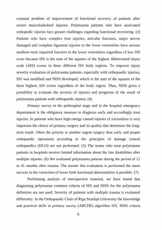
problem is the determination of trauma severity and identification of medium-
term functional outcome for patients after polytrauma. Injury severity affects
both mortality and final functional outcome. Major improvements in the
management of severely injured patients in the past decades have led to a
significant reduction of polytrauma associated mortality. (2) Therefore the is a
6
constant problem of improvement of functional recovery of patients after
severe musculoskeletal injuries. Polytrauma patients who have associated
orthopedic injuries face greater challenges regarding functional recovering. (3)
Patients who have complex foot injuries, articular fractures, major nerves
damaged and complete ligament injuries to the lower extremities have serious
medium-term impaired function in the lower extremities regardless of low ISS
score because ISS is the sum of the squares of the highest Abbreviated injury
scale (AIS) scores in three different ISS body regions. To improve injury
severity evaluation of polytrauma patients, especially with orthopaedic injuries,
ISS was modified and NISS developed, which is the sum of the squares of the
three highest AIS scores regardless of the body region. Thus, NISS gives a
possibility to evaluate the severity of injuries and prognosis of the result of
polytrauma patients with orthopaedic injures. (4)
Primary survey in the prehospital stage and in the hospital emergency
departament is the obligatory measure to diagnoze early and accordingly treat
injuries. In patients who have high energy caused injuries of extremities is very
important the choice of primary surgery and its quality that determine the long-
term result. Often the priority is another urgent surgery thus early and proper
orthopaedic operations according to the principles of damage control
orthopaedics (DCO) are not performed. (5) The teams who treat polytrauma
patients in hospitals receive limited information about the late disabilities after
multiple injuries. (6) We evaluated polytrauma patients during the period of 12
to 41 months after trauma. The sooner this evaluation is performed the more
success in the correction of lower limb functional abnormalities is possible. (7)
Performing analysis of retrospective material, we have found that
diagnosing polytrauma common criteria of ISS and NISS for the polytrauma
definition are not used. Severity of patients with multiple trauma is evaluated
differently. In the Orthopaedic Chair of Rīga Stradiņš University the knowledge
and practical skills in primary survey (ABCDE) algorithm ISS, NISS criteria

7
and DCO are included in the study course. Using primary survey decreases not
diagnozed injuries and functional limitations connected with undiagnozed
orthopaedic injuries. (8) Therefore early and proper examination of the
extremities during the primary survey in the early stage after polytrauma is very
important, regardless of the severe condition of the multiple trauma patients.
Nevertheless, during our study we have noticed that many doctors are poorly
informed about primary survey, criteria of polytrauma, significance of DCO
principles in the treatment of polytrauma patients and in their recovery. Not
always primary survey is performed immediately after the multiply injured
patient is addmitted to the hospital emergency department and in the nearest
period after trauma, which can lead to undiagnozed injuries, especially of
extremities, and late complications due to the consequences of undiagnozed
fractures and dislocations. We also noticed that after check-out of hospital not
always is the functional outcome of musculoskeletal system of patients after
multiple injuries regularly and sistematically evaluated and analyzed.
Evaluation of the functional outcome of musculoskeletal system is
important, because patients life quality and returning to previous work depends
on it. Prompt diagnosis and treatment of functional abnormalities of patients
after polytrauma are very important. Analysis of the functional outcome gives a
possibility to plan more precisely the finansial needs in the treatment of
polytrauma patients and economy of finansial resources. Clinical and
instrumental examination methods can be used to evaluate the functional results
of polytrauma patients with orthopaedic injuries. To assess the functional result
of polytrauma patients with lower limb injuries we used clinical examination
and three dimensional instrumental gait analysis (IGA). Instrumental gait
analysis is an objective instrumental examination method which is used to
examine all phases of gait cycle (GC) and identify causes of gait deviations
(GD). Evaluation of functional limitations of lower extremities in the medium-

8
term after polytrauma gives a possibility to evaluate the connection between
trauma severity and treatment, as well as plan rehabilitation measures.
Study theme is actual because it refers to doctors of different specialities
who treat polytrauma patients and provide their rehabilitation.
This study analyzes functional outcome of lower extremities in 13
years follow-up of polytrauma patients who suffered severe lower limb injuries.
We determined polytrauma severity, relation between ISS, NISS and GD
severity. We compared the data with clinical orthopaedic examination and IGA
parameters of healthy volunteers control group. We evaluated the relation
among ISS, NISS and functional limitations.
Aim of the study
The aim of our study is to analyze functional outcome of patients after
polytrauma with consequences of lower limb injuries.
Objectives of the study
1. To perform the retrospective material analysis of polytrauma patients with
musculoskeletal injuries treated in two Riga hospitals − Riga Eastern
Clinical University Hospital, Clinic „Gailezers”, Trauma and Orthopaedic
department and Hospital of Traumatology and Orthopaedics during
2008−2010 years´ period.
2. To calculate ISS and NISS for these patients and determine polytrauma
severity.
3. To perform the clinical orthopaedic examination, radiological examination
and IGA of patients with consequences of lower limb injuries in the
medium-term (13 years) after polytrauma.

9
4. To analyse GC parameters of healthy volunteers control group obtained in
IGA.
5. To analyze the results, estimate GD severity, give recommendations for the
improvement of the functional outcome obtained in IGA.
6. To compare the clinical examination data of patients and healthy
volunteers.
7. To compare IGA spatio-temporal, motions in the pelvis, hip, knee, ankle
and subtalar joints and ground reaction force (GRF) parameters of
polytrauma patients and healthy volunteers.
8. To compare IGA spatio-temporal, motions in the pelvis, hip, knee, ankle
and subtalar joints and GRF parameters in polytrauma patients with
articular and extraarticular fractures.
9. To compare IGA spatio-temporal, motions in the pelvis, hip, knee, ankle
and subtalar joints and GRF parameters of uninjured extremity of
polytrauma patients with unilateral injuries and healthy volunteers.
10. To compare polytrauma severity with GD severity and find out if there is
relation between ISS, NISS and the functional outcome and work out
proposals (algorithm) for the evaluation of polytrauma patients with lower
limb injuries and prognosis of functional outcome.
Hipothesis of the study
Polytrauma patients with consequences of lower limb injuries have not
only primary, but also secondary (compensatory) functional limitations of
pelvis and lower extremities in the uninjured side which correspond with
polytrauma definition. It is possible to prognoze functional result in polytrauma
patients with musculoskeletal injuries using NISS.

10
Scientific novelty of the study
The scientific novelty of the study is that in this study first time in
Latvia is analyzed the functional outcome of polytrauma patients with lower
extremities injuries consequences in the medium-term after trauma, by using
IGA, and studied the relation among ISS, NISS and functional limitations. New
data has been obtained about functional outcome of polytrauma patients with
consequences of lower extremities injuries and the possibilities of its prognosis.
We have found secondary (compensatory) functional limitations of pelvis and
lower extremities in the uninjured side.
Personal contribution of the author
The author personally performed all stages of study planning of the
study, collection of the retrospective material, clinical examination of the
patients, IGA, analysis, interpretation, description of the obtained results,
development of conclusions and proposals for the patients, data collection,
statistical anlysis, IGA obtained data collection and statistical anlysis of the
control group, is the author of scientific publications and presentations of
congreses, conferences and photos, presented in the thesis.
Structure of the thesis
The thesis is written in the Latvian language. It consists of introduction,
literature study, study of materials and methods, results, discussion and
conclusions. It has 10 supplements. The amount of thesis is 127 pages. One
hundred forty-three authors´ publications are analysed in the thesis.
There are 4 scientific publications about thesis theme, 1 patent, 16
abstracts published, 13 oral presentations given as well as 2 posters at
congreses and conferences.

11
1. MATERIALS AND METHODS
1.1. Definitions used in the study
1. Abbreviated injury scale (AIS) – injury severity coding system,
where injuries are coded according to the six AIS severity codes: minor,
moderate, serious, severe, critical and maximal (currently untreatable)
(Association for the Advancement of Automotive Medicine, 2008).
2. Damage control orthopaedics (DCO) – treatment tactics of
polytrauma patients with orhopaedic injuries, wich are based on primary
stabilization of long bone fractures, using external fixation (Pape HC, 2010).
3. Injury severity score (ISS) − sum of the squares of the highest AIS
scores in three different body regions (Association for the Advancement of
Automotive Medicine, 2008).
4. New injury severity score (NISS) − sum of the squares of the three
highest AIS scores anywhere in the body (Association for the Advancement of
Automotive Medicine, 2008).
5. Polytrauma − syndrome characterized by multiple injuries exceeding
a defined severity grade (ISS 17) with sequential systemic reactions that may
lead to dysfunction or failure of remote organs and vital systems, which have
not been directly injured (Trentz O, 2007).
6. Primary survey (ABCDE algorithm) – systematic and rapid
evaluation of trauma patient, to identify all life threatening injuries and treat
until next stage (American College of Surgeons, 2012).
7. Three dimensional instrumental gait analysis (IGA) − an
examination method, during which quantitative information about the
movements of body segmants is collected during GC with the aim to
understand the origin of GD and to find the ways of its prevention (Medical
technologies data base, used in medicine, Latvia).
12
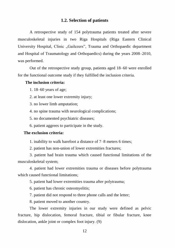
1.2. Selection of patients
A retrospective study of 154 polytrauma patients treated after severe
musculoskeletal injuries in two Riga Hospitals (Riga Eastern Clinical
University Hospital, Clinic „Gailezers”, Trauma and Orthopaedic department
and Hospital of Traumatology and Orthopaedics) during the years 2008–2010,
was performed.
Out of the retrospective study group, patients aged 18–60 were enrolled
for the functional outcome study if they fulfilled the inclusion criteria.
The inclusion criteria:
1. 18–60 years of age;
2. at least one lower extremity injury;
3. no lower limb amputation;
4. no spine trauma with neurological complications;
5. no documented psychiatric diseases;
6. patient aggrees to participate in the study.
The exclusion criteria:
1. inability to walk barefoot a distance of 7–8 meters 6 times;
2. patient has non-union of lower extremities fractures;
3. patient had brain trauma which caused functional limitations of the
musculoskeletal system;
4. patient had lower extremities trauma or diseases before polytrauma
which caused functional limitations;
5. patient had lower extremities trauma after polytrauma;
6. patient has chronic osteomyelitis;
7. patient did not respond to three phone calls and the letter;
8. patient moved to another country.
The lower extremity injuries in our study were defined as pelvic
fracture, hip dislocation, femoral fracture, tibial or fibular fracture, knee
dislocation, ankle joint or complex foot injury. (9)

13
The patients were recruited according to their residences from the
hospital case-records of the patients. The patients who corresponded inclusion
criteria were invited to undergo the evaluation of functional outcome by a
phone call or a letter that described the purpose of the study and the IGA
method.
The study was conducted on 34 polytrauma patients with orthopaedic
injuries. Evaluation of the functional outcome was performed in the Rīga
Stradiņš University Rehabilitation Research Laboratory, located in the National
Rehabilitation Centre „Vaivari” (RRL, NRC „Vaivari”).
There were two study groups: polytrauma patients group and healthy
volunteers control group.
The patients’ complaints, clinical orthopaedic examination, radiological
examination and IGA were included in the evaluation of functional outcome.
For every patient ISS and NISS values were calculated. We determined the
injured or most injured and the uninjured or less injured lower extremity. NISS
values were used to evaluate the severity of polytrauma injuries because ISS
does not give objective information about the amount of work and resources
that are required if the patient has serious multiple injuries in one of the ISS
anatomic regions, particularly orthopaedic injuries.
Clinical examination and IGA data of 34 healthy volunteers (24 women
and 10 men; age range 19–65, mean age 36.38 years) for the control group was
collected from RRL, NRC „Vaivari” volunteers data basis. IGA data were
analyzed with Visual 3-D software program.
Patients and the control group data were summerized and analyzed with
SPSS program. Clinical and IGA examination data were compared with the
control group data.
To find out if the fracture type influences CG parameters, we divided
study group patients in to two subgroups patients with articular fractures and

14
with extraarticular fractures in the injured or more injured side according to the
fracture types determined by AO classification.
To find out if there were compensatory changed GC parameters in the
uninjured side, we compared time, distance, kinematic and kinetic parameters
of the uninjured extremity of polytrauma patients from study group with
unilateral injuries of lower extremities and the control group.
Polytrauma severity was defined in the study. In the definition of
polytrauma severity we used NISS based on the AIS codes. New injury severity
score 17–26 were defined as polytrauma with moderate injuries, NISS 27–35 as
polytrauma with serious injuries, NISS 36–49 as polytrauma with severe
injuries, and NISS 50–66 as polytrauma with critical injuries.
1.3. Clinical orthopaedic and radiological examination
The patients’ complaints were examined. Patients were asked to
evaluate pain in their lower extremities according the following variables: „no
pain”, „moderate pain” or „severe pain”.
Visual examination of the posture, gait, lower extremities and foot archs
were performed. Movements in the joints were measured using goniometric
technique. Muscle power was evaluated using 5 grades scale (Medical
Research Council scale for Muscle Strength). Length and girth of the
extremities were examined using type. Neirovascular examination of the
extremities was performed. Radiological examination (consolidation stage of
the fractures, bone structure, bone fragment position and nearest joints
condition) was performed. Patients, who had talar neck fractures, were
examined, using „Method of prognosis of avascular necrosis of the talar neck
fractures” (LR patent). Hip, knee, ankle and subtalar joint movements and
mucle power parameters obtained in the clinical examination were compared
with the parameters of the control group mentioned before.

15
1.4. Instrumental gait analysis
After clinical and radiological examination patients underwent IGA. In
Latvia IGA is available only in the RRL, NRC „Vaivari”.
Instrumental gait analysis was performed using infrared light ProReflex
MCU (240 Hz) digital cameras (Qualisys Medical, Sweden), force plate
(Advanced Mechanical Technology, Inc., Watertown, USA) and Visual 3-D
software developed in the National Health Institute, USA (C-motion Inc, USA).
Light, spherical, reflective markers (19 mm) were attached to the skin to
identify bony landmarks (the first sacral vertebra, both anterior superior iliac
spinae, lateral surfaces of femur and shin, the heads of the first and the fifth
metatarsal bones and the calcaneal bones). The patients had to walk barefoot a
distance of 7–8 meters, 6–10 times at a self selected speed. The spatial
coordinates of markers′ were registered during gait recording and the markers
motion trajectories were calculated (Fig.1.1.). The patient had to put one foot
on the force plate on the floor that registered GRF. (10)
Fig. 1.1. Digital infrared light camera registers the markers′ motions,
attached in the definite anatomical landmarks during gait cycle
(photo from the author′s archive)

16
With two digital video cameras the visual gait recording was obtained.
The findings were processed with data processing programs in the form of
diagrams and numbers. The motion parameters of the pelvis and lower
extremity joints during the gait were shown in the diagrams. The force moment
of the muscles, the amount of generated and absorbed energy was shown in the
diagrams in the sagittal and frontal plane. The spatio-temporal parameters of
the GC were registered in the form of numbers.
The spatio-temporal parameters, motions in pelvis and the joints of
lower extremities in sagittal and frontal plane, anterior, posterior and vertical
load GRF of the GC of patients in the injured or more injured and uninjured or
less injured lower extremity were analyzed. We defined GC parameters used in
our study.
Gait parameters of the polytrauma patients’ lower extremities were
compared with the corresponding lower extremities gait parameters of the
control group.
The patients received the IGA description, based on the clinical and IGA
examination data.
The following patients´ group injured or more injured and uninjured or
less injured lower extremity GC parameters were analyzed in the study:
1. GC spatio-temporal time parameters (cadence, stance time, gait
speed, step lengh and step width);
2. according to the defined GC kinematic parameters:
2.1. motions in pelvis and lower extremities joints in sagittal plane;
2.2. motions in pelvis and lower extremities joints in frontal plane;
3. according to the defined GC kinetic parameters:
3.1. anterior and posterior GRF;
3.2. vertical load GRF during loading response and terminal stance.
Normal GC parameters were worked out using young healthy
volunteers´ GC parameters, which corresponded also to the literature describing

17
normal GC parameters. Phases of GC reflect functional parameters of lower
extremities. (11)
Gait deviations severity was estimated as moderate GD, serious GD,
severe GD and very severe GD according to the severity of changes of
movements in the joints in the frontal and sagittal plane and the number of
involved joints. Gait deviations severity was compared to polytrauma severity.
Data of protocols of polytrauma patients group and the control group
were summarized in the SPSS programm, with which data analysis was
performed.
The clinical examination and IGA data of polytrauma patients´ group
were compared to the control group data.
For data summarizing SPSS program was used. Statistical analysis of
the data was performed with SPSS 20.0 for Windows version. Study results
were analyzed with descriptive statistics methods. To get quantitative data,
means and SD were estimated. To compare polytrauma patients and healthy
persons clinical examination and IGA data, two groups were selected in the
study: a polytrauma patients group and a control group. Clinical examination
and IGA data in the injured or more injured and uninjured or less injured lower
extremity of the polytrauma patients were compared with mentioned data of the
corresponding extremity of the control group by using Independent Samples
T test. To find out if there are compensatory changes in the polytrauma
patients´ uninjured lower extremity, a polytrauma patients group with unilateral
injuries of lower extremities was selected. Clinical examination and IGA data
of the uninjured lower extremity was compared with the control group by using
Independent Samples T test. To compare the two dependent groups (ISS and
NISS comparison in patients with different GD severity) Wilcoxon Signed
Ranks test was used. P value<0.05 was considered significant.

18
2. RESULTS
2.1. Results of the retrospective material study of polytrauma
patients with musculoskeletal injuries
A retrospective study of 154 polytrauma patients (53 women, 101 men),
treated after severe musculoskeletal injuries (NISS17) in Trauma and
Orthopaedic department, Clinic „Gailezers”, Riga Eastern Clinical University
Hospital, (132 patients) and Hospital of Traumatology and Orthopaedics (22
patients) during the years 2008–2010 was performed. Trauma mechanisms
were high energy trauma (road traffic accidents, fall from hight etc).
The patients had NISS 1748, but ISS 948. Fifty-five patients (36%)
had NISS equal to ISS, 99 patients (64%) had NISS higher than ISS. Forty-four
patients had ISS lower than 17 (severity of trauma did not correspond the
polytrauma definition).
In accordance with the exclusion criteria of the study 61 patient from the
154 patients retrospective analysis group was excluded: 24 patients had not
lower extremities injuries, 27 patients did not correspond the age criteria of the
study, 5 patients had lower extremity amputation, 5 patients had documented
psychiatric diseases.
Ninety-three patients, who corresponded to the study inclusion criteria,
were recruited according to their residences from the hospital case-records of
the patients. They were invited to undergo the functional result evaluation by a
phone call or a letter that described the purpose of the study and the IGA
method. Out of 93 selected patients 18 patients did not respond to the phone
calls, 19 patients did not respond to the letter, 13 patients refused to undergo
the IGA, 7 patients moved to another country, 1 patient had had a fracture of
proximal segment of tibia six months before recruitment and 3 patients could
not walk the mentioned distance. These patients according to the exclusion
criteria were excluded from this study.

19
2.2. Functional results of patients with consequences
of lower limb injuries after polytrauma
2.2.1. Characteristics of patients
The study was conducted on 34 polytrauma patients (17 women and
17 men; age range 23–59, mean age 39.5011.70 years) with consequences of
lower limb injuries, 12–41 months after polytrauma, who corresponded to the
study criteria. The patients had NISS mean 25.9, ISS mean 20.7. Instrumental
gait analysis was performed in the RRL, NRC “Vaivari”. Clinical orthopaedic
examination and IGA data were compared to the control group. Fifteen patients
had polytrauma with moderate injuries (NISS 17–26), 17 patients with serious
injuries (NISS 27–35) and 2 patients had polytrauma with severe injuries
(JISS 36–48).
2.2.2. Results of the clinical orthopaedic examination and
radiological examination
Eight patients had no complaints about pain, 19 patients had moderate
pain and 7 patients had severe pain during evaluation. Six patients had open
fracture wound infection which was treated succesfully in all cases. Seventeen
patients took a rehabilitation course in the rehabilitation center within six
months after hospital discharge. Eighteen patients had true leg discrepancy in
the injured side 1−3 cm. Thirty patients had delayed fracture healing. Out of
those two patients had avascular necrosis of the femoral head 1−2 years after
C type acetabular fracture, and the evaluation of the functional outcome for
these patients was performed after total hip replacement. Two patients had non
union (one patient with a fracture of the distal segment femur, one patient with
a fracture of distal segment of tibia), and evaluation was performed after re-

20
operation and fracture healing. Two patients had fracture healing in the average
healing times.
We evaluated motion in the joints in the injured or more injured lower
extremity and in the uninjured or less injured lower extremity of polytrauma
patients group and the same side of the control group. The polytrauma patients
in the injured or more injured lower extremity had decreased hip flexion,
abduction, adduction, external and internal rotation, ankle flexion and
extension, eversion in the subtalar joint (P0.05). Two patients had knee joint
flexion contracture, two patients had ankle joint flexion contracture (after deep
peroneal nerve injury), one patient could not perform extension in the ankle
joint. The polytrauma patients in the uninjured or less injured lower
extremity had decreased hip extension, abduction, external and internal
rotation, ankle flexion and extension (P0.05). As knee extension mean value
in the polytrauma patients group and the control group was 0, P value was not
calculated.
We evaluated muscle power in the injured or more injured lower
extremity and in the uninjured or less injured lower extremity of the polytrauma
patients group and the same side of the control group. Muscle strength differed
in the polytrauma patients group in the injured or more injured lower
extremity and the control group: patients had decreased muscle strength of hip
flexors, extensors, abductors, adductors, external and internal rotators, knee and
ankle flexors and extensors, inversion and eversion providing muscles in the
subtalar joint (P0.05). Muscle strength also differed in the polytrauma patients
group in the uninjured or less injured lower extremity and the control group:
patients had decreased muscle strength of hip flexors, extensors, abductors,
adductors, external and internal rotators, knee and ankle flexors and extensors,
inversion and eversion providing muscles in the subtalar joint (P0.05). To find
out, if the clinical orthopaedic examination parameters are significantly
21
changed in the uninjured side, we compared the range of motion (ROM) in the
joints and muscle power in the uninjured side of the patients with unilateral
injuries of lower extremities with the same side of the control group. In the
uninjured side there were significantly decreased hip extension, abduction,
external and internal rotation, and ankle extension (P0.05). These patiens had
decreased muscle strength of hip extensors, abduction, adduction, external and
internal rotators, knee flexors and extensors, ankle flexors and extensors,
inversion and eversion providing muscles in the subtalar joint (P0.05).
2.2.3. Results of instrumental gait analysis
We evaluated patients´ gait using IGA. We analyzed GC spatio-
temporal, kinematic and kinetic parameters of the polytrauma patients and the
control group.
The GC spatio-temporal parameters of the polytrauma patients group
were significantly worse than the gait parameters of the same lower extremity
of the control group: in the injured or more injured and in the uninjured or
less injured side there was decreased cadence, increased stance time and
decreased step lengths (P0.05). The polytrauma patients also had decreased
walking speed and step width in comparison with the control group (P0.05).
The comparison of IGA sagittal plane motion results in the pelvis, hip,
knee and ankle joints of the injured or more injured lower extremity in the
polytrauma patients group with the same side lower extremity of the control
group showed that polytrauma patients had significantly increased pelvic
anterior tilt, a decreased hip extension and knee maximum flexion (P0.05), but
in the uninjured or less injured lower extremity in the polytrauma patients
group in comparison with the same side lower extremity of the control group,
polytrauma patients had significantly increased pelvic anterior tilt, decreased
knee maxium flexion and increased knee minimum flexion (P0.05).

22
The comparison of IGA frontal plane motion results in the pelvis, hip,
knee and subtalar joints of the injured or more injured lower extremity in the
polytrauma patients group with the same side lower extremity of the control
group showed that polytrauma patients had increased pelvic drop in the stance
and swing (P0.05), but in the uninjured or less injured lower extremity in
the polytrauma patients group in comparison with the same side lower
extremity of the control group, polytrauma patients had decreased inversion in
the subtalar joint (P 0.05).
The patients in the injured or more injured side and in the uninjured
or less injured side in comparison with the control group in the same side had
decreased anterior, posterior, vertical load GRF during loading response and
terminal stance (P0.05).
We divided the study group patients in two subgroups patients with
articular fractures and patients with extraarticular fractures in the injured or
more injured side according to the AO fracture classification types. In the study
group there were 21 patient with articular fractures and 13 patients with
extraarticular fractures. We found out that patients with articular fractures had
increased knee minimal flexion in comparison with patients with extraarticular
fractures (P=0.05). Two patients in the articular fractures subgroup were not
able to perform extension in the ankle joint and it remained in flexion during all
GC. In this subgroup patients had decreased cadence, stance time, step length,
increased pelvic anterior tilt, decreased GRF and the varus deformity of lower
leg, but without significant difference.
We compared spatio-temporal, kinematic and kinetic parameters of the
uninjured extremity of 26 polytrauma patients from study the group, who
had unilateral injuries of lower extremities and the control group. Polytrauma
patients with unilateral injuries in the uninjured side had decreased step
length, increased stance time and decreased cadence in comparison with the
23
control group (P0.05). The polytrauma patients had decreased walking speed
and step width in comparison with the control group (P0.05). Gait cycle
motion parameters of pelvis, hip, knee and ankle joint in the sagittal plane of
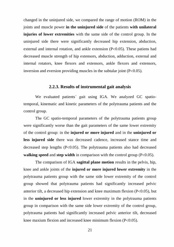
the uninjured side in patients with unilateral injuries displayed in table 2.1.
Table 2.1.
Sagittal plane motions in pelvis, hip, knee and ankle joints in the uninjured lower
extremity of polytrauma patients and of the healthy control group
during gait cycle
Parameter
Polytrauma group
(n=26)
Control group
(n=26) P value
Mean±SD(°) n Mean±SD(°) n
Pelvic anterior tilt 8.2±5.88 n=23 6.07±4.39 n=15 0.234
Pelvic posterior tilt 15.67±11.59 n=3 3.82±2.82 n=11 Not
calculated
Hip flexion, max 27.50±11.90 n=26 25.62±6.80 n=26 0.487
Hip flexion, min 8.80±6.30 n=5 − − −
Hip extension 9.24±10.15 n=21 12.27±7.77 n=26 0.253
Knee flexion, max 54.77±12.88 n=26 60.85±3.65 n=26 0.025*
Knee flexion, min 9.55±4.50 n=19 7.95±3.89 n=21 0.141
Knee extension 3.57±3.73 n=7 2.40± 2.51 n=5 Not
calculated
Ankle flexion, max 16.23±6.40 n=26 17.04±5.87 n=26 0.638
Ankle extension 10.12±4.10 n=26 10.15±1.93 n=26 0.953
SD standard deviation, * statistically significant, P independent samples T test, to compare
joint motions in the patients´ group and the control group, „” no corresponding parameter in the
control group and P value.
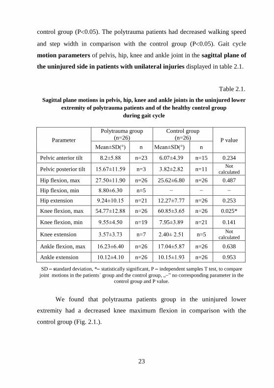
We found that polytrauma patients group in the uninjured lower
extremity had a decreased knee maximum flexion in comparison with the
control group (Fig. 2.1.).
24
Fig. 2.1. Knee joint motions in sagittal plane in the uninjured lower extremity of
polytrauma patients and of the healthy control group
Gait cycle motion parameters of pelvis, hip, knee and subtalar joint in
the frontal plane of uninjured side in patients with unilateral injuries
displayed in table 2.2.
Table 2.2.
Frontal plane motions in pelvis, hip, knee and subtalar joints in the uninjured lower
extremity of polytrauma patients and of the healthy
control group during gait cycle
Parameters
Polytrauma group
(n=26)
Control group
(n=26) P
Mean ±SD(°) n Mean±SD(°) n
Pelvis in stance 2.19 ±1.86 n=21 2.91 ±1.92 n=23 0.214
Pelvis in swing 2.14±1.23 n=14 2.27±1.48 n=22 0.780
Hip abduction 3.67±2.08 n=3 ‒ ‒ ‒
Hip adduction 9.35±5.66 n=23 9.42± 3.59 n=26 0.955
Knee abduction 5.80±3.25 n=20 5.33±2.70 n=21 0.620
Knee adduction 6.17 ±6.73 n=6 4.20±1.92 n=5 Not
calculated
Subtalat inversion 4.21±3.34 n=24 6.54 ±4.64 n=24 0.052*
Subtalar eversion 6.27±3.98 n=26 6.08±3.70 n=26 0.859
SD standard deviation, n number of patients * statistically significant, P independent samples
T test, to compare motions in the patients´ group and the control group, decreasing,
„” no corresponding parameter in the control group and P value.

25
In the evaluation of GC motion parameters in pelvis and the mentioned
joints in the frontal plane of the uninjured lower extremity of polytrauma
patients and the control group, we found that there were significantly decreased
inversion in the subtalar joint (Fig. 2.2.).
Fig. 2.2. Subtalar joint motions in frontal plane in the uninjured lower extremity of
polytrauma patients and of the healthy control group
Table 2.3.
Ground reaction force in the uninjured lower extremity of polytrauma patients and
of the healthy control group
Parameters
Polytrauma group
(n=26)
Control group
(n=26) P
Mean±SD
(GRF/weight)
Mean±SD
(GRF/weight)
GRF A 0.14±0.05 0.18±0.03 0.005*
GRF P 0.11±0.03 0.14±0.03 0.000*
GRFV1 0.97±0.06 1.05±0.09 0.001*
GRF V2 1.02±0.19 1.13 ±0.08 0.014*
GRF A maximal anterior ground reaction force in terminal stance, GRF P maximal posterior
ground reaction force in loading response, GRF V1 maximal vertical ground reaction force in
loading response, GRF V2 maximal vertical ground reaction force in terminal stance, n number
of patients, SD standard deviation, P independent samples´ T test, to compare GRF in the
patients group and control group, * statistically significant.
26
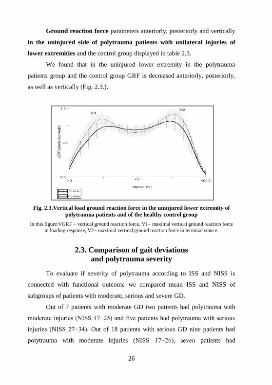
Ground reaction force parameters anteriorly, posteriorly and vertically
in the uninjured side of polytrauma patients with unilateral injuries of
lower extremities and the control group displayed in table 2.3.
We found that in the uninjured lower extremity in the polytrauma
patients group and the control group GRF is decreased anteriorly, posteriorly,
as well as vertically (Fig. 2.3.).
Fig. 2.3.Vertical load ground reaction force in the uninjured lower extremity of
polytrauma patients and of the healthy control group
In this figure:VGRF vertical ground reaction force, V1 maximal vertical ground reaction force
in loading response, V2 maximal vertical ground reaction force in terminal stance.
2.3. Comparison of gait deviations
and polytrauma severity
To evaluate if severity of polytrauma according to ISS and NISS is
connected with functional outcome we compared mean ISS and NISS of
subgroups of patients with moderate, serious and severe GD.
Out of 7 patients with moderate GD two patients had polytrauma with
moderate injuries (NISS 17−25) and five patients had polytrauma with serious
injuries (NISS 27−34). Out of 18 patients with serious GD nine patients had
polytrauma with moderate injuries (NISS 17−26), seven patients had

27
polytrauma with serious injuries (NISS 27−34) and two patients had
polytrauma with severe injuries (NISS 41−48). Out of 8 patients with severe
GD three patients had polytrauma with moderate injuries (NISS 22−26) and
five patients had polytrauma with severe injuries (NISS 27−34). One patient
with very severe GD had polytrauma with moderate injuries (NISS 22).
In the comparison of severity of GD and polytrauma severity we found
that 9 patients had appropriate polytrauma severity and GD. In the
17 polytrauma patients group with moderate injuries thirtheen had more severe
GD than the severity of polytrauma. In the 15 polytrauma patients group with
serious injuries five had more severe GD than the severity of polytrauma.
Patients who had polytrauma with moderate injuries but severe GD, had sciatic
nerve palsy, deep peroneal nerve palsy or delayed union of fractures which
caused medium-term GD. The comparison of GD and polytrauma severity is
displayed in Fig. 2.4.
Moderate injuries Serious injuries Severe injuries
Polytrauma severity
0
2
4
6
8
10
Pati
en
ts (
n)
GD severity
Moderate GD
Serious GD
Severe GD
Very severe GD
Fig.2.4. Comparison of gait deviations (GD)
and polytrauma severity
28
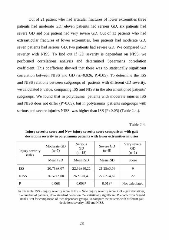
Out of 21 patient who had articular fractures of lower extremities three
patients had moderate GD, eleven patients had serious GD, six patients had
severe GD and one patient had very severe GD. Out of 13 patients who had
extraarticular fractures of lower extremities, four patients had moderate GD,
seven patients had serious GD, two patients had severe GD. We compared GD
severity with NISS. To find out if GD severity is dependant on NISS, we
performed correlations analysis and determined Speermens correlation
coefficient. This coefficient showed that there was no statistically significant
correlation between NISS and GD (rs=0.926, P0.05). To determine the ISS
and NISS relations between subgroups of patients with different GD severity,
we calculated P value, comparing ISS and NISS in the aforementioned patients´
subgroups. We found that in polytrauma patients with moderate injuries ISS
and NISS does not differ (P>0.05), but in polytrauma patients subgroups with
serious and severe injuries NISS was higher than ISS (P0.05) (Table 2.4.).
Table 2.4.
Injury severity score and New injury severity score comparison with gait
deviations severity in polytrauma patients with lower extremities injuries
Injury severity
scales
Moderate GD
(n=7)
Serious
GD
(n=18)
Severe GD
(n=8)
Very severe
GD
(n=1)
Mean±SD Mean±SD Mean±SD Score
ISS 20.71±8,07 22.39±10,22 21.25±3,69 9
NISS 26.57±5,08 26.56±8,47 27.62±4,62 22
P 0.068 0.003* 0.018* Not calculated
In this table: ISS Injury severity score, NISS New injury severity score, GD gait deviations,
n number of patients, SD standard deviation, * statistically significant, P Wilcoxon Signed
Ranks test for comparison of two dependant groups, to compare the patients with different gait deviations severity, ISS and NISS.
29
3. DISCUSSION
3.1. Connection of study with literature data and limitations
of the study
The investigation of functional outcomes in the polytrauma patients with
consequences of orthopaedic injuries using self-report questionnaires can be
used for large number of patients, but it is subjective. (12) Functional capacity
index is described as AIS based sign of functional result after trauma, but it
does not refer to the polytrauma. (13) The two largest and most respected long-
term studies of polytrauma patient outcome using questionnaires and clinical
examination: The Lower Extremity Assessment Project and The Hannover
Rehab Study are described. (14) Kubota et al, 2012 analyzed spatio-temporal,
kinematic and kinetic GC parameters 3 and 12 months after internal fixation of
fractures of acetabulum. The authors found that these patients had decreased
hip abductor strength in the injured side, therefore hip abductors should be
trained early after operation. (15) There are no data about research of
functional result after polytrauma of patients with musculoskeletal injuries in
Latvia, using IGA.
In our study we evaluated functional result of patients with
consequences of injuries of lower extremities after polytrauma, using clinical
examination and IGA and analyzed data of both of the lower extremities − the
injured or most injured and the uninjured or less injured one. Our data show
that polytrauma patients with consequences of injuries of lower extremities in
comparison with the control group had decresed cadence, increased stance
time, decreased step length, increased pelvic anterior tilt, decreased maximal
flexion in the knee joint and decreased anterior, posterior, vertical load GRF;
besides in the injured or most injured side they have decreased hip extension,
increased pelvic drop during the stance and swing, but decreased inversion in
the subtalar joint in the uninjured or less injured side.
30
Analyzing the GC parameters in the patients subgroups with articular
and extraarticular fractures, we found that in the patients subgroup with
articular fractures there was increased minimal flexion in the knee joint in
comparison with patients subgroup with extraarticular fractures.
We analyzed separately clinical examination and IGA data of the
uninjured lower extremity of patients with unilateral lower extremities injuries.
We compared GC data of the patients with the healthy control group. Using
IGA, we analyzed the connection among ISS, NISS and GD.
The age range of the polytrauma patients and the healthy control group
individuals was considered to be appropriate to compare gait changes in our
study, because the studies point to GD in the elderly after 65 years of age.
(16, 17)
We evaluated patients in 1−3 years after polytrauma. Most of the
clinical recovery outcomes of severe lower extremity trauma are attained after
one year, therefore one year after trauma was considered to be within the period
when the gait does not change significantly in the future. (18) Besides a
relatively short time after trauma gives a possibility to analyze pathology
causes found in the clinical examination and by IGA as well as provide
treatment.
We have some limitations in our study.
1. The polytrauma patients in our study had multiple injuries, including
the injuries of pelvis and lower extremities, which possibly could interfere with
the functional result. In our study there were no patients with equal diagnosis,
therefore it was not possible to divide patients into the subgroups according to
the anatomical location of the lower extremities fractures and dislocations to
compare the functional result in the subgroups of patients according to the
location of injuries. Nevertheless, we devided the patients into two groups
according to the AO classification of the fractures regarding the injury of
articular surface patients subgroups with articular and extraarticular fractures

31
in the injured or most injured side, which, we think, is one of the important
factors influencing the functional result. Injuries of other anatomical regions
might interfere with the function of lower extremities.
2. Polytrauma patients were treated with different conservative and
surgical methods, which could interfere with the functional result. Therefore
because of the small number of patients it was not possible to divide patients
into the subgroups and compare the results according to the methods of
treatment.
3. Although the period of 1−3 years after trauma is not long, we chose
this period because it is possible to correct the functional abnormalities, if they
are found.
4. There were different relations between men and women in the
polytrauma patients and the control group, but the literature data report that GC
parameters do not significantly differ between them. (19, 20)
5. We did not compare the functional result of patients, who had
rehabilitation after polytrauma and those, who had no rehabilitation because the
number of patients in subgroups would have been too small to compare the
results.
6. Social, psychological and economical factors were not analyzed in
our study, which might influence the polytrauma patients´ functional result.
3.2. Fracture union complications of patients after polytrauma
Most of the patients (88 %) had no fracture union in the normal time of
the consolidation of fractures. Fracture union delay is a frequent complication
after polytrauma and is caused by many factors. Polytrauma is caused by high
energy trauma. To compare with low energy trauma, polytrauma patients have
a large bone injury, severe soft tissue injuries, severe haemorrhagic shock,
hipoxia which is caused by lung injury and early complications, connected with

32
these injuries causing circulation disturbances in the place of the fracture,
which increase the risk of fracture union delay. In the place of high energy
injury there are severe soft tissue injuries with blood supply disturbances which
is the main cause of fracture union complications. As bone blood supply is
mainly from soft tissue blood vessels, this is the main factor of normal fracture
union. Polytrauma patients often have open fractures. Injuries of skin, muscles,
major blood vessels, nerves and periosteum in the place of open fracture
worsen the prognosis of fracture union. High energy caused fractures often are
multifragmentary. After debridement of the wound and extirpation of dead
tissue, there can be bone defects which cause fracture union disturbances.
Fracture union is influenced also by associated injuries and tactics of bone
fixation which is connected with life saving surgery priority. Using the
monolateral external fixation as the definitive treatment method, there can be
fracture union complications because of lack of fracture stability. (21) As high
energy caused fractures are with severe displacement, periosteum is detached
from a bone and there are disturbances of intramedullary blood supply.
Complications of fracture union can be caused by infection. If fractures are non
imobilized or poorly imobilized in the prehospital stage, there is an increased
risk of displacement of bone fragments which causes additional injuries of soft
tissue that increases the risk of development of fracture union complications.
(22) Taking into account the aforementioned, it is possible to explain the
fracture union complications in most of polytrauma patients.
3.3. Evaluation of the clinical orthopaedic examination
in patients after polytrauma
The motion pathology in joints found in the clinical orthopaedic
examination show that injuries of the extremities decreases ROM in the joints.
Decreasing of ROM in the injured side of uninjured joints show that patients

33
perform movements in these joints carefully. Range of motion is significantly
decreased in the ankle joint in the patients with articular fractures of distal
segment of tibia, as well as in patients with sciatic nerve and deep peroneal
nerve palsy.
Decreasing of ROM in the uninjured side in patients with unilateral
lower extremities injuries might be connected with muscle weekness which was
diagnozed in most of the patients not only in the injured, but also in the
uninjured lower extremity.
3.4. Specific features of the IGA data in the evaluation
of functional result in patients after polytrauma
The evaluation of IGA data and comparison with the control group helps
to diagnoze the motion abnormalities in polytrauma patients’ lower extremities
which often cannot be fully detected and evaluated by clinical examination
methods only. Evaluation of IGA data of patients after polytrauma is a difficult
task because the consequences of injuries interfere. We selected GC spatio-
temporal, pelvic and lower extremities´ joints sagittal and frontal plane motions
analysis because they are more responsible for pathological changes, severity of
GD and reflect the connection between motions in joints and muscle strength.
Time and distance parameters of GC were significantly changed in both sides.
Most of the patients had increased pelvic anterior tilt in both sides which
showed inability to extend the hip joints during the stance phase of the GC, or a
shorter leg in the injured side. The patients who had increased pelvic posterior
tilt had weakness of hip flexors. Increased knee minimum flexion during all GC
was due to knee joint contracture of the injured lower extremity, but the
increased knee minimum flexion during the swing was due to inadequate
function of femoral posterior group muscles. Extension in the ankle joint did
not differ from the control group, as two patients had flexion contracture

34
because of sciatic nerve or deep peroneal nerve palsy. Patients with unilateral
lower extremities injuries consequences in the uninjured lower extremity and
the same extremity in the control group in the initial swing had differed
decreasing of knee maximum flexion, that points to the weekness of hip flexors
as well as the combined weekness of quadriceps muscle and ankle flexors.
Evaluation of IGA allowed differentiations between motion abnormalities due
to functional reasons (muscle weakness) and motion abnormalities due to
permanent reasons (contracture, ankylosis). The GRF was significantly
decreased in the injured and uninjured side anteriorly, posteriorly and vertically
due to increased hip maximum flexion and decreased vertical loading force
during loading response and terminal stance. Rehabilitation of polytrauma
patients should be performed as early as possibly, in the plan of rehabilitation
including development of strengh of abdominal muscles, hip abductors and
extensors, quadriceps, hamstrings and ankle flexors, and especially the
development of the strengh of hip extensors, quadriceps, hamstrings and ankle
flexors in the uninjured side.
In the evaluation connection among ISS, NISS and severity of GD, we
found that polytrauma patients who had NISS more than ISS had serious or
severe GD. This difference might be explaned with multiple injuries of lower
extremities which cause functional limitations during GC. We advise these
patients to undergo IGA to find out these limitations. Patients with complex
foot injuries, articular fractures, major nerve damage and complete ligament
injuries to the lower extremities can have severe medium-term impaired
function in the lower extremities regardless of low ISS and NISS score,
therefore we advise to perform IGA also to these patients, to evaluate GD
severity and provide necessary treatment to improve their functional result.

35
3.5. Implementation of study results in the evaluation of polytrauma
patients in the early and medium-term after polytrauma
On the basis of our study results, we advice the following:
1. in the prehospital stage evaluate the multiply injured patients
according to the primary survey (ABCDE) algorithm and provide appropriate
emergency care according to this algorithm;
2. in the hospital emergency department evaluate the multiply injured
patients according to the primary survey algorithm, provide immediate life
saving procedures and perform secondary survey after stabilization of vital
signs;
3. in the hospital after statement of diagnosis calculate ISS un NISS;
4. in multiply injured patients with musculoskeletal injuries in the
polytrauma severity determination use NISS;
5. planning the treatment of polytrauma patients with injuries of lower
extremities, use the algorithm of the functional outcome prognosis of
polytrauma patients with musculoskeletal injuries, thus decreasing the risk of
severity of functional limitations (Fig. 3.1.);
6. introduce AIS, ISS and NISS in Latvia that would give a possibility to
evaluate the severity of polytrauma and choose appropriate treatment based on
damage control principles;
7. identify early the functional limitations of musculoskeletal system
using clinical orthopaedic examination of polytrauma patients and follow-up;
8. identify the functional limitations of musculoskeletal system in
patients after polytrauma using IGA.

36
Fig. 3.1. Algorithm of the functional outcome prognosis of polytrauma patients
with musculoskeletal injuries ISS Injury severity score, NISS New injury severity score, DCO Damage control
orthopaedics, DCS Damage control surgery, ETC early total care (definitive fracture
stabilization), FL functional limitation, IGA 3-dimension instrumental gait analysis.
*NISS less than ISS is not possible, because NISS is the sum of the squares of the three highest
AIS scores anywhere in the body. If a patient has one lower extremities injury, NISS is equal the ISS, if there are several lower extremities injuries, NISS is more than ISS.

37
4. CONCLUSIONS
1. In patients after polytrauma using IGA it is possible to obtain data about
functional limitations during GC which often cannot be fully detected and
evaluated by clinical examination methods only.
2. In the study we found, that in patients with consequences of lower
extremities injuries in the medium-term after polytrauma, in comparison
with the control group was changed:
2.1. in the injured or more injured side: GC spatio-temporal parameters;
from GC motion parameters in the sagittal plane increased pelvic
anterior tilt, decreased hip extension and knee maximum flexion, in
the frontal plane incresed pelvic dro in stance and swing; decreased
GRF;
2.2. in the uninjured or less injured side: GC spatio-temporal parameters;
from GC motion parameters in the sagittal plane is increased pelvic
anterior tilt, decreased knee maximum flexion and increased knee
minimum flexion, in the frontal plane decreased inversion in the
subtalar joint; decreased GRF;
3. Patients with articular fractures had increased knee minimum flexion in
comparison with patients, who had extraarticular fractures in the injured or
more injured side.
4. In patients with consequences of unilateral lower extremities injuries in IGA
we found functional limitations during GC also in the uninjured side:
changed GC spatio-temporal parameters; from GC motion parameters in
the sagittal plane decreased knee maximum flexion, in the frontal plane
decresed inversion in the subtalar joint; decreased GRF. These findings are
attributed to the secondary (compensatory) functional limitations in the
uninjured side.
38
5. Instrumental gait analysis gives a possibility to differ in the injured
extremity primary limitations of function from secondary functional
limitions in patients after polytrauma and identify causes of compensatory
functional limitations of uninjured lower extremity.
6. Using of IGA in the examination of polytrauma patients gives a possibility
to plan and perform rahabilitation measures, taking into account the
diagnozed functional limitations of lower extremities during GC.
7. Using ISS and NISS in polytrauma patients, it is possible to prognoze
development of functional limitations. Therefore in patients who have
incresed risk of functional limitations using ISS and NISS comparison, we
advise detalized examination of injured lower extremities and appropriate
treatment after stabilization of patients´ vital functions, taking into account
the mechanism and the severity of the injuries (according to DCO
principles).
8. Gait deviations severity in polytrauma patients does not correlate with NISS
but is connected with severe associated injuries of lower extremities which
cause late complications and functional limitations of lower extremities.

39
REFERENCES
1. Trenz O. Polytrauma: pathophysiology, priorities, and management. In Ruedi TP,
Murphy WM. AO principles of fracture management. Switzerland: AO
Publishing; 2007. p. 336‒346.
2. Stahel PF, Heyde CE, Ertel WW. Current concepts of polyrauma management.
European Journal of Trauma. 2005; 31(12): p.200‒211.
3. Tran T, Thordarson D. Functional outcome of multiple injured patients with
associated foot injury. Foot Ankle Int. 2002; 23(4): p.340‒343.
4. Sutherland AG, Johnston AT, Hutchison JD. The new injury severity score: better
prediction of functional recovery after musculoskeletal injury. Value in Health.
2006; 9(1): p. 24‒27.
5. Lerner A, Reis D, Soundry M. Severe Injuries to the Limbs. Berlin, New York:
Springer; 2007. p. 5‒27.
6. Pape HC, Zelle B, Lohse R, et al. Evaluation and outcome of patients after
polytrauma ‒ can patients be recruited for long term follow up? Injury. 2006;
37(12): p.1197‒203.
7. Pfeifer R, Pape HC. Late outcome after severe fractures. In Pape HC, Sanders R,
Borrelli J, editors. The Poly-Traumatized Patient with Fractures. A Multi-
Disciplinary Approach. Berlin, Heidelberg, Springer-Verlag; 2011. p. 349‒53.
8. Advanced Trauma Life Support for Doctors. 8th ed. USA: American College of
Surgeons; 2008. p.15‒24.
9. Gennarelly TA,Wodzin E, editors. The Abbreviated Injury Scale (AIS) 2005.
Update 2008. Association for the Advancement of Automotive Medicine.
Barrington, IL, USA; 2008. p.29.
10. Anaņjeva T, Pavāre Z, Vētra A, Lāriņš V. Gait parameters of persons with total
hip artroplasty. LASE Journal of Sport Science. 2010; 1: p. 42‒48.
11. Perry J, Burnfield JM. Gait Analysis: Normal and Pathological Function: USA:
SLACK Incorporated; 2010. p. 3‒6.
12. Binkley JM, Stratford PW, Lott SA, et al. The lower extremity functional scale
(LEFS): scale development, measurement properties, and clinical application.
Phys Ther. 1999; 79(4): p. 371‒383.
13. Mackenzie EJ, Damiano A, Miller, Luchter T. The development of the Functional
Capacity Index. J Trauma 41(5):799–807.
14. MacKenzie EJ, Bosse MJ, Pollak AN, et al. Long-term persistence of disability
following severe lower-limb trauma. Results of a seven-year follow-up. J Bone
Joint Surg Am. 2005; 87(8): p. 1801‒9.
15. Kubota M, Uchida K, Kokubo Y, Shimada S, Matsuo H, Yayama T, et al.
Changes in gait pattern and hip muscle strength after open reduction and internal
fixation of acetabular fracture. Arch Phys Med Rehabil. 2012; 93: p. 2015‒21.

40
16. Monaco V, Micera S. Age related neuromuscular adaption does not affect the
mechanical efficency of lower limbs during walking. Gait Posture. 2012; 136(3):
p. 350‒5.
17. Bleyenheuft C, Detrembleur C. Kinematic covariation in pediatric, adult and
elderly subjects: is gait control influenced by age. Clin Biomech. 2012; 27(6): p.
568‒72.
18. Castillo RC, MacKenzie EJ, Bosse MJ, LEAP Study Group. Orthopaedic trauma
clinical research: is 2-year follow up necessary? Results from a longitudinal study
of severe extremity trauma. J Trauma. 2011; 71(6): p. 1726‒31.
19. Murray MP, Drought AB, Kory RC. Walking patterns of normal men. J Bone
Joint Surg Am. 1964; 46(2): p. 335‒60.
20. Murray MP, Kory RC, Sepic SB. Walking patterns of normal women. Arch Phys
Med Rehabil. 1970; 164(51): p. 637‒50.
21. Sands S, Siska PA, Tarkin IS. Reconstructive strategies for skeletal complications
in the polytrauma patient. In Pape HC, Sanders R, Borelli J, editors. The Poly-
Traumatized Patient with Fractures. A Multi-Disciplinary Approch. Berlin,
Heidelberg: Springer-Verlag; 2011. p. 333‒44.
22. Solomon L, Warwick J, Nayagam S. Apley`s consice system of orthopaedics and
fractures. 3rd ed. London, UK: Edward Arnold; 2005. p. 292‒302.

41
PUBLICATIONS AND PRESENTATIONS
ON RESEARCH THEME
Publications about study theme
1. Jakušonoka R, Jumtiņš A, Pavāre Z. New injury severity score: availability
in evaluation of severity of multiple orthopaedic injuries. Acta Chirurgica
Latviensis. 2010; 9(1): 92‒94.
2. Jakušonoka R, Jumtiņš A, Pavāre Z, Anaņjeva T, Gorbačova K. Gaitas
izmaiņas politraumu guvušiem pacientiem. RSU zinātnisko rakstu krājums.
2010; 198‒205.
3. Jakušonoka R, Jumtiņš A, Pavāre Z, Smolovs A, Anaņjeva T.
Funkcionālais stāvoklis pacientiem ar apakšējo ekstremitāšu traumu sekām
vidējā periodā pēc pārciestas politraumas. RSU zinātnisko rakstu krājums.
2013 (accepted for publication).
4. Vikmanis A, Jakusonoka R, Jumtins A, Pavare Z. Mid-term outcome of
patients with pelvic and acetabular fractures following internal fixation
through a modified Stoppa approach. Acta Orthopaedica Belgica. 2013;
660‒6.
5. Jakušonoka R, Jumtiņš A, Pavāre Z, Smolovs A. Gait disorders in patients
after polytrauma.Proceedings of the Latvian Academy of Sciences.Section
B (submitted for publication).
6. LR Patent. Jakušonoka R, Jumtiņš A, Pavāre Z, Jodzēviča HI. Method of
prognosis of avascular necrosis after talar neck fracture. Patent publication
20.09.2010.

42
Presentations in the congress and conferences
about study theme
1. Jakušonoka R, Jumtiņš A, Pavāre Z. Funkcionālā rezultāta novērtēšana
politraumu guvušiem pacientiem ar kustību un balsta aparāta traumām.
RSU zinātniskā konference, Rīga, Latvija, 19.03.2010.
2. Jakusonoka R, Jumtins A, Pavare Z. Evaluation of functional outcome of
polytrauma patients with orthopaedic injuries. Baltijas ortopēdu un
traumatologu kongress, Viļņa, Lietuva, 22.‒24.04.2010.
3. Jakusonoka R, Jumtins A, Pavare Z. Evaluation of functional outcome of
polytrauma patients with orthopaedic injuries. Izraēlas Ortopēdu
asociācijas kongress, Tel-Aviv, Izraēla, 01.‒03.12.2010.
4. Jakušonoka R, Jumtiņš A, Pavāre Z. Gaitas izmaiņas politraumu guvušiem
pacientiem, RSU zinātniskā konference, Rīga, Latvija, 13.04. 2011.
5. Jakusonoka R, Jumtins A, Pavare Z. Gait abnormalities of polytrauma
patients with orthopaedic injuries. Safed 3.starptautiskā konference
„Traumatoloģija un rekonstruktīvā ortopēdija” Safed, Izraēla,
12.‒14.05.2011.
6. Jakusonoka R, Pavare Z, Jumtins A, Ananjeva T, Gorbacova K.
Pathological gait of patients with musculoskeletal injuries after
polytrauma. Stenda referāts. Starptautiska 20. ESMAC konference, Vīne,
Austrija, 15.‒17.09.2011.
7. Jakušonoka R, Jumtiņš A, Pavāre Z, Anaņjeva T, Zeibots E. Gaitas
izmaiņas pacientiem pēc politraumas. Apvienotais pasaules latviešu
zinātnieku III. kongress un Letonikas IV. kongress, Rīga, Latvija,
25.10.2011.
8. Jakušonoka R, Pavāre Z, Jumtiņš A. Funkcionālā rezultāta novērtēšana
pacientiem pēc politraumas ar kustību un balsta aparāta traumu sekām.
RSU zinātniskā konference, Rīga, Latvija, 30.03.2012.

43
9. Jakušonoka R, Jumtiņš A, Pavāre
Z, Anaņjeva T, Smolovs A. Soļa garuma
un kadences analīze pacientiem pēc politraumas ar apakšējo ekstremitāšu
traumu sekām. 4.Starptautiskā starpdisciplinārā zinātniskā konference
„Sabiedrība.Veselība.Labklājība”, Rīga, Latvija, 22.‒23.11. 2012.
10. Jakušonoka R, Jumtiņš A, Pavāre
Z, Anaņjeva T, Smolovs A. Apakšējo
ekstremitāšu funkcionālo traucējumu analīze pacientiem pēc politraumas.
RSU zinātniskā konference, Rīga, Latvija, 22.03.2013.
11. Jakusonoka R, Jumtins A, Pavare Z, Ananjeva T, Smolovs A. Medium-
term functional results of patients with lower limb injures after polytrauma.
Stenda referāts. Baltijas ortopēdu un traumatologu kongress, Rīga, Latvija,
20.‒21.09. 2013.
12. Vikmanis A, Jakusonoka R, Jumtins A, Pavare Z. Medium-term outcome
of patients with pelvic ring and acetabular fractures operated by the
modified Stoppa approach. Izraēlas Ortopēdu asociācijas kongress, Tel-
Aviv, Izraēla, 26.‒28.11.2013.
13. Jakušonoka R, Vikmanis A, Jumtiņš A, Pavāre Z. Funkcionālie rezultāti
pacientiem ar iegurņa traumu sekām pēc iekšējas fiksācijas ar modificēto
Stoppa pieeju. RSU zinātniskā konference, 21.‒22.03. 2014.
14. Jakušonoka R, Jumtiņš A, Pavāre Z, Anaņjeva T, Smolovs A. Gaitas
traucējumi neievainotajā pusē pacientiem ar apakšējo ekstremitāšu traumu
sekām pēc pārciestas politraumas. LSPA starptautiska konference, Rīga,
17.04.2014.
15. Jakusonoka R, Vikmanis A, Jumtins A, Pavare Z. Outcome of polytrauma
patients with pelvic fractures operated by the modified Stoppa approach.
3rd
Ziv Internatinal Trauma & Orthopaedic Conference „Treatment of
Severe Tissue Injury”. Hagoshrim, Izraēla, 26.‒28.06.2014.

44
Abstracts about study theme
1. Jumtiņš A, Briuks K, Jakušonoka R, Zēbolds S, Vikmanis A.
Intramedulāras osteosintēzes mazinvazīvās tehnikas priekšrocības. ILTOK
konference, Rīga, Latvija, 07.05.2011.
2. Jumtins A, Jakusonoka R, Zebolds S, Mednieks J. Temporary external
fixation in the pilon fractures treatment. Izraēlas Ortopēdu asociācijas
kongress, Tel-Aviv, Izraēla, 30.11.‒02.12.2011.
3. Pavare Z, Jakusonoka R, Ananjeva T, Smolovs A. Functional results of
patients with lower limb injures after polytrauma. Starptautiska 22.
ESMAC konference, Glazgova, Anglija, 05.‒07.09.2013.
4. Jumtins A, Jakusonoka R, Zebolds S. The role of external fixation in the
pilon fracture management. SICOT kongress, Hyderabad, Indija,
17.‒19.10.2013.
5. Jakusonoka R, Pavare Z, Jumtins A,
Anaņjeva T, Smolovs A. Gait
deviations in the uninjured side of patients after polytrauma with lower
limb injuries consequences. Starptautiska 23. ESMAC konference, Roma,
29.09.‒04.10.2014.

45
ACKNOWLEDGEMENTS
I express my gratitude for the possibility to study in Rīga Stradiņš
University Doctoral study program.
I thank my scientific supervisors Rīga Stradiņš University Deputy
dean/Head of Chair of Orthopaedic Surgery MD PhD, Associate Professor
Andris Jumtiņš, and leading researcher Faculty of Rehabilitation, Rehabilitation
Research Laboratory, Rīga Stradiņš University MD PhD Zane Pavāre.
My appreciations to the National Rehabilitation Center “Vaivari”,
Traumatology and Orthopaedic Department, and Archive of Clinic “Gailezers”
of Riga Eastern Clinical University Hospital and Hospital of Traumatology of
Orthopaedic.
I thank Kristīne Gorbačova for the help in the collection of study
material.
I am grateful to State Emergency Medical Service for the support
during the study.
I thank Rīga Stradiņš University Rehabilitation Research Laboratory
senior assistant Ilva Aršauska for the help in the design of the study, laboratory
assistant Tatjana Anaņjeva for the help in the data analysis, National
Rehabilitation Center “Vaivari” software and hardware specialist Aleksejs
Smolovs for the help in the figure formation.
I thank Irēna Rogovska for the help with the statistical analysis.
I am grateful to the RSU Department of Orthopaedics for the support in
the study.