Embed Size (px)
Citation preview
NPR
REVIEW
Publ
ishe
d on
12
Aug
ust 2
013.
Dow
nloa
ded
by T
echn
isch
e In
form
atio
nsbi
blio
thek
(T
IB)
on 2
6/10
/201
7 13
:53:
43.
View Article OnlineView Journal | View Issue
Targeting heat-sho
aInstitut fur Organische Chemie und Zent
(BMWZ), Leibniz Universitat Hannover,
Germany. E-mail: andreas.kirschning@oci
3011; Tel: (+49)-(0)511-762 4614bInstitut fur Biophysik, Leibniz Universita
Hannover, Germany. E-mail: zeilinger@biop
Cite this: Nat. Prod. Rep., 2013, 30,1299
Received 9th February 2013
DOI: 10.1039/c3np70012g
www.rsc.org/npr
This journal is ª The Royal Society of
ck-protein 90 (Hsp90) by naturalproducts: geldanamycin, a show case in cancer therapy
Jana Franke,a Simone Eichner,a Carsten Zeilinger*b and Andreas Kirschning*a
Covering: 2005 to 2013
In this review recent progress in the development of heat shock proteins (Hsp90) in oncogenesis is
illuminated. Particular emphasis is put on inhibitors such as geldanamycin and analogues that serve as a
natural product show case. Hsp90 has emerged as an important target in cancer therapy and/or against
pathogenic cells which elicit abnormal Hsp patterns. Competition for ATP by geldanamycin and related
compounds abrogate the chaperone function of Hsp90. In this context, this account pursues three
topics in detail: a) Hsp90 and its biochemistry, b) Hsp90 and its role in oncogenesis and c) strategies to
create compound libraries of structurally complex inhibitors like geldanamycin on which SAR studies
and the development of drugs that are currently in different stages of clinical testing rely.
1 Introduction2 Biological aspects of Hsp90 as target in cancer therapy2.1 General and structural aspects of Hsp902.2 Functional aspects of Hsp902.2.1 Role in carcinogenesis2.2.2 The ATP and conformational cycle2.2.3 Post-translational regulation of Hsp902.2.4 Client processing3 Biomedical aspects of Hsp903.1 Selection criteria for Hsp90 as a target in chemotherapy3.2 Functional domains of Hsp90 – the Achilles heel4 Hsp90 and drug design68
5 Synthetic approaches towards natural product basedlibraries of Hsp90 inhibitors
5.1 General considerations5.2 Total synthesis approaches to geldanamycin and
derivatives5.3 Semisynthetic approaches5.3.1 Semisynthetic alterations at the quinone/hydroquinone
moiety5.3.2 Semisynthetic modications in the ansa chain5.4 Geldanamycin derivatives by manipulation of its
biosynthesis5.4.1 Exchange of individual enzymes by genetic engineering
rum fur Biomolekulare Wirkstoffchemie
Schneiderberg 1B, D-30167 Hannover,
.uni-hannover.de; Fax: (+49)-(0)511-762
t Hannover, Herrenhauserstr. 2, 30419
hysik.uni-hannover.de
Chemistry 2013
5.4.2 Blocking of selected enzymes by genetic engineering5.4.3 Mutasynthetic approaches6 Assaying inhibitory effects on Hsp90 function by high
throughput screening7 Outlook8 Acknowledgements9 Notes and references
1 Introduction
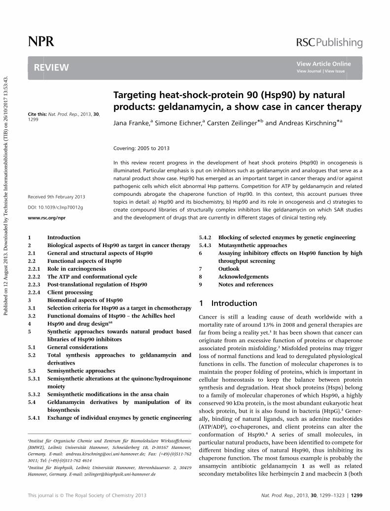
Cancer is still a leading cause of death worldwide with amortality rate of around 13% in 2008 and general therapies arefar from being a reality yet.1 It has been shown that cancer canoriginate from an excessive function of proteins or chaperoneassociated protein misfolding.2 Misfolded proteins may triggerloss of normal functions and lead to deregulated physiologicalfunctions in cells. The function of molecular chaperones is tomaintain the proper folding of proteins, which is important incellular homeostasis to keep the balance between proteinsynthesis and degradation. Heat shock proteins (Hsps) belongto a family of molecular chaperones of which Hsp90, a highlyconserved 90 kDa protein, is the most abundant eukaryotic heatshock protein, but it is also found in bacteria (HtpG).3 Gener-ally, binding of natural ligands, such as adenine nucleotides(ATP/ADP), co-chaperones, and client proteins can alter theconformation of Hsp90.4 A series of small molecules, inparticular natural products, have been identied to compete fordifferent binding sites of natural Hsp90, thus inhibiting itschaperone function. The most famous example is probably theansamycin antibiotic geldanamycin 1 as well as relatedsecondary metabolites like herbimycin 2 and macbecin 3 (both
Nat. Prod. Rep., 2013, 30, 1299–1323 | 1299
NPR Review
Publ
ishe
d on
12
Aug
ust 2
013.
Dow
nloa
ded
by T
echn
isch
e In
form
atio
nsbi
blio
thek
(T
IB)
on 2
6/10
/201
7 13
:53:
43.
View Article Online
quinone and hydroquinone forms exist) and the phenolicmetabolite reblastatin (5) (Fig. 1).5Also autolytimycin (4) wasisolated in the early 2000s, both from Streptomyces sp. S6699as well as from a culture broth of Streptomyces autolyticusJX-47 from which its name was derived.6a Only recently, Wuand coworkers reported several new metabolites 6–8 fromStreptomyces hygroscopicus 17997, all of which are functional-ised at C-19.6c–e
It is important to note that these natural products havepaved the way for a deeper understanding of heat shockproteins, and as a result Hsp90 has emerged as a remarkabletherapeutic target for the treatment of cancer. In this contextthe identication of binding sites of the target receptor, such asHsp90, that signicantly affect its function is important. Thisreview is intended to provide an overview and a story on rstlythe biochemistry of heat shock protein 90 which will be linkedto biomedical and pharmaceutical aspects and options forcancer therapy. Secondly, the preparation of geldanamycin
Jana Franke received herdiploma in chemistry from theLeibniz University of Hannoverin 2010, and was a visitingscholar with Dr Martin D. Smithat Oxford University, UK, in2009. She is currently pursuinga Ph.D. under the supervision ofProf. Dr Andreas Kirschning atthe Leibniz University of Hann-over. Her current research isfocused on natural productsynthesis.
Simone Eichner studied chem-istry at the Leibniz University ofHannover. She joined the groupof Prof. Andreas. Kirschning andreceived her PhD in 2011working in the eld of mutasyn-thesis. Aerwards she joinedratiopharm GmbH where shehas worked in chemical researchsince then.
1300 | Nat. Prod. Rep., 2013, 30, 1299–1323
libraries to uncover structure–activity relationship (SAR)knowledge will be discussed in detail as geldanamycin is ashowcase for generating libraries by chemical as well asbiosynthetic methods.
Thirdly, issues on assaying the inhibition of Hsp90 by gel-danamycin (1) will be included in the review, that along withsynthetic efforts resulted in “SAR-mapping” of this complexnatural product.
2 Biological aspects of Hsp90 as target incancer therapy2.1 General and structural aspects of Hsp90
Hsp90 is highly conserved and it is present in prokaryotes andeukaryotes, with the exception of archaea where only small heatshock proteins exist.9 The human heat shock protein 90(Hsp90a) is an 855 aa protein of 98.1 kDa, encoded by theHsp90aa1 gene on chromosome 14, whereas the cytoplasmic
Carsten Zeilinger studiedbiology at the University ofOsnabruck (Germany). InOsnabruck he joined the groupof Prof. Elmar W. Weiler andreceived his PhD in 1990working in the eld of plantphysiology. Aer a postdoctoralstay at the University ofGottingen (Germany) with Prof.R. Hedrich he started his inde-pendent research at the Univer-sity of Hannover in 1991, where
he nished his habilitation in 1998. His research interests covermembrane proteins and protein folding, temperature effects onprotein function and assay design for studying protein function.
Andreas Kirschning studiedchemistry at the University ofHamburg and at SouthamptonUniversity (UK). In Hamburg, hejoined the group of Prof. ErnstSchaumann and received hisPhD in 1989 working in the eldof organosilicon chemistry. Aera postdoctoral stay at theUniversity of Washington (Seat-tle, USA) with Prof. Heinz G.Floss, he started his independentresearch at the Clausthal
University of Technology in 1991, where he nished his habilita-tion in 1996. In 2000 he moved to the Leibniz University Hannover.His research interests cover structure elucidation as well as thesemi-, total and mutasynthesis of natural products, biomedicalbiopolymers, and synthetic technologies (solid-phase assistedsynthesis, microreactors and inductive heating).
This journal is ª The Royal Society of Chemistry 2013
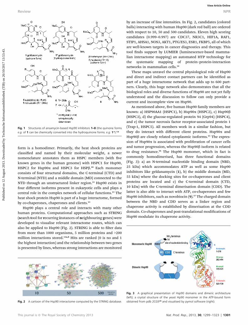
Fig. 1 Structures of ansamycin-based Hsp90 inhibitors 1–8 (the quinone formse.g. of 1 can be chemically converted into the hydroquinone forms. e.g. 1*).7,8
Review NPR
Publ
ishe
d on
12
Aug
ust 2
013.
Dow
nloa
ded
by T
echn
isch
e In
form
atio
nsbi
blio
thek
(T
IB)
on 2
6/10
/201
7 13
:53:
43.
View Article Online
form is a homodimer. Primarily, the heat shock proteins areclassied and named by their molecular weight, a newernomenclature annotates them as HSPC members (with veknown genes in the human genome) with HSPC1 for Hsp90,HSPC2 for Hsp90a and HSPC3 for HSPb.10 Each monomerconsists of four structural domains, the C-terminal (CTD) andN-terminal (NTD) and a middle domain (MD) connected to theNTD through an unstructured linker region.11 Hsp90 exists infour different isoforms present in eukaryotic cells and plays acentral role in the complex network of cellular functions.12 Theheat shock protein Hsp90 is part of a huge interactome, formedby co-chaperones, chaperones and clients.13
Hsp90 plays a central role and interacts with many otherhuman proteins. Computational approaches such as STRING(search tool for recurring instances of neighbouring genes) weredeveloped to visualise relevant interactome routes, which canalso be applied to Hsp90 (Fig. 2). STRING is able to lter datafrom more than 1000 organisms, 5 million proteins and >200million interactions stored.13a,b Hits are ranked (0 is no and 1the highest interaction) and the relationship between two genesis presented by lines, whereas strong interactions are monitored
Fig. 2 A cartoon of the Hsp90 interactome computed by the STRING database.
This journal is ª The Royal Society of Chemistry 2013
by an increase of line intensities. In Fig. 2, candidates (coloredballs) interacting with human Hsp90 (dark red ball) are orderedwith respect to 10, 50 and 500 candidates. Eleven high scoringcandidates (0.999–0.997) are CDC37, NR3C1, HIF1A, RAF1,STIP1, AHSA1, NOS3, AKT1, PTG/ES3, ESR1, FKBP5, all of whichare well-known targets in cancer diagnostics and therapy. Thistool nds support by LUMIER (luminescence-based mamma-lian interactome mapping) an automated HTP technology forthe systematic mapping of protein–protein-interactionnetworks in mammalian cells.14
These maps unravel the central physiological role of Hsp90and direct and indirect contact partners can be identied aspart of a huge interactome network that adds up to 600 part-ners. Clearly, this huge network also demonstrates that all thebiological roles and diverse functions of Hsp90 are not yet fullyunderstood and the discussion to follow can only provide acurrent and incomplete view on Hsp90.
As mentioned above, ve human Hsp90 family members areknown: a) HSP90AA1 (HSPC1), b) Hsp90a (HSPC2), c) Hsp90b(HSPC3), d) the glucose-regulated protein 94 (Grp94) (HSPC4),and e) the tumor necrosis factor receptor-associated protein 1(Trap-1, HSPC5). All members work in a similar fashion, butthey do interact with different client proteins. Hsp90a andHsp90b are closely related cytoplasmic isoforms.15 The expres-sion of Hsp90a is associated with proliferation of cancer cellsand tumor progression, whereas the Hsp90b isoform is relatedto drug resistance.16 The Hsp90 monomer, which in fact iscommonly homodimerised, has three functional domains(Fig. 3): a) an N-terminal nucleotide binding domain (NBD,25 kDa) which accommodates ATP as well as some Hsp90inhibitors like geldanamycin (1), b) the middle domain (MD,55 kDa) where the docking sites for co-chaperones and clientproteins are located and c) the C-terminal domain (CTD,10 kDa) with the C-terminal dimerisation domain (CDD). Thelatter is also able to interact with ATP, co-chaperones and fewHsp90 inhibitors, such as novobiocin (9).17 The charged domainbetween the NBD and CDD serves as a linker region andchaperone activity is established by dimerisation at the CDDdomain. Co-chaperones and post-translational modications ofHsp90 modulate its chaperone activity.
Fig. 3 A graphical presentation of Hsp90 domains and dimeric architecture(left); a crystal structure of the yeast Hp90 monomer in the ATP-bound formobtained from pdb 2CG918 and visualised by pymol software (right).
Nat. Prod. Rep., 2013, 30, 1299–1323 | 1301

NPR Review
Publ
ishe
d on
12
Aug
ust 2
013.
Dow
nloa
ded
by T
echn
isch
e In
form
atio
nsbi
blio
thek
(T
IB)
on 2
6/10
/201
7 13
:53:
43.
View Article Online
By inhibiting the Hsp90 chaperone, interference in cancercell cycles becomes possible. Thus, compounds that are able tobind to different sites in Hsp90 are potential anticancer drugs.Today three major strategies for inhibiting Hsp90 function arebeing pursued. These are:
a) the inhibition of ATPase activity by binding at theN-terminal nucleotide binding pocket for which geldanamycin(1), radicicol (13) and PU-H71 (12) are excellent examples (Fig. 7),
b) altering the conformation of Hsp90 activity with smallmolecules like novobiocin (9) (Fig. 4) that bind to the C-terminaldimerisation domain (CDD) and
c) the inhibition of co-chaperones-binding, such as Cdc37which is overexpressed in cancer cells, with Hsp90, as imposedby gedunin (10) and celastrol (11) (Fig. 4).
In addition, Hsp90 activity is regulated by targeting sitesrelevant for the association of Hsp90 with client proteins, sitesthat modulate Hsp90 activity as well as sites for the inhibition ofpost-translational modications, such as phosphorylation,acetylation and S-nitrosylation. Details on the biological back-ground of these options are discussed in the following chapters.
2.2 Functional aspects of Hsp90
2.2.1 Role in carcinogenesis. Heat shock proteins (Hsp's)are essential for the survival of organisms. In contrast to whatthe namemay suggest, heat shock proteins are not only inducedby heat but also by cellular stress situations, such as exposure toUV radiation, nutrient deprivation or oxygen deciency19,20
Hsp's and especially human heat shock protein 90 (Hsp90)strongly interfere with diverse cellular processes, which includeheat shock and other stress responses, signal transduction tochromatin-remodeling, telomerase maintenance and others.11a
Hsp90 is also located in the nucleus where it interacts withchromatin-remodeling complexes.21,22a A link to the histonemethyltransferase SMYD3 was also found.22b Additionally, Hspco-chaperones inuence DNA helicases in yeast.20f Examina-tions in yeast revealed that Hsp82p, the yeast homologue ofHsp90, is required for both DNA binding and extension of thetelomerase.23 The non-chaperone activity might provide a pointof intervention to mitigate excessive telomerase function andmay be a noteworthy aspect of cancer etiology. Shortening oftelomeres by telomerase activity is correlated with age. For thedevelopment of anti-aging concepts, interference in the foldingof telomerase by Hsp90 inhibitors may be a feasible strategy. Inthe context of therapeutic exploitation of Hsp90 inhibition, it is
Fig. 4 Structures of novobiocin (9), gedunin (10) and celastrol (11).
1302 | Nat. Prod. Rep., 2013, 30, 1299–1323
important to understand the effect and relevance of high or lowHsp90 activity for the cell. Along this line it will be important toestablish a correlation between drug-induced levels of Hsp90and associated toxic side effects.
Besides the role in the nucleosome, Hsp90 client manage-ment affects various other cellular processes. Hsp90 governs thefolding process of nascent polypeptides leading to maturationand formation of intact and functional three-dimensionalstructures.24 Not only de novo synthesised proteins are folded byHsp90, but also proteins that have been denatured by cellularstress. The cellular response to stress is highly conserved inprokaryotes and all species of eukaryotes. It represents a generalmechanism for the maintenance of cellular processes. Inaddition, it is a protection mechanism against the formation ofprotein aggregates in cytosol, whereas in some prokaryotesHsp90 is dispensable under heat stress.25 The initiation ofHsp90 synthesis and other Hsp's are evoked by specic tran-scription factors, so-called heat shock factors (HSF's). Thetrimeric form of HSF shows a high affinity for cis-acting DNAsequence elements (heat shock elements; HSEs) in thepromoter region of heat shock protein genes.26 The boundtrimer forms a complex that activates transcription of the Hsp90gene. Hsp90 is in direct contact with HSF, and stress cansignicantly increase Hsp90 expression.5a,27 The unique prop-erty of Hsp90 to fold nascent as well as denatured proteins,including mutant forms of proteins, has a special impact forcancer and therefore represents an ideal target for drugdevelopment.20a,28
Due to the antiapoptotic nature of Hsp90, cells can survivestress with the side effect that this property also helps cancercells in survival. The increased expression of Hsp90 underconditions of stress is a rescue mechanism of cells, because theproteins are protected from degradation by the proteasomicsystem. Furthermore, the “recycling” of proteins by Hsp90clearly has advantages in terms of energy balance compared tode novo protein synthesis. For several proteins, which are alsoknown as client proteins or target proteins, Hsp90 is requiredfor preserving their stability and activity. Client proteinscomprise up to 600 proteins, including proteins with excep-tional features in signal transduction, cell growth and division.For example, receptors such as connexin,29 steroid hormones,transcription factors and tumor suppressor proteins are Hsp90dependent.22d
If protein aggregates are not eliminated by the proteasome,apoptosis is initiated.20f,30 Thus, this process is a key element incancer therapy, when Hsp90 function can be suppressed byinhibitors. Besides its importance in cancer therapy, Hsp90along with its homologues (HtpG's in prokaryotes), hasemerged to be an attractive target combating other diseases,since there are homologous representatives in nearly allprokaryotic and eukaryotic cells.31 Indeed, a link to otherdiseases, like neurological disorders, malaria or leishmania, isknown. The development of different virus proteins are alsoHsp dependent.32 Yet another implication of Hsp90 (and Grp94)is in processing antigens and helping to deliver them to the cellsurface in MHC complexes or higher Hsp70 levels in humanT-cells.33 This strategy, however, can lead to a problematic
This journal is ª The Royal Society of Chemistry 2013

Review NPR
Publ
ishe
d on
12
Aug
ust 2
013.
Dow
nloa
ded
by T
echn
isch
e In
form
atio
nsbi
blio
thek
(T
IB)
on 2
6/10
/201
7 13
:53:
43.
View Article Online
situation for adherent therapies, since immune defense couldbe differently affected by high or low Hsp activity.
Under non stress conditions 1–2% Hsp90a and Hsp90b arepresent in the cell as cytosolic proteins. Hsp90a serves as theinducible and Hsp90b as the constitutive form. GRP94 is foundin the endoplasmic reticulum and TRAP-1 is localised in themitochondrial matrix. Except for Hsp90N, which has no NTD,all forms of Hsp90, including the bacterial homologue HtpG,are able to hydrolyse ATP. Unlike the other isoforms of Hsp90,the cytosolic function of Hsp90 is highly dependent on anumber of co-chaperones, as is described in the next chapter.
2.2.2 The ATP and conformational cycle. The dimeric formof Hsp90 (I/II) acts as a molecular “clip” and suffers fromconformational change by an ATP-dependent folding cycle(Fig. 5).20a,32,34 During the ATPase cycle, Hsp90 undergoesdimerisation at the N-terminus (II). Association of ATP (III) atthis domain is the driving force for this step. The target clientproteins are captured with the open form (V-shape) ofHsp90.21,35 As a result binding of ATP promotes closure of the lidof the N-terminal ATP-binding pocket, the coming together ofthe two N-termini of Hsp90 and a conformational change fromthe closed into the twisted form while “trapping” the client.Aer hydrolysis of ATP, Hsp90 folds back into its original shapeand the open molecular “clip” is regenerated. Binding andsubsequent release of the client proteins with the participationof co-chaperones is a dynamic process, whereas the rate of ATPhydrolysis and the conformational change between thedifferent Hsp-conformers varies. For the human Hsp90, this is aslow turnover rate of 3 ATP h�1.36 The conformational changedoes not necessarily depend on ATP; so the closed form ofHsp90 is also found in the absence of ATP. It is rather assumedthat ATP merely shis the balance between the “open” and“closed” form of Hsp90. Recent data obtained from singlemolecule measurements showed that ATP can bind at the N-terminus of the open and closed states (III to IV) without strictlyforcing the protein into a specic conformation. The switchesbetween the conformational and binding states are mainlythermally driven. Interestingly, ATP binds with different rates tothe two monomeric units (negative cooperativity).37 Thesestudies also revealed that the C-terminus shows a dynamicbehavior. The C-terminus (IV) opens and closes with fast
Fig. 5 A model of the ATPase and conformational cycle of Hsp90 proteins;forward and backward reaction rates are different for the individual Hsp90proteins and may be subjected to regulation by co-chaperones, such as Aha1,Hop, Cdc37 and p23 (see Fig. 6).
This journal is ª The Royal Society of Chemistry 2013
kinetics, having a modulating effect on the binding of nucleo-tides to the N-terminal domain.38
Although Hsp's and HtpG share a high degree of homology,drastic differences in structure and function between Hsp's andHtpG's are found in crystal structures, the interactome and ATPhydrolysis rates.39
The regulation of the ATPase activity and selectivity for clientproteins by cytosolic Hsp90 are signicantly inuenced by co-chaperones. In fact, more than 20 co-chaperones regulateHsp90 by modulating ATP hydrolysis (V/VI) (Aha1, Cdc37, p23),by inuencing the conformational exibility (p23, Sgt1) and byregulating complex assembly (Hop, Cdc37, Sgt1). They may alsobe required for folding other co-chaperones, such as Hsp70 andHsp40.40 During the ATPase cycle in eukaryotes different co-chaperones assemble to yield the so-called “multi-chaperone”machinery.41 The whole process was examined in detail for thematuration of a steroid hormone receptor in yeast. The cyclestarts with association of the newly synthesised and stillunfolded protein to an early complex of heat shock proteinsHsp70/Hsp40 (I) (Fig. 6).19b Complexation of this intermediatecomplex (I) to Hsp90 is facilitated by another co-chaperonecalled Hop, which contains the tetratricopeptide repeat (TPR)receptor domain and forms complex (II/II0). TPR, which hasPPIase activity, binds to the MEEVDmotif (several Hsp's and co-chaperons have this element, which is located at the CTD) inHsp90. This step prevents dimerisation of the N-termini ofHsp90 and serves as an adapter for transfer of the client proteinfromHsp70 to Hsp90 (III). This Hsp90/Hsp70/Hop complex actsas the central intermediate in the Hsp90 cycle.42 Aer bindingATP (IV) the intermediate complex forms a non-symmetriccomplex. In the presence of p23 this late complex (V) is stabi-lised, by which the co-chaperones Hop, Hsp 40 and Hsp70 arereleased. Both, p23 and Cdc37 inhibit HSP90 ATPase activityand bind near the ATP-binding pocket of Hsp90. Cdc37 xes theN-domain in an open state form, thereby preventing dimerisa-tion. Aer hydrolysis of ATP the open conformation of Hsp90(VI) is liberated. Another co-chaperone, called Aha1 (activator ofHsp90 ATPase, non TPR co-chaperone), can alternatively bind tothe Hsp90 dimer (II0). This chaperone thereby forms a linkbetween the middle domain M or the client binding site, and
Fig. 6 The folding cycle of Hsp90 with co-chaperones.
Nat. Prod. Rep., 2013, 30, 1299–1323 | 1303
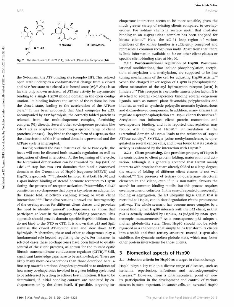
Fig. 7 The structures of PU-H71 (12), radicicol (13) and sulforaphane (14).
NPR Review
Publ
ishe
d on
12
Aug
ust 2
013.
Dow
nloa
ded
by T
echn
isch
e In
form
atio
nsbi
blio
thek
(T
IB)
on 2
6/10
/201
7 13
:53:
43.
View Article Online
the N-domain, the ATP binding site (complex III0). This relaxedopen state undergoes a conformational change from a closedand ATP free state to a closed ATP-bound state (IV).43 Aha1 is sofar the only known activator of ATPase activity by asymmetricbinding to a single Hsp90 middle domain in the open cong-uration. Its binding induces the switch of the N-domains intothe closed state, leading to the acceleration of the ATPasecycle.43 It has been proposed, that Aha1 competes for p23.Accompanied by ATP hydrolysis, the correctly folded protein isreleased from the multi-chaperone complex, furnishingcomplex (VI) directly. Several other co-chaperone proteins likeCdc37 act as adapters by recruiting a specic range of clientproteins (kinases). They bind to the open form of Hsp90, so thatthe dimerisation of the N-terminal domain is prevented and theATPase cycle is interrupted.
Having outlined the basic features of the ATPase cycle, thefocus will next be directed both towards regulation as well asintegration of client interaction. At the beginning of the cycle,the N-terminal dimerisation can be thwarted by Hop (Sti1) orCdc37.44 Hop has two TPR domains that bind a conserveddomain at the C-terminus of Hsp90 (sequence MEEVD) andHsp70, respectively.39,45 It should be noted, that both Hsp70 andHsp40 induce binding of steroid hormone receptors to Hsp90during the process of receptor activation.45Meanwhile, Cdc37constitutes a co-chaperone that plays a key role as an adapter forthe kinase fold, selectively enabling strong or weak clientinteractions.14,46 These observations unravel the heterogeneityof the co-chaperones for different client classes and provokesthe need to identify ‘general’ co-chaperones, i.e. those thatparticipate at least in the majority of folding processes. Thisapproach should provide domain-specic Hsp90 inhibitors thatdo not bind to the NTD or CTD. It is known that p23 and Sgt1stabilise the closed ATP-bound state and slow down ATPhydrolysis.44a Therefore, these and other co-chaperones play afundamental role beyond regulating the cycle. For instance, inselected cases these co-chaperones have been linked to qualitycontrol of the client proteins, as shown for the mutant cysticbrosis transmembrane conductance regulator (CFTR).48 Still,signicant knowledge gaps have to be acknowledged. There arelikely many more co-chaperones than those described here. Arst step towards a rational drug design would be to understandhow many co-chaperones involved in a given folding cycle needto be addressed by a drug to achieve best inhibition. It has to bedetermined, if initial bonding contacts are mediated by co-chaperones or by the client itself. If possible, targeting co-
1304 | Nat. Prod. Rep., 2013, 30, 1299–1323
chaperone interaction seems to be more sensible, given themuch greater variety of existing clients compared to co-chap-erones. For solitary clients a surface motif that mediatesbinding to an Hsp90–Cdc37 complex has been analysed forkinase clients.47 Here, the aC–b4 loop region of variousmembers of the kinase families is sufficiently conserved andrepresents a common recognition motif. Apart from that, thereis little information available so far on other client classes orspecic client-binding sites at Hsp90.
2.2.3 Post-translational regulation of Hsp90. Post-trans-lational modications, that include phosphorylation, acetyla-tion, nitrosylation and methylation, are supposed to be netuning mechanisms of the cell for adjusting Hsp90 activity.49
When the charged linker region of Hsp90 is phosphorylated,client maturation of the aryl hydrocarbon receptor (AHR) ishindered.50 This receptor is a cytosolic transcription factor. It isattached to several co-chaperones binding several exogenousligands, such as natural plant avonoids, polyphenolics andindoles, as well as synthetic polycyclic aromatic hydrocarbonsand dioxin-derived compounds. In addition, many kinases thatregulate Hsp90 phosphorylation are Hsp90 clients themselves.51
Acetylation can inuence client protein maturation andco-chaperone binding, and it is reported that acylation mayreduce ATP binding of Hsp90.52 S-nitrosylation at theC-terminal domain of Hsp90 leads to the reduction of Hsp90ATPase activity.53 SMYD3, a lysine methyltransferase, is upre-gulated in several cancer cells, and it was found that its catalyticactivity is enhanced by the interaction with Hsp90.54
2.2.4 Client processing. One prevalent property of Hsp90 isits contribution to client protein folding, maturation and acti-vation. Although it is generally accepted that Hsp90 mainlyinteracts with proteins that are already folded to a large degree,the extent of folding of different client classes is not welldened.19b The presence of tertiary or quarternary structuralelements in the client, even if it is non-native, favours thesearch for common binding motifs, but this process requiresco-chaperones or cofactors. In the case of repeated unsuccessfulfolding or aggregation, the E3 ubiquitin ligase CHIP, that isrecruited to Hsp90, can initiate degradation via the proteasomepathway. The whole scenario has become more complex by arecent nding that Hsp90 interacts with the p53 client, in thatp53 is actually unfolded by Hsp90a, as judged by NMR spec-troscopic measurements.55 As a consequence p53 adopts amolten globule-like state. Thus, Hsp90 should not be solelyregarded as a chaperone that simply helps transform its clientsinto a stable and xed tertiary structure. Instead, Hsp90 alsostabilises the dynamic molten globule state, which may fosterother protein interactions for those clients.
3 Biomedical aspects of Hsp903.1 Selection criteria for Hsp90 as a target in chemotherapy
Hsp90 plays a key role in a diverse range of diseases, such asischemia, reperfusion, infections and neurodegenerativediseases.56 However, from a pharmaceutical point of viewits participation in the development and control of variouscancers is most important. In cancer cells, an increased Hsp90
This journal is ª The Royal Society of Chemistry 2013

Fig. 8 Graphics of X-ray structures of Hsp90/small molecules (ATP, geldanamy-cin (1) and 17AAG 15) (left) and important molecular interactions (right): a)Hsp90N with ATP (3T0Z)65 (EC50 ¼ 200 nM); b) Hsp90N with geldanamycin(1YET)66 (IC50 ¼ 20–200 nM); c) Hsp90N with 17AAG (1OSF)67 (IC50 ¼ 24 nM). Allstructural data are deposited at www.pdb.org.
Review NPR
Publ
ishe
d on
12
Aug
ust 2
013.
Dow
nloa
ded
by T
echn
isch
e In
form
atio
nsbi
blio
thek
(T
IB)
on 2
6/10
/201
7 13
:53:
43.
View Article Online
expression parallels the overexpression of oncoproteins likeErb2, EGFR, c-RAF, HIF-1 and telomerase. These facts makeHsp90 a key target in cancer chemotherapy.57 Signal trans-duction pathways like G-protein-coupled receptors, low-molec-ular-weight GTP binding protein, tyrosine kinase, Ser/Thrkinase, ion channel receptors, nuclear pore channel andnuclear transcription factors are important elements in severaldiseases and may also expand the opportunities for Hsp90-based therapies. By identifying cancer-specic interactomes,such as the Hsp90 inhibitor PU-H71 (12), Hsp90-dependentoncogenic client proteins have been captured by pull downassays.58 This assay helped to comb through the cancer-linkedproteome using mass spectrometry and new aberrant signal-osomes in CML cells (Chronic Myelogenous Leukemia; cancercell lines of white blood cells) were found. One important keyregulator that was identied by this method is STAT5. STAT5 isa molecular regulator for proliferation, differentiation andapoptosis in hematopoietic cells, which are multipotent stemcells giving rise to all new types of blood cells.
Among the relevant oncoproteins, the transcription factorp53 is probably the most prominent one, sometimes beingreferred to as "guardian of the genome". In about 50% of allcancers, p53 is damaged and the loss of function is caused byso-called “hot spot” mutations in the DNA-binding region.59
Functionally, the detection of DNA damage and subsequentinitiation of apoptosis or repair mechanisms are the mostimportant features of p53. A rescue mechanism of a defectivep53 system is fatal, because the mechanisms of apoptosis canthus be evaded. Physiologically, tumor cells are under constantstress; so proteins actually tend to denature.60 This situationleads to an increased consumption of molecular chaperones, asreected in an increase of the cytosolic Hsp90 fraction to up to4–5%, which leads to higher heat shock protein activity intumor cells compared to healthy cells.61,62 The effect of Hsp90blockers is more pronounced in tumor cells than in healthycells due to the higher concentration of Hsp90, which isaccompanied by overproduction of Hsp90-dependent oncopro-teins. The increased presence of the “multi-chaperone”machinery in cancer cells results in a higher affinity for inhib-itors that bind to the N-terminus of Hsp90. In normal cells, themajor amount of Hsp90 is not present in multi-chaperonecomplexes and in this state it shows lower affinity for inhibitors.As a result, inhibition initiates apoptosis or stops elevatedgrowth rates.
In essence, the pronounced accumulation of Hsp90 incancer cells along with the opportunity to develop selectiveinhibitors against Hsp90 and cancer relevant multi-chaperonecomplexes are important selection criteria to identify Hsp90 asa promising target in anticancer therapy.
3.2 Functional domains of Hsp90 – the Achilles heel
The establishment of Hsp90 as a biological target in anti-tumortherapy initiated the search for inhibitors in academia and thepharmaceutical industry. The most prominent Hsp90 inhibitoris geldanamycin (1), that was rst isolated from the actinomy-cete Streptomyces hygroscopicus. Geldanamycin acts by binding
This journal is ª The Royal Society of Chemistry 2013
to the N-terminal ATP-binding site of Hsp90, the ATPase func-tion (see Fig. 6).63 The affinity of geldanamycin for the ATP-binding site is 100-fold higher than that of ATP (Fig. 8).
It binds to the site in that it adopts the structure of anunfolded polypeptide chain. The most important contacts ofATP interaction sites on the Hsp90N crystal are Met84, Asp79,Gly118, 121, 123, Phe124 and Thr170, while Asp79, Lys44, Lys98and Phe124 are relevant for geldanamycin binding and thesemisynthetic derivative 17AAG 12 (vide supra). They are notonly relevant for the inhibitory role of geldanamycin but also forthe semisynthetic derivative 17AAG 15 (vide supra). Uponbinding of geldanamycin and analogues, Hsp90 is retained inits ADP-bound conformation so that these ligands preventHsp90 from promoting the ATP cycle (Fig. 5 and 6). As a result,the client protein is ubiquitinated and then degraded by theproteasomic machinery. Moreover, the inactivation and desta-bilisation of the hypoxia-induced factor (HIF)-1a is induced.This results in the degradation of HIF-1a. As a consequence ofinhibition, apoptosis or programmed cell death is initiated.Another competitive ATPase binder is the polyketide radicicol(13). It was isolated as a secondary metabolite from the fungiMonocillium nordinii and Monosporium bonorden. Only recently,sulforaphane (14), an isothiocyanate derived from cruciferousvegetables, has been shown to possess potent chemopreventiveactivity by inhibiting pancreatic cancer cell growth (IC50 � 10–15 mM). It has been suggested that it also disrupts protein–protein interactions in Hsp90 complexes.64
4 Hsp90 and drug design68
The interest in Hsp90 inhibitors has recently found an addi-tional impetus, because heat shock proteins can also serve as a
Nat. Prod. Rep., 2013, 30, 1299–1323 | 1305
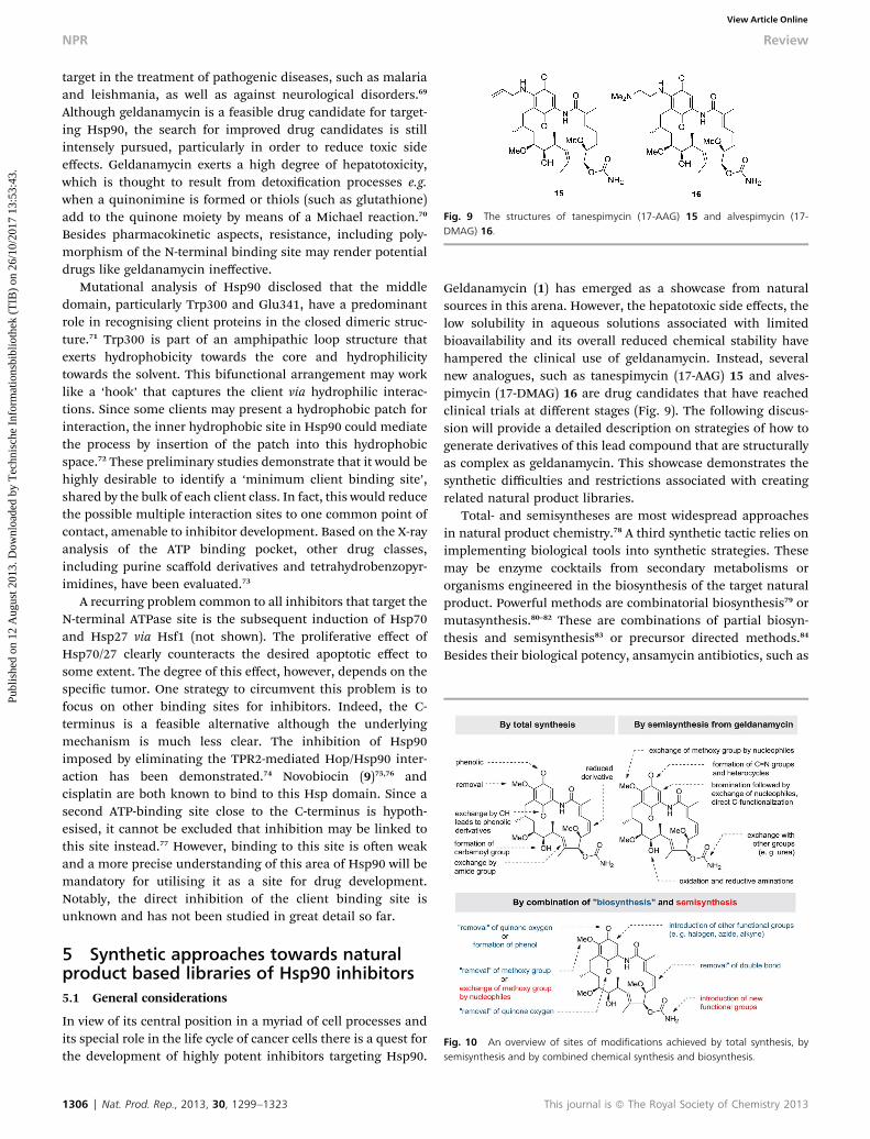
Fig. 9 The structures of tanespimycin (17-AAG) 15 and alvespimycin (17-DMAG) 16.
NPR Review
Publ
ishe
d on
12
Aug
ust 2
013.
Dow
nloa
ded
by T
echn
isch
e In
form
atio
nsbi
blio
thek
(T
IB)
on 2
6/10
/201
7 13
:53:
43.
View Article Online
target in the treatment of pathogenic diseases, such as malariaand leishmania, as well as against neurological disorders.69
Although geldanamycin is a feasible drug candidate for target-ing Hsp90, the search for improved drug candidates is stillintensely pursued, particularly in order to reduce toxic sideeffects. Geldanamycin exerts a high degree of hepatotoxicity,which is thought to result from detoxication processes e.g.when a quinonimine is formed or thiols (such as glutathione)add to the quinone moiety by means of a Michael reaction.70
Besides pharmacokinetic aspects, resistance, including poly-morphism of the N-terminal binding site may render potentialdrugs like geldanamycin ineffective.
Mutational analysis of Hsp90 disclosed that the middledomain, particularly Trp300 and Glu341, have a predominantrole in recognising client proteins in the closed dimeric struc-ture.71 Trp300 is part of an amphipathic loop structure thatexerts hydrophobicity towards the core and hydrophilicitytowards the solvent. This bifunctional arrangement may worklike a ‘hook’ that captures the client via hydrophilic interac-tions. Since some clients may present a hydrophobic patch forinteraction, the inner hydrophobic site in Hsp90 could mediatethe process by insertion of the patch into this hydrophobicspace.72 These preliminary studies demonstrate that it would behighly desirable to identify a ‘minimum client binding site’,shared by the bulk of each client class. In fact, this would reducethe possible multiple interaction sites to one common point ofcontact, amenable to inhibitor development. Based on the X-rayanalysis of the ATP binding pocket, other drug classes,including purine scaffold derivatives and tetrahydrobenzopyr-imidines, have been evaluated.73
A recurring problem common to all inhibitors that target theN-terminal ATPase site is the subsequent induction of Hsp70and Hsp27 via Hsf1 (not shown). The proliferative effect ofHsp70/27 clearly counteracts the desired apoptotic effect tosome extent. The degree of this effect, however, depends on thespecic tumor. One strategy to circumvent this problem is tofocus on other binding sites for inhibitors. Indeed, the C-terminus is a feasible alternative although the underlyingmechanism is much less clear. The inhibition of Hsp90imposed by eliminating the TPR2-mediated Hop/Hsp90 inter-action has been demonstrated.74 Novobiocin (9)75,76 andcisplatin are both known to bind to this Hsp domain. Since asecond ATP-binding site close to the C-terminus is hypoth-esised, it cannot be excluded that inhibition may be linked tothis site instead.77 However, binding to this site is oen weakand a more precise understanding of this area of Hsp90 will bemandatory for utilising it as a site for drug development.Notably, the direct inhibition of the client binding site isunknown and has not been studied in great detail so far.
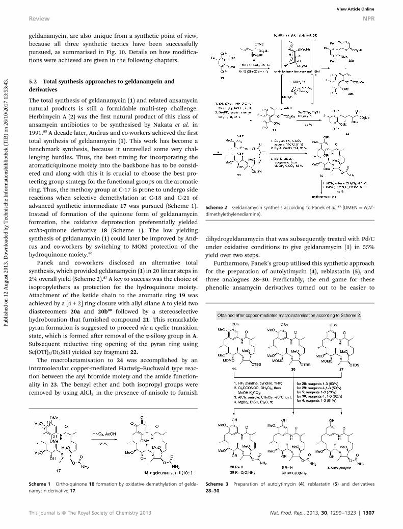
Fig. 10 An overview of sites of modifications achieved by total synthesis, bysemisynthesis and by combined chemical synthesis and biosynthesis.
5 Synthetic approaches towards naturalproduct based libraries of Hsp90 inhibitors5.1 General considerations
In view of its central position in a myriad of cell processes andits special role in the life cycle of cancer cells there is a quest forthe development of highly potent inhibitors targeting Hsp90.
1306 | Nat. Prod. Rep., 2013, 30, 1299–1323
Geldanamycin (1) has emerged as a showcase from naturalsources in this arena. However, the hepatotoxic side effects, thelow solubility in aqueous solutions associated with limitedbioavailability and its overall reduced chemical stability havehampered the clinical use of geldanamycin. Instead, severalnew analogues, such as tanespimycin (17-AAG) 15 and alves-pimycin (17-DMAG) 16 are drug candidates that have reachedclinical trials at different stages (Fig. 9). The following discus-sion will provide a detailed description on strategies of how togenerate derivatives of this lead compound that are structurallyas complex as geldanamycin. This showcase demonstrates thesynthetic difficulties and restrictions associated with creatingrelated natural product libraries.
Total- and semisyntheses are most widespread approachesin natural product chemistry.78 A third synthetic tactic relies onimplementing biological tools into synthetic strategies. Thesemay be enzyme cocktails from secondary metabolisms ororganisms engineered in the biosynthesis of the target naturalproduct. Powerful methods are combinatorial biosynthesis79 ormutasynthesis.80–82 These are combinations of partial biosyn-thesis and semisynthesis83 or precursor directed methods.84
Besides their biological potency, ansamycin antibiotics, such as
This journal is ª The Royal Society of Chemistry 2013
Review NPR
Publ
ishe
d on
12
Aug
ust 2
013.
Dow
nloa
ded
by T
echn
isch
e In
form
atio
nsbi
blio
thek
(T
IB)
on 2
6/10
/201
7 13
:53:
43.
View Article Online
geldanamycin, are also unique from a synthetic point of view,because all three synthetic tactics have been successfullypursued, as summarised in Fig. 10. Details on how modica-tions were achieved are given in the following chapters.
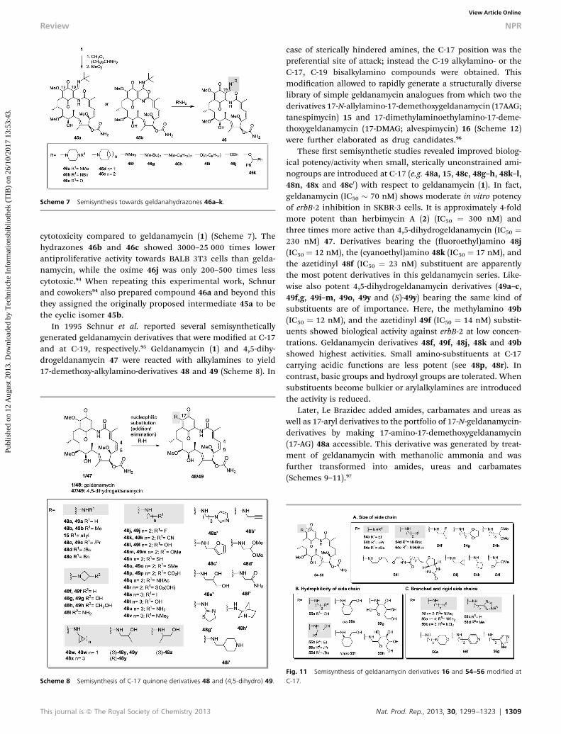
Scheme 2 Geldanamycin synthesis according to Panek et al.;87 (DMEN ¼ N,N0-dimethylethylenediamine).
5.2 Total synthesis approaches to geldanamycin andderivatives
The total synthesis of geldanamycin (1) and related ansamycinnatural products is still a formidable multi-step challenge.Herbimycin A (2) was the rst natural product of this class ofansamycin antibiotics to be synthesised by Nakata et al. in1991.85 A decade later, Andrus and co-workers achieved the rsttotal synthesis of geldanamycin (1). This work has become abenchmark synthesis, because it unravelled some very chal-lenging hurdles. Thus, the best timing for incorporating thearomatic/quinone moiety into the backbone has to be consid-ered and along with this it is crucial to choose the best pro-tecting group strategy for the functional groups on the aromaticring. Thus, the methoxy group at C-17 is prone to undergo sidereactions when selective demethylation at C-18 and C-21 ofadvanced synthetic intermediate 17 was pursued (Scheme 1).Instead of formation of the quinone form of geldanamycinformation, the oxidative deprotection preferentially yieldedortho-quinone derivative 18 (Scheme 1). The low yieldingsynthesis of geldanamycin (1) could later be improved by And-rus and co-workers by switching to MOM protection of thehydroquinone moiety.86
Panek and co-workers disclosed an alternative totalsynthesis, which provided geldanamycin (1) in 20 linear steps in2% overall yield (Scheme 2).87 A key to success was the choice ofisopropylethers as protection for the hydroquinone moiety.Attachment of the ketide chain to the aromatic ring 19 wasachieved by a [4 + 2] ring closure with allyl silane A to yield twodiastereomers 20a and 20b88 followed by a stereoselectivehydroboration that furnished compound 21. This remarkablepyran formation is suggested to proceed via a cyclic transitionstate, which is formed aer removal of the a-siloxy group in A.Subsequent reductive ring opening of the pyran ring usingSc(OTf)3/Et3SiH yielded key fragment 22.
The macrolactamisation to 24 was accomplished by anintramolecular copper-mediated Hartwig–Buchwald type reac-tion between the aryl bromide moiety and the amide function-ality in 23. The benzyl ether and both isopropyl groups wereremoved by using AlCl3 in the presence of anisole to furnish
Scheme 1 Ortho-quinone 18 formation by oxidative demethylation of gelda-namycin derivative 17.
This journal is ª The Royal Society of Chemistry 2013
dihydrogeldanamycin that was subsequently treated with Pd/Cunder oxidative conditions to give geldanamycin (1) in 55%yield over two steps.
Furthermore, Panek's group utilised this synthetic approachfor the preparation of autolytimycin (4), reblastatin (5), andthree analogues 28–30. Predictably, the end game for thesephenolic ansamycin derivatives turned out to be easier to
Scheme 3 Preparation of autolytimycin (4), reblastatin (5) and derivatives28–30.
Nat. Prod. Rep., 2013, 30, 1299–1323 | 1307

Scheme 4 Total synthesis of 18,21-diisopropyl-geldanamycin hydroquinone 36(according to Bach et al.90).
NPR Review
Publ
ishe
d on
12
Aug
ust 2
013.
Dow
nloa
ded
by T
echn
isch
e In
form
atio
nsbi
blio
thek
(T
IB)
on 2
6/10
/201
7 13
:53:
43.
View Article Online
perform than for the hydroquinonoic geldanamycin describedabove (Scheme 3).89
Bach's work90 (Scheme 4) demonstrates that future work onthe end game synthesis of quinone-based macrolactam antibi-otics will have to address an alternative protection strategy forthe hydroquinone moiety. Bach et al. based their sequence onconnecting C8 and C9 via a SmI2-mediated Reformatskycoupling reaction of a-chloro ketone 32 with aldehyde 33 toyield advanced intermediate 34. This product was transformedby Martin elimination into enone 35.90 However, aer lactamformation all efforts failed so far to remove the isopropyl groupsso that the total synthesis had to be terminated at the stage ofthe bisisopropyl geldanamycin derivative 36 (Scheme 4).Clearly, geldanamycin (1) remains a true synthetic challengeand future SAR studies cannot be expected to rely on totalsyntheses approaches alone.
Andrus et al. simplied the total synthesis approach to newgeldanamycin derivatives by installing an amide bond into theansa chain. By merging two different fragments 37 and 38 withthe advanced ansa chain fragment 39 they nally accessed
Scheme 5 A simplified total synthesis of geldanamycin derivatives 40a and 40b.
1308 | Nat. Prod. Rep., 2013, 30, 1299–1323
geldanamycin 8,9-amido-geldanamycin derivatives 40a and40b, respectively (Scheme 5). Unfortunately, both new gelda-namycin derivatives have lost their ability to promote HER2degradation in SK-Br3 cells (ED50 > 20 mM, compared to gelda-namycin with an ED50 of 5 nM).91
5.3 Semisynthetic approaches
5.3.1 Semisynthetic alterations at the quinone/hydroqui-none moiety. Semisynthesis is a widely employed technique toaccess new derivatives of complex natural products. The majorchallenge from a chemical point of view is to nd highly che-moselective transformations for a given multifunctional andoen chemically labile natural product.
Most known geldanamycin derivatives were prepared bysemisynthesis and in the majority of cases they are modied atC-17. Rinehart et al. synthesised geldanamycin derivatives,named geldanazines and geldanoxazinones by reacting gelda-namycin with o-phenylenediamines and o-aminophenols,respectively, via imine-formation at C-18 and substitution of theC-17-methoxy-group by means of an addition/eliminationprocess (Scheme 6). Both groups of derivatives showed lowinhibitory effects on the bacterial RNA polymerase but theyexerted higher potency in inhibiting the DNA polymerase oftumor cells compared to geldanamycin (1), while showingreduced cytotoxicity. Remarkably, all new geldanamycin deriv-atives except 44a, 43e and peptide 43g inhibited a new biolog-ical target, namely the RNA-dependent DNA polymerases(RDDP) of the Rauscher Leukemia Virus (RLV), which gelda-namycin does not address.92
Other “early” semisynthetic modications of geldanamycinincluded hydrazone and oxime-formation at C-19 as depictedin 46 (Scheme 7). These were obtained by Mannich conden-sation of geldanamycin (1), followed by the reaction ofthe proposed intermediate alkylimines 45a with amines orhydroxylamines, respectively. These derivatives showedgood activity against tumor viruses while exhibiting lower
Scheme 6 Semisynthesis towards geldanoxazinones 41, 43a–e,g and gelda-nazines 42 and 44a,b,d,f.
This journal is ª The Royal Society of Chemistry 2013
Scheme 7 Semisynthesis towards geldanahydrazones 46a–k.
Review NPR
Publ
ishe
d on
12
Aug
ust 2
013.
Dow
nloa
ded
by T
echn
isch
e In
form
atio
nsbi
blio
thek
(T
IB)
on 2
6/10
/201
7 13
:53:
43.
View Article Online
cytotoxicity compared to geldanamycin (1) (Scheme 7). Thehydrazones 46b and 46c showed 3000–25 000 times lowerantiproliferative activity towards BALB 3T3 cells than gelda-namycin, while the oxime 46j was only 200–500 times lesscytotoxic.93 When repeating this experimental work, Schnurand cowokers94 also prepared compound 46a and beyond thisthey assigned the originally proposed intermediate 45a to bethe cyclic isomer 45b.
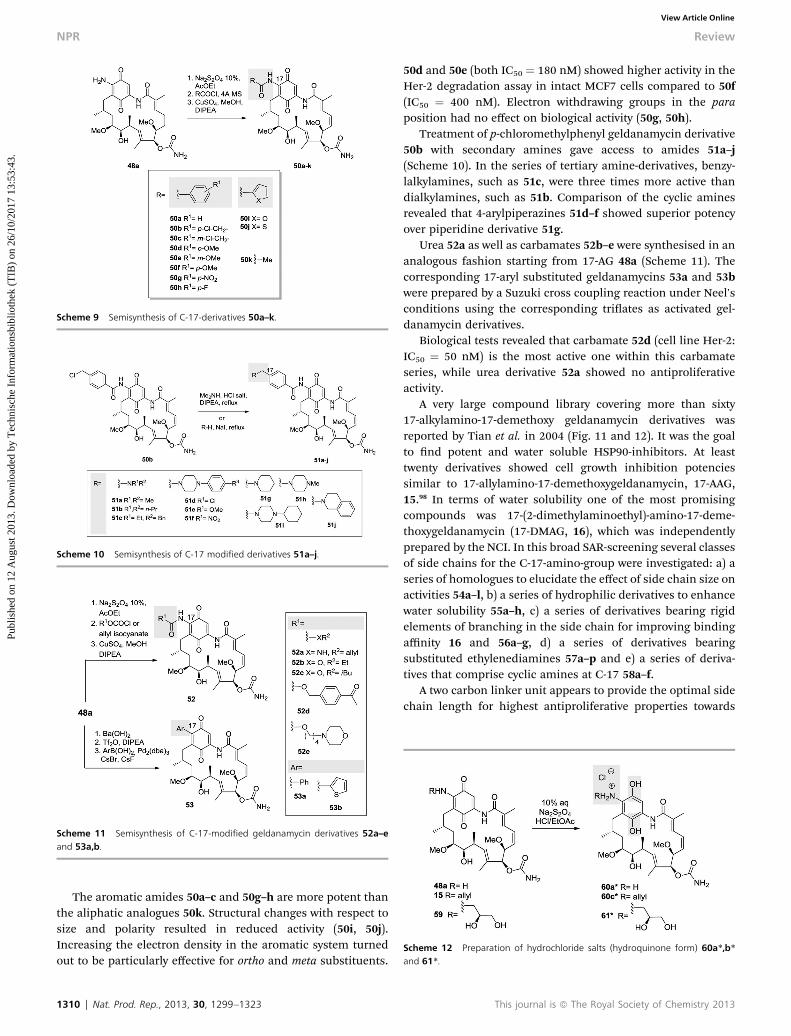
In 1995 Schnur et al. reported several semisyntheticallygenerated geldanamycin derivatives that were modied at C-17and at C-19, respectively.95 Geldanamycin (1) and 4,5-dihy-drogeldanamycin 47 were reacted with alkylamines to yield17-demethoxy-alkylamino-derivatives 48 and 49 (Scheme 8). In
Scheme 8 Semisynthesis of C-17 quinone derivatives 48 and (4,5-dihydro) 49.
This journal is ª The Royal Society of Chemistry 2013
case of sterically hindered amines, the C-17 position was thepreferential site of attack; instead the C-19 alkylamino- or theC-17, C-19 bisalkylamino compounds were obtained. Thismodication allowed to rapidly generate a structurally diverselibrary of simple geldanamycin analogues from which two thederivatives 17-N-allylamino-17-demethoxygeldanamycin (17AAG;tanespimycin) 15 and 17-dimethylaminoethylamino-17-deme-thoxygeldanamycin (17-DMAG; alvespimycin) 16 (Scheme 12)were further elaborated as drug candidates.96
These rst semisynthetic studies revealed improved biolog-ical potency/activity when small, sterically unconstrained ami-nogroups are introduced at C-17 (e.g. 48a, 15, 48c, 48g–h, 48k–l,48n, 48x and 48c0) with respect to geldanamycin (1). In fact,geldanamycin (IC50 � 70 nM) shows moderate in vitro potencyof erbB-2 inhibition in SKBR-3 cells. It is approximately 4-foldmore potent than herbimycin A (2) (IC50 ¼ 300 nM) andthree times more active than 4,5-dihydrogeldanamycin (IC50 ¼230 nM) 47. Derivatives bearing the (uoroethyl)amino 48j(IC50 ¼ 12 nM), the (cyanoethyl)amino 48k (IC50 ¼ 17 nM), andthe azetidinyl 48f (IC50 ¼ 23 nM) substituent are apparentlythe most potent derivatives in this geldanamycin series. Like-wise also potent 4,5-dihydrogeldanamycin derivatives (49a–c,49f,g, 49i–m, 49o, 49y and (S)-49y) bearing the same kind ofsubstituents are of importance. Here, the methylamino 49b(IC50 ¼ 12 nM), and the azetidinyl 49f (IC50 ¼ 14 nM) substit-uents showed biological activity against erbB-2 at low concen-trations. Geldanamycin derivatives 48f, 49f, 48j, 48k and 49bshowed highest activities. Small amino-substituents at C-17carrying acidic functions are less potent (see 48p, 48r). Incontrast, basic groups and hydroxyl groups are tolerated. Whensubstituents become bulkier or arylalkylamines are introducedthe activity is reduced.
Later, Le Brazidec added amides, carbamates and ureas aswell as 17-aryl derivatives to the portfolio of 17-N-geldanamycin-derivatives by making 17-amino-17-demethoxygeldanamycin(17-AG) 48a accessible. This derivative was generated by treat-ment of geldanamycin with methanolic ammonia and wasfurther transformed into amides, ureas and carbamates(Schemes 9–11).97
Fig. 11 Semisynthesis of geldanamycin derivatives 16 and 54–56 modified atC-17.
Nat. Prod. Rep., 2013, 30, 1299–1323 | 1309
Scheme 9 Semisynthesis of C-17-derivatives 50a–k.
Scheme 10 Semisynthesis of C-17 modified derivatives 51a–j.
Scheme 11 Semisynthesis of C-17-modified geldanamycin derivatives 52a–eand 53a,b.
Scheme 12 Preparation of hydrochloride salts (hydroquinone form) 60a*,b*and 61*.
NPR Review
Publ
ishe
d on
12
Aug
ust 2
013.
Dow
nloa
ded
by T
echn
isch
e In
form
atio
nsbi
blio
thek
(T
IB)
on 2
6/10
/201
7 13
:53:
43.
View Article Online
The aromatic amides 50a–c and 50g–h are more potent thanthe aliphatic analogues 50k. Structural changes with respect tosize and polarity resulted in reduced activity (50i, 50j).Increasing the electron density in the aromatic system turnedout to be particularly effective for ortho and meta substituents.
1310 | Nat. Prod. Rep., 2013, 30, 1299–1323
50d and 50e (both IC50 ¼ 180 nM) showed higher activity in theHer-2 degradation assay in intact MCF7 cells compared to 50f(IC50 ¼ 400 nM). Electron withdrawing groups in the paraposition had no effect on biological activity (50g, 50h).
Treatment of p-chloromethylphenyl geldanamycin derivative50b with secondary amines gave access to amides 51a–j(Scheme 10). In the series of tertiary amine-derivatives, benzy-lalkylamines, such as 51c, were three times more active thandialkylamines, such as 51b. Comparison of the cyclic aminesrevealed that 4-arylpiperazines 51d–f showed superior potencyover piperidine derivative 51g.
Urea 52a as well as carbamates 52b–e were synthesised in ananalogous fashion starting from 17-AG 48a (Scheme 11). Thecorresponding 17-aryl substituted geldanamycins 53a and 53bwere prepared by a Suzuki cross coupling reaction under Neel'sconditions using the corresponding triates as activated gel-danamycin derivatives.
Biological tests revealed that carbamate 52d (cell line Her-2:IC50 ¼ 50 nM) is the most active one within this carbamateseries, while urea derivative 52a showed no antiproliferativeactivity.
A very large compound library covering more than sixty17-alkylamino-17-demethoxy geldanamycin derivatives wasreported by Tian et al. in 2004 (Fig. 11 and 12). It was the goalto nd potent and water soluble HSP90-inhibitors. At leasttwenty derivatives showed cell growth inhibition potenciessimilar to 17-allylamino-17-demethoxygeldanamycin, 17-AAG,15.98 In terms of water solubility one of the most promisingcompounds was 17-(2-dimethylaminoethyl)-amino-17-deme-thoxygeldanamycin (17-DMAG, 16), which was independentlyprepared by the NCI. In this broad SAR-screening several classesof side chains for the C-17-amino-group were investigated: a) aseries of homologues to elucidate the effect of side chain size onactivities 54a–l, b) a series of hydrophilic derivatives to enhancewater solubility 55a–h, c) a series of derivatives bearing rigidelements of branching in the side chain for improving bindingaffinity 16 and 56a–g, d) a series of derivatives bearingsubstituted ethylenediamines 57a–p and e) a series of deriva-tives that comprise cyclic amines at C-17 58a–f.
A two carbon linker unit appears to provide the optimal sidechain length for highest antiproliferative properties towards
This journal is ª The Royal Society of Chemistry 2013
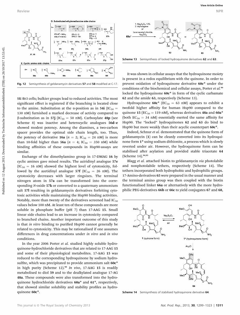
Fig. 12 Semisynthesis of geldanamycin derivatives 57 and 58 modified at C-17.
Scheme 13 Semisynthesis of locked hydroquinone derivatives 62 and 63.
Scheme 14 Semisynthesis of stabilised hydroquinone derivative 64.
Review NPR
Publ
ishe
d on
12
Aug
ust 2
013.
Dow
nloa
ded
by T
echn
isch
e In
form
atio
nsbi
blio
thek
(T
IB)
on 2
6/10
/201
7 13
:53:
43.
View Article Online
SK-Br3 cells; bulkier groups lead to reduced activities. The mostsignicant effect is registered if the branching is located closeto the amine. Substitution at the a-position as in 54i (IC50 ¼130 nM) furnished a marked decrease of activity compared tob-substitution as in 57j (IC50 ¼ 50 nM). Carboxylate 48p (seeScheme 8) was inactive and heterocyclic analogues 56d–eshowed modest potency. Among the diamines, a two-carbonspacer provides the optimal side chain length, too. Thus,the potency of derivative 16a (n ¼ 2; IC50 ¼ 24 nM) is morethan 10-fold higher than 56a (n ¼ 4; IC50 ¼ 350 nM) whilebinding affinities of these compounds in Hsp90-assays aresimilar.
Exchange of the dimethylamino group in 17-DMAG 16 bycyclic amines gave mixed results. The aziridinyl analogue 57e(IC50 ¼ 16 nM) showed the highest level of cytotoxicity, fol-lowed by the azetidinyl analogue 57f (IC50 ¼ 26 nM). Thecytotoxicity decreases with larger ringsizes. The terminalnitrogen atom in 57a can be transformed into the corre-sponding N-oxide 57k or converted to a quaternary ammoniumsalt 57l resulting in geldanamycin derivatives forfeiting cyto-toxic activities while maintaining the Hsp90 binding activities.Notably, more than twenty of the derivatives screened had IC50
values below 100 nM. At least ten of these compounds are moresoluble in phosphate buffer (pH 7) than 17-AAG 15. Smalllinear side chains lead to an increase in cytotoxicity comparedto branched chains. Another important outcome of this studyis that in vitro binding to puried Hsp90 cannot generally berelated to cytotoxicity. This may be rationalised if one assumesdifferences in drug concentrations under in vitro and in vivoconditions.
In the year 2006 Porter et al. studied highly soluble hydro-quinone-hydrochloride derivatives that are related to 17-AAG 15and some of their physiological metabolites. 17-AAG 15 wasreduced to the corresponding hydroquinone by sodium hydro-sulte, which was precipitated to provide ammonium salt 60c*in high purity (Scheme 12).99 In vivo, 17-AAG 15 is readilymetabolised to diol 59 and to the dealkylated analogue 17-AG48a. These compounds were also transformed into the hydro-quinone hydrochloride derivatives 60a* and 61*, respectively,that showed similar solubility and stability proles as hydro-quinone 60c*.
This journal is ª The Royal Society of Chemistry 2013
It was shown in cellular assays that the hydroquinone moietyis present in a redox equilibrium with the quinone. In order toprevent oxidation of hydroquinone derivative 60c* under theconditions of the biochemical and cellular assays, Porter et al.99
locked the hydroquinone 60c* in form of the cyclic carbamate62 and the amide 63, respectively (Scheme 13).
Hydroquinone 60c* (EC50 ¼ 63 nM) appears to exhibit atwofold higher affinity for human Hsp90 compared to thequinone 15 (EC50 ¼ 119 nM), whereas derivatives 48a and 60a*(both EC50 ¼ 34 nM) essentially exerted the same affinity forHsp90. The “locked” hydroquinones 62 and 63 do bind toHsp90 but more weakly than their acyclic counterpart 60c*.
Indeed, Schnur et al. demonstrated that the quinone form ofgeldanamycin (1) can be cleanly converted into its hydroqui-none form 1* using sodium dithionite, a process which is slowlyreverted under air. However, the hydroquinone form can bestabilised aer acylation and provided stable triacetate 64(Scheme 14).94,95
Blagg et al. attached biotin to geldanamycin via photolabileand nonphotolabile tethers, respectively (Scheme 14). Thetethers incorporated both hydrophobic and hydrophilic groups.17-Amino-derivatives 65 were prepared in the usual manner andthe terminal amino group was then coupled with the biotinfunctionalised linker 66a or alternatively with the more hydro-philic PEG-derivatives 66b or 66c to yield conjugates 67 and 68,
Nat. Prod. Rep., 2013, 30, 1299–1323 | 1311
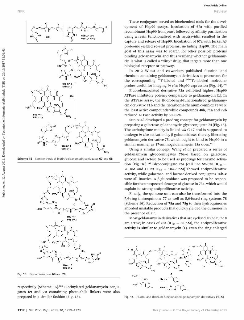
Scheme 15 Semisynthesis of biotin/geldanamycin conjugates 67 and 68.
Fig. 13 Biotin derivatives 69 and 70.
Fig. 14 Fluoro- and rhenium-functionalised geldanamycin derivatives 71–73.
NPR Review
Publ
ishe
d on
12
Aug
ust 2
013.
Dow
nloa
ded
by T
echn
isch
e In
form
atio
nsbi
blio
thek
(T
IB)
on 2
6/10
/201
7 13
:53:
43.
View Article Online
respectively (Scheme 15).100 Biotinylated geldanamycin conju-gates 69 and 70 containing photolabile linkers were alsoprepared in a similar fashion (Fig. 13).
1312 | Nat. Prod. Rep., 2013, 30, 1299–1323
These conjugates served as biochemical tools for the devel-opment of Hsp90 assays. Incubation of 67a with puriedrecombinant Hsp90 from yeast followed by affinity puricationusing a resin functionalised with neutravidin resulted in thecapture and release of Hsp90. Incubation of 67a with Jurkat A3proteome yielded several proteins, including Hsp90. The maingoal of this assay was to search for other possible proteinsbinding geldanamycin and thus verifying whether geldanamy-cin is what is called a “dirty” drug, that targets more than onebiological receptor or pathway.
In 2012 Wuest and co-workers published uorine- andrhenium-containing geldanamycin derivatives as precursors forthe corresponding 18F-labeled and 99mTc-labeled molecularprobes useful for imaging in vivo Hsp90 expression (Fig. 14).101
Fluorobenzoylated derivative 72a exhibited highest Hsp90ATPase inhibitory potency comparable to geldanamycin (1). Inthe ATPase assay, the uorobenzyl-functionalised geldanamy-cin derivative 71b and the tricarbonyl-rhenium complex 73 werethe least active compounds while compounds 48k, 71a and 72breduced ATPase activity by 50–65%.
Sun et al. developed a prodrug concept for geldanamycin bypreparing a galactose-geldanamycin glycoconjugate 74 (Fig. 15).The carbohydrate moiety is linked via C-17 and is supposed toundergo in vivo activation by b-galactosidases thereby liberatinggeldanamycin derivative 75, which ought to bind to Hsp90 in asimilar manner as 17-aminogeldanamycin 48a does.102
Using a similar concept, Wang et al. prepared a series ofgeldanamycin glycoconjugates 76a–e based on galactose,glucose and lactose to be used as prodrugs for enzyme activa-tion (Fig. 16).103 Glycoconjugate 76a (cell line SW620: IC50 ¼70 nM and HT29 IC50 ¼ 104.7 nM) showed antiproliferativeactivity, while galactose- and lactose-derived conjugates 76b–ewere all inactive. A b-glucosidase was proposed to be respon-sible for the unexpected cleavage of glucose in 73a, which wouldexplain its strong antiproliferative activity.
Finally, the quinone unit can also be transformed into the7,6-ring iminoquinone 77 as well as 5,6-fused ring systems 78(Scheme 16). Reduction of 78a and 78g to their hydroquinonesafforded unstable products that quickly yielded the quinones inthe presence of air.
Most geldanamycin derivatives that are cyclised at C-17, C-18are active; in cases of 78a (IC50 ¼ 50 nM), the antiproliferativeactivity is similar to geldanamycin (1). Even the ring enlarged
This journal is ª The Royal Society of Chemistry 2013

Fig. 15 Glycoconjugates 74–76 as potential produgs.
Scheme 16 Semisynthesis of derivatives 77 and 78.
Scheme 17 Semisynthesis of C-7 urea analogues 84a and 84b.
Scheme 18 Semisynthesis of C-19 modified quinone derivatives 86a and 86b.
Review NPR
Publ
ishe
d on
12
Aug
ust 2
013.
Dow
nloa
ded
by T
echn
isch
e In
form
atio
nsbi
blio
thek
(T
IB)
on 2
6/10
/201
7 13
:53:
43.
View Article Online
cyclic iminoquinone derivative 77 (IC50 ¼ 260 nM) revealedin vitro potency.
5.3.2 Semisynthetic modications in the ansa chain. Gel-danamycin derivatives 84modied as urea derivatives at C-7 areaccessible by a two step process and it involves a [3.3]-sigma-tropic rearrangement via intermediate 80 followed by an iso-thiocyanate (82 to 83) rearrangement via intermediate 82(Scheme 17). Starting from semisynthetically modied gelda-namycin 48g (see Scheme 9), removal of the carbamoyl groupyielded 79 and from here the resulting 7(S)-amino derivatives84a and 84b were generated with double stereocontrol andoverall retention of conguration at C-7.
An important transformation is the selective bromination atC-19 to yield bromide 85, which can be utilised in a similarmanner as described for the preparation of the geldanamycinderivatives 86. Here, the addition–elimination mechanism isfavoured for the bromide because of its better nucleofugicproperties compared to the methoxy group at C-17. Withselected amines geldanamycin derivatives 86a and 86b wereobtained (Scheme 18). When incorporating substituents atC-19 a strong reduction of antiproliferative activity takes place(see 86a,b).
This journal is ª The Royal Society of Chemistry 2013
Schnur et al. also pursued semisynthetic changes on thestraightforwardly accessible functional groups of the ansa-ring.Geldanamycin and 4,5-dihydro derivative 48 and 46 served as astarting point for further changing the macrolactam nitrogenatom as well as the hydroxyl group at C-11 (Schemes 19–23).Thus, N-substituted derivatives 87a–k and 88, 11-O-acyl prod-ucts 89a–g, 7-deamidinated analogues 90a,b, esters 91a–c,7-keto-analogue 92, 11-keto-derivatives 93a–b and 93d, 11-oximes 94a–b, 11-amino derivatives 95a–b and 11-(S)-uoroderivatives 96a–d were accessed by Schnur et al.104
These studies demonstrated that 17-azetidine and 17-allyla-mino-derivatives are among the most potent C-17 analogues.Still, the lowest IC50 were determined for the free amino deriv-atives such as aminogeldanamycine derivatives 87f (IC50 ¼230 nM) compared to 87k, (IC50 ¼ 1900 nM). N-Pyridylmethylderivative 87j was >100 fold less potent than analogues bearinga carbonyl group in the a-position of the N-alkyl group (e.g. 87aor 87i, respectively). Phenacyl-analogues 87b (IC50¼ 80 nM) and87j (IC50 ¼ 70 nM) show similar antiproliferative activity togeldanamycin (1) (IC50 ¼ 70 nM).
Acylation of the hydroxyl group at C-11 (Scheme 20) couldalso be achieved and provided new derivatives 89a–g withsimilar biological activities compared to their unsubstitutedprecursors.
Nat. Prod. Rep., 2013, 30, 1299–1323 | 1313

Scheme 19 Semisynthesis of geldanamycin derivatives 87a–k and 88.
Scheme 20 Semisynthesis of geldanamycin derivatives 89a–g.
Scheme 21 Semisynthesis of derivatives 91a–c and 92.
Scheme 22 Semisynthesis of C-11-derivatives 93a–d, 94a,b and 95a,b.
Scheme 23 Semisynthesis of fluorinated geldanamycin derivatives 96a–d and97a,b.
NPR Review
Publ
ishe
d on
12
Aug
ust 2
013.
Dow
nloa
ded
by T
echn
isch
e In
form
atio
nsbi
blio
thek
(T
IB)
on 2
6/10
/201
7 13
:53:
43.
View Article Online
Removal of the amidinoyl group gave geldanamycin deriva-tives 79 and 90a,b; both have lost their biological activity. Thesame change of biological properties occurred, when the ami-dinoyl group was exchanged by other acyl groups at C7 as ingeldanamycin derivatives 91a–c or aer oxidation of C-7, as in92 (Scheme 21). Clearly, the free carbamoyl group acts as apharmacophore. It is suggested that Asp40 and Lys44 in theATP-binding domain of Hsp90 are essential for binding of thequinone moiety of geldanamycin.
Specic exchange of selected amino acids in the highlyconserved ATP-binding site (Lys44 by arginine and Lys89 byaspartate) suggest they stabilise the quinone ring. Otherimportant positions that have an effect on geldanamycinbinding are Glu88 (demonstrated by exchange with glycine) and
1314 | Nat. Prod. Rep., 2013, 30, 1299–1323 This journal is ª The Royal Society of Chemistry 2013

Review NPR
Publ
ishe
d on
12
Aug
ust 2
013.
Dow
nloa
ded
by T
echn
isch
e In
form
atio
nsbi
blio
thek
(T
IB)
on 2
6/10
/201
7 13
:53:
43.
View Article Online
Asp92 (demonstrated by exchange by leucine).104c,d Doubleexchange mutations (Lys44 by arginine and Lys89 by aspara-gine) in the yeast chaperon homologue Hsp82 did result indecreased sensitivity towards 17-AAG 12, perhaps due to weak-ened binding to the C-12 methoxy group on (1).122
Oxidation of the hydroxyl group at C-11 yielded ketones 93a(IC50 ¼ 220 nM) and 93b (IC50 ¼ 34 nM) and 93c (IC50 ¼ 34 nM),all of which showed superior potency to the oximes 94a/IC50 ¼270 nM) and 94b (IC50 ¼ 1100 nM). Reductive aminationafforded amino derivatives 95a and 95b, respectively, whichwere less active than both the corresponding alcohols and theketones.
An interesting semisynthetic transformation allows theintroduction of a uoro substituent at C-11 with inversion ofconguration using diethylaminosulfur triuoride (DAST).In vivo studies revealed that two of the four new uorinatedderivatives, namely 96a (IC50 ¼ 46 nM) and 96b (IC50 ¼ 50 nM)showed very good antiproliferative activity. These in vivostudies are noteworthy, because many new geldanamycinderivatives obtained by semisynthesis showed hardly any in vivoactivity, such as 87h,i, 89b,f,g and 93a,d, despite being activein vitro.
Rastelli et al. also prepared geldanamycin derivatives thatvaried at C-7 (Scheme 24). The carbamoyl group is integrated ina hydrogen bonding network that involves four water mole-cules. By modifying the substituent at C-7 insight into theimportance of this network was gained. Saponication of thecarbamoyl moiety followed by reaction with carbodiimidazole(CDI) and trapping of the CDI-adduct with nucleophiles(hydrazine, hydroxylamine) furnished new 7-N-alkyl-carbamates98a–o.105 The majority of these derivatives showed no biologicalactivity and only low Hsp90 affinity. Only 98f–k and 98m wereactive towards SK-Br3 cells in the range of around 0.5 mM.
5.4 Geldanamycin derivatives by manipulation of itsbiosynthesis
Over the past decade, the combination of metabolic engi-neering of biosynthetic pathways that code for secondary
Scheme 24 Semisynthesis of C-7-derivatives 98a–o.
This journal is ª The Royal Society of Chemistry 2013
metabolism with semisynthetic modications has emerged asa powerful tool for the creation of derivatives of structurallycomplex natural products that are difficult to access other-wise. The preciseness of the biosynthetic machinery that isable to construct complex frameworks and macrocycles in arather linear fashion meets chemical synthesis with itsenormous exibility for introducing functional groups,including pharmacophoric ones that are unprecedented innature.
5.4.1 Exchange of individual enzymes by genetic engi-neering. Due to its pharmaceutical importance the biosyn-thesis of the heat shock protein inhibitor geldanamycin wasstudied in detail,106 allowing initiation of different metabolicengineering programs with the aim to generate geldanamycinderivatives.
Kosan Biosciences, Inc disclosed an elegant strategy thatrelied on the genetic manipulation of the geldanamycin poly-ketide synthase (GdmPKS) Streptomyces hygroscopicus107 thatprovided several new derivatives lacking methyl or methoxygroups in the ansa chain.108 The strategy is based on thesubstitution of acyltransferase (AT) domains in six differentGdmPKSmodules that commonly accept methylmalonyl-CoA ormethoxymalonyl-CoA, by malonyl AT domains from the rapa-mycin PKS.109 Overall this engineering of the biosynthesisprovided 2-desmethyl, 6-desmethoxy, 8-desmethyl, and 14-des-methyl geldanamycin derivatives 99–102, as well as theg,d-saturated 103 and the hydratisation products 104 and 105in sufficient amounts (Scheme 25).
Tian and Rastelli noted that Hsp90 binding is not a predic-tive indicator for cytotoxicity. The Hsp90 binding affinity ofphenol 102 (IC50¼ 860 nM, Kd (Hsp90)¼ 16 nM) is 4-fold higherthan of geldanamycin 1 (IC50 ¼ 41 nM, Kd (Hsp90) ¼ 670 nM)though the antiproliferative activity is much smaller. Further-more, the high binding affinity of 102 reveals that the quinonegroup has little signicance for the observed hepatotoxicity ofgeldanamycin (1). Derivatives 99 (IC50 ¼ 470 nM) and 100(IC50 ¼ 480 nM) exhibit moderate biological activity while101 (IC50 ¼ 3200 nM), 103 (IC50 ¼ 4900 nM) and 104 (IC50 ¼>5000 nM) exerted no cytotoxicity.
5.4.2 Blocking of selected enzymes by genetic engineering.In work by Zhang et al.macbecin (3) served as lead structure forHsp90 inhibition. By genetic engineering, mutants were createdthat are blocked in selected oxidative tailoring steps of macbe-cin biosynthesis. These mutant strains yielded novel macbecinanalogues like the nonquinone compounds 105 and 106b withsignicantly improved binding affinity to Hsp90 (Kd ¼ 3 nMvs. 240 nM for macbecin) and reduced toxicity (MTD > or ¼250 mg kg�1) (Fig. 16). The authors speculated that enhancedstructural exibility allows more facile preorganisation in theHsp90-bound conformation in solution.110
Hong and coworkers followed a similar approach. Sitedirected mutagenesis of the geldanamycin polyketide synthase(PKS) and selected post-PKS tailoring genes provided severalC-15 hydroxylated non-quinone geldanamycin analogues. Onenew derivative, 15-hydroxy-17-demethoxy analogue 107, exhibi-ted stronger inhibition of Hsp90 ATPase activity by a factor ofalmost ve than geldanamycin.111
Nat. Prod. Rep., 2013, 30, 1299–1323 | 1315

Scheme 25 Geldanamycin derivatives 99–105 produced by acyltransferase (AT)substitutions in the Gdm polyketide synthase (numbers refer to PKSmodule; areasmarked in grey show structural variations with respect to geldanamycin (1)).
Scheme 26 Semisynthesis of C-17 amino analogues 111a,c–e, 112a–d and 113.
Fig. 16 New phenolic geldanamycin derivatives isolated from mutant strainsmodified by site directed mutagenesis in selected tailoring biosynthetic steps.
NPR Review
Publ
ishe
d on
12
Aug
ust 2
013.
Dow
nloa
ded
by T
echn
isch
e In
form
atio
nsbi
blio
thek
(T
IB)
on 2
6/10
/201
7 13
:53:
43.
View Article Online
5.4.3 Mutasynthetic approaches. Hong et al. also used asemisynthetic approach that utilised the product obtained froma genetically engineered organism.112 A mutant strain fromStreptomyces hygroscopicus subsp. duamyceticus (JCM4427),which lacked the active carbamoyltransferase, provided gelda-namycin derivative 108 as the main fermentation product.Obviously, genetic interference also blocked the last biosyn-thetic step leading to the desaturation at C-4,C-5. The missing
1316 | Nat. Prod. Rep., 2013, 30, 1299–1323
carbamoyl group renders this metabolite biologically inactive(see above). Next, this 4,5-dihydro geldanamycin derivative 108was treated with trichloroacetyl isocyanate to yield bis-carba-moyl derivative 110. This compound was treated with variousamines to introduce an amino substituent at C-17 (see alsoScheme 8) to provide 4,5-dihydro derivatives 112a–d. Forcomparison purposes also the 17-aminated 7,11-bis-carbamoyl-derivatives 111a,c–e were chemically prepared from geldana-mycin (1) via 109 (Scheme 26).
As expected compound 108 is inactive as opposed to carba-moylated 4,5-dihydrogeldanamycin 47. The C-7,11-bis-carba-mates 112a–d carrying an aminoalkyl substituent at C-17 exert abroad scope of antiproliferative activities. 112c (cell line SK-Br3:IC50 ¼ 0.32 mM and cell line SK-Ov3: IC50 ¼ 5.09 mM), and 112d(SK-Br3: IC50 ¼ 0.01 mM and SK-Ov3: IC50 ¼ 1.14 mM) exhibitimproved potency compared to 4,5-dihydro derivative 11 (SK-Br3: IC50 ¼ 3.07 mM and SK-Ov3: IC50 ¼ 7.90 mM. Still, incomparison to geldanamycin (IC50 ¼ 70 nM) and the geldana-mycin derivatives 111a (SK-Br3: IC50 ¼ 0.05 mM and SK-Ov3:IC50 ¼ 6.97 mM), as well as 111c (SK-Br3: IC50 ¼ 0.020 mM andSK-Ov3: IC50 ¼ 0.67 mM), the corresponding 4,5-saturatedcompounds 112a (SK-Br3: IC50 ¼ >10 mM and SK-Ov3: IC50 ¼10.52 mM) and 112c (SK-Br3: IC50 ¼ 0.32 mM and SK-Ov3: IC50 ¼5.09 mM) carrying an additional carbamoyl group at C-11, wereless potent.
Treatment of 4,5-dihydro derivative 108 with diketenegenerated bis-diketo-derivative 113, which was inactive inantiproliferative tests (Scheme 26). This result ts well with the
This journal is ª The Royal Society of Chemistry 2013

Review NPR
Publ
ishe
d on
12
Aug
ust 2
013.
Dow
nloa
ded
by T
echn
isch
e In
form
atio
nsbi
blio
thek
(T
IB)
on 2
6/10
/201
7 13
:53:
43.
View Article Online
observations from semisynthetic studies towards similarderivatives like 91.
Several groups employed mutant strains of S. hygroscopicusthat are specically blocked in the biosynthesis of the PKSstarter building block 3-amino-5-hydroxybenzoic acid (seeScheme 25) in order to exploit the concept of mutationalbiosynthesis (mutasynthesis).83 By supplementing a culture ofthis mutant strain with different chemically modied aminobenzoic acids 114 and 115 Menzella and coworkers113 as wellas Lee and Hong114,115 isolated a series of new geldanamycinderivatives 120a–c and 121a,b in sufficient amounts forstructural analysis and biological evaluation (Scheme 27).Likewise Kirschning et al.116 could prepare new geldanamycinderivatives 122–125 aer supplementing a culture of theAHBA blocked mutant of S. hygroscopicus with mutasynthons116–119. Remarkably, also the pyridine precursor 126was accepted and processed to the aza-geldanamycin deriva-tive 127.
Cell growth inhibition assays for 120 and 121 demonstratedactivities similar to those of tanespimycin 15 and alvespimycin16 against a panel of cancer cell lines. Also new geldanamycinderivatives 122–125 showed strong antiproliferative activity,most of them having IC50 values in the nM range. Remarkably,the uoro and bromo derivatives 123 (IC50: 18–73 nM) and 122(IC50: 17–120 nM) turned out to be as active as geldanamycin. Incontrast, aza-geldanamycin 127 was inactive in assays deter-mining antiproliferative activity.
Scheme 27 Mutasynthetic preparation of geldanamycin derivatives 120–125and 127.
This journal is ª The Royal Society of Chemistry 2013
Mutasynthon 128 provided a remarkable number of differ-ently processed geldanamycin derivatives being fed to the AHBAblocked mutant of S. hygroscopicus. Besides the expected prod-ucts 129 and 130, the blocked mutant strain also provided 20-membered macrolactones 133a–c. Obviously, the benzyl alcoholcompetes as a nucleophilic function with the anilino group forthe PKS-bound seco acid during amide synthase-promotedmacrocyclisation. These results were further generalised byfeeding benzoic acids 131 and 132 containing an additionalbenzyl alcohol which resulted in the formation of macro-lactones 134 and 135, respectively (Scheme 28).117
Geldanamycin derivatives 129b and 129c and 130b showedgood (130b IC50 ¼ 22–470 nM) to moderate antiproliferativeactivities towards different cancer cell lines while 129a and 130awere >100 fold less potent than geldanamycin (1). The 20-membered lactones 133a–c, 134 and 135 exhibited no anti-proliferative properties or Hsp90 binding affinity at all.
6 Assaying inhibitory effects on Hsp90function by high throughput screening
The rising demand for compound libraries has led to the devel-opment of fully automated high-throughput screening devices(HTS). Test protocols in pharmaceutical industry are commonlytarget-oriented, i.e. the interaction of a test compound with abiological target is investigated. TheHTS results are analysed in astatistical manner and a positive assay response is called a “hit”.Clearly, the quality of HTS-derived results are associated with toomany false positive responses fromwhich the few true leads haveto be found by laborious deconvolution. In addition, no state-ment on pharmacokinetics of the ”hit”, such as toxicity,membrane permeability and solubility of the identiedsubstance, is possible at this stage. The typical HTS setup utilisesthe microplate format for assaying and trailing target–inhibitorcomplexes. It commonly relies on a biochemical reaction inwhich a substrate is functionalised with a radioactive or uo-rescent label. For identifying potential inhibitors for Hsp90,various methods are known for how HTS can be performed.These are a) cell-based assays, b) refolding assays and c) bindingassays. The classical cell-based assay addresses antiproliferation
Scheme 28 Mutasynthetic preparation of geldanamycin derivatives 129, 130and 133–135.
Nat. Prod. Rep., 2013, 30, 1299–1323 | 1317
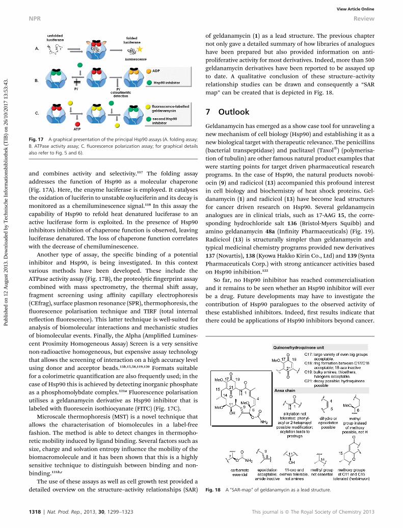
Fig. 17 A graphical presentation of the principal Hsp90 assays (A. folding assay;B. ATPase activity assay; C. fluorescence polarization assay; for graphical detailsalso refer to Fig. 5 and 6).
Fig. 18 A “SAR-map” of geldanamycin as a lead structure.
NPR Review
Publ
ishe
d on
12
Aug
ust 2
013.
Dow
nloa
ded
by T
echn
isch
e In
form
atio
nsbi
blio
thek
(T
IB)
on 2
6/10
/201
7 13
:53:
43.
View Article Online
and combines activity and selectivity.117 The folding assayaddresses the function of Hsp90 as a molecular chaperone(Fig. 17A). Here, the enzyme luciferase is employed. It catalysesthe oxidation of luciferin to unstable oxyluciferin and its decay ismonitored as a chemiluminescence signal.118 In this assay thecapability of Hsp90 to refold heat denatured luciferase to anactive luciferase form is exploited. In the presence of Hsp90inhibitors inhibition of chaperone function is observed, leavingluciferase denatured. The loss of chaperone function correlateswith the decrease of chemiluminescence.
Another type of assay, the specic binding of a potentialinhibitor and Hsp90, is being investigated. In this contextvarious methods have been developed. These include theATPase activity assay (Fig. 17B), the proteolytic ngerprint assaycombined with mass spectrometry, the thermal shi assay,fragment screening using affinity capillary electrophoresis(CEfrag), surface plasmon resonance (SPR), thermophoresis, theuorescence polarisation technique and TIRF (total internalreection uorescence). This latter technique is well-suited foranalysis of biomolecular interactions and mechanistic studiesof biomolecular events. Finally, the Alpha (Amplied Lumines-cent Proximity Homogeneous Assay) Screen is a very sensitivenon-radioactive homogeneous, but expensive assay technologythat allows the screening of interaction on a high accuracy levelusing donor and acceptor beads.13b,15,58,119,120 Formats suitablefor a colorimetric quantication are also frequently used; in thecase of Hsp90 this is achieved by detecting inorganic phosphateas a phosphomolybdate complex.121a Fluorescence polarisationutilises a geldanamycin derivative as Hsp90 inhibitor that islabeled with uorescein isothiocyanate (FITC) (Fig. 17C).
Microscale thermophoresis (MST) is a novel technique thatallows the characterisation of biomolecules in a label-freefashion. The method is able to detect changes in thermopho-retic mobility induced by ligand binding. Several factors such assize, charge and solvation entropy inuence the mobility of thebiomacromolecule and it has been shown that this is a highlysensitive technique to distinguish between binding and non-binding.121b,c
The use of these assays as well as cell growth test provided adetailed overview on the structure–activity relationships (SAR)
1318 | Nat. Prod. Rep., 2013, 30, 1299–1323
of geldanamycin (1) as a lead structure. The previous chapternot only gave a detailed summary of how libraries of analogueshave been prepared but also provided information on anti-proliferative activity for most derivatives. Indeed, more than 500geldanamycin derivatives have been reported to be assayed upto date. A qualitative conclusion of these structure–activityrelationship studies can be drawn and consequently a “SARmap” can be created that is depicted in Fig. 18.
7 Outlook
Geldanamycin has emerged as a show case tool for unraveling anew mechanism of cell biology (Hsp90) and establishing it as anew biological target with therapeutic relevance. The penicillins(bacterial transpeptidase) and paclitaxel (Taxol�) (polymerisa-tion of tubulin) are other famous natural product examples thatwere starting points for target driven pharmaceutical researchprograms. In the case of Hsp90, the natural products novobi-ocin (9) and radicicol (13) accompanied this profound interestin cell biology and biochemistry of heat shock proteins. Gel-danamycin (1) and radicicol (13) have become lead structuresfor cancer driven research on Hsp90. Several geldanamycinanalogues are in clinical trials, such as 17-AAG 15, the corre-sponding hydrochloride salt 136 (Bristol-Myers Squibb) andamino geldanamycin 48a (Innity Pharmaceuticals) (Fig. 19).Radicicol (13) is structurally simpler than geldanamycin andtypical medicinal chemistry programs provided new derivatives137 (Novartis), 138 (Kyowa Hakko Kirin Co., Ltd) and 139 (SyntaPharmaceuticals Corp.) with strong anticancer activities basedon Hsp90 inhibition.122
So far, no Hsp90 inhibitor has reached commercialisationand it remains to be seen whether an Hsp90 inhibitor will everbe a drug. Future developments may have to investigate thecontribution of Hsp90 paralogues to the observed activity ofthese established inhibitors. Indeed, rst results indicate thatthere could be applications of Hsp90 inhibitors beyond cancer.
This journal is ª The Royal Society of Chemistry 2013
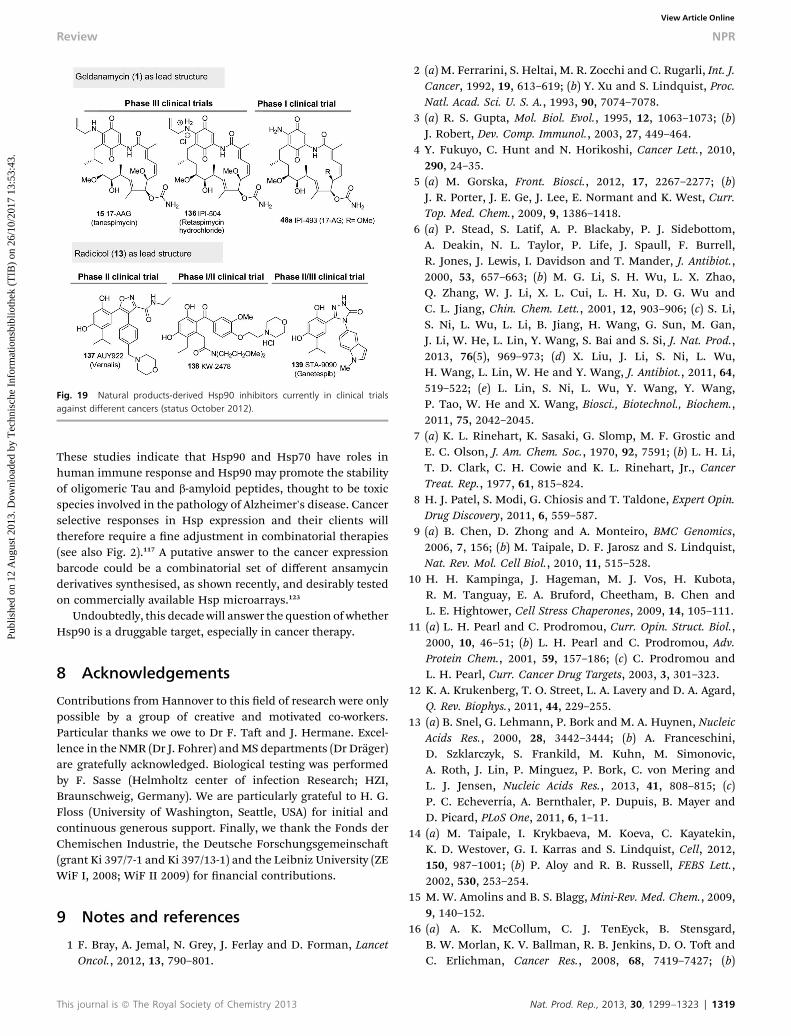
Fig. 19 Natural products-derived Hsp90 inhibitors currently in clinical trialsagainst different cancers (status October 2012).
Review NPR
Publ
ishe
d on
12
Aug
ust 2
013.
Dow
nloa
ded
by T
echn
isch
e In
form
atio
nsbi
blio
thek
(T
IB)
on 2
6/10
/201
7 13
:53:
43.
View Article Online
These studies indicate that Hsp90 and Hsp70 have roles inhuman immune response and Hsp90 may promote the stabilityof oligomeric Tau and b-amyloid peptides, thought to be toxicspecies involved in the pathology of Alzheimer's disease. Cancerselective responses in Hsp expression and their clients willtherefore require a ne adjustment in combinatorial therapies(see also Fig. 2).117 A putative answer to the cancer expressionbarcode could be a combinatorial set of different ansamycinderivatives synthesised, as shown recently, and desirably testedon commercially available Hsp microarrays.123
Undoubtedly, this decade will answer the question of whetherHsp90 is a druggable target, especially in cancer therapy.
8 Acknowledgements
Contributions from Hannover to this eld of research were onlypossible by a group of creative and motivated co-workers.Particular thanks we owe to Dr F. Ta and J. Hermane. Excel-lence in the NMR (Dr J. Fohrer) andMS departments (Dr Drager)are gratefully acknowledged. Biological testing was performedby F. Sasse (Helmholtz center of infection Research; HZI,Braunschweig, Germany). We are particularly grateful to H. G.Floss (University of Washington, Seattle, USA) for initial andcontinuous generous support. Finally, we thank the Fonds derChemischen Industrie, the Deutsche Forschungsgemeinscha(grant Ki 397/7-1 and Ki 397/13-1) and the Leibniz University (ZEWiF I, 2008; WiF II 2009) for nancial contributions.
9 Notes and references
1 F. Bray, A. Jemal, N. Grey, J. Ferlay and D. Forman, LancetOncol., 2012, 13, 790–801.
This journal is ª The Royal Society of Chemistry 2013
2 (a) M. Ferrarini, S. Heltai, M. R. Zocchi and C. Rugarli, Int. J.Cancer, 1992, 19, 613–619; (b) Y. Xu and S. Lindquist, Proc.Natl. Acad. Sci. U. S. A., 1993, 90, 7074–7078.
3 (a) R. S. Gupta, Mol. Biol. Evol., 1995, 12, 1063–1073; (b)J. Robert, Dev. Comp. Immunol., 2003, 27, 449–464.
4 Y. Fukuyo, C. Hunt and N. Horikoshi, Cancer Lett., 2010,290, 24–35.
5 (a) M. Gorska, Front. Biosci., 2012, 17, 2267–2277; (b)J. R. Porter, J. E. Ge, J. Lee, E. Normant and K. West, Curr.Top. Med. Chem., 2009, 9, 1386–1418.
6 (a) P. Stead, S. Latif, A. P. Blackaby, P. J. Sidebottom,A. Deakin, N. L. Taylor, P. Life, J. Spaull, F. Burrell,R. Jones, J. Lewis, I. Davidson and T. Mander, J. Antibiot.,2000, 53, 657–663; (b) M. G. Li, S. H. Wu, L. X. Zhao,Q. Zhang, W. J. Li, X. L. Cui, L. H. Xu, D. G. Wu andC. L. Jiang, Chin. Chem. Lett., 2001, 12, 903–906; (c) S. Li,S. Ni, L. Wu, L. Li, B. Jiang, H. Wang, G. Sun, M. Gan,J. Li, W. He, L. Lin, Y. Wang, S. Bai and S. Si, J. Nat. Prod.,2013, 76(5), 969–973; (d) X. Liu, J. Li, S. Ni, L. Wu,H. Wang, L. Lin, W. He and Y. Wang, J. Antibiot., 2011, 64,519–522; (e) L. Lin, S. Ni, L. Wu, Y. Wang, Y. Wang,P. Tao, W. He and X. Wang, Biosci., Biotechnol., Biochem.,2011, 75, 2042–2045.
7 (a) K. L. Rinehart, K. Sasaki, G. Slomp, M. F. Grostic andE. C. Olson, J. Am. Chem. Soc., 1970, 92, 7591; (b) L. H. Li,T. D. Clark, C. H. Cowie and K. L. Rinehart, Jr., CancerTreat. Rep., 1977, 61, 815–824.
8 H. J. Patel, S. Modi, G. Chiosis and T. Taldone, Expert Opin.Drug Discovery, 2011, 6, 559–587.
9 (a) B. Chen, D. Zhong and A. Monteiro, BMC Genomics,2006, 7, 156; (b) M. Taipale, D. F. Jarosz and S. Lindquist,Nat. Rev. Mol. Cell Biol., 2010, 11, 515–528.
10 H. H. Kampinga, J. Hageman, M. J. Vos, H. Kubota,R. M. Tanguay, E. A. Bruford, Cheetham, B. Chen andL. E. Hightower, Cell Stress Chaperones, 2009, 14, 105–111.
11 (a) L. H. Pearl and C. Prodromou, Curr. Opin. Struct. Biol.,2000, 10, 46–51; (b) L. H. Pearl and C. Prodromou, Adv.Protein Chem., 2001, 59, 157–186; (c) C. Prodromou andL. H. Pearl, Curr. Cancer Drug Targets, 2003, 3, 301–323.
12 K. A. Krukenberg, T. O. Street, L. A. Lavery and D. A. Agard,Q. Rev. Biophys., 2011, 44, 229–255.
13 (a) B. Snel, G. Lehmann, P. Bork and M. A. Huynen, NucleicAcids Res., 2000, 28, 3442–3444; (b) A. Franceschini,D. Szklarczyk, S. Frankild, M. Kuhn, M. Simonovic,A. Roth, J. Lin, P. Minguez, P. Bork, C. von Mering andL. J. Jensen, Nucleic Acids Res., 2013, 41, 808–815; (c)P. C. Echeverrıa, A. Bernthaler, P. Dupuis, B. Mayer andD. Picard, PLoS One, 2011, 6, 1–11.
14 (a) M. Taipale, I. Krykbaeva, M. Koeva, C. Kayatekin,K. D. Westover, G. I. Karras and S. Lindquist, Cell, 2012,150, 987–1001; (b) P. Aloy and R. B. Russell, FEBS Lett.,2002, 530, 253–254.
15 M. W. Amolins and B. S. Blagg, Mini-Rev. Med. Chem., 2009,9, 140–152.
16 (a) A. K. McCollum, C. J. TenEyck, B. Stensgard,B. W. Morlan, K. V. Ballman, R. B. Jenkins, D. O. To andC. Erlichman, Cancer Res., 2008, 68, 7419–7427; (b)
Nat. Prod. Rep., 2013, 30, 1299–1323 | 1319

NPR Review
Publ
ishe
d on
12
Aug
ust 2
013.
Dow
nloa
ded
by T
echn
isch
e In
form
atio
nsbi
blio
thek
(T
IB)
on 2
6/10
/201
7 13
:53:
43.
View Article Online
L. Whitesell and N. U. Lin, Biochim. Biophys. Acta, Mol. CellRes., 2012, 1823, 756–766.
17 (a) G. Marcu, A. Chadli, I. Bouhouche, M. Catelli andL. M. Neckers, J. Biol. Chem., 2000, 275, 37181–37186; (b)M. G. Marcu, T. W. Schulte and L. Neckers, J. Natl. CancerInst., 2000, 92, 242–248.
18 M. M. U. Ali, S. M. Roe, C. Vaughan, P. Meyer, B. Panaretou,P. W. Piper, C. Prodromou and L. H. Pearl, Nature, 2006,440, 1013–1017.
19 (a) S. Lindquist, Cold Spring Harbor Symp. Quant. Biol.,2009, 74, 103–108; (b) G. Chen, W. D. Bradford,C. W. Seidel and R. Li, Nature, 2012, 482, 246–250.
20 (a) C. Jolly and R. I. Morimoto, J. Natl. Cancer Inst., 2000, 92,1564–1572; (b) D. A. Parsell and S. Lindquist, Annu. Rev.Genet., 1993, 27, 437–496; (c) R. I. Morimoto, M. P. Kline,D. N. Bimston and J. J. Cotto, Essays Biochem., 1997, 32,17–29; (d) G. Crevel, H. Bates, H. Huikeshoven andS. Cotterill, J. Cell Science, 2001, 114, 2015–2025; (e)L. Neckers, Trends Mol. Med., 2002, 8, S55–61; (f)L. Whitesell and S. L. Lindquist, Nat. Rev. Cancer, 2005, 5,761–772.
21 (a) R. Zhao and W. A. Houry, Biochem. Cell Biol., 2005, 83,703–710; (b) R. F. de Zoeten, L. Wang, K. Butler,U. H. Beier, T. Akimova, H. Sai, J. E. Bradner,R. Mazitschek, A. P. Kozikowski, P. Matthias andW. W. Hancock, Mol. Cell. Biol., 2011, 31, 2066–2078.
22 (a) T. H. Davies, Y. M. Ning and E. R. Sanchez, J. Biol. Chem.,2002, 277, 4597–4600; (b) R. Hamamoto, Y. Furukawa,M. Morita, Y. Iimura, F. P. Silva, M. Li, R. Yagyu andY. Nakamura, Nat. Cell Biol., 2004, 6, 731–740.
23 (a) O. A. Toogun, D. C. Dezwaan and B. C. Freeman, Mol.Cell. Biol., 2008, 28, 457–467; (b) B. R. Keppler,A. T. Grady and M. B. Jarstfer, J. Biol. Chem., 2006, 281,9840–19848; (c) R. Villa, M. Folini, C. D. Porta,A. Valentini, M. Pennati, M. G. Daidone and N. Zaffaroni,Carcinogenesis, 2003, 24, 851–859.
24 (a) F. U. Hartl, A. Bracher and M. Hayer-Hartl, Nature, 2011,475, 324–332; (b) F. U. Hartl and M. Hayer-Hartl, Science,2002, 295, 1852–1858.
25 J. G. Thomas and F. Baneyxm, J. Bacteriol., 1998, 180, 5165–5172.
26 (a) M. Tanabe, Y. Kawazoe, S. Takeda, R. I. Morimoto,K. Nagata and A. Nakai, EMBO J., 1998, 16, 1750–1758; (b)R. I. Morimoto, Genes Dev., 1998, 12, 3788–3796.
27 (a) X. Wang, M. A. Khaleque, M. J. Zhao, R. Zhong,M. Gaestel and S. K. Calderwood, J. Biol. Chem., 2006,281, 782–791; (b) R. Baler, G. Dahl and R. Voellmy, Mol.Cell. Biol., 1993, 13, 2486–2496; (c) Y. Guo, T. Guettouche,M. Fenna, F. Boellmann, W. B. Pratt, D. O. To,D. F. Smith and R. Voellmy, J. Biol. Chem., 2001, 276,45791–45799; (d) R. Bagatell, G. D. Paine-Murrieta,C. W. Taylor, E. J. Pulcini, S. Akinaga, I. J. Benjamin andL. Whitesell, Clin. Cancer Res., 2000, 6, 3312–3318; (e)C. Wu, Annu. Rev. Cell Dev. Biol., 1995, 11, 441–469.
28 L. Neckers, Curr. Med. Chem., 2003, 10, 733–739.29 A. Rodriguez-Sinovas, K. Boengler, A. Cabestrero, P. Gres,
M. Morente, M. Ruiz-Meana, I. Konietzka, E. Miro,
1320 | Nat. Prod. Rep., 2013, 30, 1299–1323
A. Totzeck, G. Heusch, R. Schulz and D. Garcia-Dorado,Circ. Res., 2006, 7, 93–101.
30 (a) M. A. Theodoraki and A. J. Caplan, Biochim. Biophys.Acta, Mol. Cell Res., 2012, 1823, 683–688; (b)P. N. Munster, A. Basso, D. Solit, L. Norton and N. Rosen,Clin. Cancer Res., 2001, 7, 2155–2158.
31 (a) R. Kumar, A. Musiyenko and S. Barik, Malar. J., 2003, 2,1–11; (b) M. A. Morales, R. Watanabe, M. Dacher, P. Chafey,J. Osorio y Fortea, D. A. Scott, S. M. Beverley, G. Ommen,J. Clos, S. Hem, P. Lenormand, J. C. Rousselle, A. Namaneand G. F. Spath, Proc. Natl. Acad. Sci. U. S. A., 2010, 107,8381–8386; (c) L. E. Cowen, PLoS Pathog., 2009, 5,e1000471; (d) G. Homuth, S. Domm, D. Kleiner andW. Schumann, J. Bacteriol., 2000, 182, 4257–4263; (e)R. Kumar, S. R. Pavithra and U. Tatu, J. Biosci., 2007, 32,531–536.
32 A. Zuehlke and J. L. Johnson, Biopolymers, 2010, 93, 211–217.33 (a) A. Granelli-Piperno, L. Andrus and R. M. Steinman,
J. Exp. Med., 1986, 163, 922–937; (b) L. H. Pearl,C. Prodromou and P. Workman, Biochem. J., 2008, 410,439–453.
34 L. H. Pearl and C. Prodromou, Annu. Rev. Biochem., 2006,75, 271–294.
35 A. K. Shiau, S. F. Harris, D. R. Southworth and D. A. Agard,Cell, 2006, 127, 329–340.
36 S. H. McLaughlin, L. A. Ventouras, B. Lobbezoo andS. E. Jackson, J. Mol. Biol., 2004, 344, 813–826.
37 C. Ratzke, F. Berkemeier and T. Hugel, Proc. Natl. Acad. Sci.U. S. A., 2012, 109, 161–16.
38 C. Ratzke, M. Mickler, B. Hellenkamp, J. Buchner andT. Hugel, Proc. Natl. Acad. Sci. U. S. A., 2010, 107, 16101–16106.
39 D. R. Southworth and D. A. Agard, Mol. Cell, 2008, 32, 631–640.
40 T. Makhnevych and W. A. Houry, Biochim. Biophys. Acta,Mol. Cell Res., 2012, 1823, 674–682.
41 J. L. Johnson and C. Brown, Cell Stress Chaperones, 2009, 14,83–94.
42 D. R. Southworth and D. A. Agard, Mol. Cell, 2011, 42, 771–781.
43 M. Retzlaff, F. Hagn, L. Mitschke, M. Hessling, F. Gugel,H. Kessler, K. Richter and J. Buchner, Mol. Cell, 2010, 37,344–354.
44 (a) S. Chen and D. F. Smith, J. Biol. Chem., 1998, 273, 35194–35200; (b) C. Scheuer, A. Brinker, G. Bourenkov,S. Pegoraro, L. Moroder, H. Bartunik, F. U. Hartl andI. Moare, Cell, 2000, 101, 199–210.
45 J. C. Young, I. Moare and F. U. Hartl, J. Cell Biol., 2001, 154,267–273.
46 W. B. Pratt and K. D. Dittmar, Trends Endocrinol. Metab.,1998, 9, 254–252.
47 A. Citri, D. Harari, G. Shohat, P. Ramakrishnan, J. Gan,S. Lavi, M. Eisenstein, A. Kimchi, D. Wallach,S. Pietrokovski and Y. Yarden, J. Biol. Chem., 2006, 281,14361–14369.
48 N. V. Marozkina, S. Yemen, M. Borowitz, L. Liu, M. Plapp,F. Sun, R. Islam, P. Erdmann-Gilmore, R. R. Townsend,
This journal is ª The Royal Society of Chemistry 2013
Review NPR
Publ
ishe
d on
12
Aug
ust 2
013.
Dow
nloa
ded
by T
echn
isch
e In
form
atio
nsbi
blio
thek
(T
IB)
on 2
6/10
/201
7 13
:53:
43.
View Article Online
C. F. Lichti, S. Mantri, P. W. Clapp, S. H. Randell, B. Gastonand K. Zaman, Proc. Natl. Acad. Sci. U. S. A., 2010, 107,11393–11398.
49 J. Li, J. Soroka and J. Buchner, Biochim. Biophys. Acta, Mol.Cell Res., 2012, 1823, 624–635.
50 H. Ogiso, N. Kagi, E. Matsumoto, M. Nishimoto, R. Arai,M. Shirouzu, J. Mimura, Y. Fujii-Kuriyama andS. Yokoyama, Biochemistry, 2004, 43, 15510–15519.
51 (a) M. Mollapour and L. Neckers, Methods Mol. Biol., 2011,787, 67–74; (b) Y. Miyata, Cell. Mol. Life Sci., 2009, 66,1840–1849.
52 B. T. Scroggins, K. Robzyk, D. Wang, M. G. Marcu,S. Tsutsumi, K. Beebe, R. J. Cotter, S. Felts, D. To,L. Karnitz, N. Rosen and L. Neckers, Mol. Cell, 2007, 25,151–159.
53 M. Retzlaff, M. Stahl, H. C. Eberl, S. Lagleder, J. Beck,H. Kessler and J. Buchner, EMBO Rep., 2009, 10, 1147–1153.
54 (a) R. Hamamoto, Y. Furukawa, M. Morita, Y. Iimura,F. P. Silva, M. Li, R. Yagyu and Y. Nakamura, Nat. CellBiol., 2004, 6, 731–740; (b) R. Hamamoto, F. P. Silva,M. Tsuge, T. Nishidate, T. Katagiri, Y. Nakamura andY. Furukawa, Cancer Sci., 2006, 97, 113–118.
55 S. J. Park, B. N. Borin, M. A. Martinez-Yamout andH. J. Dyson, Nat. Struct. Mol. Biol., 2011, 18, 537–541.
56 (a) J. S. Hahn, BMB Rep., 2009, 42, 623–630; (b) W. Luo,W. Sun, T. Taldone, A. Rodina and G. Chiosis, Mol.Neurodegener., 2010, 5, 1–8.
57 (a) L. Neckers, J. Biosci., 2007, 32, 517–530; (b) H. Yamaki,M. Nakajima, K. W. Shimotohno and N. Tanaka,J. Antibiot., 2011, 64, 635–644.
58 K. Moulick, J. H. Ahn, H. Zong, A. Rodina, L. Cerchietti,E. M. Gomes DaGama, E. Caldas-Lopes, K. Beebe,F. Perna, K. Hatzi, L. P. Vu, X. Zhao, D. Zatorska,T. Taldone, P. Smith-Jones, M. Alpaugh, S. S. Gross,N. Pillarsetty, T. Ku, J. S. Lewis, S. M. Larson, R. Levine,H. Erdjument-Bromage, M. L. Guzman, S. D. Nimer,A. Melnick, L. Neckers and G. Chiosis, Nat. Chem. Biol.,2011, 7, 818–826.
59 X. Yu, Z. S. Guo, M. G. Marcu, L. Neckers, D. M. Nguyen,G. A. Chen and D. S. Schrump, J. Natl. Cancer Inst., 2002,94, 504–513.
60 (a) L. Muller, A. Schaupp, D. Walerych, H. Wegele andJ. Buchner, J. Biol. Chem., 2004, 279, 48846–48854; (b)M. V. Blagosklonny, J. Toretsky, S. Bohen and L. Neckers,Proc. Natl. Acad. Sci. U. S. A., 1996, 93, 8379–8383.
61 M. Ferrarini, S. Heltai, M. R. Zocchi and C. Rugarli, Int. J.Cancer, 1992, 51, 613–619.
62 (a) A. Maloney, P. A. Clarke and P. Workman, Curr. CancerDrug Targets, 2003, 3, 331–341; (b) P. Workman, TrendsMol. Med., 2004, 10, 47–51.
63 C. E. Stebbins, A. A. Russo, C. Schneider, N. Rosen,F. U. Hartl and N. P. Pavletich, Cell, 1997, 89, 239–250.
64 Y. Li, G. I. Karagoz, Y. H. Seo, T. Zhang, Y. Jiang, Y. Yu,A. M. S. Duarte, S. J. Schwartz, R. Boelens, K. Caroll,S. G. D. Rudiger and D. Sun, J. Nutr. Biochem., 2012, 23,1617–1626.
This journal is ª The Royal Society of Chemistry 2013
65 J. Li, L. Sun, C. Xu, F. Yu, H. Zhou, Y. Zhao, J. Zhang, J. Cai,C. Mao, L. Tang, Y. Xu and J. He, Acta Biochim. Biophys. Sin.,2012, 44, 300–306.
66 C. E. Stebbins, A. A. Russo, C. Schneider, N. Rosen,F. U. Hartl and N. P. Pavletich, Cell, 1997, 89, 239–250.
67 J. M. Jez, J. C. Chen, G. Rastelli, R. M. Stroud and D. V. Santi,Chem. Biol., 2003, 10, 361–368.
68 (a) M. A. Biamonte, R. Van de Water, J. W. Arndt,R. H. Scannevin, D. Perret and W.-C. Lee, J. Med. Chem.,2010, 53, 3–17; (b) Y. L. Janin, J. Med. Chem., 2005, 48,7503–7512.
69 (a) K. D. Corbett and J. M. Berger, Proteins: Struct., Funct.,Bioinf., 2010, 78, 2738–2744; (b) R. Pallavi, N. Roy,R. K. Nageshan, P. Talukdar, S. R. Pavithra, R. Reddy,S. Venketesh, R. Kumar, A. K. Gupta, R. K. Singh,S. C. Yadav and U. Tatu, J. Biol. Chem., 2010, 285, 37964–37975; (c) A. L. Petersen, C. E. Guedes, C. L. Versoza,J. G. Lima, L. A. de Freitas, V. M. Borges and P. S. Veras,PLoS One, 2012, 7, e49496.
70 (a) R. L. Cysyk, R. J. Parker, J. J. Barchi, Jr., P. S. Steeg,N. R. Hartmann and J. M. Strong, Chem. Res. Toxicol.,2006, 19, 376–381.
71 P. Meyer, C. Prodromou, B. Hu, C. Vaughan, S. M. Roe,B. Panaretou, P. W. Piper and L. H. Pearl, Mol. Cell, 2003,11, 647–658.
72 L. H. Pearl, C. Prodromou and P. Workman, Biochem. J.,2008, 410, 439–453.
73 J. Trepel, M. Mollapour, G. Giaccone and L. Neckers, Nat.Rev. Cancer, 2010, 10, 537–549.
74 F. Yi and L. Regan, ACS Chem. Biol., 2008, 3, 645–654.75 M. G. Marcu, A. Chadli, I. Bouhouche, M. Catelli and
L. M. Neckers, J. Biol. Chem., 2000, 275, 37181–37186.76 M. G. Marcu, T. W. Schulte and L. Neckers, J. Natl. Cancer
Inst., 2000, 92, 242–248.77 C. M. Palermo, C. A. Westlake and T. A Gasiewicz,
Biochemistry, 2005, 44, 5041–5052.78 (a) M. Q. Zhang and B. Wilkinson, Curr. Opin. Biotechnol.,
2007, 18, 478–488; (b) F. von Nussbaum, M. Brands,B. Hinzen, S. Weigand and D. Habich, Angew. Chem.,2006, 118, 5194–5254; Angew. Chem., Int. Ed., 2006, 45,5072; (c) I. Paterson and E. A. Anderson, Science, 2005,310, 451–453; (d) D. J. Newman, G. M. Cragg andK. M. Snader, J. Nat. Prod., 2003, 66, 1022–1037.
79 J. Rohr, Angew. Chem., Int. Ed. Engl., 1995, 34, 881–888;Angew. Chem., 1995, 107, 963.
80 (a) S. Weist and R. D. Sussmuth, Appl. Microbiol. Biotechnol.,2005, 68, 141–150; (b) A. Kirschning, F. Ta andT. Knobloch, Org. Biomol. Chem., 2007, 5, 3245–3295.
81 (a) M.-C. Wu, B. Law, B. Wilkinson and J. Mickleeld, Curr.Opin. Biotechnol., 2012, 23, 931–940; (b) R. J. M. Goss,S. Shankar and A. A. Fayad, Nat. Prod. Rep., 2012, 29, 870–889; (c) K. Weisman, Trends Biotechnol., 2007, 25, 139–42.
82 The rst time, this concept was mentioned by (a) A. J. Birch,Pure Appl. Chem., 1963, 7, 527–537; in a later paper thisconcept was further specied: (b) W. T. Shier,K. L. Rinehart Jr. and D. Gottlieb, Proc. Natl. Acad. Sci.U. S. A., 1969, 63, 198–204.
Nat. Prod. Rep., 2013, 30, 1299–1323 | 1321
NPR Review
Publ
ishe
d on
12
Aug
ust 2
013.
Dow
nloa
ded
by T
echn
isch
e In
form
atio
nsbi
blio
thek
(T
IB)
on 2
6/10
/201
7 13
:53:
43.
View Article Online
83 A. Kirschning and F. Hahn, Angew. Chem., 2012, 124, 4086–4096; Angew. Chem., Int. Ed., 2012, 51, 4012.
84 I. Sattler, R. Thiericke and S. Grabley in Drug Discovery fromNature, ed. S. Grabley and R. Thiericke, Springer Verlag,Berlin, Heidelberg, 1999, ch. 11, pp. 191–214.
85 M. Nakata, T. Osumi, A. Ueno, T. Kimura, T. Tamai andK. Tatsuta, Tetrahedron Lett., 1991, 32, 6015–6018.
86 (a) M. B. Andrus, E. L.Meredith, B. L. Simmons, B. B. V. SomaSekkar and E. J. Hicken, Org. Lett., 2002, 4, 3549–3552; (b)M. B. Andrus, E. J. Hicken, E. L. Meredith, B. L. Simmonsand J. F. Canon, Org. Lett., 2003, 5, 3859–3862.
87 H. Qin and J. S. Panek, Org. Lett., 2008, 10, 2477–2479.88 H. Huang and J. S. Panek, J. Am. Chem. Soc., 2000, 122,
9836–9837 (different proposals for the transition state arediscussed in ref. 87 and 88).
89 (a) I. E. Wrona, A. Gozman, T. Taldone, G. Chiosis andJ. S. Panek, J. Org. Chem., 2010, 75, 2820–2835; (b)I. E. Wrona, A. E. Gabarda, G. Evano and J. S. Panek,J. Am. Chem. Soc., 2005, 127, 15026–15027.
90 T. Hampel, T. Neubauer, T. van Leeuwen and T. Bach,Chem.–Eur. J., 2012, 18, 10382–10392.
91 M. B. Andrus, Y. Wong, J. Liu, K. Beebe and L. M. Neckers,Tetrahedron Lett., 2009, 50, 6705–6708.
92 L. Rinehart Jr., M. W. McMillan, T. R. Witty, C. D. Tiptonand L. S. Shield, Bioorg. Chem., 1977, 6, 353–369.
93 K. L. Rinehart Jr., W. Sobiczewski, J. F. Honegger,R. M. Enanoza, T. R. Witty, V. J. Lee and L. S. Shield,Bioorg. Chem., 1977, 6, 341–351.
94 R. C. Schnur and M. L. Corman, J. Org. Chem., 1994, 59,2581–2584.
95 R. C. Schnur, M. L. Corman, R. J. Gallaschun, B. A. Cooper,M. F. Dee, J. L. Doty, M. L. Muzzi, J. D. Moyer, C. I. DiOrio,E. G. Barbacci, P. E. Miller, A. T. O'Brien, M. J. Morin,B. A. Foster, V. A. Pollack, D. M. Savage, D. E. Sloan,L. R. Pustilnik and M. P. Moyer, J. Med. Chem., 1995, 38,3806–3812.
96 (a) E. A. Sausville, J. E. Tomaszewski and P. Ivy, Curr. CancerDrug Targets, 2003, 3, 377–383; (b) U. Banerji, A. O'Donnell,M. Scurr, S. Pacey, S. Stapleton, Y. Asad, L. Simmons,A. Maloney, F. Raynaud, M. Campbell, M. Walton,S. Lakhani, S. Kaye, P. Workman and I. Judson, J. Clin.Oncol., 2005, 23, 4152–4161; (c) R. K. Ramanathan,D. L. Trump, J. L. Eiseman, C. P. Belani, S. S. Agarwala,E. G. Zuhowski, J. Lan, D. M. Potter, S. P. Ivy,S. Ramalingam, A. M. Brufsky, M. K. K. Wong, S. Tutchkoand M. J. Egorin, Clin. Cancer Res., 2005, 11, 3385–3391;(d) M. P. Goetz, D. To, J. Reid, M. Ames, B. Stensgard,S. Safgren, A. A. Adjei, J. Sloan, J. Atherton, P. Vasile,V. Salazaar, S. Adjei, A. Croghan and G. Erlichman, J. Clin.Oncol., 2005, 23, 1078–1087.
97 J.-Y. Le Brazidec, A. Kamal, D. Busch, L. Thao, L. Zhang,G. Timony, R. Grecko, K. Trent, R. Lough, T. Salazar,S. Khan, F. Burrows and M. F. Boehm, J. Med. Chem.,2004, 47, 3865–3873.
98 Z.-Q. Tian, Y. Liu, D. Zhang, Z. Wang, S. D. Dong,C. W. Carreras, Y. Zhou, G. Rastelli, D. V. Santi andD. C. Myles, Bioorg. Med. Chem., 2004, 12, 5317–5329.
1322 | Nat. Prod. Rep., 2013, 30, 1299–1323
99 J. Ge, E. Normant, J. R. Porter, J. A. Ali, M. S. Dembski,Y. Gao, A. T. Georges, L. Grenier, R. H. Pak, J. Patterson,J. R. Sydor, T. T. Tibbitts, J. K. Tong, J. Adams andV. J. Palombella, J. Med. Chem., 2006, 49, 4606–4615.
100 R. C. Clevenger, J. M. Raibel, A. M. Peck and B. S. J. Blagg,J. Org. Chem., 2004, 69, 4375–4380.
101 F. Wuest, V. Bouvet, B. Mai and P. LaPointe, Org. Biomol.Chem., 2012, 10, 6724–6731.
102 L. Fang, R. F. Battisti, H. Cheng, P. Reigan, Y. Xin, J. Shen,D. Ross, K. K. Chan, E. W. Martin Jr., P. G. Wang andD. Sun, J. Med. Chem., 2006, 49, 6290–6297.
103 H. Cheng, X. Cao, M. Xian, L. Fang, T. B. Cai, J. J. Ji,J. B. Tunac, D. Sun and P. G. Wang, J. Med. Chem., 2005,48, 645–652.
104 (a) R. C. Schnur, M. L. Corman, R. J. Gallaschun,B. A. Cooper, M. F. Dee, J. L. Doty, M. L. Muzzi,C. I. DiOrio, E. G. Barbacci, P. E. Miller, V. A. Pollack,D. M. Savage, D. E. Sloan, L. R. Pustilnik, J. D. Moyerand M. P. Moyer, J. Med. Chem., 1995, 38, 3813–3820;(b) C. W. Zapf, J. D. Bloom, Z. Li, R. G. Dushin,T. Nittoli, M. Otteng, A. Nikitenko, J. M. Golas, H. Liu,J. Lucas, F. Boschelli, E. Vogan, A. Olland, M. Johnsonand J. I. Levin, Bioorg. Med. Chem. Lett., 2011, 21,4602–4607; (c) S. H. Millson, C. S. Chua, S. M. Roe,S. Polier, S. Solovieva, L. H. Pearl, T. S. Sim,C. Prodromou and P. W. Piper, FASEB J., 2011, 25,3828–3837; (d) P. W. Piper and S. H. Millson, OpenBiol., 2012, 2, 120138.
105 G. Rastelli, Z.-Q. Tian, Z. Wang, D. Myles and Y. Liu, Bioorg.Med. Chem. Lett., 2005, 15, 5016–5021.
106 (a) C.-G. Kim, T. W. Yu, C. B. Fryhle, S. Handa andH. G. Floss, J. Biol. Chem., 1997, 11, 6030–6040; (b)K. Arakawa, R. Muller, T. Mahmud, T.-W. Yu andH. G. Floss, J. Am. Chem. Soc., 2002, 124, 10644–10645.
107 For biosynthetic details of geldanamycin see (a) A. Rascher,Z. Hu, N. Viswanathan, A. Schirmer, R. Reid,W. C. Nierman, M. Lewis and C. R. Hutchinson, FEMSMicrobiol. Lett., 2003, 218, 223–230; (b) A. Rascher, Z. Hu,G. O. Buchanan, R. Reid and C. R. Hutchinson, Appl.Environ. Microbiol., 2005, 71, 4862–4871; (c) Y.-S. Hong,D. Lee, W. Kim, J. K. Jeong, C. G. Kim, J. K. Sohng,J. H. Lee, S. G. Paik and J. J. Lee, J. Am. Chem. Soc., 2004,126, 11142–11143; (d) D. Lee, K. Lee, X. F. Cai, N. T. Dat,S. K. Boovanahalli, M. Lee, J. C. Shin, W. Kim, J. K. Jeong,J. S. Lee, C. H. Lee, J. H. Lee, Y.-S. Hong and J. J. Lee,ChemBioChem, 2006, 7, 246–248.
108 K. Patel, M. Piagentini, A. Rascher, Z. Q. Tian,G. O. Buchanan, R. Regentin, Z. Hu, C. R. Hutchinsonand R. McDaniel, Chem. Biol., 2004, 11, 1625–1633.
109 T. Schwecke, J. F. Aparicio, I. Molnar, A. Konig, L. E. Khaw,S. F. Haydock, M. Oliynyk, P. Caffrey, J. Cortes, J. B. Lester,G. A. Bohm, J. Staunton and P. F. Leadlay, Proc. Natl. Acad.Sci. U. S. A., 1995, 92, 7839–7843.
110 M. Q. Zhang, S. Gaisser, M. Nur-E-Alam, L. S. Sheehan,W. A. Vousden, N. Gaitatzis, G. Peck, N. J. Coates,S. J. Moss, M. Radzom, T. A. Foster, R. M. Sheridan,M. A. Gregory, S. M. Roe, C. Prodromou, L. Pearl,
This journal is ª The Royal Society of Chemistry 2013
Review NPR
Publ
ishe
d on
12
Aug
ust 2
013.
Dow
nloa
ded
by T
echn
isch
e In
form
atio
nsbi
blio
thek
(T
IB)
on 2
6/10
/201
7 13
:53:
43.
View Article Online
S. M. Boyd, B. Wilkinson and C. J. Martin, J. Med. Chem.,2008, 51, 5494–4947.
111 W. Kim, D. Lee, S. S. Hong, Z. Na, J. C. Shin, S. H. Roh,C. Z. Wu, O. Choi, K. Lee, Y. M. Shen, S. G. Paik, J. J. Leeand Y. S. Hong, ChemBioChem, 2009, 10, 1243–1251.
112 K. Lee, J. S. Ryn, Y. Jin, W. Kim, N. Kaur, S. J. Chung,Y.-J. Jeon, J.-T. Park, J. S. Bang, H. S. Lee, T. Y. Kim,J. J. Lee and Y.-S. Hong, Org. Biomol. Chem., 2008, 6, 340–348.
113 (a) H. G. Menzella, T.-T. Tran, J. R. Carney, J. Lau-Wee,J. Galazzo, C. D. Reeves, C. Carreras, S. Mukadam,S. Eng, Z. Zhong, P. B. W. M. Timmermans, S. Murliand G. W. Ashley, J. Med. Chem., 2009, 52, 1518–1521;(b) G. W. Ashley, J. R. Carney, H. Menzella and J. Lau-Wee, PCT Int. Appl., 2008, p. 53, Pub. No.: US 2008/0188450 A1.
114 W. Kim, J. S. Lee, D. Lee, X. F. Cai, J. C. Shin, K. Lee,C.-H. Lee, S. Ryu, S.-G. Paik, J. J. Lee and Y.-S. Hong,ChemBioChem, 2007, 8, 1491–1494.
115 W. Kim, D. Lee, S. S. Lee, Z. Na, J. C. Shin, S. H. Roh,C.-Z. Wu, O. Choi, K. Lee, Y.-M. Shen, S.-G. Paik, J. J. Leeand Y.-S. Hong, ChemBioChem, 2009, 10, 1243–1251.
116 S. Eichner, H. G. Floss, F. Sasse and A. Kirschning,ChemBioChem, 2009, 10, 1801–1805.
117 S. Eichner, T. Eichner, H. G. Floss, J. Fohrer, E. Hofer,F. Sasse, C. Zeilinger and A. Kirschning, J. Am. Chem. Soc.,2012, 134, 1673–1679.
This journal is ª The Royal Society of Chemistry 2013
118 L. Galam, M. K. Hadden, Z. Ma, Q. Z. Ye, B. G. Yun,B. S. Blagg and R. L. Matts, Bioorg. Med. Chem., 2007, 15,1939–1946.
119 (a) R. L. Matts, G. E. Brandt, Y. Lu, A. Dixit, M. Mollapourm,S. Wang, A. C. Donnelly, L. Neckers, G. Verkhivker andB. S. Blagg, Bioorg. Med. Chem., 2011, 19, 684–692; (b)R. L. Matts, A. Dixit, L. B. Peterson, L. Sun, S. Voruganti,P. Kalyanaraman, S. D. Hartson, G. M. Verkhivker andB. S. Blagg, ACS Chem. Biol., 2011, 6, 800–807; (c)R. L. Matts and J. R. Manjarrez, Curr. Top. Med. Chem.,2009, 9, 1462–1478.
120 M. W. Amolins and B. S. Blagg,Mini-Rev. Med. Chem., 2009,9, 140–152.
121 (a) W. Aherne, A. Maloney, C. Prodromou, M. G. Rowlands,A. Hardcastle, K. Boxall, P. Clarke, M. I. Walton, L. Pearl andP. Workman, Methods Mol. Med., 2003, 85, 149–161; (b)C. Austin, S. N. Pettit, S. K. Magnolo, J. Sanvoisin,W. Chen, S. P. Wood, L. D. Freeman, R. J. Pengelly andD. E. Hughes, J. Biomol. Screening, 2012, 17, 868–876; (c)S. A. Seidel, P. M. Dijkman, W. A. Lea, G. van den Bogaart,M. Jerabek-Willemsen, A. Lazic, J. S. Joseph, P. Srinivasan,P. Baaske, A. Simeonov, I. Katritch, F. A. Melo,J. E. Ladbury, G. Schreiber, A. Watts, D. Braun, S. Duhr,2013, http://dx.doi.org/10.1016/j.ymeth.2012.12.005.
122 G. Chiosis, C. A. Dickey and J. L. Johnson, Nat. Struct. Mol.Biol., 2013, 20, 1–4.
123 C. Zeilinger and F. Stahl, unpublished results.
Nat. Prod. Rep., 2013, 30, 1299–1323 | 1323