Embed Size (px)
Citation preview

Review ArticleEndovascular Treatment of Venous Sinus Stenosis inIdiopathic Intracranial Hypertension: Complications,Neurological Outcomes, and Radiographic Results
Robert M. Starke,1 Tony Wang,1 Dale Ding,1 Christopher R. Durst,2
R. Webster Crowley,1,2 Nohra Chalouhi,3 David M. Hasan,4 Aaron S. Dumont,5
Pascal Jabbour,3 and Kenneth C. Liu1,2
1Department of Neurological Surgery, University of Virginia, Charlottesville, VA 22908, USA2Department of Radiology, University of Virginia, Charlottesville, VA 22908, USA3Department of Neurosurgery, Thomas Jefferson University, Philadelphia, PA 19107, USA4Department of Neurological Surgery, University of Iowa, Iowa City, IA 52242, USA5Department of Neurological Surgery, Tulane University, New Orleans, LA 70112, USA
Correspondence should be addressed to Robert M. Starke; [email protected]
Received 10 September 2014; Accepted 16 March 2015
Academic Editor: Ahmad Beydoun
Copyright © 2015 Robert M. Starke et al. This is an open access article distributed under the Creative Commons AttributionLicense, which permits unrestricted use, distribution, and reproduction in any medium, provided the original work is properlycited.
Introduction. Idiopathic intracranial hypertension (IIH) may result in a chronic debilitating disease. Dural venous sinus stenosiswith a physiologic venous pressure gradient has been identified as a potential etiology in a number of IIH patients. Intracranialvenous stenting has emerged as a potential treatment alternative.Methods. A systematic review was carried out to identify studiesemploying venous stenting for IIH. Results. From 2002 to 2014, 17 studies comprising 185 patients who underwent 221 stentingprocedures were reported. Mean prestent pressure gradient was 20.1mmHg (95% CI 19.4–20.7mmHg) with a mean poststentgradient of 4.4mmHg (95% CI 3.5–5.2mmHg). Complications occurred in 10 patients (5.4%; 95% CI 4.7–5.4%) but were major inonly 3 (1.6%). At a mean clinical follow-up of 22 months, clinical improvement was noted in 130 of 166 patients with headaches(78.3%; 95% CI 75.8–80.8%), 84 of 89 patients with papilledema (94.4%; 95% CI 92.1–96.6%), and 64 of 74 patients with visualsymptoms (86.5%; 95% CI 83.0–89.9%). In-stent stenosis was noted in six patients (3.4%; 95% CI 2.5–4.3%) and stent-adjacentstenosis occurred in 19 patients (11.4%; 95% CI 10.4–12.4), resulting in restenting in 10 patients. Conclusion. In IIH patients withvenous sinus stenosis and a physiologic pressure gradient, venous stenting appears to be a safe and effective therapeutic option.Further studies are necessary to determine the long-term outcomes and the optimal management of medically refractory IIH.
1. Introduction
Idiopathic intracranial hypertension (IIH) has long beenassociated with the hallmark clinical triad of headaches,papilledema, and visual loss in the absence of neurologicsigns (with the exception of possible CN VI palsy), ven-triculomegaly, or intracranial masses on CT or MRI [1].While the incidence of IIH is relatively low among thegeneral population at 1-2 per 100000 [1], it can be as highas 19–21 per 100000 in overweight, young adolescent tomiddle aged females [2]. To date, a variety of etiologies have
been suggested to explain the pathophysiology behind IIH,including meningeal inflammation, metabolic disturbances(e.g., hyper- or hypoadrenalism and hypoparathyroidism),medication effects (e.g., excess vitamin A, corticosteroids,and tetracycline), and cerebral venous hypertension [3].
The first line treatment for IIH consists of weight lossand/or medical therapy including diuretics such as aceta-zolamide. When medical treatment fails, surgical optionsinclude cerebrospinal fluid (CSF) diversion via ventricu-loperitoneal (VP) or lumboperitoneal (LP) shunting or opticnerve sheath fenestration. Recently, another etiology of
Hindawi Publishing Corporatione Scientific World JournalVolume 2015, Article ID 140408, 8 pageshttp://dx.doi.org/10.1155/2015/140408
2 The Scientific World Journal
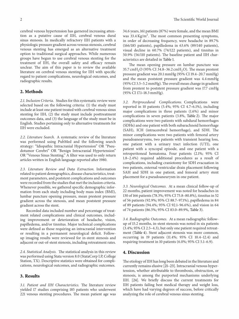
cerebral venous hypertension has garnered increasing atten-tion as a putative cause of IIH, cerebral venous duralsinus stenosis. In medically refractory IIH patients with aphysiologic pressure gradient across venous stenosis, cerebralvenous stenting has emerged as an alternative treatmentoption to traditional surgical approaches. While numerousgroups have begun to use cerebral venous stenting for thetreatment of IIH, the overall safety and efficacy remainunclear. The aim of this paper is to review the availableliterature on cerebral venous stenting for IIH with specificregard to patient complications, neurological outcomes, andradiographic results.
2. Methods
2.1. Inclusion Criteria. Studies for this systematic reviewwereselected based on the following criteria: (1) the study mustinclude at least one patient treated with cerebral venous sinusstenting for IIH, (2) the study must include posttreatmentoutcomes data, and (3) the language of the study must be inEnglish. Studies pertaining only to alternative treatments forIIH were excluded.
2.2. Literature Search. A systematic review of the literaturewas performed using PubMed and the following searchstrategy: “Idiopathic Intracranial Hypertension” OR “Pseu-dotumor Cerebri” OR “Benign Intracranial Hypertension”OR “Venous Sinus Stenting.” A filter was used to only returnarticles written in English language reported after 1980.
2.3. Literature Review and Data Extraction. Informationrelated to patient demographics, disease characteristics, treat-ment parameters, and poststent complications and outcomeswere recorded from the studies thatmet the inclusion criteria.Whenever possible, we gathered specific demographic infor-mation from each study including body mass index (BMI),lumbar puncture opening pressure, mean prestent pressuregradient across the stenosis, and mean poststent pressuregradient across the stent.
Recorded data included number and percentage of treat-ment related complications and clinical outcomes, includ-ing improvement or deterioration of headache, vision,papilledema, and/or tinnitus. Major technical complicationswere defined as those requiring an intracranial interventionor resulting in a permanent neurological deficit. Follow-up imaging results were reviewed for in-stent stenosis andadjacent or out-of-stent stenosis, including retreatment rates.
2.4. Statistical Analysis. The statistical analysis in this reviewwas performed using Stata version 8.0 (StataCorp LP, CollegeStation, TX). Descriptive statistics were obtained for compli-cations, neurological outcomes, and radiographic outcomes.
3. Results
3.1. Patient and IIH Characteristics. The literature reviewyielded 17 studies comprising 185 patients who underwent221 venous stenting procedures. The mean patient age was
34.6 years, 161 patients (87%) were female, and themean BMIwas 33.4 Kg/m2. The most common presenting symptoms,in order of decreasing frequency, were headache in 89.7%(166/185 patients), papilledema in 63.6% (89/140 patients),visual decline in 60.7% (74/122 patients), and tinnitus in50.9% (56/110 patients). The baseline patient and IIH char-acteristics are detailed in Table 1.
The mean opening pressure on lumbar puncture was35.7 cmH
2O (95% CI 34.8–36.2 cmH
2O). The mean prestent
pressure gradient was 20.1mmHg (95% CI 19.4–20.7mmHg)and the mean poststent pressure gradient was 4.4mmHg(95%CI 3.5–5.2mmHg).The overallmean change in gradientfrom prestent to poststent pressure gradient was 17.7 cmHg(95% CI 17.1–18.3mmHg).
3.2. Periprocedural Complications. Complications werereported in 10 patients (5.4%; 95% CI 4.7–6.1%), includingmajor complications in three patients (1.6%) and minorcomplications in seven patients (3.8%, Table 2). The majorcomplications were two patients with subdural hemorrhages(SDHs) and one patient with both subarachnoid hemorrhage(SAH), ICH (intracerebral hemorrhage), and SDH. Theminor complications were two patients with femoral arterypseudoaneurysms, two patients with transient hearing loss,one patient with a urinary tract infection (UTI), onepatient with a syncopal episode, and one patient with aretroperitoneal hematoma. Four patients (2.1%; 95% CI1.8–2.4%) required additional procedures as a result ofcomplications, including craniotomy for SDH evacuation intwo patients, external ventricular drain placement followingSAH and SDH in one patient, and femoral artery stentplacement for a pseudoaneurysm in one patient.
3.3. Neurological Outcomes. At a mean clinical follow-up of22 months, patient improvement was noted for headaches in130 of 166 patients (78.3%; 95% CI 75.8–80.8%), tinnitus in 52of 56 patients (92.9%; 95% CI 88.7–97.1%), papilledema in 84of 89 patients (94.4%; 95% CI 92.1–96.6%), and vision in 64of 74 patients (86.5%; 95% CI 83.0–89.9%, Table 3).
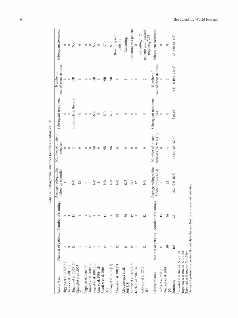
3.4. Radiographic Outcomes. At a mean radiographic follow-up of 15.2 months, in-stent stenosis was noted in six patients(3.4%; 95% CI 2.5–4.3), but only one patient required retreat-ment (Table 4). Stent adjacent stenosis was more common,occurring in 19 patients (11.4%; 95% CI 10.4–12.4) andrequiring treatment in 10 patients (6.0%; 95% CI 5.1–6.9).
4. Discussion
Theetiology of IIHhas long been debated in the literature andcurrently remains elusive [21–23]. Intracranial venous hyper-tension, whether attributable to thrombosis, obstruction, orstenosis, is among the purported mechanisms underlyingIIH. [24]. We briefly discuss the current treatments forIIH patients failing best medical therapy and weight loss,which have had varying degrees of success, before criticallyanalyzing the role of cerebral venous sinus stenting.

The Scientific World Journal 3
Table1:Ch
aracteris
ticso
fpatientsa
ndcasesinvolvedin
stentingforIIH
.
Author/year
Num
bero
fpatie
nts
Num
bero
fste
nts
Age
Female
gend
erBM
I(Kg
/m2 )
CSFop
eningpressure
(cm
H20)
Meanprestent
pressure
gradient
(mmHG)
Meanpo
ststent
pressure
gradient
(mmHG)
Meangradient
change
(mmHG)
Higgins
etal.2002[4]
11
301/1
30.1
3518
315
Owlere
tal.2003
[5]
44
27(17–38)
3/4
30(23–48)
29(22–35)∗
19(12–25)
0.3(0-1)
18.7
Higgins
etal.2003[6]
1214
33(19
–52)
12/12
36.9(29–
45)
33.7(25–36)
18.9(8–37)
11.3(2–23)
7.6Ogu
ngbo
etal.2003
[7]
11
371/1
26.1
>40
25NR
NR
Rajpaletal.2005[8]
11
150/1
26.9
3725
NR
NR
Don
netetal.2008
[9]
1011
41(28–60)
8/10
27.3(22–37)
40.2(29–
59)
19.1(12–34)
NR
NR
Paqu
etetal.2008[10]
11
601/1
NR
3015
NR
NR
Arace
tal.2009
[11]
11
511/1
2931
132
11Bu
ssiere
etal.2010
[12]
1013
34(16–
65)
10/10
35.9(27–47)
NR
28.3(11–50)
11.3(2–23)
17
Zhengetal.2010[13]
11
341/1
26.1
4022.5
6.5
16Ahm
edetal.2011[14]
5260
34(10–
64)
47/52
>30
in47
32.9(25–73)∧
19.1(4–4
1)0.6(0–14)
18.5
Albuq
uerque
etal.
2011[15]
1530
32.3(15–51)
12/15
NR
NR
NR
NR
NR
Kumpe
etal.2012[16]
1819
37.9(16–
62)
12/18
31.6(22.6–
38)
39.6(25–55)
21.4(4–39)
2.6(0–7)
18.8
Teleb
etal.2012[17]
11
221/1
2848
260
26Ra
dvanyetal.2013
[18]
1212
39(21–55)
11/12
32.6
(27.3
–45.7)
39.4(29–
55)
12.4(5–28)
1.3(0–4
)11.1
Fields
etal.2013[19
]15
1534
(20–
56)
15/15
39(30–
73)
NR
24(13–40
)4(0–9
)20
Ducruetetal.2014
[20]
3036
33(14
–52)
25/30
NR
NR
NR
NR
21.4(10–
56)
Characteris
tics
Num
bero
fpatie
nts
Num
bero
fste
nts
Meanage
(95%
CI)
Female
gend
er(%
;95%
CI)
BMI
(95%
CI)
CSFop
eningpressure
(95%
CI)
Meanprestent
pressure
gradient
(95%
CI)
Meanpo
ststent
pressure
gradient
(95%
CI)
Meangradient
(95%
CI)
Summary
185
221
34.6
(34.0–
35.1)
161(87.0;
0.86–0
.89)
33.4
(32.6–
34.3)
35.7(34.8–36.2)
20.1(19
.4–20.7)
4.4(3.5–5.2)
17.7(17.1–18.3)
∗Not
repo
rted
in1p
atient.
∧Not
repo
rted
in11patie
nts.

4 The Scientific World Journal
Table 2: Complications following stenting for IIH.
Author/year Number ofpatients
Number ofstentings Complications Complication rate
Complicationsrequiring additionalprocedure
Higgins et al. 2002 [4] 1 1 0 0% N/AOwler et al. 2003 [5] 4 4 0 0% N/AHiggins et al. 2003 [6] 12 14 0 0% N/AOgungbo et al. 2003[7] 1 1 0 0% N/A
Rajpal et al. 2005 [8] 1 1 0 0% N/ADonnet et al. 2008 [9] 10 11 0 0% N/APaquet et al. 2008 [10] 1 1 0 0% N/AArac et al. 2009 [11] 1 1 0 0% N/ABussiere et al. 2010[12] 10 13 0 0% N/A
Zheng et al. 2010 [13] 1 1 0 0% N/A
Ahmed et al. 2011 [14] 52 60 2 major (SDH); 2 minor(transient hearing loss) 7.7%
2 (1 SDH, 1SDH/ICH/SAH bothrequiring emergentcraniotomy)
Albuquerque et al.2011 [15] 15 30 1 minor RPH not
requiring transfusion 3.3% 0
Kumpe et al. 2012 [16] 18 191 major (SAH/SDH); 2minor (UTI andsyncope)
16.7%1 (SAH/SDHhematoma requiringEVD)
Teleb et al. 2012 [17] 1 1 0 0% N/ARadvany et al. 2013[18] 12 12 0 0% N/A
Fields et al. 2013 [19] 15 15 1 minor (femoralpseudoaneurysm) 6.7%
0 (femoralpseudoaneurysmresolvedcompression)
Ducruet et al. 2014[20] 30 36 1 minor (femoral
pseudoaneurysm 2.8
1 (femoralpseudoaneurysmrequiring femoralartery stent)
Characteristics Number ofpatients
Number ofstents Complications Complication rate %
(95% CI)
Complicationsrequiring additionalprocedure (%; 95%CI)
Summary 185 221 10 5.4% (4.7–6.1) 4 (2.1%; 1.8–2.4%)SAH: subarachnoid hemorrhage.SDH: subdural hemorrhage.ICH: intracerebral hemorrhage.RPH: retroperitoneal hematoma.
4.1. Surgical Interventions for IIH. Surgical therapies aretypically considered after medical therapy has failed andgenerally consist of CSF diversion (serial lumbar puncture,lumboperitoneal shunt, or ventriculoperitoneal shunt) oroptic nerve sheath fenestration (ONSF). With regard toCSF diversion procedures, LP shunting is often preferredin IIH patients due to their characteristic silt-like ventricleswhich increase the difficulty of ventriculoperitoneal shuntplacement. However, CSF diversion is fraught with hardwarefailure and repeated need for revisions along with infections.
A recent review showed that, while LP and VP shunting arehighly effective inmitigating IIH symptoms in the immediatepostoperative period, both procedures have a fairly highfailure rate.The revision rates for both forms of CSF diversionprocedure were 60% for LP and 30% for VP shunts [25, 26].
ONSF is typically indicated in IIH patients with visualloss who endorse mild to no headaches. A small duralwindow created in the optic nerve sheath serves to drainCSF and relieve pressure on the optic disc, thereby helping topreserve vision. Another theory suggests that ONSF serves

The Scientific World Journal 5
Table3:Neurologico
utcomes
follo
wingste
ntingforIIH
.
Author/year
Num
bero
fpatients
Num
bero
fstents
Improved
headache
Improved
tinnitus
Improved
papilledema
Improved
visio
nFo
llow-up
Higgins
etal.2002[4]
11
1/1NA
1/11/1
12Owlere
tal.2003
[5]
44
3/4
1/14/4
4/4
9.8(5–12)
Higgins
etal.2003[6]
1214
7/12
NR
5/8
7/12
14.2(2–26)
Ogu
ngbo
etal.2003
[7]
11
1/1NA
1/11/1
6
Rajpaletal.2005[8]
11
1/1NA
1/11/1
6Don
netetal.2008
[9]
1011
8/10
9/9
10/10
9/10
17.2(6–36)
Paqu
etetal.2008[10]
11
1/1NA
1/11/1
NR
Arace
tal.2009
[11]
11
1/10/1
NA
NA
2Bu
ssiere
etal.2010
[12]
1013
10/10
3/3
9/9
7/8
20.1(4–6
0)
Zhengetal.2010[13]
11
1/1NA
1/11/1
3Ahm
edetal.2011[14]
5260
40/43
17/17
9/9
19/19
24(2–108)
Albuq
uerque
etal.
2011[15]
1530
12/15
(1:w
orse,2:n
ochange)
NR
NR
NR
20(2–4
0)
Kumpe
etal.2012[16]
1819
10/12
(2:n
ochange)
NR
15/16
NR
43.7(11–136)
Teleb
etal.2012[17]
11
1/1NA
1/11/1
6Ra
dvanyetal.2013
[18]
1212
5/12
(5:nochange)
11/11
11/12
10/12
16(9–36)
Fields
etal.2013[19
]15
1510/15
(1:nochange,2:
worse,2:different)
11/14
15/15
2/3
14(1–
49)
Characteris
tics
Num
bero
fpatients
Num
bero
fstents
Improved
headache
(%;95%
CI)
Improved
tinnitus(%;
95%CI
)
Improved
papilledema(
%;95%
CI)
Improved
visio
n(%
;95%CI
)Meanfollo
w-up(95%
CI)
Ducruetetal.2014
[20]
3036
18/26(8:nochange)
NR
NR
NR
23(0–58)
Summary
185
221
130(78.3;75.8–80.8)
52(92.9;88.7–
97.1)
84/89(94.4;
92.1–
96.6)
64/74(86.5;
83.0–89.9
)22.0(20.7–23.2)
6 The Scientific World Journal
Table4:Ra
diograph
icou
tcom
esfollo
wingste
ntingforIIH
.
Author/year
Num
bero
fpatients
Num
bero
fstentings
Averager
adiographic
follo
w-up(m
onths)
Num
bero
fin-ste
ntste
noses
Subsequent
treatment
Num
bero
fou
t-of-s
tent
steno
ses
Subsequent
treatment
Higgins
etal.2002[4]
11
10
00
0Owlere
tal.2003
[5]
94
30
01
0Higgins
etal.2003[6]
1214
NR
2Th
rombo
lytic
therapy
NR
NR
Ogu
ngbo
etal.2003
[7]
11
120
00
0
Rajpaletal.2005[8]
11
60
00
0Don
netetal.2008
[9]
1011
60
00
0Paqu
etetal.2008[10]
11
NR
NR
NR
NR
NR
Arace
tal.2009
[11]
11
20
00
0Bu
ssiere
etal.2010
[12]
1013
NR
NR
NR
NR
NR
Zhengetal.2010[13]
11
NR
NR
NR
NR
NR
Ahm
edetal.2011[14]
5260
NR
00
6Re
sentingin
6patie
nts
Albuq
uerque
etal.
2011[15]
1530
12.5
00
1Re
stenting
Kumpe
etal.2012[16]
1819
25.3
00
4Re
stentingin
1patient
Teleb
etal.2012[17]
11
120
00
0
Radvanyetal.2013
[18]
1212
NA
00
2Re
stentingin
2patie
ntsa
nd1p
atient
requ
iring
VPS
Characteris
tics
Num
bero
fpatients
Num
bero
fstentings
Averager
adiographic
follo
w-up(95%
CI)
Num
bero
fin-ste
ntste
noses(%;95%
CI)
Subsequent
treatment
(%)
Num
bero
fou
t-of-s
tent
steno
ses
Subsequent
treatment
Fields
etal.2013[19
]15
159
00
00
Ducruetetal.2014
[20]
3036
224
05
0
Summary
185
221
15.2(13.6–
16.8)1
6(3.4;2.5–4
.3)2
1(0.6)∧
19(11.4
;10.4–
12.4)3
10(6.0;5.1–
6.9)
3
1 Reportedin
11stu
dies
(𝑁=102).
2 Reportedin
14stu
dies
(𝑁=178).
3 Reportedin
13stu
dies
(𝑁=166).
∧Re
fersto
1patient
who
received
thrombo
lytic
therapy.Not
patie
ntsreceivedreste
nting.

The Scientific World Journal 7
to elicit an inflammatory response that results in fibrosis ofthe optic nerve sheath, thereby preventing the transduction ofintracranial pressure via the subarachnoid space to the opticdisc.While ONSF often stabilizes vision function in the acutepostprocedural period, it has been shown to have failure rates(defined as progressive vision loss after surgery) of 34% at1 year and 45% at 3 years [15]. Thus, the current treatmentoptions for IIH are limited by their lack of durability andrelatively high long-term failure rates.
4.2. Role of Cerebral Venous Sinus Stenting in theManagementof IIH. With the increasing recognition of cerebral venousstenosis as an etiology of IIH, dural venous sinus stentinghas emerged as a potentially effective treatment. Recentpublications have demonstrated promising clinical resultswith regard to headache and tinnitus resolution, papilledemareduction, and visual function improvement.
While the majority of the data focuses on headacheimprovement, more recent literatures have also focused onvisual outcomes which may improve in a significant numberof patients following stenting. However, many of the earlystudies simply state that treated patients’ visual complaintsimproved without further quantification of pre- and post-procedural visual acuity or visual fields, and therefore it ishard to draw concrete conclusions from these studies [5–8, 13, 17]. More recent data have provided objective measuresof ophthalmologic outcomes, including visual acuity andvisual field testing [14, 18, 19]. As such, more rigorousophthalmologic data will be needed in future studies to bettersubstantiate the use of dural venous sinus stenting to improvevision in patients with IIH.
While venous stenting often obviates the need for CSFdiversion, it is not without its own set of risks. Many of thereported complications arise from the angiography proce-dure rather than from stent placement. The most commoncomplications were access related and include a retroperi-toneal hematoma and 2 femoral pseudoaneurysms [15, 19,20]. A more serious complication in the form of SDH andSAH was observed in 1 of 18 patients as reported by Kumpeet al. [16]. During stent placement of the right transversesinus in this patient, there was stasis of flow in the rightsigmoid sinus leading to a left SDH and SAH. The patientwas managed successfully with an external ventricular drain.Similar complications were seen in a large series of 52 patients[14]. In this series, two patients had postprocedural SDHs.One patient developed a SDH after guidewire perforation ofa dural sinus, while the other patient suffered a SDH alongwith SAH and intracerebral hemorrhage during emergentstent placement for fulminant IIH. Both patients underwentemergent craniotomies and made a full recovery. Althoughrisks are inherent to any procedure, venous stenting for IIHremains a relatively safe procedure with numerous studiesreporting no intraoperative complications [4–6, 11, 12, 18].
As with any stenting procedure, there exist complicationsinherently related to the stent, namely, in-stent stenosis.Two separate processes have been described for stent-relatedstenosis in the setting of IIH: in-stent stenosis and stentadjacent stenosis. Stent thrombosis may lead to in-stentstenosis or occlusion [27]. This, in general, would likely
cause the return of the presenting symptoms. However, stentthrombosis may theoretically be disastrous if the thrombusoccludes the drainage of the vein of Labbe. The increasinguse of periprocedural dual antiplatelet therapy has led to adecrease in the incidence of in-stent stenosis [15, 19], althoughit has not been totally eliminated [20]. Stent adjacent stenosisis defined as a venous sinus stenosis which develops adjacentto the stent, often in the segment from the torcula to thestent, and is somewhat unique to the dural venous sinusesfollowing stenting. This phenomenon has been described in19 cases in this review [14–16], of which 10 underwent furtherstenting. However, some groups were elected to not treatasymptomatic stent adjacent stenosis. The phenomenon ofout-of-stent stenosis in IIH raises the question as to whetherthere exists an inherent pressure from the brain parenchymaitself that serves to push on the venous sinus, giving thema stenosed appearance. Thus, venous sinus stenosis may bea result of idiopathic increased intracranial hypertensionrather than a cause of it. Long-term radiographic outcomesand further delineation of the pathophysiology behind duralvenous sinus stenosis are indicated in future studies.
Finally, it is important to consider that radiographic evi-dence of venous sinus stenosis alone is inadequate to justifystenting for IIH. There must also be physiologic evidence ofa significant pressure gradient across the stenosis in orderfor stenting to be clinically efficacious. In our literaturereview, we found the mean prestent pressure gradient to be20mmHg. Further studies are necessary to determine theoptimal gradient for stenting in IIH patients.
4.3. Study Limitations. This review is limited by the hetero-geneity of the case series of which it is comprised. Specifically,there were no reporting standards for the baseline clinicaland radiographic characteristics and for the posttreatmentoutcomes. Additionally, all studieswere retrospective, and thenumber of patients per series was relatively small. Finally,the stent type and design varied across different series, thuslimiting further the generalizability of our findings. Giventhese limitations, venous sinus stenting for patients withmedically refractory IIH in whom a radiographic venoussinus stenosis and physiologic pressure gradient are bothevident is a Class IIa Recommendation, Level of Evidence C.
5. Conclusions
Cerebral venous dural sinus stenting affords a favorable risk-to-benefit profile for appropriately selected IIH patients whoare refractory to medical management and are demonstratedto have both a venous sinus stenosis and a physiologicpressure gradient. The available literature demonstrates thatvenous stenting is effective, but further long-term, prospec-tive evaluation of this treatment approach is necessary. Specif-ically, additional studies that define ophthalmologic andradiographic baseline parameters and outcomes are requisitefor defining the optimal patient population. Additionally,further work is necessary to determine the best therapeuticoption for IIH patients.

8 The Scientific World Journal
Conflict of Interests
The authors report no direct conflict of interests. PascalJabbour is a consultant for ev3, Codman, andMizuho. AaronS. Dumont is a consultant for ev3 and Stryker.
References
[1] K. Radhakrishnan, J. E. Ahlskog, J. A. Garrity, and L. T. Kurland,“Idiopathic intracranial hypertension,”MayoClinic Proceedings,vol. 69, no. 2, pp. 169–180, 1994.
[2] K. Radhakrishnan, J. E. Ahlskog, S. A. Cross, L. T. Kurland,and W. M. O’Fallon, “Idiopathic intracranial hypertension(Pseudotumor cerebri): descriptive epidemiology in Rochester,Minn, 1976 to 1990,”Archives of Neurology, vol. 50, no. 1, pp. 78–80, 1993.
[3] K. B. Digre, “Epidemiology of idiopathic intracranial hyper-tension,” in Proceedings of the Annunal Meeting of the NorthAmerican Neuro-Ophthalmoligical Society (NANOS ’92), June1992.
[4] J. N. P. Higgins, B. K. Owler, C. Cousins, and J. D. Pickard,“Venous sinus stenting for refractory benign intracranial hyper-tension,”The Lancet, vol. 359, no. 9302, pp. 228–230, 2002.
[5] B. K. Owler, G. Parker, G. M. Halmagyi et al., “Pseudotumorcerebri syndrome: venous sinus obstruction and its treatmentwith stent placement,” Journal of Neurosurgery, vol. 98, no. 5,pp. 1045–1055, 2003.
[6] J. N. P. Higgins, C. Cousins, B. K. Owler, N. Sarkies, and J. D.Pickard, “Idiopathic intracranial hypertension: 12 cases treatedby venous sinus stenting,” Journal of Neurology, Neurosurgery &Psychiatry, vol. 74, no. 12, pp. 1662–1666, 2003.
[7] B. Ogungbo, D. Roy, A. Gholkar, and A. D. Mendelow,“Endovascular stenting of the transverse sinus in a patient pre-senting with benign intracranial hypertension,” British Journalof Neurosurgery, vol. 17, no. 6, pp. 565–568, 2003.
[8] S. Rajpal, D. B. Niemann, and A. S. Turk, “Transverse venoussinus stent placement as treatment for benign intracranialhypertension in a young male. Case report and review of theliterature,” Journal of Neurosurgery, vol. 102, no. 3, pp. 342–346,2005.
[9] A. Donnet, P. Metellus, O. Levrier et al., “Endovascular treat-ment of idiopathic intracranial hypertension: clinical and radi-ologic outcome of 10 consecutive patients,” Neurology, vol. 70,no. 8, pp. 641–647, 2008.
[10] C. Paquet, M. Poupardin, M. Boissonnot, J. P. Neau, andJ. Drouineau, “Efficacy of unilateral stenting in idiopathicintracranial hypertension with bilateral venous sinus stenosis: acase report,” European Neurology, vol. 60, no. 1, pp. 47–48, 2008.
[11] A. Arac,M. Lee, G. K. Steinberg,M.Marcellus, andM. P.Marks,“Efficacy of endovascular stenting in dural venous sinus stenosisfor the treatment of idiopathic intracranial hypertension,”Neurosurgical Focus, vol. 27, no. 5, p. E14, 2009.
[12] M. Bussiere, R. Falero, D. Nicolle, A. Proulx, V. Patel, andD. Pelz, “Unilateral transverse sinus stenting of patients withidiopathic intracranial hypertension,” The American Journal ofNeuroradiology, vol. 31, no. 4, pp. 645–650, 2010.
[13] H. Zheng,M. Zhou, B. Zhao, D. Zhou, and L.He, “Pseudotumorcerebri syndrome and giant arachnoid granulation: treatmentwith venous sinus stenting,” Journal of Vascular and Interven-tional Radiology, vol. 21, no. 6, pp. 927–929, 2010.
[14] R. M. Ahmed, M. Wilkinson, G. D. Parker et al., “Transversesinus stenting for idiopathic intracranial hypertension: a review
of 52 patients and of model predictions,” American Journal ofNeuroradiology, vol. 32, no. 8, pp. 1408–1414, 2011.
[15] F. C. Albuquerque, S. R. Dashti, Y. C. Hu et al., “Intracranialvenous sinus stenting for benign intracranial hypertension:clinical indications, technique, and preliminary results,”WorldNeurosurgery, vol. 75, no. 5-6, pp. 648–652, 2011.
[16] D. A. Kumpe, J. L. Bennett, J. Seinfeld, V. S. Pelak, A. Chawla,and M. Tierney, “Dural sinus stent placement for idiopathicintracranial hypertension,” Journal of Neurosurgery, vol. 116, no.3, pp. 538–548, 2012.
[17] M. S. Teleb, H. Rekate, S. Chung, and F. C. Albuquerqu,“Psuedotumor cerebri presenting with ataxia and hyper-reflexiain a non-obese woman treated with sinus stenting,” Journal ofNeuroInterventional Surgery, vol. 4, no. 5, p. e22, 2012.
[18] M. G. Radvany, D. Solomon, S. Nijjar et al., “Visual andneurological outcomes following endovascular stenting forpseudotumor cerebri associated with transverse sinus stenosis,”Journal of Neuro-Ophthalmology, vol. 33, no. 2, pp. 117–122, 2013.
[19] J. D. Fields, P. P. Javedani, J. Falardeau et al., “Dural venoussinus angioplasty and stenting for the treatment of idio-pathic intracranial hypertension,” Journal of NeuroInterven-tional Surgery, vol. 5, no. 1, pp. 62–68, 2013.
[20] A. F. Ducruet, R. W. Crowley, C. G. McDougall, and F. C.Albuquerque, “Long-term patency of venous sinus stents foridiopathic intracranial hypertension,” Journal of NeuroInterven-tional Surgery, vol. 6, no. 3, pp. 238–242, 2014.
[21] J. O. Donaldson, “Pathogenesis of pseudotumor cerebri syn-dromes,” Neurology, vol. 31, no. 7, pp. 877–880, 1981.
[22] R. A. Fishman, “The pathophysiology of pseudotumor cerebri.An unsolved puzzle,” Archives of Neurology, vol. 41, no. 3, pp.257–258, 1984.
[23] B. Ireland, J. J. Corbett, and R. B. Wallace, “The search forcauses of idiopathic intracranial hypertension: a preliminarycase-control study,”Archives of Neurology, vol. 47, no. 3, pp. 315–320, 1990.
[24] B. S. Ray and H. S. Dunbar, “Thrombosis of the dural venoussinuses as a cause of pseudotomor cerebri,” Annals of Surgery,vol. 134, no. 3, pp. 376–386, 1951.
[25] K. Abubaker, Z. Ali, K. Raza, C. Bolger, D. Rawluk, andD. O’Brien, “Idiopathic intracranial hypertension: lumboperi-toneal shunts versus ventriculoperitoneal shunts—case seriesand literature review,” British Journal of Neurosurgery, vol. 25,no. 1, pp. 94–99, 2011.
[26] M. J. McGirt, G. Woodworth, G. Thomas, N. Miller, M.Williams, and D. Rigamonti, “Cerebrospinal fluid shunt place-ment for pseudotumor cerebri-associated intractable headache:Predictors of treatment response and an analysis of long-termoutcomes,” Journal of Neurosurgery, vol. 101, no. 4, pp. 627–632,2004.
[27] J. N. P. Higgins and J. D. Pickard, “Lateral sinus stenosesin idiopathic intracranial hypertension resolving after CSFdiversion,” Neurology, vol. 62, no. 10, pp. 1907–1908, 2004.

Submit your manuscripts athttp://www.hindawi.com
Stem CellsInternational
Hindawi Publishing Corporationhttp://www.hindawi.com Volume 2014
Hindawi Publishing Corporationhttp://www.hindawi.com Volume 2014
MEDIATORSINFLAMMATION
of
Hindawi Publishing Corporationhttp://www.hindawi.com Volume 2014
Behavioural Neurology
EndocrinologyInternational Journal of
Hindawi Publishing Corporationhttp://www.hindawi.com Volume 2014
Hindawi Publishing Corporationhttp://www.hindawi.com Volume 2014
Disease Markers
Hindawi Publishing Corporationhttp://www.hindawi.com Volume 2014
BioMed Research International
OncologyJournal of
Hindawi Publishing Corporationhttp://www.hindawi.com Volume 2014
Hindawi Publishing Corporationhttp://www.hindawi.com Volume 2014
Oxidative Medicine and Cellular Longevity
Hindawi Publishing Corporationhttp://www.hindawi.com Volume 2014
PPAR Research
The Scientific World JournalHindawi Publishing Corporation http://www.hindawi.com Volume 2014
Immunology ResearchHindawi Publishing Corporationhttp://www.hindawi.com Volume 2014
Journal of
ObesityJournal of
Hindawi Publishing Corporationhttp://www.hindawi.com Volume 2014
Hindawi Publishing Corporationhttp://www.hindawi.com Volume 2014
Computational and Mathematical Methods in Medicine
OphthalmologyJournal of
Hindawi Publishing Corporationhttp://www.hindawi.com Volume 2014
Diabetes ResearchJournal of
Hindawi Publishing Corporationhttp://www.hindawi.com Volume 2014
Hindawi Publishing Corporationhttp://www.hindawi.com Volume 2014
Research and TreatmentAIDS
Hindawi Publishing Corporationhttp://www.hindawi.com Volume 2014
Gastroenterology Research and Practice
Hindawi Publishing Corporationhttp://www.hindawi.com Volume 2014
Parkinson’s Disease
Evidence-Based Complementary and Alternative Medicine
Volume 2014Hindawi Publishing Corporationhttp://www.hindawi.com