Embed Size (px)
Citation preview

Review ArticleDevelopment of Synthetic and Natural Materials for TissueEngineering Applications Using Adipose Stem Cells
Yunfan He and Feng Lu
Department of Plastic and Cosmetic Surgery, Nanfang Hospital, Southern Medical University, Guangzhou, Guangdong 510515, China
Correspondence should be addressed to Feng Lu; [email protected]
Received 16 October 2015; Revised 9 January 2016; Accepted 12 January 2016
Academic Editor: Norbert Pallua
Copyright © 2016 Y. He and F. Lu. This is an open access article distributed under the Creative Commons Attribution License,which permits unrestricted use, distribution, and reproduction in any medium, provided the original work is properly cited.
Adipose stem cells have prominent implications in tissue regeneration due to their abundance and relative ease of harvest fromadipose tissue and their abilities to differentiate into mature cells of various tissue lineages and secrete various growth cytokines.Development of tissue engineering techniques in combination with various carrier scaffolds and adipose stem cells offers greatpotential in overcoming the existing limitations constraining classical approaches used in plastic and reconstructive surgery.However, as most tissue engineering techniques are new and highly experimental, there are still many practical challenges thatmust be overcome before laboratory research can lead to large-scale clinical applications. Tissue engineering is currently a growingfield of medical research; in this review, we will discuss the progress in research on biomaterials and scaffolds for tissue engineeringapplications using adipose stem cells.
1. Introduction
Adipose stem cells (ASCs) have the potential to differentiateinto various cell phenotypes if there is a specific inducingmicroenvironment using suitable inductive substances [1–3]. Meanwhile, their abundance and relative ease of harvest,along with their autogenous immune-privileged status, havealsomade them an attractive candidate for tissue engineeringand regenerative therapies [4, 5].
Tissue engineering enables the regeneration or repairof tissues and organs through combinations of stem cells,biomaterial scaffolds, and regulatory growth factors [6, 7].ASC-based tissue engineering strategies depend primarily onthe quality of theASC fraction used. Although a large numberof studies have been conducted to assess the differentiationpotential of ASCs in different carriers, there are still someunclear aspects regarding the basic knowledge ofASCbiologyand the clinical applications. These include the following: (i)What is the best definition for ASCs, and what is the truenature of the fractions of ASCs used by the various investi-gators? (ii) Does heterogeneity exist between freshly isolatedASCs and ASCs expanded in several culture passages? (iii)What are the best procedures for harvesting adipose tissue,
preparing the stromal vascular fraction (SVF), and isolatingASCs? (iv) What are the best procedures for cell banking andcellular cryopreservation?The past few years have seen excit-ing progress in tissue engineering and regenerative medicineusing various biomaterials and scaffold [8–11]. Natural andsynthetic materials have been developed to provide a carrierscaffold that is ideally supposed to mimic the extracellularmatrix (ECM) properties of an in vivo microenvironment toinduce tissue formation [12, 13]. Nowadays, the developmentof efficient biomaterials and scaffolds is still in high demandfor the production of clinically useable volumes of new tissuesto replace lost or malfunctioning body parts and to achieveuncomplicated wound healing.
It is crucial that scaffolding materials can positivelyinteract with surrounding tissue to not only fill the defect,but also facilitate the natural regeneration of stem cells.Significant efforts have been made to develop such scaffoldsfor tissue engineering applications [14–16]. For example, elec-trospinning, lithography,microfabrication, and self-assemblytechniques have been widely explored for the fabrication ofengineered scaffolds appropriate for specific tissue applica-tions. Considering the usage of these engineered scaffoldsin the body, they should have the following characteristics:
Hindawi Publishing CorporationStem Cells InternationalVolume 2016, Article ID 5786257, 12 pageshttp://dx.doi.org/10.1155/2016/5786257

2 Stem Cells International
(i) possession of appropriate surface properties to promotethe adhesion, proliferation, and differentiation of stem cells;(ii) low toxicity and immunogenicity; (iii) high porosity; and(iv) degradability that is adequate for specific tissues, withan interconnected pore network for cell growth and flowtransport of nutrients and metabolic waste. The objectiveof this review is to recapitulate the progress in the fields ofbiomaterial and scaffold development and various proceduresfor ASC selection for tissue engineering applications and toreview several clinical cases for the advancement of ASCs-based tissue engineering strategies.
2. Basic Knowledge on the Biology of ASCs inRegenerative Medicine
In the past decade, a number of cell characterization studieshave described the underlying biology of ASCs [17–33].Preclinical studies on the use of ASCs both in vitro and invivo have been performed, and the efficacy of ASCs has beendetermined in several clinical trials [34–40]. Compared withbone marrow or umbilical cord stem cells, ASCs have a simi-lar self-renewal ability in vitro, and the ability of ASCs to dif-ferentiate in other mesodermal and ectodermal lineages hasbeen demonstrated on several occasions [1, 41–45].Moreover,ASCs release multiple growth factors, such as the two keyfactors vascular endothelial growth factor (VEGF) and hep-atocyte growth factor (HGF). Other factors include VEGF-B, VEGF-C, fibroblast growth factor- (FGF-) 2, angiopoietin-(Ang-) 1, Ang-2, SPARC/osteonectin, platelet-derived growthfactor- (PDGF-) b, transforming growth factor (TGF), andstromal cell-derived factor-1 (SDF-1) [46]. However, thespecific population of ASCs with the greatest therapeuticpotential remains unclear. Since the initial reports in the late1960s [47], many researchers have established that stromalstem cells similar to those identified in bone marrow can beisolated from adipose tissue that is either resected as intacttissue or aspirated using tumescent liposuction [17, 48, 49]. Ingeneral, the obtained adipose tissue is digestedwith one of thefollowing: collagenase, dispase, trypsin, or related enzymes.A consensus exists regarding temperature (37∘C), digestionduration times (range, 30min to >1 h), and ratios of tissueweight to volume; however, protease concentrations are farmore variable. Following the neutralization of the enzymesand differential centrifugation, the released elements, whichare separated from the mature adipocytes, are defined asthe SVF. The SVF consists of a heterogeneous mesenchy-mal population of cells that includes not only adiposestromal and hematopoietic stem and progenitor cells butalso endothelial cells, erythrocytes, fibroblasts, lymphocytes,monocyte/macrophages, and pericytes [17, 50, 51]. For thephenotypic characterization of the SVF, the InternationalFederation for Adipose Therapeutics and Science (IFATS)and the International Society for Cellular Therapy (ISCT)proposed a stromal cell population, excluding hematopoieticand endothelial cells, based on the following combination:CD45−CD235a−CD31−CD34+ and additional markers usedto identify SVF are CD13 (APN), CD73 (L-VAP-2), CD90(Thy-1), and CD105 (Endoglin). Based on the existing liter-ature, this population combination represents at least 20% of
the cells in the SVF [52–56], and the percentage of CD34+cells mainly depends on the method used to harvest theadipose tissue, the degree of vascular hemorrhage, and thesubsequent digestion and isolation techniques. In addition tothe enzyme digestion, methods for isolating SVF cells usingmechanical, nonenzymatic techniques have been developedrecently, and some have been applied in clinical practice [57–59].
When SVF pellets are seeded into culture, a subset ofelongated cells begins to adhere to the bottom of the plastictissue culture plate. After a combination of washing stepsand culture expansion with media to remove most of thehematopoietic cell population from the SVF cells, these cellsare purified as an adherent cell population termed ASCs.ASCs are less heterogeneous than SVF cells and have theability to undergo self-renewal and the capacity to undergomultilineage differentiation and generate multiple terminallydifferentiated cells when cultured in specific lineage-inducingculture media. One main difference between SVF cells andASC suspensions is the high percentage of CD45+ cells in theSVF cell population (30–70%) and the low or undetectablepercentage in ASC population (2–30%). ASCs generallyexpress CD34+ during the early phase of culture (within 8–12 population doublings after culture of the SVF), but thenits expression decreases with continued cell division [51, 55].A joint statement by the IFATS and ISCT recommendedthat the surface antigens used to characterize ASCs shouldinclude CD73, CD90, and CD34 without CD45 and CD31.In addition, CD13 has also been proposed as an alternativeor supplement to CD105 [17]. To date, most experimentalresearch groups have isolated ASCs by tissue digestion,centrifugation, and the capacity of ASCs to adhere to cellculture plastic surfaces [9, 43, 60]. However, the adherentcell population also contains other cell types that are notmultipotential [61–63]. In order to overcome the problem of“contamination,” a number of alternative methods have beenproposed, including magnetic-activated cell sorting (MACS)and fluorescence-activated cell sorting (FACS). FACS is atypical cell enrichment method that utilizes complementaryfluorochrome conjugated antibodies to label cells of interest.However, the sorted cells obtained from FACS can be utilizedfor diagnostic and experimental purposes but not for ther-apeutics due to problems with safety and efficacy [64, 65].MACS is an antibody-aided technique based on immuno-magnetic beads coated with specific antibodies against stemcell surface molecules, and it is technically accessible andaffordable. From the view of clinical application, MACSwith biodegradable magnetic beads wins over FACS on thegrounds of safety, and it is the only method approved for usein clinical settings [61, 66–68].
Clinical research on adult stromal cell populations hasaccelerated, andmultiple clinical investigations are underwayto examine the use of ASCs and SVF cells for tissue engi-neering and regenerative medical applications [22, 69, 70].To achieve the large numbers of ASCs required for clinicalapplications, either the cells need to be expanded in cultureor ASCs must be pooled from multiple donors. Therefore,the development of stem cell banks is necessary. These banksmust assure the quality and safety of these cell products,

Stem Cells International 3
Table 1: Synthesis of synthetic materials for tissue engineering applications using ASCs.
Materials Properties Principal uses References
TiO2
nanofiber High degree of crystallinity andsurface wettability Bone tissue engineering [85]
Poly(3-hydroxybutyrate-co-hydroxyvalerate) (PHBV)scaffold
Good integration with the surroundingtissue, stiff character, and degradability Skin tissue engineering [10]
Copolymer PEGylated fibrin (P-fibrin)gels
Stable urethane (carbamate) linkage,degradability
Cardiovascular and skintissue engineering [91]
Poly(glycerol sebacate)(PGS)/poly(L-lactic acid) (PLLA)blend scaffolds
Favorable porous microstructures,good hydrophilicity, appropriatemechanical properties for soft tissueapplications, and degradability
Adipose tissue engineering [93]
Poly(lactic-coglycolicacid)/multiwalled carbonnanotubes/silk fibroin(PLGA/MWCNTs/SF) nanofibrousscaffolds
Nonwoven structures and randomfiber distribution with smooth andbeadless fibers morphology
Nerve tissue engineering [95]
[Poly-D,L-lactic acid/polyethyleneglycol/poly-D,L-lactic acid(PDLLA-PEG)]/hyaluronic acid (HA)matrix
Designed architectures, highmechanical strength andbiodegradability, biocompatibility, andwater solubility
Cartilage tissue engineering [99]
especially when the stored ASCs are intended for clinicaluse in cell therapy and regenerative medicine. Cryopreser-vation may be an ideal option for this and is currentlythe only method to preserve ASCs while maintaining theirfunctional properties and genetic characteristics in the longterm [71–73]. Slow freezing and vitrification are the currentlyavailable methods for the cryopreservation of stem cells inlaboratories and clinics [74–78]. Vitrification only works wellwith the cryopreservation of human cells in small volumes,such as oocytes, but it is ill-suited for large volumes ofASCs [79, 80]. Moreover, vitrification techniques requirehigher cryoprotectant agent concentrations, which inducestoxicity and osmotic stress in cells and tissues. Slow freezingis an established technique pioneered in the early 1970sand involves cryopreserving biological samples at controlledfreezing rates to avoid intracellular ice formation and mini-mize structural damage to the cell membrane and cytoskele-ton [81, 82]. Cryoprotectant agents are used at relativelylow concentrations [21] in slow freezing, which has becomethe standard method for cell and tissue cryopreservation.However, formation of ice crystals, extreme hyperosmolarity,and dehydration are still reported when cells undergo theslow freezing process. Dimethyl sulfoxide (DMSO) is themost widely used cryopreservant for cells, but it is knownto be toxic at room temperature. Trehalose is a nontoxicdisaccharide of glucose that may stabilize and preserve cellsand cellular structures during the freezing procedure. Acryopreservation method using trehalose as a cryoprotectiveagent is recommended for the long-term preservation ofASCs compared to simple cryopreservation or to cryop-reservation using DMSO alone. Other cryoprotective agentssuch as polyvinylpyrrolidone and methylcellulose have beendeveloped to replace DMSO; however, they are less efficientthan DMSO in terms of maintaining ASC viability [75].
Adipose-derived stem cells are a promising cell sourcefor regenerative medicine. It is important to understand thebasic knowledge and biology behind stem cells, and furtherresearch is needed to guarantee the safety of ASCs and theeffectiveness of tissue engineering using ASCs.
3. Advancement in Synthetic Materials forTissue Engineering Applications Using ASCs
In the provision of an appropriate microenvironment forcellular components to interact with, the extracellular matrix(ECM) is an important component of normal tissue thatmustbe considered. Various synthetic materials have been devel-oped to provide carrier scaffolds that mimic ECM propertiesfor tissue regeneration and reconstruction in combinationwith ASCs (Table 1). The advantages of synthetic materialsand scaffolds rely on the technical possibility that chemicaland physical properties (e.g., porosity, surface characteris-tics, and degradation products nature) can be specificallyoptimized for a particular application [83, 84]. Ideally, apolymeric material used for tissue engineering should be ableto regulate cell proliferation without the loss of pluripotencyand to direct differentiation into a specific cell lineage whendesired. Tan et al. described the influence of TiO
2nanofibrous
surface structures, which were produced in situ onto Ti-6Al-4V substrate via a thermal oxidation process, on the regula-tion of proliferation and preservation of stemness of ASCs.The results show thatASCs exhibit better adhesion and signif-icantly enhanced proliferation on TiO
2nanofibrous surfaces
than on flat control surfaces, thus presenting a promisingpotential for the application of TiO
2nanofibrous surfaces in
the field of bone tissue engineering and regenerative therapies[85]. Although much has been done to develop tissue-engi-neered skin substitutes in the past decade, poor visualization,
4 Stem Cells International
hypertrophic scarring, and keloid formation are still possiblenegative outcomes for current skin graft strategies [86–88].In an effort to overcome these limitations, Zonari et al.proposed the combination of poly(3-hydroxybutyrate-co-hydroxyvalerate) (PHBV) structures with ASCs to induceskin regeneration in a full-thickness model. In this work,PHBV scaffolds demonstrated good integration with thesurrounding tissue, allowing exudation and infiltration byinflammatory cells, which may contribute to rapid degrada-tion over time. Furthermore, PHBV scaffolds offered a moistenvironment combined with a stiff character that withstandscontraction and simultaneously stimulates the secretion ofvarious growth factors by seeded ASCs; these factors enhancevascularization and ECM deposition with reduced scarring.Ultimately, this study revealed the great advantages of PHBVloaded with ASCs to improve wound healing and skinregeneration with reduced scarring in skin tissue engineering[10].The advancement of tissue engineering as a regenerativetherapy relies on rapid vascularization of tissue constructs,and engineered three-dimensional biomaterials are known toaffect the angiogenic capacity of seeded stem cells [89–91].Copolymer PEGylated fibrin (P-fibrin) gels were introducedby Chung et al. as an ASC-carrying scaffold for encouraginglocal angiogenesis in an in vitro culture model without addedsoluble factors. In P-fibrin gels, ASCs elicited higher vonWillebrand factor expression than the two commonly usedhydrogels (i.e., collagen and fibrin). After seven days ofcultivation, vascular endothelial growth factor (VEGF) wassecreted more in fibrin and P-fibrin gels than in collagen;several other angiogenic and immunomodulatory cytokineswere similarly enhanced. Moreover, P-fibrin matrices wereuniquely able to drive a vessel-like phenotype in ASCsand induce formation of well-organized vascular networksrelative to other gels. Thus, it can be speculated that theresearch on ASCs’ regenerative potential in a carrier scaffoldcan be expanded to include cardiovascular and skin tissueengineering applications based on the observed angiogenicproperties of ASCs in P-fibrin [91]. Seeding cells on mechan-ically appropriate scaffolds and applying specific mechanicalstimulation to these cells have been found to be beneficial interms of proliferation and differentiation [92, 93]. Frydrychet al. reported a large and flexible 3D porous poly(glycerolsebacate) (PGS)/poly(L-lactic acid) (PLLA) blend scaffoldwithmechanical properties comparable to adipose tissue thatwas fabricated via a freeze-drying and a subsequent curingprocess. In vitro cell test results provided clear evidence thatPGS/PLLA scaffolds are suitable for the culture of ASCs, asthey are characterized by deep cell penetration and ECMgrowth.This work demonstrates that the PGS/PLLA scaffoldsprovided favorable porousmicrostructures, good hydrophiliccharacteristics, and appropriate mechanical properties forsoft tissue applications [93]. Neural tissue possesses a verylimited capacity to regenerate new functional neurons afternerve injuries, and tissue-engineered neural tissues usingstem cells may serve as a promising alternative for neuralregeneration. However, such stem cells would need to pro-liferate and differentiate into the desired phenotype with theaid of adequate chemical, mechanical, or biological stimuliregeneration [94–96]. Catalpol is a natural active ingredient
extracted from a traditional Chinese medicine. Guo et al.evaluated the effects of a catalpol-loaded scaffold on guidingthe neuronal differentiation of hASCs. In their study, theprocess for catalpol loading into the electrospun poly(lactic-coglycolic acid)/multiwalled carbon nanotubes/silk fibroin(PLGA/MWCNTs/SF) nanofibrous scaffolds was successfullyestablished. As a result of adding catalpol, the diametersof the nanofibers decreased and the porosity increased.Moreover, the mechanical properties of the composite scaf-folds were improved, and more neuronal-like cells werefound on scaffolds with catalpol [95]. The poor self-healingability of cartilage necessitates the development of meth-ods for cartilage regeneration. Fabrication of scaffolds withlive stem cell incorporation and subsequent differentiationpresents a promising route [97, 98]. Sun et al. [99] reportedthe use of a visible-light-based PSL (VL-PSL) system toencapsulate hASCs into a biodegradable polymer [poly-D,L-lactic acid/polyethylene glycol/poly-D,L-lactic acid (PDLLA-PEG)]/hyaluronic acid (HA) matrix to produce live cell con-structs with customized architectures. In the chondrogenicmedium-treated group (TGF-𝛽3 group), hASCs showed highviability (84%) and expressed the chondrogenic genes Sox9,collagen type II, and aggrecan at 11, 232, and 2.29 × 10⁵fold increases, respectively, compared to levels at day 0 innonchondrogenic medium. After 28 days, the mechanicalstrength of the TGF-𝛽3 group remained high at 240 kPa.Thus, PSL and PDLLA-PEG/HA-based fabrication methodusing ASCs is a promising approach for producing mechani-cally competent engineered cartilage.
Thus, synthetic materials provide greater control overthe mechanical and biochemical properties of the carrierscaffolds and represent a promising tool in tissue engineeringand regeneration medicine [100].
4. Development of Natural Materials forTissue Engineering Applications Using ASCs
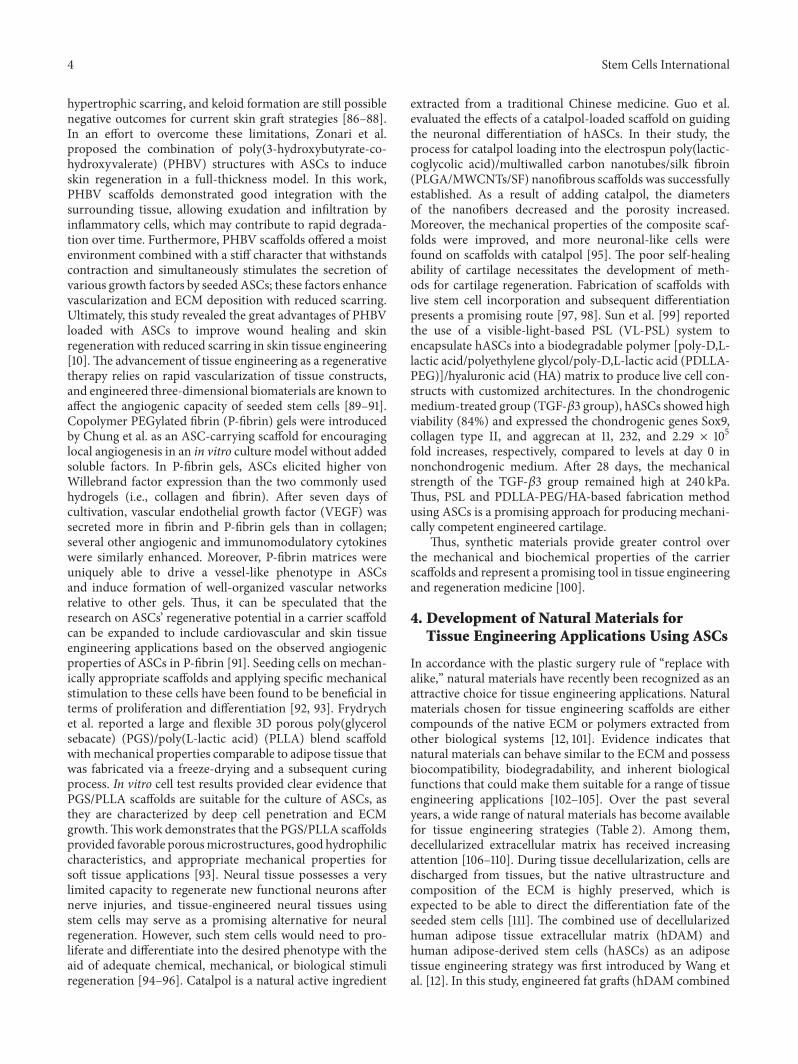
In accordance with the plastic surgery rule of “replace withalike,” natural materials have recently been recognized as anattractive choice for tissue engineering applications. Naturalmaterials chosen for tissue engineering scaffolds are eithercompounds of the native ECM or polymers extracted fromother biological systems [12, 101]. Evidence indicates thatnatural materials can behave similar to the ECM and possessbiocompatibility, biodegradability, and inherent biologicalfunctions that could make them suitable for a range of tissueengineering applications [102–105]. Over the past severalyears, a wide range of natural materials has become availablefor tissue engineering strategies (Table 2). Among them,decellularized extracellular matrix has received increasingattention [106–110]. During tissue decellularization, cells aredischarged from tissues, but the native ultrastructure andcomposition of the ECM is highly preserved, which isexpected to be able to direct the differentiation fate of theseeded stem cells [111]. The combined use of decellularizedhuman adipose tissue extracellular matrix (hDAM) andhuman adipose-derived stem cells (hASCs) as an adiposetissue engineering strategy was first introduced by Wang etal. [12]. In this study, engineered fat grafts (hDAM combined
Stem Cells International 5
Table2:Synthesis
ofnaturalm
aterialsfortiss
ueengineeringapplications
usingASC
s.
Materials
Prop
ertie
sPrincipalu
ses
References
Decellulariz
edhu
man
adipose
tissuee
xtracellu
larm
atrix
(hDAM)
Maintains
them
ajor
adiposetissue
ECM
compo
nents
and3D
structure
andinclu
desc
ollagen,
sulfated
glycosam
inoglycan,
andandvascular
endo
thelial
grow
thfactor
butlacks
major
histo
compatib
ility
complex
antig
enI
Adiposetissue
engineering
[12]
Acellularc
artilagem
atric
es(A
CMs)
Ideal3Dstr
ucture
andph
ysicochemicalprop
ertie
sand
good
biocom
patib
ility
Cartilage
tissuee
ngineerin
g[112]
Liverd
ecellulariz
edextracellular
matrix
(DCM
)
Preservesm
acroscop
ic3D
architecturea
ndthen
ative
compo
sition,
andultrastructure
remains
avisc
ous
liquidatlowtemperatures(ator
underroo
mtemperature)a
ndbecomes
gelationat37∘
C
Livertissue
engineering
[113]
Paper-basedbioactives
caffo
ldMicrofib
rous
porous
3Darchitecturea
ndbiocom
patib
le,cost-
effectiv
e,mechanicalrob
ustness
andwater
resistance
Bone
tissuee
ngineerin
g[117]
Hyaluronica
cidscaffold
Biocom
patib
ility,non
immun
ogenicity,high
hygroscopicity,and
capacityto
degradeintosafe
prod
ucts
Muscle
tissuee
ngineerin
g[121]
Collagen
Non
toxic,biocom
patib
le,andbioabsorbable,anditis
FDAapproved
foru
sein
humans
Adiposer
egenerationand
adiposetissue
engineering
[122,124,125]
Matrig
elNaturalpo
lymer
andbiocom
patib
leAd
iposetissue
engineering
[128]
Chito
san
Biod
egradable,biocom
patib
le,andan
excellent
hemostatic
andanalgesic
agentw
ithantio
xidant
prop
ertie
s
Skin
reconstructio
nandskin
tissuee
ngineerin
g[128,129]
6 Stem Cells International
with hASCs) were implanted subcutaneously in nude rats.The results showed that hASCs seeded in hDAM contributedto adipose tissue formation; the implanted engineered fatgrafts maintained their volume for eight weeks. Hence,this study provides a platform and novel scaffold designfor adipose tissue engineering of hDAM-hASC constructs.Current cartilage tissue engineering technology has devel-oped quickly and efforts have focused on the creation of asuitable chondrocyte scaffold. Acellular cartilaginous matrix(ACM), which is obtained from fresh cartilage using a seriesof acellular manipulations, is a recently developed naturalmatrix material. Wang et al. reported that the repair ofarticular cartilage defects had been achieved with ASCs andacellular cartilaginous matrix in rabbits [112]. In the tissue-engineered cartilage group (ACM combined with ASCs)after 12 weeks, articular cartilage defects were filled withchondrocyte-like tissue with a smooth surface and were richin glucan and type II collagen, similar to normal articu-lar cartilage. Although the development of cartilage tissueengineering is still in its infancy, the acellular cartilaginousmatrix obtained in this study offers tremendous potentialin cartilage regeneration medicine. Recently, a decellularizedliver 3D matrix scaffold has been proved to be able tofacilitate the activity and function of the hepatic cells andstem cells [113–115]. Zhang and Dong [113] compared thehepatogenic differentiation-inducing effect of decellularizedliver 3D matrix scaffold and several extracellular matrices,including collagen, fibronectin, and Matrigel in combinationwithmouse adipose-derivedmesenchymal stem cells in vitro.The results clearly demonstrated that decellularized liverECM gel, either on its own or in the presence of growfactors, could significantly enhance hepatic differentiationfrom ASCs compared with other matrix scaffolds; thisdemonstrates the feasibility of liver DCM as a bioscaffoldfor liver regenerative medicine and tissue engineering. Paper,which is produced from natural sources, can be supplied inlarge quantities with fair properties of biocompatibility andcost-effectiveness [116]. Hence, paper may have the potentialin establishing tissue engineering scaffolds for therapeuticapplication of stem cells. Park et al. reported the feasibilityof a paper-based bioactive scaffold for hASCs application torepair bone tissue defects for the first time [117]. In this study,paper scaffolds were prepared from three types of commer-cial paper materials: weighing paper (WP), chromatographypaper (CP), and wiping tissue (WT), after which a polymer-coating method called initiated chemical vapor deposition(iCVD) was employed to coat the paper scaffold to achievefavorable biochemical surface properties (e.g., adhesivenessand water resistance), without damaging the scaffolds. Theresults showed that osteogenic differentiation of hASCs wasinduced on the paper scaffolds under osteogenesis-inducingconditions in vivo, indicating that paper material possessesgreat potential as a bioactive, functional, and cost-effectivenatural scaffold for adipose stem cell-mediated bone tissueengineering. Insoluble (derivatized or crosslinked) forms ofHA have been extensively investigated for tissue engineeringpurposes due to HA’s role in the extracellular matrix as wellas its biocompatibility, nonimmunogenicity, high hygroscop-icity, and capacity to degrade into safe products [118–121].
Desiderio et al. evaluated the differentiation potential ofconstructs made from a new crosslinked HA (XHA) scaffoldon which NG2+ ASCs were loaded.Thirty days after engraft-ment in mice, NG2+ ASCs underwent a complete myogenicdifferentiation and fabricated human skeletal muscle tissue,indicating a significant step in muscle regeneration withoutthe need for a prior in vitromuscle differentiation step [121].
In summary, the application of natural materials in thefield of regeneration medicine is currently progressing. Theadvantages of natural materials are biocompatibility andmechanical and biological properties consistent with in vivofeatures, making them perfect candidates for tissue engineer-ing field. Apart from the neoteric materials mentioned above,other commonly used natural materials in tissue engineeringinclude collagen [122–125], hyaluronan [126], Matrigel [127,128], and chitosan [129].
5. Clinical Applications of DifferentCarrier Scaffolds in Combination withASCs in Tissue Engineering Strategies
The major role of regenerative medicine in this century isbased on cell therapy, in which ASCs hold a key position[130–135]. Recently, a number of in vitro and a few in vivostudies using ASCs in combination with carrier scaffolds canbe found through searches and on clinical trial websites.However, the use of cultured stem cells in clinical settingsis strictly controlled by governmental regulations aroundthe world, which largely restrict the application of ASCs inregenerative medicine. Meanwhile, plastic surgeons in Koreaand Japan have played a leading role in pioneering the useof ASCs in tissue engineering more than any other Westernnations due to less stringent government regulations.
It is well known that diabetic ulcers and chronic radiationulcers are notorious for their recurrence, or failure to healdue to patient debilitation or poor local blood supply con-ditions. Several conventional reconstructive surgeries havebeen introduced for patients with chronic nonhealing cuta-neous lesions [136, 137]. Presently, Kim and Jeong reported aless invasive method using adipose stem cell-based therapyand a collagen sponge scaffold, which was covered withan artificial dermis (Terudermis®) to deal with a chronicdiabetic ulcer on the knee area. Two weeks after application,vascular tissue ingrowth was seen in the lesion area andthus a skin graft could be placed on the newly engineeredvascular bed [5]. Complex fistulas are difficult to manage.Currently limited surgical procedures often result in highrecurrence rates, whereas extensive surgical procedures maycause fecal incontinence. One recent improvement in treatingcomplex fistulas may be the use of ASCs in combinationwith a fibrin glue scaffold described by Garcia-Olmo et al.[138]. The fibrin glue used in this study contained humanfibrinogen, bovine aprotinin, and human thrombin, and theASCs were isolated from lipoaspirated fat tissue. Eight weeksafter the final treatment, fistula healing was observed in 17(71 percent) of 24 patients who received fibrin glue plusASCs, in comparison to 4 (16 percent) of 25 patients whoreceived fibrin glue alone. The proportion of patients with

Stem Cells International 7
healing strongly indicates that the combination of the fibringlue scaffold and ASCs is an effective and safe treatment forcomplex perianal fistulas. Autogenous bone graft has beenconsidered to be the gold standard for reconstructive bonesurgery. However, harvesting bone for grafting is associatedwith significant donor site morbidity that requires additionaloperative and anesthetic time [139]. An alternative approachis bone tissue engineering, through which in situ boneformation by using combinations of biomaterials, bioactivemolecules, and stem cells can be achieved [140, 141]. Sandoret al. reported a case that used an ASC tissue-engineeredconstruct to treat a large anterior mandibular defect. Inthis report, expanded ASCs were seeded on 𝛽-tricalciumphosphate (𝛽-TCP) granular scaffolds consisting of recom-binant human bone morphogenetic protein-2; the constructswere implanted into a U-shaped titaniummesh that spannedthe parasymphyseal defect. Ten months after reconstruction,dental implants were integrated into the grafted site suc-cessfully with a dental implant-supported overdenture. Thepatient has been followed for three years since the tissue-engineered constructs were placed; he has been pleased withthe aesthetic outcome of the procedure and continues to besatisfied with the function of his dental implants.
Although the applications of scaffolding materialstogether with ASCs technologies are a rapidly developingfield of regeneration medicine, they are highly experimentalso far. Thus, there still remains a significant need to developefficient carrier materials that may bridge the gap and leadtowards clinical applications in tissue engineering.
6. Conclusions and Future Perspectives
In the past several years, evidence has demonstrated thatthe ECM not only offers structural support for cells butalso profoundly influences the major cellular programs ofgrowth, differentiation, and apoptosis [142, 143]. An idealscaffold structure must accomplish the roles of the extra-cellular matrix for the seeded cells, which will be usedto form a tissue-engineered construct and to promote therepair/regeneration of damaged tissue [144, 145]. Hence, thedesign of carrier materials that can regulate cell behaviorssuch as proliferation and differentiation is the main purposefor the fabrication of tissue engineering scaffolds. Moreover,we should begin to understand that biomaterials and scaffoldsused in tissue engineering strategies are dynamic, mobile,and multifunctional regulators of cellular behavior, and notjust mere carriers for stem cells or storehouses for cytokines.Indeed, as the field of tissue engineering is still in its infancy,possible biomechanics and biomechanical effects created bydifferent types of scaffolds on seeded ASCs should be furtherelucidated so that carrier scaffolds with special properties canbe created. The creation of such scaffolds would help us tooptimize cellular activities including changes in morphology,proliferation, and differentiation.
In general, carrier materials for the successful generationand maintenance of engineered tissue constructs shouldhave several necessary properties including biocompatibility,degradability, low toxicity, and immunogenicity. However,most carrier scaffolds possess only some of these desirable
properties. If a scaffold could embody all of these propertiessuccessfully, it would provide an ideal platform for tissueregeneration.
In conclusion, ASCs have a prominent and strong rolein tissue engineering and regenerative medicine due to theirhigh cell yield in adipose tissue, their ability to differentiateinto multiple lineages and secrete various cytokines, andtheir immunomodulatory effects. The field of ASCs-basedtissue engineering therapy is still young. Ongoing and futuredevelopment of carrier scaffolds together with reasonablepromotion of stem cell research and clinical studies willno doubt gradually bring ASC-based tissue engineeringtechnology down from the ivory tower and make it clinicallyaccessible on a larger scale, thereby benefiting more patients.
Conflict of Interests
The authors confirm that there is no known conflict ofinterests associated with this paper.
Acknowledgments
The authors gratefully thank Feng Lu for his support andcontribution during the conducting of this study. This workwas supported by the National Natural Science Founda-tion of China (81471881, 81372083, 81171834, and 81201482),Key Clinical Specialty Discipline Construction Program,and Health Collaborative Innovation Major Projects ofGuangzhou (7414275040815).
References
[1] E. Anghileri, S. Marconi, A. Pignatelli et al., “Neuronal differen-tiation potential of human adipose-derived mesenchymal stemcells,” Stem Cells and Development, vol. 17, no. 5, pp. 909–916,2008.
[2] D. A. Young, Y. S. Choi, A. J. Engler, and K. L. Christman,“Stimulation of adipogenesis of adult adipose-derived stem cellsusing substrates that mimic the stiffness of adipose tissue,”Biomaterials, vol. 34, no. 34, pp. 8581–8588, 2013.
[3] B. F. Seo, K. J. Kim, M. K. Kim, and J. W. Rhie, “The effects ofhuman keratinocyte coculture on human adipose-derived stemcells,” International Wound Journal, 2014.
[4] D.-C. Yeh, T.-M. Chan,H.-J. Harn et al., “Adipose tissue-derivedstem cells in neural regenerative medicine,” Cell Transplanta-tion, vol. 24, no. 3, pp. 487–492, 2015.
[5] Y.-J. Kim and J.-H. Jeong, “Clinical application of adipose stemcells in plastic surgery,” Journal of Korean Medical Science, vol.29, no. 4, pp. 462–467, 2014.
[6] S. Giannitelli, P. Mozetic, M. Trombetta, and A. Rainer, “Com-bined additive manufacturing approaches in tissue engineer-ing,” Acta Biomaterialia, vol. 24, pp. 1–11, 2015.
[7] K. H. Moon, I. K. Ko, J. J. Yoo, and A. Atala, “Kidney diseasesand tissue engineering,”Methods, 2015.
[8] B. Ozcelik, A. Blencowe, J. Palmer et al., “Highly porous andmechanically robust polyester poly(ethylene glycol) sponges asimplantable scaffolds,” Acta Biomaterialia, vol. 10, no. 6, pp.2769–2780, 2014.

8 Stem Cells International
[9] F. M. Ghorbani, B. Kaffashi, P. Shokrollahi, E. Seyedjafari, andA. Ardeshirylajimi, “PCL/chitosan/Zn-doped nHA electrospunnanocomposite scaffold promotes adipose derived stem cellsadhesion and proliferation,”Carbohydrate Polymers, vol. 118, pp.133–142, 2015.
[10] A. Zonari, T. M. Martins, A. C. Paula et al., “Polyhydroxy-butyrate-co-hydroxyvalerate structures loaded with adiposestem cells promote skin healing with reduced scarring,” ActaBiomaterialia, vol. 17, pp. 170–181, 2015.
[11] Y. Acil, X. Zhang, T. Nitsche et al., “Effects of different scaffoldson rat adipose tissue derived stroma cells,” Journal of Cranio-Maxillo-Facial Surgery, vol. 42, no. 6, pp. 825–834, 2014.
[12] L.Wang, J. A. Johnson,Q. Zhang, andE.K. Beahm, “Combiningdecellularized human adipose tissue extracellular matrix andadipose-derived stem cells for adipose tissue engineering,” ActaBiomaterialia, vol. 9, no. 11, pp. 8921–8931, 2013.
[13] M. Tallawi, E. Rosellini, N. Barbani et al., “Strategies for thechemical and biological functionalization of scaffolds for car-diac tissue engineering: a review,” Journal of the Royal SocietyInterface, vol. 12, no. 108, 2015.
[14] J. Liao, K. Shi, Q. Ding, Y. Qu, F. Luo, and Z. Qian, “Recentdevelopments in scaffold-guided cartilage tissue regeneration,”Journal of Biomedical Nanotechnology, vol. 10, no. 10, pp. 3085–3104, 2014.
[15] L. Ghasemi-Mobarakeh, M. P. Prabhakaran, L. Tian et al.,“Structural properties of scaffolds: crucial parameters towardsstem cells differentiation,”World Journal of Stem Cells, vol. 7, no.4, pp. 728–744, 2015.
[16] N. Shadjou andM.Hasanzadeh, “Bone tissue engineering usingsilica-based mesoporous nanobiomaterials: recent progress,”Materials Science and Engineering C: Materials for BiologicalApplications, vol. 55, pp. 401–409, 2015.
[17] P. Bourin, B. A. Bunnell, L. Casteilla et al., “Stromal cells fromthe adipose tissue-derived stromal vascular fraction and cultureexpanded adipose tissue-derived stromal/stem cells: a jointstatement of the International Federation for Adipose Thera-peutics and Science (IFATS) and the International Society forCellular Therapy (ISCT),” Cytotherapy, vol. 15, no. 6, pp. 641–648, 2013.
[18] N. Pallua and B. S. Kim, “Commentary on: interaction betweenbreast cancer cells and adipose tissue cells derived from fatgrafting,” Aesthetic Surgery Journal, 2015.
[19] N. Pallua, M. Serin, and T. P. Wolter, “Characterisation ofangiogenetic growth factor production in adipose tissue-derivedmesenchymal cells,” Journal of Plastic Surgery andHandSurgery, vol. 48, no. 6, pp. 412–416, 2014.
[20] A. Schellenberg, T. Stiehl, P. Horn et al., “Population dynam-ics of mesenchymal stromal cells during culture expansion,”Cytotherapy, vol. 14, no. 4, pp. 401–411, 2012.
[21] D. Cholewa, T. Stieh, A. Schellenberg et al., “Expansion ofadiposemesenchymal stromal cells is affected by humanplateletlysate and plating density,” Cell Transplantation, vol. 20, no. 9,pp. 1409–1422, 2011.
[22] F. De Francesco, G. Ricci, F. D’Andrea, G. F. Nicoletti, and G.A. Ferraro, “Human adipose stem cells: from bench to bedside,”Tissue Engineering Part B: Reviews, vol. 21, no. 6, pp. 572–584,2015.
[23] A. De Rosa, F. De Francesco, V. Tirino et al., “A new methodfor cryopreserving adipose-derived stem cells: an attractive andsuitable large-scale and long-term cell banking technology,”Tissue Engineering Part C: Methods, vol. 15, no. 4, pp. 659–667,2009.
[24] F. De Francesco, V. Tirino, V. Desiderio et al., “Human CD34+/CD90+ ASCs are capable of growing as sphere clusters, produc-ing high levels of VEGF and forming capillaries,” PLoS ONE,vol. 4, no. 8, Article ID e6537, 2009.
[25] F. D’Andrea, F. De Francesco, G. A. Ferraro et al., “Large-scale production of human adipose tissue from stem cells: anew tool for regenerative medicine and tissue banking,” TissueEngineering—Part C: Methods, vol. 14, no. 3, pp. 233–242, 2008.
[26] M. Tobita, S. Tajima, and H. Mizuno, “Adipose tissue-derivedmesenchymal stem cells and platelet-rich plasma: stem celltransplantation methods that enhance stemness,” Stem CellResearch &Therapy, vol. 6, article 215, 2015.
[27] Y. Shingyochi,H.Orbay, andH.Mizuno, “Adipose-derived stemcells for wound repair and regeneration,” Expert Opinion onBiological Therapy, vol. 15, no. 9, pp. 1285–1292, 2015.
[28] F. Josh, M. Tobita, R. Tanaka et al., “Concentration of PDGF-AB, BB and TGF-𝛽1 as valuable human serum parametersin adipose-derived stem cell proliferation,” Journal of NipponMedical School, vol. 80, no. 2, pp. 140–147, 2013.
[29] H. Orbay, M. Tobita, and H. Mizuno, “Mesenchymal stemcells isolated from adipose and other tissues: basic biologicalproperties and clinical applications,” Stem Cells International,vol. 2012, Article ID 461718, 9 pages, 2012.
[30] H. Mizuno, M. Tobita, and A. C. Uysal, “Concise review:adipose-derived stem cells as a novel tool for future regenerativemedicine,” Stem Cells, vol. 30, no. 5, pp. 804–810, 2012.
[31] M. Tobita, H. Orbay, and H. Mizuno, “Adipose-derived stemcells: current findings and future perspectives,” DiscoveryMedicine, vol. 11, no. 57, pp. 160–170, 2011.
[32] H. Mizuno, “Adipose-derived stem and stromal cells for cell-based therapy: current status of preclinical studies and clinicaltrials,” Current Opinion in MolecularTherapeutics, vol. 12, no. 4,pp. 442–449, 2010.
[33] S. Tajima, M. Tobita, H. Orbay, H. Hyakusoku, and H. Mizuno,“Direct and indirect effects of a combination of adipose-derivedstemcells andplatelet-rich plasmaonbone regeneration,”TissueEngineering Part A, vol. 21, no. 5-6, pp. 895–905, 2015.
[34] S. Bohr, H. O. Rennekampff, and N. Pallua, “Cell-enrichedlipoaspirate arthroplasty: a novel approach to first carpom-etacarpal joint arthritis,” Hand Surgery, vol. 20, no. 3, pp. 479–481, 2015.
[35] P. J. Dıaz-Agero Alvarez, Y. A. Bellido-Reyes, J. G. Sanchez-Giron, D. Garcıa-Olmo, and M. Garcıa-Arranz, “Novel bron-choscopic treatment for bronchopleural fistula using adipose-derived stromal cells,” Cytotherapy, vol. 18, no. 1, pp. 36–40,2016.
[36] R. Sanz-Baro, M. Garcia-Arranz, H. Guadalajara, P. de laQuintana, M. D. Herreros, and D. Garcia-Olmo, “First-in-human case study: pregnancy in women with Crohn’s perianalfistula treated with adipose-derived stem cells: a safety study,”Stem Cells Translational Medicine, vol. 4, no. 6, pp. 598–602,2015.
[37] F. de la Portilla, F. Alba, D. Garcıa-Olmo, J. M. Herrerıas, F.X. Gonzalez, and A. Galindo, “Expanded allogeneic adipose-derived stem cells (eASCs) for the treatment of complex peri-anal fistula in Crohn’s disease: results from a multicenter phaseI/IIa clinical trial,” International Journal of Colorectal Disease,vol. 28, no. 3, pp. 313–323, 2013.
[38] T. Georgiev-Hristov, M. Garcıa-Arranz, and D. Garcıa-Olmo,“Adipose tissue-derived products for complex fistula treatment,”Techniques in Coloproctology, vol. 17, no. 6, pp. 675–676, 2013.

Stem Cells International 9
[39] H. Guadalajara, D. Herreros, P. De-La-Quintana, J. Trebol,M. Garcia-Arranz, and D. Garcia-Olmo, “Long-term follow-upof patients undergoing adipose-derived adult stem cell adminis-tration to treat complex perianal fistulas,” International Journalof Colorectal Disease, vol. 27, no. 5, pp. 595–600, 2012.
[40] M. D. Herreros, M. Garcia-Arranz, H. Guadalajara, P. De-La-Quintana, and D. Garcia-Olmo, “Autologous expandedadipose-derived stem cells for the treatment of complex cryp-toglandular perianal fistulas: a phase III randomized clinicaltrial (FATT 1: fistula advanced therapy trial 1) and long-termevaluation,” Diseases of the Colon & Rectum, vol. 55, no. 7, pp.762–772, 2012.
[41] C. Romagnoli and M. L. Brandi, “Adipose mesenchymal stemcells in the field of bone tissue engineering,” World Journal ofStem Cells, vol. 6, no. 2, pp. 144–152, 2014.
[42] J.-P. Stromps, N. E. Paul, B. Rath, M. Nourbakhsh, J. Bern-hagen, and N. Pallua, “Chondrogenic differentiation of humanadipose-derived stem cells: a new path in articular cartilagedefect management?” BioMed Research International, vol. 2014,Article ID 740926, 7 pages, 2014.
[43] T. T.Han, S. Toutounji, B.G.Amsden, andL. E. Flynn, “Adipose-derived stromal cellsmediate in vivo adipogenesis, angiogenesisand inflammation in decellularized adipose tissue bioscaffolds,”Biomaterials, vol. 72, pp. 125–137, 2015.
[44] F. Colazzo, F. Alrashed, P. Saratchandra et al., “Shear stress andVEGF enhance endothelial differentiation of human adipose-derived stem cells,” Growth Factors, vol. 32, no. 5, pp. 139–149,2014.
[45] J.M. Lasso, R. Perez Cano, Y. Castro, L. Arenas, J. Garcıa, andM.E. Fernandez-Santos, “Xenotransplantation of human adipose-derived stem cells in the regeneration of a rabbit peripheralnerve,” Journal of Plastic, Reconstructive&Aesthetic Surgery, vol.68, no. 12, pp. e189–e197, 2015.
[46] Q.Chang and F. Lu, “Anovel strategy for creating a large amountof engineered fat tissue with an axial vascular pedicle and aprefabricated scaffold,” Medical Hypotheses, vol. 79, no. 2, pp.267–270, 2012.
[47] C. H. Hollenberg and A. Vost, “Regulation of DNA synthesisin fat cells and stromal elements from rat adipose tissue,” TheJournal of Clinical Investigation, vol. 47, no. 11, pp. 2485–2498,1969.
[48] J. M. Gimble, A. J. Katz, and B. A. Bunnell, “Adipose-derivedstem cells for regenerative medicine,” Circulation Research, vol.100, no. 9, pp. 1249–1260, 2007.
[49] A. S. Zanetti, C. Sabliov, J. M. Gimble, and D. J. Hayes,“Human adipose-derived stem cells and three-dimensionalscaffold constructs: a review of the biomaterials and modelscurrently used for bone regeneration,” Journal of BiomedicalMaterials Research Part B: Applied Biomaterials, vol. 101, no. 1,pp. 187–199, 2013.
[50] W. P. Cawthorn, E. L. Scheller, andO. A.MacDougald, “Adiposetissue stem cells meet preadipocyte commitment: going back tothe future,” Journal of Lipid Research, vol. 53, no. 2, pp. 227–246,2012.
[51] J. B. Mitchell, K. McIntosh, S. Zvonic et al., “Immunophenotypeof human adipose-derived cells: temporal changes in stromal-associated and stem cell-associatedmarkers,” StemCells, vol. 24,no. 2, pp. 376–385, 2006.
[52] V. Planat-Benard, J.-S. Silvestre, B. Cousin et al., “Plasticity ofhuman adipose lineage cells toward endothelial cells: physio-logical and therapeutic perspectives,” Circulation, vol. 109, no.5, pp. 656–663, 2004.
[53] G. Pachon-Pena, G. Yu, A. Tucker et al., “Stromal stem cellsfrom adipose tissue and bone marrow of age-matched femaledonors display distinct immunophenotypic profiles,” Journal ofCellular Physiology, vol. 226, no. 3, pp. 843–851, 2011.
[54] C. Sengenes, K. Lolmede, A. Zakaroff-Girard, R. Busse, andA. Bouloumie, “Preadipocytes in the human subcutaneousadipose tissue display distinct features from the adult mes-enchymal and hematopoietic stem cells,” Journal of CellularPhysiology, vol. 205, no. 1, pp. 114–122, 2005.
[55] M. Maumus, J.-A. Peyrafitte, R. D’Angelo et al., “Native humanadipose stromal cells: localization,morphology andphenotype,”International Journal of Obesity, vol. 35, no. 9, pp. 1141–1153, 2011.
[56] F. Lanza, L. Healy, and D. R. Sutherland, “Structural andfunctional features of the CD34 antigen: an update,” Journal ofBiological Regulators and Homeostatic Agents, vol. 15, no. 1, pp.1–13, 2001.
[57] M. T. Friji, “Nanofat grafting: basic research and clinical appli-cations,” Plastic and Reconstructive Surgery, vol. 134, no. 2, pp.333e–334e, 2014.
[58] O. Memar, A. Nezamabadi, B. Y. Milani, F. Y. Milani, and A.Djalilian, “Nanofat grafting: basic research and clinical applica-tion,” Plastic and Reconstructive Surgery, vol. 133, no. 5, article728e, 2014.
[59] P. Tonnard, A. Verpaele, G. Peeters, M. Hamdi, M. Cornelissen,and H. Declercq, “Nanofat grafting: basic research and clinicalapplications,” Plastic and Reconstructive Surgery, vol. 132, no. 4,pp. 1017–1026, 2013.
[60] P. A. Zuk, M. Zhu, H. Mizuno et al., “Multilineage cells fromhuman adipose tissue: implications for cell-based therapies,”Tissue Engineering, vol. 7, no. 2, pp. 211–228, 2001.
[61] M. Gierloff, L. Petersen, H.-H.Oberg, E. S. Quabius, J.Wiltfang,and Y. Acil, “Adipogenic differentiation potential of rat adiposetissue-derived subpopulations of stromal cells,” Journal of Plas-tic, Reconstructive & Aesthetic Surgery, vol. 67, no. 10, pp. 1427–1435, 2014.
[62] S. S. Tholpady, R. Llull, R. C. Ogle, J. P. Rubin, J. W. Futrell, andA. J. Katz, “Adipose tissue: stem cells and beyond,” Clinics inPlastic Surgery, vol. 33, no. 1, pp. 55–62, 2006.
[63] N. Quarto and M. T. Longaker, “FGF-2 inhibits osteogenesis inmouse adipose tissue-derived stromal cells and sustains theirproliferative and osteogenic potential state,” Tissue Engineering,vol. 12, no. 6, pp. 1405–1418, 2006.
[64] R. W. Mays, W. Van’t Hof, A. E. Ting, R. Perry, and R. Deans,“Development of adult pluripotent stem cell therapies forischemic injury and disease,” Expert Opinion on BiologicalTherapy, vol. 7, no. 2, pp. 173–184, 2007.
[65] M.Mimeault, R.Hauke, and S.K. Batra, “Stem cells: a revolutionin therapeutics-recent advances in stem cell biology and theirtherapeutic applications in regenerative medicine and cancertherapies,” Clinical Pharmacology &Therapeutics, vol. 82, no. 3,pp. 252–264, 2007.
[66] H. Valli, M. Sukhwani, S. L. Dovey et al., “Fluorescence- andmagnetic-activated cell sorting strategies to isolate and enrichhuman spermatogonial stem cells,” Fertility and Sterility, vol.102, no. 2, pp. 566–580, 2014.
[67] S. Indumathi, R. Mishra, R. Harikrishnan, J. S. Rajkumar, N.Kantawala, and M. Dhanasekaran, “Lineage depletion of stro-mal vascular fractions isolated from human adipose tissue: anovel approach towards cell enrichment technology,” Cytotech-nology, vol. 66, no. 2, pp. 219–228, 2014.

10 Stem Cells International
[68] S. Miltenyi, W. Muller, W. Weichel, and A. Radbruch, “Highgradient magnetic cell separation with MACS,” Cytometry, vol.11, no. 2, pp. 231–238, 1990.
[69] L. Casteilla, V. Planat-Benard, P. Laharrague, and B. Cousin,“Adipose-derived stromal cells: their identity anduses in clinicaltrials, an update,” World Journal of Stem Cells, vol. 3, no. 4, pp.25–33, 2011.
[70] M. Locke, V. Feisst, and S. Meidinger, “From bench to bedside:use of human adipose-derived stem cells,” Stem Cells andCloning: Advances and Applications, vol. 8, pp. 149–162, 2015.
[71] O. G. Davies, A. J. Smith, P. R. Cooper, R. M. Shelton, and B. A.Scheven, “The effects of cryopreservation on cells isolated fromadipose, bonemarrow and dental pulp tissues,”Cryobiology, vol.69, no. 2, pp. 342–347, 2014.
[72] K. W. Yong, B. Pingguan-Murphy, F. Xu et al., “Phenotypic andfunctional characterization of long-term cryopreserved humanadipose-derived stem cells,” Scientific Reports, vol. 5, article9596, 2015.
[73] K. W. Yong, W. S. W. K. Zaman, F. Xu et al., “Cryopreservationof humanmesenchymal stem cells for clinical applications: cur-rent methods and challenges,” Biopreservation and Biobanking,vol. 13, no. 4, pp. 231–239, 2015.
[74] G. Liu, H. Zhou, Y. Li et al., “Evaluation of the viability andosteogenic differentiation of cryopreserved human adipose-derived stem cells,” Cryobiology, vol. 57, no. 1, pp. 18–24, 2008.
[75] S.Thirumala, J. M. Gimble, and R. V. Devireddy, “Evaluation ofmethylcellulose and dimethyl sulfoxide as the cryoprotectantsin a serum-free freezingmedia for cryopreservation of adipose-derived adult stem cells,” Stem Cells and Development, vol. 19,no. 4, pp. 513–522, 2010.
[76] S. Thirumala, X. Wu, J. M. Gimble, and R. V. Devireddy, “Eval-uation of polyvinylpyrrolidone as a cryoprotectant for adiposetissue-derived adult stem cells,” Tissue Engineering—Part C:Methods, vol. 16, no. 4, pp. 783–792, 2010.
[77] E. A. Ozudogru and E. Kaya, “Cryopreservation of Thymuscariensis and T. vulgaris shoot tips: comparison of threevitrification-based methods,” Cryo-Letters, vol. 33, no. 5, pp.363–375, 2012.
[78] Y. Li, J.-C. Tan, and L.-S. Li, “Comparison of three methods forcryopreservation of human embryonic stem cells,” Fertility andSterility, vol. 93, no. 3, pp. 999–1005, 2010.
[79] Y. S. Song, D. Adler, F. Xu et al., “Vitrification and levitation ofa liquid droplet on liquid nitrogen,” Proceedings of the NationalAcademy of Sciences of the United States of America, vol. 107, no.10, pp. 4596–4600, 2010.
[80] X. Zhang, P. N. Catalano, U. A. Gurkan, I. Khimji, and U.Demirci, “Emerging technologies in medical applications ofminimum volume vitrification,”Nanomedicine, vol. 6, no. 6, pp.1115–1129, 2011.
[81] P. Desrosiers, C. Legare, P. Leclerc, and R. Sullivan, “Membra-nous and structural damage that occur during cryopreservationof human sperm may be time-related events,” Fertility andSterility, vol. 85, no. 6, pp. 1744–1752, 2006.
[82] J. Boldt, N. Tidswell, A. Sayers, R. Kilani, and D. Cline, “Humanoocyte cryopreservation: 5-year experience with a sodium-depleted slow freezing method,” Reproductive BioMedicineOnline, vol. 13, no. 1, pp. 96–100, 2006.
[83] J. F. FitzGerald andA. S. Kumar, “Biologic versus syntheticmeshreinforcement: what are the pros and cons?”Clinics inColon andRectal Surgery, vol. 27, no. 4, pp. 140–148, 2014.
[84] J. Znaleziona, P. Ginterova, J. Petr et al., “Determination andidentification of synthetic cannabinoids and theirmetabolites indifferent matrices by modern analytical techniques—a review,”Analytica Chimica Acta, vol. 874, pp. 11–25, 2015.
[85] A. W. Tan, L. Tay, K. H. Chua, R. Ahmad, S. A. Akbar, and B.Pingguan-Murphy, “Proliferation and stemness preservation ofhuman adipose-derived stem cells by surface-modified insitu TiO
2
nanofibrous surfaces,” International Journal ofNanomedicine, vol. 9, no. 1, pp. 5389–5401, 2014.
[86] H. Lagus, M. Sarlomo-Rikala, T. Bohling, and J. Vuola,“Prospective study on burns treated with Integra®, a cellulosesponge and split thickness skin graft: comparative clinical andhistological study—randomized controlled trial,” Burns, vol. 39,no. 8, pp. 1577–1587, 2013.
[87] S. Aarabi, M. T. Longaker, and G. C. Gurtner, “Hypertrophicscar formation following burns and trauma: new approaches totreatment,” PLoS Medicine, vol. 4, no. 9, article e234, 2007.
[88] W. Haslik, L.-P. Kamolz, G. Nathschlager, H. Andel, G. Meissl,andM. Frey, “First experiences with the collagen-elastin matrixmatriderm® as a dermal substitute in severe burn injuries of thehand,” Burns, vol. 33, no. 3, pp. 364–368, 2007.
[89] S. Levenberg, J. Rouwkema, M. Macdonald et al., “Engineeringvascularized skeletal muscle tissue,” Nature Biotechnology, vol.23, no. 7, pp. 879–884, 2005.
[90] S. Natesan, G. Zhang, D. G. Baer, T. J. Walters, R. J. Christy, andL. J. Suggs, “A bilayer construct controls adipose-derived stemcell differentiation into endothelial cells and pericytes withoutgrowth factor stimulation,” Tissue Engineering—Part A, vol. 17,no. 7-8, pp. 941–953, 2011.
[91] E. Chung, J. A. Rytlewski, A. G. Merchant, K. S. Dhada, E.W. Lewis, and L. J. Suggs, “Fibrin-based 3D matrices induceangiogenic behavior of adipose-derived stem cells,” Acta Bio-materialia, vol. 17, pp. 78–88, 2015.
[92] P. M. Crapo and Y. Wang, “Physiologic compliance in engi-neered small-diameter arterial constructs based on an elas-tomeric substrate,” Biomaterials, vol. 31, no. 7, pp. 1626–1635,2010.
[93] M. Frydrych, S. Roman, S. MacNeil, and B. Chen, “Biomimeticpoly(glycerol sebacate)/poly(l-lactic acid) blend scaffolds foradipose tissue engineering,” Acta Biomaterialia, vol. 18, pp. 40–49, 2015.
[94] I. Faravelli, M. Bucchia, P. Rinchetti et al., “Motor neuronderivation from human embryonic and induced pluripotentstem cells: experimental approaches and clinical perspectives,”Stem Cell Research andTherapy, vol. 5, no. 4, article 87, 2014.
[95] J. H. Guo, Y. Liu, Z. J. Lv et al., “Potential neurogenesis of humanadipose-derived stem cells on electrospun catalpol-loaded com-posite nanofibrous scaffolds,” Annals of Biomedical Engineering,vol. 43, no. 10, pp. 2597–2608, 2015.
[96] S. Sahoo, L. T. Ang, J. C.-H. Goh, and S.-L. Toh, “Growth factordelivery through electrospun nanofibers in scaffolds for tis-sue engineering applications,” Journal of Biomedical MaterialsResearch Part A, vol. 93, no. 4, pp. 1539–1550, 2010.
[97] R. S. Tuan, A. F. Chen, and B. A. Klatt, “Cartilage regeneration,”The Journal of the American Academy of Orthopaedic Surgeons,vol. 21, no. 5, pp. 303–311, 2013.
[98] M. Demoor, D. Ollitrault, T. Gomez-Leduc et al., “Cartilagetissue engineering: molecular control of chondrocyte differen-tiation for proper cartilage matrix reconstruction,” Biochimicaet Biophysica Acta, vol. 1840, no. 8, pp. 2414–2440, 2014.
[99] A. X. Sun, H. Lin, A. M. Beck, E. J. Kilroy, and R. S. Tuan, “Pro-jection stereolithographic fabrication of human adipose stem
Stem Cells International 11
cell-incorporated biodegradable scaffolds for cartilage tissueengineering,” Frontiers in Bioengineering and Biotechnology, vol.3, article 115, 2015.
[100] M. Hosseinkhani, D. Mehrabani, M. H. Karimfar, S. Bakhtiyari,A. Manafi, and R. Shirazi, “Tissue engineered scaffolds inregenerative medicine,”World Journal of Plastic Surgery, vol. 3,no. 1, pp. 3–7, 2014.
[101] M. Wang and L. Yu, “Transplantation of adipose-derived stemcells combined with decellularized cartilage ECM: a novelapproach to nasal septum perforation repair,”Medical Hypothe-ses, vol. 82, no. 6, pp. 781–783, 2014.
[102] X. Liu, J. M. Holzwarth, and P. X.Ma, “Functionalized syntheticbiodegradable polymer scaffolds for tissue engineering,”Macro-molecular Bioscience, vol. 12, no. 7, pp. 911–919, 2012.
[103] Y. Li, H. Meng, Y. Liu, and B. P. Lee, “Fibrin gel as an injectablebiodegradable scaffold and cell carrier for tissue engineering,”The Scientific World Journal, vol. 2015, Article ID 685690, 10pages, 2015.
[104] E. Taghiabadi, S. Nasri, S. Shafieyan, S. J. Firoozinezhad, and N.Aghdami, “Fabrication and characterization of spongy denudedamniotic membrane based scaffold for tissue engineering,” CellJournal, vol. 16, no. 4, pp. 476–487, 2015.
[105] E. Hosseinzadeh, M. Davarpanah, N. H. Nemati, and S. A.Tavakoli, “Fabrication of a hard tissue replacement usingnatural hydroxyapatite derived from bovine bones by thermaldecomposition method,” International Journal of Organ Trans-plantation Medicine, vol. 5, no. 1, pp. 23–31, 2014.
[106] A. Porzionato, M. M. Sfriso, A. Pontini et al., “Decellularizedhuman skeletal muscle as biologic scaffold for reconstructivesurgery,” International Journal of Molecular Sciences, vol. 16, no.7, pp. 14808–14831, 2015.
[107] D. Rana, H. Zreiqat, N. Benkirane-Jessel, S. Ramakrishna, andM. Ramalingam, “Development of decellularized scaffolds forstem cell-driven tissue engineering,” Journal of Tissue Engineer-ing and Regenerative Medicine, 2015.
[108] R. M. Wang and K. L. Christman, “Decellularized myocardialmatrix hydrogels: in basic research and preclinical studies,”Advanced Drug Delivery Reviews, vol. 96, pp. 77–82, 2016.
[109] N. Y. Anisimova, M. V. Kiselevsky, I. V. Sukhorukova, N.Shvindina, and D. Shtansky, “Fabrication method, structure,mechanical, and biological properties of decellularized extra-cellular matrix for replacement of wide bone tissue defects,”Journal of the Mechanical Behavior of Biomedical Materials, vol.49, pp. 255–268, 2015.
[110] G. M. Cunniffe, T. Vinardell, J. M. Murphy et al., “Porous decel-lularized tissue engineered hypertrophic cartilage as a scaffoldfor large bone defect healing,” Acta Biomaterialia, vol. 23,pp. 82–90, 2015.
[111] R. Fu, Y. Wang, S. Liu et al., “Decellularization and recellular-ization technologies in tissue engineering,”Cell Transplantation,vol. 23, no. 4, pp. 621–630, 2014.
[112] Z. J. Wang, R. Z. An, J. Y. Zhao et al., “Repair of articularcartilage defects by tissue-engineered cartilage constructedwithadipose-derived stem cells and acellular cartilaginous matrixin rabbits,” Genetics and Molecular Research, vol. 13, no. 2, pp.4599–4606, 2014.
[113] X. Zhang and J. Dong, “Direct comparison of different coatingmatrix on the hepatic differentiation fromadipose-derived stemcells,” Biochemical and Biophysical Research Communications,vol. 456, no. 4, pp. 938–944, 2015.
[114] Y. Wang, C.-B. Cui, M. Yamauchi et al., “Lineage restriction ofhuman hepatic stem cells to mature fates is made efficient by
tissue-specific biomatrix scaffolds,”Hepatology, vol. 53, no. 1, pp.293–305, 2011.
[115] B. E. Uygun, A. Soto-Gutierrez, H. Yagi et al., “Organ reengi-neering through development of a transplantable recellularizedliver graft using decellularized liver matrix,” Nature Medicine,vol. 16, no. 7, pp. 814–820, 2010.
[116] M. C. Barr, J. A. Rowehl, R. R. Lunt et al., “Direct monolithicintegration of organic photovoltaic circuits on unmodifiedpaper,” Advanced Materials, vol. 23, no. 31, pp. 3500–3505, 2011.
[117] H.-J. Park, S. J. Yu, K. Yang et al., “Paper-based bioactivescaffolds for stem cell-mediated bone tissue engineering,” Bio-materials, vol. 35, no. 37, pp. 9811–9823, 2014.
[118] A. La Gatta, M. De Rosa, I. Marzaioli, T. Busico, and C.Schiraldi, “A complete hyaluronan hydrodynamic characteri-zation using a size exclusion chromatography-triple detectorarray system during in vitro enzymatic degradation,” AnalyticalBiochemistry, vol. 404, no. 1, pp. 21–29, 2010.
[119] C. Tonello, V. Vindigni, B. Zavan et al., “In vitro reconstructionof an endothelialized skin substitute provided with a microcap-illary network using biopolymer scaffolds,”The FASEB Journal,vol. 19, no. 11, pp. 1546–1548, 2005.
[120] S. Mathews, S. A. Mathew, P. K. Gupta, R. Bhonde, and S.Totey, “Glycosaminoglycans enhance osteoblast differentiationof bone marrow derived human mesenchymal stem cells,”Journal of Tissue Engineering and Regenerative Medicine, vol. 8,no. 2, pp. 143–152, 2014.
[121] V. Desiderio, F. De Francesco, C. Schiraldi et al., “HumanNg2+ adipose stem cells loaded in vivo on a new crosslinkedhyaluronic acid-lys scaffold fabricate a skeletal muscle tissue,”Journal of Cellular Physiology, vol. 228, no. 8, pp. 1762–1773,2013.
[122] J. H. Choi, J. M. Gimble, K. Lee et al., “Adipose tissue engi-neering for soft tissue regeneration,” Tissue Engineering Part B:Reviews, vol. 16, no. 4, pp. 413–426, 2010.
[123] A. V. Vashi, E. Keramidaris, K. M. Abberton et al., “Adiposedifferentiation of bone marrow-derived mesenchymal stemcells using pluronic F-127 hydrogel in vitro,” Biomaterials, vol.29, no. 5, pp. 573–579, 2008.
[124] J. P. Rubin, J. M. Bennett, J. S. Doctor, B. M. Tebbets, andK. G. Marra, “Collagenous microbeads as a scaffold for tissueengineeringwith adipose-derived stem cells,”Plastic and Recon-structive Surgery, vol. 120, no. 2, pp. 414–424, 2007.
[125] G. A. Ferraro, F. De Francesco, G. Nicoletti et al., “Human adi-pose CD34+ CD90+ stem cells and collagen scaffold constructsgrafted in vivo fabricate loose connective and adipose tissues,”Journal of Cellular Biochemistry, vol. 114, no. 5, pp. 1039–1049,2013.
[126] L. Flynn, G. D. Prestwich, J. L. Semple, and K. A. Woodhouse,“Adipose tissue engineering in vivo with adipose-derived stemcells on naturally derived scaffolds,” Journal of BiomedicalMaterials Research—Part A, vol. 89, no. 4, pp. 929–941, 2009.
[127] A. C. H. Ting, R. O. Craft, J. A. Palmer et al., “The adipogenicpotential of various extracellular matrices under the influenceof an angiogenic growth factor combination in a mouse tissueengineering chamber,” Acta Biomaterialia, vol. 10, no. 5, pp.1907–1918, 2014.
[128] J. A. Rophael, R. O. Craft, J. A. Palmer et al., “Angiogenic growthfactor synergism in a murine tissue engineering model ofangiogenesis and adipogenesis,”TheAmerican Journal of Pathol-ogy, vol. 171, no. 6, pp. 2048–2057, 2007.
[129] S. Gomathysankar, A. S. Halim, and N. S. Yaacob, “Proliferationof keratinocytes induced by adipose-derived stem cells on a
12 Stem Cells International
chitosan scaffold and its role in wound healing, a review,”Archives of Plastic Surgery, vol. 41, no. 5, pp. 452–457, 2014.
[130] K. Sawada, M. Takedachi, S. Yamamoto et al., “Trophic fac-tors from adipose tissue-derived multi-lineage progenitor cellspromote cytodifferentiation of periodontal ligament cells,” Bio-chemical and Biophysical Research Communications, vol. 464,no. 1, pp. 299–305, 2015.
[131] J. A. Plock, J. T. Schnider, W. Zhang et al., “Adipose- and bonemarrow-derived mesenchymal stem cells prolong graft survivalin vascularized composite allotransplantation,”Transplantation,vol. 99, no. 9, pp. 1765–1773, 2015.
[132] P. H. Wu, H. Y. Chung, J. H. Wang et al., “Amniotic membraneand adipose-derived stem cell co-culture system enhances boneregeneration in a rat periodontal defect model,” Journal of theFormosan Medical Association, 2015.
[133] S. Riis, V. Zachar, S. Boucher, M. Vemuri, C. P. Pennisi, and T.Fink, “Critical steps in the isolation and expansion of adipose-derived stem cells for translational therapy,” Expert Reviews inMolecular Medicine, vol. 17, article e11, 2015.
[134] W. Feng, S. Lv, J. Cui et al., “Histochemical examination of adi-pose derived stem cells combined with 𝛽-TCP for bone defectsrestoration under systemic administration of 1𝛼,25(OH)2D3,”Materials Science and Engineering C: Materials for BiologicalApplications, vol. 54, pp. 133–141, 2015.
[135] P. Barba-Recreo, J. L. Del Castillo Pardo de Vera, T. Georgiev-Hristov et al., “Adipose-derived stem cells and platelet-richplasma for preventive treatment of bisphosphonate-relatedosteonecrosis of the jaw in a murine model,” Journal of Cranio-Maxillo-Facial Surgery, vol. 43, no. 7, pp. 1161–1168, 2015.
[136] H. Jeon, J. Kim, H. Yeo, H. Jeong, D. Son, and K. Han, “Treat-ment of diabetic foot ulcer usingmatriderm in comparisonwitha skin graft,” Archives of Plastic Surgery, vol. 40, no. 4, pp. 403–408, 2013.
[137] Y. Zayakova, A. Stanev, H. Mihailov, and N. Pashaliev, “Appli-cation of local axial flaps to scalp reconstruction,” Archives ofPlastic Surgery, vol. 40, no. 5, pp. 564–569, 2013.
[138] D. Garcia-Olmo, D. Herreros, I. Pascual et al., “Expandedadipose-derived stem cells for the treatment of complex peri-anal fistula: a phase II clinical trial,” Diseases of the Colon &Rectum, vol. 52, no. 1, pp. 79–86, 2009.
[139] X. F. Ling and X. Peng, “What is the price to pay for a free fibulaflap? A systematic review of donor-site morbidity following freefibula flap surgery,” Plastic and Reconstructive Surgery, vol. 129,no. 3, pp. 657–674, 2012.
[140] R. Kuang, Z. Zhang, X. Jin et al., “Nanofibrous spongy micro-spheres enhance odontogenic differentiation of human dentalpulp stem cells,” Advanced Healthcare Materials, vol. 4, no. 13,pp. 1993–2000, 2015.
[141] S. Amadori, P. Torricelli, S. Panzavolta, A. Parrilli, M. Fini, andA. Bigi, “Multi-layered scaffolds for osteochondral tissue engi-neering: in vitro response of co-cultured human mesenchymalstem cells,”Macromolecular Bioscience, vol. 15, no. 11, pp. 1535–1545, 2015.
[142] H. Sano, H. Orbay, H. Terashi, H. Hyakusoku, and R. Ogawa,“Acellular adipose matrix as a natural scaffold for tissue engi-neering,” Journal of Plastic, Reconstructive & Aesthetic Surgery,vol. 67, no. 1, pp. 99–106, 2014.
[143] V. Hruschka, A. Saeed, P. Slezak et al., “Evaluation of athermoresponsive polycaprolactone scaffold for in vitro three-dimensional stem cell differentiation,” Tissue Engineering PartA, vol. 21, no. 1-2, pp. 310–319, 2015.
[144] K. Skrlec, B. Strukelj, and A. Berlec, “Non-immunoglobulinscaffolds: a focus on their targets,” Trends in Biotechnology, vol.33, no. 7, pp. 408–418, 2015.
[145] J. K. Kular, S. Basu, and R. I. Sharma, “The extracellular matrix:structure, composition, age-related differences, tools for anal-ysis and applications for tissue engineering,” Journal of TissueEngineering, vol. 5, 2014.
Submit your manuscripts athttp://www.hindawi.com
Hindawi Publishing Corporationhttp://www.hindawi.com Volume 2014
Anatomy Research International
PeptidesInternational Journal of
Hindawi Publishing Corporationhttp://www.hindawi.com Volume 2014
Hindawi Publishing Corporation http://www.hindawi.com
International Journal of
Volume 2014
Zoology
Hindawi Publishing Corporationhttp://www.hindawi.com Volume 2014
Molecular Biology International
GenomicsInternational Journal of
Hindawi Publishing Corporationhttp://www.hindawi.com Volume 2014
The Scientific World JournalHindawi Publishing Corporation http://www.hindawi.com Volume 2014
Hindawi Publishing Corporationhttp://www.hindawi.com Volume 2014
BioinformaticsAdvances in
Marine BiologyJournal of
Hindawi Publishing Corporationhttp://www.hindawi.com Volume 2014
Hindawi Publishing Corporationhttp://www.hindawi.com Volume 2014
Signal TransductionJournal of
Hindawi Publishing Corporationhttp://www.hindawi.com Volume 2014
BioMed Research International
Evolutionary BiologyInternational Journal of
Hindawi Publishing Corporationhttp://www.hindawi.com Volume 2014
Hindawi Publishing Corporationhttp://www.hindawi.com Volume 2014
Biochemistry Research International
ArchaeaHindawi Publishing Corporationhttp://www.hindawi.com Volume 2014
Hindawi Publishing Corporationhttp://www.hindawi.com Volume 2014
Genetics Research International
Hindawi Publishing Corporationhttp://www.hindawi.com Volume 2014
Advances in
Virolog y
Hindawi Publishing Corporationhttp://www.hindawi.com
Nucleic AcidsJournal of
Volume 2014
Stem CellsInternational
Hindawi Publishing Corporationhttp://www.hindawi.com Volume 2014
Hindawi Publishing Corporationhttp://www.hindawi.com Volume 2014
Enzyme Research
Hindawi Publishing Corporationhttp://www.hindawi.com Volume 2014
International Journal of
Microbiology


![[Poster] Natural to Synthetic : Biophilic Pattern Language](https://img.dokumen.tips/doc/110x75/626b5e329e53f62b4102ac48/poster-natural-to-synthetic-biophilic-pattern-language-.jpg)