Embed Size (px)
Citation preview
TELKOMNIKA, Vol.10, No. 3, July 2012, pp. 537~544 e-ISSN: 2087-278X accredited by DGHE (DIKTI), Decree No: 51/Dikti/Kep/2010 � 537
Received December 19, 2011; Revised April 21, 2012; Accepted May 8, 2012
Retinal Image Preprocessing: Background and Noise Segmentation
Ibaa Jamal1, M. Usman Akram*2, Anam Tariq3
1,2,3Bahria University, National University of sciences & Technology, Islamabad, Pakistan e-mail: [email protected], [email protected]*2, [email protected]
Abstrak Citra medis merupakan area riset yang sangat populer sekarang ini yang melibatkan
diagnosa berbantuan komputer untuk banyak penyakit, dengan menggunakan citra digital sebagai masukan. Citra retina digital digunakan untuk penapisan dan diagnosa penyakit retina mata akibat diabetes. Suatu sistem otomatis untuk diagnosis penyakit retina akibat diabetes seharusnya menonjolkan semua tanda penyakit yang ada dalam citra dan untuk meningkatkan akurasi sistem, kualitas citra retina harus ditingkatkan. Makalah ini mempresentasikan suatu metode peningkatan kualitas citra retina masukan dan metode ini merupakan langkah pemrosesan awal dalam diagnosa otomatis penyakit retina akibat diabetes. Pemrosesan awal terdiri dari estimasi latar belakang dan penghilangan derau dari citra retina dengan mengaplikasikan segmentasi kasar dan halus. Pengujian yang ekstensif dilakukan untuk validasi teknik pemrosesan awal yang diajukan menggunakan basis data citra baku permukaan belakang mata (fundus). Kata kunci: pemrosesan awal, penyakit retina akibat diabetes, segmentasi latar belakang, segmentasi derau
Abstract Medical imaging is very popular research area these days and includes computer aided
diagnosis of different diseases by taking digital images as input. Digital retinal images are used for the screening and diagnosis of diabetic retinopathy, an eye disease. An automated system for the diagnosis of diabetic retinopathy should highlight all signs of disease present in the image and in order to improve the accuracy of the system, the retinal image quality must be improved. In this article, we present a method to improve the quality of input retinal image and we consider this method as a preprocessing step in automated diagnosis of diabetic retinopathy. The preprocessing consists of background estimation and noise removal from retinal image by applying coarse and fine segmentation. We perform extensive results to check the validity of proposed preprocessing technique using standard fundus image database.
Keywords: background segmentation, diabetic retinopathy, noise segmentation, preprocessing
Copyright © 2012 Universitas Ahmad Dahlan. All rights reserved.
1. Introduction One out of ten is affected by diabetes, which result in vision loss, stroke and heart
failure. Group of eye problems that people with diabetes may face is known as diabetic eye disease. People affected by diabetic are more facing eye problems like contracts and glaucoma and the main reason for vision loss is the disease affecting on retina[1]. The complications of diabetes affect the vascular structure of human retina and cause the leakage of blood on surface of retina which leads to blindness and it is known as diabetic retinopathy [1]. Diabetic retinopathy is a progressive disease as there are no such signs of disease at its early stages but as the time passes the disease turns into severe [2]. The common symptoms of diabetic retinopathy are blurred vision and even loss of vision if not treated in time [2].
By using retinal image we can check whether a patient suffers from diabetic retinopathy or not. The regular screening of diabetic retinopathy results in a large number of retinal images that need to be examined by the ophthalmologists. Since the cost of manual examination is high
� e-ISSN: 2087-278X
TELKOMNIKA Vol. 10, No. 3, July 2012 : 537 – 544
538
so the best solution is an automated screening system for retinal images [1]. This system can be differentiating between normal and abnormal retinal images and will result in reducing the workload for the ophthalmologists. Since the ophthalmologists will examine only the abnormal images which is diagnosed by the system [3].
Healthy retina contains blood vessels, optic disc, macula and fovea as main features. An automated system for screening and diagnosis of diabetic retinopathy should detect all these features automatically [5], [6], [7] and all signs of diabetic retinopathy such as microaneurysms [4], [8], [9], hemorrhages, edema [4], hard exudates and cotton wool spots [10-11]. The accuracy of all these depends on the quality of acquired retinal image as some times it contains uneven illumination, blurry and noisy areas. The center region of a retinal image is usually highly illuminated while the noise increases closer to the edge of the retina [17]. So, Illumination equalization and noise removal are required to enhance the image quality.
In the beginning, before the detection of abnormalities and features in retinal image we must remove the noise and background from retinal image which will increase the quality of the image. This is done in preprocessing step and without this step the automated system will give poor result for feature extraction and abnormality detection.
The aim of preprocessing is to increase the quality of an image by reducing the amount of noise appearing in the image and highlighting features that are used in image segmentation. Two typical techniques used in preprocessing are filtering and contrast enhancing. Standard contrast stretching techniques have been applied by [4], [13] and [23] for segmentation and noise reduction. In [14], [15] and [16] the local contrast enhancement method is used for equalizing uneven illumination in the intensity channel of retinal images. A large mean filter, large median filter and collectively are used for retinal image have used intensity channel values to detect the dark regions from retinal image. Thresholding is also an important and widely used technique in image segmentation [19], because thresholding is effective and simple to implement. In thresholding, pixels within a defined range are selected as belonging to the foreground whereas gray-levels outside the range are rejected to the background [19].
In this paper, we present the retinal image preprocessing technique that detects the background using local mean and variance and removes noise using HSI color space. The proposed preprocessing method consists of two steps i.e. coarse and fine segmentation. In the first step, it does coarse segmentation that creates binary masks for background estimation and noise removal. In the second step, it does fine segmentation that combines background segmented mask and noise segmented mask and applies morphological operations to remove single pixel noise and edge pixels.
This paper is organized in four sections. Section 2 presents the step by step techniques required for color retinal image segmentation. Experimental results are discussed in section 3 followed by conclusion in section 4.
2. System Overview Computer assisted diagnosis for various diseases is very common now a days and
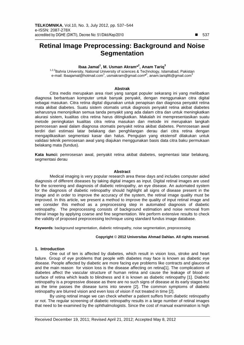
medical imaging is playing a vital role in such computer assisted diagnosis. We present an automated preprocessing system to improve the quality of retinal images so that the accuracy of automated screening of diabetic retinopathy can be improved. The proposed preprocessing method is used to improve the quality of image by extracting background and removing the noisy area from the retinal image. In automatic diagnosis of diabetic retinopathy, the processing of the surrounding background and noisy areas in retinal image is not necessary and consumes more processing time in all stages. Cutting or cropping out the region that contains the retinal image feature minimizes the number of operations on the retinal image. Moreover the noisy pixels may cause false features and reduce the accuracy of the automated classification so it is necessary to remove the noisy pixels. Figure 1 shows the flow diagram of our preprocessing technique. It shows the step by step outputs of proposed preprocessing technique.
2.1 Coarse Segmentation
Coarse segmentation creates mask for background and noise estimation using mean and variance method and HSI (Hue, Saturation, Intensity) color model respectively.
.
TELKOMNIKA
Retinal Image Preprocessing: Background and Noise Segmentation
Figure 1.
2.1.1 Background EstimationA color retinal image consists of a (semi) circular region of interest
background. This dark background is initially never really black.feature extraction and lesion detection algorithms on this area and it consumes more time so it is important to remove the background from input retin
We present a local meancreates a binary background segmentation maskgreater than threshold value, the block is considered as belongs to background. The algorithm for the background extraction mask is as below:Step 1: Divide the acquired retinal image into nonStep 2: Compute the local mean value
Step 3: Use local mean value computed in step 2 to compute the local standard deviation value std(I) using equation 2
Step 4: Select threshold value empiricallyStep 5: for each pixel,
Calculate std(I)> if true, add pixel in original retinal image area pixels if false, add pixel in background area pixels
end_for 2.1.2 Noise Segmentation Mask
Noise in color retinal image is normally due to noise pixelsdistorted. Both seem to exist in regions where illumination has been inadequate.illumination is usually adequate in the center of the image, poor image quality regions are located near the edge of the retinal image. Regionabnormality detection. That is why they should be detected and removed before detection of abnormalities.
In our technique, we create binary noise segmentation mask which includes the noisy area and it is applied on retinal image to ensure not to process the noisy area in upcoming steps i.e. feature extraction and abnormality detection. In this segmentation technique, we convert RGB (Red, Green, Blue) retinal image into HSI color space because firstly it is the way a human experiences colors and secondly noise can be easily removed in HSI color space [19]. The RGB retinal image
e-ISSN: 2087-278X
Retinal Image Preprocessing: Background and Noise Segmentation
Figure 1. Flow Diagram of Retinal Image Preprocessing
Estimation A color retinal image consists of a (semi) circular region of interest
background. This dark background is initially never really black. It is not necessary to apply feature extraction and lesion detection algorithms on this area and it consumes more time so it is important to remove the background from input retinal image.
local mean and variance based method [19] for background creates a binary background segmentation mask by applying a threshold on std(I)greater than threshold value, the block is considered as original retinal image area otherwise it
The algorithm for the background extraction mask is as below: retinal image into non-overlapping blocks
: Compute the local mean value M(I) using equation 1
: Use local mean value computed in step 2 to compute the local standard deviation value using equation 2
: Select threshold value empirically
>Threshold? , add pixel in original retinal image area pixels , add pixel in background area pixels
Noise Segmentation Mask Noise in color retinal image is normally due to noise pixels and pixels whose color is
distorted. Both seem to exist in regions where illumination has been inadequate.illumination is usually adequate in the center of the image, poor image quality regions are located near the edge of the retinal image. Regions with poor image quality may cause errors in abnormality detection. That is why they should be detected and removed before detection of
In our technique, we create binary noise segmentation mask which includes the noisy pplied on retinal image to ensure not to process the noisy area in upcoming
steps i.e. feature extraction and abnormality detection. In this segmentation technique, we convert RGB (Red, Green, Blue) retinal image into HSI color space because firstly it is the way a human experiences colors and secondly noise can be easily removed in HSI color
RGB retinal image is converted into HSI color space using equations
�
Retinal Image Preprocessing: Background and Noise Segmentation (Ibaa Jamal)
539
A color retinal image consists of a (semi) circular region of interest on a dark It is not necessary to apply
feature extraction and lesion detection algorithms on this area and it consumes more time so it
for background estimation. It by applying a threshold on std(I). If the std(I) is
original retinal image area otherwise it
(1)
: Use local mean value computed in step 2 to compute the local standard deviation value
(2)
and pixels whose color is distorted. Both seem to exist in regions where illumination has been inadequate. Since illumination is usually adequate in the center of the image, poor image quality regions are
s with poor image quality may cause errors in abnormality detection. That is why they should be detected and removed before detection of
In our technique, we create binary noise segmentation mask which includes the noisy pplied on retinal image to ensure not to process the noisy area in upcoming
steps i.e. feature extraction and abnormality detection. In this segmentation technique, we convert RGB (Red, Green, Blue) retinal image into HSI color space because firstly it is closer to the way a human experiences colors and secondly noise can be easily removed in HSI color
SI color space using equations 3, 5 and 6

�
TELKOMNIKA Vol. 10, No. 3
540
.
where
here R, G and B represent RED, GREEN and BLUE components of RGB retinal image.
The algorithm for noise removal mask is as below:Step 1: Divide the input retinal image Step 2: Use histogram equalization to enhance the contrast Step 3: Use a 3x3 median filter to reduce the noise in background of image.Step 4: Convert the equalized and filtered RGB retinal image into Step 5: Calculate N (noise factor) which is a ratio of Hue and IntensityStep 6: Select a threshold value empirically.Step 7: for each pixel, Calculate N(I)<Threshold? if true, add pixel in normal retinal image area pixels if false, add pixel in noisy area pixels
end_for Figure 2 shows the
images. These segmentation masks contain single pixel and edge pixel noise which will be removed in fine segmentation.
Figure 2. Coarse segmentation. a) Original retinal image; b) Background segmentation mask; c)
2.2 Fine Segmentation Background and noise segmentation masks that are formed by coarse segmentation
contain single pixel noise and edge pixels. Fine segmentation is done to remove these noises from segmentation masks. In fine segmentation, morphological operations i.e. morphological dilation, morphological erosion and morphological opening are applied to remove noise from binary masks [19]. We have used 5 x 5 square structuring element for morphological operations [19]. Background segmentation mask contains black single pixel noise (order to remove the black pixel noise, square structurremoves all black single pixel noise and edge pixels and it gives a finsegmentation mask. Noise segmentation mask contains white single pixel noise and edge pixels (Figure 2). In order to remove the wopening followed by erosion. Opening removes all edge pixels and erosion removes all white single pixel noise and it gives a fisegmentation masks and fine segmentation masks.
e-
3, July 2012 : 537 – 544
here R, G and B represent RED, GREEN and BLUE components of RGB retinal image.
The algorithm for noise removal mask is as below: : Divide the input retinal image I(i,j) into non-overlapping blocks with size : Use histogram equalization to enhance the contrast : Use a 3x3 median filter to reduce the noise in background of image. : Convert the equalized and filtered RGB retinal image into HSI color space.
(noise factor) which is a ratio of Hue and Intensity : Select a threshold value empirically.
<Threshold? , add pixel in normal retinal image area pixels
, add pixel in noisy area pixels
shows the coarse background and noise segmentation masks for retinal segmentation masks contain single pixel and edge pixel noise which will be segmentation.
Coarse segmentation. a) Original retinal image; b) Background segmentation mask; c)
Noise segmentation mask
Background and noise segmentation masks that are formed by coarse segmentation single pixel noise and edge pixels. Fine segmentation is done to remove these noises
from segmentation masks. In fine segmentation, morphological operations i.e. morphological dilation, morphological erosion and morphological opening are applied to remove noise from binary masks [19]. We have used 5 x 5 square structuring element for morphological operations [19]. Background segmentation mask contains black single pixel noise (order to remove the black pixel noise, square structuring element is used for dilation. Dilation removes all black single pixel noise and edge pixels and it gives a fin
Noise segmentation mask contains white single pixel noise and edge pixels ). In order to remove the white pixel noise, square structuring element is used for
opening followed by erosion. Opening removes all edge pixels and erosion removes all white single pixel noise and it gives a fine noise segmentation mask. Figure
fine segmentation masks.
-ISSN: 2087-278X
(3)
(4)
here R, G and B represent RED, GREEN and BLUE components of RGB retinal image.
(5)
(6)
overlapping blocks with size w x w.
HSI color space.
noise segmentation masks for retinal segmentation masks contain single pixel and edge pixel noise which will be
Coarse segmentation. a) Original retinal image; b) Background segmentation mask; c)
Background and noise segmentation masks that are formed by coarse segmentation single pixel noise and edge pixels. Fine segmentation is done to remove these noises
from segmentation masks. In fine segmentation, morphological operations i.e. morphological dilation, morphological erosion and morphological opening are applied to remove single pixel noise from binary masks [19]. We have used 5 x 5 square structuring element for morphological operations [19]. Background segmentation mask contains black single pixel noise (Figure 2). In
ing element is used for dilation. Dilation removes all black single pixel noise and edge pixels and it gives a fine background
Noise segmentation mask contains white single pixel noise and edge pixels hite pixel noise, square structuring element is used for
opening followed by erosion. Opening removes all edge pixels and erosion removes all white 3 shows coarse
TELKOMNIKA
Retinal Image Preprocessing: Background and Noise Segmentation
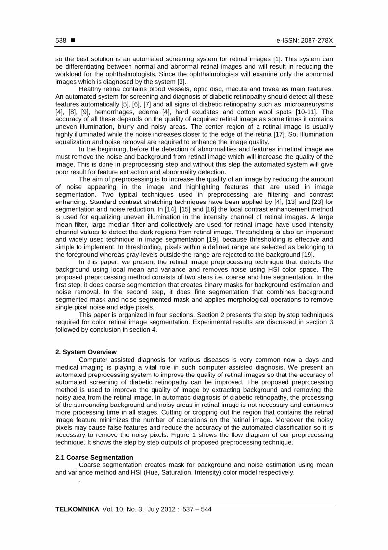
Figure 3. Fine segmentation. a)Segmentation Masks
2.3 Final Segmentation Mask
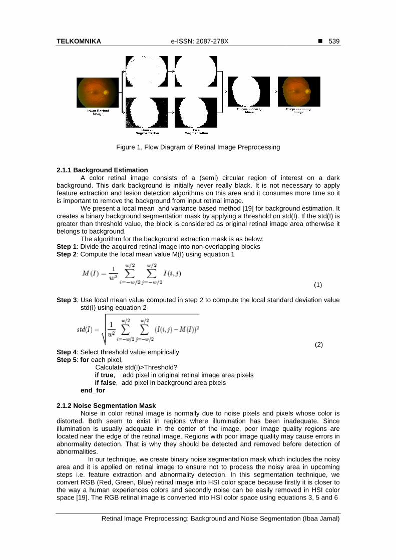
Final segmentation mask is prepaid by combining fine background and fine noise segmentation mask. For more fine segmentation mask, small regions are removed by filtering the combined mask by a medium size median filter [19]. Final noise free segmentation mask is then applied on retinal imfinal segmentation masks for retinal image segmentation.
Figure 4. (a): Original Color Retinal Image
from database; (b): Fine Background Segmentation Mask; (c): Fine Noise
Segmentation Mask; (d): Final Segmentation Mask
3. Experimental Results
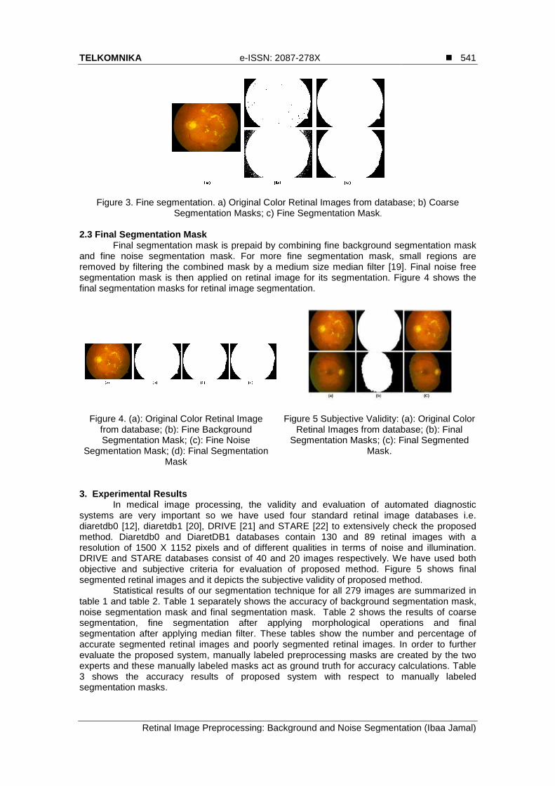
In medical image processing, the validity and evaluation of automated diagnostic systems are very important so we have used four standard retinal image databases i.e. diaretdb0 [12], diaretdb1 [20], DRIVE [21] and STARE [22] to extensively check the proposmethod. Diaretdb0 and DiaretDB1 dresolution of 1500 X 1152 pixels and of different qualities in terms of noise and illumination. DRIVE and STARE databases consist of 40 and 20 images respectively. We havobjective and subjective criteria for evaluation of proposed method. segmented retinal images and it depicts the subjective validity of proposed method.
Statistical results of our segmentation technique table 1 and table 2. Table 1 separately shows the accuracy of background segmentation mask, noise segmentation mask and final segmentation mask.segmentation, fine segmentation after applying morphological operations and final segmentation after applying median filter. These tables show the number and percentage of accurate segmented retinal images and poorly segmented evaluate the proposed system, manually labeled preprocessing masks are created by the two experts and these manually labeled masks act as ground truth for accuracy calculations. Table 3 shows the accuracy results of propsegmentation masks.
e-ISSN: 2087-278X
Retinal Image Preprocessing: Background and Noise Segmentation
Fine segmentation. a) Original Color Retinal Images from database; b) Coarse Segmentation Masks; c) Fine Segmentation Mask.
Final Segmentation Mask Final segmentation mask is prepaid by combining fine background segmentation mask
and fine noise segmentation mask. For more fine segmentation mask, small regions are removed by filtering the combined mask by a medium size median filter [19]. Final noise free segmentation mask is then applied on retinal image for its segmentation. Figfinal segmentation masks for retinal image segmentation.
Figure 4. (a): Original Color Retinal Image from database; (b): Fine Background Segmentation Mask; (c): Fine Noise
Segmentation Mask; (d): Final Segmentation
Figure 5 Subjective Validity: (a): Original Color
Retinal Images from database; (b): Final Segmentation Masks; (c): Final Segmented
Mask.
In medical image processing, the validity and evaluation of automated diagnostic systems are very important so we have used four standard retinal image databases i.e.
[20], DRIVE [21] and STARE [22] to extensively check the proposand DiaretDB1 databases contain 130 and 89 retinal images with a
resolution of 1500 X 1152 pixels and of different qualities in terms of noise and illumination. DRIVE and STARE databases consist of 40 and 20 images respectively. We havobjective and subjective criteria for evaluation of proposed method. Figure
and it depicts the subjective validity of proposed method.Statistical results of our segmentation technique for all 279 images are summarized in
separately shows the accuracy of background segmentation mask, noise segmentation mask and final segmentation mask. Table 2 shows the results of coarse segmentation, fine segmentation after applying morphological operations and final segmentation after applying median filter. These tables show the number and percentage of accurate segmented retinal images and poorly segmented retinal images. In order to further evaluate the proposed system, manually labeled preprocessing masks are created by the two experts and these manually labeled masks act as ground truth for accuracy calculations. Table 3 shows the accuracy results of proposed system with respect to manually labeled
�
Retinal Image Preprocessing: Background and Noise Segmentation (Ibaa Jamal)
541
Original Color Retinal Images from database; b) Coarse
segmentation mask and fine noise segmentation mask. For more fine segmentation mask, small regions are removed by filtering the combined mask by a medium size median filter [19]. Final noise free
egmentation. Figure 4 shows the
Figure 5 Subjective Validity: (a): Original Color Retinal Images from database; (b): Final
Segmentation Masks; (c): Final Segmented
In medical image processing, the validity and evaluation of automated diagnostic systems are very important so we have used four standard retinal image databases i.e.
[20], DRIVE [21] and STARE [22] to extensively check the proposed retinal images with a
resolution of 1500 X 1152 pixels and of different qualities in terms of noise and illumination. DRIVE and STARE databases consist of 40 and 20 images respectively. We have used both
ure 5 shows final and it depicts the subjective validity of proposed method.
are summarized in separately shows the accuracy of background segmentation mask,
shows the results of coarse segmentation, fine segmentation after applying morphological operations and final segmentation after applying median filter. These tables show the number and percentage of
In order to further evaluate the proposed system, manually labeled preprocessing masks are created by the two experts and these manually labeled masks act as ground truth for accuracy calculations. Table
osed system with respect to manually labeled
�
TELKOMNIKA Vol. 10, No. 3
542
TableType of
Segmentation Mask
Accurately Processed (Numbers)
Background Segmentation
275
Noise Segmentation 267
Final Segmentation 264
TableType of
Segmentation Mask
Accurately Processed (Numbers)
Coarse Segmentation 257
Fine Segmentation
265
Final Segmentation 269
Table 3. Accuracy results for proposed preprocessing method
Retinal images of different illumination and noise values are shown in
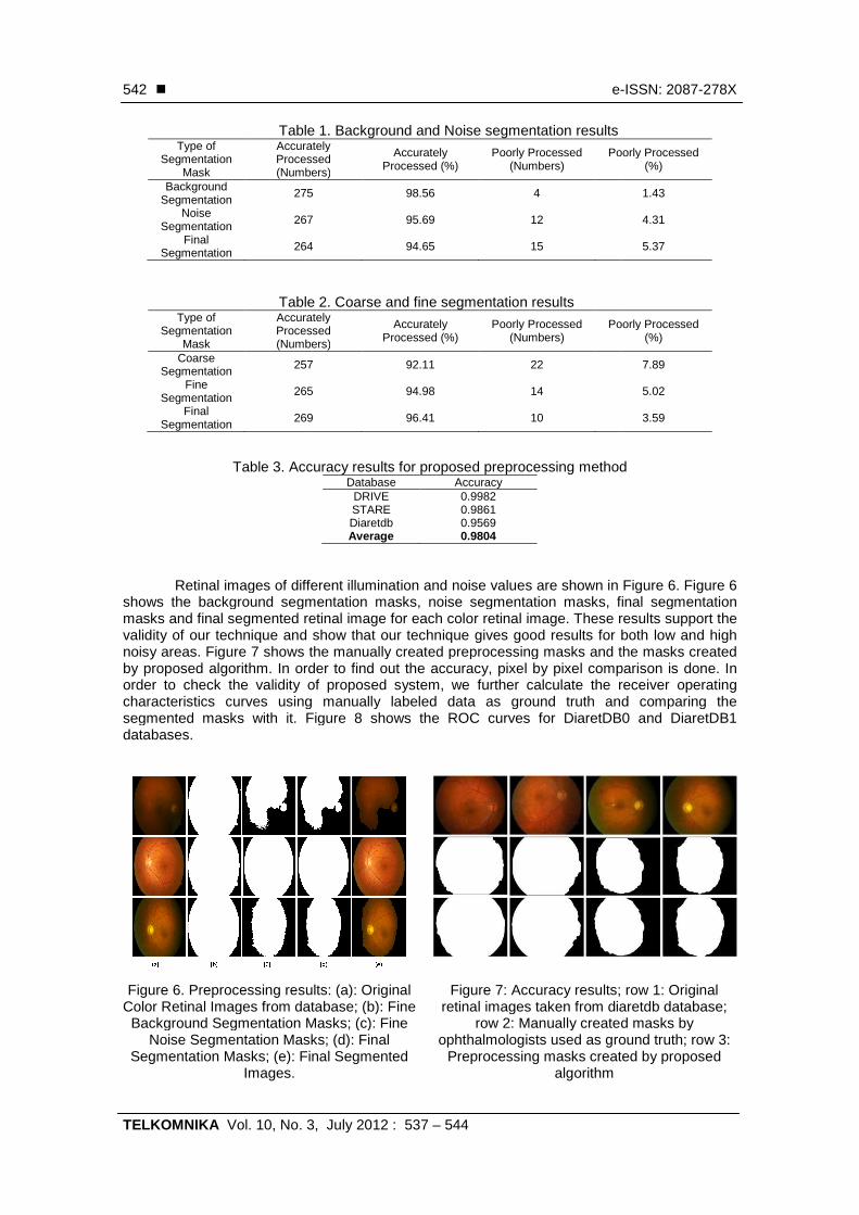
shows the background segmentation masks, noise segmentation masks, final segmentation masks and final segmented retinal image for each color retinal image. These results support the validity of our technique and show that our technique gives good results noisy areas. Figure 7 shows the manually created preprocessing masks and the masks created by proposed algorithm. In order to find out the accuracy, pixorder to check the validity of proposed system,characteristics curves using manually labeled data as ground truth and comparing the segmented masks with it. Figure 8 shows the ROC curves for DiaretDB0 and DiaretDB1 databases.
Figure 6. Preprocessing results: (a): Original Color Retinal Images from database; (b): Fine
Background Segmentation Masks; (c): Fine Noise Segmentation Masks; (d): Final
Segmentation Masks; (e): Final Segmented Images.
e-
3, July 2012 : 537 – 544
Table 1. Background and Noise segmentation results Accurately Processed (Numbers)
Accurately Processed (%)
Poorly Processed (Numbers)
Poorly Processed
275 98.56 4
67 95.69 12
64 94.65 15
Table 2. Coarse and fine segmentation results Accurately Processed (Numbers)
Accurately Processed (%)
Poorly Processed (Numbers)
Poorly Processed
57 92.11 22
265 94.98 14
9 96.41 10
Table 3. Accuracy results for proposed preprocessing methodDatabase Accuracy
DRIVE 0.9982 STARE 0.9861 Diaretdb 0.9569 Average 0.9804
Retinal images of different illumination and noise values are shown in shows the background segmentation masks, noise segmentation masks, final segmentation masks and final segmented retinal image for each color retinal image. These results support the validity of our technique and show that our technique gives good results for both low and high
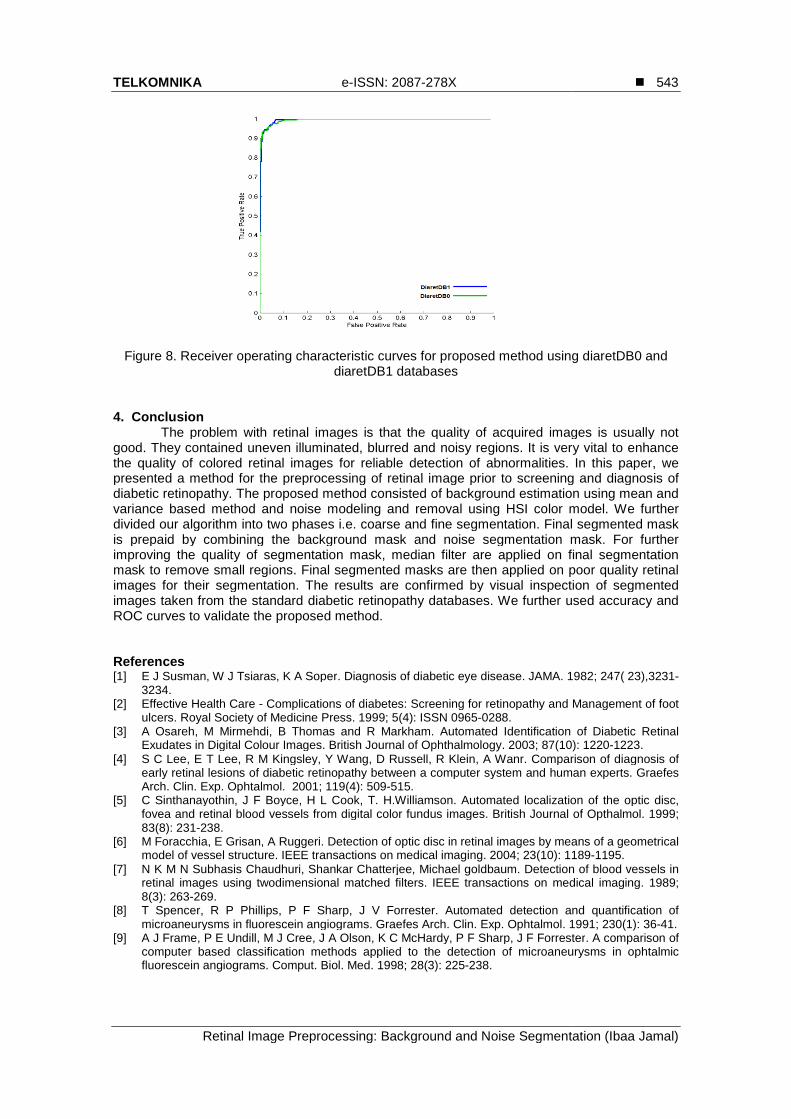
shows the manually created preprocessing masks and the masks created by proposed algorithm. In order to find out the accuracy, pixel by pixel comparison is done. In order to check the validity of proposed system, we further calculate the receiver operating characteristics curves using manually labeled data as ground truth and comparing the segmented masks with it. Figure 8 shows the ROC curves for DiaretDB0 and DiaretDB1
Figure 6. Preprocessing results: (a): Original Color Retinal Images from database; (b): Fine
Background Segmentation Masks; (c): Fine Noise Segmentation Masks; (d): Final
Segmentation Masks; (e): Final Segmented
Figure 7: Accuracy results; row 1: Original retinal images taken from diaretdb database;
row 2: Manually created masks by ophthalmologists used as ground truth; row 3:
Preprocessing masks created by proposed algorithm
-ISSN: 2087-278X
Poorly Processed (%)
1.43
4.31
5.37
Poorly Processed (%)
7.89
5.02
3.59
Table 3. Accuracy results for proposed preprocessing method
Retinal images of different illumination and noise values are shown in Figure 6. Figure 6 shows the background segmentation masks, noise segmentation masks, final segmentation masks and final segmented retinal image for each color retinal image. These results support the
for both low and high shows the manually created preprocessing masks and the masks created
el by pixel comparison is done. In we further calculate the receiver operating
characteristics curves using manually labeled data as ground truth and comparing the segmented masks with it. Figure 8 shows the ROC curves for DiaretDB0 and DiaretDB1
Figure 7: Accuracy results; row 1: Original retinal images taken from diaretdb database;
row 2: Manually created masks by ophthalmologists used as ground truth; row 3:
Preprocessing masks created by proposed
TELKOMNIKA
Retinal Image Preprocessing: Background and Noise Segmentation
Figure 8. Receiver operating
4. Conclusion The problem with retinal images is that the quality of acquired images is usually not
good. They contained uneven illuminated, blurred and noisy regions. It is very vital to enhance the quality of colored retinal images for reliable detection of abnormalitpresented a method for the preprocessing of retinal image prior to screening and diagnosis of diabetic retinopathy. The proposed method consisted of background estimation using mean and variance based method and noise modeling and redivided our algorithm into two phases i.e. coarse and fine segmentation. is prepaid by combining the background mask and noise segmentation mask. For further improving the quality of segmentationmask to remove small regions. Final segmented masks are then applied on poor quality retinal images for their segmentation. The results are confirmed by visual inspection of segmented images taken from the standard diabetic retinopathy databases.ROC curves to validate the proposed method.
References [1] E J Susman, W J Tsiaras, K A Soper
3234. [2] Effective Health Care - Complications of diabetes: Screening for retinopathy and Management of foot
ulcers. Royal Society of Medicine Press[3] A Osareh, M Mirmehdi, B Thomas and R
Exudates in Digital Colour Images[4] S C Lee, E T Lee, R M Kingsley, Y
early retinal lesions of diabetic retinopathy between a coArch. Clin. Exp. Ophtalmol.
[5] C Sinthanayothin, J F Boyce, H L Cook, T. H.Williamsonfovea and retinal blood vessels from digital color fundus images83(8): 231-238.
[6] M Foracchia, E Grisan, A Ruggeri. model of vessel structure. IEEE transa
[7] N K M N Subhasis Chaudhuri, Shankar Chatterjeeretinal images using twodimensional matched filters8(3): 263-269.
[8] T Spencer, R P Phillips, P F Sharp, J V Forrestermicroaneurysms in fluorescein angiograms
[9] A J Frame, P E Undill, M J Cree, J A Olson, K C McHardy, P F Sharp, J F Forrestercomputer based classification methods applied to the detection of microaneurysms in opfluorescein angiograms. Comput. Biol. Med
e-ISSN: 2087-278X
Retinal Image Preprocessing: Background and Noise Segmentation
Receiver operating characteristic curves for proposed method using diaretDB0 and diaretDB1 databases
The problem with retinal images is that the quality of acquired images is usually not good. They contained uneven illuminated, blurred and noisy regions. It is very vital to enhance the quality of colored retinal images for reliable detection of abnormalities. In this paper, presented a method for the preprocessing of retinal image prior to screening and diagnosis of diabetic retinopathy. The proposed method consisted of background estimation using mean and variance based method and noise modeling and removal using HSI color model. We further divided our algorithm into two phases i.e. coarse and fine segmentation. Final segmented mask is prepaid by combining the background mask and noise segmentation mask. For further improving the quality of segmentation mask, median filter are applied on final segmentation mask to remove small regions. Final segmented masks are then applied on poor quality retinal images for their segmentation. The results are confirmed by visual inspection of segmented
the standard diabetic retinopathy databases. We further used accuracy and ROC curves to validate the proposed method.
Tsiaras, K A Soper. Diagnosis of diabetic eye disease. JAMA. 1982
Complications of diabetes: Screening for retinopathy and Management of foot Royal Society of Medicine Press. 1999; 5(4): ISSN 0965-0288.
hdi, B Thomas and R Markham. Automated Identification of Diabetic Retinal Exudates in Digital Colour Images. British Journal of Ophthalmology. 2003; 87(10S C Lee, E T Lee, R M Kingsley, Y Wang, D Russell, R Klein, A Wanr. Comparison of diagnosis of early retinal lesions of diabetic retinopathy between a computer system and human experts
ch. Clin. Exp. Ophtalmol. 2001; 119(4): 509-515. Sinthanayothin, J F Boyce, H L Cook, T. H.Williamson. Automated localization of the optic disc,
fovea and retinal blood vessels from digital color fundus images. British Journal of
Grisan, A Ruggeri. Detection of optic disc in retinal images by means of a IEEE transactions on medical imaging. 2004; 23(10): 1189
M N Subhasis Chaudhuri, Shankar Chatterjee, Michael goldbaum. Detection of blood vessels in twodimensional matched filters. IEEE transactions on medical imaging
T Spencer, R P Phillips, P F Sharp, J V Forrester. Automated detection and quantification of ysms in fluorescein angiograms. Graefes Arch. Clin. Exp. Ophtalmol. 1991;P E Undill, M J Cree, J A Olson, K C McHardy, P F Sharp, J F Forrester
computer based classification methods applied to the detection of microaneurysms in opComput. Biol. Med. 1998; 28(3): 225-238.
�
Retinal Image Preprocessing: Background and Noise Segmentation (Ibaa Jamal)
543
characteristic curves for proposed method using diaretDB0 and
The problem with retinal images is that the quality of acquired images is usually not good. They contained uneven illuminated, blurred and noisy regions. It is very vital to enhance
ies. In this paper, we presented a method for the preprocessing of retinal image prior to screening and diagnosis of diabetic retinopathy. The proposed method consisted of background estimation using mean and
moval using HSI color model. We further Final segmented mask
is prepaid by combining the background mask and noise segmentation mask. For further mask, median filter are applied on final segmentation
mask to remove small regions. Final segmented masks are then applied on poor quality retinal images for their segmentation. The results are confirmed by visual inspection of segmented
We further used accuracy and
1982; 247( 23),3231-
Complications of diabetes: Screening for retinopathy and Management of foot
Automated Identification of Diabetic Retinal ): 1220-1223.
Comparison of diagnosis of mputer system and human experts. Graefes
Automated localization of the optic disc, ournal of Opthalmol. 1999;
Detection of optic disc in retinal images by means of a geometrical 1189-1195.
Detection of blood vessels in transactions on medical imaging. 1989;
Automated detection and quantification of 1991; 230(1): 36-41.
P E Undill, M J Cree, J A Olson, K C McHardy, P F Sharp, J F Forrester. A comparison of computer based classification methods applied to the detection of microaneurysms in ophtalmic
� e-ISSN: 2087-278X
TELKOMNIKA Vol. 10, No. 3, July 2012 : 537 – 544
544
[10] A Osareh, M Mirmehdi, B Thomas, R Markham. Automatic recognition of exudative maculopathy using fuzzy c-means clustering and neural networks. Proc. Medical Image Understanding Analysis Conf. 2001: 49-52.
[11] R Phillips, J Forrester, P Sharp. Automated detection and quantification of retinal exudates. Graefes Arch. Clin. Exp. Ophtalmol. 1993; 231(2): 90-94.
[12] Kauppi, T Kalesnykiene, V Kamarainen, J K Lensu, L Sorri, I Uusitalo, H Kälviäinen, H Pietilä, J DIARETDB0. Evaluation database and methodology for diabetic retinopathy algorithms. Technical Report. 2006
[13] M H Goldbaum, N P Katz, S Chaudhuri, M Nelson, P Kube. Digital image processing for ocular fundus images. Ophthalmol. Clin. North Amer. 1990; 3(3): 447-466.
[14] D Usher, M Dumskyj, M Himaga, T H Williamson, S Nussey, J Boyce. Automated detection of diabetic retinopathy in digital retinal images: a tool for diabetic retinopathy screening. Diabetes UK. Diabetic Medicine. 2003; 21(1): 84-90.
[15] C Sinthanayothin, V Kongbunkiat, S P Ruenchanachain, A Singlavanija. Automated screening system for diabetic retinopathy. Proc. of the 3rd International Symposium on Image and Signal Processing and Analysis, 2003: 915-920.
[16] A Osareh, M Mirmehdi, B Thomas, R Markham. Classification and localisation of diabetic-related eye disease. Proc. 7th European Conference on Computer Vision. Springer LNCS. 2002; 2353: 502-516,.
[17] N P Ward, S Tomlinson, C J.Taylor. Image analysis of fundus photographs. Ophthalmology. 1989; 96(1): 80-86.
[18] H Wang, W Hsu, KG Goh, M L Lee. An effective approach to detect lesions in color retinal images. Proc. IEEE Conference on Computer Vision and Pattern Recognition (CVPR). 2000: 181-187.
[19] R C Gonzalez and R E Woods. Digital Image Processing, 2nd ed. Prentice Hall. 2002. [20] Kauppi, T Kalesnykiene, V Kamarainen, J K Lensu, L Sorri, I Raninen A, Voutilainen R, Uusitalo, H
Kälviäinen, H Pietilä, J DIARETDB1. Diabetic retinopathy database and evaluation protocol. Technical report. 2007.
[21] Hoover A, Goldbaum M. Locating the optic nerve in a retinal image using the fuzzy convergence of the blood vessels. Transaction on Medical Imaging. 2003; 22(8): 951–958.
[22] Staal, J Abramoff, M D Niemeijer, M Viergever M A , van Ginneken B. Ridge-based vessel segmentation in color images of the retina. IEEE Transaction on Medical Imaging. 2004; 23(4): 501–509.
[23] K Firdausy, T Sutikno, E Prasetyo. Image Enhancement Using Contrast Stretching on RGB And IHS Digital Image. TELKOMNIKA. 2007; 5(1): 45-50.