Embed Size (px)
Citation preview

Vieira et al. Parasites & Vectors (2015) 8:135 DOI 10.1186/s13071-015-0736-2
RESEARCH Open Access
Rhodnius prolixus interaction with Trypanosomarangeli: modulation of the immune system andmicrobiota populationCecilia S Vieira1, Débora P Mattos1, Peter J Waniek1, Jayme M Santangelo3, Marcela B Figueiredo1, Marcia Gumiel1,Fabio F da Mota1,2, Daniele P Castro1,2, Eloi S Garcia1,2 and Patrícia Azambuja1,2*
Abstract
Background: Trypanosoma rangeli is a protozoan that infects a variety of mammalian hosts, including humans. Itsmain insect vector is Rhodnius prolixus and is found in several Latin American countries. The R. prolixus vectorcompetence depends on the T. rangeli strain and the molecular interactions, as well as the insect’s immuneresponses in the gut and haemocoel. This work focuses on the modulation of the humoral immune responses ofthe midgut of R. prolixus infected with T. rangeli Macias strain, considering the influence of the parasite on theintestinal microbiota.
Methods: The population density of T. rangeli Macias strain was analysed in different R. prolixus midgutcompartments in long and short-term experiments. Cultivable and non-cultivable midgut bacteria were investigatedby colony forming unit (CFU) assays and by 454 pyrosequencing of the 16S rRNA gene, respectively. The modulationof R. prolixus immune responses was studied by analysis of the antimicrobial activity in vitro against different bacteriausing turbidimetric tests, the abundance of mRNAs encoding antimicrobial peptides (AMPs) defensin (DefA, DefB, DefC),prolixicin (Prol) and lysozymes (LysA, LysB) by RT-PCR and analysis of the phenoloxidase (PO) activity.
Results: Our results showed that T. rangeli successfully colonized R. prolixus midgut altering the microbiota populationand the immune responses as follows: 1 - reduced cultivable midgut bacteria; 2 - decreased the number ofsequences of the Enterococcaceae but increased those of the Burkholderiaceae family; the families Nocardiaceae,Enterobacteriaceae and Mycobacteriaceae encountered in control and infected insects remained the same;3 - enhanced midgut antibacterial activities against Serratia marcescens and Staphylococcus aureus; 4 - down-regulated LysB and Prol mRNA levels; altered DefB, DefC and LysA depending on the infection (short and long-term); 5 - decreased PO activity.
Conclusion: Our findings suggest that T. rangeli Macias strain modulates R. prolixus immune system and modifiesthe natural microbiota composition.
Keywords: Rhodnius prolixus, Trypanosoma rangeli, Immune system, Prophenoloxidase, Antimicrobial peptide
* Correspondence: [email protected]ório de Bioquímica e Fisiologia de Insetos, Instituto Oswaldo Cruz,Fundação Oswaldo Cruz (IOC/FIOCRUZ), Rio de Janeiro, RJ, Brazil2Departamento de Entomologia Molecular, Instituto Nacional deEntomologia Molecular (INCT-EM), Rio de Janeiro, RJ, BrazilFull list of author information is available at the end of the article
© 2015 Vieira et al.; licensee BioMed Central. This is an Open Access article distributed under the terms of the CreativeCommons Attribution License (http://creativecommons.org/licenses/by/4.0), which permits unrestricted use, distribution, andreproduction in any medium, provided the original work is properly credited. The Creative Commons Public DomainDedication waiver (http://creativecommons.org/publicdomain/zero/1.0/) applies to the data made available in this article,unless otherwise stated.

Vieira et al. Parasites & Vectors (2015) 8:135 Page 2 of 13
BackgroundThe haemoflagellate, Trypanosoma rangeli, is a proto-zoan parasite that infects a large number of mammals,including humans, and it is vectored by triatomineinsects, especially the genus Rhodnius [1-4]. The in-teraction with triatomine hosts, such as Rhodnius pro-lixus, begins with the ingestion of an infective bloodmeal containing T. rangeli. After ingestion, the para-sites transform into epimastigotes, then multiplies inthe insect gut, and invades the haemolymph. To per-petuate the infection they transform into the metacyc-lic forms in the salivary glands [1,2,5]. The life cycle ofT. rangeli is completed with the transmission of theparasite to vertebrate hosts by the vector through itssalivary gland secretions during a blood meal [6,7].The establishment of T. rangeli infections in both
the digestive tract and haemocoel is regulated byphysiological processes of the triatomine vector [8].The parasites survive despite the activation of innateimmune reactions and complete their life cycle in theinsect host [9-11]. Once inside the midgut, the para-sites must interact with blood digestion products aswell as midgut components including bacterial micro-biota [12,13], haemolytic factors [14,15], lectins [16],the prophenoloxidase (PPO) system [17], antimicrobialpeptides (AMPs) [18,19] and reactive nitrogen andoxygen species [20].Some of these factors act as biological barriers to the
infection of T. rangeli in the vector gut. However, theT. rangeli infection may lead to immunedepression ofthe insect host by the inhibition of phagocytosis,haemocyte microaggregation, PPO activation and ei-cosanoids synthesis [11,21,22]. These physiological alter-ations allow the parasites to overcome the immuneresponse, reach the salivary glands and complete their lifecycle.Knowledge of the modulation of the triatomine im-
mune system by the numerous strains of T. rangeli isstill poorly understood. Thus, the aim of the presentstudy was to investigate the effects of T. rangeli Maciasstrain infection on the midgut immune responses,parasite development and bacteria population of 5th in-star nymphs of R. prolixus orally infected with para-sites. In addition to the evaluation of the effects of T.rangeli in short-term infections, long-term infectionswere analysed in the 5th instar nymphs previously in-fected in the 4th instar stage. The present results suggestthat the parasites modulate the R. prolixus immune re-sponses, affecting the intestinal microbiota by inhibiting ac-tivation of prophenoloxidase, altering the abundance ofantimicrobial peptide transcripts and enhancing antimicro-bial activities against Serratia marcescens. These resultsprovide further elucidation of the T. rangeli-R. prolixusinteraction.
MethodsEthics StatementDefibrinated rabbit blood provided by the AnimalsCreation Center Laboratory (Cecal/Fiocruz) was providedto the insects through an artificial apparatus respectingthe guidelines of the Ethics Committee on Animal Use(Ceua/Fiocruz). CEUA follows the Ethical Principles inAnimal Experimentation composed by Fiocruz researchersand external consultants. The protocol number L-0061/08was established by CONCEA/MCT [23].
Maintenance of Trypanosoma rangeli epimastigotesT. rangeli Macias strain, first isolated from a human inVenezuela [24,25] and later characterized as genotypeKP1+ [26], was kindly supplied by Dr. Suzete Gomes,Universidade Federal Fluminense (Rio de Janeiro, Brazil).The parasites were maintained at 28°C in brain heart in-fusion (BHI) media (Sigma-Aldrich, São Paulo, Brazil)supplemented with 20% heat-inactivated bovine foetalserum [27] and subcultured twice a week. This proced-ure keeps the parasite in the log phase growth resultingpredominantly in short epimastigotes (99%). The num-ber of parasites was quantified in a Neubauer chamber.
Bacteria preparationStaphylococcus aureus 9518, and Escherichia coli K124401 were purchased from the National Collection ofIndustrial and Marine Bacteria (NCIMB), Aberdeen, UK.S. marcescens RPH was previously isolated from R. pro-lixus [12] and maintained at Laboratório de Bioquímicae Fisiologia de Insetos. The bacteria were maintainedat -70°C in tryptone agar and 10% glycerol. For all ex-perimental procedures, bacteria were grown as pre-viously described [28]. Briefly, bacteria were grownovernight in tryptone soy broth (TSB) at 30°C and thencultured in fresh TSB for a further 4 h under the sameconditions. The bacteria were then washed in phos-phate buffered saline (PBS, 0.01 M phosphate buffer,2.7 mM potassium chloride and 0.137 M sodium chloride,pH 7.4) and resuspended in TSB to a final concentrationof 1 × 10⁴ cells/ml.
Insect oral infection: long and short-term infectionsInsects were kept at 27°C and fed artificially with defi-brinated rabbit blood [27]. All insects were fed on blood,after heat-inactivation of the plasma. The blood was cen-trifuged at 1,890 x g for 10 min at 4°C and the super-natant (plasma) was incubated for 30 min at 55°C. Theplasma was added back to the erythrocytes and then re-ceived T. rangeli epimastigotes obtained from culture.The same procedure of plasma inactivation was under-taken for control insects.For long-term experiments, inactivated blood contain-
ing 1 × 10⁶ epimastigotes/mL (infected group) or blood

Vieira et al. Parasites & Vectors (2015) 8:135 Page 3 of 13
without parasites (control uninfected group) was givento 4th instar nymphs. After moulting to 5th instars, bothinsect groups received a non-infective blood meal whichoccurred 38 days after feeding (DAF) of the 4th instarnymphs. For short-term experiments, 5th instar nymphswere fed on blood containing 1×10⁶ epimastigotes/mLor with parasite-free blood. Only fully engorged insectswere used for all experiments.
Quantification of parasites in the digestive tractFifth instar nymphs obtained from long or short-termexperiments were dissected. The anterior midgut (stom-ach) was collected and homogenized in 1.0 mL PBS andthe posterior midgut (intestine) plus rectum was placed in50 μL in PBS. Samples were macerated and the number ofparasites was determined by counting in a Neubauerhaemocytometer and expressed as parasites/mL. Parasiteswere quantified in three experiments with five insects each(n = 15).
Analysis of intestinal microbiotaColony forming unit (CFU)Anterior midgut contents obtained from 5th instarnymphs infected or uninfected with parasites (short andlong-term infections) were analysed for microbiota bacter-ial population using CFU at 12 DAF. The midgut contentswere serially tenfold diluted with PBS and 20 μL wasspread on a Petri dish in BHI agar (Sigma-Aldrich) cul-ture medium. The plates were incubated at 30°C for24 h and the CFU counted. As a control, PBS was alsoplated to check the sterility of all experiments.
Metagenomic DNA extractionSeven days after insect feeding (long-term infection),metagenomic DNA was extracted from the anterior mid-gut contents of four T. rangeli infected insects and fouruninfected R. prolixus 5th instar nymphs by an unbiasedand efficient mechanical lysis method [29]. The extrac-tion was carried out using the commercial Fast-DNA™Spin Kit for Soil (Qbiogene, MP Biomedicals, USA) fol-lowing the manufacturer's instructions. DNA extractswere visualized on 1% agarose gels to assess their integ-rity and purity.
Amplification and 454 sequencing of targeted 16S rRNAgene variable regionFor quantitative analysis of bacterial microbiota inlong-term infected insects, ribosomal genes from meta-genomic DNA samples were amplified using bar-codedprimers for the 16S variable region V3-V1, cleaned up,quantified and normalized according to the HMP 373016S protocol version 4.2 [30], which is available on theHMP Data Analysis and Coordination Center website[31]. The PCR products obtained were then submitted
to FLX-Titanium pyrosequencing in a GS Junior System(Roche).The raw sequences were analysed using the RDP Pipe-
line with default parameters. Sequences with a scorebelow the quality threshold were discarded and the se-quence portions devoted to 454 sequencing were trimmedout. Sequences with more than 400 bases were thenaligned using the INFERNAL aligner [32] and chimeric se-quences detected (and removed) with UCHIME [33].Taxonomical classification was assigned using the RDPclassifier [34,35] with a minimum confidence level for rec-ord assignment set to 0.80.
Turbidimetric antibacterial assayIn long and short-term experiments, the antibacterial ac-tivities of the anterior midgut contents from 5th instarnymphs infected or not with T. rangeli were tested at 7DAF, according to previous studies which have shownthat the maximal antibacterial activity is reached at thistime [17,36]. Fifth instar nymphs of R. prolixus were dis-sected to collect the anterior midgut. The midgut wallswere removed and the midgut contents pooled (3 in-sects) in 200 μl ultrapure water, homogenized, centri-fuged at 10,000 x g for 10 min at 4°C and filtered by asterile PVDF membrane (Millipore) and stored at −20°Cuntil use. Before assaying, the midgut content sampleswere diluted ten times in sterile water. Subsequently,10 μl of E. coli, S. aureus or S. marcescens bacterial sus-pensions (104 cells/mL) were added to each well of asterile flat bottom 96-well microtiter plate (Nunc, FisherScientific, Leicestershire, UK) with 45 μl of dilutedmidgut samples and 5 μl of peptone 10% and incubatedat 37°C for 19 h. The optical densities were measured at550 nm (OD550) at hourly intervals using a SpectraMax 190 Plate Reader (Molecular Devices, Sunnyvale,USA). Control wells, run without anterior midgutsamples, contained 10 μl of bacteria in a final concen-tration of 1% peptone in ultrapure water. Ampicillin(80 μg/ml) was included in each experiment as an anti-biotic control. To exclude the opacity of the midgutsamples all data points were blanked against time zero.The antibacterial activity was calculated by subtractingthe bacterial growth readings (control wells) from therespective values of anterior midgut samples incubatedwith bacteria.
Transcript abundance of antimicrobial peptidesTranscript abundance of genes encoding antibacterialpeptides in short and long-term experiments with T.rangeli infected and uninfected 5th instar nymphs wasanalysed by reverse transcription PCR (RT-PCR) asdescribed previously [36]. In brief, the anterior andposterior midgut walls of a pool of ten insects weredissected from 5th instar nymphs 1 and 7 DAF. Total
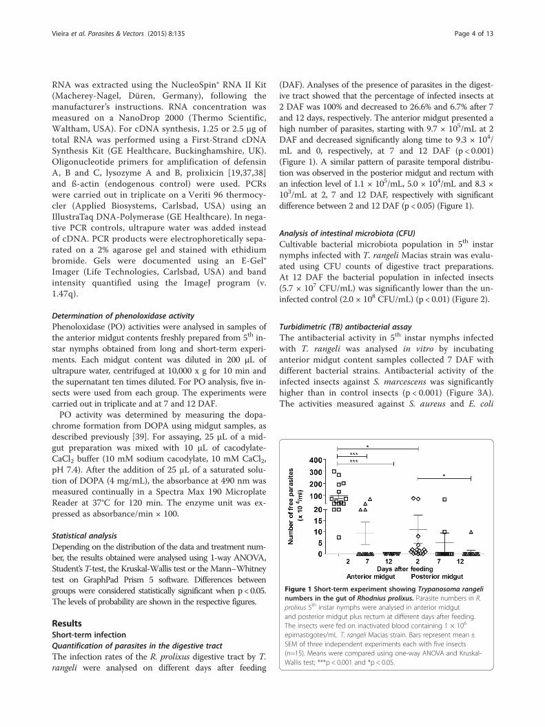
Figure 1 Short-term experiment showing Trypanosoma rangelinumbers in the gut of Rhodnius prolixus. Parasite numbers in R.prolixus 5th instar nymphs were analysed in anterior midgutand posterior midgut plus rectum at different days after feeding.The insects were fed on inactivated blood containing 1 × 10⁶
epimastigotes/mL T. rangeli Macias strain. Bars represent mean ±SEM of three independent experiments each with five insects(n=15). Means were compared using one-way ANOVA and Kruskal-Wallis test; ***p < 0.001 and *p < 0.05.
Vieira et al. Parasites & Vectors (2015) 8:135 Page 4 of 13
RNA was extracted using the NucleoSpin® RNA II Kit(Macherey-Nagel, Düren, Germany), following themanufacturer’s instructions. RNA concentration wasmeasured on a NanoDrop 2000 (Thermo Scientific,Waltham, USA). For cDNA synthesis, 1.25 or 2.5 μg oftotal RNA was performed using a First-Strand cDNASynthesis Kit (GE Healthcare, Buckinghamshire, UK).Oligonucleotide primers for amplification of defensinA, B and C, lysozyme A and B, prolixicin [19,37,38]and ß-actin (endogenous control) were used. PCRswere carried out in triplicate on a Veriti 96 thermocy-cler (Applied Biosystems, Carlsbad, USA) using anIllustraTaq DNA-Polymerase (GE Healthcare). In nega-tive PCR controls, ultrapure water was added insteadof cDNA. PCR products were electrophoretically sepa-rated on a 2% agarose gel and stained with ethidiumbromide. Gels were documented using an E-Gel®Imager (Life Technologies, Carlsbad, USA) and bandintensity quantified using the ImageJ program (v.1.47q).
Determination of phenoloxidase activityPhenoloxidase (PO) activities were analysed in samples ofthe anterior midgut contents freshly prepared from 5th in-star nymphs obtained from long and short-term experi-ments. Each midgut content was diluted in 200 μL ofultrapure water, centrifuged at 10,000 x g for 10 min andthe supernatant ten times diluted. For PO analysis, five in-sects were used from each group. The experiments werecarried out in triplicate and at 7 and 12 DAF.PO activity was determined by measuring the dopa-
chrome formation from DOPA using midgut samples, asdescribed previously [39]. For assaying, 25 μL of a mid-gut preparation was mixed with 10 μL of cacodylate-CaCl2 buffer (10 mM sodium cacodylate, 10 mM CaCl2,pH 7.4). After the addition of 25 μL of a saturated solu-tion of DOPA (4 mg/mL), the absorbance at 490 nm wasmeasured continually in a Spectra Max 190 MicroplateReader at 37°C for 120 min. The enzyme unit was ex-pressed as absorbance/min × 100.
Statistical analysisDepending on the distribution of the data and treatment num-ber, the results obtained were analysed using 1-way ANOVA,Student’sT-test, the Kruskal-Wallis test or the Mann–Whitneytest on GraphPad Prism 5 software. Differences betweengroups were considered statistically significant when p< 0.05.The levels of probability are shown in the respective figures.
ResultsShort-term infectionQuantification of parasites in the digestive tractThe infection rates of the R. prolixus digestive tract by T.rangeli were analysed on different days after feeding
(DAF). Analyses of the presence of parasites in the digest-ive tract showed that the percentage of infected insects at2 DAF was 100% and decreased to 26.6% and 6.7% after 7and 12 days, respectively. The anterior midgut presented ahigh number of parasites, starting with 9.7 × 10⁵/mL at 2DAF and decreased significantly along time to 9.3 × 10⁴/mL and 0, respectively, at 7 and 12 DAF (p < 0.001)(Figure 1). A similar pattern of parasite temporal distribu-tion was observed in the posterior midgut and rectum withan infection level of 1.1 × 10⁵/mL, 5.0 × 10⁴/mL and 8.3 ×10³/mL at 2, 7 and 12 DAF, respectively with significantdifference between 2 and 12 DAF (p < 0.05) (Figure 1).
Analysis of intestinal microbiota (CFU)Cultivable bacterial microbiota population in 5th instarnymphs infected with T. rangeli Macias strain was evalu-ated using CFU counts of digestive tract preparations.At 12 DAF the bacterial population in infected insects(5.7 × 10⁷ CFU/mL) was significantly lower than the un-infected control (2.0 × 10⁸ CFU/mL) (p < 0.01) (Figure 2).
Turbidimetric (TB) antibacterial assayThe antibacterial activity in 5th instar nymphs infectedwith T. rangeli was analysed in vitro by incubatinganterior midgut content samples collected 7 DAF withdifferent bacterial strains. Antibacterial activity of theinfected insects against S. marcescens was significantlyhigher than in control insects (p < 0.001) (Figure 3A).The activities measured against S. aureus and E. coli

Figure 2 Short-term experiment showing bacteria populationin Rhodnius prolixus midgut infected with Trypanosoma rangeli.Fifth instar nymphs were fed on inactivated blood containing 1 × 10⁶
epimastigotes/mL of T. rangeli. CFU counts were made 12 days afterfeeding. Bars represent mean ± SEM of three independent experimentswith three pools of insects (n = 9). Each point represents the CFU fromone pool of three insects. Means were compared using Student T-testor Mann–Whitney test; **p < 0.01.
Vieira et al. Parasites & Vectors (2015) 8:135 Page 5 of 13
were similar in infected insects compared with the con-trols (Figure 3B, C).
Figure 3 Short-term experiment showing antibacterial activityin the midgut of Rhodnius prolixus infected with Trypanosomarangeli. The antibacterial activities of anterior midgut of R. prolixus5th instar nymphs 7 days after infection with T. rangeli were testedagainst (A) S. marcescens (B) S. aureus (C) E. coli. Treatments: whitecolumns – control, uninfected group; black columns – infectedgroup. Fifth instar nymphs were fed on inactivated blood with orwithout 1 × 10⁶ epimastigotes/mL. Antibacterial activity value wasmeasured using the turbidimetric assay (TB) (OD550 nm) after 19 hincubation of midgut samples with different bacteria and calculatedby the difference between the optical densities of the midgutsamples and bacterial control. Bars represent mean ± SEM of threeindependent experiments. Each experiment consisted of three poolsof three insects (n = 9). Means were compared using t-test;***p < 0.001 and NS = not significant.
Transcript abundance of antimicrobial peptides (AMPs)The modification of antimicrobial activities in the an-terior and posterior midguts of the 5th instar nymphs in-fected with T. rangeli was analysed by the transcriptabundance profiles of AMPs 1 and 7 DAF. The relativeabundance of lysozyme A (LysA), lysozyme B (LysB),prolixicin (Prol), defensins A (DefA), B (DefB) and C(DefC) was also quantified (Figure 4).In the anterior midgut at 1 DAF, the expression of
DefB was significantly higher (p < 0.05) and LysB wassignificantly lower (p < 0.05) in comparison betweeninfected and control insects, respectively (Figure 4A). Incontrast, at 7 DAF, three AMPs (DefB, LysB and Prol)had significantly lower levels (p < 0.001; p < 0.05;p < 0.001, respectively) and only one (DefC) had a signifi-cantly higher level (p < 0.001) of transcripts in infectedinsects when compared to control (Figure 4C). However,in the posterior midgut the differences in levels of theAMPs between the infected and control insects wereless striking. Compared to control the infected insectspresented higher levels (p < 0.001) of DefC transcriptsat 1 DAF and lower levels (p < 0.001) of Prol at 7DAF(Figure 4B and D).

Figure 4 Short-term experiment showing relative transcript abundance of antimicrobial peptides and lysozymes in Rhodnius prolixusmidgut. Relative AMP mRNA levels from R. prolixus 5th instar nymphs were analysed 1 and 7 days after feeding (DAF) A – anterior midgut 1 DAF;B – posterior midgut 1 DAF; C – anterior midgut 7 DAF; D – posterior midgut 7 DAF. Treatments: white columns – control, uninfected group;black columns – infected group. Insects were fed on inactivated blood with or without 1 × 10⁶ epimastigotes/mL. Bars represent mean ± SEM ofthree independent experiments with a pool of 10 insects (n = 3). Means were compared using one-way ANOVA and Student T-test; ***p < 0.001and *p < 0.05.
Vieira et al. Parasites & Vectors (2015) 8:135 Page 6 of 13
Determination of phenoloxidase activityPO activities measured in the anterior midgut contentsof the 5th instar nymphs at 7 DAF did not show signifi-cant differences, when comparing T. rangeli infected andcontrol groups. However, at 12 DAF, the PO activity wassignificantly lower in infected insects than in the control(p < 0.01) (Figure 5). The PO activity inhibition by T.rangeli infection was significantly higher at 12 DAFwhen compared with 7 DAF (p < 0.001) (Figure 5).
Long term infectionQuantification of parasites in the digestive tractThe T. rangeli infection in the insects was also inves-tigated in long-term experiments. Parasites were quan-tified in the digestive tract from the 4th instar nymphswhen infection occurred and after insects moulted to 5th
instar followed by a second feeding with parasite freeblood. The percentages of infected insects in 4th instarnymphs at 2 and 7 days after infection were 86.7% and93.3%, respectively. In this infected group, the numberof parasites encountered in the anterior midgut was
significantly higher than in the posterior midgut on bothdays analysed (Figure 6A). The parasite numbers reached28.9 × 104/mL and 32.8 × 10⁴/mL in the anterior midgutat 2 and 7 DAF, respectively, and 3.4 × 10⁴/mL and 4.4 ×10⁴/mL in the posterior midgut at 2 and 7 DAF, respec-tively (Figure 6A).The percentages of 5th instar nymphs which showed T.
rangeli infection in the digestive tract were 46.7% and73.3% at 2 and 7 DAF, respectively, after an uninfectedblood meal. In these 5th instar nymphs, the results wereopposite to those observed in the 4th instar nymphs, inwhich the anterior midgut presented significantly lowernumbers of parasites than the posterior midgut onboth days analysed (Figure 6B). The infection level inthe anterior midgut was 0 and 0.17 × 10⁴/mL at 2and 7 DAF respectively and in the posterior midgutand rectum was 22.4 × 10⁴/mL and 17.7 × 10⁴/mL at2 and 7 DAF, respectively (Figure 6B). These resultsshowed that T. rangeli successfully colonized R. prolixusmidgut, even after moulting and a second blood meal(Figure 6).

Figure 5 Short-term experiment showing phenoloxidaseactivity in the midgut of Rhodnius prolixus infected withTrypanosoma rangeli. PO activities were measured in the anteriormidgut of R. prolixus 5th instar nymphs at 7 and 12 days afterinfection with T. rangeli. The insects were fed on inactivated bloodwith or without 1 × 10⁶ epimastigotes/mL. Treatments: whitecolumns – control, uninfected group; black columns – infectedgroup. Bars represent mean ± SEM of three independentexperiments each with five insects (n=15). Means were comparedusing Student T-test; ***p < 0.001 and **p < 0.01.
Figure 7 Long-term experiment showing bacteria population inRhodnius prolixus midgut infected with Trypanosoma rangeli.CFU counts were made with R. prolixus 5th instar nymphs 12 daysafter feeding (DAF) on blood without parasites. Previously, 4th instarnymphs were fed on inactivated blood with or without 1 × 10⁶
epimastigotes/mL. Bars represent mean ± SEM of three independentexperiments. Each point represents the CFU from three pools ofthree insects (n = 9). Means were compared using Student T-test orMann–Whitney test; **p < 0.01 and *p < 0.05.
Vieira et al. Parasites & Vectors (2015) 8:135 Page 7 of 13
Analysis of intestinal microbiotaColony forming unit (CFU)The cultivable bacterial microbiota population of R. pro-lixus 5th instar nymphs, infected as 4th instars with T.rangeli was significantly lower than control insects at 12DAF (p < 0.01) (Figure 7).
Amplification and 454 sequencing of targeted 16S rRNAgene variable regionThe bacterial microbiota in the anterior midgut waspredominantly composed of Enterobacteriaceae andEnterococcaceae families, which include Serratia and
Figure 6 Long-term experiment showing Trypanosoma rangeli numbein R. prolixus (A) 4th and (B) 5th instar nymphs: anterior midgut and posterifed on inactivated blood containing 1 × 10⁶ epimastigotes/mL. After moulrepresent mean ± SEM of three independent experiments each with five inKruskal-Wallis test; ***p < 0.001 and *p < 0.05.
Enterococcus species, respectively, as well as Nocardiaceae(Figure 8). Seven days after feeding, there was a significantdecrease of Enterococcaceae in the R. prolixus 5th instarnymphs, infected at 4th instar with T. rangeli while therewas significant increase of Burkholderiaceae in the in-fected 5th instar nymphs (Figure 8).
Turbidimetric antibacterial assayThe anterior midgut antibacterial activity of insects in-fected over the long-term was investigated. Comparing tothe control group, infected insects presented significantly
rs in the gut of Rhodnius prolixus. Parasite numbers were analysedor midgut at 2 and 7 days after feeding. Fourth instar nymphs wereting, 5th instar nymphs were fed on blood without parasites. Barssects (n = 15). Means were compared using one-way ANOVA and

Figure 8 Bacterial composition identified by 16S ribosomal pyrosequencing in Rhodnius prolixus midgut infected with Trypanosomarangeli. Long-term experiment showing bacterial composition at the family levels. Pyrosequencing 454 experiments of anterior midgutpreparations from R. prolixus 5th instar nymphs 7 days after feeding (DAF) on blood without parasites. Previously, 4th instar nymphs were fedon inactivated blood with or without 1 × 10⁶ epimastigotes/mL of T. rangeli. Each bar graph presents the mean number of sequence readsassigned to a given bacterial family in four insect samples. Others: represent families with only one or two sequences (Pseudomonadaceae,Comamonadaceae, Rhodobacteraceae, Phyllobacteriaceae, Bradyrhizobiaceae, Staphylococcaceae, Bacillaceae, Nitrospiraceae, Flavobacteriaceae).Means were compared using t-test or Mann–Whitney test; *p < 0.05.
Vieira et al. Parasites & Vectors (2015) 8:135 Page 8 of 13
higher antibacterial activity against S. marcescens (p <0.001) and S. aureus (p < 0.05) and lower activity againstE. coli (p < 0.001) (Figure 9).
Transcript abundance of antimicrobial peptides (AMPs)The relative transcript abundance of AMPs and lyso-zymes encoding mRNA in the 5th instar R. prolixusnymphs that were infected with T. rangeli as 4th instarnymphs was investigated (Figure 10). The expression ofLysB was significantly lower in both compartments ofthe midgut at 1 and 7 DAF in infected insects whencompared to the uninfected control group. This differ-ence in abundance of LysB was more significant at 1DAF (p < 0.001) both in the anterior and posterior mid-guts of the infected insects (Figure 10A and 10B). Theabundance of LysA transcripts was significantly loweronly in the anterior midgut at 7 DAF of the infectedinsects (p < 0.05) in comparison to the control insects(Figure 10C). Compared to the control group, DefCtranscripts in infected insects were less abundant in theanterior midgut at 1 DAF (p < 0.001). In the posteriormidgut at 7 DAF abundance of the DefB transcripts waslower (p < 0.01) in infected insects than in the control in-sects (Figure 10D). Moreover, the abundance of Prol wassignificantly lower in the anterior midgut at 7 DAF (p <0.001) and in the posterior midgut at 1 and 7 DAF (p <0.01) of infected insects when compared to control(Figures 10 B, C and D). Only DefC mRNA levels in theposterior midgut were up-regulated (57-fold, p < 0.001)
at 1 DAF in infected insects when compared to the con-trol group (Figure 10B).
Prophenoloxidase (PPO) activityThe anterior midgut of 5th instar nymphs, previously in-fected with T. rangeli at 4th instar, were investigated. ThePO activities of infected insects were significantly lowerthan the control insects at 7 and 12 DAF (p < 0.001 andp < 0.01, respectively) (Figure 11). Moreover, the PO ac-tivity in the control insects was lower at 12 DAF whencompared to 7 DAF (p < 0.01) (Figure 11).
DiscussionExperiments in which R. prolixus were infected with T.rangeli H14 or Choachi strains have demonstrated thatthe modulation of the insect’s immune responses andsubsequent establishment of the infection in the digest-ive tract depends on the strain of the parasite [25,40-44].It is also known that gut microbiota can be correlated tothe success of the parasite infection in diverse inverte-brate hosts [17,45-48]. Therefore, we infected R. prolixuswith the T. rangeli Macias strain and investigated themodulation of the immune system and bacteria popula-tion of the insect’s digestive tract. Our results demon-strated that the percentage of insects with intestinalparasites varied within days after infection and midgutcompartments examined. In the short-term infection, T.rangeli predominantly colonized the anterior midgut andthe number of parasites decreased over time.
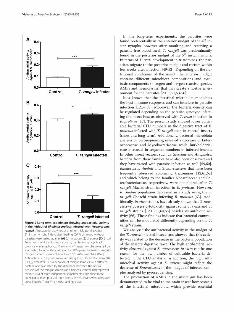
Figure 9 Long-term experiment showing antibacterial activityin the midgut of Rhodnius prolixus infected with Trypanosomarangeli. Antibacterial activities of anterior midgutof R. prolixus5th instar nymphs 7 days after feeding (DAF) on blood withoutparasiteswere tested against (A) S. marcescens(B) S. aureus (C) E. coli.Treatments: white columns – control, uninfected group; blackcolumns – infected group. Previously, 4th instar nymphs were fed oninactivated blood with or without 1 × 10⁶ epimastigotes/mL. Anteriormidgut contents were collected from 5th instar nymphs 7 (DAF).Antibacterial activity was measured using the turbidimetric assay (TB)(OD550 nm) after 19 h incubation of midgut samples with differentbacteria and calculated by the difference between the opticaldensities of the midgut samples and bacterial control. Bars representmean ± SEM of three independent experiments. Each experimentconsisted of three pools of three insects (n = 9). Means were comparedusing Student T-test; ***p < 0.001 and *p < 0.05.
Vieira et al. Parasites & Vectors (2015) 8:135 Page 9 of 13
In the long-term experiments, the parasites werefound preferentially in the anterior midgut of the 4th in-star nymphs; however after moulting and receiving aparasite-free blood meal, T. rangeli was predominantlyfound in the posterior midgut of the 5th instar nymphs.In terms of T. cruzi development in triatomines, the par-asites migrate to the posterior midgut and rectum withinfew weeks after infection [49-52]. Depending on the nu-tritional conditions of the insect, the anterior midgutcontains different microbiota compositions and cyto-toxic components (nitrogen and oxygen reactive species,AMPs and haemolysins) that may create a hostile envir-onment for the parasites [20,36,51,53-56].It is known that the intestinal microbiota modulates
the host immune responses and can interfere in parasiteinfection [12,57,58]. Moreover, the bacteria density canbe regulated depending on the parasite genotype infect-ing the insect host as observed with T. cruzi infection inR. prolixus [17]. The present study showed lower cultiv-able bacterial CFU numbers in the digestive tract of R.prolixus infected with T. rangeli than in control insects(short and long-term). Additionally, bacterial microbiotaanalysis by pyrosequencing revealed a decrease of Enter-ococcaceae and Mycobacteriaceae while Burkholderia-ceae increased in sequence numbers in infected insects.In other insect vectors, such as Glossina and Anopheles,bacteria from these families have also been observed andthey have varied with parasite infection as well [59,60].Rhodococcus rhodnii and S. marcenscens that have beenfrequently observed colonizing triatomines [12,61,62]and which belong to the families Nocardiaceae and En-terobacteriaceae, respectively, were not altered after T.rangeli Macias strain infection in R. prolixus. However,R. rhodnii population decreased in a study using the T.rangeli Choachi strain infecting R. prolixus [63]. Add-itionally, in vitro studies have already shown that S. mar-cescens possess cytotoxicity against some T. cruzi and T.rangeli strains [12,13,55,64,65] besides its antibiotic ac-tivity [66]. These findings indicate that bacterial commu-nities can be modulated differently depending on the T.rangeli strain.We analysed the antibacterial activity in the midgut of
the T. rangeli infected insects and showed that this activ-ity was related to the decrease in the bacteria populationof the insect’s digestive tract. The high antibacterial ac-tivity observed against S. marcescens in vitro can be onereason for the low number of cultivable bacteria de-tected in the CFU analysis. In addition, the high anti-microbial activity against S. aureus might reflect thedecrease of Enterococcus in the midgut of infected sam-ples analysed by pyrosequencing.The production of AMPs in the insect gut has been
demonstrated to be vital to maintain insect homeostasisof the intestinal microbiota which provide essential

Figure 10 Long-term experiment showing relative transcript abundance of antimicrobial peptides and lysozymes in Rhodnius prolixusmidgut. Relative AMP mRNA levels from R. prolixus 5th instar nymphs were analysed 1 and 7 days after feeding (DAF) on blood without parasites.A – anterior midgut 1 DAF; B – posterior midgut 1 DAF; C – anterior midgut 7 DAF; D – posterior midgut 7 DAF. Previously, 4th instar nymphswere fed on inactivated blood with or without 1 × 10⁶ epimastigotes/mL. Treatments: white columns – control, uninfected group; black columns –infected group. Bars represent mean ± SEM of three independent experiments with a pool of 10 insects (n = 3). Means were compared using one-wayANOVA and Student T-test; ***p < 0.001, **p < 0.01 and *p < 0.05.
Figure 11 Long-term experiment showing phenoloxidaseactivity in the midgut of Rhodnius prolixus infected withTrypanosoma rangeli. PO activities were measured in the anteriormidgut of R. prolixus 5th instar nymphs at 7 and 12 days after feedingon blood without parasites. Previously, 4th instar nymphs were fedon inactivated blood with or without 1 × 10⁶ epimastigotes/mL.Treatments: white columns – control, uninfected group; blackcolumns – infected group. Bars represent mean ± SEM of threeindependent experiments each with five insects (n = 15). Means werecompared using Student T-test; ***p < 0.001 and **p < 0.01.
Vieira et al. Parasites & Vectors (2015) 8:135 Page 10 of 13
nutrients, promote digestion and control pathogenicmicroorganisms by modulating the immune responses[55,67-69]. In Drosophila the activation of signallingpathways of the immunity depends on the type of pre-dominant microorganisms in the digestive tract [70-72].An important immune response in the midgut lumen
of insect vectors to control natural microbiota growthand pathogens is the production of AMPs [55,69]. AMPsare effectors molecules of the humoral immune systemof insects that control microorganisms by disrupting cellmembranes [73-75]. Analysis of the relative expressionof mRNAs encoding lysozymes and AMPs in T. rangeliinfected insects showed a different pattern in short andlong-term infections. However, in general there was asuppression of most AMP genes. For example, LysB andLysA down-regulation was observed in the anterior andposterior midgut compartments. A previous work sug-gested that LysA is mainly expressed in the midgut with adigestive function while LysB is expressed in the fat bodywith an immune role [38]. Nevertheless, S. aureus oral in-fection in R. prolixus increased LysA mRNA levels in themidgut [36]. Combining these results with the suppression

Vieira et al. Parasites & Vectors (2015) 8:135 Page 11 of 13
of LysA by T. rangeli infection observed herein, we suggestits involvement in the immune response.Regarding prolixicin, a previous work showed that this
peptide presented antimicrobial activities against Gram-negative and Gram-positive bacteria, but no toxicityagainst T. cruzi was detected [19]. In the present work,Prol was down-regulated in both midgut compartments,in both the short and long-term infections with T. rangeli.Although cytotoxicity of prolixicin against T. rangeli hasnot been described in the literature, the present resultssuggest that the modulation of Prol expression by T. ran-geli could be one possible mechanism that, indirectly ben-efits the parasite’s development in R. prolixus.Another group of AMPs extensively studied in insects
are the defensins. These peptides are known to act mainlyon Gram-positive bacteria, but also show some activityagainst Gram-negative bacteria [76,77] and some pro-tozoans such as Plasmodium and Trypanosoma [78-81].Considering the short-term infection and the parasitepopulation dynamics in the insect’s midgut, a rapid in-crease of DefB levels and its subsequent down-regulationin the anterior midgut suggests a possible role of the re-spective peptides in the control of microorganism densityin this compartment. The role of defensins in the controlof trypanosomatid infections in the vector has been sug-gested previously [80,82]. On the other hand, the increaseof DefC in both midgut compartments represents an im-mune modulation caused by T. rangeli that could repre-sent a strategy to facilitate the establishment of T. rangeliin the gut of R. prolixus. Combined, these results suggestthat an increase of antimicrobial activities and a decreaseof CFU numbers detected in the anterior midgut in shortterm infection might be a result of the increased DefB andDefC levels observed. Long-term infection resulted in amassive up-regulation of DefC in the posterior midgut,which can explain the decrease of bacteria population en-countered and the parasite’s preference to develop in thismidgut compartment. These results indicate that the para-site infection can modulate the insect’s immune system,which consequently can influence the microbiota popula-tion in the insect’s digestive tract.Another important biological event in the T. rangeli
cycle, in its invertebrate host, is its ability to modulatethe PPO system in the triatomine haemolymph [83-85].The presence of T. rangeli also reduced the level of PPOactivation in vitro [86] and in vivo in R. prolixus haemo-lymph [22,84]. The present study is the first to demon-strate that the PO activity in the R. prolixus midgut wasalso inhibited after oral infection with T. rangeli. ThePO activity in the midgut seems to be differently regu-lated accordingly to the trypanosomatid species. WhileT. rangeli has the ability to inhibit the insect PPO sys-tem, T. cruzi infection induces an increase in this im-mune response [17]. Other immune modulated factors
such as reactive oxygen and nitrogen species may be in-volved in the development of the parasite in insect’smidgut [17,87-89].
ConclusionParasite-microbiota competition for nutrients can changethe bacteria composition in the R. prolixus midgut andsubsequently modulate the insect’s immune system. A dir-ect modulation of the immune system by the parasite canalso affect the microbiota population. The strategy of cer-tain trypanosome species for successful infection of the in-vertebrate host is a complex interplay and depends on atripartite interaction between parasite, insect immune sys-tem and bacteria [46,59,60,90,91]. These interactions arean important field for research, opening up new insightsinto the understanding of parasite-vector relationships[92]. A better understanding of the role of bacterialspecies composing the gut microbiota on host immun-ity against pathogens can lead to the development ofnew strategies to control vector-borne diseases.
Competing interestsThe authors declare that they have no competing interests.
Authors’ contributionsConceived and designed the experiments: CSV, PJW, DPC, ESG and PA.Carried out the biochemical experiments: CSV, DPM, MBF and DPC.Performed the molecular experiments: CSV, PJW, MG, FFM. Analysis andinterpretation of data: CSV, DPM, PJW, JMS, MBF, MG, FFM, and PA.Contributed reagents/materials: ESG and PA. Wrote the manuscript: CSV,PJW, DPC and PA. All authors read and approved the final manuscript.
AcknowledgmentsThis work was supported by grants from Conselho Nacional deDesenvolvimento Científico e Tecnológico (CNPq), Fundação Oswaldo Cruz(FIOCRUZ) (PAPES Project), Fundação de Amparo à Pesquisa do Estado doRio de Janeiro (FAPERJ) and INCT-EM. We thank Dr. Norman Ratcliffe forEnglish revision of this work.
Author details1Laboratório de Bioquímica e Fisiologia de Insetos, Instituto Oswaldo Cruz,Fundação Oswaldo Cruz (IOC/FIOCRUZ), Rio de Janeiro, RJ, Brazil.2Departamento de Entomologia Molecular, Instituto Nacional deEntomologia Molecular (INCT-EM), Rio de Janeiro, RJ, Brazil. 3Departamentode Ciências Ambientais, Instituto de Florestas, Universidade Federal Rural doRio de Janeiro (UFRRJ), Seropédica, RJ, Brazil.
Received: 10 November 2014 Accepted: 13 February 2015
References1. Watkins R. Histology of Rhodnius prolixus infected with Trypanosoma rangeli.
J Invertebr Pathol. 1971;17:59–66.2. Hoare CA. The trypanosomes of mammals: a zoological monograph. Oxford:
Blackwell Scientific Publications; 1972.3. Tobie EJ. Observations on development of Trypanosoma rangeli in
hemocoel of Rhodnius prolixus. J Invertebr Pathol. 1970;15:118–25.4. Guhl F, Vallejo GA. Trypanosoma (Herpetosoma) rangeli Tejera, 1920 - An
updated review. Mem Inst Oswaldo Cruz. 2003;98:435–42.5. Hecker H, Schwarzenbach M, Rudin W. Development and interactions of
Trypanosoma rangeli in and with the reduviid bug Rhodnius prolixus.Parasitol Res. 1990;76:311–8.
6. Garcia ES, Mello CB, Azambuja P, Ribeiro JMC. Rhodnius prolixus - salivaryantihemostatic components decrease with Trypanosoma rangeli infection.Exp Parasitol. 1994;78:287–93.
Vieira et al. Parasites & Vectors (2015) 8:135 Page 12 of 13
7. Azambuja P, Garcia ES. Trypanosoma rangeli interactions within the vectorRhodnius prolixus: a mini review. Mem Inst Oswaldo Cruz. 2005;100:567–72.
8. Garcia ES, Castro DP, Figueiredo MB, Azambuja P. Parasite mediatedinteractions within the insect vector: Trypanosoma rangeli strategies. ParasitVectors. 2012;5:105.
9. Garcia ES, Ratcliffe NA, Whitten MM, Gonzalez MS, Azambuja P. Exploringthe role of insect host factors in the dynamics of Trypanosomacruzi-Rhodnius prolixus interactions. J Insect Physiol. 2007;53:11–21.
10. Garcia ES, Castro DP, Figueiredo MB, Genta FA, Azambuja P. Trypanosomarangeli: a new perspective for studying the modulation of immunereactions of Rhodnius prolixus. Parasit Vectors. 2009;2:33.
11. Figueiredo MB, Genta FA, Garcia ES, Azambuja P. Lipid mediators and vectorinfection: Trypanosoma rangeli inhibits Rhodnius prolixus hemocytephagocytosis by modulation of phospholipase A2 and PAF-acetylhydrolaseactivities. J Insect Physiol. 2008;54:1528–37.
12. Azambuja P, Feder D, Garcia ES. Isolation of Serratia marcescens in themidgut of Rhodnius prolixus: impact on the establishment of the parasiteTrypanosoma cruzi in the vector. Exp Parasitol. 2004;107:89–96.
13. Azambuja P, Garcia ES, Ratcliffe NA. Gut microbiota and parasitetransmission by insect vectors. Trends Parasitol. 2005;21:568–72.
14. Azambuja PG, Guimarães JA, Garcia ES. Haemolytic factor from the crop ofRhodnius prolixus: evidence and partial characterization. J Insect Physiol.1983;29:5.
15. Pulido XC, Perez G, Vallejo GA. Preliminary characterization of a Rhodniusprolixus hemolymph trypanolytic protein, this being a determinant ofTrypanosoma rangeli KP1(+) and KP1(−) subpopulations vectorial ability.Mem Inst Oswaldo Cruz. 2008;103:172–9.
16. Mello CB, Nigam Y, Garcia ES, Azambuja P, Newton RP, Ratcliffe NA. Studieson a haemolymph lectin isolated from Rhodnius prolixus and its interactionwith Trypanosoma rangeli. Exp Parasitol. 1999;91:289–96.
17. Castro DP, Moraes C, Gonzalez M, Ratcliffe N, Azambuja P, Garcia E.Trypanosoma cruzi immune response modulation decreases microbiota inRhodnius prolixus gut and is crucial for parasite survival and development.PLoS One. 2012;7:e36591.
18. Waniek PJ, Castro HC, Sathler PC, Miceli L, Jansen AM, Araujo CAC. Twonovel defensin-encoding genes of the Chagas disease vector Triatomabrasiliensis (Reduviidae, Triatominae): gene expression and peptide-structuremodeling. J Insect Physiol. 2009;55:840–8.
19. Ursic-Bedoya R, Buchhop J, Joy JB, Durvasula R, Lowenberger C. Prolixicin: anovel antimicrobial peptide isolated from Rhodnius prolixus with differentialactivity against bacteria and Trypanosoma cruzi. Insect Mol Biol.2011;20:775–86.
20. Whitten M, Sun F, Tew I, Schaub G, Soukou C, Nappi A, et al. Differentialmodulation of Rhodnius prolixus nitric oxide activities following challengewith Trypanosoma rangeli, T. cruzi and bacterial cell wall components.Insect Biochem Mol Biol. 2007;37:440–52.
21. Garcia ES, Machado EM, Azambuja P. Inhibition of hemocytemicroaggregation reactions in Rhodnius prolixus larvae orally infected withTrypanosoma rangeli. Exp Parasitol. 2004;107:31–8.
22. Garcia ES, Machado EM, Azambuja P. Effects of eicosanoid biosynthesisinhibitors on the prophenoloxidase-activating system and microaggregationreactions in the hemolymph of Rhodnius prolixus infected with Trypanosomarangeli. J Insect Physiol. 2004;50:157–65.
23. Sociedade Brasileira de Ciências de Animais em Laboratório. COBEA.http://www.cobea.org.br. (2014). Accessed 10 Oct 2014.
24. Schottelius J. Neuraminidase fluorescence test for the differentiation ofTrypanosoma cruzi and Trypanosoma rangeli. Trop Med Parasitol.1987;38:323–7.
25. Grisard EC, Steindel M, Guarneri AA, Eger-Mangrich I, Campbell DA,Romanha AJ. Characterization of Trypanosoma rangeli strains isolated inCentral and South America: an overview. Mem Inst Oswaldo Cruz.1999;94:203–9.
26. Vallejo GA, Guhl F, Carranza JC, Lozano LE, Sanchez JL, Jaramillo JC, et al.kDNA markers define two major Trypanosoma rangeli lineages in Latin-America. Acta Trop. 2002;81:77–82.
27. Azambuja P, Garcia ES. Care and maintenance of triatomine colonies.In: Crampton JM, Beard CB, Louis C, editors. Molecular biology ofinsect disease vectors: a methods manual. London: Chapman and Hall;1997. p. 56–64.
28. Bexfield A, Nigam Y, Thomas S, Ratcliffe NA. Detection and partialcharacterisation of two antibacterial factors from the excretions/secretions
of the medicinal maggot Lucilia sericata and their activity against methicillinresistant Staphylococcus aureus (MRSA). Microbes Infect. 2004;6:1297–304.
29. Ferrand J, Patron K, Legrand-Frossi C, Frippiat JP, Merlin C, Alauzet C, et al.Comparison of seven methods for extraction of bacterial DNA from fecaland cecal samples of mice. J Microbiol Methods. 2014;105:180–5.
30. Andersson AF, Lindberg M, Jakobsson H, Backhed F, Nyren P, Engstrand L.Comparative analysis of human gut microbiota by barcodedpyrosequencing. PLoS One. 2008;3:e2836.
31. National Institute of Health Common Fund Human Microbiome Project(HMP) http://www.hmpdacc.org. (2014). Accessed 15 Sept 14.
32. Nawrocki EP, Eddy SR. Query-dependent banding (QDB) for faster RNAsimilarity searches. PLoS Comput Biol. 2007;3:e56.
33. Edgar RC, Haas BJ, Clemente JC, Quince C, Knight R. UCHIMEimproves sensitivity and speed of chimera detection. Bioinformatics.2011;27:2194–200.
34. Wang Q, Garrity GM, Tiedje JM, Cole JR. Naive Bayesian classifier for rapidassignment of rRNA sequences into the new bacterial taxonomy.Appl Environ Microbiol. 2007;73:5261–7.
35. Cole JR, Wang Q, Cardenas E, Fish J, Chai B, Farris RJ, et al. The ribosomaldatabase project: improved alignments and new tools for rRNA analysis.Nucleic Acids Res. 2009;37:D141–5.
36. Vieira CS, Waniek PJ, Mattos DP, Castro DP, Mello CB, Ratcliffe NA, et al.Humoral responses in Rhodnius prolixus: bacterial feeding inducesdifferential patterns of antibacterial activity and enhances mRNA levels ofantimicrobial peptides in the midgut. Parasit Vectors. 2014;7:232.
37. Lopez L, Morales G, Ursic R, Wolff M, Lowenberger C. Isolation andcharacterization of a novel insect defensin from Rhodnius prolixus, a vectorof Chagas disease. Insect Biochem Mol Biol. 2003;33:439–47.
38. Ursic-Bedoya RJ, Nazzari H, Cooper D, Triana O, Wolff M, Lowenberger C.Identification and characterization of two novel lysozymes from Rhodniusprolixus, a vector of Chagas disease. J Insect Physiol. 2008;54:593–603.
39. Genta FA, Souza RS, Garcia ES, Azambuja P. Phenol oxidases from Rhodniusprolixus: temporal and tissue expression pattern and regulation byecdysone. J Insect Physiol. 2010;56:1253–9.
40. Machado PE, Eger-Mangrich I, Rosa G, Koerich LB, Grisard EC, Steindel M.Differential susceptibility of triatomines of the genus Rhodnius toTrypanosoma rangeli strains from different geographical origins.Int J Parasitol. 2001;31:632–4.
41. Urrea DA, Carranza JC, Cuba CA, Gurgel-Goncalves R, Guhl F, Schofield CJ,et al. Molecular characterisation of Trypanosoma rangeli strains isolated fromRhodnius ecuadoriensis in Peru, R. colombiensis in Colombia and R. pallescensin Panama, supports a co-evolutionary association between parasites andvectors. Infect Genet Evol. 2005;5:123–9.
42. Whitten MM, Mello CB, Gomes SA, Nigam Y, Azambuja P, Garcia ES, et al.Role of superoxide and reactive nitrogen intermediates in Rhodnius prolixus(Reduviidae)/Trypanosoma rangeli interactions. Exp Parasitol. 2001;98:44–57.
43. Vallejo GA, Guhl F, Carranza JC, Triana O, Perez G, Ortiz PA, et al. Trypanosomarangeli parasite-vector-vertebrate interactions and their relationship to thesystematics and epidemiology of American trypanosomiasis. Biomedica.2007;27 Suppl 1:110–8.
44. Vallejo GA, Guhl F, Schaub GA. Triatominae-Trypanosoma cruzi/T. rangeli.Vector-parasite interactions. Acta Trop. 2009;110:137–47.
45. Tchioffo MT, Boissiere A, Churcher TS, Abate L, Gimonneau G, Nsango SE,et al. Modulation of malaria infection in Anopheles gambiae mosquitoesexposed to natural midgut bacteria. PLoS One. 2013;8:e81663.
46. Weiss BL, Wang J, Maltz MA, Wu Y, Aksoy S. Trypanosome infectionestablishment in the tsetse fly gut is influenced by microbiome regulatedhost immune barriers. PLoS Pathog. 2013;9:e1003318.
47. Clayton AM, Dong Y, Dimopoulos G. The Anopheles innate immune systemin the defense against malaria infection. J Innate Immun. 2014;6:169–81.
48. Sant'Anna MR, Diaz-Albiter H, Aguiar-Martins K, Al Salem WS, Cavalcante RR,Dillon VM, et al. Colonisation resistance in the sand fly gut: Leishmaniaprotects Lutzomyia longipalpis from bacterial infection. Parasit Vectors.2014;7:329.
49. Kollien AH, Schaub GA. The development of Trypanosoma cruzi intriatominae. Parasitol Today. 2000;16:381–7.
50. Carvalho-Moreira CJ, Spata MC, Coura JR, Garcia ES, Azambuja P, GonzalezMS, et al. In vivo and in vitro metacyclogenesis tests of two strains ofTrypanosoma cruzi in the triatomine vectors Triatoma pseudomaculata andRhodnius neglectus: short/long-term and comparative study. Exp Parasitol.2003;103:102–11.

Vieira et al. Parasites & Vectors (2015) 8:135 Page 13 of 13
51. Cortez MR, Provencano A, Silva CE, Mello CB, Zimmermann LT, Schaub GA,et al. Trypanosoma cruzi: effects of azadirachtin and ecdysone on thedynamic development in Rhodnius prolixus larvae. Exp Parasitol.2012;131:363–71.
52. Araujo CAC, Waniek PJ, Jansen AM. TcI/TcII co-infection can enhanceTrypanosoma cruzi growth in Rhodnius prolixus. Parasit Vectors. 2014;7:94.
53. Terra WR. Evolution of digestive system of insects – review. Annu RevEntomol. 1990;35:181–200.
54. Kollien AH, Gonçalves TC, De Azambuja P, Garcia ES, Schaub GA. The effectof azadirachtin on fresh isolates of Trypanosoma cruzi in different species oftriatomines. Parasitol Res. 1998;84:286–90.
55. Garcia ES, Genta FA, de Azambuja P, Schaub GA. Interactions betweenintestinal compounds of triatomines and Trypanosoma cruzi. TrendsParasitol. 2010;26:499–505.
56. Waniek PJ, Pacheco Costa JE, Jansen AM, Costa J, Araujo CAC. Cathepsin Lof Triatoma brasiliensis (Reduviidae, Triatominae): sequence characterization,expression pattern and zymography. J Insect Physiol. 2012;58:178–87.
57. Lee WJ. Bacterial-modulated signaling pathways in gut homeostasis.Sci Signal. 2008;1:pe24.
58. Ha EM, Lee KA, Seo YY, Kim SH, Lim JH, Oh BH, et al. Coordination ofmultiple dual oxidase regulatory pathways in responses to commensal andinfectious microbes in drosophila gut. Nat Immunol. 2009;10:949–57.
59. Geiger A, Fardeau ML, Njiokou F, Ollivier B. Glossina spp. gut bacterial floraand their putative role in fly-hosted trypanosome development. Front CellInfect Microbiol. 2013;3:34.
60. Gendrin M, Christophides G. The Anopheles mosquito microbiota and theirimpact on pathogen transmission. In: Manguin S, editor. Anophelesmosquitoes - New insights into malaria vectors. Croatia: InTech; 2013.doi:10.5772/55107.
61. Wigglesworth VB. ymbiotic bacteria in a blood-sucking insect, Rhodniusprolixus Stal (Hemiptera, Triatomidae). Parasitology. 1936;28:284–9.
62. da Mota FF, Marinho LP, Moreira CJ, Lima MM, Mello CB, Garcia ES, et al.Cultivation-independent methods reveal differences among bacterial gutmicrobiota in triatomine vectors of Chagas disease. PLoS Negl Trop Dis.2012;6:e1631.
63. Eichler S, Schaub GA. Development of symbionts in triatomine bugs andthe effects of infections with trypanosomatids. Exp Parasitol. 2002;100:17–27.
64. Castro DP, Moraes CS, Garcia ES, Azambuja P. Inhibitory effects ofd-mannose on trypanosomatid lysis induced by Serratia marcescens.Exp Parasitol. 2007;115:200–4.
65. Castro DP, Seabra SH, Garcia ES, de Souza W, Azambuja P. Trypanosomacruzi: ultrastructural studies of adhesion, lysis and biofilm formation bySerratia marcescens. Exp Parasitol. 2007;117:201–7.
66. Thomson NR, Crow MA, McGowan SJ, Cox A, Salmond GP. Biosynthesis ofcarbapenem antibiotic and prodigiosin pigment in Serratia is under quorumsensing control. Mol Microbiol. 2000;36:539–56.
67. Dillon RJ, Dillon VM. The gut bacteria of insects: nonpathogenic interactions.Annu Rev Entomol. 2004;49:71–92.
68. Leulier F, Royet J. Maintaining immune homeostasis in fly gut. Nat Immunol.2009;10:936–8.
69. Schaub GA. Interactions of Trypanosomatids and Triatomines. Adv InsectPhys. 2009;37:177–242.
70. Nehme NT, Liegeois S, Kele B, Giammarinaro P, Pradel E, Hoffmann JA, et al.A model of bacterial intestinal infections in Drosophila melanogaster.PLoS Pathog. 2007;3:e173.
71. Ryu JH, Kim SH, Lee HY, Bai JY, Nam YD, Bae JW, et al. Innate immunehomeostasis by the homeobox gene caudal and commensal-gut mutualismin Drosophila. Science. 2008;319:777–82.
72. Marmaras VJ, Lampropoulou M. Regulators and signalling in insecthaemocyte immunity. Cell Signal. 2009;21:186–95.
73. Zasloff M. Antimicrobial peptides of multicellular organisms. Nature.2002;415:389–95.
74. Bulet P, Stocklin R. Insect antimicrobial peptides: structures, properties andgene regulation. Protein PepT Lett. 2005;12:3–11.
75. Ferrandon D, Imler JL, Hetru C, Hoffmann JA. The Drosophila systemicimmune response: sensing and signalling during bacterial and fungalinfections. Nat Rev Immunol. 2007;7:862–74.
76. Bulet P, Cociancich S, Reuland M, Sauber F, Bischoff R, Hegy G, et al. A novelinsect defensin mediates the inducible antibacterial activity in larvae of thedragonfly Aeschna cyanea (Paleoptera, Odonata). Eur J Biochem.1992;209:977–84.
77. Lamberty M, Ades S, Uttenweiler-Joseph S, Brookhart G, Bushey D,Hoffmann JA, et al. Insect immunity. Isolation from the lepidopteranHeliothis virescens of a novel insect defensin with potent antifungal activity.J Biol Chem. 1999;274:9320–6.
78. Shahabuddin M, Fields I, Bulet P, Hoffmann JA, Miller LH. Plasmodiumgallinaceum: differential killing of some mosquito stages of the parasite byinsect defensin. Exp Parasitol. 1998;89:103–12.
79. Vizioli J, Richman AM, Uttenweiler-Joseph S, Blass C, Bulet P. The defensinpeptide of the malaria vector mosquito Anopheles gambiae: antimicrobialactivities and expression in adult mosquitoes. Insect Biochem Mol Biol.2001;31:241–8.
80. McGwire BS, Olson CL, Tack BF, Engman DM. Killing of Africantrypanosomes by antimicrobial peptides. J Infect Dis. 2003;188:146–52.
81. Kleschenko YE, Karpenko LP, Villalta F. Effects of human defensin-α1 onTrypanosoma cruzi trypomastigotes in vitro. Bull Exp Biol Med.2010;149:731–3.
82. Waniek PJ, Jansen AM, Araujo CAC. Trypanosoma cruzi infection modulatesthe expression of Triatoma brasiliensis def1 in the midgut. Vector BorneZoonotic Dis. 2011;11:845–7.
83. Gomes SA, Feder D, Thomas NE, Garcia ES, Azambuja P. Rhodnius prolixusinfected with Trypanosoma rangeli: In vivo and in vitro experiments.J Invertebr Pathol. 1999;73:289–93.
84. Gomes SA, Feder D, Garcia ES, Azambuja P. Suppression of theprophenoloxidase system in Rhodnius prolixus orally infected withTrypanosoma rangeli. J Insect Physiol. 2003;49:829–37.
85. Machado EM, Azambuja P, Garcia ES. WEB 2086, a platelet-activating factorantagonist, inhibits prophenoloxidase-activating system and hemocytemicroaggregation reactions induced by Trypanosoma rangeli infection inRhodnius prolixus hemolymph. J Insect Physiol. 2006;52:685–92.
86. Gregorio EA, Ratcliffe NA. The prophenoloxidase system and in vitrointeraction of Trypanosoma rangeli with Rhodnius prolixus and Triatomainfestans haemolymph. Parasite Immunol. 1991;13:551–64.
87. Ascenzi P, Gradoni L. Nitric oxide limits parasite development in vectors andin invertebrate intermediate hosts. IUBMB Life. 2002;53:121–3.
88. Rivero A. Nitric oxide: an antiparasitic molecule of invertebrates. TrendsParasitol. 2006;22:219–25.
89. Cosentino-Gomes D, Rocco-Machado N, Meyer-Fernandes JR. Rhodniusprolixus: Modulation of antioxidant defenses by Trypanosoma rangeli.Exp Parasitol. 2014;145:118–24.
90. Cirimotich CM, Dong Y, Clayton AM, Sandiford SL, Souza-Neto JA, MulengaM, et al. Natural microbe-mediated refractoriness to Plasmodium infection inAnopheles gambiae. Science. 2011;332:855–8.
91. Ramirez JL, Souza-Neto J, Torres Cosme R, Rovira J, Ortiz A, Pascale JM, et al.Reciprocal tripartite interactions between the Aedes aegypti midgutmicrobiota, innate immune system and dengue virus influences vectorcompetence. PLoS Negl Trop Dis. 2012;6:e1561.
92. Weiss B, Aksoy S. Microbiome influences on insect host vector competence.Trends Parasitol. 2011;27:514–22.
Submit your next manuscript to BioMed Centraland take full advantage of:
• Convenient online submission
• Thorough peer review
• No space constraints or color figure charges
• Immediate publication on acceptance
• Inclusion in PubMed, CAS, Scopus and Google Scholar
• Research which is freely available for redistribution
Submit your manuscript at www.biomedcentral.com/submit


![THE CUTICULAR PATTERN IN AN INSECT, RHODNIUS ...[ 45 ]9 THE CUTICULAR PATTERN IN AN INSECT,RHODNIUS PROLIXUS STAL BY M. LOCKE Department of Zoology, University College of the West](https://img.dokumen.tips/doc/110x75/60d8dfdd6bafa25aa5444dad/the-cuticular-pattern-in-an-insect-rhodnius-45-9-the-cuticular-pattern-in.jpg)