Embed Size (px)
Citation preview
BioMed Central
Reproductive Biology and Endocrinology
ss
Open AcceResearchRemoval of spermatozoa with externalized phosphatidylserine from sperm preparation in human assisted medical procreation: effects on viability, motility and mitochondrial membrane potentialCorinne de Vantéry Arrighi1, Hervé Lucas2, Didier Chardonnens3 and Ariane de Agostini*1Address: 1Unit of Reproductive Medicine, Department of Obstetrics and Gynaecology, Geneva University Hospitals and University of Geneva, 30, bd de la Cluse, 1211 Geneva 14, Switzerland, 2AB-Biology, AMP74 Center, Hospital Center of Annemasse-Bonneville, France and 3Reproductive Medecine Center Medixy, La Tour Hospital, Geneva, Switzerland
Email: Corinne de Vantéry Arrighi - [email protected]; Hervé Lucas - [email protected]; Didier Chardonnens - [email protected]; Ariane de Agostini* - [email protected]
* Corresponding author
AbstractBackground: Externalization of phosphatidylserine (EPS) occurs in apoptotic-like spermatozoaand could be used to remove them from sperm preparations to enhance sperm quality for assistedmedical procreation. We first characterized EPS in sperms from infertile patients in terms offrequency of EPS spermatozoa as well as localization of phosphatidylserine (PS) on spermatozoa.Subsequently, we determined the impact of depleting EPS spermatozoa on sperm quality.
Methods: EPS were visualized by fluorescently-labeled annexin V binding assay. Double stainingwith annexin V and Hoechst differentiates apoptotic from necrotic spermatozoa. We usedmagnetic-activated cell sorting using annexin V-conjugated microbeads (MACS-ANMB) techniqueto remove EPS spermatozoa from sperm prepared by density gradient centrifugation (DGC). Theimpact of this technique on sperm quality was evaluated by measuring progressive motility, viability,and the integrity of the mitochondrial membrane potential (MMP) by Rhodamine 123.
Results: Mean percentages of EPS spermatozoa were 14% in DGC sperm. Four subpopulations ofspermatozoa were identified: 70% alive, 3% early apoptotic, 16% necrotic and 11% late apoptoticor necrotic. PS were localized on head and/or midpiece or on the whole spermatozoa. MACSefficiently eliminates EPS spermatozoa. MACS combined with DGC allows a mean reduction of 70%in EPS and of 60% in MMP-disrupted spermatozoa with a mean increase of 50% in sperm survivalat 24 h.
Conclusion: Human ejaculates contain EPS spermatozoa which can mostly be eliminated by DGCplus MACS resulting in improved sperm long term viability, motility and MMP integrity. EPS may beused as an indicator of sperm quality and removal of EPS spermatozoa may enhance fertilitypotential in assisted medical procreation.
Published: 8 January 2009
Reproductive Biology and Endocrinology 2009, 7:1 doi:10.1186/1477-7827-7-1
Received: 8 September 2008Accepted: 8 January 2009
This article is available from: http://www.rbej.com/content/7/1/1
© 2009 de Vantéry Arrighi et al; licensee BioMed Central Ltd. This is an Open Access article distributed under the terms of the Creative Commons Attribution License (http://creativecommons.org/licenses/by/2.0), which permits unrestricted use, distribution, and reproduction in any medium, provided the original work is properly cited.
Page 1 of 12(page number not for citation purposes)

Reproductive Biology and Endocrinology 2009, 7:1 http://www.rbej.com/content/7/1/1
BackgroundHuman sperm quality is defined by the classical parame-ters, concentration, motility and morphology, accordingto standard World Health Organisation diagnostic semenanalysis [1]. Nevertheless, hidden anomalies affectingspermatozoa membranes such as externalization of thephosphatidylserine (EPS), or spermatozoa organellessuch as the disruption of mitochondrial membranepotential (MMP), caspases activation and/or DNA frag-mentation, are common somatic features of apoptosis.Such features are not routinely detected in ejaculated sper-matozoa but they have been proven to have a negativeimpact on assisted medical procreation (AMP) outcome[2-8]. Indeed, successful assisted reproduction is mainlydependent on the quality of the sperm plasma membrane,requiring normal integrity and function to provide motil-ity, acrosome reaction, and fertilization [9]. Spermatozoawith impaired membrane integrity occur more frequentlyin infertile men, partly explaining suboptimal results inAMP. However, striking modifications of sperm plasmamembrane occur physiologically in ejaculated sperm.During capacitation, there is an AC/cAMP/PKA-depend-ent lipid remodelling of the sperm plasma membrane dueto phospholipids translocation that lead to externaliza-tion of phosphatidylserine (PS) and phosphatidyleth-anolamine, and to an albumin mediated efflux ofcholesterol resulting in an increase in membrane fluidity[10-16].
Apoptosis plays a critical role in many normal biologicalprocesses such as embryogenesis and tissue homeostasis,as well as in pathological states and diseases. Apoptosiscauses disruption of membrane, phospholipid asymmetryand translocation of PS, a signal for specific recognitionand removal of apoptotic cells by phagocytosis [17-20].Annexin V, an anticoagulant protein of 35.8 kDa, bindsoutwardly exposed PS with high affinity in a calcium-dependent manner [21]. Thus, the combined use ofannexin V and of a supravital fluorescent dye, such asHoechst 33258, allows the simultaneous detection ofapoptotic and necrotic cells [22]. Currently, it is knownthat germinal cells apoptosis is an underlying mechanismof normal spermatogenesis and an altered apoptotic proc-ess has been found to be closely associated with abnormalspermatozoa in semen and thus with male infertility [23-26]. Indeed, Maeda et al. [27] inhibited sperm productionin mice by annexin V microinjection into seminiferoustubules, showing that the phagocytic clearance of apop-totic spermatogenic cells by Sertoli cells is PS-mediatedand required for a normal production of sperm. In con-trast, apoptosis in ejaculated sperm is less well understoodand remains controversial [28]. Few reports applied thedetection of EPS, by annexin V-labeling, for the evaluationof cryopreservation techniques on ram [29,30], boar[31,32], bull [33-35], and human [36-43] spermatozoa.Other human sperm studies attempted to relate EPS, DNA
fragmentation and/or apoptotic proteins with spermquality parameters [6,7,15,44-56]. It comes out from theliterature that EPS on ejaculated mature spermatozoa iseither the result of a plasma membrane modificationbecause of capacitation and/or acrosome reaction[11,15,54] or the sign of an early apoptotic phenotype[45,57]. Sakkas et al. [2,58] proposed that apoptosis inejaculated sperm is the result of a spermatogenetic failure,thus an abortive apoptotic process that started before ejac-ulation [59]. It has also been described that inflammation[60-62] and bacteria can induce EPS in human spermato-zoa [63]. Nevertheless, the link between EPS and spermapoptosis is still controversial, although it is accepted thatannexin V binding to spermatozoa characterizes modifiedsperm plasma membrane. Interesting results on EPS inrelationship with infertility problems in AMP are those ofcollaborators at the Reproductive Research Center of theCleveland Clinic (USA), and at the Department of Derma-tology and Andrology of Leipzig University (Germany)[39,51,64-70] who adapted the technique of magnetic-activated cell sorting using annexin V-conjugatedmicrobeads (MACS-ANMB) to sperm preparations inorder to evaluate the impact of EPS on sperm parametersand fertility potential.
In the present study, we have characterized EPS on sper-matozoa after DGC sperm preparation and tested itsimpact on the quality of sperm prepared for AMP, anessential element for successful fertilization and reproduc-tive success. EPS is the main sperm surface apoptoticmarker but its role in ejaculated sperm remains ambigu-ous. We have investigated the effects of removal of EPS-positive spermatozoa on the motility and MMP integrityof prepared spermatozoa in patients consulting for infer-tility. To this end, we have depleted sperm preparations ofEPS spermatozoa by affinity to annexin V. The patientpopulation studied, consulting for infertility, presentedsubobptimal semen characteristics and could benefit fromremoval of EPS sperm, using ANMB-MACS from spermpreparations. This study has tested the feasibility of suchpurification given the limited amounts of viable sperma-tozoa available in the ejaculates of these patients. Thesedata allow to draw guidelines for the clinical applicationof this purification in low concentration and/or bad qual-ity sperm.
Firstly, we determined the proportion of EPS-positivespermatozoa in noncapacitated and capacitated sperma-tozoa, and secondly we precisely localized PS on sperma-tozoa among sperms of patients consulting our infertilityunit. Further, we compared raw sperm with two spermpreparations, DGC and DGC combined with MACS-ANMB, for percentages of EPS-positive spermatozoa, ofviability, of progressive motility and of spermatozoa withdisrupted MMP. Collectively, these data allowed to evalu-ate in details the quality of sperm preparations after elim-
Page 2 of 12(page number not for citation purposes)

Reproductive Biology and Endocrinology 2009, 7:1 http://www.rbej.com/content/7/1/1
ination of EPS-positive spermatozoa by MACS-ANMB inour population of infertile patients in AMP conditions.
MethodsSperm preparationThis study was approved by the ethic committee of theGynaecology and Obstetrics Department, Geneva Univer-sity Hospital, Switzerland.
Human sperm, collected following an abstinence of 3–5days, were obtained from men attending the Unit ofReproductive Medicine (Geneva University Hospital,Switzerland), after signing a written informed consent.Semen (standard) parameters were thus obtained. Afterliquefaction, ejaculated sperm were prepared using PureS-perm® (NidaCon laboratories AB, Gothenburg, Sweden)or SpermFilter® (Cryos, Denmark) DGC (90–45%). Thepellet of the 90% fraction was washed in BM1 medium(Synthetic medium for human in vitro cell culture, NMSBIO MEDICAL AG, Suisse), centrifuged for 10 min at 300g and resuspended in BM1 medium and placed in an incu-bator at 37°C in a humidified atmosphere of 6% CO2,before applying on MACS-ANMB according to the experi-mental design. Selection of sperm by DGC was conductedin BM1, a capacitating medium. For capacitation experi-ments, sperm suspensions were further incubated for 3–5h at 37°C in a humidified atmosphere of 6% CO2. Forsperm survival analysis, sperm suspensions in BM1medium were maintained in the incubator at 37°C in ahumidified atmosphere of 6% CO2 for 24 h.
Sperm parameters were determined in accordance withthe World Health Organization guidelines [1]. Spermconcentration was assessed using a Makler® Chamber.Sperm motility was assessed with 10 μl of suspensionbetween slide and cover-slide. Sperm viability and mor-phology were assessed using eosin-nigrosin and Papani-colaou staining, respectively [1].
Experimental designSperm count (concentration in million spermatozoa perml), progressive motility, EPS-positive spermatozoa andMMP-disrupted spermatozoa were evaluated in liquefiedsperm (raw samples) after sperm preparations by DGCalone or by combining DGC with MACS-ANMB. We alsoevaluated sperm survival 24 h following DGC combinedwith MACS. We determined the sperm recovery rate afterMACS preparation. We also applied MACS to low amountof spermatozoa and/or bad quality sperm in order todefine the limit of application of this technique.
Detection and evaluation of EPS-positive spermatozoa in DGC sperm preparationPrepared spermatozoa were adjusted at a concentration of0.5–1 million/ml and washed with 1× Binding Buffer (BD
Biosciences Pharmingen, San Diego, CA). Spermatozoawere then resuspended in 100 μl 1× Binding Buffer con-taining 5–10 μl fluorescein isothiocyanate (FITC)-labeledannexin V, according to the manufacturer's instructions(BD Biosciences Pharmingen), and 0.5 μg/ml Hoechst33258 (Sigma-Aldrich, Switzerland) and incubated for 15min at room temperature in the dark. Spermatozoa sus-pensions were washed with Binding Buffer and centri-fuged at 400 g. Supernatants were removed and pelletsresuspended in 10–15 μl of Binding Buffer and depositedon glass slides. Two types of preparations were done, withor without mounting media for fluorescence, Vectashield(Vector Laboratories, Burlingame, CA), before coveringthe slide with a coverslip. The first preparation used forthe localization of annexin V staining on spermatozoaand the second one for the double labeling with annexinV-FITC and vital dye Hoechst allowing the distinctionbetween spermatozoa with or without EPS, and betweenintact and dead spermatozoa, respectively. Spermatozoawere analysed under a Nikon fluorescent microscopeusing a dual filter set for FITC and Hoechst. The specificityof binding of annexin V-FITC to human spermatozoa wasdemonstrated in control experiments by preincubatingspermatozoa with excess purified recombinant annexin V(BD Bioscience Pharmingen) before performing the testor by performing the assay in Binding Buffer in theabsence of calcium. Both control conditions preventedthe binding of annexin V-FITC to PS-positive spermato-zoa. The sensitivity of the assay was tested by usingdecreasing amounts of spermatozoa and of annexin-V-FITC concentrations.
Depletion of EPS-positive spermatozoa by magnetic-activated cell sorting (MACS) method using annexin V magnetic beads (ANMB)The sperm suspensions were divided into two fractions bypassage through a magnetic field (miniMACS, MiltenyiBiotec GmbH, Germany) based on the binding of para-magnetic annexin V-microbeads to PS present on the sur-face of spermatozoa [39,50,71]. Briefly, DGC-preparedspermatozoa were incubated with annexin V-conjugatedmicrobeads (Miltenyi Biotec GmbH) for 15 min at roomtemperature. About 100 μl of annexin V microbeads sus-pension were used for each 10 million spermatozoa. Thespermatozoa/microbeads suspension was loaded on aseparation MS column containing iron balls, which wasfitted in a miniMACS separator (magnet) attached to amultistand (Miltenyi Biotec GmbH). The fraction withintact membranes that passed through the column waslabeled as MACS-ANMB-negative fraction, depleted in PS,whereas the fraction composed of apoptotic or deterio-rated PS-positive membranes spermatozoa was retainedin the separation column and labeled as MACS-ANMB-positive fraction. After the column was removed from the
Page 3 of 12(page number not for citation purposes)
Reproductive Biology and Endocrinology 2009, 7:1 http://www.rbej.com/content/7/1/1
magnetic field, the retained fraction was eluted usingannexin V-binding buffer (Miltenyi BiotecGmbH).
Evaluation of EPSRaw semen samples were washed in BM1 medium beforeannexin V-FITC staining. Samples of 0.5 to 1 million sper-matozoa from either washed raw spermatozoa, DGC orDGC combined with MACS preparations were washedwith Binding Buffer, centrifuged at 400 g for 5 min andresuspended in 100 μl Binding Buffer containing 5 μg/mlannexin V-FITC (BD Bioscience Pharmingen) for 15 minat room temperature in the dark. At the end of the labe-ling, 500 μl Binding Buffer were added to the samples andcentrifuged at 400 g for 5 min. 5–10 μl of the antifadingVectashield were added to 10–15 μl of the pellets anddeposited on glass slides and rapidly observed under aNikon fluorescence microscope with a 100-fold objective.At least 200 spermatozoa per slide were examined.
Evaluation of Mitochondrial Membrane Potential (MMP)Rhodamine 123 (Rh123) is a cell-permeant, cationic, flu-orescent dye that is readily sequestered by active mito-chondria without inducing cytotoxic effects. Uptake andequilibration of Rh123 is rapid, within a few minutes.Viewed through a fluorescein longpass optical filter, fluo-rescence of mitochondria stained by Rh123 appears yel-low-green or red with a Rhodamine filter. The stainingprocedure was performed according to Auger et al. [72].Briefly, samples of 0.5 to 1 million spermatozoa preparedby either DGC or DGC combined with MACS were incu-bated in BM1 containing 10 μg/ml Rh123 (MolecularProbes, Leiden, The Nederlands) for 10 min at room tem-perature in the dark; samples were then pelleted by cen-trifugation at 400 g, resuspended in BM1 and incubatedfor 30 min in dye-free BM1 to eliminate the dye not spe-cifically bound to mitochondria. Samples were then pel-leted and 15–20 μl were deposited on glass slides andrapidly observed under a Nikon fluorescence microscopewith a 100-fold objective. At least 200 spermatozoa perslide were examined.
Statistical analysisResults were expressed as mean ± SEM. To compareparameters between raw, DGC and DGC combined withMACS paired or non-paired Student's t-test were used.Potential correlations between sperm annexin V-positivityand MMP integrity and between the amount of spermato-zoa loaded on MACS and MACS recovery rate were evalu-ated by determining Pearson's correlation coefficients andlinear regression analysis. P values < 0.05 were consideredstatistically significant. Statistical analysis were performedusing Excel on a HP computer.
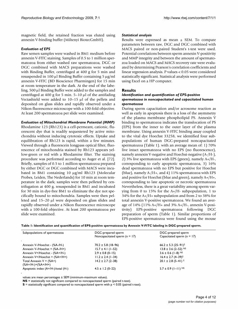
ResultsIdentification and quantification of EPS-positive spermatozoa in noncapacitated and capacitated human spermatozoaDuring sperm capacitation and/or acrosome reaction aswell as early in apoptosis there is a loss of the asymmetryof the plasma membrane phospholipid PS. Annexin Vbinding to spermatozoa indicates the translocation of PS(EPS) from the inner to the outer layer of the plasmamembrane. Using annexin V-FITC binding assay coupledto the vital dye Hoechst 33258, we identified four sub-populations of human DGC-prepared noncapacitatedspermatozoa (Table 1). with an average mean of: 1) 70%live intact spermatozoa with no EPS (no fluorescence),namely annexin V-negative and Hoechst-negative (A-/H-),2) 3% live spermatozoa with EPS (green), namely A+/H-,corresponding to early apoptotic spermatozoa, 3) 16%dead spermatozoa with no EPS but positive for Hoechst(blue), namely A-/H+, and 4) 11% spermatozoa with EPSand positive for Hoechst (blue and green), namely A+/H+,corresponding to late apoptotic or necrotic spermatozoaNevertheless, there is a great variability among sperm var-ying from 0 to 15% for the A+/H- subpopulation, 1 to34% for the A+/H+ subpopulation and from 2 to 38% fortotal annexin V-positive spermatozoa. We found an aver-age of 14% (11% A+/H+ and 3% A+/H-, annexin V-posi-tivity) EPS-positive spermatozoa following DGCpreparation of sperm (Table 1). Similar proportions ofEPS-positive spermatozoa were found using the mouse
Table 1: Identification and quantification of EPS-positive spermatozoa by Annexin V-FITC labeling in DGC-prepared sperm.
Subpopulations of spermatozoa DGC-prepared spermNoncapacitated sperm (n = 17)
DGC-prepared spermCapacitated sperm (n = 17)
Annexin V-/Hoechst - (%A-/H-) 70.2 ± 5.8 (18–96) 66.2 ± 5.3 (25–91)S
Annexin V-/Hoechst + (%A-/H+) 15.7 ± 4.1 (1–52) 13.8 ± 3.6 (2–52) NS
Annexin V+/Hoechst - (%A+/H-) 2.9 ± 0.8 (0–15) 3.6 ± 0.6 (1–8) NS
Annexin V+/Hoechst + (%A+/H+) 11.2 ± 2.4 (1–34) 16.4 ± 2.7 (4–39)S
Total Annexin V + (%A+)(%A+/H-)+(%A+/H+)
14.2 ± 2.7 (2–38) 20.1 ± 2.8 (5–41) S
Apoptotic index (A+/H-)/total (H-) 4.5 ± 1.2 (0–22) 5.7 ± 0.9 (1–11) NS
values are mean percentages ± SEM (minimum-maximum values).NS = statistically not significant compared to noncapacitated sperm (paired t-test).S = statistically significant compared to noncapacitated sperm with p < 0.05 (paired t-test).
Page 4 of 12(page number not for citation purposes)
Reproductive Biology and Endocrinology 2009, 7:1 http://www.rbej.com/content/7/1/1
monoclonal IgG anti-Phosphatidylserine, clone 1H6(Upstate, NY, USA) coupled to an anti mouse IgG-FITC(Sigma-Aldrich) (data not shown).
Our results showed that there is a moderate increase in thepercentage of total annexin V-positive (A+/H- and A+/H+)spermatozoa, following incubation in capacitating condi-tions. This increase concerns only the A+/H+ subpopula-tion since there is not a statistically significant increase inthe early apoptotic annexin V-positive and Hoechst-nega-tive (A+/H-) subpopulation of spermatozoa nor in theapoptotic index, (A+/H-)/total H-, following capacitation(Tables 1 and 2). The increase in EPS-positivity duringcapacitation involves 47% and 18% (fold increase supe-rior to 1.5 and 2, respectively) of the sperm analysed witha mean percentage increase from 14 to 20% (Table 1).
Localization of PS on human spermatozoaPS was localized on head alone, head plus midpiece, mid-piece alone, tail or on the entire spermatozoon (Figure 1and Table 2). Among the annexin V-positive spermatozoapopulation (13%), about 15% are labeled on head, 8%on midpiece and 12% on head and midpiece (Table 2).Head PS labeling can concern the apical, basal, equatorialregion or entire head, and for midpiece PS labeling, theupper, lower region or whole midpiece. PS localizationthus vary among spermatozoa involving either the headand/or midpiece region or the entire spermatozoa. Aslight significative increase in annexin V-positivity in thecategories head and head plus midpiece was observedunder capacitation conditions (Table 2).
Study design and sperm samples evaluationWe thus studied 44 sperms of patients consulting ourAMP laboratory of the Unit of Reproductive Medicine atour institution. Our study focused on the evaluation ofthe quality of spermatozoa prepared by MACS-ANMB.Sperm parameters such as concentration and motility,and the apoptotic markers, EPS and MMP, were deter-mined on raw sperm samples, and on sperm samples fol-
lowing DGC or DGC combined with MACS-ANMBpreparations (Figure 2). Two fractions were obtained fol-lowing MACS-ANMB, the non apoptotic, annexin V-nega-tive and the apoptotic, annexin V-positive (Figure 2). Rawsperm parameters are presented in Table 3. Insufficientamount and/or bad quality of some sperm samples led usto partial analysis of our parameters. We thus formed sub-groups of sperm according to the parameters analysed, i.e.EPS, MMP, progressive motility, and survival at 24 h(Tables 4 and 5). All classical sperm parameters, i.e. vol-ume, concentration, total count, motility and morphol-ogy are shown in Table 3 and were not statisticallydifferent between these subgroups. In raw sperm, EPS-positivity and MMP integrity were inversely correlated (R= -0.37).
DGC combined with MACS-ANMB was efficient in reducing the sperm population in EPS-positive spermatozoaThe mean percentage of EPS-positive spermatozoa in rawsperm samples was 22% (n = 21), this value fall to 13%following DGC preparation and to 7% after DGC com-bined with MACS-ANMB (Table 4). DGC sperm prepara-tion, which is the routinely used method of spermpreparation in AMP laboratories, allowed a reduction ofabout 40% in EPS-positive spermatozoa. This reductionin EPS-positive spermatozoa was similar to that found forthe larger sperm samples subgroup, n = 36 (Table 4). Theuse of the MACS-ANMB technique allowed a furtherreduction of about 50% of EPS-positive spermatozoa inthe non apoptotic annexin V-negative fraction. In otherwords, we saw a decrease of minus 9% EPS-positive sper-matozoa in DGC compared to raw sperms, and a furtherdecrease minus 6.4% EPS-positive spermatozoa in MACS-ANMB-DGC compared to DGC-prepared sperms (Table4). The sensitivity and specificity of the MACS-ANMBtechnique for the separation of EPS-positive spermatozoawas evaluated by labeling the unbound annexin V-nega-tive fraction and the bound annexin V-positive fractionfrom each sperm sample with annexin V-FITC and by vis-
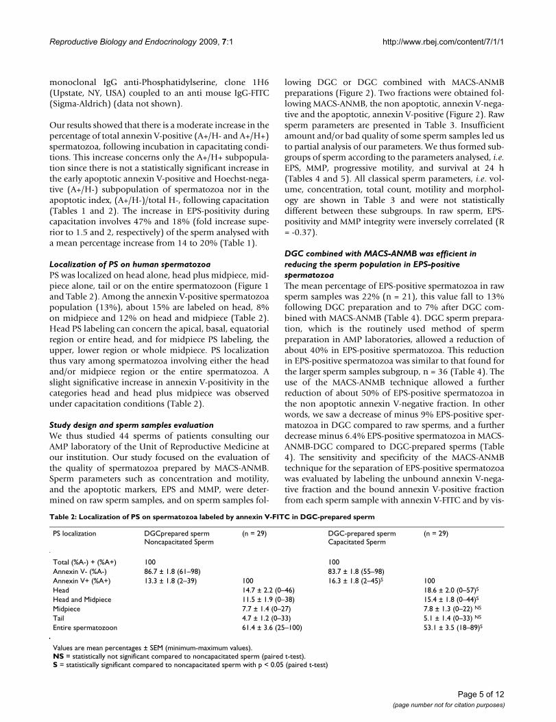
Table 2: Localization of PS on spermatozoa labeled by annexin V-FITC in DGC-prepared sperm
PS localization DGCprepared spermNoncapacitated Sperm
(n = 29) DGC-prepared spermCapacitated Sperm
(n = 29)
Total (%A-) + (%A+) 100 100Annexin V- (%A-) 86.7 ± 1.8 (61–98) 83.7 ± 1.8 (55–98)Annexin V+ (%A+) 13.3 ± 1.8 (2–39) 100 16.3 ± 1.8 (2–45)S 100Head 14.7 ± 2.2 (0–46) 18.6 ± 2.0 (0–57)S
Head and Midpiece 11.5 ± 1.9 (0–38) 15.4 ± 1.8 (0–44)S
Midpiece 7.7 ± 1.4 (0–27) 7.8 ± 1.3 (0–22) NS
Tail 4.7 ± 1.2 (0–33) 5.1 ± 1.4 (0–33) NS
Entire spermatozoon 61.4 ± 3.6 (25–100) 53.1 ± 3.5 (18–89)S
Values are mean percentages ± SEM (minimum-maximum values).NS = statistically not significant compared to noncapacitated sperm (paired t-test).S = statistically significant compared to noncapacitated sperm with p < 0.05 (paired t-test)
Page 5 of 12(page number not for citation purposes)
Reproductive Biology and Endocrinology 2009, 7:1 http://www.rbej.com/content/7/1/1
ualization under a fluorescent microscope. On average,67.8% spermatozoa were recognized as EPS-positive inthe annexin V-positive fraction and 6.7% were recognizedas EPS-positive in the annexin V-negative fraction (Table4).
DGC combined with MACS-ANMB had an effect on the quality of spermatozoa in terms of MMP integrity and progressive motilityThe mean percentage of MMP-disrupted spermatozoa inraw sperm samples (n = 15) was 58% and this value fallto 37% following DGC preparation and to 25% after DGCcombined with MACS-ANMB (Table 4). DGC sperm prep-aration allowed a reduction of about 30% in MMP-dis-rupted spermatozoa. A comparable result was obtainedfor the larger subgroup with 31 sperm samples (Table 4).The use of the MACS-ANMB technique allowed a furtherreduction of about 30% in MMP-disrupted spermatozoain the non apoptotic annexin V-negative fraction. In otherwords, we observed a decrease of minus 21% MMP-dis-rupted spermatozoa in DGC compared to raw sperms,and a further decrease of minus 12% MMP-disruptedspermatozoa in MACS-ANMB compared to DGC-pre-pared sperms (Table 4).
DGC sperm preparations greatly enriched the populationof progressive motile spermatozoa, categories 3 and 2
according to World Health Organisation [1]. In our exper-imental conditions, the addition of the MACS-ANMBtechnique to DGC preparations only slightly improvedthe sperm motility. In other words, we report an increaseof plus 16% (n = 28) and plus 20% (n = 20) spermatozoawith progressive motility in DGC compared to rawsperms, and an increase of plus 3.5% (n = 28) and plus6% (n = 20) spermatozoa with progressive motility inMACS-ANMB compared to DGC-prepared sperms) (Table4 and 5).
Positive effect of DGC combined with MACS-ANMB on sperm survivalMean progressive motility of raw sperms (n = 20) wasincreased by 40% by DGC preparation (69% compared to49%) and further increased by 10% by passage throughMACS-ANMB (76% compared to 69%) (Table 5). To fur-ther study the increased viability and motility afterremoval of EPS spermatozoa from prepared sperm, weevaluated the motility 24 h following sperm preparationby DGC or DGC combined with MACS-ANMB (Table 5).The results showed that sperm survival at 24 h expressedin mean percentage of progressive motility was higher inDGC combined with MACS-ANMB preparations than inDGC preparations alone, with approximately 48% against32% progressive motile spermatozoa, respectively. Spermsurvival at 24 h was thus increased by 50% by passagethrough MACS-ANMB, an increase considerably morepronounced than the 10% increase in progressive motilityobserved rapidly following the passage through MACS-ANMB (Table 5).
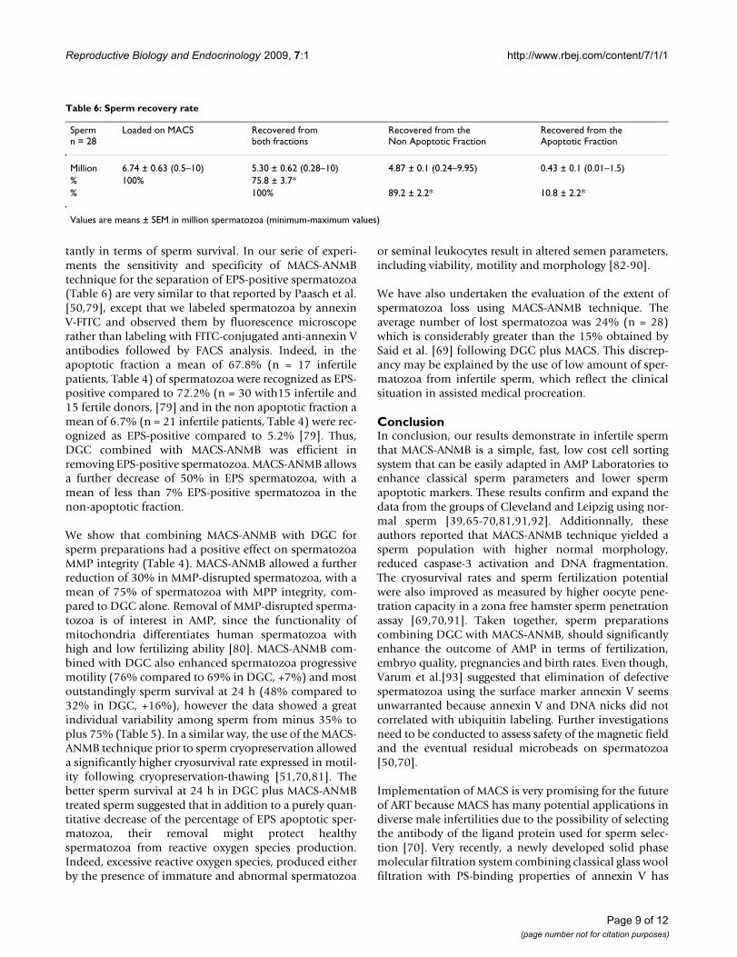
Sperm recovery rate after MACS-ANMB preparationThe MACS-ANMB technique in our experimental condi-tions, including infertile sperms of either low motilityrates, poor morphology and/or low amount of spermato-zoa (0.5 to 10 millions), allowed us to recover a mean of76% of the quantity (million) of spermatozoa loaded onMACS-ANMB column (Table 6). Nevertheless, the recov-ery rate of spermatozoa expressed in percentage of sper-matozoa charged on the MACS-ANMB column, washingand resuspension in BM1 medium, greatly vary amongsperm, from a minimum of 36% to a maximum of 100%recovery when compared with spermatozoa recovered atthe end of the preparation after passage through the col-umn. There is a significant positive correlation betweenthe amount of spermatozoa (in million) loaded on MACSand the MACS recovery rate (R = 0.48). The percentage ofspermatozoa collected in the MACS-ANMB-negative frac-tion, corresponding to the non apoptotic fraction, washigher than in the MACS-ANMB-positive fraction, corre-sponding to the apoptotic fraction, 89% and 11%, respec-tively (Table 6).
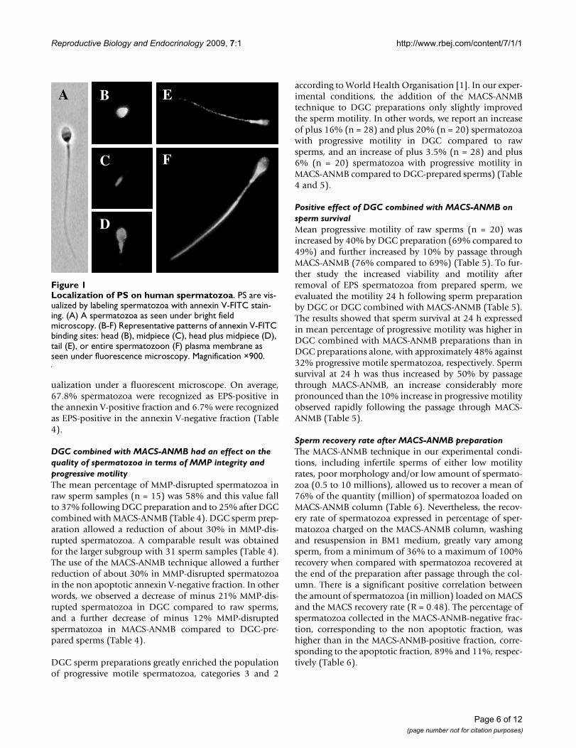
Localization of PS on human spermatozoaFigure 1Localization of PS on human spermatozoa. PS are vis-ualized by labeling spermatozoa with annexin V-FITC stain-ing. (A) A spermatozoa as seen under bright field microscopy. (B-F) Representative patterns of annexin V-FITC binding sites: head (B), midpiece (C), head plus midpiece (D), tail (E), or entire spermatozoon (F) plasma membrane as seen under fluorescence microscopy. Magnification ×900.
A
D
B
C
E
F
Page 6 of 12(page number not for citation purposes)
Reproductive Biology and Endocrinology 2009, 7:1 http://www.rbej.com/content/7/1/1
DiscussionSeveral studies have shown that ejaculated spermatozoado exhibit changes consistent with apoptosis in particularEPS, decrease in MMP integrity, DNA fragmentation andexpression of a number of pro- and anti-apoptotic pro-teins [73]. Unfortunately, DGC preparation, which is thetechnique currently used for sperm preparations in AMPlaboratories [74], does not eliminate all spermatozoa withapoptotic features. The present study was carried out toquantify EPS-positive spermatozoa and localize PS onspermatozoa in a population of infertile patients attend-ing our in vitro Fertilization laboratory and to measure theimpact of removing these EPS-positive spermatozoa byMACS-ANMB on sperm quality.
We determined in raw and DGC-prepared sperm frominfertile patients the proportion of spermatozoa withcompromised survival and fertility potential. In raw
sperm, a mean of 20% (min 5%–max 68%) are annexinV-positive (Table 4). Following DGC, about 30% of thespermatozoa are still in late apoptotic or necrotic state andabout 14% are annexin V-positive (Table 1).
The moderate increase in annexin-V-positive spermatozoaobserved following incubation in capacitating conditions(involving only the A+/H+ subpopulation, Table 1) mightbe due to the in vitro incubation. Indeed, the number oflive sperm drops quickly over time in samples frompatients with poor semen quality. We cannot rule out thepossibility that the A+/H- is a quick and transitory sub-population being converted in the A+/H+ subpopulation[49], as also suggested by Sion et al. [43]. Kotwicka et al.[12] detected an increase from 15% to 35% annexin V-positive spermatozoa following incubation in capacitat-ing medium. Martin et al. [54] showed that EPS is mainlyrelated to the acrosome reaction because calcium iono-
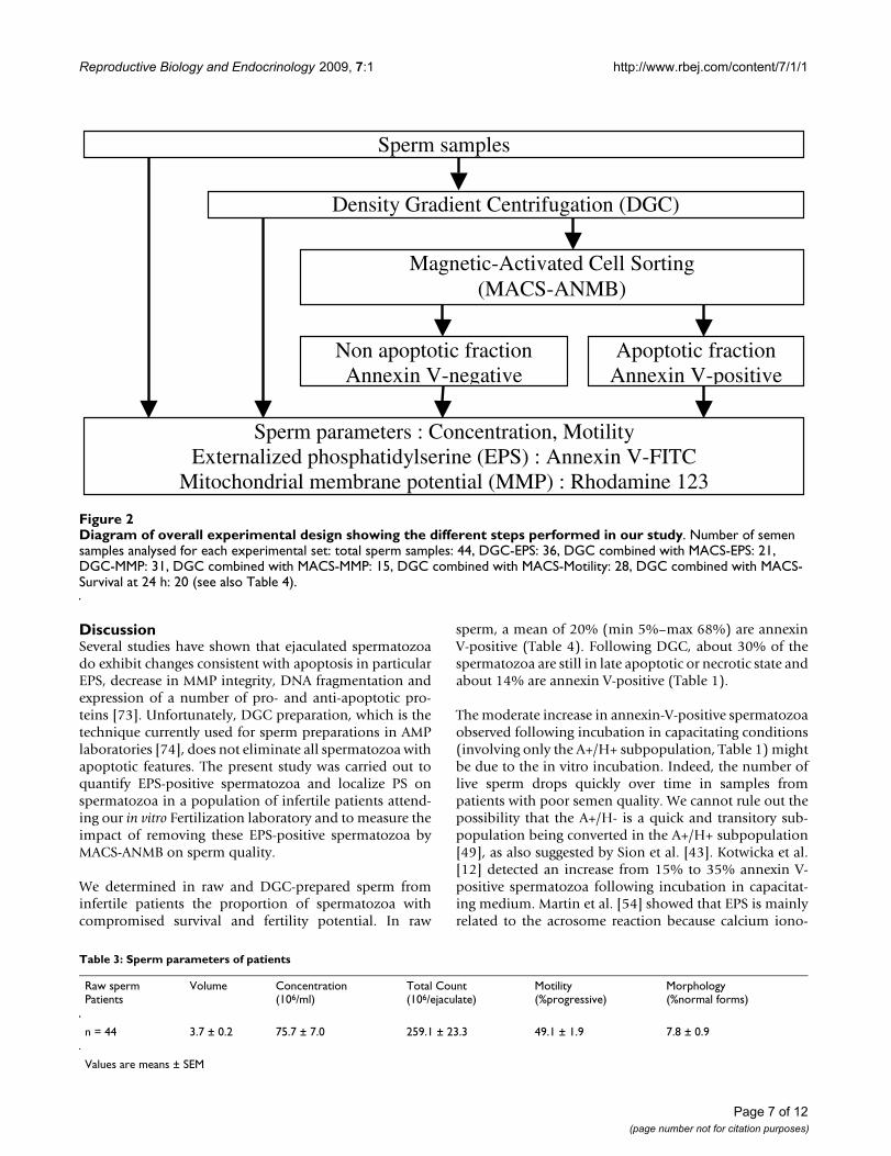
Diagram of overall experimental design showing the different steps performed in our studyFigure 2Diagram of overall experimental design showing the different steps performed in our study. Number of semen samples analysed for each experimental set: total sperm samples: 44, DGC-EPS: 36, DGC combined with MACS-EPS: 21, DGC-MMP: 31, DGC combined with MACS-MMP: 15, DGC combined with MACS-Motility: 28, DGC combined with MACS-Survival at 24 h: 20 (see also Table 4).
Density Gradient Centrifugation (DGC)
Sperm samples
Magnetic-Activated Cell Sorting (MACS-ANMB)
Non apoptotic fraction Annexin V-negative
Apoptotic fraction Annexin V-positive
Sperm parameters : Concentration, Motility Externalized phosphatidylserine (EPS) : Annexin V-FITC
Mitochondrial membrane potential (MMP) : Rhodamine 123
Table 3: Sperm parameters of patients
Raw spermPatients
Volume Concentration(106/ml)
Total Count(106/ejaculate)
Motility(%progressive)
Morphology(%normal forms)
n = 44 3.7 ± 0.2 75.7 ± 7.0 259.1 ± 23.3 49.1 ± 1.9 7.8 ± 0.9
Values are means ± SEM
Page 7 of 12(page number not for citation purposes)

Reproductive Biology and Endocrinology 2009, 7:1 http://www.rbej.com/content/7/1/1
phore A23187 induced a significant increase in the pro-portion of living EPS-positive spermatozoa from 7 to48%.
Results from the literature are still controversial. Someauthors reported that EPS was related to sperm capacita-tion process due to a bicarbonate-activated outward trans-location of PS [11,15,54]. In contrast, other reports foundcorrelations between EPS and apoptotic events such asDNA fragmentation, caspases activities and/or loss ofMMP integrity [6,7,44-47,49,50,52,53]. Taken together,EPS spermatozoa could be survivors of a testis abortiveapoptotic process or the result of oxidative stresses initi-ated during the transit or storage in the male genital tractor be linked to a physiological event in the post-ejacula-tion period. We could also speculate that spermatozoamight enter an apoptotic-like pathway, giving a physio-logical role to EPS during capacitation/acrosome reaction,for the acquisition of fertilizing ability, in a natural mech-anistic economy. Such mechanism would be dependenton a correct schedule for successful fertilization [75], orotherwise it would be converted in the apoptotic signal forelimination by phagocytic cells. Indeed, it is known thatmany boar spermatozoa are phagocytized after insemina-tion, thus reducing the risk of eliciting a harmful immunereaction that may interfere with fertilization or embryosimplantation [76]. Altogether, regardless of whether EPSrepresent, an apoptotic marker or a capacitated/acrosomereacted marker, EPS-positive spermatozoa in fresh ejacu-lates are potentially harmful in AMP being in apoptosis oroff schedule for fertilization, respectively.
Concerning the localization of PS on human spermatozoa(Figure 1 and Table 2), we found several categories ofannexin V-positive spermatozoa. Our results are similar tothose of Shen et al. [49] who also reported annexin V-labeling around head, midpiece and certain parts of thetail. Following incubation in capacitating conditions wefound an increase in both head and head plus midpiececategories. Kotwicka et al. [12] also found a change in thelocalization of PS during capacitation, firstly mainly in themidpiece and then in the acrosomal region of the head. deVries et al. [15] reported that PS in living spermatozoa wasrestricted to the apical area of the head plasma membrane,whereas deteriorated spermatozoa presented PS at themidpiece. The various PS localizations on spermatozoacould thus be interpreted as different stages of thedynamic physiological process of capacitation/acrosomereaction or apoptosis versus necrosis status.
Numerous reports linked fertilization failures to apop-totic markers [8,45,77,78]. Infertile patients have higherrates of spermatozoa with EPS, activated caspases and/orloss of MMP integrity [7,45,50]. Therefore MACS-ANMB,which eliminate EPS-positive spermatozoa, is adapted tohuman sperm in the context of seeking new techniqueswith better criteria of selection for sperm preparations inAMP [39]. Our study on sperm from infertile patientsshows that DGC combined with MACS-ANMB is: 1)applicable to low amount, 43% of sperm tested with lessthan 5 million spermatozoa, and/or bad quality sperma-tozoa, 2) efficient in removing EPS spermatozoa and, 3)efficient in enhancing the quality of spermatozoa in termof MMP integrity, progressive motility and most impor-
Table 4: EPS, MMP and progressive motility in raw sperm, in DGC and in DGC combined with MACS-ANMB prepared sperm
Criteria, subgroups Raw sperm DGC sperm DGC+MACS spermNon Apoptotic Fraction
DGC+MACS spermApoptotic Fraction
EPS (%A+), n = 36 20.0 ± 2.3 (5–68) 11.8 ± 1.4 (1–32) - -EPS (%A+), n = 21 22.1 ± 3.4 (5–68) 13.1 ± 2.0 (1–32) 6.7 ± 1.2 (1–22) 67.8 ± 4.2 (25–94)MMP disruption (%MMP-), n = 31 61.7 ± 3.2 (30–95) 44.2 ± 3.7 (16–86) - NDMMP disruption (%MMP-), n = 15 57.9 ± 4.5 (30–91) 37.0 ± 5.0 (16–86) 24.7 ± 5.6 (6–82) NDMotility, n = 28 (%progressive) 48.4 ± 2.4 (17–71) 64.4 ± 3.8 (22–91) 67.9 ± 5.4 (0–97) 8.0 ± 2.6 (0–53)
Values are means ± SEM (minimum-maximum values).All values are statistically significantly different when comparing DGC+MACS, DGC, raw samples (p < 0.05, paired t-test) except for the value of motility 67.9 compared to 64.4.
Table 5: Sperm survival at 24 h in DGC and DGC plus MACS-ANMB preparations
n = 20 Rawsperm
DGC sperm DGC+MACS sperm Non Apoptotic Fraction
DGC sperm Survival at 24 h DGC+MACS sperm Non Apoptotic Fraction Survival at 24 h
Motility (%progressive) 48.9 ± 3.0(17–71)
69.3 ± 4.2(22–91)
75.6 ± 5.2(20–97)
32.4 ± 6.1(0–78)
47.5 ± 7.0(0–88)
Values are means ± SEM (minimum-maximum values).All values are statistically significantly different (p < 0.05, paired t-test)
Page 8 of 12(page number not for citation purposes)
Reproductive Biology and Endocrinology 2009, 7:1 http://www.rbej.com/content/7/1/1
tantly in terms of sperm survival. In our serie of experi-ments the sensitivity and specificity of MACS-ANMBtechnique for the separation of EPS-positive spermatozoa(Table 6) are very similar to that reported by Paasch et al.[50,79], except that we labeled spermatozoa by annexinV-FITC and observed them by fluorescence microscoperather than labeling with FITC-conjugated anti-annexin Vantibodies followed by FACS analysis. Indeed, in theapoptotic fraction a mean of 67.8% (n = 17 infertilepatients, Table 4) of spermatozoa were recognized as EPS-positive compared to 72.2% (n = 30 with15 infertile and15 fertile donors, [79] and in the non apoptotic fraction amean of 6.7% (n = 21 infertile patients, Table 4) were rec-ognized as EPS-positive compared to 5.2% [79]. Thus,DGC combined with MACS-ANMB was efficient inremoving EPS-positive spermatozoa. MACS-ANMB allowsa further decrease of 50% in EPS spermatozoa, with amean of less than 7% EPS-positive spermatozoa in thenon-apoptotic fraction.
We show that combining MACS-ANMB with DGC forsperm preparations had a positive effect on spermatozoaMMP integrity (Table 4). MACS-ANMB allowed a furtherreduction of 30% in MMP-disrupted spermatozoa, with amean of 75% of spermatozoa with MPP integrity, com-pared to DGC alone. Removal of MMP-disrupted sperma-tozoa is of interest in AMP, since the functionality ofmitochondria differentiates human spermatozoa withhigh and low fertilizing ability [80]. MACS-ANMB com-bined with DGC also enhanced spermatozoa progressivemotility (76% compared to 69% in DGC, +7%) and mostoutstandingly sperm survival at 24 h (48% compared to32% in DGC, +16%), however the data showed a greatindividual variability among sperm from minus 35% toplus 75% (Table 5). In a similar way, the use of the MACS-ANMB technique prior to sperm cryopreservation alloweda significantly higher cryosurvival rate expressed in motil-ity following cryopreservation-thawing [51,70,81]. Thebetter sperm survival at 24 h in DGC plus MACS-ANMBtreated sperm suggested that in addition to a purely quan-titative decrease of the percentage of EPS apoptotic sper-matozoa, their removal might protect healthyspermatozoa from reactive oxygen species production.Indeed, excessive reactive oxygen species, produced eitherby the presence of immature and abnormal spermatozoa
or seminal leukocytes result in altered semen parameters,including viability, motility and morphology [82-90].
We have also undertaken the evaluation of the extent ofspermatozoa loss using MACS-ANMB technique. Theaverage number of lost spermatozoa was 24% (n = 28)which is considerably greater than the 15% obtained bySaid et al. [69] following DGC plus MACS. This discrep-ancy may be explained by the use of low amount of sper-matozoa from infertile sperm, which reflect the clinicalsituation in assisted medical procreation.
ConclusionIn conclusion, our results demonstrate in infertile spermthat MACS-ANMB is a simple, fast, low cost cell sortingsystem that can be easily adapted in AMP Laboratories toenhance classical sperm parameters and lower spermapoptotic markers. These results confirm and expand thedata from the groups of Cleveland and Leipzig using nor-mal sperm [39,65-70,81,91,92]. Additionnally, theseauthors reported that MACS-ANMB technique yielded asperm population with higher normal morphology,reduced caspase-3 activation and DNA fragmentation.The cryosurvival rates and sperm fertilization potentialwere also improved as measured by higher oocyte pene-tration capacity in a zona free hamster sperm penetrationassay [69,70,91]. Taken together, sperm preparationscombining DGC with MACS-ANMB, should significantlyenhance the outcome of AMP in terms of fertilization,embryo quality, pregnancies and birth rates. Even though,Varum et al.[93] suggested that elimination of defectivespermatozoa using the surface marker annexin V seemsunwarranted because annexin V and DNA nicks did notcorrelated with ubiquitin labeling. Further investigationsneed to be conducted to assess safety of the magnetic fieldand the eventual residual microbeads on spermatozoa[50,70].
Implementation of MACS is very promising for the futureof ART because MACS has many potential applications indiverse male infertilities due to the possibility of selectingthe antibody of the ligand protein used for sperm selec-tion [70]. Very recently, a newly developed solid phasemolecular filtration system combining classical glass woolfiltration with PS-binding properties of annexin V has
Table 6: Sperm recovery rate
Spermn = 28
Loaded on MACS Recovered fromboth fractions
Recovered from theNon Apoptotic Fraction
Recovered from theApoptotic Fraction
Million 6.74 ± 0.63 (0.5–10) 5.30 ± 0.62 (0.28–10) 4.87 ± 0.1 (0.24–9.95) 0.43 ± 0.1 (0.01–1.5)% 100% 75.8 ± 3.7*% 100% 89.2 ± 2.2* 10.8 ± 2.2*
Values are means ± SEM in million spermatozoa (minimum-maximum values)
Page 9 of 12(page number not for citation purposes)

Reproductive Biology and Endocrinology 2009, 7:1 http://www.rbej.com/content/7/1/1
been proven to enrich spermatozoa free of apoptosismarkers to the same extent as MACS but without thepotential inconvenient of an accidental transmission ofsupermagnetic microbeads into oocytes [94]. Overall, PSmight become the prognostic marker of sperm fertilitypotential easy to use for sorting spermatozoa in ART. Inconsequence, discarding EPS spermatozoa may optimizeAMP outcomes.
AbbreviationsAMP: assisted medical procreation; DGC: density gradientcentrifugation; EPS: externalization of the phosphatidyl-serine; FITC: fluorescein isothiocyanate; MACS-ANMB:magnetic-activated cell sorting using annexin V-conju-gated microbeads; MMP: mitochondrial membranepotential; PS: phosphatidylserine; and Rh123: rhodamine123.
Competing interestsThe authors declare that they have no competing interests.
Authors' contributionsCDVA did the work of acquisition of funding, conception,design, and acquisition of data, analysed and interpretedthe data, drafted the manuscript, tables and figures, andrevised the manuscript. HL did the work of acquisition offunding, contributed to conception and design, analysedand interpreted the data, and revised the manuscript. DCparticipated to the discussions, and revised the manu-script for content and language. ADA did the work ofacquisition of funding, contributed to conception anddesign, analysed and interpreted the data, and revised themanuscript. All authors read and approved the final man-uscript.
AcknowledgementsWe would like to thank Dr V. Ibecheole and the technicians I. Wagner, J. Fournier, V. Laplana and R. Luthi of the Unit of Reproductive Medicine, Geneva University Hospitals for their help, essentially in recruiting patients, and to V. Crettenand Dupertuis for her technical assistance. This work was supported by the « Fond National Suisse de la Recherche Sientifique » attributed to H. Lucas n° 3200-063739.00/1 and by the Geneva University Hospitals (PRD 05-27-II).
References1. World Health Organization (WHO): Laboratory manual for the
examination of human semen and sperm-cervical mucusinteraction. 4th edition. Cambridge: Cambridge University Press;1999.
2. Sakkas D, Mariethoz E, St John JC: Abnormal sperm parametersin humans are indicative of an abortive apoptotic mecha-nism linked to the Fas-mediated pathway. Exp Cell Res 1999,251:350-305.
3. Sakkas D, Seli E, Bizzaro D, Tarozzi N, Manicardi GC: Abnormalspermatozoa in the ejaculate: abortive apoptosis and faultynuclear remodelling during spermatogenesis. RBM Online2003, 7:428-432.
4. Oosterhuis GJ, Mulder AB, Kalsbeek-Batenburg E, Lambalk CB, Sch-oemaker J, Vermes I: Measuring apoptosis in human spermato-
zoa: a biological assay for semen quality. Fertil Steril 2000,74:245-250.
5. Kasai T, Ogawa K, Mizuno K, Nagai S, Uchida Y, Ohta S, Fujie M,Suzuki K, Hirata S, Hoshi K: Relationship between sperm mito-chondrial membrane potential, sperm motility, and fertilitypotential. Asian J Androl 2002, 4:97-103.
6. Weng SL, Taylor SL, Morshedi M, Schuffner A, Duran EH, Beebe S,Oehninger S: Caspase activity and apoptotic markers in ejacu-lated human sperm. Mol Hum Reprod 2002, 8:984-991.
7. Taylor SL, Weng SL, Fox P, Duran EH, Morshedi MS, Oehninger S,Beebe SJ: Somatic cell apoptosis markers and pathways inhuman ejaculated sperm: potential utility as indicators ofsperm quality. Mol Hum Reprod 2004, 10:285-834.
8. Seli E, Gardner DK, Schoolcraft WB, Moffatt O, Sakkas D: Extend ofnuclear DNA damage in ejaculated spermatozoa impacts onblastocyst development after in vitro fertilization. Fertil Steril2004, 82:378-383.
9. Flesch FM, Gadella BM: Dynamics of the mammalian spermplasma membrane in the process of fertilization. Biochim Bio-phys Acta 2000, 1469:197-235.
10. Flesch FM, Brouwers JF, Nievelstein PF, Verkleij AJ, van Golde LM,Colenbrander B, Gadella BM: Bicarbonate stimulated phosphol-ipid scrambling induces cholesterol redistribution and ena-bles cholesterol depletion in the sperm plasma membrane. JCell Sci 2001, 114:3543-3555.
11. Gadella BM, Harrison RA: Capacitation induces cyclic adenosine3',5'-monophosphate-dependent, but apoptosis-unrelated,exposure of aminophospholipids at the apical head plasmamembrane of boar sperm cells. Biol Reprod 2002, 67:340-350.
12. Kotwicka M, Jendraszak M, Warchol JB: Plasma membrane trans-location of phosphatidylserine in human spermatozoa. FoliaHistochem Cytobiol 2002, 40:111-112.
13. Lefièvre L, Jha KN, de Lamirande E, Visconti PE, Gagnon C: Activa-tion of protein kinase A during human sperm capacitationand acrosome reaction. J Androl 2002, 23:709-716.
14. Cross NL: Decrease in order of human sperm lipids duringcapacitation. Biol Reprod 2003, 69:529-534.
15. de Vries KJ, Wiedmer T, Sims PJ, Gadella BM: Caspase-independ-ent exposure of aminophospholipids and tyrosine phosphor-ylation in bicarbonate responsive human sperm cells. BiolReprod 2003, 68:2122-2134.
16. Salicioni AM, Platt MD, Wertheimer EV, Arcelay E, Allaire A, SosnikJ, Visconti PE: Signalling pathways involved in sperm capacita-tion. Soc Reprod Fertil Suppl 2007, 65:245-259.
17. Martin SJ, Reutelingsperger CP, McGahon AJ, Rader JA, van Schie RC,LaFace DM, Green DR: Early redistribution of plasma mem-brane phosphatidylserine is a general feature of apoptosisregardless of initiating stimulus: inhibition by overexpressionof Bcl-2 and Abl. J Exp Med 1995, 182:1545-1556.
18. Schlegel RA, Williamson P: Phosphatidylserine, a death knell.Cell Death Differ 2001, 8:545-548.
19. Fadeel B: Plasma membrane alterations during apoptosis:role in corpse clearance. Antioxid Redox Signal 2004, 6:269-275.
20. Wu Y, Tibrewal N, Birge RB: Phosphatidylserine recognition byphagocytes: a view to a kill. Trends Cell Biol 2006, 16:189-197.
21. Van Heerde WL, de Groot PG, Reutelingsperger CP: The complex-ity of the phospholipid binding protein Annexin V. ThrombHaemost 1995, 73:172-179.
22. Vermes I, Haanen C, Steffens-Nakken H, Reutelingsperger C: Anovel assay for apoptosis. Flow cytometric detection ofphosphatidylserine expression on early apoptotic cells usingfluorescein labelled Annexin V. J Immunol Methods 1995,184:39-51.
23. Rodriguez I, Ody C, Araki K, Garcia I, Vassalli P: An early and mas-sive wave of germinal cell apoptosis is required for the devel-opment of functional spermatogenesis. EMBO J 1997,16:2262-2270.
24. Tesarik J, Greco E, Cohen-Bacrie P, Mendoza C: Germ cell apop-tosis in men with complete and incomplete spermiogenesisfailure. Mol Hum Reprod 1998, 4:757-762.
25. Kawasaki Y, Nakagawa A, Nagaosa K, Shiratsuchi A, Nakanishi Y:Phosphatidylserine binding of class B scavenger receptortype I, a phagocytosis receptor of testicular sertoli cells. J BiolChem 2002, 277:27559-27566.
26. Sakkas D, Seli E, Bizzaro D, Tarozzi N, Manicardi GC: Abnormalspermatozoa in the ejaculate: abortive apoptosis and faulty
Page 10 of 12(page number not for citation purposes)
Reproductive Biology and Endocrinology 2009, 7:1 http://www.rbej.com/content/7/1/1
nuclear remodelling during spermatogenesis. RBM Online2003, 7:428-432.
27. Maeda Y, Shiratsuchi A, Namiki M, Nakanishi Y: Inhibition of spermproduction in mice by annexin V microinjected into seminif-erous tubules: possible etiology of phagocytic clearance ofapoptotic spermatogenic cells and male infertility. Cell DeathDiffer 2002, 9:742-749.
28. Oehninger S, Morshedi M, Weng SL, Taylor S, Duran H, Beebe S:Presence and significance of somatic cell apoptosis markersin human ejaculated spermatozoa. Reprod Biomed Online 2003,7:469-476.
29. Müller K, Pomorski T, Muller P, Herrmann A: Stability of transbi-layer phospholipid asymmetry in viable ram sperm cellsafter cryotreatment. J Cell Sci 1999, 112:11-20.
30. Marti E, Pérez-Pé R, Colas C, Muino-Blanco T, Cebrian-Pérez JA:Study of apoptosis-related markers in ram spermatozoa.Anim Reprod Sci 2008, 106:113-132.
31. Pena FJ, Johannisson A, Wallgren M, Rodriguez-Martinez H: Assess-ment of fresh and frozen-thawed boar semen using anannexin-V assay: a new method of evaluating sperm mem-brane integrity. Theriogenology 2003, 60:677-689.
32. Kurz A, Viertel D, Herrmann A, Müller K: Localization of phos-phatidylserine in boar sperm cell membranes during capaci-tation and acrosome reaction. Reproduction 2005, 130:615-626.
33. Anzar M, He L, Buhr MM, Kroetsch TG, Pauls KP: Sperm apoptosisin fresh and cryopreserved bull semen detected by flowcytometry and its relationship with fertility. Biol Reprod 2002,66:354-360.
34. Januskauskas A, Johannisson A, Rodriguez-Martinez H: Subtle mem-brane changes in cryopreserved bull semen in relation withsperm viability, chromatin structure, and field fertility. The-riogenology 2003, 60:743-758.
35. Martin G, Sabido O, Durand P, Levy R: Cryopreservation inducesan apoptosis-like mechanism in bull sperm. Biol Reprod 2004,71:28-37.
36. Glander HJ, Schaller J: Binding of annexin V to plasma mem-branes of human spermatozoa: a rapid assay for detection ofmembrane changes after cryostorage. Mol Hum Reprod 1999,5:109-115.
37. James PS, Wolfe CA, Mackie A, Ladha S, Prentice A, Jones R: Lipiddynamics in the plasma membrane of fresh and cryopre-served human spermatozoa. Hum Reprod 1999, 14:1827-1832.
38. Duru NK, Morshedi M, Schuffner A, Oehninger S: Cryopreserva-tion-thawing of fractionated human spermatozoa is associ-ated with membrane phosphatidylserine externalization andnot DNA fragmentation. J Androl 2001, 22:646-651.
39. Grunewald S, Paasch U, Glander HJ: Enrichment of non-apop-totic human spermatozoa after cryopreservation by immu-nomagnetic cell sorting. Cell and Tissue Bank 2001, 2:127-133.
40. Grunewald S, Baumann T, Paasch U, Glander HJ: Capacitation andacrosome reaction in nonapoptotic human spermatozoa.Ann NY Acad Sci 2006, 1090:138-146.
41. Schuffner A, Morshedi M, Oehninger S: Cryopreservation of frac-tionated, highly motile human spermatozoa: effect on mem-brane phosphatidylserine externalization and lipidperoxidation. Hum Reprod 2001, 16:2148-2153.
42. Paasch U, Sharma RK, Gupta AK, Grunewald S, Mascha EJ, ThomasAJ, Glander HJ, Agarwal A: Cryopreservation and thawing isassociated with varying extent of activation of apoptoticmachinery in subsets of ejaculated human spermatozoa. BiolReprod 2004, 71:1828-1837.
43. Sion B, Janny L, Boucher D, Grizard G: Annexin V binding toplasma predicts the quality of human cryopreserved sper-matozoa. Int J Androl 2004, 27:108-114.
44. Barroso G, Morshedi M, Oehninger S: Analysis of DNA fragmen-tation, plasma membrane translocation of phosphatidylser-ine and oxidative stress in human spermatozoa. Hum Reprod2000, 15:1338-1344.
45. Barroso G, Taylor S, Morshedi M, Manzur F, Gavino F, Oehninger S:Mitochondrial membrane potential integrity and plasmamembrane translocation of phosphatidylserine as earlyapoptotic markers: a comparison of two different spermpopulations. Fertil Steril 2006, 85:149-154.
46. Oosterhuis GJ, Mulder AB, Kalsbeek-Batenburg E, Lambalk CB, Sch-oemaker J, Vermes I: Measuring apoptosis in human spermato-
zoa: a biological assay for semen quality. Fertil Steril 2000,74:245-250.
47. Ricci G, Perticarari S, Fragonas E, Giolo E, Canova S, Pozzobon C,Guaschino S, Presani G: Apoptosis in human sperm: its correla-tion with semen quality and the presence of leukocytes. HumReprod 2002, 17:2665-2672.
48. Schuffner A, Morshedi M, Vaamonde D, Duran EH, Oehninger S:Effect of different incubation conditions on phosphatidylser-ine externalization and motion parameters of purified frac-tions on highly motile human spermatozoa. J Androl 2002,23:194-201.
49. Shen HM, Dai J, Chia SE, Lim A, Ong CN: Detection of apoptoticalterations in sperm in subfertile patients and their correla-tions with sperm quality. Hum Reprod 2002, 17:1266-1273.
50. Paasch U, Grunewald S, Fitzl G, Glander HJ: Deterioration ofplasma membrane is associated with activated caspases inhuman spermatozoa. J Androl 2003, 24:246-252.
51. Paasch U, Grunewald S, Wuendrich K, Jope T, Glander HJ: Immu-nomagnetic removal of cryo-damage human spermatozoa.Asian J of Androl 2005, 7:61-69.
52. Lachaud C, Tesarik J, Canadas ML, Mendoza C: Apoptosis andnecrosis in human ejaculated spermatozoa. Hum Reprod 2004,19:607-610.
53. Moustafa MH, Sharma RK, Thornton J, Mascha E, Abdel-Hafez MA,Thomas AJ Jr, Agarwal A: Relationship between ROS produc-tion, apoptosis and DNA denaturation in spermatozoa frompatients examined for infertility. Hum Reprod 2004, 19:129-138.
54. Martin G, Sabido O, Durand P, Levy R: Phosphatidylserine exter-nalization in human sperm induced by calcium ionophoreA23187: relationship with apoptosis, membrane scramblingand the acrosome reaction. Human Reprod 2005, 20:3459-3468.
55. Chen Z, Hauser R, Trbovich AM, Shifren JL, Dorer DJ, Godfrey-BaileyL, Singh NP: The relationship between human semen charac-teristics and sperm apoptosis: a pilot study. J Androl 2006,27:112-120.
56. Zhang HB, Chen ZJ, Ma CY, Lu SM, Wang L, Li X: Early apoptoticchanges in human spermatozoa and their relationships withconventional semen parameters and sperm DNA fragmen-tation. Asian J Androl 2008, 10:227-235.
57. Muratori M, Porazzi I, Luconi M, Marchiani S, Forti G, Baldi E: Annex-inV binding and merocyanine staining fail to detect humansperm capacitation. J Androl 2004, 25:797-810.
58. Sakkas D, Moffatt O, Manicardi GC, Mariethoz E, Tarozzi N, BizzaroD: Nature of DNA damage in ejaculated human spermato-zoa and the possible involvement of apoptosis. Biol Reprod2002, 66:1061-1067.
59. Cayli S, Sakkas D, Vigue L, Demir R, Huszar G: Cellular maturityand apoptosis in human sperm: creatine kinase, caspase-3and Bcl-XL levels in mature and diminished maturity sperm.Mol Hum Reprod 2004, 10:365-372.
60. Aitken RJ, Buckingham D, West K, Wu FC, Zikopoulos K, RichardsonDW: Differential contribution of leukocytes and spermato-zoa to the generation of reactive oxygen species in the ejac-ulates of oligozoospermic patients and fertile donors. JReprod Fertil 1992, 94:451-462.
61. Ochsendorf FR: Infections in the male genital tract and reac-tive oxygen species. Hum Reprod Update 1999, 5:399-420.
62. Lessig J, Spalteholz H, Reibetanz U, Salavei P, Fischlechner M, GlanderHJ, Arnhold J: Myeloperoxidase binds to non-vital spermato-zoa on phosphatidylserine epitopes. Apoptosis 2007,12:1803-1812.
63. Villegas J, Schulz M, Soto L, Sanchez R: Bacteria induce expressionof apoptosis in human spermatozoa. Apoptosis 2005,10:105-110.
64. Glander HJ, Schiller J, Süss R, Paasch U, Grunewald S, Arnhold J:Deterioration of spermatozoal plasma membrane is associ-ated with an increase of sperm lyso-phosphatidylcholines.Andrologia 2002, 34:360-366.
65. Said TM, Grunewald S, Paasch U, Glander HJ, Baumann T, Kriegel C,Li L, Agarwal A: Advantage of combining magnetic cell separa-tion with sperm preparation techniques. RBM Online 2005,10:740-746.
66. Said TM, Grunewald S, Paasch U, Rasch M, Agarwal A, Glander HJ:Effects of magnetic-activated cell sorting on sperm motilityand cryosurvival rates. Fertil Steril 2005, 83:1442-1446.
Page 11 of 12(page number not for citation purposes)
Reproductive Biology and Endocrinology 2009, 7:1 http://www.rbej.com/content/7/1/1
Publish with BioMed Central and every scientist can read your work free of charge
"BioMed Central will be the most significant development for disseminating the results of biomedical research in our lifetime."
Sir Paul Nurse, Cancer Research UK
Your research papers will be:
available free of charge to the entire biomedical community
peer reviewed and published immediately upon acceptance
cited in PubMed and archived on PubMed Central
yours — you keep the copyright
Submit your manuscript here:http://www.biomedcentral.com/info/publishing_adv.asp
BioMedcentral
67. Said TM, Aziz N, Sharma RK, Lewis-Jones I, Thomas AJ, Agarwal A:Novel association between sperm deformity index and oxi-dative stress-induced DNA damage in infertile malepatients. Asian J Androl 2005, 7:121-126.
68. Said TM, Agarwal A, Grunewald S, Rasch M, Glander HJ, Paasch U:Evaluation of sperm recovery following annexin V magnetic-activated cell sorting separation. RBM Online 2006, 13:336-339.
69. Said TM, Agarwal A, Grunewald S, Rasch M, Baumann T, Kriegel C, LiL, Glander HJ, Thomas AJ Jr, Paasch U: Selection of nonapoptoticspermatozoa as a new tool for enhancing assisted reproduc-tion outcomes: an in vitro model. Biol Reprod 2006, 74:530-537.
70. Said TM, Agarwal A, Zborowski M, Grunewald S, Glander HJ, PaaschU: Utility of magnetic cell separation as a molecular spermpreparation technique. J Androl 2008, 29:134-142.
71. Miltenyi S, Muller W, Weichel W, Radbruch A: High gradient mag-netic cell separation with MACS. Cytometry 1990, 11:231-238.
72. Auger J, Leonce S, Jouannet P, Ronot X: Flow cytometric sortingof living, highly motile spermatozoa based on evaluation oftheir mitochondrial activity. J Histochem Cytochem 1993,41:1247-1251.
73. Marchetti C, Marchetti P: Place des marqueurs de l'apoptosedans l'exploration de l'infertilité masculine. Gynecol Obstet Fertil2005, 33:669-677.
74. Henkel RR, Schill WB: Sperm preparation for ART. Reprod BiolEndocrinol 2003, 1:108.
75. Tesarik J: Appropriate timing of the acrosome reaction is amajor requirement for the fertilizing spermatozoon. HumReprod 1989, 4:957-961.
76. Matthijs A, Harkema W, Engel B, Woelders H: In vitro phagocyto-sis of boar spermatozoa by neutrophils from peripheralblood of sows. J Reprod Fertil 2000, 120:265-273.
77. Henkel R, Hajimohammad M, Stalf T, Hoogendijk C, Mehnert C,Menkveld R, Gips H, Schill WB, Kruger TF: Influence of deoxyri-bonucleic acid damage on fertilization and pregnancy. FertilSteril 2004, 81:965-972.
78. Host E, Lindenberg S, Smidt-Jensen S: The role of DNA strandbreaks in human spermatozoa used for IVF and ICSI. ActaObstet Gynecol Scand 2000, 79:559-563.
79. Paasch U, Grunewald S, Glander HJ: Sperm selection in assistedreproductive techniques. Soc Reprod Fertil Suppl 2007,65:515-525.
80. Gallon F, Marchetti C, Jouy N, Marchetti P: The functionality ofmitochondria differentiates human spermatozoa with highand low fertilizing capability. Fertil Steril 2006, 86:1526-1530.
81. Grunewald S, Paasch U, Said TM, Rasch M, Agarwal A, Glander HJ:Magnetic-activated cell sorting before cryopreservation pre-serves mitochondrial integrity in human spermatozoa. CellTissue Bank 2006, 7:99-104.
82. Aitken RJ, Buckingham D, West K, Wu FC, Zikopoulos K, RichardsonDW: Differential contribution of leukocytes and spermato-zoa to the generation of reactive oxygen species in the ejac-ulates of oligozoospermic patients and fertile donors. JReprod Fertil 1992, 94:451-462.
83. Sharma RK, Pasqualotto FF, Nelson DR, Thomas AJ Jr, Agarwal A:The reactive oxygen species-total antioxidant capacity scoreis a new measure of oxidative stress to predict male infertil-ity. Hum Reprod 1999, 14:2801-2807.
84. Saleh RA, Agarwal A, Kandirali E, Sharma RK, Thomas AJ, Nada EA,Evenson DP, Alvarez JG: Leukocytospermia is associated withincreased reactive oxygen species production by humanspermatozoa. Fertil Steril 2002, 78:1215-1224.
85. Wang X, Sharma RK, Sikka SC, Thomas AJ Jr, Falcone T, Agarwal A:Oxidative stress is associated with increased apoptosis lead-ing to spermatozoa DNA damage in patients with male fac-tor infertility. Fertil Steril 2003, 80:531-535.
86. Nallella KP, Sharma RK, Allamanene SSR, Agarwal A: Identificationof male factor infertility using a novel semen quality scoreand reactive oxygen species levels. Clinics 2005, 60:317-324.
87. Athayde KS, Cocuzza M, Agarwal A, Krajcir N, Lucon AM, Srougi M,Hallak J: Development of normal reference values for seminalreactive oxygen species and their correlation with leuko-cytes and semen parameters in a fertile population. J Androl2007, 28:613-620.
88. Deepinder F, Chowdary HT, Agarwal A: Role of metabolic analy-sis of biomarkers in the management of male infertility.Expert Rev Mol Diagn 2007, 7:351-358.
89. Moskovtsev SI, Willis J, White J, Mullen JB: Leukocytospermia:relationship to sperm deoxyribonucleic and integrity inpatients evaluated for male factor infertility. Fertil Steril 2007,88:737-740.
90. Agarwal A, Makler K, Sharma R: Clinical relevance of oxidativestress in male factor infertility: an update. Am J Reprod Immunol2008, 59:2-11.
91. Grunewald S, Said TM, Paasch U, Glander HJ, Agarwal A: Relation-ship between sperm apoptosis signalling and oocyte penetra-tion capacity. Int J Androl 2008, 31:325-330.
92. Aziz N, Said T, Paasch U, Agarwal A: The relationship betweenhuman sperm apoptosis, morphology and the spermdeformity index. Hum Reprod 2007, 22:1413-1419.
93. Varum S, Bento C, Sousa APM, Gomes-Santos CSS, Henriques NP,Almeida-Santos T, Teodosio C, Paiva A, Ramalho-Santos J: Charac-terization of human sperm populations using conventionalparameters, surface ubiquitination, and apoptotic markers.Fertil Steril 2007, 87:572-583.
94. Grunewald S, Miska W, Miska G, Rasch M, Reinhardt M, Glander HJ,Paasch U: Molecular glass wool filtration as a new tool forsperm preparation. Hum Reprod 2007, 22:1405-1412.
Page 12 of 12(page number not for citation purposes)