Embed Size (px)
Citation preview

Premenopause: The endocrinology of reproductive decline
Menelaos L. Batrinos
Professor Emeritus of Endocrinology, Athens University Medical School, Athens, Greece
Review
HORMONES 2013, 12(3):334-349
Address for correspondence:Menelaos L. Batrinos, 8 Evripidou Str., 14563 Politeia-Kifissia, Greece, Tel.: +30 210 6204041, e-mail: [email protected]
Received 03-10-2012, Accepted 11-02-2013
The ovaries follow a different course of aging from the other endocrine glands and organs. Instead of an inevitable progressive deterioration with advancing age, like all other structures and functions of the organism, including the male gonad, the testis, in the late thirties the ovaries undergo an abrupt diminu-tion of their functional units, the follicles, that leads to their extinction 10 to 14 years later marking the end of ovarian reproductive and endocrine function. It has been estimated by mathematical models that the ovaries would be functioning until the age of 71 years if this rapid exhaustion of the follicles did not occur.1 The phenomenon of this major biological change, dividing the life of women into a pre- and postmenopausal period, is universal among all hu-man females.
The period after the complete cessation of men-struation, characterized as menopause and which represents almost 1/3 of a woman’s lifetime and therefore a large part of the human female popula-tion, has been the subject of intense and continuous medico-social interest, therapeutic trials and research
since the second half of the last century. National and international societies, journals and congresses and specialized scientists have zealously devoted their activities to the study of menopause. In contrast, the endocrine and other functional aspects and problems during the years before the total cessation of menses have long been ignored and have only gained grow-ing attention during the last few decades (Figure 1).
While interest in the perimenopausal transition was steadily gaining momentum, this term was often used by authors with variable chronological definition of its borders, necessitating in 1996 a consensus by a WHO scientific group,2 which delineated its limits as “the period immediately before the menopause”. This definition proposed by the WHO scientific authorities was accepted for some years but it soon became obvi-ous that it did not serve a practical purpose among researchers or for the epidemiological studies.
Key words: Aging, Menstrual irregularities, Premenopause, Ovarian hormones, Ovaries, Uterus bleeding
Figure 1. Number of publications per year (total number of the decade divided by 10) obtained by entering in the PubMed the key-word premenopause.

Endocrinology of reproductive decline 335
The definition of the time when premenopause commences has been problematic. The vague term “immediately before menopause” being unsatisfac-tory, several other limits were proposed. From a clinical point of view, menstrual irregularities are the first sign of underlying ovarian dysfunction. The abnormal menses, however, occur with considerable individual chronological variability that degrades their importance as a signal of premenopause initiation. Much earlier than the clinical manifestations and more consistent are the anatomical changes of the ovaries and the endocrinological alterations concerning the hypothalamo-pituitary-ovarian axis that are respon-sible for the clinical effects. This was recognized in 2001 by the experts of the Stages of Reproductive Aging Workshop (STRAW) of the World Health Organization who, still using the term menopausal transition, defined premenopause as the time of an increase of FSH and increased variability in menstrual length.3 The experts of the workshop determined that the premenopause period can be divided into 5 age stages: 3 reproductive, that is with ovulatory cycles (early, middle and late), and 2 premenopausal (early and late). The STRAW aging criteria were strongly criticized the following year in a letter to the editor with a severe title characterization “not less, but more confusion” and the problems of staging reproduc-tive aging remained unresolved.4 Nevertheless, the STRAW aging criteria have continued to be used despite their obvious weakness of uncertain limits and overlapping between the stages of age. Later, the report of the NIH State-of-the-Science Panel held at the NIH in 2005 clarified, within parenthe-ses, that the period immediate prior to menopause is when the “biological” and clinical features of ap-proaching menopause begin,5 and recently (June 2012) the results of a Herculean effort by 10 experts and 47 collaborators from 5 countries and multiple disciplines to address the problem of staging the reproductive aging were published.6 The committee adopted, by consensus modifications of STRAW called STRAW+10 concerning the late reproductive stage, the late transition and early postmenopause stages of STRAW with the hope that the comparability of studies will improve.
The biological features of premenopause are the alterations of the hormonal circuit that regulate
the endocrine and reproductive functions of the ovarian follicles. The endocrine changes arise from the genetically programmed short duration of the human female genital organ lifetime. The anatomic changes of the ovaries during the course of premeno-pause are difficult to assess in vivo. In contrast, the ensuing hormonal characteristics at any stage of the premenopausal period are easily detectable and absolutely measurable, furnishing useful informa-tion about the process of ovarian follicular aging. Moreover, the reproductive status, which presents considerable variability among women, is more ac-curately ascertained by the hormonal profile than the chronological age. With the tendency in modern societies of postponing child-bearing, a much larger number of women than in the past, who still desire to procreate, will enter the premenopausal period with its problems as to fertility, conception and delivery. This explains the revived and intense medico-social interest in premenopause and the abundance of clinical investigations: these have been summarized in an excellent review by Prior in 19987 but have since necessitated comprehensive periodic reviewing of the data in 2002,8 2005,9 200810 and 2009.11
The purpose of the present discussion is to focus on the complex, irregular and unpredictable endo-crine events and the anomalous hormone secretion provoked by the anatomic changes of the ovaries in the period of reproductive decline at premenopause.
anatomIc changes of the ovaRIes In PRemenoPause
A limited number of histological studies of the ovaries from women who underwent ovariectomy and hysterectomy for various reasons at the ages of premenopause have all documented an accelerated rate of follicle depletion beginning in the late thirties or early forties. Βlock et al reported quantitative data on the evolution of ovarian follicle with aging.12 In 5 pubertal cases (12-16 years) the average number of primary follicles was 382,000, in 8 women aged 32-38 years it was diminished to 74,000 and then in the early forties (40-44 years) the primary follicles had plummeted to 8,300 (7 cases). Several sections of one ovary in 17 women aged 45-55 yrs showed significant changes in the number of follicles between

336 M.L. BAtriNos
the women with normal cycles (n=6, 1,392±72) and those with irregular cycles13 (n=7, 142±72). Follicles were virtually absent in the ovary of 4 postmenopausal woman. A steep depletion of the follicles at the same age period was noticed by Gougeon et al14 in a series of 43 pairs of ovaries obtained from women undergo-ing surgery for various gynecological disorders. In 10 cases aged 25-39 years, the number of small follicles in each ovary ranged from 12,900 to 50,000 (mean 28,700), as compared with 14 cases aged 40-45 years in whom they had dropped precipitously to 390-9,150 (mean 3,537). In the same age group, two cases aged 40 and 45 years retained a relatively high number of small follicles, i.e. 12,000-22,000. In 18 cases aged 46-50 years with ovulatory cycles of normal length, the number of small follicles ranged from 230 to 6,250, mean 2,309 (data displayed in Table 1). Evaluating only a small number of ovarian sections, Westhoff et al15 also showed a decrease of ovarian follicles from
31.4±22.8 in 4 cases 25-34 years old to 10±14.5 in 20 women 35-44 years old and 2.3±3.2 in 65 women 45-54 years old. There was near absence of follicles in postmenopausal women. It is to be noted, however, that primordial follicles have been demonstrated in women aged up to 57 years and the verified oldest age of spontaneous pregnancy is 57 years.
In recent years, the sonographic assessment of the number of visible (≥2mm) antral follicles was used to compensate for the lack of new histological studies of the number of ovarian follicles in old age and the need to know the degree of ovarian aging and its reproductive reserve in order to estimate an individual’s fertility. Reuss et al16 in 1996 were the first to estimate the antral follicle count (AFC) in the follicular and luteal phases in 31 volunteers be-longing to 3 age groups 22 to 25, 30 to 33 and 39 to 42 years, demonstrating a decrease of AFC by about 60% between 22 and 42 years. The authors pointed
table 1. Anatomical changes of the aging ovaries in the premenopause
Authors subjects Method Age (yrs) Number of follicles
Block12 1952 587
12-1632-3840-44
382.000 (average)74.0008.300
Richardson et al13 1987 6 Several sections of one ovary 45-55Normal cycles
1392±72
7 Irregular cycles 142±72
4 Post menopause Virtually absent
Gougeon et al14 1994 10 Each ovary 25-39 28.700 (range 12900-50000)
14 40-44 3537 (390-9250)
18 46-50 Ovulatory
2309 (230-6250)
Westhoff et al15 2000 3 Only small number of ovarian section
25-34 31,4±22,8
20 35-44 10±14,5
65 45-52 2,3±3,2
Reuss et al16 1996 31 Sonographic assessment of follicles 2-5 mm (AFC)
22-42 Decrease of 60% between 22 and 42 years
Hansen et al17 2008 31 Stereology 19-34 219938±205914
22 Sonographic 36-40 28298±24140
16 Technique 41-45 10661±15040
15 46-50 2898±3917

Endocrinology of reproductive decline 337
out the correspondence of their data with those of Block’s histological cases and suggested that AFC rates indicate reproductive age more accurately than does chronological age. Using a modern stereology technique to determine the ovarian follicles in 122 women aged from immediately after birth to the 51st year of age, Hansen et al17 arrived at the conclusion that their method predicts no sudden decay rate but rather a constantly accelerating rate, unlike the previous reported steep ovarian follicle depletion by the abovementioned publications. However, the ovaries of the 31 cases (numbers 19-49 of Table 1) aged 19-34 years and presenting a huge range in the number of follicles (9,405 to 804,036), this explaining the high standard deviations, contained a mean of 219,928±205,914 follicles, whereas the immediately next age group of 22 women aged 36-40 years (n=50 to 71 of Table 1) presented an almost tenfold reduction of the mean number of the follicles 28,298±24,140 (range 346 to 90,554). The depletion of the follicles in the following two age groups of 41-45 years (n=36) and 46-51 (n=15) was gradual, with the mean number of follicles being 10,661±15,047 and 2,898±3,917, respectively (Table 1).
the clInIcal symPtoms of PRemenoPause
There are several clinical manifestations that sig-nal the onset of the premenopausal period arousing the awareness of both woman and physician that the course to the termination of the reproductive period has begun.
The irregularities of the menstrual cycle are the most obvious and are directly related to ovarian mulfunction.
Dysfunctional uterine bleeding, sometimes taking the form of serious hemorrhage, is another common symptom of premenopause that may occur with regular menses but is more frequently associated clinically and pathophysiologically with oligomenorrhea.
Subfertility manifested by delay or lack of concep-tion due to infrequent ovulation or insufficient luteal phase in cycles of regular length are also indirect markers of ovarian insufficiency.
The high frequency of abortions and malforma-tions observed with increasing incidence in the fifth
decade of a woman’s life are also indicative of the deterioration of the genital system, including the genetic quality of the ovum.
Vasomotor symptoms, hot flushes and sweats during normal cycles are an early but inconsistent symptom of the premenopause.
a. Irregularities of the menstrual cycle
Abnormal menstrual cycle lengths comprise the most common clinical symptom that signals to the woman the changing status of ovarian function. They are also the marker of premenopause onset for the ma-jority of investigators because of their easy detection and reference. Two classical prospective observations have demonstrated the natural course of menstrual cycle intervals. Treloar studied the characteristics of the menstrual cycle of a large cohort of women prospectively and persuaded a group of their college mates to record the onset of menstruation from their twenties until their menopause.18,19 This outstanding longitudinal study offers a comprehensive overview of menstrual cycle length evolution. Four to five years after menarche, longer cycles are frequent followed by more regular cycles of 25 to 28 days in the years 25-35 of age. In the 40s, seven to three years (mean 4.5 years) before menopause the length of cycles in-creases again. Chiazze et al, studying 30,655 cycles of 23,116 women, reported similar results.20 The lesser differences in cycle length are noted in women at the ages of 25-35 years, whereas at the ages of 40-44 a slight lengthening of the cycles begins. The length of follicular phase measured from the onset of menstrua-tion up to but not including the day of LH peak was studied in 293 women.21 The mean length was 12.9 days and a significant decrease was found in women aged 40-44 years (10.4 days) compared with women aged 18-24 years (14.2 days). One theory regarding the shorter follicular phase in the early forties sug-gested that the increase of FSH secretion starting at this age period may stimulate a more rapid follicular development.
Data of a 5-year longitudinal study of 1,550 women aged 45-53 years from the Massachusetts Women’s Health Study who responded to mail questionnaires and telephone interviews demonstrated that increased menstrual irregularity was one of the two factors that best defined the inception of premenopause, the sec-

338 M.L. BAtriNos
ond being 3-11 months of amenorrhea.22 The mean age at inception of premenopause based on the same criteria was found to be 48 years in a cohort of 1,166 British women replying to annual questionnaires.23 Those with the most children entered the premeno-pause at a later age. The use of contraceptives did not affect the time of onset of premenopause. The onset of premenopause was defined when a woman reported that her menstrual cycle length had become more irregular in the preceding 12 months or if periods had stopped for between 3 and 12 months.
The results of a study of 628 women who filled out a questionnaire and recorded prospectively a menstrual calendar every two years did not cor-roborate the assumption that women with a late age at menopause have a longer premenopause period with irregular cycles than women with an earlier age at menopause.24
The pattern of menstrual cycle length was inves-tigated in an epidemiological study of 3,743 Danish women aged 15-49 years.25 The normal cycle length (5th -95th percentile) increased from 23-33 days in the 15-39 age group to 33-30 days in the 40-44 years group. At least one cycle length of less than 21 days was experienced by 18% and one cycle of more than 35 days by 29.5% of the women. Menstrual cycle variation was reported in 29.3% of all women.
A classification of the ovulatory cycles proposed in 2008 by Robertson et al26 seems to be rational and practical for the study of the endocrine and reproductive decline of the ovaries. Three patterns of ovulatory cycles were distinguished in 56 women 45 to 55 years old. Fourteen (33%) had hormonal concentrations similar to 21 women aged 21-35 years except for 20-fold lower AMH levels; 24 women (53%) had increased FSH, decreased inhibin-B but normal E2 and PG levels; and 5 women (12%) had the same hormonal characteristics as the 24 women but lower luteal phase progesterone and increased LH. The pattern of anomalous and unpredictable endocrine and reproductive function in the pre-menopause characterized by oligomenorrhea with anovulatory or ovulatory cycles, periods of amenor-rhea followed by anovulatory or ovulatory cycles of normal or elongated or short lengths with normal or deficient luteal phase starting around the 42nd year of
age and prevailing more frequently at the approach of menopause is illustrated by a sample case report presented by Brown.27
Based on the above epidemiological studies and the self-reported data, it may be estimated that menstrual irregularities begin 3 to 7 years before the permanent cessation of menses, the average being 4.5 years, and that they are more frequent as the age at menopause approaches.
b. Dysfunctional uterine bleeding
Excessive flow and duration of menses in cycles of normal intervals (menorrhagia) or irregular intervals (metrorrhagia) are frequent in premenopause, this arousing deep concern in women over the possibil-ity of developing ovarian cancer and their need to entrust to the gynecologist the task of excluding such an occurrence.
Unopposed estradiol in the case of anovulation provokes proliferation and hyperplasia of the en-dometrium that will bring about a breakthrough bleeding whenever there is a diminution of blood estradiol levels. The volume of the ensuing blood flow is usually related to the amount of circulating estra-diol and the duration of progesterone insufficiency. Increased levels or fluctuations of estradiol secretion, which are not uncommon in the premenopause and prolonged periods of amenorrhea can be followed by acute and or heavy flow that may necessitate hospitalization and blood transfusions. Secretion of small amounts of estradiol sustained for a long time can also provoke serious uterine hemorrhage. Even ovulatory cycles with a short luteal phase may have abundant menstrual bleeding because the insufficient progesterone is unable to counteract the proliferative action of estradiol on the endometrium. The major-ity of cases of prolonged uterine bleeding or sudden heavy flow occurring in 17 to 24% of women during premenopause is the result of anovulation and is produced by the aforementioned mechanism.7,28 The high statistical prevalence of this etiology, however, should by no means discourage the clinician from tak-ing the necessary measures to exclude more serious pathology (polyps submucosal leiomyomas, cancer). Moreover, the clinician who is aware of the great incidence of unavoidable anovulation in the years of premenopause and the risk of hemorrhage should

Endocrinology of reproductive decline 339
inform a patient manifesting clinical or endocrino-logical signs of anovulatory cycles of this possibility and offer his advice.
The hormonal and ovarian anatomical changes during a normal menstrual cycle as well as the ovula-tion and luteal phase function can be ascertained via hormonal assays and ultrasonography permitting the detection of any deviation from the normal at an early stage of occurrence or at any other time in premeno-pause. The impact therefore of anovulation on the endometrium resulting from a high or sustained action of estradiol that has not been controlled because of the lack or insufficient progesterone can be promptly diagnosed and etiologically treated with a replace-ment therapy with progesterone or its homologues, the progestins.29-31
The addition of progestin to estrogens in hor-mone replacement treatment after the menopause, as reported in the large longitudinal 5.2-year study of the Women’s Health Initiative, showed a relative risk of 1.26 (1-1.59) for breast cancer after the first 5 years of use. However, reinstatement for short periods of the ovulatory cycles or of sufficient luteal phases in the premenopause by progestins, or bet-ter by progesterone, repairs the abnormal hormonal status, thus enabling women receiving such treatment to mimic the status of women of the same age who have normal ovulatory cycles.
c. Subfertility in the premenopause
Both age-related decrease in fertility and the significant individual variations that characterize its occurrence are phenomena that are historically well recognized. The relatively high pregnancy rate in older women with donor oocytes indicates that the cause of fertility decline with advancing age is ovarian. Postponing motherhood in recent decades beyond the age of 35 increases the risk for conception difficulties and pregnancy outcome.32,33
After reviewing the published literature, the Re-productive and Endocrine Committee formulated in 2011 the following recommendations: Women should be informed that the risk of spontaneous pregnancy loss and chromosomal abnormalities in-creases with age. Because of the decline in fertility and the increased time of conception that occurs
after the age of 35, women >35 years of age should be referred for infertility work-up after 6 months of trying to conceive.34
The need to know the fertility capacity of women desiring to conceive in the premenopausal years has promoted a series of hormonal tests which, together with the sonographic investigation of the ovarian fol-licular pool, can provide a fairly accurate evaluation of the ovarian functional and anatomical state and the prospect of fertility.
d. Abortions and malformations
It is also well known that, with increasing age, the capacity of women to maintain an ongoing pregnancy and deliver a child is significantly and proportion-ally reduced. The risk of abortion is 40% greater in women 35-44 years old and up to 74.7% after the age of 45 years.35 The increasing rate of aneuploidy and the creation of unviable offspring and children with abnormalities by the age of 35 years and over has also been statistically documented.32
e. Vasomotor symptoms in the premenopause
Vasomotor symptoms (VMS) in the form of episodic hot flushes and sweating occurring in still menstruating women have been more directly related to menopause than to menstrual irregularities and, along with the discomfort they provoke, they have a psychological effect on women as they are taken as a pessimistic message of entering another stage of female life. Vasomotor phenomena are more com-mon in periods of amenorrhea with fluctuations of estrogen levels.36
In the past and before the availability of hormone assays, the VMS were considered by physicians a sign of impending menopause based on the appear-ance of hot flushes and sweats, sometimes extremely severe, after the final cessation of menses and im-mediately after bilateral ovariectomy. They were correctly related to hypoestrogenism, but several studies have indicated that VMS may also occur at the premenopause in women with high estrogen levels.7 A plausible explanation for this controversy is that a relative hypoestrogenism resulting from a steep reduction of estradiol from whatever levels may initiate the neuroendocrine mechanisms of VMS manifestations. The exact prevalence of hot flushes
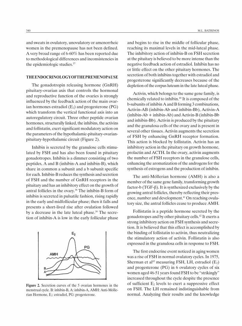
Figure 2. Secretion curves of the 5 ovarian hormones in the menstrual cycle. B: inhibin-B, A: inhibin-A, AMH: Anti-Mülle-rian Hormone, E2: estradiol, PG: progesterone.
340 M.L. BAtriNos
and sweats in ovulatory, unovulatory or amenorrheic women in the premenopause has not been defined. A very broad range of 6-60% has been reported due to methodological differences and inconsistencies in the epidemiologic studies.37
the endocRInology of the PRemenoPause
The gonadotropin releasing hormone (GnRH) pituitary-ovarian axis that controls the hormonal and reproductive function of the ovaries is strongly influenced by the feedback action of the main ovar-ian hormones estradiol (E2) and progesterone (PG) which transform the vertical functional axis into an autoregulatory circuit. Three other peptide ovarian hormones, structurally linked, the inhibins, the activins and follistatin, exert significant modulatory action on the parameters of the hypothalamic-pituitary-ovarian-pituitary-hypothalamic circuit (Figure 2).
Inhibin is secreted by the granulose cells stimu-lated by FSH and has also been found in pituitary gonadotropes. Inhibin is a dimmer consisting of two peptides, A and B (inhibin-A and inhibin-B), which share in common a subunit and a b subunit specific for each. Inhibin-B reduces the synthesis and secretion of FSH and the number of GnRH receptors in the pituitary and has an inhibitory effect on the growth of antral follicles in the ovary.38 The inhibin-B form of inhibin is secreted in pulsatile fashion, rising rapidly in the early and midfollicular phase; then it falls and presents a short-lived rise after ovulation followed by a decrease in the late luteal phase.39 The secre-tion of inhibin-A is low in the early follicular phase
and begins to rise in the middle of follicular phase, reaching its maximal levels in the mid-luteal phase. The inhibitory action of inhibin-B on FSH secretion at the pituitary is believed to be more intense than the negative feedback action of estradiol. Inhibin has no or little effect on the other pituitary hormones. The secretion of both inhibins together with estradiol and progesterone significantly decreases because of the depletion of the corpus luteum in the late luteal phase.
Activin, which belongs to the same gene family, is chemically related to inhibin.40 It is composed of the b-subunits of inhibin A and B forming 3 combinations, Activin-AB (inhibin-Ab and inhibin-Bb), Activin-A (inhibin-Ab + inhibin-Ab) and Activin-B (inhibin-Bb and inhibin-Bb). Activin is produced by the pituitary and the granulosa cells of the ovary and is present in several other tissues. Activin augments the secretion of FSH by enhancing GnRH receptor formation. This action is blocked by follistatin. Activin has an inhibitory action in the pituitary on growth hormone, prolactin and ACTH. In the ovary, activin augments the number of FSH receptors in the granulose cells, enhancing the aromatization of the androgens for the synthesis of estrogens and the production of inhibin.
The anti-Müllerian hormone (AMH) is also a member of the same gene family, transforming growth factor-b (TGF-β). It is synthesized exclusively by the growing antral follicles, thereby reflecting their pres-ence, number and development.41 On reaching ovula-tory size, the antral follicles cease to produce AMH.
Follistatin is a peptide hormone secreted by the gonadotropes and by other pituitary cells.42 It exerts a strong inhibitory action on FSH synthesis and secre-tion. It is believed that this effect is accomplished by the binding of follistatin to activin, thus neutralizing the stimulatory action of activin. Follistatin is also expressed in the granulosa cells in response to FSH.
The first endocrine event noticed in aging women was a rise of FSH in normal ovulatory cycles. In 1975, Sherman et al43 measuring FSH, LH, estradiol (E2) and progesterone (PG) in 6 ovulatory cycles of six women aged 46-51 years found FSH to be “strikingly” increased throughout the cycle despite the presence of sufficient E2 levels to exert a suppressive effect on FSH. The LH remained indistinguishable from normal. Analyzing their results and the knowledge

Endocrinology of reproductive decline 341
on the interrelationships between FSH, LH and E2, the authors arrived at a hypothesis that is worth quoting: “These data have led us to hypothesize an ovarian regulatory hormone, an inhibin, which would exert a negative feedback control over FSH secre-tion and which would be reduced in the years before menopause.” Inhibin was isolated a few years later and proved to be an important factor in the ovarian-pituitary interplay.
At the same period another hormonal paradox was recognized, namely, the increased secretion of estrogen in certain cycles of many women at the pre-menopause at an age when diminution of estrogen is normally expected because of the aging ovaries and their course to the terminal phase of their function. Prior7 analysis of the data of 12 different publications constructed the mean of follicular phase estradiol from 415 premenopausal women and compared it with the mean E2 levels in 292 controls. Estradiol in the premenopausal women was significantly higher (p=0.041) than the controls (225±98 pmol/L vs 175±57 pmol/L).
In 1988, Lee et al44 studied a larger cohort of ovulatory women aged 24-50 years with cycles of normal length separated into 3 age groups 36-40 (n=19), 41-45 (n=18) and 46-50 (n=16) with daily estimations throughout the menstrual cycle of FSH, LH, estradiol (E2) and progesterone (PG). They found elevated values of FSH beginning at the age of 39 years and becoming prominent in the mid-follicular and post-ovulatory phases at a time when in the normal menstrual time inhibin concentrations appear to be minimal. LH, E2 and PG changed little with advancing age. The role of inhibin A and B in the suppression of FSH during the cycles was inves-tigated by Lahlou et al45 by measuring FSH, inhibin A and B and E2 during a control cycle and 4 days of the following cycle and in a cycle treated with percu-taneous 0.1mg E2 from day 10 of luteal phase to day 4 of the next cycle. The results showed that E2 and not inhibin-A was responsible for FSH suppression in the luteal phase, and inhibin-B and not E2 for FSH suppression in the follicular phase. Reame et al46 studied the pulsatile pattern of LH in aging women via a well designed paradigm of frequent sampling of LH (every 10 minutes for 8 hrs (09.00-17.00)) and hourly of FSH, E2 and PG at 3 time points of the
menstrual cycle (day 6 of the follicular phase and days 7 and 12 after LH surge) in 3 ovulatory age groups (19-34 yrs, n=8, 35-39, n=8 and 40-50 n=16). They demonstrated significantly higher mean LH values and pulse amplitude at the late luteal phase in the age group of 40-50 years and a tendency to the same LH effect in the age group 35-39 years. FSH was higher in all three study days in the older group, while E2 and PG showed no significant difference among the groups except for a trend of E2 to be higher in the follicular phase of the older group.
The monotropic increase of FSH in women with menstrual irregularities in the perimenopausal years was determined in a large population-based sample of 380 women aged 45.6-56.9 years (mean 49.4 yrs). Women who experienced changes in frequency of the cycles and flow (28%) had 53% and those with at least 3 months of amenorrhea (13%) 253% higher FSH than the women with continuing regular cycles (27%). The decrease of E2 and inhibin in the group with at least 3 months of amenorrhea was 54% and 53%, respectively.47 The hormone levels of the cycles in 150 women who experienced menopause during 6 years of follow-up were studied by the same Australian group.48 Mean FSH and E2 levels measured annually started to decrease from about 2 years before the final menstrual period (FMP). Levels of both inhibin A and B were undetectable in the majority of the cases by the time of menopause and in almost all women 4 years after FMP.
Six months’ daily voided urine was assayed for FSH, LH, estrone (E1) and pregnandiol (PGd) in 11 midreproductive women aged 19-38 years, 5 older reproductive (43-47 years old) and 6 cycling women 47 years and older.49 FSH excretion was higher in the women aged ≥47 years than in the two reproductive groups and E1 was also significantly higher in the older group (Figure 3). Urine samples collected daily for a period of 6-18 months by 34 women 40-53 years old who had at least one long cycle (mean length 38.4 days, range 167) were assayed for FSH, estrone and pregnandiol. The elongated cycles were characterized by increased periods of progressive FSH increase before estrogen take-off and progres-sive luteal pregnandiol decline. The authors believe that there is a temporary lack of ovarian response to FSH in old age.50
Figure 3. Schematic presentation of a graph from Santoro’s publication49 that illustrates the irregular and unpredictable endocrine events in the premenopause. It demonstrates the urinary values of FSH, LH, estrone (E1) and pregnandiol (PGd) measured by a women daily for 180 days. Increased FSH with several high values is observed in days 0 to 40 followed by low or normal FSH (days 40-50). At the same period two ovulations, the second with lower PGd excretion occurred (day 40-50 lower panel) followed by a series of high FSH values reaching postmenopausal levels (days 50-70). Then a sustained for 40 days increase in E1 appears, probably due to follicular cyst formulation by the intense FSH stimulation (days 80-120). A small increase in PGd follows (days 120-130), paradoxi-cally without an obvious LH surge, and then the course to the permanent cessation of menses is shown by the gradual increase of FSH and the E1 decrease.
342 M.L. BAtriNos
The findings of all the above studies were sup-ported by population-based data from 3,388 women aged 35-60 years of NHANES III, 1988-1994, that demonstrated an increase of FSH and LH in aging women beginning in their late forties.51
Two more elaborate studies have provided a clearer picture of the endocrine and reproductive status in premenopause. A detailed study of the longitudinal characteristics of menstrual cycles was performed in 2004 in 13 women between 4 and 9 years before menopause and 1 year after. FSH, LH, E2, PG, prolactine and inhibins A and B were de-termined annually 3 times weekly for 1 month. This study, reporting a panorama of individual cycles together with the levels of the 7 hormones, clarified the hormonal characteristics of women approaching menopause.52 Prolonged cycles were not identified in any woman earlier than 27 cycles (2 years, 3 months) from menopause. They appeared as menopause
approached, reaching 62% in the last 10 cycles and showed elevated FSH and LH and increased FSH/inhibin A and B ratios. The FSH in cycles remaining ovulatory did not consistently increase, contrary to previous studies indicating a progressive increase in follicular phase FSH. According to the authors, the determinant of elevated FSH is an anovulatory cycle or delayed ovulation. The integrated endocrinology of the menstrual cycle in 76 women classified according to the STRAW staging criteria by measuring all the involved hormones FSH, LH, E2, PG, inhibin A and B and AMH was reported in 2007 by the Australian school.53 Twenty-one women (aged 28.9±4.4 yrs) were at the mid-reproductive stage (MR), 16 women at the late reproductive (LR) stage (46.6±1.4 yrs), 16 women at the early menopause (EMT) transition stage (49.9±2.3 yrs) and 23 at the late menopause (LMT) transition (49.7±2.3 yrs). Two, zero, 1 and 9 anovulatory cycles were identified in the MR, LR, EMT and LMT, respectively. FSH, LH and E2 in-

Figure 4. Schematic presentation of 3 typical irregular men-strual cycles occurring in the premenopause. Upper graph, in-creased FSH value in menstrual cycles with normal estradiol secretion. Middle graph, increased estradiol (E2) secretion in several menstrual cycles throughout the period of premeno-pause. Lower graph, cycles with a luteal out-of-phase (LOOP) increase and fall of estradiol superimposed on progesterone se-cretion occurring in many cycles in the premenopause (normal cycles-----).
Endocrinology of reproductive decline 343
creased with progression of STRAW stage and PG decreased. Inhibin-B decreased steadily and AMH decreased markedly (10-15 fold).
Another pattern of erratic hormonal secretion of the menstrual cycles in premenopause was dem-onstrated in a study of cycles with increased or de-creased lengths in 77 women aged 21 to 55 years classified according to the STRAW aging stages as mid- (n=21) and late-reproductive stage (n=16), early (n=17) and late (n=23) menopausal transi-tion.54 Blood samples taken 3 times per week over 1 and 1/3 menstrual cycles were assayed for FSH, LH, E2, PG and inhibin A and B. In 37% (11 of 29) of the cycles of menstrual transition, a second rise and fall of E2 during the mid- and late-luteal phase, resembling the follicular phase and superimposed on the luteal phase, as well as decreased progesterone secretion were observed. This luteal out-of-phase (LOOP) increase of E2 appeared to be triggered by prolonged high follicular phase FSH levels and lower inhibin early in the cycles (Figure 4).
hoRmonal IndIcatoRs of ovaRIan ReseRve
The increase of FSH in women in the late thirties or early forties was considered a precocious sign of ovarian aging independent of the chronological age and was soon used in clinical practice as an indicator of the ovarian capacity to respond to the treatment of infertility by in vitro fertilization. Several studies demonstrated that the estimation of FSH on day 3 of the cycle was associated with the chances of preg-nancy and was used as a guide to IVF treatment. The predictive value of day 3 FSH was evaluated in 738 consecutive cycles in 444 women.55 Patients with less FSH than 15mU/ml had higher pregnancy rates and 17% ongoing pregnancies, those with FSH levels 15-24.9 mU/ml had 9.3% ongoing pregnancies and those with greater than 25mU/ml only 3.6%. Pregnancy rate in women with 3-day FSH less than 15IU/L was found to be 25% and with increasing levels of FSH the possibility of gestation and ongoing pregnancies are greatly reduced, reaching almost zero, with a FSH of 40 IU/L. The pregnancy rate in 1,750 women with an arbitrary threshold of day 3 FSH less than 20 IU/L was found by Martin et al to be 16.5% per
cycle.56 The authors express extreme pessimism as to further treatment in women with even one day-3 FSH more than 20 IU/L. A cut-off point of 13 IU/L FSH was reported by the 1999-2000 NHANES study between women with regular cycles and those with irregularities of the cycle in a cohort of 576 women aged 35-60 years.57
The predictive performance of day 3 FSH concen-trations reported in the above and other studies was not considered sufficient in clinical practice because of the great individual variations and the overlapping
344 M.L. BAtriNos
between the arbitrarily set cut-off points of FSH levels. This is the negative conclusion reached by Bancsi et al58 in 2003 in a meta-analysis of 21 relevant publications. Despite its limitations, the 3-day FSH has been widely used in IVF programs because of its practicability. However, the need for better identifica-tion of the chance of responding to an IVF treatment remains a priority of great importance both to the physician and the patient as it may help arrive at a more individualized and thus more correct decision. Three other tests, each with keen advocates and sup-porting evidence, have been proposed to meet this challenge, the antral follicle count (AFC), the day 3 inhibin B estimation and AMH measurement at the follicular phase.
The value of antral follicle count (AFC) as a predictor of pregnancy in IVF was demonstrated in 130 couples divided into 3 groups according to the AFC: ≥11, 4-10 and £ 3. The AFC correlated signifi-cantly with day 3 FSH and the number of oocytes retrieved.59 The group with the lower AFC (£3) had the higher rate of cycle cancelations compared with the two other groups. To compensate for the lack of systematic assessment of the predictive capacity of AFC for the outcome of IVF, Hendricks et al60 per-formed a meta-analysis of the 11 studies published in the first Reuss report of AFC, from 1996 through 2003, and compared their findings with the results of a meta-analysis concerning the predictive value of poor ovarian response assessed by basal FSH levels. The authors concluded that the predictive performance of AFC is significantly better than that of basal FSH. The study of 120 women undergoing their first IVF cycle arrived at the same conclusions. Measurement of the number of antra follicles (AFC) proved to be the best single predictor for poor ovarian response compared to day 3 FSH and inhibin-B estimation. However, addition of FSH and inhibin-B levels sig-nificantly improved the prediction of poor response.61
Inhibin was shown to decrease with advancing age earlier than FSH and to have a stronger inhibi-tory action on FSH than estradiol. Its decrease at an early age is believed to be the cause of subsequent FSH increase since concomitant E2 levels are not substantially reduced. Follicular phase inhibin-B was found to be inversely associated with FSH and this introduced the use of inhibin-B as a direct in-
dicator of ovarian aging and responsiveness to IVF treatment by Danforth et al62 in 1998. Day 3 of the inhibin-B cycle in 25 women aged 39-52 years proved to be more sensitive than FSH indicator of ovarian reserve. Luteal phase inhibin-A was also connected inversely with age.
The effect of increasing age in women with regular cycles on inhibin, FSH, LH, estradiol (E2) and pro-gesterone (PG) measured on days 4-6 of the follicular phase and on days 3-12 before the next menses was determined in 4 age groups of 20-29, 30-39, 40-44 and 45-49 years of age.63 Women 45-49 years old had significantly lower inhibin-B and higher FSH levels than the other 3 groups (Inhibin-B 128U/L Vs 239, 235 and 209 U/L and FSH 13 IU/L vs 4.9, 5.5 and 5.3 IU/L) in the 20-29, 30-39 and the 40-45 yrs groups, respectively. LH did not differ across age groups. E2 was not different between the 45-49 year age group and the 40-45 year age group, indicating that FSH increase was independent of E2 levels and related to inhibin decrease. The changing point of FSH increase was estimated to be at 42.97 years. Progesterone falls slowly with increasing age.
The exact role of inhibins and activin-A in the hormonal dynamics of the menstrual cycle in old age was investigated by Klein et al64 in 16 women aged 40-45 years compared with 13 younger women aged 20-25 years. Daily assays of FSH, LH, E2, inhibin A and B and activin-A were performed throughout one menstrual cycle and the follicular phase of the subsequent cycle. This detailed study demonstrated significant FSH elevation in old subjects, a decrease of inhibin-B and no deficiencies in inhibin-A, activin-A or E2.
Over the last decade, the era of a third test for ovarian anatomic and functional reserve that of AMH, was initiated based on sound evidence.65 AMH is solely produced by granulosa cells of the small grow-ing antral follicles and its expression ceases when these cells reach the dominant size. Serum AMH levels therefore correlate with the number of antral follicles and have emerged as a good predictor of ovarian responsiveness and chances of pregnancy. Van Rooij et al,66 from the department of Reproduc-tive Medicine at Utrecht, demonstrated in 2002 that this particularity of AMH could be used as a measure

Endocrinology of reproductive decline 345
of ovarian reserve and a marker of ovarian aging. In a prospective study in 119 patients undergoing IVF treatment, the number of the antral follicles and the number of oocytes retrieved were highly correlated with serum AMH levels. The superiority of AMH over the other markers of ovarian aging was demonstrated by another prospective study of 75 women.67 Day 3 AMH levels were more strongly correlated with the follicular count than inhibin-B, FSH and E2. The correlation with LH was not significant. The mean and range of day 3 AMH in 75 women was 1.39ng/mL (0.24-6.40). In 2005, Van Rooij et al68 reaffirmed their previous findings with a longitudinal study. Eighty-one women were examined twice with on average a 4-year interval and AFC, FSH, inhibin, E2 and AMH were assessed on both occasions. The AMH and AFC were highly correlated with age at both time periods, whereas FSH and inhibin-B changed mainly in women more than 40 years old. AMH showed the best consistency over time at an individual level, AFC showing the second best. FSH and inhibin-B showed only modest consistency and E2 none. The results of this study represent the typical average hormonal changes occurring at premenopause. In 56 women aged < 38 years with normal day-3 FSH, a study of E2, inhibin-B and AMH and the estimation of AFC before and after administration of 300 IU of recom-binant FSH demonstrated that the best models for predicting oocyte numbers were AFC, basal AMH and stimulated inhibin-B.69
In 2008, a longitudinal study was performed of 50 premenopausal women who made 6 consecutive annual visits and measurement of AMH, inhibin-B and FSH on days 2-7 of the follicular phase prior to their subsequent documented FMP.70 The hormones at the initial evaluation were as follows: mean AMH 0.62±0.51ng/ml, mean inhibin-B 69.8±45.2 pg/ml, mean FSH 7.6±2.4 mIU/ml, E2 58.9±24.5 and the mean age of the women was 42±2.7 years at the start and 50.5 years at FMP. AMH showed a linear decline to a time point 5 years prior to the FMP; inhibin-B declined, being undetectable 5 years before FMP at a time when FSH had doubled (15mIU/ml). The authors consider AMH a good endocrine marker of follicle depletion.
The value of AMH as a test compared with antral follicle count (AFC) performance was reviewed in
a meta-analysis of 13 studies on AMH and 17 on AFC, with the main outcome being poor response (less than 4-6 oocytes) and non-pregnancy after IVF. It was shown that AMH has at least the same level of accuracy and clinical value for the prediction of poor response and non-pregnancy as AFC after IVF stimulation.71 The authors point out that the application of the AMH test may have some major advantages as it does not need to be performed on a specific day of the cycle and does not necessitate skilled ultrasound operators to measure the ovarian follicles. The routine use of AMH as a test of ovarian reserve prediction by IVF clinics is increasing.72-74 In a comprehensive review of all the evidence-based literature on ovarian reserve tests, Broekmans et al75 concluded that tests like AFC, AMH and stimulated inhibin-B may have some predictive ability but the accuracy of predicting the chance of pregnancy is limited (Table 2).
the endocRIne envIRonment In PRemenoPause
Concurrently with the decline in reproductive function of the ovaries, important alterations in the function of the hypothalamic-dependent endo-crine glands take place.76 The secretion of growth hormone, a strong anabolic hormone essential in women who have small quantities of the stronger anabolic hormone, testosterone, begins to decline in the 3rd decade and continues in the years of pre-menopause, reaching levels 50% lower at menopause than those at a younger age. The adrenal androgens, dehydroepiandrosteronbe and Δ4-androstendione, which are the source by transformation of the 50% of circulating testosterone in women, present a steeper diminution in the same age period, whereas cortisol secretion remains unchanged, reviving the old theory of an independent of ACTH regulation of adrenal androgens. These changes, designated somatopause and adrenopause, together with menopause create a state of relative multiple endocrine insufficiency in women reaching the fifth decade of their life.
conclusIons
In women, the reproductive decline and the ac-companying endocrine deterioration begin in the late

table 2. Comparative studies of ovarian reserve hormone indicators at premenopause
Authors subjects Age Group Design results
Danforthet al.62 1998
25 39-52 Follicular day-3 and days 6 and 8 after LH surge. Measurements of FSH, inhibin A and B estradiol (E2) and progesterone (PG)
Day-3 inhibin-B more sensitive than FSH indicator of ovarian reserve. Luteal phase inhibin-A correlated inversely with age
Lahlouet al.45 1994
6 28-38 Measurement of FSH, E2 and inhibin A and B during a control cycle and 4 days of the following cycle compared to a cycle treated with 0.1mg E2 from day 10 of luteal phase to day 4 of the next cycle
The results showed that E2 and not inhibin-A is responsible for FSH in the luteal phase and inhibin-B and not E2 for the suppression of FSH in the follicular phase
Bancsiet al.58 2002
120 Women undergoing
IVF
Measurement of day-3 FSH, inhibin-B E2 and antral follicle count (AFC)
AFC the best single predictor. Addition of FSH and inhibin-B improves prediction
Fanchinet al. 67 2003
75 Measurement on day-3 of FSH, LH, E2, inhibin-B AMH and AFC
Day-3 AMH levels more strongly correlated with AFC than FSH, E2, LH, inhibin-B
Hendrikset al. 60 2005
Meta-analysis of 11 studies on AFC compared with a meta-analysis on basal FSH
Antral follicular count (AFC) performance significantly better than that of basal FSH
Vam Roojet al. 68 2005
81 Basal meanage 39,6 yrsAfter 4 yrs
43.6 yrs
Twice assessment with on average a 4-year interval of AMH, FSH, inhibin-B, E2 and AFC
High correlation of AMH with AFC. Best-consistency of AMH over time with AFC the second best
Broekmanset al. 75 2006
Literature review of predicting value of the ovarian reserve tests
Statistical analysis showed that the best tests are AFC, follicular phase AMH and stimulated inhibin-B
Elder-Gevaet al. 69 2012
56 <38 yrs Measurement of E2, AMH and inhibin-B and evaluation of AFC before and after administration of 300 IU FSH
Best predictive value have AFC, basal AMH and stimulated activin-B
346 M.L. BAtriNos
thirties or early forties, with an abrupt depletion of the ovarian functional units, the follicles, entailing a unique period of transition until the final cessation of the menses.
The declining number of follicles, which are the source of ovarian hormones that exert a modulatory feed-back action on the hypothalamus and the pitu-itary, results in hormonal irregularities that disorganize the hypothalamic-pituitary-ovarian axis, creating an erratic and unpredictable endocrine status followed by clinical abnormalities.
The ovarian hormonal secretion subsequent to the anatomic changes of the ovaries is characterized by an increase of FSH throughout the menstrual cycle, early decrease of inhibin-B and later of inhibin-A, great decrease of anti-Müllerian hormone (AMH)
which at the approach of menopause may become undetectable, lack of increase or low levels of proges-terone and, latest of all, decrease of estradiol which may present intermediate unpredictable phases of increased secretion in normal cycles or during amen-orrheic periods.
This pattern of hormonal secretion in the premeno-pause forms the foundation of clinical manifestations. Menstrual irregularities are common, unpredictable and highly individually variable. Ovulatory cycles of normal length or prolonged duration may be followed by or alternate with anovulatory cycles or cycles with insufficient luteal phases or periods of amenorrhea. The lack of ovulation, the insufficient luteal phases and the periods of amenorrhea frequently create conditions of unopposed proliferative estrogen ac-tion on the endometrium, while the risk of abnormal

Endocrinology of reproductive decline 347
bleeding, which may sometimes be heavy, can neces-sitate urgent medical care and treatment.
The parallel aging of the oocytes is responsible for the decline in fertility and the difficulty of maintaining a pregnancy in the premenopause. The subfertility of these years, which decreases further with the ap-proach of menopause, concerns a large part of the female population in the civilized world because of the postponement of pregnancy due to the years devoted to education and the professional career. These difficulties encountered in late child-bearing have contributed to the gigantic growth and advance-ment of assisted reproductive technologies (ART) and the parallel investigations into hormonal tests for the assessment of ovarian aging and the chance of pregnancy, now offer invaluable help to women desiring pregnancy in the premenopause.
Continuous and intense investigations and research into all the aspects of premenopause are needed with the aim of improving the health and the psychological well-being of the women who go through this difficult period of their life and arrive at menopause, in effect a state of relative multiple endocrine insufficiency, having lost 80-90% of estradiol, 70% of adrenal an-drogens and 50% of growth hormone.
RefeRences1. Faddy MJ, godsen rg, gougeon A, richardson sJ,
Nelson JF, 1992 Accelerated disappearance of ovarian follicles in mid-life: implications for forecasting meno-pause. Hum reprod 7: 1342-1346.
2. WHo scientific group 1996 research on the Menopause in the 1990’s. a report of the WHo scientific group. World Health organization, geneva, switzerland; pp, 866: 1-79.
3. soules Mr, sherman s, Parrott E, et al, 2001 Execu-tive summary: stages of reproductive Aging Workshop (strAW). Climacteric 4: 267-272.
4. Den tokelaar i, de Boer EJ, Broekmans FJ, te Velde Er, 2002 Executive summary: stages of reproductive Aging Workshop (strAW): Not less, but more confu-sion. Climacteric 5: 399-401.
5. National institutes of Health, 2005 National institutes of health state-of-the-science conference statement: Management of menopause-related symptoms. Ann intern Med 142: 1003-1013.
6. Harlow sD, gass M, Hall JE, et al, 2012 Executive summary of the stages of reproductive Aging Work-shop + 10: addressing the unfinished agenda of staging reproductive aging. Fertile steril 97: 843-951.
7. Prior JC, 1998 Perimenopause: the complex endocri-nology of the menopause transition. Endocrine reviews 19: 397-428.
8. Burger Hg, Dudley EC, robertson DM, Dennerstein L, 2002 Hormonal changes in the menopause transition. recent Prog Horm res 57: 257-275.
9. Prior JC, 2005 ovarian aging and the perimenopausal transition. Endocrine 26:297-300.
10. Burger H 2008 the menopausal transition – endocrinol-ogy. J sex Med 5: 2266-2273.
11. Broekmans FJ, soules Mr, Fauser BC, 2009 ovarian aging: Mechanisms and clinical consequences. Endocrine reviews 30: 465-493.
12. Block E, 1952 Quantitative morphological investigation of the follicular system. Variations in different age. Acta Anat 14: 108-123.
13. richardson sJ, senikas V, Nelson F, 1987 Follicular depletion during the menopausal transition: Evidence for accelerated loss and ultimate exhaustion. J Clin Endocrinol Metab 65: 1231-1237.
14. gougeon A, Ecochard r, thalabard JC, 1994 Age-related changes of the population of human ovarian follicles: increase in the disappearance rate of non-growing and early-growing follicles in aging women. Biol reprod 50: 653-663.
15. Westhoff C, Murphy P, Heller D, 2000 Predictors of ovarian follicle number. Fertil steril 74: 624-628.
16. reuss ML, kline J, santos r, Levin B, timor-tritsch i, 1996 Age and the ovarian follicle pool assessed with transvaginal ultrasonography. Am J obstet gynecol 174: 624-627.
17. Hansen kr, knowlton Ns, thyer AC, Charleston Js, soules Mr, klein NA, 2008 A new model of reproductive aging: the decline in ovarian non-growing follicle number from birth to menopause. Human reprod 23: 699-708.
18. treloar AE, Boyton rE, Behn Bg, Brown BW, 1967 Variations of the human menstrual cycle through repro-ductive life. int J Fertil steril 12: 77-126.
19. treloar AE, 1981 Menstrual cyclicity and the premeno-pause. Maturitas 3: 249-264.
20. Chiazze L, Brayer Ft, Macisco JJ, Parker MP, Duffy BJ, 1968 the length and variability in the human menstrual cycle. J Am Med Assoc 203: 377-380.
21. Lenton EA, Landgren BM, sexton L, Harper r, 1984 Normal variation in the length of the follicular phase of the menstrual cycle: effect of chronological age. Br J obstet gynaecol 91: 681-684.
22. Brambilla DJ, Mckinlay sM, Johannes CB, 1994 Defin-ing the perimenopause for application in epidemiologic investigations. Am J Epidemiol 140: 1091-1095.
23. Hardy H, kuh D, 1999 reproductive characteristics and the age at inception of the perimenopause in a British National Cohort. Am J Epidemiol 149: 612-620.
24. Den tonkelaar i, te Velde Er, Looman CW, 1998 Men-strual cycle length preceding menopause in relation to age at menopause. Maturitas 29: 115-123.

348 M.L. BAtriNos
25. Münster k, schimdt L, Helm P, 1992 Length and varia-tion in the menstrual cycle – a cross sectional study from a Danish county. Br J obstet gynaecol 99: 422-429.
26. robertson DM, Hale gE, Fraser is, Hughes CL, Burger Hg, 2008 A proposed classification system for menstrual cycles in the menopause transition based on changes in serum hormone profiles. Menopause 15: 1139-1144.
27. Brown JB, 2011 types of ovarian activity in women and their significance: the continuum (a reinterpretation of early findings). Human reprod Update 17: 141-158.
28. Donnez J, 2011 Menometrorrhagia during the premeno-pause: an overview. gynecol Endocrinol 27: suppl 1: 1114-1119.
29. Bouchard P, 2011 Current and future medical treatments for menometrorrhagia during the premenopause. gynecol Endocrinol 27: suppl 1: 1120-1125.
30. Campagnoli C, Ambroggio s, Lotano Mr, Peris C, 2009 Progestogen use in women approaching the menopause and breast cancer risk. Maturitas 62: 338-342.
31. ortmann o, Doren M, Windler E, 2011 Hormone therapy in perimenopause and postmenopause (Ht). Arch gy-necol obstet 284: 343-355.
32. Balasch J, gratacos E, 2012 Delayed childbearing: ef-fects on fertility and the outcome of pregnancy. Curr opin obstet gynecol 24: 187-193.
33. shufaro Y, schenker Jg, 2012 Pregnancies beyond the human biological fecundity. Womens Health (Lond Engl) 8: 49-55.
34. reproductive Endocrinology and infertility Committee 2011 Advanced reproductive age and fertility. J obstet gynaecol Can 33: 1165-1175.
35. Nybo Andersen Am, Wohlfahrt J, Christens P, olsen J, Melbye M, 2000 Maternal age and fetal loss: popula-tion based register linkage study. BMJ 320: 1708-1712.
36. Hale gE, Hitchcock CL, Williams LA, Vigna YM, Prior JC, 2003 Cyclicity of breast tenderness and night-time vasomotor symptoms in mid-life women: information collected using the Daily Perimenopause Diary. Cli-macteric 6: 128-139.
37. kronenberg F 1990 Hot flashes: epidemiology and physiology. Ann N Y Acad sci 592: 52-86.
38. Makanji Y, Harrison CA, robertson DM, 2011 Feedback regulation by inhibins A and B of the pituitary secretion of follicle-stimulating hormone. Vitam Horm 85: 299-321.
39. groome NP, illingworth PJ, o’ Brien M 1996 Meas-urement of dimeric inhibin B throughout the human menstrual cycle. J Clin Endocrinol Metab 81: 1401-1405.
40. Xia Y, schneyer AL, 2009 the biology of activin: re-cent advances in structure, regulation and function. J Endocrinol 202: 1-12.
41. La Marca A, Volpe A, 2006 Anti-Müllerian hormone (AMH) in female reproduction: is measurement of circulating AMH a useful tool? Clin Endocrinol (oxf) 64: 603-610.
42. Bilezikjian LM, Justice NJ, Blacker AN, Wiater E, Vale WW, 2012 Cell- type specific modulation of pituitary
cells by activin, inhibin and follistatin. Mol Cell Endo-crinol 359: 43-52.
43. sherman BM, korenman sC, 1979 Hormonal char-acteristics of the human menstrual cycle throughout reproductive life. J Clin investigation 55: 699-706.
44. Lee sJ, Lenton EA, sexton L, Cooke iD, 1988 the ef-fect of age on the cyclical patterns of plasma LH, FsH, oestradiol and progesterone in women with regular menstrual cycles. Human reproduction 3: 851-855.
45. Lahlou N, Chabbert-Buffet N, Christin-Maitre s, Le Nestour E, roger M, Bouchard P, 1999 Main inhibitor of follicle stimulating hormone in the luteal-follicular: inhibin A, oestradiol, or inhibin B? Hum reprod 14: 1190-1193.
46. reame NE, kelch rP, Beitins iZ, et al, 1996 Age effects on follicle-stimulating hormone and pulsatile luteinizing hormone secretion across the menstrual cycle of premeno-pausal women. J Clin Endocrinol Metab 81: 1512-1518.
47. Burger Hg, Dudley EC, Hopper JL, et al, 1995 the endocrinology of the menopausal transition: a cross-sectional study of a population-based sample. J Clin Endocrinol Metab 80: 3537-3545.
48. Burger Hg, Dudley EC, Hopper JL, et al, 1999 Pro-spectively measured levels of serum follicle-stimulating hormone, estradiol, and the dimeric inhibins during the menopausal transition in a population-based cohort of women. J Clin Endocrinol Metab 85: 4025-4030.
49. santoro N, Brown Jr, Adel t, skurnick JH, 1996 Char-acterization of reproductive hormonal dynamics in the perimenopause. J Clin Endocrinol Metab 81: 1495-1501.
50. Miro F, Parker sW, Aspinall LJ, Coley J, Perry PW, Ellis JE, 2004 origins and consequences of the elongation of the human menstrual cycle during the menopausal transition: the FrEEDoM study. J Clin Endocrinol Metab 89: 4910-4915.
51. Backer LC, rubin Cs, Marcus M, kieszak sM, schober se, 1999 serum follicle-stimulating hormone and lu-teinizing hormone levels in women aged 35-60 in the U.s. population: the third National Health and Nutri-tion Examination survey (NHANEs iii, 1988-1994). Menopause 6: 29-35.
52. Landgren BM, Collins A, Csemiczky g, 2004 Menopause transition: Annual changes in serum hormonal patterns over the menstrual cycle in women during a nine-year period prior to menopause. J Clin Endocrinol Metab 89: 2763-2769.
53. Hale gE, Zhao X, Hughes CL, et al, 2007 Endocrine fea-tures of menstrual cycles in middle and late reproductive age and the menopausal transition classified according to the staging of reproductive aging workshop (strAW) staging system. J Clin Endocrinol Metab 92: 3060-3067.
54. Hale gE, Hughes CL, Burger Hg, 2009 Atypical estra-diol secretion and ovulation patterns caused by luteal out-of-phase (LooP) events underlying irregular ovu-latory menstrual cycles in the menopausal transition. Menopause 16: 50-59.

Endocrinology of reproductive decline 349
55. scott rt, toner JP, Muasher sJ, oehninger s, robinson s, rosenwaks Z, 1989 Follicle-stimulating hormone levels on cycle day 3 are predictive of in vitro fertiliza-tion outcome. Fertil steril 51: 651-654.
56. Martin Js, Nisker JA, tummon is, Daniel sA, Auckland JL, Feyles V, 1996 Future in vitro fertilization pregnancy potential of women with variable elevated day 3 follicle-stimulating hormone levels. Fertil steril 65: 1238-1240.
57. Henrich JB, Hughes JP, kaufman sC, 2006 Limitations of follicle-stimulating hormone in assessing menopause status: findings from the National Health and Nutrition Examination survey (NHANEs 1999-2000). Menopause 13: 171-177.
58. Bancsi LF, Boekmans FJ, Eijkemans MJ, 2002 Predic-tors of poor ovarian response in in-vitro fertilization: a prospective study comparing basal markers of ovarian reserve. Fertil steril 77: 328-336.
59. Chang MY, Chiang CH, Hsieh tt, soong Yk, Hsu kH, 1998 Use of the antral follicle count to predict the outcome of assisted reproductive technologies. Fertil steril 69: 505-510.
60. Hendriks DJ, Mol BW, Bancsi LF, et al, 2005 Antral follicle count in the prediction of poor ovarian response and pregnancy after in vitro fertilization: a meta-analysis and comparison with basal follicle-stimulating hormone level. Fertil steril 83: 291-301.
61. Bancsi LF, Broekmans FJ, Mol BW, Habbema JD, te Velde Er, 2003 Performance of basal follicle-stimulating hormone in the prediction of poor ovarian response and failure to become pregnant after in vitro fertilization: a meta-analysis. Fertil steril 79: 1091-1100.
62. Danforth Dr, Arbogast Lk, Mroueh J, et al, 1998 Di-meric inhibin: a direct marker of ovarian aging. Fertil steril 70: 119-123.
63. MacNaughton J, Banah M, McCloud P, Hee J, Burger H, 1992 Age related changes in follicle stimulating hormone, luteinizing hormone, oestradiol and immunoreactive inhibin in women of reproductive age. Clin Endocrinol (oxf) 36: 339-345.
64. klein NA, Houmard Bs, Hansen kr, et al, 2004 Age-related analysis of inhibin A, inhibin B and activin a relative to the intercycle monotropic follicle-stimulating hormone rise in normal ovulatory women. J Clin Endo-crinol Metab 89: 2977-2981.
65. karkanaki A, Vosnakis Ch, Panidis D, 2011 the clinical significance of anti-Müllerian hormone in gynecological
endocrinology. Hormones 10: 95-103.66. Van rooij iA, Broekmans FJ, te Velde Er, et al, 2002
serum anti-Müllerian hormone levels: a novel measure of ovarian reserve. Hum reprod 17: 3065-3071.
67. Fanchin r, schonäuer LM, righini C, guibourdenche J, Frydman r, taieb J, 2003 serum anti-Müllerian hormone is more strongly related to ovarian follicular status than serum inhibin B, estradiol, FsH and LH on day 3. Hum reprod 18: 323-327.
68. Van rooij iA, Broekmans FJ, scheffer gL, et al, 2005 serum anti-Müllerian hormone levels best reflect the reproductive decline with age in normal women with proven fertility: a longitudinal study. Fertil steril 83: 979-987.
69. Eldar-geva t, Ben-Chetrit A, spitz iM, et al, 2005 Dynamic assays of inhibin B, anti-Müllerian hormone and estradiol following FsH stimulation and ovarian ultrasonography as predictors of iVF outcome. Hum reprod 20: 3178-3183.
70. sowers Mr, Eyvazzadeh A, McConnell D, et al, 2008 Anti-Müllerian hormone and inhibin B in the definition of ovarian aging and the menopause transition. J Clin Endocrinol Metab 93: 3478-3483.
71. Broer sL, Mol BWJ, Hendriks D, Broekmans FJM, 2009 the role of anti-müllerian hormone in prediction of outcome after iVF: comparison with the antral follicle count. Fertil steril 91: 705-714.
72. ocal P, sahmay s, Cetin M, irez t, guralp o, Cepni i, 2011 serum anti-Müllerian hormone and antral follicle count as predictive markers of oHss in Art cycles. J Assist reprod genet 28: 1197-1203.
73. steiner AZ, Herring AH, kesner Js, et al, 2011 Anti-Müllerian hormone as a predictor of natural fecund-ability in women aged 30-42 years. obstet gynecol 117: 798-804.
74. Anderson rA, Nelson sM, Wallace WH, 2012 Measuring anti-Müllerian hormone for the assessment of ovarian reserve: when and for whom is it indicated? Maturitas 71: 28-33.
75. Broekmans FJ, kwee J, Hendriks DJ, Mol BW, Lam-balk CB, 2006 A systematic review of tests predicting ovarian reserve and iVF outcome. Hum reprod Update 12: 685-718.
76. Batrinos ML, 2012 the aging of the endocrine hypo-thalamus and its dependent endocrine glands. Hormones (Athens) 11: 241-253.