Embed Size (px)
Citation preview
Replicative DNA Polymerases
Erik Johansson1 and Nicholas Dixon2
1Department of Medical Biochemistry and Biophysics, Umea University, SE-90187 Umea, Sweden2School of Chemistry, University of Wollongong, NSW 2522, Australia
Correspondence: [email protected]; [email protected]
In 1959, Arthur Kornberg was awarded the Nobel Prize for his work on the principles bywhich DNA is duplicated by DNA polymerases. Since then, it has been confirmed in allbranches of life that replicative DNA polymerases require a single-stranded template to builda complementary strand, but they cannot start a new DNA strand de novo. Thus, they alsodepend on a primase, which generally assembles a short RNA primer to provide a 30-OH thatcan be extended by the replicative DNA polymerase. The general principles that (1) a heli-case unwinds the double-stranded DNA, (2) single-stranded DNA-binding proteins stabilizethe single-stranded DNA, (3) a primase builds a short RNA primer, and (4) a clamp loaderloads a clamp to (5) facilitate the loading and processivity of the replicative polymerase, arewell conserved among all species. Replication of the genome is remarkably robust and isperformed with high fidelity even in extreme environments. Work over the last decade or sohas confirmed (6) that a common two-metal ion-promoted mechanism exists for the nucleo-tidyltransferase reaction that builds DNA strands, and (7) that the replicative DNA poly-merases always act as a key component of larger multiprotein assemblies, termed replisomes.Furthermore (8), the integrity of replisomes is maintained by multiple protein–protein andprotein–DNA interactions, many of which are inherently weak. This enables large confor-mational changes to occur without dissociation of replisome components, and also meansthat in general replisomes cannot be isolated intact.
The genomes, from the smallest to the larg-est, provide an enormous challenge for the
replicative DNA polymerases to faithfully copyto give the many generations that follow acomparable condition for life. In this article,we discuss the structural and functional basesby which replicative DNA polymerases areable to efficiently and faithfully build new cop-ies of genomes in eubacteria, archaea, and eu-karyotes.
BUILDING DNA
Considerable progress in the description of thechemical and structural basis for DNA synthesishas been made in recent years. A large variety ofDNA polymerases have been isolated and stud-ied since the initial discovery of Escherichia coliDNA Pol I. The DNA polymerases have beendivided into families based on homology oftheir primary sequences. The replicative DNA
Editors: Stephen D. Bell, Marcel Mechali, and Melvin L. DePamphilis
Additional Perspectives on DNA Replication available at www.cshperspectives.org
Copyright # 2013 Cold Spring Harbor Laboratory Press; all rights reserved; doi: 10.1101/cshperspect.a012799
Cite this article as Cold Spring Harb Perspect Biol 2013;5:a012799
1
on July 8, 2018 - Published by Cold Spring Harbor Laboratory Press http://cshperspectives.cshlp.org/Downloaded from
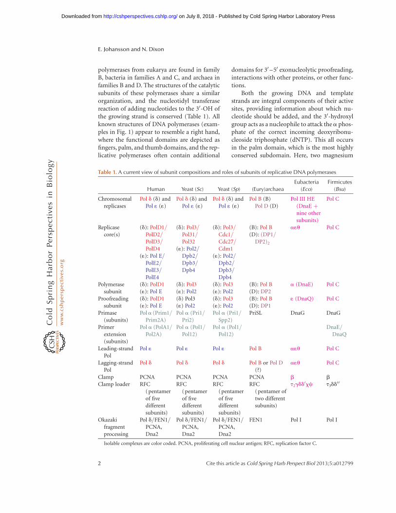
polymerases from eukarya are found in familyB, bacteria in families A and C, and archaea infamilies B and D. The structures of the catalyticsubunits of these polymerases share a similarorganization, and the nucleotidyl transferasereaction of adding nucleotides to the 30-OH ofthe growing strand is conserved (Table 1). Allknown structures of DNA polymerases (exam-ples in Fig. 1) appear to resemble a right hand,where the functional domains are depicted asfingers, palm, and thumb domains, and the rep-licative polymerases often contain additional
domains for 30 –50 exonucleolytic proofreading,interactions with other proteins, or other func-tions.
Both the growing DNA and templatestrands are integral components of their activesites, providing information about which nu-cleotide should be added, and the 30-hydroxylgroup acts as a nucleophile to attack thea phos-phate of the correct incoming deoxyribonu-cleoside triphosphate (dNTP). This all occursin the palm domain, which is the most highlyconserved subdomain. Here, two magnesium
Table 1. A current view of subunit compositions and roles of subunits of replicative DNA polymerases
Human Yeast (Sc) Yeast (Sp) (Eury)archaea
Eubacteria
(Eco)
Firmicutes
(Bsu)
Chromosomalreplicases
Pol d (d) andPol 1 (1)
Pol d (d) andPol 1 (1)
Pol d (d) andPol 1 (1)
Pol B (B)Pol D (D)
Pol III HE(DnaE þnine othersubunits)
Pol C
Replicasecore(s)
(d): PolD1/PolD2/PolD3/PolD4
(1): Pol E/PolE2/PolE3/PolE4
(d): Pol3/Pol31/Pol32
(1): Pol2/Dpb2/Dpb3/Dpb4
(d): Pol3/Cdc1/Cdc27/Cdm1
(1): Pol2/Dpb2/Dpb3/Dpb4
(B): Pol B(D): (DP1/
DP2)2
a1u Pol C
Polymerasesubunit
(d): PolD1(1): Pol E
(d): Pol3(1): Pol2
(d): Pol3(1): Pol2
(B): Pol B(D): DP2
a (DnaE) Pol C
Proofreadingsubunit
(d): PolD1(1): Pol E
(d) Pol3(1) Pol2
(d): Pol3(1): Pol2
(B): Pol B(D): DP1
1 (DnaQ) Pol C
Primase(subunits)
Pola (Prim1/Prim2A)
Pol a (Pri1/Pri2)
Pol a (Pri1/Spp2)
PriSL DnaG DnaG
Primerextension(subunits)
Pol a (PolA1/Pol2A)
Pol a (Pol1/Pol12)
Pol a (Pol1/Pol12)
DnaE/DnaQ
Leading-strandPol
Pol 1 Pol 1 Pol 1 Pol B a1u Pol C
Lagging-strandPol
Pol d Pol d Pol d Pol B or Pol D(?)
a1u Pol C
Clamp PCNA PCNA PCNA PCNA b b
Clamp loader RFC(pentamerof fivedifferentsubunits)
RFC(pentamerof fivedifferentsubunits)
RFC(pentamerof fivedifferentsubunits)
RFC(pentamer oftwo differentsubunits)
t2gdd0xc t3dd
00
Okazakifragmentprocessing
Pol d/FEN1/PCNA,Dna2
Pol d/FEN1/PCNA,Dna2
Pol d/FEN1/PCNA,Dna2
FEN1 Pol I Pol I
Isolable complexes are color coded. PCNA, proliferating cell nuclear antigen; RFC, replication factor C.
E. Johansson and N. Dixon
2 Cite this article as Cold Spring Harb Perspect Biol 2013;5:a012799
on July 8, 2018 - Published by Cold Spring Harbor Laboratory Press http://cshperspectives.cshlp.org/Downloaded from

ions are coordinated by two invariable asparticacids. This is the basis for the two-metal cata-lytic mechanism (Steitz 1993), conserved amongall replicative polymerases studied so far. Thefingers domain is adjacent to the palm domain,contacts the incoming nucleotide, and goesthrough large conformational changes whenthe nucleotide is positioned near the metals,template, and the 30 end of the growing strandin the active site (Yang et al. 1999; Franklin et al.2001). The thumb domain, which interacts withthe duplex DNA upstream of the polymeraseactive site, has been shown in A- and B-familypolymerases to influence the partitioning be-tween editing and polymerization modes (Dou-blie et al. 1998; Franklin et al. 2001). Mutationsin the thumb domain can act both as antimu-tators and mutators, depending on how the bal-ance has been shifted either toward the editingor the polymerization mode (Stocki et al. 1995;Wu et al. 1998). The thumb domain in C-fami-ly polymerases may have a similar function tothat in A- and B-family polymerases, because it
also contacts the duplex DNA (Bailey et al. 2006;Lamers et al. 2006; Wing et al. 2008).
Replicative DNA polymerases are highly ac-curate during the synthesis of DNA. In additionthey have either a built-in 30 –50 exonucleasesite, located at a distance from the polymeraseactive site, or an associated subunit with 30 –50
exonuclease activity (reviewed in Patel and Loeb2001; Kunkel 2004; McHenry 2011). In eithercase, the distance from the polymerase site to theexonuclease site necessitates that at least threenucleotides of the double-stranded DNA areunwound to allow editing. This is a particularlyimportant facet of the editing function becausethe exonuclease sites only accommodate single-stranded DNA.
At every replication fork, replicative poly-merases cooperate in copying both DNA strands.Because the directions of the two templatestrands are opposite, and polymerases can onlyextend preexisting primers from their 30-OHend, only one strand can be made continuously(the leading strand). The other (lagging) strand
Amino-terminal domain
1T7PBacteriophage T7 gp43
Family A
A B C3IAYSaccharomyces cerevisiae Pol δ
Family B
2HNHEscherichia coli Pol III
Family C
Palm
Fingers
Thumb
Exonuclease domain Divalent metal ions
Incoming nucleotide
DNA
PHP domain
β-Binding domain
Figure 1. Representative structures and domain architecture of DNA polymerases from the A, B, and C families.(A) Bacteriophage T7 gene 5 protein with primer-template DNA (family A; pdb 1T7P). (Data from Doublieet al. 1998.) (B) Saccharomyces cerevisiae Pol d (family B; pdb 3IAY). (Data from Swan et al. 2009.) (C) E. coli PolIII a subunit, residues 1–917 (family C; pdb 2HNH). (Data from Lamers et al. 2006.) Figure drawn usingPyMOL.
Replicative DNA Polymerases
Cite this article as Cold Spring Harb Perspect Biol 2013;5:a012799 3
on July 8, 2018 - Published by Cold Spring Harbor Laboratory Press http://cshperspectives.cshlp.org/Downloaded from

has to be synthesized in the direction oppositethe fork movement as a series of short (Oka-zaki) fragments that are processed and subse-quently joined together. In general, leading-and lagging-strand synthesis is coordinated, buthow this is achieved varies somewhat among or-ganisms.
Replicases from all domains of life functionwithin larger molecular machines called repli-somes that contain helicase(s), which separatethe two DNA strands at the apex of the replica-tion fork, a primase for repeated RNA primingin lagging-strand synthesis, sliding clamps thatencircle the nascent double-stranded product totether polymerases onto the DNA to ensure theydissociate rarely, clamp loaders that load theclamps, various single-stranded DNA-bindingproteins, etc. The ring-shaped sliding clamps,called the b subunit (a homodimer) in eubac-teria and PCNA (a homotrimer) in archaea andeukaryotes, have very similar structures (Konget al. 1992; Krishna et al. 1994). They interactat the same site in each of their subunits withmany other proteins including the replicativepolymerases, via short peptide motifs calledthe clamp-binding motif in eubacteria (Dal-rymple et al. 2001) and the PIP box in higherorganisms (Warbrick 1998). The structures andfunctions of the sliding clamps and other ac-cessory replisomal components are describedin detail in the literature.
So far we have highlighted a few well-con-served features of the replicative polymerases.However, as we now proceed with a more de-tailed comparison, you will find that they havespecial features to function in their specific en-vironments.
QUATERNARY STRUCTURE
The quaternary structures of replicative poly-merases vary significantly (Table 1). In part,this variation rather arbitrarily depends onwhich subunits are isolated together to definethe polymerase. From some species (e.g., Pol IIIin Escherichia coli [Eco]), a holoenzyme can bepurified as an entity with 10 different subunits.In eukaryotes, the replicative polymerases ap-pear to be more loosely associated within the
replisome, and they are thus isolated in as-semblies that contain fewer accessory subunits.We will discuss here the core catalytic subunitand accessory subunits that are considered to beparts of the DNA polymerase.
Eubacteria
Pol III holoenzyme is the E. coli chromosomalreplicase that synthesizes both leading and lag-ging strands simultaneously. As isolated directlyfrom cells, it has an average composition closeto (a1u)2–(t2gdd
0cx)–(b2)2 (17 subunits),where a1u is the polymerase core discussed inmore detail below, b2 is the sliding clamp, andt2gdd
0cx is the clamp loader complex that maycontain two to three t and one to zero g sub-units (McHenry 2011).
The a1u core of Pol III is a tightly associatedcomplex. The large a subunit is a family C po-lymerase, and 1 is a separate 30 –50 editing exo-nuclease subunit from the DnaQ family. Thesmall u subunit has a role in stabilizing 1, butit only occurs in a limited range of bacterialspecies. The a subunit is made up of a seriesof domains (Fig. 1C) (Bailey et al. 2006; Lamerset al. 2006): the amino-terminal PHP domainseems to be a vestigial exonuclease domain thatmay still be functional as a proofreader in somespecies (Stano et al. 2006). In E. coli, it hasevolved to be the site of interaction of the 1 sub-unit (Wieczorek and McHenry 2006). This do-main is followed by the usual polymerase palm,thumb, and fingers domains, and a b-bindingdomain that contains a conserved clamp-bind-ing motif that interacts with the b2 clamp totether a to the product DNA to ensure its highprocessivity. This is followed by an OB-folddomain that is likely to interact with the sin-gle-stranded template DNA, and a carboxy-terminal domain that interacts tightly withthe carboxy-terminal domain of the t subunitof the clamp loader. Because the clamp loadercontains two (or three) t subunits, at least twoa1u cores are maintained in the replicase com-plex, one each for leading- and lagging-strandsynthesis.
The 1 subunit has a globular amino-ter-minal exonuclease domain (residues 7–180),
E. Johansson and N. Dixon
4 Cite this article as Cold Spring Harb Perspect Biol 2013;5:a012799
on July 8, 2018 - Published by Cold Spring Harbor Laboratory Press http://cshperspectives.cshlp.org/Downloaded from
the structure of which is reminiscent of manyediting exonuclease domains from other poly-merases (Hamdan et al. 2002). It contains abinuclear metal site for processing nucleotidesmisincorporated by a, followed by a poorlyconserved 63-residue carboxy-terminal segmentthat by itself is unstructured. The last �40 res-idues are now known to be involved directlyin binding to the PHP domain of a (Ozawa etal. 2008); a short segment that immediately fol-lows the exonuclease domain contains a secondclamp-binding motif (S Jergic and N Dixon,unpubl.), and the unstructured segment in be-tween remains flexible even in the a1u complex(Ozawa et al. 2008). This may suggest an unusu-al mechanism in which 1 can occupy quite dif-ferent positions in the complex when it is or isnot being used for proofreading.
Most (or all) eubacterial species seem tohave replicase subunits closely related to a
(DnaE family) and 1, but some Gram-positivebacteria like Bacillus subtilis (Bsu) also have asecond more distantly related polymerase/exo-nuclease called Pol C (Table 1). These latter en-zymes have the proofreading and polymeraseactivities in a single polypeptide, and a differentdomain organization, with putative t-bindingand OB-fold domains preceding a discontinu-ous PHP domain that incorporates the proof-reader, and the b-binding motif being rightat the carboxyl terminus (Evans et al. 2008).Although it was for some time believed thatPol C was dedicated to leading-strand synthe-sis and DnaE to the lagging strand (Dervyn etal. 2001), recent work suggests that most chro-mosomal DNA synthesis in these bacteria isperformed by Pol C. However, because onlythe DnaE polymerase can extend RNA primerslike those made by the DnaG primase, it has acritical role in lagging-strand synthesis. The cur-rent model is that DnaE extends RNA primersfor some length before passing them to Pol C(Sanders et al. 2010). This has some parallelwith the eukaryotic Pol a (below). Note thatin eubacteria, RNA priming is performed by adedicated primase (DnaG) that does not as-sociate directly with the polymerase; it is asso-ciated, sometimes firmly and often more tran-siently, with the replicative helicase to enable
primer synthesis on both the leading and lag-ging strands.
There is no crystal structure available of acomplete Pol III core from any bacterium, butavailable structures of a subunits include E. colia(1–917), which misses its internal clamp-binding motif and domains that follow it(Lamers et al. 2006), and full-length Thermusaquaticus (Taq) a (DnaE), both by itself andin complex with primer-template DNA (Baileyet al. 2006; Wing et al. 2008). The most recentstructure is of a ternary complex containingprimer-template DNA and incoming dNTPbound to Pol C from Geobacillus kaustophilus(Gka) (Evans et al. 2008). The Taq and Eco struc-tures without DNA are very similar in the re-gions that can be compared (Fig. 1C), but DNAbinding induces large conformational changesto a closed state, especially in the b-bindingregion of the Taq protein. The Gka Pol C terna-ry complex is also similar to the Taq a–DNAstructure in this respect. Interestingly, the struc-ture of a b2–DNA complex (Georgescu et al.2008a) can be docked neatly into both thea and Pol C–DNA structures to give plausiblemodels of the a or Pol C–b2–DNA replicasesin the polymerization mode (Evans et al. 2008;Wing et al. 2008), and it has been suggested thatthe open structures might mimic the replicasestructure in the editing mode (Fig. 2A). Theposition of the proofreading exonuclease hasnot been defined (it was deleted from the Pol Cconstruct used for crystallization), but it is pre-sumably between the PHP domains and b2 inthe modeled structures. A second interesting as-pect of the a/Pol C structures is that the palmdomain (active site) architecture is distinct fromthat found in the A- and B-family polymerases,being closer to that of X family members like theeukaryotic repair polymerase, Pol b. This sug-gests that the replicative polymerases of eubac-teria evolved independently of those in eukary-otes and archaea.
The other polymerase that plays a significantrole in bacterial DNA replication is Pol I, thefounding member of the family A polymer-ases (representative Pol A structure is shown inFig. 1A). Its primary function in replication is inOkazaki fragment processing on the lagging
Replicative DNA Polymerases
Cite this article as Cold Spring Harb Perspect Biol 2013;5:a012799 5
on July 8, 2018 - Published by Cold Spring Harbor Laboratory Press http://cshperspectives.cshlp.org/Downloaded from

strand. Pol III is capable of synthesizing Okazakifragments right up to the 50 end of a precedingRNA primer, whereupon it is recycled to a newprimer terminus, leaving behind a nick or shortgap (discussed in Dohrmann et al. 2011). Pol Ihas three separate activities in a single polypep-tide chain. The amino-terminal domain is a50 –30 exonuclease capable of excising the RNAprimers at the same time as the carboxy-termi-nal polymerase domain (with thumb, palm, andfingers subdomains) extends the DNA primerbehind it. The central domain is a DnaQ family
proofreading exonuclease used to ensure highfidelity. Thus, Pol I uses a process of “nick trans-lation” to replace RNA primers with DNA, leav-ing a nick with a 30-OH and 50-phosphate that isa substrate for DNA ligase, which seals the nickto make a contiguous lagging strand.
Archaea
The replisome in archaebacteria is less com-plex than in eubacteria, and in most aspects isa simplified version of the eukaryotic system.
3′–5′ exonuclease
β-Clamp
Pol IV
ssDNA
5′
Exo/TLS
30°
β-Bindingmotif
3′–5′ exonuclease
β-Bindingmotif β-Clamp
ssDNA
5′
Pol
30°
A Pol C
B Pol B
Polymerization mode Proofreading mode
PIP
Polymeraseactive site
Exonucleaseactive site
PIP
Polymeraseactive site
Exonucleaseactive site
PCNA
PCNA
Figure 2. Modeled structures of ternary complexes of polymerases, clamps, and DNA in the polymerization andediting modes. (A) Geobacillus kaustophilus Pol C with DNA and b sliding clamp. (Panel A is from Evans et al.2008; reprinted, with express permission, from the authors.) (B) Pyrococcus furiosus Pol B with DNA and PCNA(Mayanagi et al. 2011). (Panel B is from Mayanagi et al. 2011; reprinted, with permission, from The NationalAcademy of Science # 2011.)
E. Johansson and N. Dixon
6 Cite this article as Cold Spring Harb Perspect Biol 2013;5:a012799
on July 8, 2018 - Published by Cold Spring Harbor Laboratory Press http://cshperspectives.cshlp.org/Downloaded from

All archaea contain single-subunit Pol B poly-merases, with the proofreading activity includ-ed in the catalytic subunit as a domain near theamino terminus (Table 1). Some genera havemultiple Pol Bs (family B), whose separate rolesin chromosomal replication are yet to be clear-ly defined, whereas the euryarchaea uniquelyalso contain a more complex heterotetramericPol D polymerase, with subunit composition(DP1)2–(DP2)2. The smaller DP1 subunit con-tains a 30 –50 exonuclease domain, whereas thelarger DP2 is the (family D) polymerase. Al-though Pol D is a processive and efficient en-zyme that, in common with Pol B, also interactswith the PCNA sliding clamp, the role of Pol Das a chromosomal replicase in euryarchaeal spe-cies is still uncertain (Tori et al. 2007). It hasbeen suggested that in Pyrococcus abyssi, Pol Bsynthesizes the leading and Pol D the laggingstrand (Henneke et al. 2005). High-resolutionstructural information on Pol D is limited to thecrystal structure of the amino-terminal domainof Pyrococcus horikoshii DP2, which has a rolein Pol D subunit oligomerization (Matsui et al.2011).
The crenarchaea do not have Pol Ds, but of-ten have multiple Pol Bs. For example, Sulfolobussolfataricus hasthree of them, and Pol B1 (Dpo1)is a high-fidelity polymerase thought to carryout both leading- and lagging-strand replication(Choi et al. 2011). The archaeal Pol Bs have beenstudied extensively, in partbecausesome of themfrom thermophilic species are used extensivelyfor high-fidelity PCR applications. Althoughcrystal structures of Pol Bs from several archaealspecies have been determined (the earliest are inHopfner et al. 1999, Zhao et al. 1999, Rodriguezet al. 2000, and Hashimoto et al. 2001), there isnoneyet that shows primer-template DNA in thepolymerization or proofreading modes. Thesestructures can, however, be modeled using thestructures of the T4-related phage RB69 Pol B,bound to DNA in both of these modes (Franklinet al. 2001).
The archaeal Pol Bs seem generally anduniquely among family B polymerases (Wardleet al. 2008) to contain an additional amino-terminal domain that precedes the proofreadingexonuclease domain, whose function is to scan
the template DNA ahead of the polymerase forthe uracil base in dUMP produced by cytosinedeamidation (and hypoxanthine, from adeno-sine) that has escaped repair by uracil-N-glyco-sylase (Greagg et al. 1999, Fogg et al. 2002). Onrecognition of dUMP in the template, the poly-merase stalls four nucleotides before the lesionis encountered. Although how this might leadto subsequent repair of the lesion is not known,crystal structures have been reported of stalledcomplexes containing primer templates withappropriately incorporated dUMP or dIMP(Firbank et al. 2008, Killelea et al. 2010). Thebases are buried within a specific pocket in theamino-terminal domain, and the primer-tem-plate DNA is thought to be in a position in thestructure close to that in the editing mode, bycomparison to the RB69 Pol B structure (Frank-lin et al. 2001).
In a recent study (Mayanagi et al. 2011),a series of available crystal structures was com-bined with low-resolution reconstructions fromelectron microscopy and single-particle analy-sis to generate plausible models of the structuresof archaeal Pol B–DNA–clamp ternary com-plexes in both the polymerization and proof-reading modes (Fig. 2B). As for the bacterialPol III complex (Fig. 2A), these models generatenew hypotheses that will be tested in the future,leading to further progressive improvement ofour understanding of replisome structure andfunction.
A significant difference between the archae-al replisomes and those in eukaryotes (see be-low) is that archaea, like the bacteria, use pri-mases that are not physically associated withthe polymerases. Nevertheless, the archaeal pri-mases contain two subunits (p41 and p46) thatare homologs of the PriS and PriL subunits ofthe eukaryotic Pola–primase complex (Table 1).Recent in vitro studies with purified proteinshave shown quite clearly that the Thermococcuskodakarensis primase can carry out (DNA) pri-mer synthesis in the total absence of ribonu-cleoside triphosphates (rNTPs), and in a roll-ing-circle assay with a synthetically primedminicircle DNA template, it can prime efficientlagging-strand Okazaki-fragment synthesis de-pendent on the hexameric MCM replicative
Replicative DNA Polymerases
Cite this article as Cold Spring Harb Perspect Biol 2013;5:a012799 7
on July 8, 2018 - Published by Cold Spring Harbor Laboratory Press http://cshperspectives.cshlp.org/Downloaded from
helicase and Pol B (Chemnitz Galal et al. 2012).Thus, the stage is now set to use in vitro studieswith the archaeal replisome to probe mechanis-tic questions in the way that has already beenperformed with the model bacterial systems.
Eukarya
Eukaryotes have evolved a more complex repli-some that depends on three different B-fami-ly DNA polymerases: Pol a, Pol d, and Pol 1(Kunkel and Burgers 2008; Burgers 2009). Eachof these multisubunit polymerases (Table 1) hasone catalytic core subunit and two to four ac-cessory subunits. Pola is built up by four differ-ent subunits: PriS, PriL, Pol1, and Pol12 (Muzi-Falconi et al. 2003); PriL has primase activityand Pol1 has DNA polymerase activity. Nega-tive-stain EM reconstructions combined withhigh-resolution structures of domains of theSaccharomyces cerevisiae (Sc) Pol1–Pol12 com-plex have revealed two globular domains con-nected via a flexible linker (Klinge et al. 2009). Itwas suggested that an intramolecular hand-offoccurs from the primase domain to the poly-merase domain when the initial RNA primerhas reached a specific length (Klinge et al. 2009;Nunez-Ramirez et al. 2011). The final result is a30–35 nucleotide primer with RNA at the 50
end and DNA at the 30 end. Thus, Pol a synthe-sizes a substantial amount of DNA because theeukaryotic Okazaki fragments are on averageonly about 165 nucleotides long (Smith andWhitehouse 2012). Thus, the absence of a proof-reading activity in Pol a requires that errorsmade by it are corrected by other mechanisms.For instance Pol d, but not Pol 1, proofreadserrors made by Pol a, in addition to error cor-rection by the mismatch repair system (Pavlovet al. 2006; Nick McElhinny et al. 2010a).
Pol 1 and Pol d are responsible for the bulksynthesis of DNA on the leading and lag-ging strands (Kunkel and Burgers 2008; Burgers2009). They are both highly accurate DNA po-lymerases with built-in 30 –50 exonuclease ac-tivities for proofreading (Morrison et al. 1991;Morrison and Sugino 1994). They are also high-ly processive enzymes when interacting withthe clamp, PCNA (Burgers 1991; Lee et al. 1991;
Chilkova et al. 2007). The exact roles of Pols 1and d at the replication fork have been debated(Kunkel and Burgers 2008; Burgers 2009). Overthe past few years, evidence has accumulated thatin S. cerevisiae and Schizosaccharomyces pombe(Sp), Pol 1 is primarily responsible for leading-strand and Pol d for lagging-strand synthesis(Pursell et al. 2007; Nick McElhinny et al. 2008;Miyabe et al. 2011). This suggests that the rolesof Pol 1 and Pol d are evolutionarily conservedamong eukaryotes.
Pol d is constituted of three subunits (Pol3,Pol31, and Pol32) in S. cerevisiae, and four inS. pombe and H. sapiens (Table 1) (Gerik et al.1998; Reynolds et al. 1998; Liu et al. 2000). Thismay be species variations but could also reflecthow tightly specific subunits are associated withthe catalytic core subunit. The hydrodynamicproperties of ScPol d and SpPol d suggestedthat Pol d has an elongated structure, heavilyinfluenced by the Pol32 subunit (Johanssonet al. 2001; Bermudez et al. 2002). This was laterconfirmed by small-angle X-ray scattering(SAXS) analysis of ScPol3 in complex withPol31 and the amino terminus of Pol32 and ahigh-resolution structure of the human acces-sory subunits Pold2 together with the aminoterminus of Pold3 (Baranovskiy et al. 2008;Jain et al. 2009). The catalytic subunit ScPol3is located at one end of the complex; Pol31 actsas a bridge to the very elongated Pol32 that car-ries an important interaction motif with PCNAat the far end of the elongated structure ex-tending away from the catalytic core subunitPol3 (Johansson et al. 2004; Baranovskiy et al.2008; Jain et al. 2009). A high-resolution struc-ture of ScPol3 (amino acid residues 68–985)was solved which included both the exonucleaseand polymerase domains (Fig. 1B), but exclud-ed the carboxy-terminal domain with the CysAand CysB motifs discussed below. Overall, thestructure of ScPol3 is highly reminiscent of atypical family B polymerase, with a fold verysimilar to that of RB69 despite the limited se-quence identity (Swan et al. 2009).
Pol 1 consists of four subunits. In S. cerevi-siae, these are called Pol2, Dpb2, Dpb3, andDpb4 (Hamatake et al. 1990; Chilkova et al.2003). Structural information on Pol 1 is limited
E. Johansson and N. Dixon
8 Cite this article as Cold Spring Harb Perspect Biol 2013;5:a012799
on July 8, 2018 - Published by Cold Spring Harbor Laboratory Press http://cshperspectives.cshlp.org/Downloaded from
to a low-resolution cryo-electron microscopy(cryo-EM) structure. ScPol 1 has two domains,a globular domain with the catalytic subunitand an extended tail domain that has been sug-gested to harbor the three accessory subunitsDpb2, Dpb3, and Dpb4 (Asturias et al. 2006).Dpb2 is essential in S. cerevisiae and S. pombe(Araki et al. 1991; Feng et al. 2003). However, theDpb2 subunit does not appear to influence thepolymerase activity in human Pol 1 (Bermudezet al. 2011). It is possible that the essential func-tion of Dpb2 is during the initiation of DNAreplication, as described in Tanaka and Araki(2013) (also reviewed in Araki 2010). Dpb3and Dpb4 form a heterodimer that has a highaffinity for double-stranded DNA (Tsubota et al.2006). In contrast to Dpb2, ScPol 1 depends onDpb3 and Dpb4 for full processivity in the ab-sence of PCNA (Aksenova et al. 2010).
The eukaryotic replicative polymerases allhave two conserved motifs with cysteines (CysAand CysB) located at the carboxyl terminus ofthe catalytic subunit (Netz et al. 2012). For along time they have been considered to be Zn-finger motifs and Zn was found in both Pol 1and Pol a (Dua et al. 2002; Klinge et al. 2009).Their primary role was suggested to form aninteraction surface with the B subunit of theDNA polymerase. Recently it was shown thatthe two motifs CysA and CysB have differentialfunctions and coordinate different metals inScPol d (Netz et al. 2012). CysA coordinates Znand is required to support processive synthesisby Pol d via a direct interaction with PCNA,whereas CysB coordinates a Fe–S center and isimportant for the interaction with the Pol31subunit. Loss of interaction with Pol31 led toloss of Pol32, and as a result an important in-teraction with the PCNA clamp was lost. Thus,Pol d has at least two separate interactions withPCNA, CysA in Pol3 and the PIP box in Pol32,to support efficient loading and synthesis ofDNA (Netz et al. 2012). Experiments with car-boxy-terminal domains of Pol a and Pol 1 im-plied that these replicative polymerases may alsohave an Fe–S cluster. However, conflicting re-sults with expression of the carboxy-terminaldomains of the catalytic subunits from humanPols a and 1 in E. coli were recently published
(Baranovskiy et al. 2012) and it remains to beshown whether the ScPols a and 1 holoenzymescarry Fe–S clusters that mediate the interactionsbetween the catalytic subunit and Pol12 andDpb2, respectively. The study of the carboxy-terminal domain of the catalytic subunit in hu-man Pold confirmed that there is an Fe–S centercoordinated by CysB (Baranovskiy et al. 2012).
As discussed above, ScPol d has at leasttwo well-defined motifs that mediate the con-tact with PCNA. Two additional motifs, one inPol3 and one in Pol31, were recently reported,emphasizing that ScPol d has multiple surfacesto stabilize the interaction with PCNAwhen syn-thesizing DNA (Acharya et al. 2011). Less isknown about how Pol 1 interacts with PCNA.In S. cerevisiae, the catalytic subunit and Dpb2both have a PIP box, but they are not located atthe amino or carboxyl terminus as found typi-cally in PCNA-interacting proteins (Dua et al.2002). Biochemical characterizations and ge-netic experiments in yeast suggested that theseare not functional motifs during DNA replica-tion (Dua et al. 2002; Chilkova et al. 2007). Nev-ertheless, Pol 1 is stimulated by PCNA in vitro,suggesting that either the exact motif(s) remainsto be found or there are multiple weak interac-tions that together stabilize the interaction withPCNA (Maga et al. 1999). Pol 1 also has an un-usually high intrinsic processivity in part medi-ated by the Dpb3 and Dpb4 subunits (Aksenovaet al. 2010). In addition, the catalytic subunit hasby itself an unusually high processivity and itwas speculated that a 66 amino acid insertionin the vicinity of the palm domain might givePol 1 this unusual property (Shcherbakovaet al. 2003).
FIDELITY OF THE REPLICATIVEPOLYMERASES
The high fidelity of replicative polymerases isdetermined by the tolerance for the incomingnucleotide when performing chemistry and the30 –50 exonucleolytic proofreading activity. Inaddition, the concentration of each dNTP in thecell and the quality of the template influence therate at which errors are made (Kunkel 2011).Until recently, not much attention was paid to
Replicative DNA Polymerases
Cite this article as Cold Spring Harb Perspect Biol 2013;5:a012799 9
on July 8, 2018 - Published by Cold Spring Harbor Laboratory Press http://cshperspectives.cshlp.org/Downloaded from

how the relatively large pools of rNTPs in eu-karyotic cells influence the fidelity of the rep-licative DNA polymerases. The polymeraseshave a built-in steric gate that should select fordNTPs and against rNTPs (Joyce 1997). How-ever, the concentration of dNTPs is very low incomparison to the rNTPs and it appears thatPols a, d, and 1 misincorporate ribonucleotidesat a surprisingly high rate when synthesizingDNA (Nick McElhinny et al. 2010c). All threePols also incorporated rNTPs in vivo in S. cer-evisiae and S. pombe and gave a significantlyincreased elevation of 2–5 nucleotide deletions(Nick McElhinny et al. 2010b; Miyabe et al.2011). To avoid mutagenic events, ribonucleo-tides must be removed by a repair mechanismthat involves RNase H2 before the next S phase(Nick McElhinny et al. 2010b; Miyabe et al.2011). The incorporation of ribonucleotidesand the dependence on RNase H2 for the re-moval of ribonucleotides was also found inmice, suggesting that this repair mechanism isconserved among all eukaryotes (Reijns et al.2012). The exact mechanism behind the errorsignature in S. cerevisiae is unknown, but Pol a,Pol d, and Pol 1 stall when encountering a ri-bonucleotide in the template and this may leadto deletions in repetitive sequences (Watt et al.2011). In case ribonucleotides are not removed,postreplication repair will be activated duringthe next S phase (Lazzaro et al. 2012). Pol z iscapable of replicating across 1–4 ribonucleo-tides in the template and also MMS2-depen-dent template switching contributes to the tol-erance for unrepaired ribonucleotide lesions(Lazzaro et al. 2012).
It is yet unclear if archaeal DNA polymer-ases also frequently misinsert ribonucleotides inthe genome. However, as described earlier, thearchaeal replicative polymerases (in, e.g., P. fur-iosus and S. solfataricus) have a unique capacityto proofread the template for uracil and otherdeamidated bases (Greagg et al. 1999; Choi et al.2011). A binding pocket for uracil is found intheir unique amino-terminal domains wherethe single-stranded template is scanned beforeentering the polymerase catalytic site (Fogget al. 2002; Shuttleworth et al. 2004; Firbanket al. 2008).
ANTIBIOTICS/INHIBITORS
DNA replication is an essential process in allorganisms. Although there are sufficient varia-tions in structures among replisomal proteinsfrom bacteria and humans to make replisomes avery good target for discovery of new antibacte-rial therapeutics, it is a target that is surprisinglyunderexploited both by pharmaceutical compa-nies and in nature by other organisms (reviewedin Robinson et al. 2010, 2012). Although thereare no known inhibitors of DnaE-type Pol IIIs,there have been substantial efforts to target Pol Cfrom Gram-positive bacteria, including Staph-ylococcus aureus, using a range of 6-anilinoura-cils (6-AUs) and quinazolin-2-ylamino-quina-zolin-4-ols (BisQuinols). The 6-AUs act as Pol Cinhibitors by competing with dGTP for basepairing with cytosines in the template DNA,whereas the BisQuinols appear to interferewith template DNA binding. Another promis-ing target is the b clamp, which makes essentialinteractions with the clamp-binding motifs ina large number of other proteins (including allfive DNA polymerases, the clamp loader, andDNA ligase in E. coli, for example) at a singlehighly conserved binding site that is structurallydistinct from the PIP-binding sites in PCNA(Dalrymple et al. 2001). Development of resis-tance to antibacterials that inhibit interactionsat this site by target mutagenesis would be ex-pected to be slow because it would require si-multaneous mutations to arise in genes encod-ing several different proteins. There have beenrecent reports of identification through screen-ing campaigns of new inhibitors of interactionswith b, including two crystal structures (Geor-gescu et al. 2008b; Wijffels et al. 2011), but thereis much to be done to develop these compoundsinto useful leads.
CONCLUDING REMARKS
Almost 60 years have passed since the first iso-lation of a replicative DNA polymerase by Ar-thur Kornberg in the mid-1950s. In this longhistory of research, many modern tools in struc-tural and molecular biology have provided frag-ments of information for progressive synthesis
E. Johansson and N. Dixon
10 Cite this article as Cold Spring Harb Perspect Biol 2013;5:a012799
on July 8, 2018 - Published by Cold Spring Harbor Laboratory Press http://cshperspectives.cshlp.org/Downloaded from

of coherent pictures of how these complex mul-tifunctional enzymes work in organisms fromall three domains of life. There are common as-pects that are well established, like the commoncatalytic mechanism of the nucleotidyltrans-ferase reaction, but our pictures are still far fromcomplete. The main reason for this is that theseenzymes never work alone. They are key partsof larger, very dynamic nucleoprotein mach-ines (replisomes) that undergo large structuralchanges as they function, with proteins comingand going and often changing binding partners.Sometimes this occurs at common binding sites(as on the sliding clamps) to ensure ordered se-quences of events, and often interactions involveintrinsically unstructured regions of proteinsthat only become ordered as they interact withtheir binding partners. This latter fact alone hasmade structural studies slow and difficult, and italso means that multiple structures need to besolved to really understand function. A secondbasic operating principle for replisomes is thattheir integrity is maintained by a very largenumber of protein–protein and protein–nu-cleic acid interactions, many of which are inher-ently weak and can easily be transiently broken.These interactions are often difficult to detectindividually, but sum to be strong enough tohold the replisomes together as they function.
ACKNOWLEDGMENTS
We apologize to those colleagues whose workis not cited because of formatting and spacerestrictions. We thank Matthew Hogg for helpwith Figure 1, and Rick Lewis for comments onthe manuscript. Research in our laboratoriesis funded by the Swedish Cancer Society, theSwedish Research Council, the Knut and AliceWallenberg foundation (E.J.), the AustralianResearch Council, including an Australian Pro-fessorial Fellowship, and the Australian Nation-al Health and Medical Research Council (N.D.).
REFERENCES�Reference is also in this collection.
Acharya N, Klassen R, Johnson RE, Prakash L, Prakash S.2011. PCNA binding domains in all three subunits of
yeast DNA polymerase d modulate its function in DNAreplication. Proc Natl Acad Sci 108: 17927–17932.
Aksenova A, Volkov K, Maceluch J, Pursell ZF, Rogozin IB,Kunkel TA, Pavlov YI, Johansson E. 2010. Mismatch re-pair—Independent increase in spontaneous mutagenesisin yeast lacking non-essential subunits of DNA polymer-ase 1. PLoS Genet 6: e1001209.
Araki H. 2010. Cyclin-dependent kinase-dependent initia-tion of chromosomal DNA replication. Curr Opin CellBiol 22: 766–771.
Araki H, Hamatake RK, Johnston LH, Sugino A. 1991.DPB2, the gene encoding DNA polymerase II subunitB, is required for chromosome replication in Saccharo-myces cerevisiae. Proc Natl Acad Sci 88: 4601–4605.
Asturias FJ, Cheung IK, Sabouri N, Chilkova O, Wepplo D,Johansson E. 2006. Structure of Saccharomyces cerevisiaeDNA polymerase epsilon by cryo-electron microscopy.Nat Struct Mol Biol 13: 35–43.
Bailey S, Wing RA, Steitz TA. 2006. The structure ofT. aquaticus DNA polymerase III is distinct from eukary-otic replicative DNA polymerases. Cell 126: 893–904.
Baranovskiy AG, Babayeva ND, Liston VG, Rogozin IB,Koonin EV, Pavlov YI, Vassylyev DG, Tahirov TH. 2008.X-ray structure of the complex of regulatory subunits ofhuman DNA polymerase d. Cell Cycle 7: 3026–3036.
Baranovskiy AG, Lada AG, Siebler HM, Zhang Y, Pavlov YI,Tahirov TH. 2012. DNA polymerase d and z switch bysharing accessory subunits of DNA polymerase d. J BiolChem 287: 17281–17287.
Bermudez VP, MacNeill SA, Tappin I, Hurwitz J. 2002. Theinfluence of the Cdc27 subunit on the properties of theSchizosaccharomyces pombe DNA polymerase d. J BiolChem 277: 36853–36862.
Bermudez VP, Farina A, Raghavan V, Tappin I, Hurwitz J.2011. Studies on human DNA polymerase 1 and GINScomplex and their role in DNA replication. J Biol Chem286: 28963–28977.
Burgers PM. 1991. Saccharomyces cerevisiae replication fac-tor C. II. Formation and activity of complexes with theproliferating cell nuclear antigen and with DNA polymer-ases d and 1. J Biol Chem 266: 22698–22706.
Burgers PM. 2009. Polymerase dynamics at the eukaryoticDNA replication fork. J Biol Chem 284: 4041–4045.
Chemnitz Galal W, Pan M, Kelman Z, Hurwitz J. 2012.Characterization of DNA primase complex isolatedfrom the archaeon, Thermococcus kodakearaensis, J BiolChem 287: 16209–16219.
Chilkova O, Jonsson BH, Johansson E. 2003. The quaternarystructure of DNA polymerase 1 from Saccharomyces cer-evisiae. J Biol Chem 278: 14082–14086.
Chilkova O, Stenlund P, Isoz I, Stith CM, Grabowski P,Lundstrom EB, Burgers PM, Johansson E. 2007. The eu-karyotic leading and lagging strand DNA polymerases areloaded onto primer-ends via separate mechanisms buthave comparable processivity in the presence of PCNA.Nucleic Acids Res 35: 6588–6597.
Choi JY, Eoff RL, Pence MG, Wang J, Martin MV, Kim EJ,Folkmann LM, Guengerich FP. 2011. Roles of the fourDNA polymerases of the crenarchaeon Sulfolobus solfa-taricus and accessory proteins in DNA replication. J BiolChem 286: 31180–31193.
Replicative DNA Polymerases
Cite this article as Cold Spring Harb Perspect Biol 2013;5:a012799 11
on July 8, 2018 - Published by Cold Spring Harbor Laboratory Press http://cshperspectives.cshlp.org/Downloaded from
Dalrymple BP, Kongsuwan K, Wijffels G, Dixon NE, Jenn-ings PA. 2001. A universal protein-protein interactionmotif in the eubacterial DNA replication and repair sys-tems. Proc Natl Acad Sci 98: 11627–11632.
Dervyn E, Suski C, Daniel R, Bruand C, Chapuis J, Erring-ton J, Janniere L, Ehrlich SD. 2001. Two essential DNApolymerases at the bacterial replication fork. Science 294:1716–1719.
Dohrmann PR, Manhart CM, Downey CD, McHenry CS.2011. The rate of polymerase release upon filling the gapbetween Okazaki fragments is inadequate to support cy-cling during lagging strand synthesis. J Mol Biol 414:15–27.
Doublie S, Tabor S, Long AM, Richardson CC, Ellenberger T.1998. Crystal structure of a bacteriophage T7 DNA rep-lication complex at 2.2 A resolution. Nature 391: 251–258.
Dua R, Levy DL, Li CM, Snow PM, Campbell JL. 2002. Invivo reconstitution of Saccharomyces cerevisiae DNA po-lymerase 1 in insect cells. Purification and characteriza-tion. J Biol Chem 277: 7889–7896.
Evans RJ, Davies DR, Bullard JM, Christensen J, Green LS,Guiles JW, Pata JD, Ribble WK, Janjic N, Jarvis TC. 2008.Structure of PolC reveals unique DNA binding and fidel-ity determinants. Proc Natl Acad Sci 105: 20695–20700.
Feng W, Rodriguez-Menocal L, Tolun G, D’Urso G. 2003.Schizosacchromyces pombe Dpb2 binds to origin DNAearly in S phase and is required for chromosomal DNAreplication. Mol Biol Cell 14: 3427–3436.
Firbank SJ, Wardle J, Heslop P, Lewis RJ, Connolly BA. 2008.Uracil recognition in archaeal DNA polymerases cap-tured by X-ray crystallography. J Mol Biol 381: 529–539.
Fogg MJ, Pearl LH, Connolly BA. 2002. Structural basis foruracil recognition by archaeal family B DNA polymer-ases. Nat Struct Biol 9: 922–927.
Franklin MC, Wang J, Steitz TA. 2001. Structure of the rep-licating complex of a pol a family DNA polymerase. Cell105: 657–667.
Georgescu RE, Kim S-S, Yurieva O, Kuriyan J, Kong X-P,O’Donnell M. 2008a. Structure of a sliding clamp onDNA. Cell 132: 43–54.
Georgescu RE, Yurieva O, Kim S-S, Kuriyan J, Kong X-P,O’Donnell M. 2008b. Structure of a small-molecule in-hibitor of a DNA polymerase sliding clamp. Proc NatlAcad Sci 105: 11116–11121.
Gerik KJ, Li X, Pautz A, Burgers PM. 1998. Characterizationof the two small subunits of Saccharomyces cerevisiaeDNA polymerase d. J Biol Chem 273: 19747–19755.
Greagg MA, Fogg MJ, Panayotou G, Evans SJ, Connolly BA,Pearl LH. 1999. A read-ahead function in archaeal DNApolymerases detects promutagenic template-strand ura-cil. Proc Natl Acad Sci 96: 9045–9050.
Hamatake RK, Hasegawa H, Clark AB, Bebenek K,Kunkel TA, Sugino A. 1990. Purification and characteri-zation of DNA polymerase II from the yeast Saccharomy-ces cerevisiae. Identification of the catalytic core and apossible holoenzyme form of the enzyme. J Biol Chem265: 4072–4083.
Hamdan S, Carr PD, Brown SE, Ollis DL, Dixon NE. 2002.Structural basis for proofreading during replication ofthe Escherichia coli chromosome. Structure 10: 535–546.
Hashimoto H, Nishioka M, Fujiwara S, Takagi M, Ima-naka T, Inoue T, Kai Y. 2001. Crystal structure of DNApolymerase from hyperthermophilic archaeon Pyrococ-cus kodakaraensis KOD1. J Mol Biol 306: 469–477.
Henneke G, Flament D, Hubscher U, Querellou J, Raffin JP.2005. The hyperthermophilic euryarchaeota Pyrococcusabyssi likely requires the two DNA polymerases D andB for DNA replication. J Mol Biol 350: 53–64.
Hopfner K-P, Eichinger A, Engh RA, Laue F, Ankenbauer W,Huber R, Angerer B. 1999. Crystal structure of a thermo-stable type B DNA polymerase from Thermococcus gorgo-narius. Proc Natl Acad Sci 96: 3600–3605.
Jain R, Hammel M, Johnson RE, Prakash L, Prakash S,Aggarwal AK. 2009. Structural insights into yeast DNApolymerase d by small angle X-ray scattering. J Mol Biol394: 377–382.
Johansson E, Majka J, Burgers PM. 2001. Structure of DNApolymerase d from Saccharomyces cerevisiae. J Biol Chem276: 43824–43828.
Johansson E, Garg P, Burgers PM. 2004. The Pol32 subunitof DNA polymerase d contains separable domains forprocessive replication and proliferating cell nuclear anti-gen (PCNA) binding. J Biol Chem 279: 1907–1915.
Joyce CM. 1997. Choosing the right sugar: How polymer-ases select a nucleotide substrate. Proc Natl Acad Sci 94:1619–1622.
Killelea T, Ghosh S, Tan SS, Heslop P, Firbank SJ, Kool ET,Connolly BA. 2010. Probing the interaction of archaealDNA polymerases with deaminated bases using X-raycrystallography and non-hydrogen bonding isostericbase analogues. Biochemistry 49: 5772–5781.
Klinge S, Nunez-Ramirez R, Llorca O, Pellegrini L. 2009. 3Darchitecture of DNA Pol a reveals the functional core ofmulti-subunit replicative polymerases. Embo J 28: 1978–1987.
Kong X-P, Onrust R, O’Donnell M, Kuriyan J. 1992. Three-dimensional structure of the b subunit of E. coli DNApolymerase III holoenzyme: A sliding DNA clamp. Cell69: 425–437.
Krishna TSR, Kong X-P, Gary S, Burgers PM, Kuriyan J.1994. Crystal structure of the eukaryotic DNA polymer-ase processivity factor PCNA. Cell 79: 1233–1243.
Kunkel TA. 2004. DNA replication fidelity. J Biol Chem 279:16895–16898.
Kunkel TA. 2011. Balancing eukaryotic replication asymme-try with replication fidelity. Curr Opin Chem Biol 15:620–626.
Kunkel TA, Burgers PM. 2008. Dividing the workload at aeukaryotic replication fork. Trends Cell Biol 18: 521–527.
Lamers MH, Georgescu RE, Lee SG, O’Donnell M, Kuri-yan J. 2006. Crystal structure of the catalytic a subunit ofE. coli replicative DNA polymerase III. Cell 126: 881–892.
Lazzaro F, Novarina D, Amara F, Watt DL, Stone JE, Cos-tanzo V, Burgers PM, Kunkel TA, Plevani P, Muzi-Falconi M. 2012. RNase H and postreplication repairprotect cells from ribonucleotides incorporated inDNA. Mol Cell 45: 99–110.
Lee SH, Pan ZQ, Kwong AD, Burgers PM, Hurwitz J. 1991.Synthesis of DNA by DNA polymerase 1 in vitro. J BiolChem 266: 22707–22717.
E. Johansson and N. Dixon
12 Cite this article as Cold Spring Harb Perspect Biol 2013;5:a012799
on July 8, 2018 - Published by Cold Spring Harbor Laboratory Press http://cshperspectives.cshlp.org/Downloaded from

Liu L, Mo J, Rodriguez-Belmonte EM, Lee MY. 2000. Iden-tification of a fourth subunit of mammalian DNA poly-merase delta. J Biol Chem 275: 18739–18744.
Maga G, Jonsson ZO, Stucki M, Spadari S, Hubscher U.1999. Dual mode of interaction of DNA polymerase 1with proliferating cell nuclear antigen in primer bindingand DNA synthesis. J Mol Biol 285: 259–267.
Matsui I, Urushibata Y, Shen Y, Matsui E, Yokoyama H. 2011.Novel structure of an N-terminal domain that is crucialfor the dimeric assembly and DNA-binding of an archae-al DNA polymerase D large subunit from Pyrococcushorikoshii. FEBS Lett 585: 452–458.
Mayanagi K, Kiyonari S, Nishida H, Saito M, Kohda D,Ishino Y, Shirai T, Morikawa K. 2011. Architecture ofthe DNA polymerase B-proliferating cell nuclear antigen(PCNA)-DNA ternary complex. Proc Natl Acad Sci 108:1845–1849.
McHenry CS. 2011. DNA replicases from a bacterial per-spective. Annu Rev Biochem 80: 403–436.
Miyabe I, Kunkel TA, Carr AM. 2011. The major roles ofDNA polymerases 1 and d at the eukaryotic replicationfork are evolutionarily conserved. PLoS Genet 7:e1002407.
Morrison A, Sugino A. 1994. The 30 ! 50 exonucleases ofboth DNA polymerases d and 1 participate in correctingerrors of DNA replication in Saccharomyces cerevisiae.Mol Gen Genet 242: 289–296.
Morrison A, Bell JB, Kunkel TA, Sugino A. 1991. EukaryoticDNA polymerase amino acid sequence required for 30 –50
exonuclease activity. Proc Natl Acad Sci 88: 9473–9477.
Muzi-Falconi M, Giannattasio M, Foiani M, Plevani P.2003. The DNA polymerase a-primase complex: Multi-ple functions and interactions. ScientificWorldJournal 3:21–33.
Netz DJ, Stith CM, Stumpfig M, Kopf G, Vogel D,Genau HM, Stodola JL, Lill R, Burgers PM, Pierik AJ.2012. Eukaryotic DNA polymerases require an iron-sul-fur cluster for the formation of active complexes. NatChem Biol 8: 125–132.
Nick McElhinny SA, Gordenin DA, Stith CM, Burgers PM,Kunkel TA. 2008. Division of labor at the eukaryotic rep-lication fork. Mol Cell 30: 137–144.
Nick McElhinny SA, Kissling GE, Kunkel TA. 2010a. Differ-ential correction of lagging-strand replication errorsmade by DNA polymerases a and d. Proc Natl Acad Sci107: 21070–21075.
Nick McElhinny SA, Kumar D, Clark AB, Watt DL,Watts BE, Lundstrom EB, Johansson E, Chabes A,Kunkel TA. 2010b. Genome instability due to ribonucle-otide incorporation into DNA. Nat Chem Biol 6: 774–781.
Nick McElhinny SA, Watts BE, Kumar D, Watt DL,Lundstrom EB, Burgers PM, Johansson E, Chabes A,Kunkel TA. 2010c. Abundant ribonucleotide incorpora-tion into DNA by yeast replicative polymerases. Proc NatlAcad Sci 107: 4949–4954.
Nunez-Ramirez R, Klinge S, Sauguet L, Melero R, Recuero-Checa MA, Kilkenny M, Perera RL, Garcia-Alvarez B,Hall RJ, Nogales E, et al. 2011. Flexible tethering of pri-mase and DNA Pol a in the eukaryotic primosome. Nu-cleic Acids Res 39: 8187–8199.
Ozawa K, Jergic S, Park AY, Dixon NE, Otting G. 2008. Theproofreading exonuclease subunit 1 of Escherichia coliDNA polymerase III is tethered to the polymerase sub-unit alpha via a flexible linker. Nucleic Acids Res 36:5074–5082.
Patel PH, Loeb LA. 2001. Getting a grip on how DNA po-lymerases function. Nat Struct Biol 8: 656–659.
Pavlov YI, Frahm C, Nick McElhinny SA, Niimi A, Suzuki M,Kunkel TA. 2006. Evidence that errors made by DNApolymerase a are corrected by DNA polymerase d. CurrBiol 16: 202–207.
Pursell ZF, Isoz I, Lundstrom EB, Johansson E, Kunkel TA.2007. Yeast DNA polymerase 1 participates in leading-strand DNA replication. Science 317: 127–130.
Reijns MA, Rabe B, Rigby RE, Mill P, Astell KR, Lettice LA,Boyle S, Leitch A, Keighren M, Kilanowski F, et al. 2012.Enzymatic removal of ribonucleotides from DNA is es-sential for mammalian genome integrity and develop-ment. Cell 149: 1008–1022.
Reynolds N, Watt A, Fantes PA, MacNeill SA. 1998. Cdm1,the smallest subunit of DNA polymerase d in the fissionyeast Schizosaccharomyces pombe, is non-essential forgrowth and division. Curr Genet 34: 250–258.
Robinson A, Brzoska AJ, Turner KM, Withers R, Harry EJ,Lewis PJ, Dixon NE. 2010. Essential biological processesof an emerging pathogen: DNA replication, transcriptionand cell division in Acinetobacter spp. Microbiol Mol BiolRev 74: 273–297.
Robinson A, Causer RJ, Dixon NE. 2012. Architecture andconservation of the bacterial DNA replication machinery,an underexploited drug target. Curr Drug Targets 13:352–372.
Rodriguez AC, Park HW, Mao C, Beese LS. 2000. Crystalstructure of a pol a family DNA polymerase from thehyperthermophilic archaeon Thermococcus sp. 98 N-7.J Mol Biol 299: 447–462.
Sanders GM, Dallmann HG, McHenry CS. 2010. Reconsti-tution of the B. subtilis replisome with 13 proteins in-cluding two distinct replicases. Mol Cell 37: 273–281.
Shcherbakova PV, Pavlov YI, Chilkova O, Rogozin IB,Johansson E, Kunkel TA. 2003. Unique error signatureof the four-subunit yeast DNA polymerase 1. J BiolChem 278: 43770–43780.
Shuttleworth G, Fogg MJ, Kurpiewski MR, Jen-Jacobson L,Connolly BA. 2004. Recognition of the pro-mutagenicbase uracil by family B DNA polymerases from archaea.J Mol Biol 337: 621–634.
Smith DJ, Whitehouse I. 2012. Intrinsic coupling of lagging-strand synthesis to chromatin assembly. Nature 483:434–438.
Stano NM, Chen J, McHenry CS. 2006. A coproofreadingZn2þ-dependent exonuclease within a bacterial replicase.Nat Struct Mol Biol 13: 458–459.
Steitz TA. 1993. DNA- and RNA-dependent DNA polymer-ases. Curr Opin Struct Biol 3: 31–38.
Stocki SA, Nonay RL, Reha-Krantz LJ. 1995. Dynamics ofbacteriophage T4 DNA polymerase function: Identifica-tion of amino acid residues that affect switching betweenpolymerase and 30 ! 50 exonuclease activities. J Mol Biol254: 15–28.
Replicative DNA Polymerases
Cite this article as Cold Spring Harb Perspect Biol 2013;5:a012799 13
on July 8, 2018 - Published by Cold Spring Harbor Laboratory Press http://cshperspectives.cshlp.org/Downloaded from

Swan MK, Johnson RE, Prakash L, Prakash S, Aggarwal AK.2009. Structural basis of high-fidelity DNA synthesis byyeast DNA polymerase d. Nat Struct Mol Biol 16: 979–986.
� Tanaka S, Araki H. 2013. Helicase activation and establish-ment of replication forks at chromosomal origins of rep-lication. Cold Spring Harb Perspect Biol doi: 10.1101/cshperspect.a010371.
Tori K, Kimizu M, Ishino S, Ishino Y. 2007. DNA polymeraseB1 and D from the hyperthermophilic archaeon Pyrococ-cus furiosus both bind proliferating cell nuclear antigenwith their C-terminal PIP-box motifs. J Bacteriol 189:5652–5657.
Tsubota T, Tajima R, Ode K, Kubota H, Fukuhara N,Kawabata T, Maki S, Maki H. 2006. Double-strandedDNA binding, an unusual property of DNA polymerase1, promotes epigenetic silencing in Saccharomyces cerevi-siae. J Biol Chem 281: 32898–32908.
Warbrick E. 1998. PCNA binding through a conserved mo-tif. BioEssays 20: 195–199.
Wardle J, Burgers PM, Cann IK, Darley K, Heslop P,Johansson E, Lin LJ, McGlynn P, Sanvoisin J, Stith CM,et al. 2008. Uracil recognition by replicative DNA poly-merases is limited to the archaea, not occurring withbacteria and eukarya. Nucleic Acids Res 36: 705–711.
Watt DL, Johansson E, Burgers PM, Kunkel TA. 2011. Rep-lication of ribonucleotide-containing DNA templates byyeast replicative polymerases. DNA Repair (Amst) 10:897–902.
Wieczorek A, McHenry CS. 2006. The NH2-terminal phpdomain of the a subunit of the Escherichia coli replicasebinds the 1 proofreading subunit. J Biol Chem 281:12561–12567.
Wijffels G, Johnson WM, Oakley AJ, Turner K, Epa VC,Briscoe SJ, Polley M, Liepa A, Hofmann A, Buchardt J,et al. 2011. Binding inhibitors of the bacterial slidingclamp by design. J Med Chem 54: 4831–4838.
Wing RA, Bailey S, Steitz TA. 2008. Insights into thereplisome from the structure of a ternary complex ofthe DNA polymerase III a-subunit. J Mol Biol 382:859–869.
Wu P, Nossal N, Benkovic SJ. 1998. Kinetic characterizationof a bacteriophage T4 antimutator DNA polymerase. Bi-ochemistry 37: 14748–14755.
Yang G, Lin T, Karam J, Konigsberg WH. 1999. Steady-statekinetic characterization of RB69 DNA polymerase mu-tants that affect dNTP incorporation. Biochemistry 38:8094–8101.
Zhao Y, Jeruzalmi D, Moarefi I, Leighton L, Lasken R,Kuriyan J. 1999. Crystal structure of an archaebacterialDNA polymerase. Structure 7: 1189–1199.
E. Johansson and N. Dixon
14 Cite this article as Cold Spring Harb Perspect Biol 2013;5:a012799
on July 8, 2018 - Published by Cold Spring Harbor Laboratory Press http://cshperspectives.cshlp.org/Downloaded from
2013; doi: 10.1101/cshperspect.a012799Cold Spring Harb Perspect Biol Erik Johansson and Nicholas Dixon Replicative DNA Polymerases
Subject Collection DNA Replication
Barr Viral DNA−Replication of EpsteinWolfgang Hammerschmidt and Bill Sugden
Endoreplication
DePamphilisNorman Zielke, Bruce A. Edgar and Melvin L.
Replication Proteins and Human Disease
Nicholas ColemanAndrew P. Jackson, Ronald A. Laskey and
Replication-Fork Dynamics
Antoine M. van Oijen, et al.Karl E. Duderstadt, Rodrigo Reyes-Lamothe,
Break-Induced DNA Replication
HaberRanjith P. Anand, Susan T. Lovett and James E.
ReplicationReplication Forks at Chromosomal Origins of Helicase Activation and Establishment of
Seiji Tanaka and Hiroyuki ArakiRegulating DNA Replication in Eukarya
Khalid Siddiqui, Kin Fan On and John F.X. DiffleyPoxvirus DNA Replication
Bernard MossArchaeology of Eukaryotic DNA Replication
Kira S. Makarova and Eugene V. Koonin HelicaseThe Minichromosome Maintenance Replicative
Stephen D. Bell and Michael R. BotchanTranslesion DNA Polymerases
Myron F. Goodman and Roger WoodgateDNA Replication Origins
Alan C. Leonard and Marcel Méchali
Cancer?Human Papillomavirus Infections: Warts or
Louise T. Chow and Thomas R. BrokerBacteria, Archaea, and EukaryaPrinciples and Concepts of DNA Replication in
StillmanMichael O'Donnell, Lance Langston and Bruce
Chromatin and DNA ReplicationDavid M. MacAlpine and Geneviève Almouzni
DNA Replication TimingNicholas Rhind and David M. Gilbert
http://cshperspectives.cshlp.org/cgi/collection/ For additional articles in this collection, see
Copyright © 2013 Cold Spring Harbor Laboratory Press; all rights reserved
on July 8, 2018 - Published by Cold Spring Harbor Laboratory Press http://cshperspectives.cshlp.org/Downloaded from