Embed Size (px)
Citation preview

EUKARYOTIC DNA POLYMERASES
Ulrich Hubscher,1 Giovanni Maga,1,2 and Silvio Spadari21Institute of Veterinary Biochemistry and Molecular Biology, University of Zurich,Winterthurerstrasse 190, CH-8057 Zurich, Switzerland; e-mail:[email protected] di Genetica Biochimica ed Evoluzionistica, IGBE-CNR, Via Abbiategrasso 207,I-27100 Pavia, Italy; e-mail: [email protected], [email protected]
Key Words replication, repair, recombination, translesion synthesis,holoenzyme, clamp, clamp loaders
f Abstract Any living cell is faced with the fundamental task of keeping thegenome intact in order to develop in an organized manner, to function in a complexenvironment, to divide at the right time, and to die when it is appropriate. To achievethis goal, an efficient machinery is required to maintain the genetic informationencoded in DNA during cell division, DNA repair, DNA recombination, and thebypassing of damage in DNA. DNA polymerases (pols) �, �, �, �, and � are the keyenzymes required to maintain the integrity of the genome under all these circum-stances. In the last few years the number of known pols, including terminaltransferase and telomerase, has increased to at least 19. A particular pol might havemore than one functional task in a cell and a particular DNA synthetic event mayrequire more than one pol, which suggests that nature has provided various safetymechanisms. This multi-functional feature is especially valid for the variety of novelpols identified in the last three years. These are the lesion-replicating enzymes pol �,pol �, pol �, pol , and Rev1, and a group of pols called pol , pol �, pol �, pol ,and pol � that fulfill a variety of other tasks.
CONTENTS
INTRODUCTION . . . . . . . . . . . . . . . . . . . . . . . . . . . . . . . . . . . . . . . . 134THE CLASSICAL DNA POLYMERASES �, �, �, , AND � . . . . . . . . . . . . 136
An Evolutionary Perspective: DNA Polymerases Have a Very ConservedActive Site . . . . . . . . . . . . . . . . . . . . . . . . . . . . . . . . . . . . . . . . . . 136
A Mechanistic Perspective: DNA Polymerases Are Built by Addition ofSpecific Domains to a Conserved Core with Essential Catalytic Activity. . . . . 138
Coordinated Leading and Lagging Strand Synthesis and the DNA PolymeraseSwitch Mechanism: Distinct Roles for DNA Polymerases �, �, and � . . . . . . 141
Fidelity of DNA Synthesis: Novel Roles for Accessory Proteins . . . . . . . . . . 144The Matchmaker Concept for Establishing a Moving Platform: ReplicationFactor C and Proliferating Cell Nuclear Antigen . . . . . . . . . . . . . . . . . . . 144
DNA Polymerase �, The Mitochondrial Replicase . . . . . . . . . . . . . . . . . . . 146
Annu. Rev. Biochem. 2002. 71:133–63DOI: 10.1146/annurev.biochem.71.090501.150041
Copyright © 2002 by Annual Reviews. All rights reservedFirst published as a Review in Advance on March 15, 2002
1330066-4154/02/0707-0133$14.00

Further Functions of DNA Polymerases �/Primase, , �, and �:A Coordinated Interplay . . . . . . . . . . . . . . . . . . . . . . . . . . . . . . . . . . 147
DNA Polymerase �, The Prototype of a Repair Enzyme . . . . . . . . . . . . . . . 150THE NOVEL DNA POLYMERASES. . . . . . . . . . . . . . . . . . . . . . . . . . . . 151
Discovery . . . . . . . . . . . . . . . . . . . . . . . . . . . . . . . . . . . . . . . . . . . 151Functions of DNA Polymerases �, �, �, , and Rev1, The
Lesion-Replicating Enzymes . . . . . . . . . . . . . . . . . . . . . . . . . . . . . . . 151How Are the Functions of Lesion-Bypassing DNA Polymerases Coordinated? . . 156DNA Polymerases , �, �, , � and Terminal DeoxynucleotidylTransferase, Enzymes with Further Distinct Functions . . . . . . . . . . . . . . . . 156
How Many DNA Polymerases Are Involved in the Immune System? . . . . . . . 157FUTURE DIRECTIONS . . . . . . . . . . . . . . . . . . . . . . . . . . . . . . . . . . . . 158
INTRODUCTION
Complex cellular functions are performed by networks of protein machines. Theseprotein assemblies contain highly coordinated moving parts, whose functions are ingeneral temporally and spatially regulated by a series of ordered conformationalchanges that and are powered by chemical energy derived from hydrolysis ofnucleoside triphosphates (1). The eukaryotic replisome is an example of how proteincomponents interact and communicate with one another, acting in a coordinatedfashion in order to duplicate the genetic information of the cell (2). At the heart ofthe replisome are template-directed machines for phosphoryl transfer (3): the DNApolymerases (pols). Since the discovery of pol � in 1957, the number of eukaryoticpols identified has grown (reviewed in 4). In the early 1970s pols � and � werediscovered, leading to the simple concept that pol � was the enzyme responsible fornuclear DNA replication, pol � for DNA repair, and pol � for mitochondrial DNAreplication (Table 1). The discovery of pol � and pol � and the intensive work doneon them during the 1980s suggested that a particular pol might have more than onefunctional task and that a particular DNA synthetic event may require more than onepol (reviewed in 5). Moreover since 1999, at least 10 novel pols have beendiscovered (for details see below).
Since both DNA replication and repair are of primary importance for cells, itappears that nature created safety mechanisms by employing different pols forsimilar functional tasks. For example, DNA replication requires pol �, pol �, and pol�, while translesion DNA synthesis depends at least on pol �, pol �, pol �, pol , andRev1 (6). In many cases pols are multipolypeptide complexes that contain otherfunctional subunits in addition to the polymerizing subunit, which often displays aproofreading 3�3 5� exonuclease. The other functional subunits are responsible forother enzymatic activities (e.g. DNA primase to synthesize RNA primers) or allowthe pol to interact with other proteins. An impressive example of these multiplefunctions is the finding that the replicative pols � and � are chaperoned by twoaccessory proteins, replication factor C and proliferating cell nuclear antigen(reviewed in 7 and 8, respectively), which allow accurate and fast DNA synthesis. At
134 HUBSCHER y MAGA y SPADARI

the structural level, all classical pols share a similar active site (9). Polymerizationoccurs through a mechanism catalyzed by two metal ions; this mechanism guaranteesthe incorporation of the correctly base paired deoxyribonucleoside monophosphateonto a growing primer/template duplex, with the exception of some of the translesionpols (e.g. pol �) (6). This review first summarizes the assembled knowledge of thefive “classical” and accurate pols �, �, �, �, and � and the two important accessoryproteins replication factor C and proliferating cell nuclear antigen. A second partaddresses the recently discovered and mostly inaccurate pol �, pol �, pol , pol �, pol, pol �, pol �, pol , pol �, and Rev1.
TABLE 1 The classical DNA polymerases and telomerasea
Pol Functional tasks
� Initiator pol
POLAb Lagging strand pol
Bc
� Base excision repair pol
POLB Recombination pol
X Meiosis pol
Translesion pol
Role in neurogenesis
� Mitochondrial replication pol
POLG
A
� Main pol at the leading and lagging strand
POLD Base excision repair pol
B Nucleotide excision repair pol
Mismatch repair pol
Double-strand break repair pol
Recombination pol
� Leading and lagging strand pol
POLE Base excision repair pol
B Recombination pol
Checkpoint control pol
Telomerase Telomere maintenance pol
Homologous to reverse transcriptase
aFor details see text and references therein.bHuman Genome Organization (HUGO) nomenclature.cDNA polymerase family A, B, X, or Y (see References 10 and 11).
135EUKARYOTIC DNA POLYMERASES

THE CLASSICAL DNA POLYMERASES �, �, �, �, AND �
An Evolutionary Perspective: DNA Polymerases Have aVery Conserved Active Site
Based on sequence homology and structural similarities, pols have been groupedin five different families: A, B, C, X, and Y (10, 11). The eukaryotic replicativepols (pol �, pol �, and pol �) belong to family B, and the mitochondrial pol � tofamily A. Albeit pols from different families are structurally quite dissimilar,several common features have emerged (9, 12–14). They fold into a conforma-tion resembling a human right hand composed of three distinct domains desig-nated as palm, thumb, and fingers. Figure 1A shows the structure of pol frombacteriophage RB69 (a prototype of the family B pol and consequently related topol �, pol �, and pol �).
On the basis of the pol � sequence, six highly conserved regions termed I–VIhave been identified among eukaryotic, prokaryotic, and viral pols. Their relativeposition along the primary sequence is also conserved; region IV is at the Nterminus, followed by regions II, VI, III, I, and V. The functional roles of theseregions have been extensively described elsewhere (12, 14). The highly con-served region I is located in the palm, close to the thumb domain, and containsone of the conserved aspartic residues that form the catalytic diad of all knownpols in family B (-YGDTDS- motif). The other invariant aspartic acid belongs toregion II and is located at the tip of a �-sheet that is part of the palm subdomain(-DxxSLYPS- II region). Included in this region is the highly conservedSLYPS-II region, which is important for deoxynucleoside triphosphate (dNTP)binding. Although these motifs are absolutely conserved in pol � and � subfam-ilies, pol � has considerably diverged from the consensus, so that the region Imotif of pol � is -ELDTDG- and region II has become -DxxAMYPN-. Otherresidues important for dNTP binding are in region III, and they fold into an�-helix located in the fingers subdomain. Region IV is located at the N terminus,in extending into a domain that is part of the 3�3 5� exonuclease active site. Theother two conserved regions, V and VI, are located in the thumb and fingerssubdomains, respectively.
The high degree of structural and sequence conservation of these domainsbetween eukaryotic, prokaryotic, and viral pols suggests that these pols derivefrom a common ancestor gene. The palm subdomain, which contains thecatalytically important residues, can be superimposed among members of the pol� subfamily, Escherichia coli pol I family, and HIV-1 reverse transcriptase. Thissubdomain consists of four- to six-stranded �-sheets flanked by two �-helices.Comparison with the structure of the RB69 pol showed that the most conservedresidues are located 10 Å from the active site, into three regions that emanatefrom the palm, the fingers, and the thumb and converge at the catalytic site,forming a continuous conserved surface.
136 HUBSCHER y MAGA y SPADARI

Figure 1 Structure of a replicative DNA polymerase. (A) Structure of ternarycomplex of RB69 pol with primer/template duplex DNA and dTTP. (From Reference19.) (B) Structure of RB69 pol complexed with the sliding clamp gp45. (FromReference 20.) For details see text. (Reproduced with permission from T. Steitz.)
137EUKARYOTIC DNA POLYMERASES

Unlike the palm, the other two subdomains, thumb and fingers, are unrelatedamong the four families, but they function similarly using analogous secondarystructural elements. The fingers are involved in correctly positioning the templateand the incoming complementary dNTP, and the thumb is important in DNAbinding and processivity (13).
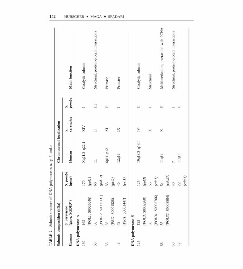
As discussed in the following section, eukaryotic pols are heteromultimerscomposed of a large subunit and a variety of smaller subunits (see also Table 2).The latter have been implicated in stabilization of the catalytic subunit, estab-lishment of protein-protein interactions, cell-cycle regulation, and even check-point function.
The large subunits of pols �, �, and � can be grouped into three distinctsubfamilies within the B-type pols. Pol � is the most conserved, with identityranging from 93% between human and mouse, to 49% between human and yeast(15). The identity between human and yeast genes for pol � is only 35% and forpol � 39%. The N-terminal part of pol � (amino acid residues 1–305) is generallypoorly conserved among �-like pols, but it contains three regions of highhomology: a nuclear targeting signal (NTS) and the so-called NT-1 and NT-2regions. The C-terminal part (amino acids 850–1105) is more conserved, withthree highly identical regions termed CT-1 to 3 and a zinc-finger domain (ZnF2),which is 89% identical between human and yeast pol �. C-terminal zinc-fingerdomains are also present in all �- and �-like pols. Both the N- and C-terminalparts of pol � show about 25% identity between human and yeast. The C-terminalregion of pol � contains 1000 extra amino acids, is found only in pol � subfamilymembers, and contains a highly acidic region (residues 1918–1948) and thezinc-finger domain (residues 2125–2222). Thus, both from sequence and struc-tural analysis, it appears that the catalytic subunits of eukaryotic pols arecomposed of a central domain that is evolutionarily very conserved.
A Mechanistic Perspective: DNA Polymerases Are Built byAddition of Specific Domains to a Conserved Core withEssential Catalytic Activity
STRUCTURAL AND FUNCTIONAL CONSERVATION WITHIN THE CATALYTIC CORE
Pols are template-directed enzymes that catalyze for phosphoryl transfer. Hencesince they have the ability to synthesize long polymers of nucleoside monophos-phates, whose linear spatial disposition is dictated by the sequence of thecomplementary template DNA strand (16). Their overall structure has beenoptimized through evolution to suit the specialized tasks each pol performswithin the cell. The most conserved domains are usually responsible for essentialbasic catalytic functions, whereas more divergent parts have evolved indepen-dently to fulfill specific roles (14).
The phosphoryl transfer reaction that lies at the heart of the polymerizationmechanism is catalyzed by a two-metal-ion mechanism (13, 17). Two Mg2� ionsform a pentacoordinated transition state with the phosphate groups of the
138 HUBSCHER y MAGA y SPADARI

incoming nucleotide, through interaction with conserved carboxylate residues inregion I and region II. Besides the conserved chemical step catalyzed by twometal ions, another common feature of all pols is the concerted movement of thefinger subdomains that rotate toward the palm to switch from an “open” to a“closed” conformation, forming the binding pocket for the incoming dNTP.Resolution of the structure of RB69 pol complexed with primer/template DNAand dTTP shows that upon formation of the ternary complex, the fingers domainrotated 60° toward the palm, resulting in a movement of the finger tips of 30 Å.The thumb domain also rotated toward the palm by 8°. The resulting closedconformation allows the interaction of conserved residues of the fingers with thedNTP binding site and the exonuclease domain. In addition the thumb is wrappedaround the minor groove of the primer/template DNA duplex. The pol � and �catalytic subunits both contain a 3� 3 5� proofreading exonuclease domain attheir N terminus. In the known crystal structures of family B members, thisdomain is folded around a central �-sheet that contains the active site and,together with the pol domain, creates a ring-shaped structure with a central hole,where the template/primer duplex DNA is positioned. The catalytic mechanismleading to the removal of the last incorporated nucleotide by the exonucleaseactivity is a phosphoryl transfer catalyzed by two metal ions, analogous to theone responsible for polymerization. The 3�3 5� exonuclease activity allows thepol to remove misincorporated nucleotides, ensuring the high fidelity of DNAsynthesis required for faithful genome replication.
Thus, during DNA synthesis, pol � and � repetitively shuttle between apolymerizing and an editing mode, and the balance between these two activitiesis regulated by a competition for the 3� end of the primer between the exonu-clease and polymerase active sites (18). These two different functional states ofexonuclease-containing pols are also reflected at the structural level. The duplexDNA occupies the same position adjacent to the thumb in either the editing or thepolymerizing mode, whereas the 3� end is bound to the exonuclease or polymer-ase active sites, respectively, which can be separated by more than 30 Å. A modelfor the coordinated action between the polymerase and exonuclease activities offamily B pols can be constructed by comparing the structure of RB69 in itspolymerizing (19) and editing (20) modes. A mismatched base pair prevents thefingers from rotating toward the palm to bind the incoming dNTP. This leaves the3� mismatched end available for binding to the exonuclease active site, whichremoves the wrong nucleotide. During the switch between polymerizing andediting modes, the DNA moves toward the exonuclease active site with a rotationin the double-helix axis. This movement is aided by the tip of the thumbsubdomain, which holds contact with the DNA during the movement, guiding iton a path between the two sites.
MOLECULAR ARCHITECTURE OF POL � In all eukaryotic organisms, pol � is aheterotetrameric enzyme (Table 2). Three separate domains were identified in thecatalytic p180 subunit: (a) an N-terminal domain (amino acids 1–329), which
139EUKARYOTIC DNA POLYMERASES

appears dispensable for both the catalytic activity and the assembly of thetetrameric complex; (b) a central domain (amino acids 330–1279), whichcontains all the conserved regions responsible for DNA binding, dNTP binding,and phosphoryl transfer (see above); and (c) a C-terminal domain (amino acids1235–1465), which is dispensable for catalysis but necessary for the interactionwith the other subunits. The heterotetrameric pol � is unique among eukaryoticpols, since two of the three small subunits have DNA primase activity. Theeukaryotic DNA primase is a heterodimeric enzyme with subunits (in humancells) of 49 and 55 kilodaltons (kDa) (21). Since DNA primases were reviewedlast year in this series we do not discuss these enzymes further (22). Theheterodimeric DNA primase is associated with the catalytic 180-kDa subunit andthe B subunit (21). Like the corresponding polypeptides in pol � and pol �heteromultimers, the B subunit of pol � has no detectable enzymatic activity, butis essential in yeast and appears to have a role in maintaining a functionalheterotetrameric complex. In addition, the finding that it is phosphorylated in acell-cycle-dependent manner suggests regulatory functions (23).
Studies of coexpression of all four mammalian subunits led to a model for thedynamic assembly of the heterotetrameric pol �/primase. The mouse primase p55subunit directly interacts with the catalytic p180 subunit and the second primasesubunit p48. In addition, the p68 B subunit directly contacts p180 and p55/p48.These interactions are essential for tethering the heterodimeric primase to thelarge catalytic subunit. The p55/p48 can translocate into the nucleus indepen-dently from the other subunits, by virtue of its own nuclear localization signallocated in p55, but interaction of p180 and p68 is required for their nuclearlocalization, since both subunits are cytoplasmic when expressed separately.
MOLECULAR ARCHITECTURE OF POL � Five subunits have been identified inSchizosaccharomyces pombe pol � (p125, p55, p54, p40 and p22) (24) four inmammalian cells (p125, p66, p50, and p12) (25, 26), and three in Saccharomycescerevisiae (p125, p58, and p55) (27) (see Table 2, pp 142–43). Recently, aunified nomenclature for these subunits was proposed (28). Pol � is presentwithin the eukaryotic cell in high-molecular-weight complexes and the smallsubunits might be critical for their maintenance. In S. cerevisiae it has beenshown that a complex of the p125 and p58 subunits is a dimer in solution,whereas addition of the third p55 subunit generates a high-molecular-weightcomplex that is a dimer of the heterotrimer p125/p58/p55. In S. pombe it has beenshown that the complex of the p125, p55, and p22 subunits is a trimer in solutionand that addition of the p54 subunit generated a dimer of the heterotetramer. Bothdimeric forms of the S. cerevisiae heterotrimer (27) and of the S. pombeheterotetramer (29) were more efficient and processive in DNA synthesis thanthe corresponding monomeric forms. In mammalian cells, the three smallsubunits identified so far, p66, p50, and p12, are homologous to, respectively, thep55, p40, and p22 subunits of the S. pombe enzyme. A study of purified pol �from mammalian tissue revealed that the native form of the enzyme had a
140 HUBSCHER y MAGA y SPADARI

molecular weight between 250,000 and 500,000, which suggests that in mam-malian cells pol � also forms a dimeric pol (26).
MOLECULAR ARCHITECTURE OF POL � Pol � is composed of four subunits, p261,p59, p17, and p12 in human cells (30, 31) and p256, p80, p23, and p22 in S.cerevisiae (32) (see Table 2). In both cases the two smaller subunits interact withthe larger two, forming a heterotetrameric complex. Biochemical analysisrevealed that the p80 subunit alone or the heterodimer p256/p80 can form dimers,which suggests that p80 might be responsible for the formation of a dimeric pol.
Coordinated Leading and Lagging Strand Synthesis and theDNA Polymerase Switch Mechanism: Distinct Roles forDNA Polymerases �, �, and �
Pol �/primase associates with the initiation complex at the DNA origin (33) andstarts to synthesize a short RNA/DNA hybrid of approximatively 10 RNAnucleotides followed by 20 to 30 DNA nucleotides. This oligonucleotide is thenutilized by pol � or � for processive elongation on both the leading and thelagging strand (34). Replication on the lagging strand is characterized by smallDNA pieces called Okazaki fragments, with a length of about 200 bases. Inmammalian cells an initiation event has to happen 4 � 104 times on the leadingstrand (approximately the number of origins of DNA replication in a mammaliancell), but it has to be repeated at the beginning of each Okazaki fragment (about2 � 107 times in mammalian cells).
The substitution of pol �/primase by the more processive pol � holoenzymeis called pol switch and is dependent upon the synthesis of the RNA/DNA primerby pol �. The pol switch is coordinated and regulated by an ATP switchcatalyzed by the auxiliary protein RF-C (35) and involves a complex network ofinteractions among pol �/primase, pol �, RF-C, and the protein that bindssingle-stranded DNA, replication protein A (RP-A) (reviewed in 5).
Both pol �/primase and pol � are perfectly suited for their respective roles: Pol�/primase can initiate synthesis de novo, whereas pol �, through its interactionwith proliferating cell nuclear antigen (PCNA, a processivity factor), has theability to synthesize long stretches of DNA. The proposed dimerization of pol �(27, 29) might play a role in the coordination of leading and lagging strandsynthesis (like in the pol III holoenzyme in E. coli) and in establishing anasymmetric replication fork, possibly through association of pol �/primase to oneof the two halves of the dimeric pol �.
Genetic analysis in budding yeast has shown that pol � is required for DNAreplication (36). In addition, UV cross-linking studies with replicating chromatinin mammalian cells detected pol � along with pol � and pol � (37). Experimentsin human cells (38) and in Xenopus egg extracts (39) further suggested that pol� is involved in DNA replication. The N-terminal part of yeast pol �, includingthe conserved core domains with all the catalytically important residues, is
141EUKARYOTIC DNA POLYMERASES
TA
BL
E2
Subu
nit
stru
ctur
eof
DN
Apo
lym
eras
es�
,�
,�,
and
�
Subu
nit
com
posi
tion
(kD
a)C
hrom
osom
allo
caliz
atio
n
Mai
nfu
ncti
onH
uman
S.ce
revi
siae
(gen
e,SG
DID
a)
S.po
mbe
(gen
e)H
uman
S. cere
visi
aeS. po
mbe
DN
Apo
lym
eras
e�
180
165
170
Xq2
1.3–
q22.
1X
IVI
Cat
alyt
icsu
buni
t
(PO
L1,
S000
5046
)(p
ol1)
6886
6611
IIII
ISt
ruct
ural
,pr
otei
n-pr
otei
nin
tera
ctio
ns
(PO
L12
,S0
0001
31)
(pol
12)
5558
556p
11–p
12X
III
Prim
ase
(PR
I2,
S000
1528
)(p
ri2)
4849
4512
q13
IXI
Prim
ase
(PR
I1,
S000
1447
)(p
ri1)
DN
Apo
lym
eras
e�
125
125
125
19q1
3.3–
q13.
4IV
IIC
atal
ytic
subu
nit
(PO
L3,
S000
2260
)(p
ol3)
5855
XI
Stru
ctur
al
(PO
L31
,S0
0037
66)
(cdc
1)
6655
5411
q14
XII
Mul
timer
izat
ion,
inte
ract
ion
with
PCN
A
(PO
L32
,S0
0038
04)
(cdc
27)
5040
7I
Stru
ctur
al,
prot
ein-
prot
ein
inte
ract
ions
1222
11q1
3II
(cdm
1)
142 HUBSCHER y MAGA y SPADARI

TA
BL
E2
Con
tinu
ed
Subu
nit
com
posi
tion
(kD
a)C
hrom
osom
allo
caliz
atio
n
Mai
nfu
ncti
onH
uman
S.ce
revi
siae
(gen
e,SG
DID
a)
S.po
mbe
(gen
e)H
uman
S. cere
visi
aeS. po
mbe
DN
Apo
lym
eras
e�
261
256
250
12q2
4.3
XIV
IIC
atal
ytic
subu
nit
(PO
L2,
S000
5206
)(p
ol2/
cdc2
0)
5980
6714
q21–
q22
XV
IM
ultim
eriz
atio
n
(DP
B2,
S000
6379
)(d
pb2)
1723
9q33
IISt
ruct
ural
,pr
otei
n-pr
otei
nin
tera
ctio
ns
(DP
B3,
S000
0482
)
1222
232p
12IV
IISt
ruct
ural
,pr
otei
n-pr
otei
nin
tera
ctio
ns
(DP
B4,
S000
2528
)(d
pb4)
DN
Apo
lym
eras
e�
139.
514
3.5
116.
315
q25
XV
III
Cat
alyt
icsu
buni
t
(MIP
1,S0
0058
57)
(pol
G)
5517
qPr
oces
sivi
ty
a SGD
ID,
Sacc
haro
myc
esce
revi
siae
geno
me
data
base
iden
tifica
tion.
143EUKARYOTIC DNA POLYMERASES

dispensable for viability, whereas the C-terminal part, which is involved inprotein-protein interactions and checkpoint control, is essential in S. cerevisiae(40). S. pombe mutants with N-terminal deletions in pol � are viable, as in S.cerevisiae, but show accumulation of DNA damage and need expression of thecheckpoint genes rad3, hus1, and chk1 (41). These data do not necessarily meanthat the catalytic activity of pol � is not involved in DNA replication, but rathersuggest than it can be substituted for the basic synthetic function and not for thespecialized checkpoint function. During evolution pol � could have acquiredspecialized functions as a “sensor” pol for quality control of DNA replication,whereas the function of pol � remained as a DNA synthesizing machineexclusively.
Fidelity of DNA Synthesis: Novel Roles for AccessoryProteins
As fidelity of pols has been reviewed in depth recently (42), we concentrate ona few recent developments. Genetic analysis has shown that fidelity is relevantfor DNA replication (43), since mutations affecting the exonuclease activity ofpol � and pol � result in high mutation rates in vivo. Moreover, transgenic micewith exo mutations of alleles for pol � showed a striking increase in cancersusceptibility within 12 months of age (44). Because pol � is essential for DNAreplication in vivo but lacks a proofreading activity, its infidelity poses a risk tothe cell of accumulating dangerous mutations during DNA replication. Biochem-ical studies have revealed that RP-A can physically interact with pol � andstabilize its binding to a primer end, at the same time reducing the ability of pol� to incorporate a wrong nucleotide (45). Thus, it is conceivable that both theerror-free Okazaki fragment maturation and fidelity clamp function of RP-Acooperate to prevent misincorporation by pol �. Interestingly, the same biochem-ical parameter (increased affinity of the enzyme for the DNA template) can havetwo opposite effects: increase in fidelity in the case of the RP-A/pol � interactionand decrease in fidelity in the case of the PCNA/pol � interaction (46). From anevolutionary point of view, it is meaningful that maximization of processivity ofpol � might have been the main goal to be achieved. The higher rate ofmisincorporation could be compensated for by the associated proofreadingexonuclease activity. In the case of pol �, the crucial target may have been toachieve a higher level of fidelity, whereas the increase in processivity was lessimportant because of the limited size of the products synthesized.
The Matchmaker Concept for Establishing a MovingPlatform: Replication Factor C and Proliferating CellNuclear Antigen
REPLICATION FACTOR C Replication factor C (RF-C) is a heteropentamericcomplex composed of one large subunit (p140/RFC1) and four smaller ones
144 HUBSCHER y MAGA y SPADARI

(p40/RFC4, p38/RFC5, p37/RFC2, and p36/RFC3), which share considerablesequence similarity with each other as well as with their bacterial clamp loadercounterparts in the pol III �-complex (reviewed in 5). RF-C is a clamp loader anda matchmaker ATPase, which can load the sliding clamp PCNA onto DNA.RF-C-catalyzed PCNA loading is obligatory for the assembly of pol � onto theDNA template to form the processive holoenzyme that acts during DNAsynthesis of both leading and lagging strands at the replication fork (reviewed in47). RF-C dissociates from PCNA after loading it onto the DNA and does notremain directly associated with the pol � core (48). Electron microscopic studiessuggested that in the absence of ATP, RF-C has a closed two-finger structurecalled the U form. This U form is converted into a more open C form uponbinding of ATP. PCNA can be held between these two fingers, and a structuralchange in the RF-C conformation can open the PCNA ring so that it can encirclethe DNA (49). Studies of the homologous system in E. coli (50), as well as thecrystal structure of the �� subunit of the E. coli �-complex (51), have giveninsights into how this may take place. The �-complex can, upon ATP binding,associate with and open up the �-clamp (the structural and functional homologof PCNA). The �-complex/�-clamp/ATP complex then associates with theprimer terminus and forms a ternary complex with the DNA. The DNA bindingstimulates ATP hydrolysis, which ejects the �-complex and leaves � on theDNA. In addition, the roles of the five subunits in the �-complex (�, �.��, � and�) have been identified to a great extent, and it seems that sequential ATPhydrolysis can drive the exact assembly of the �-clamp around the DNA (52).The �-subunit of the �-complex was identified as the clamp unloader (53) andmight have an analogous role as the RF-C p40 subunit, which is also capable ofunloading PCNA from the DNA (54). Moreover, as discussed above, RF-C islikely responsible for the polymerase switch.
DNA replication is not the only pathway requiring RF-C. As discussed inmore detail below, PCNA is also involved in a number of DNA repair pathways,distinct from DNA replication, and since PCNA loading is required for all thesefunctions, RF-C is also an important component of these pathways. Twowell-documented examples are nucleotide excision repair (NER) (55) and long-patch base excision repair (BER) (56). Finally, RF-C may have a role incheckpoint control, since the S. cerevisiae Rfc5p has been shown to interact withSpk1p, an essential protein kinase for the transition from S phase to mitosis (57),whereas the S. cerevisiae RFC2 gene is required for an S-phase checkpoint (58)and the S. pombe Rfc2p has been shown to play a key role in a DNA replicationcheckpoint (59).
PROLIFERATING CELL NUCLEAR ANTIGEN PCNA is a homotrimeric ring-shapedprotein with a molecular mass of 29 kDa for each monomer that occupies 120°in the ring (reviewed in 60). Each monomer is composed of two domains. Thecrystal structure of S. cerevisiae (61) and human PCNA (62) revealed how PCNAcan carry out its sliding clamp function on DNA by forming a trimeric ring that
145EUKARYOTIC DNA POLYMERASES

encircles the DNA strand without making direct contact with it. In addition to itscrystal structure being solved, a wealth of information about the involvement ofPCNA in replication has accumulated and the list of PCNA interactors continuesto grow rapidly. At least 30 proteins appear to interact with PCNA (60), althoughthe exact function of most of these interactions has not been clarified. Here wediscuss only the interactions of PCNA with pols and RF-C.
PCNA contacts the three multisubunit proteins pol �, pol �, and RF-C(63–65), and the common interaction domain has been mapped to a region on theouter front surface of PCNA, involving the loop that connects the two domainsof each PCNA monomer and a loop immediately preceding the C terminus. Inaddition, pol � has been shown to interact with a loop on the back side of PCNA(66). An interesting question involves the site of PCNA interaction in the pol �holoenzyme, and apparently there are multiple sites of interaction. The structureof the prokaryotic and bacteriophage sliding clamps are very similar to eukary-otic PCNA and have almost superimposable structures. The �-subunit of pol IIIholoenzyme is a dimer with 180° for each monomer (67), and the gp45 ofbacteriophage T4 is a trimer (68). An important role for the linker in theattachment of the pol to the clamp ring has been demonstrated by using amodeling approach to a molecular replacement search, combining the structure ofthe sliding clamp of gp45 of bacteriophage RB69 (a close relative of T4 and tothe family of B pols such as pol �, pol �, and pol �), with the RB69 pol structure.This analysis indicated that the interaction between the pol and the sliding clampresembles the interaction of the human kinase inhibitor p21 and PCNA, whichsuggests that the pol/sliding clamp interaction has been conserved in evolutionover 109 years. Since the clamp structure is perfectly conserved from bacteria tohuman, two properties of the E. coli �-subunit could also be extended to humanPCNA and pol �. First, the �-complex (the RF-C homolog) can load the�-subunit (the PCNA homolog) onto a primer with a length of 10 nucleotidesonly if no steric hindrance by another protein is there. In such a situation 14–16nucleotides are required. The complete pol III holoenzyme needs 22 nucleotidesfor a successful loading followed by DNA synthesis (69). Second, clamps aredynamic in handling secondary structures. Small obstacles (stems, loops, flaps,and bubbles) can be overcome by the pol III holoenzyme; this is likely to occursince this enzyme has strand displacement activity (70). Because pol � also hasstrand displacement activity (71), one could expect an analogous handling offrequently occurring obstacles in eukaryotic chromosomes.
DNA Polymerase �, The Mitochondrial Replicase
Pol � is a heterodimeric protein composed of a large subunit, responsible for thecatalytic activities, and a small accessory subunit. The large subunit of human pol� has been cloned and the gene mapped to the chromosomal location 15q25(Table 2) (72). It contains 1239 amino acids, with a calculated molecular mass of139.5 kDa. The amino acid sequence in human cells is 42%, 43%, 49%, and 78%identical to those of S. pombe, S. cerevisiae, D. melanogaster, and the C-terminal
146 HUBSCHER y MAGA y SPADARI

half of G. gallus, respectively. The large subunit was recombinantly expressedand the enzyme (p140) demonstrated DNA polymerase and both 5�3 3� and 3�3 5� exonuclease activities (73). Pol � is also able to catalyze the removal of a5�-deoxyribose phosphate (74). The p55 subunit was shown to stimulate both thepolymerase and exonuclease activities of p140. The crystallographic structure ofthe pol � p55 subunit from mouse cells showed that the dimerization domainfolded into a four-helix bundle and that dimerization was necessary for pol �stimulation (75). Thus the p55 subunit contains separate domains for binding top140, dimerization, and DNA binding.
Further Functions of DNA Polymerases �/Primase, �, �,and �: A Coordinated Interplay
CELL-CYCLE REGULATION DNA replication and chromosome segregation arehighly coordinated and interdependent. DNA replication enzymes (and theiraccessory factors) can trigger S-phase arrest when they are stalled at DNA lesionsin damaged DNA. However, they are also the targets of inhibitory signalstriggered by external stimuli, which can block S-phase progression to allow DNArepair or, alternatively, apoptosis. Increasing evidence points to a critical role forpol � in integrating DNA replication and cell-cycle regulation (reviewed in 76).The S. cerevisiae protein kinase Rad53 modulates the replication apparatus forthe lagging strand by controlling phosphorylation of the pol �/primase complexin response to intra-S-phase DNA damage (77). According to these results, the S.pombe Rad53 homolog cdc1� was shown to be a suppressor of a temperature-sensitive mutant of pol � (78). Genetic studies in S. cerevisiae (79) and in S.pombe (80) suggested that pol � is likely also a target of checkpoint controls,through its two associated primase subunits. Additional evidence came from astudy in Xenopus laevis extracts, where chromatin was found to be competent toinitiate a checkpoint response only after the DNA was unwound and pol � hadbeen loaded. Checkpoint induction did not require de novo DNA synthesis on thetemplate strand, but did require RNA primer synthesis by primase (81).
Pol � is another important sensor for UV damage and DNA replication blocksduring S phase. This checkpoint function has been mapped to the C-terminaldomain of pol � (see above) (82). As already mentioned, a pol � mutant with adeletion encompassing the entire N-terminal half is viable in yeast, whichsuggests that another pol can compensate for the catalytic role of pol � (83).Interestingly, S. pombe mutants with an N-terminal deletion in pol � were able toreplicate but accumulated damages, which suggests a major defect during DNAsynthesis. In contrast, mutations affecting the C-terminal region do not present anintact S-phase checkpoint (41). In S. cerevisiae cells, a physical complexcontaining the checkpoint protein Dpb11 and pol � has been detected (84).Moreover, mutations affecting the small subunit Dpb4p of pol � are lethal in aDpb11 mutant genetic background (85). During the S phase of the cell cycle,Dpb11 associated preferentially with DNA fragments containing autonomous
147EUKARYOTIC DNA POLYMERASES
replicating sequences (ARSs) at the same time as pol � does. Association ofDpb11 and pol � with these fragments was found to be mutually dependent.Moreover, Dpb11 was shown to be required for the association of pol �/primasewith the ARS (86), providing a possible link between the checkpoint functions ofthese two pols.
Activity of replicative pols is also regulated in a cell-cycle-dependent manner(87). Posttranslational modification by phosphorylation is well documented forpol �/primase (88). S. pombe pol �/primase is a phosphoprotein; serine is itsexclusive phosphoamino acid. Pol �/primase was found to be phosphorylated toa threefold higher level in late-S-phase cells compared with cells in the G2 andM phases. Moreover, the phosphorylation sites of pol �/primase in late-S-phasecells were not the same as those in G2/M-phase cells. The p180 catalytic subunitis phosphorylated throughout the cell cycle and the p66 subunit only in mitosis.The p68 subunit homolog in yeast undergoes a complex regulation (89). It ispresent in two forms, of 86 and 91 kDa: The p91 form results from cell-cycle-regulated phosphorylation of p86, whereas the p86 subunit present in G1 arisesby dephosphorylation of p91 while cells are exiting from mitosis, becomesphosphorylated in early S phase, and is competent and sufficient to initiate DNAreplication. However, another pol of p86 is also transiently synthesized as aconsequence of periodic transcription of the POL12 gene and is phosphorylatedno earlier than G2. This phosphorylation event requires binding of p86 to thep180 subunit.
Pol � is also a phosphoprotein and is most actively phosphorylated during theS phase. Physical interaction between pol � and cyclin-dependent kinases hasbeen detected and the interaction domains mapped (90).
DNA REPAIR Three types of excision repair are acting in the cell (91): BER,NER, and mismatch repair (MMR), each of them targeted to specific DNAlesions. A common feature of all these mechanisms is that DNA synthesis isrequired in order to replace the damaged DNA with a faithful copy of the intactstrand. Genetic and biochemical studies are consistent with an involvement ofboth pols � and � in NER and BER (92). For example, either pol � or � can beused to reconstitute long-patch BER (56) and NER in vitro (55). Because of itslimited activity in strand displacement, for in vitro reconstitution studies pol �seemed to be more efficient in long-patch BER, whereas pol � achieved a higherefficiency in gap-filling synthesis in NER reactions. In vitro complementationassays also showed the ability of pol � to reconstitute MMR in deficient extracts(93). Finally, genetic evidence from S. cerevisiae suggested that pol �/primase isinvolved in double-strand break repair (DSBR), which could be accomplishedthrough generation of a structure like a replication fork (94).
In contrast to the chromosomal DNA, the mitochondrial DNA (mtDNA) hasa 10-fold higher rate of nucleotide substitution. Pol �, with its associated 3� 35� proofreading exonuclease (95), participates in mtDNA repair. When muta-genic uracil residues are incorporated in mtDNA, subsequent to actions of
148 HUBSCHER y MAGA y SPADARI

uracil-DNA glycosylase and apurinic/apyrimidinic endonuclease, human pol � isable to fill a single nucleotide gap in the presence of a 5�-terminal deoxyribosephosphate flap (96). In addition, the p140 subunit of human pol �, thanks to itsintrinsic 5�-deoxyribose phosphatase activity, catalyzes the release of the phos-phate residue from incised apurinic/apyrimidinic sites, thus producing a substratefor DNA ligase (74, 97).
DNA RECOMBINATION A particular kind of DNA repair is DSBR, which occursthrough a recombination-type mechanism (98). Genetic studies of yeast withtemperature-sensitive (ts) pol � mutants suggested that pol � might be involvedin DSBR. As mentioned above, there is genetic evidence that pol �/primase isalso important in DSBR (94). The repair of DSBs can be accomplished througha homologous recombination pathway termed break-induced replication (BIR),involving DNA synthesis initiated by the free end of the chromosomal fragment.The recipient chromosome has both strands newly synthesized, with the gener-ation of structures like a replication fork, requiring coordinated leading andlagging strand synthesis. Thus, the indications for a requirement of differentreplicative pols in DSBR might reflect the need to establish a fully functionalreplication fork at the site of the lesion.
TELOMERE MAINTENANCE Maintenance of the physical integrity of chromo-somal ends, the telomeres, is another critical event coupled to DNA replication(99). Although a specialized DNA polymerase, telomerase (Table 1), is respon-sible for the synthesis of telomeres in eukaryotic cells, recent evidence points toan active role of DNA replication enzymes as well in controlling telomere length(100). Mutations in the structural gene for pol � in S. cerevisiae caused anincrease in telomere length. Telomeric DNA synthesis by telomerase should betightly coregulated with the production of the opposite strand to prevent telo-merase from generating excessively long single-strand tails, which may bedeleterious to chromosome stability. Using a synthetic telomere DNA template,synthesis of the telomere complementary strand in whole mammalian cellextracts was shown to be inhibited by neutralizing antibodies to pol �. Purifiedpol �/primase was capable of catalyzing synthesis of the lagging strand with thesame requirements as those observed in crude cell extracts. In addition, mam-malian pol �/primase was shown to precisely initiate de novo synthesis of anRNA primer with adenosine, opposite the 3�-side thymidine in the G-richtelomere repeat 5�-(TTAGGG)(n)-3� and to synthesize the nascent DNA frag-ments by extending the primer. Moreover, pol �/primase extends the productDNA far beyond the length of the template DNA, which suggests a role intelomere expansion. Further evidence for a role of pol �/primase in telomeremetabolism comes from the observation that the S. cerevisiae telomere-bindingprotein Cdc13p interacts with the catalytic subunit of pol � and RP-A (101).
149EUKARYOTIC DNA POLYMERASES

DNA Polymerase �, The Prototype of a Repair Enzyme
Pol � is the smallest eukaryotic pol and is composed of a single 39-kDapolypeptide containing 335 amino acid residues (reviewed in 102). It consists oftwo domains: The 8-kDa N-terminal domain performs the 5�-deoxyribose phos-phatase activity (to remove the 5�-deoxyribose phosphate) and single-strandedDNA binding, whereas the large 31-kDa domain performs the pol activity. Pol �is able to fill short gaps in a distributive way and these gaps contain a5�-phosphate. The structure of pol � has been solved by X-ray crystallography(17, 103) and some of its structural characteristics were discussed in the section“A Mechanistic Perspective.”
PHYSIOLOGICAL FUNCTIONS OF POL � More than 20 years ago pol � wasproposed as a DNA repair enzyme (104). The constant danger of damagingDNA is counteracted by a variety of repair mechanisms such as NER, BER,DSBR, MMR, and recombinational repair (RR). Among these, BER as anessential mechanism relies to a great extent on pol � (105). BER involves theremoval of a single base and its replacement. Efficient repair of a uracil-guanine base pair present in a duplex oligonucleotide can be achieved invitro, via replacement of a single nucleotide (short-patch BER), by thesequential action of the human proteins uracil-DNA glycosylase, the apurinic/apyrimidinic endonuclease HAP 1, pol �, and either DNA ligase I or III. The5�-deoxyribose phosphatase activity of pol � removes the 5�-phosphate andrenders the unphosphorylated 5�-OH group into a substrate for DNA ligase.DNA synthesis as well as the 5�-deoxyribose phosphatase are coupled, andboth are essential for short-patch BER (106). The second BER pathway,which involves filling a gap of several nucleotides (long-patch BER) specif-ically requires the clamp PCNA, its clamp loader RF-C, and the Flapendonuclease 1 (Fen1). The efficiency of long-patch BER detected in extractsfrom pol �– deleted mouse cells, as well as the PCNA dependency of thispathway, strongly suggested the involvement of either pol � and/or � in theresynthesis step (see above). In this pathway Fen1 is required to cleave areaction intermediate generated by displacement of the template strand duringgap filling. This Fen1- and PCNA-dependent pathway can be reconstitutedwith pol � and � (56), but recent experiments also suggested that pol � mayact in this pathway in vivo, as also suggested by the reduced repair activityin pol �– deficient cells (107). Finally, Fen1 stimulates the strand displace-ment activity of pol �, which suggests a communication between these twoenzymes (108). In sum, it appears that the vital BER pathway has developedseveral strategies to repair damaged DNA and these strategies involvedifferent sets of DNA synthesis machineries (e.g. pol � alone, pol � withFen1, or pol � or pol � holoenzymes with Fen1).
Pol � has other roles besides BER (102). First, it has been implicated inmeiotic events associated with synapsis and recombination. Second, the 67-kDaS. cerevisiae homolog of mammalian pol � encoded by the nonessential POL4
150 HUBSCHER y MAGA y SPADARI

gene has been implicated in DSBR; it probably utilizes a nonhomologousend-joining mechanism. Third, mice carrying a targeted disruption of the pol �gene had growth retardation and died of a respiratory failure immediately afterbirth (109). The increased apoptotic cell death observed in the developing centraland peripheral nervous systems suggest that pol � plays an essential role inneurogenesis.
THE NOVEL DNA POLYMERASES
Discovery
The classical pols have inherent high fidelity and perform accurate DNAsynthesis. During DNA replication, however, there are situations where lesionsin DNA can impede the replication machinery. We have learned in the last fewyears that a variety of so-called lesion-replicating pols can overcome thereplication blocks (reviewed in 4, 110, and also in this volume, 111). These polsbelong to a group of four structurally related proteins that are found in all threedomains of life, the prokaryotes, the archaea, and the eukaryotes. Initially,genetic studies in E. coli showed that UmuC and DinB are involved in translesionDNA synthesis, whereas in S. cerevisiae, Rev1 and Rad30 were found to havesimilar roles (reviewed in 6). These genes were found to code for novel pols thatare able to replicate damaged DNA; this group is now called the Y-family ofDNA polymerases (11). The first identified translesion pol was the Rev3 andRev7 holoenzyme in yeast called pol � (112), which was shown to performthymidine dimer bypass (113). Translesion synthesis required an additionalenzyme called Rev1 (114), containing a template-directed deoxycytidyltrans-ferase activity, mainly incorporating C in front of abasic sites. The product ofRev1 can be extended by pol �.
Later, the Rad30 pathway that facilitates translesion synthesis was identifiedin yeast. This gene belongs to the Rad6 epistasis group, which is involved inpostreplication repair. Rad30 was identified as a pol and named pol �. Soonthereafter paralogs of pol � were identified in human cells: Rad30A for pol �,Rad30B for pol �, and DinB1 for pol .
Functions of DNA Polymerases �, �, �, , and Rev1, TheLesion-Replicating Enzymes
Replicative pols stop before a DNA lesion; at that point translesion pols are likelyattracted to damaged DNA. The duties to be covered by these pols include (a) theverification of the type of DNA damage [e.g. an abasic site, a thymine-thyminecyclobutane pyrimidine dimer, a cis-platinum adduct, an 8-oxoguanine adduct, oran N-2-acetyl aminofluorene (AAF) adduct], (b) the way the DNA is synthesizedover the lesion, and (c) how a lesion-terminated primer is extended so that the
151EUKARYOTIC DNA POLYMERASES

TABLE 3 The novel DNA polymerases and terminal deoxynucleotidyltransferasea
Pol Genes Functional tasks
� Rev3/Rev7 Developmental pol (nonredundant)
POLZb Cell proliferation pol
Bc Mismatch extender pol
Somatic hypermutation pol (low errors)
� Rad30A (polV)d Xeroderma pigmentosum variant pol
POLH Accurate mismatch pol
Y Somatic hypermutation pol (strand-biased hot-spot Amutations, less than pol )
Repair of interstrand cross-links
POLQ
A
� Rad30B Meiosis pol
POLI Pol that can incorporate opposite to lesions
Y Most error-prone pol
Violates Watson-Crick base pair rule
Somatic hypermutation pol (high errors)
DinB1 (polIV)d Deletion and base substitution pol
POLK Low fidelity and moderate processivity
Y Somatic hypermutation pol (strand-biased hot-spot Amutations, more than pol �)
� Repair in meiosis
POLL Homologous to pol �
X
� Homologous to TdT
POLM Lymphoid formation pol
X Somatic hypermutation pol?
1 Trf4-1 Homologous to pol �
POLS1 Stimulated by PCNA
X Fourth essential pol in yeast
Sister chromatid cohesion pol
2 Trf4-2 Homologous to pol �
POLS2 Sister chromatid cohesion pol
X
� POL5 Fifth essential pol in yeast
POLF Stimulated by RF-C and PCNA
B Not involved in replication
152 HUBSCHER y MAGA y SPADARI

replication machinery of pol � holoenzyme can resume DNA synthesis. SeeTable 3.
POL � Human pol � is the product of the XPV gene, which is mutated inpatients with xeroderma pigmentosum variant (XP-V), who have a predispositionfor skin cancer (115, 116). Cells from these patients are defective in thereplication of DNA synthesis over damaged DNA. Even though pol � is alimited-fidelity pol (117), if compared to the more accurate pol �, pol �, pol �,pol �, and pol �, it is a translesion pol with a high fidelity in replicating overdamaged DNA with certain types of lesions (118). Pol � incorporates the correctnucleotide over lesions such as a thymine-thymine cyclobutane pyrimidine dimer(119), a cis-platinum adduct, or an AAF adduct and continues chain elongation,whereas replicative pols cannot, which suggests that pol � has a dual function intranslesion synthesis and in elongation from a lesion (118). On the other hand, ifthe translesion synthesis by pol � is incorrect, elongation cannot be performed.This inability to elongate suggests that the fidelity of translesion synthesisincludes the two steps of DNA synthesis per se and subsequent elongation fromthe lesion. Pol � is also very efficient in bypassing O6-methylguanine formed bythe action of alkylating agents (120). Pol � can also bypass O6-methylguanine,but this bypass is less accurate since the chance that the right C base isincorporated opposite O6-methylguanine is twofold higher with pol � than withpol �.
Pol � was reported to be capable of performing translesion synthesis that iserror-free in general (121), but also of performing translesion synthesis that iserror-prone on 8-oxoguanine (122). The latter study also compared its ability tosynthesize over abasic sites and on (�)-trans-anti-benzo(a)pyrene-N2-dGuanine,which suggests a second role for pol � in translesion synthesis of mutagenicbypass in mammalian cells (122, 123). Both synthetic activity and lesion bypassare stimulated by the physical interaction between pol � and PCNA, which ismediated by a consensus PCNA-binding motif (124).
TABLE 3 Continued
Pol Genes Functional tasks
Eso1 Cohesion factor containing a domain similar to pol �
Rev1 Synthesis opposite an abasic site
REV1L
TdT Template-independent pol
aFor details see text and references therein.bHuman Genome Organization (HUGO) nomenclature.cDNA polymerase family A, B, X, or Y (see References 10 and 11).dCorreponding E. coli genes.
153EUKARYOTIC DNA POLYMERASES

Finally, it is interesting to note that pol � differs in the ability to bypass lesionsif one compares the corresponding enzymes from the single cellular organismyeast to multicellular organisms. Whereas yeast pol � predominantly incorpo-rates C opposite 8-oxoguanine, human pol � inserts C and A with similarefficiencies. Pol � from yeast favors incorporation of G opposite an apurinic site,whereas human pol � prefers A under the same conditions (120, 121).
Pol � is the first Y-family pol for which the crystal structure has been resolved(125) and the amino acids critical for activity and biological functions identified(126). This pol shows small and stubby fingers and thumb domains, with respectto the known pols.
Pol � has been found to colocalize uniformly in the nucleus and is associatedwith replication foci during S phase (127). When the cells were treated withDNA-damaging agents (UV, carcinogens), pol � accumulated at replication focithat stalled at DNA damages. Furthermore it was found that the C-terminal 70amino acids are essential for nuclear localization and the next 40 amino acids forrelocalization into the replication foci. These localization domains are importantsince two mutations in this region of pol � were found in XP-V patients.Inactivation of pol � could be a promising strategy to enhance the anticancerpotency of alkylating agents in cancer chemotherapy. Moreover, pol � deficiencyin XP-V uncovered an overlap between the S-phase checkpoint and double-strand break repair (128). It has also been postulated that replication errorsintroduced by pol � can be corrected by extrinsic exonucleases (129). Finally, acohesion molecule from fission yeast S. pombe called Eso1p consists of a domainthat is very similar to pol � (130) but its function is unknown.
POL Pol is the product of the DINB1 gene (131). Pol has a low fidelityof about 1:200 and performs a predominant T3 G transversion mutation at a rateof about 1:147 (132). Moreover, pol creates mismatches with high frequencyon undamaged DNA. Pol can neither bypass cis-syn or (6–4) thymine-thyminedimers nor cis-platin adducts. As for pol �, it was found that pol can passcertain lesions in an error-free and others in an error-prone way. Error-pronebypass was measured at abasic sites and at 8-oxoguanine lesions (132). Inerror-prone and error-free bypasses, A was preferentially incorporated. Error-freebypass is achieved with the AAF adduct, where pol incorporates either C or Tand less efficiently A, and with the (�)-trans-anti-benzo(a)pyrene-N2-dG adduct,where preferentially a C is incorporated opposite the lesion (133). Pol hasanother unique property: On the one hand it possesses a very low fidelity, but onthe other hand it is moderately processive (25 or more nucleotides). This propertysuggests an important role in spontaneous mutagenesis (134).
POL � Pol � is the gene product of RAD30B (135, 136). In sharp contrast to pol� and to a certain extent pol , pol � is a much less accurate translesion pol. Ithas been found that pol � can even violate the Watson-Crick base pairing rule,since it preferentially incorporates a G instead of the correct A opposite a
154 HUBSCHER y MAGA y SPADARI

template T (137, 138). This unexpected property might be of crucial importanceto replicate over 5MeC, which after deamination becomes a T. So the inaccuratepol � can incorporate a G opposite a T that might have been produced from a5MeC (139). Moreover, pol � can replicate over a thymine-thymine cyclobutanepyrimidine dimer and under certain conditions (e.g. GA instead of the correctAA) even extend wrongly synthesized lesions (140). This is in contrast to theaccurate translesion pol �. In a new model for mutagenic bypass it was proposedthat pol � acts in concert with pol � (see below): Pol � incorporates deoxyribo-nucleotides opposite DNA lesions, and pol � functions as a mispair extender(141) (see below), a property that the accurate replicative pols �, �, and � do nothave.
Human pol � can respond differently to various DNA lesions (138). First, itstops at an 8-oxoguanine lesion, and this is in contrast to pol �, pol , and evenpol �. Second, it can preferentially incorporate G opposite an abasic site. Third,it can preferentially incorporate a C opposite an AAF-adducted G, and fourth, itis largely unresponsive to TT dimers.
Pol � might contain a loose and flexible pocket as an active site, resulting inan extra low fidelity that can even violate the Watson-Crick base pair rule. On theother hand, this loose active pocket is able to fit certain types of damagedtemplates, resulting in the correct nucleotide incorporation opposite certainlesions. Finally, pol � could have its role in BER since it contains a secondenzymatic activity, the 5�-deoxyribose phosphatase, as do pol � (139), pol � (74),and pol � (see below).
POL � REV1 It was initially assumed that pol � is an error-prone translesion pol(113 and reviewed in 142). Subsequent experiments identified pol � as a mispairextending enzyme rather than a mispair inserting enzyme (141). It is likely thatpol � will extend translesion products that are not accurate, such as the manymisincorporations by pol � and the few misincorporations by pol � (see above).Rev1, on the other hand, is an enzyme that can incorporate a C at an abasic siteand has been shown to act in concert with pol � (114). Like pol � and pol �, Rev1is a translesion inserter and pol � again is the extender for the translesionproducts. A human homolog of the S. cerevisiae REV3 gene was shown toencode the catalytic subunit of pol � (143), and a human homolog of the S.cerevisiae REV1 also codes for a dCMP transferase that is dependent upon aDNA template (144). When the pol � catalytic subunit, the REV3 gene, wasdisrupted in transgenic mice it resulted in early embryonic lethality between days9.5 and 12.5 (145–147). One explanation for such an effect is that duringpropagation and differentiation through many cell divisions the cells mightgradually accumulate DNA damages in the absence of pol �. In utero, moreover,DNA lesions constantly form in the genome as a result of oxidative andhydrolytic processes. The experiments with transgenic mice clearly indicate thatpol � has a nonredundant function in development and this is likely the reasonwhy genetic diseases of pol � have never been detected.
155EUKARYOTIC DNA POLYMERASES

How Are the Functions of Lesion-Bypassing DNAPolymerases Coordinated?
Taking together the properties of all these pols, it is likely that pol � plays thepivotal role in error-free and accurate translesion replication. In wild-type cellsthe normal situation would first include accurate translesion replication by pol �with subsequent continuation of replication by pol � holoenzyme. Second,efficient bypass of an apurinic site was demonstrated by the combined action ofpol � and pol � (148). Third, when pol � is inaccurate, pol � has to extend thestrand before the replicative pol � can resume. Finally, the very error-prone pol� incorporates mismatches that are first extended by pol �, before replication bypol � can continue. It is conceivable that the latter two pathways lead toerror-prone bypasses. This could explain the predisposition of the XP-V patientsto cancer, since they lack the accurate translesion bypass by pol �. Why hasnature maintained pols that are inaccurate? It is quite obvious that DNA damageis deleterious to the cells. Therefore, it is not surprising that cells evolvedmultiple ways to repair damage, and some of these methods have redundantspecialties. In experiments performed in S. cerevisiae it was found that the cellrepairs spontaneous DNA damages by employing redundant functions of NER,BER, MMR, RR, DSBR, recombination, and translesion synthesis pathways.These experiments also suggested that the translesion pol � can introducemultiple mutations when bypassing spontaneous DNA damages in S. cerevisiae(149). As indicated above, the error-prone role of pol � is likely a consequenceof its property to extend mispaired bases rather than to misincorporate oppositelesions (141).
DNA Polymerases , �, �, , � and TerminalDeoxynucleotidyl Transferase, Enzymes with FurtherDistinct Functions
The other novel pols identified so far and terminal deoxynucleotidyl transferaselikely have roles other than translesion replication.
POL Pol is proposed to have a role in DNA repair of interstrand cross-links,but its mechanism is not known (150).
POL � Pol � shows homology to pol �. It has conserved critical amino acidresidues for DNA binding, nucleotide binding and selection, catalysis, anddeoxyribose 5�-phosphatase activity, which suggests a role in BER. Since pol �was preferentially expressed in testis, and it appears to be developmentallyregulated and associated to pachytene spermatocytes, a potential role in DNArepair during meiosis has been suggested (151).
POL � The discovery of pol � (152, 153) indicated that this pol has 41%identity to terminal deoxynucleotidyltransferase (TdT), but in contrast to TdT can
156 HUBSCHER y MAGA y SPADARI

efficiently be stimulated by adding a template DNA. Moreover, pol � ispreferentially expressed in peripheral lymphoid tissues, and in human cells alarge proportion of the expressed sequence tags correponding to this enzymederived from germinal center B cells. This makes pol � a candidate hypermutase,possibly involved in somatic hypermutation of immunoglobulin genes (152).Moreover, since the expressions of both pol � and pol � are down-regulated afterDNA damage occurred, it has been suggested that these enzymes are unlikely tohave a role in translesion DNA synthesis (154).
POL In S. cerevisiae the Trf4 gene encodes for pol , which appears to beinvolved in DNA synthesis during sister chromatid cohesion, thus having a rolein mitosis (155) and in chromosome segregation. Pol is required for buildingthe connection between sister chromatids. Moreover, it is encoded by an essentialgene, thus increasing the number of essential pols in eukaryotes to five (pol �, pol�, pol �, pol �, and pol ). Pol was initially called pol (155), but this nameis now reserved for the DINB1 gene product (see above). Finally, a second formof a Trf4 gene product has been found so that these enzymes are now called pol 1 and pol 2.
POL � Pol � has been identified in S. cerevisiae and its function has in part beenelucidated (A. Sugino, personal communication). The POL5 gene is essential foryeast cell growth. It encodes a polypeptide of about 130 kDa, which has a weaksimilarity to family B pols. Its product, when purified from S. cerevisiae cells,has an aphidicolin-sensitive DNA polymerization activity. Pol � is very muchdistributive, is error-free, but has no 3� 3 5� exonuclease activity. The activityis stimulated by addition of PCNA and RF-C on singly primed single-strandedDNA. The pol � exclusively localizes in the nucleolus. The cell morphology oftemperature-sensitive mutants of POL5 does not show a dumbbell shape, whichsuggests that this pol is not required for chromosomal DNA replication. S. pombehas a homologous gene to S. cerevisiae POL5, but so far no homologs in othereukaryotic cells have been identified.
TERMINAL DEOXYNUCLEOTIDYLTRANSFERASE This enzyme (abbreviated TdT) isa template-independent pol. It was first detected in connection with an unusualdeoxynucleotide-polymerizing activity in pol preparation of calf thymus in 1960.TdT has been found only in lymphoid tissues and is proposed to have a role insomatic hypermutation of immunoglobulin genes (156).
How Many DNA Polymerases Are Involved in the ImmuneSystem?
Somatic hypermutation of immunoglobulin genes increases variability and thusleads to an increase in antibody diversity. Recent data from several groups
157EUKARYOTIC DNA POLYMERASES

suggest that besides TdT (see above), at least the novel pol �, pol �, pol �, pol ,and pol � share the task of creating mutations in the immunoglobulin loci (157).
This topic is covered extensively by another review in this volume (111) andis therefore only briefly summarized here. (a) Pol � is an A-T mutator in somatichypermutation of immunoglobulin variable genes (158), and the mutation hotspots correlate with the pol � error spectrum (159). (b) Pol appears to have aneven greater bias to the A-T mutation, which suggests that it could contributeeven more to the variability of the immunoglobulin variable genes (159). (c) Thetranslesion extender pol � was found to play an important role in hypermutationof the immunoglobulin and the bcl-6 genes, since it was found that pol � isup-regulated and pol down-regulated by the B-cell-receptor engagement (160).(d) Pol � has an extremely low fidelity for nucleotide incorporation at the very endof a DNA template, which suggests its participation in hypermutation ofimmunoglobulin genes (161). (e) Double-strand break repair has been implicatedin somatic hypermutation. DSBs are usually repaired by homologous recombi-nation, which recruits an error-prone DNA polymerase (162). This modelproposes that a nonprocessive, low-fidelity DNA polymerase, such as pol �,performs the initial extension from hypermutation DSBR, resulting in clusteringof mutations near the DSBR. Pol � might then be replaced by a more lesion-processive DNA polymerase such as pol � or pol . (f) Finally, pol � might actas a hypermutator in somatic hypermutation (152, 153).
FUTURE DIRECTIONS
In the last few years we have witnessed quite an enlargement of the pol family.In particular, the members of the novel pol family await the elucidation of theirfunctional tasks. In the near future we would like to learn how the translesionpols can bypass the many lesions occurring in DNA, how pols are engaged incohesion DNA synthesis, and how pols are involved in immunoglobulin recom-bination. Knockout transgenic mice may yield informative phenotypes thatsuggest roles for these pols. Additional structural studies will reveal the activesites and their surroundings for pols that can bypass DNA lesions. For thereplicative pol �, pol �, and pol �, the exact subunit structures and their roles inDNA replication and the various DNA repair processes have to be worked out.The methods of choice include conditional knockout technologies and mutationaland biochemical analysis of higher-order DNA replication complexes.
ACKNOWLEDGMENTS
We thank Z. O. Jonsson, R. Freire, and F. Focher for critically reading themanuscript. The work in the authors’ labs was supported by an EU-TMR grant(ERBMRXCT 970125), by the Swiss National Science Foundation (to UH), by
158 HUBSCHER y MAGA y SPADARI

the Kanton of Zurich (to UH and GM), and by the CNR Project on Biotechnology(to SS).
The Annual Review of Biochemistry is online at http://biochem.annualreviews.org
LITERATURE CITED
Due to strict space and reference limitations it was impossible to cite manyoriginal references.
1. Alberts B. 1998. Cell 92:291–942. Waga S, Stillman B. 1998. Annu. Rev.
Biochem. 67:721–513. Baker TA, Bell SP. 1998. Cell 92:295–
3054. Hubscher U, Nasheuer HP, Syvaoja J.
2000. Trends Biochem. Sci. 25:143–475. Stucki M, Stagliar I, Jonsson ZO, Hub-
scher U. 2000. Prog. Nucleic Acid Res.Mol. Biol. 65:261–98
6. Woodgate R. 1999. Genes Dev.13:2191–95
7. Jonsson ZO, Hubscher U. 1997. BioEs-says 19:967–75
8. Mossi R, Hubscher U. 1998. Eur. J. Bio-chem. 254:209–16
9. Steitz TA. 1999. J. Biol. Chem. 274:17395–98
10. Braithwaite DK, Ito J. 1993. NucleicAcids Res. 21:787–802
11. Ohmori H, Friedberg EC, Fuchs RPP,Goodman MF, Hanaoka F, et al. 2001.Mol. Cell 7:7–8
12. Joyce CM, Steitz TA. 1994. Annu. Rev.Biochem. 63:777–822
13. Steitz TA. 1998. Nature 391:231–3214. Brautigam CA, Steitz TA. 1998. Curr.
Opin. Struct. Biol. 8:54–6315. Hindges R, Hubscher U. 1997. Biol.
Chem. 378:345–6216. Kelman Z, O’Donnell M. 1994. Curr.
Opin. Genet. Dev. 4:185–9517. Sawaya MR, Pelletier H, Kumar A, Wil-
son SH, Kraut J. 1994. Science 264:1930–35
18. Johnson KA. 1993. Annu. Rev. Biochem.62:685–713
19. Franklin MC, Wang J, Steitz TA. 2001.Cell 105:657–67
20. Shamoo Y, Steitz TA. 1999. Cell99:155–66
21. Arezi B, Kuchta RD. 2000. Trends Bio-chem. Sci. 25:572–76
22. Frick DN, Richardson CC. 2001. Annu.Rev. Biochem. 70:39–80
23. Mizuno T, Yamagishi K, Miyazawa H,Hanaoka F. 1999. Mol. Cell. Biol.19:7886–96
24. Zuo S, Gibbs E, Kelman Z, Wang TS,O’Donnell M, et al. 1997. Proc. Natl.Acad. Sci. USA 94:11244–49
25. Hughes P, Tratner I, Ducoux M, Piard K,Baldacci G. 1999. Nucleic Acids Res.27:2108–14
26. Mo JY, Liu L, Leon A, Mazloum N, LeeMY. 2000. Biochemistry 39:7245–54
27. Burgers PM, Gerik KJ. 1998. J. Biol.Chem. 273:19756–62
28. MacNeill SA, Burgers PM, Baldacci G,Hubscher U. 2001. Trends Biochem. Sci.26:16–17
29. Zuo S, Bermudez V, Zhang G, KelmanZ, Hurwitz J. 2000. J. Biol. Chem. 275:5153–62
30. Jokela M, Makiniemi M, Lehtonen S,Szpirer C, Hellman U, Syvaoja JE. 1998.Nucleic Acids Res. 26:730–34
31. Li Y, Pursell ZF, Linn S. 2000. J. Biol.Chem. 275:23247–52
32. Dua R, Edwards S, Levy DL, CampbellJL. 2000. J. Biol. Chem. 275:28816–25
33. Bell SP. 2002. Annu. Rev. Biochem.71:333–74
34. Waga S, Bauer G, Stillman B. 1994.J. Biol. Chem. 269:10923–34
159EUKARYOTIC DNA POLYMERASES

35. Maga G, Stucki M, Spadari S, HubscherU. 2000. J. Mol. Biol. 295:791–801
36. D’Urso G, Nurse P. 1997. Proc. Natl.Acad. Sci. USA 94:12491–96
37. Zlotkin T, Kaufmann G, Jiang Y, LeeMY, Uitto L, et al. 1996. EMBO J.15:2298–305
38. Pospiech H, Kursula I, Abdel-Aziz W,Malkas L, Uitto L, et al. 1999. NucleicAcids Res. 27:3799–804
39. Waga S, Masuda T, Takisawa H, SuginoA. 2001. Proc. Natl. Acad. Sci. USA98:4978–83
40. Kesti T, Flick K, Keranen S, Syvaoja JE,Wittenberg C. 1999. Mol. Cell 3:679–85
41. Feng W, D’Urso G. 2001. Mol. Cell.Biol. 21:4495–504
42. Kunkel TA, Bebenek K. 2000. Annu.Rev. Biochem. 69:497–529
43. Tran HT, Gordenin DA, Resnick MA.1999. Mol. Cell. Biol. 19:2000–7
44. Goldsby RE, Lawrence NA, Hays LE,Olmsted EA, Chen X, et al. 2001. Nat.Med. 7:638–39
45. Maga G, Frouin I, Spadari S, HubscherU. 2001. J. Biol. Chem. 276:18235–42
46. Mozzherin DJ, McConnell M, JaskoMV, Krayevsky AA, Tan CK, et al.1996.J. Biol. Chem. 271:31711–17
47. Hubscher U, Maga G, Podust VN. 1996.In DNA Replication Accessory Proteins,ed. ML De Pamphilis, pp. 525–43. ColdSpring Harbor, NY: Cold Spring HarborLab. Press
48. Podust VN, Tiwari N, Stephan S, Fan-ning E. 1998. J. Biol. Chem.273:31992–99
49. Shiomi Y, Usukura J, Masamura Y,Takeyasu K, Nakayama Y, et al. 2000.Proc. Natl. Acad. Sci. USA 97:14127–32
50. Turner J, Hingorani MM, Kelman Z,O’Donnell M. 1999. EMBO J.18:771–83
51. Guenther B, Onrust R, Sali A, O’DonnellM, Kuriyan J. 1997. Cell 91:335–45
52. Hingorani MM, Bloom LB, GoodmanMF, O’Donnell M. 1999. EMBO J.18:5131–44
53. Leu FP, Hingorani MM, Turner J,O’Donnell MO. 2000. J. Biol. Chem.275:34609–18
54. Cai JS, Gibbs E, Uhlmann F, Phillips B,Yao N, et al. 1997. J. Biol. Chem. 272:18974–81
55. Aboussekhra A, Biggerstaff M, ShivjiMK, Vilpo JA, Moncollin V, et al. 1995.Cell 80:859–68
56. Stucki M, Pascucci B, Parlanti E, FortiniP, Wilson SH, et al. 1998. Oncogene17:835–43
57. Sugimoto K, Shimomura T, HashimotoK, Araki H, Sugino A, Matsumoto K.1996. Proc. Natl. Acad. Sci. USA93:7048–52
58. Noskov VN, Araki H, Sugino A. 1998.Mol. Cell. Biol. 18:4914–23
59. Reynolds N, Fantes PA, MacNeill SA.1999. Nucleic Acids Res. 27:462–69
60. Warbrick E. 2000. BioEssays 22:997–1006
61. Krishna TSR, Kong X-P, Gary S,Burgers PM, Kuriyan J. 1994. Cell79:1233–43
62. Gulbis JM, Kelman Z, Hurwitz J,O’Donnell M, Kuriyan J. 1996. Cell87:297–306
63. Fotedar R, Mossi R, Fitzgerald P, Rous-selle T, Maga G, et al. 1996. EMBO J.15:4423–33
64. Mossi R, Jonsson ZO, Allen BL, HardinSH, Hubscher U. 1997. J. Biol. Chem.272:1769–76
65. Eissenberg JC, Ayyagari R, Gomes XV,Burgers PM. 1997. Mol. Cell. Biol.17:6367–78
66. Maga G, Jonsson ZO, Stucki M, SpadariS, Hubscher U. 1999. J. Mol. Biol. 285:259–67
67. Kong XP, Onrust R, O’Donnell M,Kuriyan J. 1992. Cell 69:425–37
68. Moarefi I, Jeruzalmi D, Turner J,O’Donnell M, Kuriyan J. 2000. J. Mol.Biol. 296:1215–23
69. Yao N, Hurwitz J, O’Donnell M. 2000.J. Biol. Chem. 275:1421–32
70. Yao N, Leu FP, Anjelkovic J, Turner J,
160 HUBSCHER y MAGA y SPADARI
O’Donnell M. 2000. J. Biol. Chem. 275:11440–50
71. Podust VN, Hubscher U. 1993. NucleicAcids Res. 21:841–46
72. Zullo SJ, Butler L, Zahorchak RJ,Macville M, Wilkes C, Merril CR. 1997.Cytogenet. Cell Genet. 78:281–84
73. Graves SW, Johnson AA, Johnson KA.1998. Biochemistry 37:6050–58
74. Longley MJ, Prasad R, Srivastava DK,Wilson SH, Copeland WC. 1998. Proc.Natl. Acad. Sci. USA 95:12244–48
75. Carrodeguas JA, Theis K, BodenhagenDF, Kisker C. 2001. Mol. Cell 7:43–54
76. Foiani M, Lucchini G, Plevani P. 1997.Trends Biochem. Sci. 22:424–27
77. Pellicioli A, Lucca C, Liberi G, MariniF, Lopes M, et al. 1999. EMBO J.18:6561–72
78. D’Urso G, Grallert B, Nurse P. 1995.J. Cell Sci. 108:3109–18
79. Marini F, Pellicioli A, Paciotti V, Luc-chini G, Plevani P, et al. 1997. EMBO J.16:639–50
80. Tan S, Wang TS. 2000. Mol. Cell. Biol.20:7853–66
81. Michael WM, Ott R, Fanning E, New-port J. 2000. Science 289:2133–37
82. Navas TA, Zhou Z, Elledge SJ. 1995.Cell 80:29–39
83. Kesti T, Flick K, Keranen S, Syvaoja JE,Wittenberg C. 1999. Mol. Cell 3:679–85
84. Araki H, Leem SH, Phongdara A,Sugino A. 1995. Proc. Natl. Acad. Sci.USA 92:11791–95
85. Ohya T, Maki S, Kawasaki Y, Sugino A.2000. Nucleic Acids Res. 28:3846–52
86. Matsumoto H, Sugino A, Araki H. 2000.Mol. Cell. Biol. 20:2809–17
87. Merrill GF, Morgan BA, Lowndes NF,Johnston LH. 1992. BioEssays14:823–30
88. Voitenleitner C, Rehfuess C, Hilmes M,O’Rear L, Liao PC, et al. 1999. Mol.Cell. Biol. 19:646–56
89. Ferrari M, Lucchini G, Plevani P, FoianiM. 1996. J. Biol. Chem. 271:8661–66
90. Wu SM, Zhang P, Zeng XR, Zhang SJ,
Mo J, et al. 1998. J. Biol. Chem. 273:9561–69
91. Lindahl T, Wood RD. 1999. Science286:1899–905
92. Wang Z, Wu X, Friedberg EC. 1993.Mol. Cell. Biol. 13:1051–58
93. Kolodner RD, Marsischky GT. 1999.Curr. Opin. Gen. Dev. 9:89–96
94. Holmes AM, Haber JE. 1999. Cell96:415–24
95. Vanderstraeten S, Van den Brule S,Hu J, Foury F. 1998. J. Biol. Chem.273:23690–97
96. Pinz KG, Bogenhagen DF. 1998. Mol.Cell. Biol. 18:1257–65
97. Pinz KG, Bogenhagen DF. 2000. J. Biol.Chem. 275:12509–14
98. Kanaar R, Hoeijmakers JH, VanGentDC. 1998. Trends Cell Biol. 8:483–89
99. Blackburn EH. 2000. Nature 408:53–55100. Martin AD, Dionne I, Wellinger RJ,
Holm C. 2000. Mol. Cell. Biol.20:786–96
101. Qi HY, Zakian VA. 2000. Genes Dev.14:1777–88
102. Wilson SH. 1998. Mutat. Res.407:203–15
103. Pelletier H, Sawaya MR, Amalendra K,Wilson SH, Kraut J. 1994. Science 264:1891–903
104. Hubscher U, Kuenzle CC, Spadari S.1979. Proc. Natl. Acad. Sci. USA76:2316–20
105. Sobol RW, Horton JK, Kuhn R, Gu H,Singhal RK, et al. 1996. Nature 379:183–86
106. Sobol RW, Prasad R, Evenski A, BakerA, Yang XP, et al. 2000. Nature 405:807–10
107. Dianov GL, Prasad R, Wilson SH, BohrVA. 1999. J. Biol. Chem. 274:13741–43
108. Prasad R, Dianov GL, Bohr VA, WilsonSH. 2000. J. Biol. Chem. 275:4460–66
109. Sugo N, Aratani Y, Nagashima Y, Kub-ota Y, Koyama H. 2000. EMBO J.19:1397–404
110. Lehmann AF. 2000. Gene 253:1–12
161EUKARYOTIC DNA POLYMERASES

111. Goodman MF. 2002. Annu. Rev. Bio-chem. 71:17–50
112. Morrison A, Christensen RB, Alley J,Beck AK, Bernstine EG, et al. 1989. J.Bacteriol. 171:5659–67
113. Nelson JR, Lawrence CW, Hinkle DC.1996. Science 272:1646–49
114. Nelson JR, Lawrence CW, Hinkle DC.1996. Nature 382:729–31
115. Washington MT, Johnson RE, PrakashL, Prakash S. 2001. Proc. Natl. Acad.Sci. USA 98:8355–60
116. Masutani C, Kusumoto R, Yamada A,Dohmae N, Yokoi M, et al. 1999. Nature399:700–4
117. Matsuda T, Bebenek K, Masutani C,Hanaoka F, Kunkel TA. 2000. Nature404:1011–13
118. Masutani C, Araki M, Yamada A,Kasumoto R, Nogomori T, et al. 1999.EMBO J. 18:3491–501
119. Johnson RE, Prakash S, Prakash L. 1999.Science 283:1001–4
120. Haracska L, Prakash S, Prakash L. 2000.Mol. Cell. Biol. 20:8001–7
121. Haracska L, Yu S-L, Johnson RE,Prakash L, Prakash S. 2000. Nat. Genet.25:458–61
122. Zhang Y, Yuan F, Wu X, Wang M,Rechkoblit O, et al. 2000. Nucleic AcidsRes. 28:4138–46
123. Yuan F, Zhang Y, Rajpal DK, Wu X,Guo D, et al. 2000. J. Biol. Chem. 275:8233–39
124. Haracska L, Kondratick CM, Unk I,Prakash S, Prakash L. 2001. Mol. Cell8:407–15
125. Trincao J, Johnson RE, Escalante CR,Prakash S, Prakash L, Aggarwal AK.2001. Mol. Cell 8:417–26
126. Kondratick CM, Washington MT,Prakash S, Prakash L. 2001. Mol. Cell.Biol. 21:2018–25
127. Kannouche P, Broughton BC, Volker M,Hanaoka F, Mullenders LHF, LehmannAR. 2001. Genes Dev. 15:158–72
128. Limoli CL, Giedzinski E, Morgan WF,
Cleaver JE. 2000. Proc. Natl. Acad. Sci.USA 97:7939–46
129. Bebenek K, Matsuda T, Masutani C,Hanaoka F, Kunkel TA. 2001. J. Biol.Chem. 276:2317–20
130. Tanaka K, Yonekawa T, Kawasaki Y,Kai M, Furuya K, et al. 2000. Mol. Cell.Biol. 20:3459–69
131. Livneh Z. 2001. J. Biol. Chem. 276:25639–42
132. Zhang Y, Yuan F, Xin H, Wu X, RajpalDK, et al. 2000. Nucleic Acids Res.28:4147–56
133. Ohashi E, Ogi T, Kusomoto R, Iwai S,Masutani C, et al. 2000. Genes Dev.14:1589–94
134. Ohashi E, Bebenek K, Matsuda T,Feaver WJ, Gerlach VL, et al. 2000.J. Biol. Chem. 275:39678–84
135. McDonald JP, Tissier A, Frank EG, IwaiS, Hanaoka F, Woodgate R. 2001. Phi-los. Trans. R. Soc. London Ser. B 356:53–60
136. McDonald JP, Rapic-Otrin V, EpsteinJA, Broughton BC, Wang XY, et al.1999. Genomics 60:20–30
137. Tissier A, McDonald JP, Frank EG,Woodgate R. 2000. Genes Dev.14:1642–50
138. Zhang Y, Yuan F, Wu X, Taylor J-S,Wang Z. 2001. Nucleic Acids Res.29:928–35
139. Bebenek K, Tissier A, Frank EK,McDonald JP, Prasad R, et al. 2001.Science 291:2156–59
140. Tissier A, Frank EG, McDonald JP, IwaiS, Hanaoka F, Woodgate R. 2000.EMBO J. 19:5259–66
141. Johnson RE, Washington MT, HaracskaL, Prakash S, Prakash L. 2000. Nature406:1015–19
142. Lawrence CW, Maher VM. 2001. Phi-los. Trans. R. Soc. London Ser. B 356:41–46
143. Gibbs PEM, Wang XD, Li ZQ, McMa-nus TP, McGregor WG, et al. 2000.Proc. Natl. Acad. Sci. USA 97:4186–91
144. Lin WS, Xin H, Zhang YB, Yuan FH,
162 HUBSCHER y MAGA y SPADARI

Wu XH, Wang ZG. 1999. Nucleic AcidsRes. 27:4468–75
145. Esposito G, Texido G, Betz UA, Gu H,Muller W, et al. 2000. Proc. Natl. Acad.Sci. USA 97:1166–71
146. Esposito G, Godin I, Klein U, YaspoM-L, Cumano A, Rajewsky K. 2000.Curr. Biol. 10:1221–24
147. Wittschieben J, Shivji MKK, Lalani E,Jakobs MA, Marini F, et al. 2000. Curr.Biol. 10:1217–20
148. Haracska L, Unk I, Johnson RE, Johan-sson E, Burgers PMJ, et al. 2001. GenesDev. 15:945–54
149. Harfe BD, Jinks-Robertson S. 2000.Mol. Cell 6:1491–99
150. Sharief FS, Vojta P-J, Ropp PA, Cope-land WC. 1999. Genomics 59:90–96
151. Garcia-Diaz M, Dominguez O, Lopez-Fernandez LA, de Lera LT, Sangier ML,et al. 2000. J. Mol. Biol. 301:851–67
152. Dominguez O, Ruiz JF, de Lera TL,Garcia-Diaz M, Gonzalez MA, et al.2000. EMBO J. 19:1731–42
153. Ruiz JF, Dominguez O, de Lera TL,Garcia-Diaz M, Bernad A, Blanco L.
2001. Philos. Trans. R. Soc. London Ser.B 356:99–109
154. Aoufouchi S, Flatter E, Dahan A, FailiA, Bertocci B, et al. 2000. Nucleic AcidsRes. 28:3684–93
155. Wang ZH, Castano IB, De Las Penas A,Adams C, Christman MF. 2000. Science289:774–79
156. Bollum FJ. 1978. Adv. Enzymol.47:347–74
157. Ishikawa T, Uematsu N, Mizukoshi T,Iwai S, Iwasaki H, et al. 2001. J. Biol.Chem. 276:15155–63
158. Zeng X, Winter DB, Kasmer C, KraemerKH, Lehman AR, Gearhart PJ. 2001.Nat. Immunol. 2:537–41
159. Rogozin IB, Pavlov YI, Bebenek K,Matsuda T, Kunkel TA. 2001. Nat.Immunol. 2:530–36
160. Zan H, Komori A, Li ZD, Cerutti A,Schaffer A, et al. 2001. Immunity14:643–53
161. Frank EG, Tissier A, McDonald JP,Rapic-Otrin V, Zeng XM, et al. 2001.EMBO J. 20:2914–22
162. Papavasilliou FN, Schatz DG. 2000.Nature 408:216–21
163EUKARYOTIC DNA POLYMERASES