Embed Size (px)
Citation preview
Translesion-synthesis DNA polymerases participatein replication of the telomeres in StreptomycesHsiu-Hui Tsai1, Hung-Wei Shu2, Chien-Chin Yang3 and Carton W. Chen1,*
1Department of Life Sciences and Institute of Genome Sciences, 2Department of Biotechnology and LaboratoryScience in Medicine, Institute of Biotechnology in Medicine, National Yang-Ming University, Shih-Pai,Taipei 11221, and 3Department of Chemistry, Chung-Yuan Christian University, Chung-li 32023, Taiwan
Received August 8, 2011; Revised September 22, 2011; Accepted September 23, 2011
ABSTRACT
Linear chromosomes and linear plasmids ofStreptomyces are capped by terminal proteins thatare covalently bound to the 50-ends of DNA.Replication is initiated from an internal origin,which leaves single-stranded gaps at the 30-ends.These gaps are patched by terminal protein-primedDNA synthesis. Streptomyces contain five DNApolymerases: one DNA polymerase I (Pol I), twoDNA polymerases III (Pol III) and two DNA polymer-ases IV (Pol IV). Of these, one Pol III, DnaE1, is es-sential for replication, and Pol I is not required forend patching. In this study, we found the two Pol IVs(DinB1 and DinB2) to be involved in end patching.dinB1 and dinB2 could not be co-deleted fromwild-type strains containing a linear chromosome,but could be co-deleted from mutant strains con-taining a circular chromosome. The resulting"dinB1 "dinB2 mutants supported replication ofcircular but not linear plasmids, and exhibited in-creased ultraviolet sensitivity and ultraviolet-induced mutagenesis. In contrast, the second PolIII, DnaE2, was not required for replication, endpatching, or ultraviolet resistance and mutagenesis.All five polymerase genes are relatively syntenous inthe Streptomyces chromosomes, including a 4-bpoverlap between dnaE2 and dinB2. Phylogeneticanalysis showed that the dinB1-dinB2 duplicationoccurred in a common actinobacterial ancestor.
INTRODUCTION
The linear chromosomes and linear plasmids ofStreptomyces are capped by terminal proteins (TPs) thatare covalently bound to the 50-ends of the DNA. Most TPsidentified in Streptomyces belong to an archetypal Tpgfamily with highly conserved amino acid sequences and
sizes (�185 aa) (1,2). The Tpg proteins cap a family ofhighly conserved telomere DNA sequences found in mostchromosomes and some linear plasmids of Streptomyces.There are atypical telomeres with heterologous sequences,such as those of linear plasmid SCP1 of Streptomycescoelicolor (3). So far, only one atypical TP has been identi-fied, i.e. Tpc that caps SCP1 (4). Tpc is distinct from Tpgsin sequence and size (259 aa).
Replication of these linear replicons is initiated from aninternal origin and proceeds bidirectionally to the telo-meres, which results in a 30-single-stranded gap at eachend. The gaps are presumably filled by DNA synthesis(‘end patching’) using the TPs as protein primers [reviewedin (5,6)]. That the Streptomyces TP acts as a primer forDNA synthesis has been supported by in vitro deoxy-nucleotidylation, in which dCTP (the first nucleotide ofthe conserved telomere sequences) was specifically linkedto a Thr residue of Tpg (7). In this system, a crude extractof Streptomyces was used as the source of the participatingenzymes, and therefore it was not clear which DNA poly-merase catalyzed the reaction.
TP-primed DNA synthesis was initially discovered inreplication of other TP-capped linear viral replicons, ofwhich adenoviruses [reviewed in (8)] and Bacillus phagef29 [reviewed in (9)] are best studied. These systemsdiffer from that of Streptomyces in that the TP-primedsynthesis initiates replication at a telomere and proceedsto the other end without invoking discontinuous laggingstrand synthesis. The DNA polymerases catalyzing theTP-primed DNA synthesis in these systems are ofFamily B.
No Family-B DNA polymerase is encoded by theStreptomyces genome. Instead, five DNA polymerases be-longing to three other families are found in the sequencedStreptomyces genomes—one Pol I enzyme (encoded bypolA) of Family A, two Pol III enzymes (encoded bydnaE) of Family C, and two Pol IV enzymes (encodedby dinB) of Family Y. One or more of these is presumedto catalyze the end patching synthesis. Of these, deletionof polA was achieved in S. coelicolor strains with linear
*To whom correspondence should be addressed. Tel: +886-2-28267040; Fax: +886-2-28264930; Email: [email protected]
1118–1130 Nucleic Acids Research, 2012, Vol. 40, No. 3 Published online 17 October 2011doi:10.1093/nar/gkr856
� The Author(s) 2011. Published by Oxford University Press.This is an Open Access article distributed under the terms of the Creative Commons Attribution Non-Commercial License (http://creativecommons.org/licenses/by-nc/3.0), which permits unrestricted non-commercial use, distribution, and reproduction in any medium, provided the original work is properly cited.
Downloaded from https://academic.oup.com/nar/article-abstract/40/3/1118/1145938by gueston 30 January 2018

chromosomes (10), indicating either that it is not involvedin TP-primed end patching, or that it is, but its functionmay be substituted by another DNA polymerase(s).
In S. coelicolor, dnaE1 has been previously shown to beessential for chromosome replication (11). The role ofdnaE2 in Streptomyces has not been investigated. InFirmicutes, a second Pol III is encoded by polC, whichcatalyzes leading strand synthesis of the chromosome,while DnaE catalyzes lagging strand synthesis (12). It ispossible that the two DnaE homologs also divide theirresponsibility similarly in Streptomyces. Alternatively,DnaE2 may be involved in translesion synthesis duringDNA repair as in Mycobacterium tuberculosis (13).Lastly, DnaE2 may catalyze TP-primed end patchingsynthesis.
Multiple copies of dinB homologs are more commonthan those of dnaE homologs in bacterial genomes.Interestingly, in all available Streptomyces sequences, oneof the duplicate dinB homologs (dinB2) is tightly coupledwith dnaE2, in that the initiation codon (ATG) of dinB2(SCO1738 in S. coelicolor) overlaps the stop codon (TGA)of dnaE2 (SCO1739) to from an ATGA overlappingsequence. The other homolog, dinB1 (SCO1380), standsalone. dinB-encoded Pol IV catalyzes error-prone trans-lesion synthesis (TLS) in Escherichia coli and severalbacteria. However, in M. tuberculosis, deletion of twodinB homologs individually or in combination had noeffect on the susceptibility to compounds that form N2-dG adducts and alkylating agents, and the rate andthe spectrum of spontaneous mutations (14). It was sug-gested that the DinB homologs in Mycobacterium differ inbiological functions from their counterparts in otherbacteria.
Which one(s) of these DNA polymerases is involved inend patching synthesis? Thus far, only polA and dnaE1had been studied. In this study, we therefore investigateddnaE2, dinB1 and dinB2 for possible roles in end patching.We found that dnaE2 may be deleted without affectingreplication of linear chromosomes. dinB1 and dinB2could also be deleted singly, but deletion of both geneswas possible only on a circular chromosome but noton a linear chromosome. These results indicate thatthese Pol IV homologs participate in end patching DNAsynthesis. Moreover, dinB1 and dinB2, but not dnaE2,were found to be involved in ultraviolet radiation resist-ance and mutagenesis. Phylogenetic studies indicated thatthe dinB1-dinB2 duplication and evolution occurred onlyin actinobacteria, while independent duplications occurredsporadically in various other bacterial clades. In contrast,the duplication of dnaE appeared to have occurred in anearlier bacterial ancestor, leading to widespread dnaEhomologs.
MATERIALS AND METHODS
Bacterial strains and plasmids
Bacterial strains and plasmids used in this study are listedin Table 1.
Microbiological and genetic manipulations
Genetic manipulations of E. coli and Streptomyces wereperformed according to the methods of Kieser et al. (18).
Gene disruption
The PCR-targeting system of Gust et al. (16) was used forgene disruption in Streptomyces. The gene disruptioncassette was generated by PCR using a pair of primerscontaining sequences flanking the target gene on atemplate containing oriT and a resistance marker. For dis-ruption of dnaE2 and dinB2, the template was pIJ773 [con-taining apramycin resistance gene aac(3)IV]. Fordisruption of dinB1, the template was pIJ778 (containingspectinomycin resistance gene aadA). The PCR productwas used to transform E. coli BW25113/pIJ790 harboringa plasmid or cosmid of S. coelicolor containing a kanamy-cin resistance (aph) gene and the target gene to replace thelatter by the gene cassette. The resulting vectors were usedfor targeted gene replacement in S. coelicolor via conjuga-tion from E. coli ET12567/pUZ8002. Transformants re-sistant to kanamycin, spectinomycin, or apramycin wereselected initially. From spores of these transformants,kanamycin-sensitive segregants were scored for possiblecandidates, in which the wild type alleles had beenremoved by a second crossover.
UV sensitivity and mutagenesis
For UV sensitivity tests, diluted spore suspensions werespread on R2YE medium, irradiated at various dosageswith a UV Stratalinker 1800 (Stratagene), and incubatedat 30�C for 4 days in the dark to minimize photoreactiva-tion repair. For the mutagenesis test, the UV irradiatedplates were incubated at 30�C for 24 h to allow mutationfixation, and overlaid with 11 mg/ml rifampicin to scorerifampicin-resistant mutants (13,19).
Reverse transcription polymerase chain reaction(RT–PCR) assay for gene expression
RT-PCR assay was performed on Streptomyces culturestreated with ultraviolet (UV) irradiation or methylmethanesulfonate (MMS). For UV irradiation,S. coelicolor M145 was grown on a cellophane membranelaid on R2YE agar for 2 to 3 days. The plates wereirradiated with a UV Stratalinker 1800 (Stratagene,200 J/m2), and the mycelial mass was collected, and dis-persed in STE buffer (0.1M NaCl, 10mM Tris–HCl,pH8.0 and 1mM EDTA). For MMS treatment,S. coelicolor M145 was cultivated in liquid YEMEmedium containing 0.5% glycine to log phase, andMMS was added to a final concentration of 25 mg/ml.After different lengths of time, the mycelium was har-vested by centrifugation, and resuspended in STE. Thecollected mycelium was treated with lysozyme (1mg/ml)at 37�C for 10min, and RNA was extracted using theRNeasy Mini Kit (Qiagen) according to the manufactur-er’s instructions. The reverse transcription reaction wascarried out by using QuantiTect Rev. Transcription Kit(Qiagen) according to the manufacturer’s instructions. A5-ml aliquot of the RT reaction product was used as a
Nucleic Acids Research, 2012, Vol. 40, No. 3 1119
Downloaded from https://academic.oup.com/nar/article-abstract/40/3/1118/1145938by gueston 30 January 2018

template and amplified with FastStart Taq DNA polymer-ase (Roche). The program used for the PCR consisted of2min of initial denaturation at 95�C, followed by 25 cyclesof 95�C for 30 s, 55�C for 30 s and 72�C for 1min/kb. Thefinal extension step was done at 72�C for 7min. The oligo-nucleotide primers are listed in Supplementary Table S1.
Phylogenetic analysis
Sixty-eight bacterial strains were used to assess the phyl-ogeny of DinB and DnaE homologs in them. These se-quences were retrieved from KEGG Orthology (KO)database (http://www.genome.jp/kegg/ko.html). Thehomologous sequences were aligned using MAFFT(PMID: 18372315, PMID: 16362903, PMID: 15661851),and the aligned sequences were used to reconstruct thephylogenetic trees in a maximum-likelihood manner withPhyML (PMID: 14530136). To acquire accurate andreliable phylogeny, equilibrium frequencies and propor-tion of invariable sites were optimized and estimated inthe substitution model. In addition, the tree topology wassearched using SPR moves (PMID: 16234323). For evalu-ation of the branching significance, the aLRT statisticaltest was applied to compute the branch supports (PMID:16785212).
Ka/Ks analysis
The coding sequences of dinB and dnaE were retrievedfrom KEGG Orthology (KO) database (http://www.genome.jp/kegg/ko.html), and aligned by the codon-based alignment using MAFFT and RevTrans (PMID:18372315, PMID: 16362903, PMID: 15661851, PMID:12824361). The Li93 method (PMID: 8433381) was thenexploited to calculate the Ka/Ks values. For sliding Ka/Ks
calculations, the windows size was 90 bases and the stepwas 15 bases.
RESULTS AND DISCUSSION
DnaE2 is not essential and is not involved in end patching
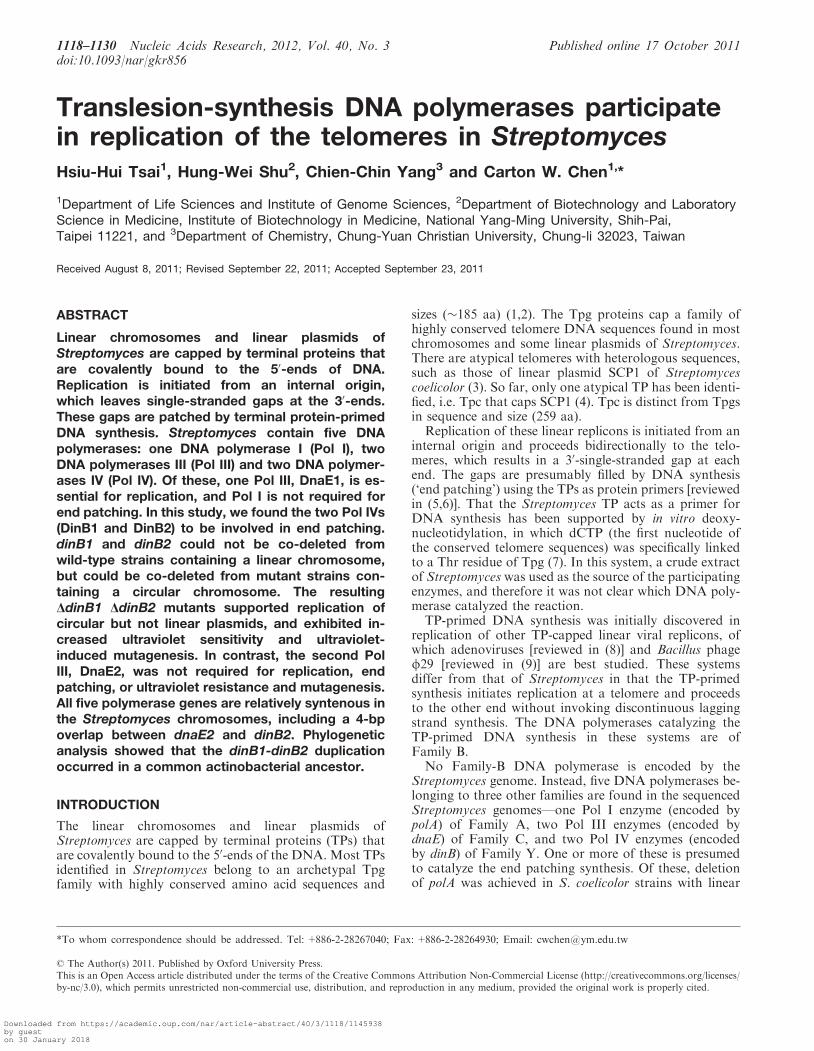
To examine the role of DnaE2 in Streptomyces, theREDIRECT procedure of Gust et al. (16) was used todelete dnaE2 (SCO1739) and replace it with theaac(3)IV (apramycin resistance, Amr) gene cassette onthe chromosome of S. coelicolor M145. �dnaE2 mutantswere readily isolated, in which the deletion was confirmedby Southern hybridization (Figure 1a). The mutants ex-hibited no detectable difference in growth or morphology.The result indicated that DnaE2 was not essential forchromosome replication in S. coelicolor. A representativedesignated E2ko was chosen for further studies.
To check the possibility that DnaE2 might be involvedin end patching, and that the chromosomes of the �dnaE2mutants might be circularized, genomic DNA of E2ko wassubjected to restriction and Southern hybridization usingthe telomere DNA as the probe (‘end probe’). The re-sults showed the presence of intact telomere sequences(Figure 1c), indicating that DnaE2 was not necessary forend patching.
dinB1 and dinB2 can be individually knocked out inS. coelicolor
To test the roles of DinB1 and DinB2 in Streptomyces,attempt was made to delete dinB1 (SCO1380) and dinB2(SCO1738) individually from S. coelicolor M145. Fordeletion of dinB2, dinB2 on cosmid StI11 (containingaph gene conferring kanamycin resistance, Kmr; http://streptomyces.org.uk/) was replaced by the aac(3)IVgene cassette in E. coli, and the cosmid was conjugallytransferred to S. coelicolor M145. From the resultingAmr transconjugants, kanamycin-sensitive (Kms) segre-gants (putative double-crossover products) were readilyisolated after one sporulation cycle at a frequency of
Table 1. Bacterial strains and plasmids used in this study
Strains/plasmids Description Source/reference
S. coelicolor M145 SCP1� SCP2� (15)E2ko �dnaE2::aac(3)IV mutant of M145 This studyHH9 �dinB2::aac(3)IV mutant of M145 This studyHH10 �dinB1:: aadA mutant of M145 This studyHH11 Spr Kmr exoconjugant isolated from HH9 that has received pLUS898 This studyHH12 Chloramphenicol-sensitive derivative of HH11 containing a circular chromosome This studyHH13 �dinB2::aac(3)IV and �dinB1:: aadA segregant of HH12 This study
S. lividans TK64hyg Spontaneous hygromycin resistant mutant of TK64, pro-2 str-6 This studyE. coli BW25113/pIJ790 K12 derivative; �araBAD �rhaBAD/�RED (gam bet exo) cat araC rep101(Ts) (16)E. coli ET12567/pUZ8002 dam-13::Tn9 dcm cat tet hsdM hsdR zjj-201::Tn10/tra neo RP4 (16)pIJ773 aac(3)IV oriT (16)pIJ778 aadA oriT (16)StI11 S. coelicolor cosmid containing dnaE2 and dinB2 (17)pLUS897 pCRII-TOPO plasmid containing SCO1379-SCO1382 This studypLUS898 pLUS897 derivative in which dinB1 is replaced by the aadA gene cassette Figure 2pLUS899 Plasmid containing the ARS of pSLA2, tsr, tap-tpg and a pair of S. lividans telomeres Figure 3pLUS899L Linear version of pLUS899 Figure 3pLUS899dinB1 pLUS899 derivative containing dinB1 Figure 3pLUS899dinB1L Linear version of pLUS899dinB1 Figure 3pLUS899dinB2 pLUS899 derivative containing dinB2 Figure 3pLUS899dinB2L Linear version of pLUS899dinB2 Figure 3
1120 Nucleic Acids Research, 2012, Vol. 40, No. 3
Downloaded from https://academic.oup.com/nar/article-abstract/40/3/1118/1145938by gueston 30 January 2018
10%. The deletion of dinB2 in the Amr Kms segregantswas confirmed by Southern hybridization (Figure 1a). Arepresentative of the �dinB2 mutants, HH9, was chosenfor further studies.
For deletion of dinB1, E. coli plasmid pLUS898 con-taining aph and SCO1379-SCO1382, in which dinB1(SCO1380) had been replaced by an aadA gene cassette(conferring spectinomycin resistance, Spr; Figure 2a) wasconjugally transferred to M145 by conjugation. Spr
exoconjugants were isolated, from which Kms segregantswere readily isolated at a 5% frequency. The deletion ofdinB1 in these segregants was confirmed by Southern hy-bridization (Figure 1b). A representative of the �dinB1mutants, HH10, was chosen for further studies.
The chromosomes in HH9 and HH10 remained linear,as evident by the presence of intact telomeres in thesestrains (Figure 1c). These results indicated that dinB1and dinB2 were not essential for end patching. However,there was a possibility that the two genes might comple-ment each other in end patching function. To test this,attempts were made to delete both genes together.
dinB1 and dinB2 can be deleted together only on circularchromosomes
To create such a double mutant, we attempted to deletedinB1 in HH9 (�dinB2) using pLUS898 (Figure 2a). Spr
Kmr exoconjugants (putative single-crossover products)were readily isolated. However, no Spr Kms segregants(putative double-crossover products) could be isolatedafter screening about 104 colony-forming units fromspores of the Spr Kmr exoconjugants. The failure todelete dinB1 in HH9, compared to the relative ease of
deleting it in M145, suggested that dinB1 and dinB2could not be deleted together on the linear S. coelicolorchromosome.The substrate mycelium of Streptomyces contains mul-
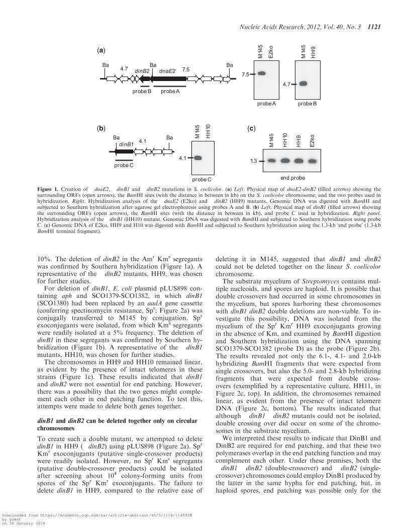
tiple nucleoids, and spores are haploid. It is possible thatdouble crossovers had occurred in some chromosomes inthe mycelium, but spores harboring these chromosomeswith dinB1 dinB2 double deletions are non-viable. To in-vestigate this possibility, DNA was isolated from themycelium of the Spr Kmr HH9 exoconjugants growingin the absence of Km, and examined by BamHI digestionand Southern hybridization using the DNA spanningSCO1379-SCO1382 (probe D) as the probe (Figure 2b).The results revealed not only the 6.1-, 4.1- and 2.0-kbhybridizing BamHI fragments that were expected fromsingle crossovers, but also the 5.0- and 2.8-kb hybridizingfragments that were expected from double cross-overs (exemplified by a representative culture, HH11, inFigure 2c, top). In addition, the chromosomes remainedlinear, as evident from the presence of intact telomereDNA (Figure 2c, bottom). The results indicated thatalthough �dinB1 �dinB2 mutants could not be isolated,double crossing over did occur on some of the chromo-somes in the substrate mycelium.We interpreted these results to indicate that DinB1 and
DinB2 are required for end patching, and that these twopolymerases overlap in the end patching function and maycomplement each other. Under these premises, both the�dinB1 �dinB2 (double-crossover) and �dinB2 (single-crossover) chromosomes could employDinB1 produced bythe latter in the same hypha for end patching, but, inhaploid spores, end patching was possible only for the
(a)
(b) (c)
Figure 1. Creation of �dnaE2, �dinB1 and �dinB2 mutations in S. coelicolor. (a) Left. Physical map of dnaE2-dinB2 (filled arrows) showing thesurrounding ORFs (open arrows), the BamHI sites (with the distance in between in kb) on the S. coelicolor chromosome, and the two probes used inhybridization. Right. Hybridization analysis of the �dnaE2 (E2ko) and �dinB2 (HH9) mutants. Genomic DNA was digested with BamHI andsubjected to Southern hybridization after agarose gel electrophoresis using probes A and B. (b) Left. Physical map of dinB1 (filled arrows) showingthe surrounding ORFs (open arrows), the BamHI sites (with the distance in between in kb), and probe C used in hybridization. Right panel.Hybridization analysis of the �dinB1 (HH10) mutant. Genomic DNA was digested with BamHI and subjected to Southern hybridization using probeC. (c) Genomic DNA of E2ko, HH9 and H10 was digested with BamHI and subjected to Southern hybridization using the 1.3-kb ‘end probe’ (1.3-kbBamHI terminal fragment).
Nucleic Acids Research, 2012, Vol. 40, No. 3 1121
Downloaded from https://academic.oup.com/nar/article-abstract/40/3/1118/1145938by gueston 30 January 2018
�dinB2 chromosomes, but not for the �dinB1 �dinB2chromosomes.
This model predicted that deletion of double deletionsof dinB1and dinB2 were possible on a circularized chromo-some. To test this, a chloramphenicol-sensitive mutant,HH12, containing a circularized chromosome (lackingthe chromosomal telomeres; Figure 2c, bottom) was iso-lated from HH11. As expected from the model, Spr Kms
mutants were isolated readily (at frequencies of �5%)from the spores of HH12. Restriction and hybridizationanalysis of the genomic DNA of the Spr Kms mutants(exemplified by HH13) revealed only the 5.0- and 2.8-kbBamHI fragments as expected from double crossovers(Figure 2c, top). Thus, double deletion of dinB1 anddinB2 was readily achieved on a circular chromosome.
Replication of linear plasmids requires dinB1 or dinB2
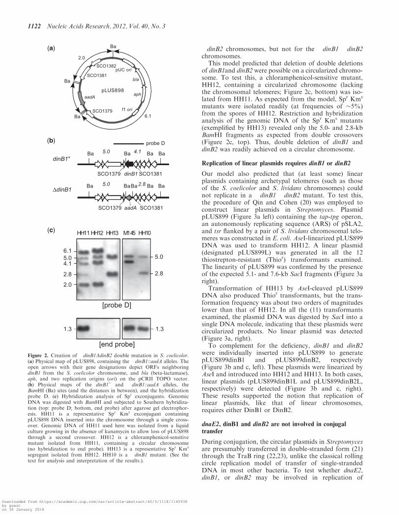
Our model also predicted that (at least some) linearplasmids containing archetypal telomeres (such as thoseof the S. coelicolor and S. lividans chromosomes) couldnot replicate in a �dinB1 �dinB2 mutant. To test this,the procedure of Qin and Cohen (20) was employed toconstruct linear plasmids in Streptomyces. PlasmidpLUS899 (Figure 3a left) containing the tap-tpg operon,an autonomously replicating sequence (ARS) of pSLA2,and tsr flanked by a pair of S. lividans chromosomal telo-meres was constructed in E. coli. AseI-linearized pLUS899DNA was used to transform HH12. A linear plasmid(designated pLUS899L) was generated in all the 12thiostrepton-resistant (Thior) transformants examined.The linearity of pLUS899 was confirmed by the presenceof the expected 5.1- and 7.6-kb SacI fragments (Figure 3aright).
Transformation of HH13 by AseI-cleaved pLUS899DNA also produced Thior transformants, but the trans-formation frequency was about two orders of magnitudeslower than that of HH12. In all the (11) transformantsexamined, the plasmid DNA was digested by SacI into asingle DNA molecule, indicating that these plasmids werecircularized products. No linear plasmid was detected(Figure 3a, right).
To complement for the deficiency, dinB1 and dinB2were individually inserted into pLUS899 to generatepLUS899dinB1 and pLUS899dinB2, respectively(Figure 3b and c, left). These plasmids were linearized byAseI and introduced into HH12 and HH13. In both cases,linear plasmids (pLUS899dinB1L and pLUS899dinB2L,respectively) were detected (Figure 3b and c, right).These results supported the notion that replication oflinear plasmids, like that of linear chromosomes,requires either DinB1 or DinB2.
dnaE2, dinB1 and dinB2 are not involved in conjugaltransfer
During conjugation, the circular plasmids in Streptomycesare presumably transferred in double-stranded form (21)through the TraB ring (22,23), unlike the classical rollingcircle replication model of transfer of single-strandedDNA in most other bacteria. To test whether dnaE2,dinB1, or dinB2 may be involved in replication of
2.8
5.06.1
4.1
2.8
5.0
pLUS898
SCO1379
SCO1381
SCO1382
aadA
bla
aph
f1 ori
Ba
Ba
Ba
2.0
6.1
pUC ori
Ba Ba Ba5.0 4.1
dinB1
Ba
5.0 2.8 BaBaBaBa
aadASCO1379 SCO1381
Ba
(b)
(a)
2.0
M145HH13HH12HH11
[end probe]
HH10(c)
1.3
[probe D]
probe D
dinB1+
ΔdinB1
1.3
SCO1379 SCO1381
Figure 2. Creation of �dinB1DdinB2 double mutation in S. coelicolor.(a) Physical map of pLUS898, containing the �dinB1::aadA alleles. Theopen arrows with their gene designations depict ORFs neighboringdinB1 from the S. coelicolor chromosome, and bla (beta-lactamase),aph, and two replication origins (ori) on the pCRII TOPO vector.(b) Physical maps of the dinB1+ and �dinB1::aadA alleles, theBamHI (Ba) sites (and the distances in between), and the hybridizationprobe D. (c) Hybridization analysis of Spr exconjugants. GenomicDNA was digested with BamHI and subjected to Southern hybridiza-tion (top: probe D; bottom, end probe) after agarose gel electrophor-esis. HH11 is a representative Spr Kmr exconjugant containingpLUS898 DNA inserted into the chromosome through a single cross-over. Genomic DNA of HH11 used here was isolated from a liquidculture growing in the absence of kanamycin to allow loss of pLUS898through a second crossover. HH12 is a chloramphenicol-sensitivemutant isolated from HH11, containing a circular chromosome(no hybridization to end probe). HH13 is a representative Spr Kms
segregant isolated from HH12. HH10 is a �dinB1 mutant. (See thetext for analysis and interpretation of the results.).
1122 Nucleic Acids Research, 2012, Vol. 40, No. 3
Downloaded from https://academic.oup.com/nar/article-abstract/40/3/1118/1145938by gueston 30 January 2018

(a)
(b)
(c)
Figure 3. dinB1 or dinB2 is required for linear plasmid replication. (a) pLUS899. Left panels. pLUS899 contains an ARS of the pSLA2 linearplasmid, tsr (thiostrepton resistant gene), tap-tpg and a pair of the 365-bp telomere sequences of the S. lividans chromosome (filled arrows). The AseI(As)-containing sequence between the telomere sequences is the E. coli vector pMTL23. The expected linear derivative of pLUS899, designatedpLUS899L, is shown below. The unique SacI site and the sizes of the SacI fragments are shown. Right panel. AseI-linearized pLUS899 DNA wasused to transform HH12 and HH13. HH13 was transformed at an efficiency about one hundred fold lower than HH12. Thiostrepton-resistanttransformants were isolated and their genomic DNA was fractionated by agarose gel electrophoresis with (�) or without (+) SacI (Sc) digestion. Inall HH12 transformants, linear pLUS899L DNA was evident by the digestion of the 12.7-kb DNA into 7.6- and 5.1-kb fragments. In all HH13transformants, the plasmid DNA appeared to be circular as evident by the production of a single SacI fragment of �13 kb. (b) pLUS899dinB1. (Leftpanel) pLUS899dinB1 is a derivative of pLUS899 containing a copy of dinB1 and its upstream ORF (SCO1381). The expected linear derivative,designated pLUS899dinB1L, is shown with the unique SacI site. (Right panel) AseI-linearized pLUS899dinB1 DNA transformed HH12 and HH13 atabout the same efficiency. The transformants of both strains harbored linear DNA (pLUS899dinB1L), as evident by the cleavage of the uncutplasmid DNA into the expected 7.9- and 6.2-kb SacI fragments. pLUS899dinB1. (C) (Left panel) pLUS899dinB2 is a derivative of pLUS899containing a copy of dinB2 and the upstream overlapping dnaE2. The expected linear derivative, designated pLUS899dinB2L, is shown with theunique SacI site. (Right panel) AseI-linearized pLUS899dinB2 DNA transformed HH12 and HH13 at about the same efficiency. The transformantsof both strains harbored linear DNA (pLUS899dinB2L), as evident by the cleavage of the uncut plasmid DNA into the expected 8.0- and 6.0-kb SacIfragments.
Nucleic Acids Research, 2012, Vol. 40, No. 3 1123
Downloaded from https://academic.oup.com/nar/article-abstract/40/3/1118/1145938by gueston 30 January 2018

circular plasmids during conjugal transfer, circular plasmidpIJ303 was introduced into M145, E2ko HH9, HH10,HH11, HH12 and HH13 by conjugal transfer, and testedfor conjugal transfer of the plasmid to TK64hyg (a spon-taneous hygromycin resistant mutant isolated in thisstudy). No significant difference in the plasmid transferfrequencies were observed among these matings (datanot shown), indicating that transfer of these the circularplasmid did not depend on dnaE2, dinB1 or dinB2.Similarly, SLP2tsr, a SLP2 derivative containing an
insert of tsr (24), was introduced to M145, E2ko HH9,HH10, HH11 and HH12, and tested for transfer toTK64hyg. Again, the mutations in the DNA polymerasegenes did not cause defects in plasmid transfers (data notshown). In E. coli, replication of circular plasmids duringconjugal transfer is catalyzed by Pol III (25). InStreptomyces, this role is probably also mainly played bythe Pol III composed of DnaE1.
dinB1 and dinB2 are involved in translesion repair
None of the �dinB1 (HH10), �dinB2 (HH9) and �dinB1�dinB2 (HH13) mutants exhibit any detectable anomalyin morphology or growth characteristics. In other bacteria,dinB-encoded DNA polymerase IV is involved in trans-lesion repair of DNA damage, and dinB mutants exhibithigher sensitivity to UV and increased UV-induced muta-genesis [reviewed in (26,27)]. Are dinB1 and dinB2 alsoinvolved in these processes in Streptomyces? Comparedwith the wild-type parent M145, the �dinB1 and �dinB2single mutants did not exhibit increased sensitivity to UV(Figure 4a). However, the �dinB1 �dinB2 double muta-tions caused a slight increase in UV sensitivity. Theseresults indicated that dinB1 and dinB2 also assume com-plementary roles in repair of UV damage. They apparentlyplay only a relatively minor role in the repair due to thepresence of multiple other repair systems, such as excision
repair, photoreactivation and recombinational repair, inStreptomyces.
Mutation to rifampicin resistance was used to test UV-induced mutagenesis in these mutants (Figure 4b), andthe results showed that the mutagenesis rates in HH10(�dinB1) and HH9 (�dinB2) were reduced to 50%,compared to M145. The mutation rate in HH13(�dinB1 �dinB2) was further reduced to about 10% ofthat in M145. It is noteworthy that HH13 differs fromM145 not only in the dinB1 and dinB2 mutations butalso in possessing a circular chromosome with large dele-tions. These results indicated that both DinB1 and DinB2polymerases produced about the same extent of errorsduring TLS repair of UV damage.
E2ko exhibited no differences in UV damage repair andUV-induced mutagenesis as M145 (data not shown),indicating that DnaE2 was not involved in these processes.
It was possible that DNA repair in spores and myceliainvolved different DNA polymerases and different mech-anisms in Streptomyces. These repair and mutation studieswere performed using UV-irradiated spores, and any UVdamages on the chromosomes that required translesionrepair by DinB1 and/or DinB2 polymerases must be re-paired at the germination stage for the cultures to survive.Therefore, the observed effects of �dinB1 and �dinB2mutations on UV damage repair (Figure 4a) reflectedthe involvement of these polymerases at the germinationstage. This was also true for UV-induced mutagenesis,which accompanied the translesion repair. The involve-ment of these polymerases in DNA repair during mycelialgrowth remained to be investigated.
The M. tuberculosis genome also carries two dinBhomologs, dinB1 (dinX) and dinB2 (dinP). Recently,Kana et al. (14) discovered that deletion of them singlyor in combination did not appear to cause increases insensitivity to DNA damaging agents or mutation fre-quencies. The authors suggest that the DinB homologs
(a) (b)
Figure 4. UV sensitivity and UV-induced mutagenesis of the DNA polymerase mutants. (a) Sensitivity of the polymerase mutants to UV. Spores ofthe mutant cultures were irradiated with ultraviolet radiation to various dosages, and the survivals were scored by plating and incubating on R2YEagar for 4 days. Filled circles, M145; open circles, HH9; filled triangles, HH10; open triangles, HH11; filled squares, HH12; open squares, HH13.The results shown are the means of five independent experiments. The standard deviations were all <2.6% of the means, and therefore not shown.(b) Frequency of UV-induced rifampicin-resistant mutations in the mutants. Spores of the mutant cultures were irradiated with 200 J/m2 of UV, andthe numbers of rifampicin resistant mutants were scored (filled bars) and compared with unirradiated spores (open bars). The results shown are themeans of five independent experiments, and the error bars indicate the standard deviations.
1124 Nucleic Acids Research, 2012, Vol. 40, No. 3
Downloaded from https://academic.oup.com/nar/article-abstract/40/3/1118/1145938by gueston 30 January 2018

in Mycobacterium differ significantly in biological func-tions from their homologs in other bacteria. It is likelythat one or both of these DinB homologs also take partin end patching for replication of TP-capped linearplasmids found in some Mycobacterium species, such asM. xenopi, M. branderi and M. celatum (28,29).
dnaE2 and dinB2 are induced by UV irradiationand DNA alkylation
The involvement of dinB1 and dinB2 in resistance to UVand UV-induced mutagenesis suggested that they are in-ducible by the SOS response. In E. coli, the single dinBgene is induced by the SOS response (30). A LexA-bindingSOS box is present in the promoter of dinB in E. coli.A Gram-positive bacterial SOS box, GAACN4RTTY,(31–33) is present in the promoter region of dnaE2 inS. coelicolor, S. avermitilis, S. scabiei and S. griseus (withone mismatch). This further suggests that the putativednaE2-dinB2 operon is under SOS response.
To test this notion, M145 was irradiated with UV andthe expression of dinB1, dinB2 and dnaE2 was determinedby RT–PCR. Interestingly, the results (Figure 5) showthat the expression of dinB2 and dnaE2 occurred at a rela-tively low level, and was increased by �6-fold in 60minafter UV irradiation. In contrast, dinB1 was constitutivelyexpressed and insensitive to UV irradiation. recA, which isknown to be under SOS regulation, was induced to aboutthe same extent as dinB2 and dnaE2 in M145. Treatmentof M145 cultures with MMS gave similar results withabout the same levels of induction for dinB2, dnaE2 and
recA, and none for dinB1. In comparison, the expressionof the TP gene tpg appeared to be constitutive and notinduced by UV or MMS treatment.Constitutive expression of the two Pol IV enzymes
during active growth of S. coelicolor is expected for theirroles in replication of the chromosomes. However, thelack of SOS response by dinB1 is surprising consideringits involvement in repair of UV damage and UV-inducedmutagenesis. The identical constitutive expression andSOS response patterns of dnaE2 and dinB2 are consistentwith the notion that these two genes form an operon underthe same transcriptional regulation.
TLS DNA polymerases are biochemically suitable forend patching
TP-primed replication of the linear genomes of Bacillusphage f29 and adenoviruses is catalyzed by a viral-encoded DNA polymerase that belongs to the sameFamily B as Pol II. In bacteria, both Pol II and Pol IVare TLS DNA polymerases involved in DNA repair andinduced by the SOS response (34,35). Perhaps it makesbiochemical sense that TLS DNA polymerases were ado-pted to catalyze TP-primed DNA synthesis during evolu-tion. The use of TP as primer to carry out patchingsynthesis at the terminal 30-overhangs is not unlike theswitch of Pol III to a TLS polymerase in bypassing abulky adduct in that proper Watson-Crick base pairingbetween the template and the incoming nucleotides isnot present, and TLS polymerases have been shown topossess a more spacious active site that could accommo-date bulky adducts or non-Watson-Crick base pairing(36,37).Then why do f29 and adenoviruses adopt Pol II, while
Streptomyces adopts Pol IV for TP-primed synthesis?There are two possible considerations. First, Pol II pos-sesses a 30–50 proofreading function that Pol IV lacks, andis therefore less error-prone than Pol IV. Second, Pol IIhas a higher processivity than Pol IV. These two fea-tures of Pol II would be advantageous for achievinghigh fidelity in replication of whole viral genomes. In con-trast, patching of the relatively short (�300-nt) ofStreptomyces telomeres probably does not demand highfidelity. Interestingly, there is no Pol II homolog inStreptomyces.Since a single deletion of either dinB1 or dinB2 did not
prevent replication of the linear replicons, it is apparentthat one of them is sufficient for end patching. It is notclear whether both of them participate in end patchingin vivo, or one plays the main role, while the other repre-sents a backup. In the latter case, which one is the majorplayer?
The DNA polymerase genes are relatively conserved insynteny in Streptomyces
A total of five DNA polymerase homologs (dnaE1, dnaE2,polA, dinB1 and dinB2) are identified in nine sequencedStreptomyces chromosomes (S. coelicolor, S. lividans,S. avermitilis, S. scabiei, S. griseus, S. bingchenggensis,S. cattleya, S. flavogriseus and S. venezuelae).Interestingly, they are all located on the ‘left arm’ of the
Figure 5. Expression of dnaE2, dinB1 and dinB2. M145 cultures wereirradiated with UV or treated with MMS as described in ‘Materials andMethods’ section. RNA was isolated from the cultures and 0.5 mg ofRNA each was used for RT-PCR (‘+RT’) using primers specific fordnaE2, dinB1 and dinB2 (Supplementary Table S1). The time (0, 20, 40and 60min) after UV or MMS treatment is indicated. Controls withoutthe reverse transcription step (‘-RT’) are included. ‘DNA’, control PCRwith genomic DNA as template.
Nucleic Acids Research, 2012, Vol. 40, No. 3 1125
Downloaded from https://academic.oup.com/nar/article-abstract/40/3/1118/1145938by gueston 30 January 2018
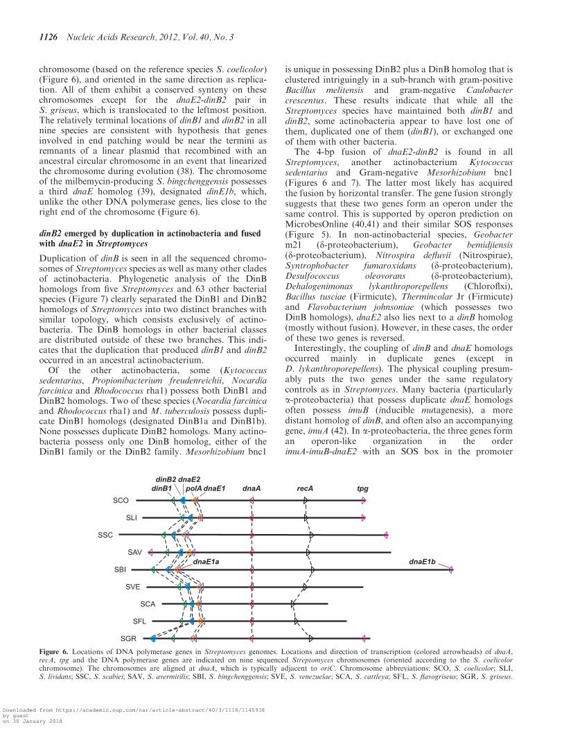
chromosome (based on the reference species S. coelicolor)(Figure 6), and oriented in the same direction as replica-tion. All of them exhibit a conserved synteny on thesechromosomes except for the dnaE2-dinB2 pair inS. griseus, which is translocated to the leftmost position.The relatively terminal locations of dinB1 and dinB2 in allnine species are consistent with hypothesis that genesinvolved in end patching would be near the termini asremnants of a linear plasmid that recombined with anancestral circular chromosome in an event that linearizedthe chromosome during evolution (38). The chromosomeof the milbemycin-producing S. bingchenggensis possessesa third dnaE homolog (39), designated dinE1b, which,unlike the other DNA polymerase genes, lies close to theright end of the chromosome (Figure 6).
dinB2 emerged by duplication in actinobacteria and fusedwith dnaE2 in Streptomyces
Duplication of dinB is seen in all the sequenced chromo-somes of Streptomyces species as well as many other cladesof actinobacteria. Phylogenetic analysis of the DinBhomologs from five Streptomyces and 63 other bacterialspecies (Figure 7) clearly separated the DinB1 and DinB2homologs of Streptomyces into two distinct branches withsimilar topology, which consists exclusively of actino-bacteria. The DinB homologs in other bacterial classesare distributed outside of these two branches. This indi-cates that the duplication that produced dinB1 and dinB2occurred in an ancestral actinobacterium.Of the other actinobacteria, some (Kytococcus
sedentarius, Propionibacterium freudenreichii, Nocardiafarcinica and Rhodococcus rha1) possess both DinB1 andDinB2 homologs. Two of these species (Nocardia farcinicaand Rhodococcus rha1) and M. tuberculosis possess dupli-cate DinB1 homologs (designated DinB1a and DinB1b).None possesses duplicate DinB2 homologs. Many actino-bacteria possess only one DinB homolog, either of theDinB1 family or the DinB2 family. Mesorhizobium bnc1
is unique in possessing DinB2 plus a DinB homolog that isclustered intriguingly in a sub-branch with gram-positiveBacillus melitensis and gram-negative Caulobactercrescentus. These results indicate that while all theStreptomyces species have maintained both dinB1 anddinB2, some actinobacteria appear to have lost one ofthem, duplicated one of them (dinB1), or exchanged oneof them with other bacteria.
The 4-bp fusion of dnaE2-dinB2 is found in allStreptomyces, another actinobacterium Kytococcussedentarius and Gram-negative Mesorhizobium bnc1(Figures 6 and 7). The latter most likely has acquiredthe fusion by horizontal transfer. The gene fusion stronglysuggests that these two genes form an operon under thesame control. This is supported by operon prediction onMicrobesOnline (40,41) and their similar SOS responses(Figure 5). In non-actinobacterial species, Geobacterm21 (d-proteobacterium), Geobacter bemidjiensis(d-proteobacterium), Nitrospira defluvii (Nitrospirae),Syntrophobacter fumaroxidans (d-proteobacterium),Desulfococcus oleovorans (d-proteobacterium),Dehalogenimonas lykanthroporepellens (Chloroflxi),Bacillus tusciae (Firmicute), Thermincolar Jr (Firmicute)and Flavobacterium johnsoniae (which possesses twoDinB homologs), dnaE2 also lies next to a dinB homolog(mostly without fusion). However, in these cases, the orderof these two genes is reversed.
Interestingly, the coupling of dinB and dnaE homologsoccurred mainly in duplicate genes (except inD. lykanthroporepellens). The physical coupling presum-ably puts the two genes under the same regulatorycontrols as in Streptomyces. Many bacteria (particularlya-proteobacteria) that possess duplicate dnaE homologsoften possess imuB (inducible mutagenesis), a moredistant homolog of dinB, and often also an accompanyinggene, imuA (42). In a-proteobacteria, the three genes forman operon-like organization in the orderimuA-imuB-dnaE2 with an SOS box in the promoter
Figure 6. Locations of DNA polymerase genes in Streptomyces genomes. Locations and direction of transcription (colored arrowheads) of dnaA,recA, tpg and the DNA polymerase genes are indicated on nine sequenced Streptomyces chromosomes (oriented according to the S. coelicolorchromosome). The chromosomes are aligned at dnaA, which is typically adjacent to oriC. Chromosome abbreviations: SCO, S. coelicolor; SLI,S. lividans; SSC, S. scabiei; SAV, S. avermitilis; SBI, S. bingchenggensis; SVE, S. venezuelae; SCA, S. cattleya; SFL, S. flavogriseus; SGR, S. griseus.
1126 Nucleic Acids Research, 2012, Vol. 40, No. 3
Downloaded from https://academic.oup.com/nar/article-abstract/40/3/1118/1145938by gueston 30 January 2018

Figure 7. Phylogenetic analysis of DinB homologs. DinB homologs in 68 bacteria are subjected to multiple sequence alignment followed byphylogenetic tree construction as described in ‘Materials and Methods’ section. The bootstrap numbers are indicated at the branch junctions. ‘1’and ‘2’ following the species names depict DinB1 (shaded in brown) and DinB2 (shaded in blue) in Streptomyces, respectively. ‘I’, ‘II’ and ‘III’following the species names depict DinB homologs in other species. For those species in which dnaE and dinB2 appear to form an operon, thesynteny of the dnaE and dinB homologs relative to other DNA polymerase genes is displayed by the arrays of colored arrows: Dark red, dnaE1; lightred, dnaE2; Dark blue, dinB1; light blue, dinB2; white, other dinB homologs (outside of actinobacteria except in Mesorhizobium bnc1).
Nucleic Acids Research, 2012, Vol. 40, No. 3 1127
Downloaded from https://academic.oup.com/nar/article-abstract/40/3/1118/1145938by gueston 30 January 2018

region. In contrast to dnaE2 of Streptomyces, dnaE2 (aswell as imuA and imuB) of C. crescentus are involved inerror-prone DNA repair (42).
Some dnaE homologs are acquired by horizontal transfer
The biological role of DnaE2 in Streptomyces is a mystery.This study shows that it is not essential for chromosomereplication or end patching, and deletion of dnaE2 did notcause any changed sensitivity to UV-induced killing andmutagenesis despite the fact that it is induced by UV ir-radiation and exposure to MMS. In contrast, the down-stream gene dinB2, which most likely is co-transcribed andco-regulated, is involved in end patching, repair ofUV-induced damage, as well as UV-induced mutagenesis.
Despite its cryptic biological function in Streptomyces,dnaE2 homologs are present in diverse clades of bacteria.Of 23 actinobacterial chromosome sequences examined,all except Tropheryma whipplei and Thermobifida fuscacontain a danE homolog. Multiple (two or more) copiesof dnaE homologs are also found in a-, b- and g-proteo-bacteria, acidobacteria, planctomycete and chloroflexus.Phylogenetic analysis of DnaE homologs in 68 bacteriaincluding 19 actinobacteria (Figure 8) separated theminto two large families (herein designated DnaE1 andDnaE2 families) represented by DnaE1 and DnaE2 ofStreptomyces, respectively, and a small family. TheDnaE1 family includes those homologs present singly inbacterial genomes. This suggests that dnaE1 representsthe primordial archetype, and that dnaE2 emerged byduplication.
In the DnaE1 branch, while all the DnaE1 homologsencoded by various Streptomyces chromosomes are clus-tered together in a single sub-branch, the DnaE1 homologencoded by the SCP1 plasmid is clustered with the secondDnaE1 homolog of S. bingchenggensis (designatedDnaE1b) in a separate sub-branch. Unlike the otherDNA polymerase genes, dnaE1b is located near theright-hand telomere (104 kb from the end; Figure 6). Thephylogenetic distances and the unique genome loca-tions (on a linear plasmid and near the end of the chromo-some) suggest that dnaE1 of SCP1 and dnaE1b ofS. bingchenggensis were acquired by horizontal transfer.It is not known whether dnaE1b plays a biological role inS. bingchenggensis, but dnaE1 (SCP1.224) on SCP1 isaccompanied by dnaN (SCP1.119; encoding b slidingclamp), and both of these genes appear to be importantfor replication of SCP1 (H.-H. Tsai, unpublished results).
dnaE2 and dinB2 evolved rapidly in Streptomyces
The longer branch lengths in the phylogenetic treesshowed that dinB2 has evolved more rapidly than dinB1in Streptomyces (Figure 7). Analysis of synonymous vs.non-synonymous substitutions shows relatively low(<0.5) values of Ka/Ks ratios for dinB1 (SupplementaryFigure S1a). These results indicate that it has evolved rela-tively rapidly toward a higher degree of purifying(stabilizing) selection. In contrast, dinB2 exhibits signifi-cantly larger and more variable Ka/Ks. Sliding Ka/Ks
analysis between each dinB1 and dinB2 pair in fiveStreptomyces species further revealed Ka/Ks ratios
Figure 8. Phylogenetic analysis of DnaE homologs. DnaE homologs in68 bacteria are subjected to multiple sequence alignment followed byphylogenetic tree construction as described in ‘Materials and Methods’section. The bootstrap numbers are indicated at the branch junctions.‘1’ and ‘2’ following the species names depict DnaE1 (shaded in blue)and DnaE2 (shaded in brown) in Streptomyces, respectively. PolCfamily is shaded in green. The Streptomyces homologs are emphasizedby dark reverse colors.
1128 Nucleic Acids Research, 2012, Vol. 40, No. 3
Downloaded from https://academic.oup.com/nar/article-abstract/40/3/1118/1145938by gueston 30 January 2018
significantly larger than 1 in specific regions, suggestingthese regions had undergone positive selection in dinB2(Supplementary Figure S1b).
dnaE1, which encodes the chromosomal replicase,exhibits very low Ka/Ks ratios as expected for a conservedreplicase (Supplementary Figure S1c). In comparison,dnaE2 has larger and more variable Ka/Ks ratios, evenmore so than dinB2 (Supplementary Figure S1b). Thisand the longer branch length in the DnaE phylogenetictree (Figure 8) indicate that dnaE2 has also evolvedmore rapidly toward a new but unclear function. SlidingKa/Ks analysis between each dnaE1 and dnaE2 pair in fiveStreptomyces species revealed positive selection in specificregions, notably the ‘thumb’ domain and the regionbetween the PHP and ‘palm’ domains, of dnaE2(Supplementary Figure S1c).
SUPPLEMENTARY DATA
Supplementary Data are available at NAR Online:Supplementary Table S1, Supplementary Figure S1.
ACKNOWLEDGEMENTS
The authors thank David Hopwood for reading the manu-script and suggesting improvements.
FUNDING
National Professorship from Ministry of Education,R. O. C. to C.W.C. National Science Council, R. O. C.(NSC98-2311-B-010-004-MY3; NSC99-2811-B-010-027);Ministry of Education, R. O. C. (Aim for the TopUniversity Plan). Funding for open access charge:National Yang-Ming University.
Conflict of interest statement. None declared.
REFERENCES
1. Yang,C.-C., Huang,C.-H., Li,C.-Y., Tsay,Y.-G., Lee,S.-C. andChen,C.W. (2002) The terminal proteins of linear Streptomyceschromosomes and plasmids: A novel class of replication primingproteins. Mol. Microbiol., 43, 297–305.
2. Bao,K. and Cohen,S.N. (2001) Terminal proteins essential for thereplication of linear plasmids and chromosomes in Streptomyces.Genes Dev., 15, 1518–1527.
3. Kinashi,H., Shimaji-Murayama,M. and Hanafusa,T. (1991)Nucleotide sequence analysis of the unusually long terminalinverted repeats of a giant linear plasmid, SCP1. Plasmid, 26,123–130.
4. Huang,C.-H., Tsai,H.-H., Tsay,Y.-G., Chien,Y.-N., Wang,S.-L.,Cheng,M.-Y., Ke,C.-H. and Chen,C.W. (2007) The telomeresystem of the Streptomyces linear plasmid SCP1 represents anovel class. Mol. Microbiol., 63, 1710–1718.
5. Chaconas,G. and Chen,C.W. (2005) In Higgins,N.P. (ed.),The Bacterial Chromosome. American Society for Microbiology,Washington, D. C., pp. 525–539.
6. Chen,C.W. (2007) In Meinhardt,F. and Klassen,R. (eds),Microbial Linear Plasmids. Springer-Verlag, Berlin, Heidelberg,pp. 33–61.
7. Yang,C.C., Chen,Y.H., Tsai,H.H., Huang,C.H., Huang,T.W. andChen,C.W. (2006) In vitro deoxynucleotidylation of the terminalprotein of Streptomyces linear chromosomes. Appl. Environ.Microbiol., 72, 7959–7961.
8. Liu,H., Naismith,J.H. and Hay,R.T. (2003) Adenovirus DNAreplication. Curr. Top. Microbiol. Immunol., 272, 131–164.
9. Meijer,W.J., Horcajadas,J.A. and Salas,M. (2001) j29 Family ofPhages. Microbiol. Mol. Biol. Rev., 65, 261–287.
10. Huang,T.W. and Chen,C.W. (2008) DNA polymerase I is notrequired for replication of linear chromosomes in Streptomyces.J. Bacteriol., 190, 755–758.
11. Flett,F., de Mello Jungmann-Campello,D., Mersinias,V.,Koh,S.L., Godden,R. and Smith,C.P. (1999) A’gram-negative-type’ DNA polymerase III is essential forreplication of the linear chromosome of Streptomyces coelicolorA3(2). Mol. Microbiol., 31, 949–958.
12. Dervyn,E., Suski,C., Daniel,R., Bruand,C., Chapuis,J.,Errington,J., Janniere,L. and Ehrlich,S.D. (2001) Two essentialDNA polymerases at the bacterial replication fork. Science, 294,1716–1719.
13. Boshoff,H.I., Reed,M.B., Barry,C.E. 3rd and Mizrahi,V. (2003)DnaE2 polymerase contributes to in vivo survival and theemergence of drug resistance in Mycobacterium tuberculosis. Cell,113, 183–193.
14. Kana,B.D., Abrahams,G.L., Sung,N., Warner,D.F.,Gordhan,B.G., Machowski,E.E., Tsenova,L., Sacchettini,J.C.,Stoker,N.G., Kaplan,G. et al. (2010) Role of the DinB homologsRv1537 and Rv3056 in Mycobacterium tuberculosis. J. Bacteriol.,192, 2220–2227.
15. Hopwood,D.A., Kieser,T., Wright,H.M. and Bibb,M.J. (1983)Plasmids, recombination, and chromosomal mapping inStreptomyces lividans 66. J. Gen. Microbiol., 129, 2257–2269.
16. Gust,B., Challis,G.L., Fowler,K., Kieser,T. and Chater,K.F.(2003) PCR-targeted Streptomyces gene replacement identifies aprotein domain needed for biosynthesis of the sesquiterpene soilodor geosmin. Proc. Natl Acad. Sci. USA, 100, 1541–1546.
17. Redenbach,M., Kieser,H.M., Denapaite,D., Eichner,A., Cullum,J.,Kinashi,H. and Hopwood,D.A. (1996) A set of ordered cosmidsand a detailed genetic and physical map for the 8 MbStreptomyces coelicolor A3(2) chromosome. Mol. Microbiol., 21,77–96.
18. Kieser,T., Bibb,M., Buttner,M.J., Chater,K.F. andHopwood,D.A. (2000) Practical Streptomyces Genetics. The JohnInnes Foundation, Norwich.
19. Newell,K.V., Thomas,D.P., Brekasis,D. and Paget,M.S. (2006)The RNA polymerase-binding protein RbpA confers basal levelsof rifampicin resistance on Streptomyces coelicolor. Mol.Microbiol., 60, 687–696.
20. Qin,Z. and Cohen,S.N. (1998) Replication at the telomeres of theStreptomyces linear plasmid pSLA2. Mol. Microbiol., 28, 893–904.
21. Possoz,C., Ribard,C., Gagnat,J., Pernodet,J.L. and Guerineau,M.(2001) The integrative element pSAM2 from Streptomyces:kinetics and mode of conjugal transfer. Mol. Microbiol., 42,159–166.
22. Vogelmann,J., Ammelburg,M., Finger,C., Guezguez,J., Linke,D.,Flotenmeyer,M., Stierhof,Y.-D., Wohlleben,W. and Muth,G.(2011) Conjugal plasmid transfer in Streptomyces resemblesbacterial chromosome segregation by FtsK/SpoIIIE. EMBO J.,30, 2246–2254.
23. Reuther,J., Gekeler,C., Tiffert,Y., Wohlleben,W. and Muth,G.(2006) Unique conjugation mechanism in mycelial streptomycetes:a DNA-binding ATPase translocates unprocessed plasmid DNAat the hyphal tip. Mol. Microbiol., 61, 436–446.
24. Hsu,C.C. and Chen,C.W. (2010) Linear plasmid SLP2 ismaintained by partitioning, intrahyphal spread, and conjugaltransfer in Streptomyces. J. Bacteriol., 192, 307–315.
25. Kingsman,A. and Willetts,N. (1978) The requirements forconjugal DNA synthesis in the donor strain during Flac transfer.J. Mol. Biol., 122, 287–300.
26. Andersson,D.I., Koskiniemi,S. and Hughes,D. (2010) Biologicalroles of translesion synthesis DNA polymerases in eubacteria.Mol. Microbiol., 77, 540–548.
27. Sutton,M.D. (2010) Coordinating DNA polymerase trafficduring high and low fidelity synthesis. Biochim. Biophys.Acta –Prot. Proteom., 1804, 1167–1179.
28. Picardeau,M. and Vincent,V. (1998) Mycobacterial linear plasmidshave an invertron-like structure related to other linear repliconsin actinomycetes. Microbiology, 144, 1981–1988.
Nucleic Acids Research, 2012, Vol. 40, No. 3 1129
Downloaded from https://academic.oup.com/nar/article-abstract/40/3/1118/1145938by gueston 30 January 2018

29. Picardeau,M., Le Dantec,C. and Vincent,V. (2000) Analysis of theinternal replication region of a mycobacterial linear plasmid.Microbiology, 146, 305–313.
30. Kenyon,C.J. and Walker,G.C. (1980) DNA-damaging agentsstimulate gene expression at specific loci in Escherichia coli.Proc. Natl Acad. Sci. USA, 77, 2819–2823.
31. Cheo,D.L., Bayles,K.W. and Yasbin,R.E. (1992)Molecular characterization of regulatory elements controllingexpression of the Bacillus subtilis recA+ gene. Biochimie, 74,755–762.
32. Walker,G.C., Marsh,L. and Dodson,L. (1985) Cellular responsesto DNA damage. Environ. Health Perspect., 62, 115–117.
33. Vierling,S., Weber,T., Wohlleben,W. and Muth,G. (2001)Evidence that an additional mutation is required to tolerateinsertional inactivation of the Streptomyces lividans recA gene.J. Bacteriol., 183, 4374–4381.
34. Napolitano,R., Janel-Bintz,R., Wagner,J. and Fuchs,R.P. (2000)All three SOS-inducible DNA polymerases (Pol II, Pol IV andPol V) are involved in induced mutagenesis. EMBO J., 19,6259–6265.
35. Wagner,J., Gruz,P., Kim,S.R., Yamada,M., Matsui,K.,Fuchs,R.P. and Nohmi,T. (1999) The dinB gene encodes a novelE. coli DNA polymerase, DNA pol IV, involved in mutagenesis.Mol. Cell., 4, 281–286.
36. Yang,W. (2003) Damage repair DNA polymerases Y.Curr. Opin. Struct. Biol., 13, 23–30.
37. Zhou,B.L., Pata,J.D. and Steitz,T.A. (2001) Crystal structure of aDinB lesion bypass DNA polymerase catalytic fragment reveals aclassic polymerase catalytic domain. Mol. Cell., 8, 427–437.
38. Chen,C.W., Huang,C.-H., Lee,H.-H., Tsai,H.-H. and Kirby,R.(2002) Once the circle has been broken: dynamics and evolutionof Streptomyces chromosomes. Trends Genet., 18, 522–529.
39. Wang,X.J., Yan,Y.J., Zhang,B., An,J., Wang,J.J., Tian,J.,Jiang,L., Chen,Y.H., Huang,S.X., Yin,M. et al. (2010) Genomesequence of the milbemycin-producing bacterium Streptomycesbingchenggensis. J. Bacteriol., 192, 4526–4527.
40. Dehal,P.S., Joachimiak,M.P., Price,M.N., Bates,J.T.,Baumohl,J.K., Chivian,D., Friedland,G.D., Huang,K.H.,Keller,K., Novichkov,P.S. et al. (2010) MicrobesOnline: anintegrated portal for comparative and functional genomics.Nucleic Acids Res., 38, D396–D400.
41. Alm,E.J., Huang,K.H., Price,M.N., Koche,R.P., Keller,K.,Dubchak,I.L. and Arkin,A.P. (2005) The MicrobesOnline Website for comparative genomics. Genome Res., 15, 1015–1022.
42. Galhardo,R.S., Rocha,R.P., Marques,M.V. and Menck,C.F.(2005) An SOS-regulated operon involved in damage-induciblemutagenesis in Caulobacter crescentus. Nucleic Acids Res., 33,2603–2614.
1130 Nucleic Acids Research, 2012, Vol. 40, No. 3
Downloaded from https://academic.oup.com/nar/article-abstract/40/3/1118/1145938by gueston 30 January 2018