Embed Size (px)
Citation preview
RELAPSING PLASMODIUM VIVAX MALARIA WITH ATYPICAL PARASITE FORMS AND PHAGOCYTOSIS
BY PERIPHERAL NEUTROPHILS
MAZARS E.* .**, LABERENNE J.E.***, SNOUNOU G.****, DEI-CAS E.**.*****, CATTOEN C.* & POIRRIEZ J . * * * * *
Summary :
A case of atypical Plasmodium vivax malaria is presented. The clinical follow-up has allowed to characterize three consecutive malaria clinical episodes within one year. At the first attack, 39 % of the infected red blood cells were parasitized by gametocytes. Furthermore, rare crisis forms, exceptional « pseudo-parthenogenesis » forms, a few equatorial trophozoites, malaria pigment-containing leucocytes and phagocytized parasites were also found in the thin blood smears. At the second malaria episode, morphological aspects were quite similar, but the gametocyte percentage decreased and that of the equatorial trophozoite forms increased. Only at the third attack, was the morphology typical of P. vivax. The Plasmodium species and the absence of mixed infection were unequivocally confirmed using polymerase chain reaction. Atypical strains of P. vivax are relatively frequent. Nevertheless, to our knowledge, neither so high a gametocyte percentage, nor extensive P. vivax peripheral phagocytosis were previously reported.
KEY WORDS : P. vivox, atypical morphology, peripheral phagocytosis.
INTRODUCTION
A mong human malaria species, Plasmodium falciparum is the most fearsome pathogen, whereas P. vivax has the most widespread
* Service de Biologie Clinique, Centre Hospitalier Général, avenue Désandrouins, 59322 Valenciennes Cedex. ** INSERM (U42), 369, rue Jules-Guesde, BP 39, 59651 Villeneuve-d'Ascq Cedex. *** Service de Gastroentérologie, Centre Hospitalier Général, avenue Désandrouins, 59322 Valenciennes Cedex. **** Imperial College School of Medicine, Department of Infection and Tropical Medicine, Lister unit, Northwick Park Hospital, Harrow, Middlesex, HA1 3UJ, UK. ***** Service de Parasitologie-Mycologie, CHRU et Faculté de Médecine, 1, place Verdun, 59045 Lille. ****** Laboratoire de Biologie, Centre Hospitalier, 130, avenue Louis-Herbeaux, BP 6367, 59385 Dunkerque Cedex 1, France. Correspondence : Dr. Dei-Cas, INSERM (U42), 369, rue Jules-Guesde, BP 39, 59651 Villeneuve-d'Ascq Cedex - Tel : 03 20 47 25 73 - Fax : 03 20 05 91 72.
Résumé : RECHUTES DE PALUDISME À PLASMODIUM VIVAX AVEC FORMES PARASITAIRES ATYPIQUES ET PHAGOCYTOSE PAR LES NEUTROPHILES PÉRIPHÉRIQUES
Nous présentons un cas d'infection palustre à Plasmodium vivax atypique. Le patient a contracté 3 accès palustres en 1 an. Lors du premier accès, les gamétocytes représentaient 39 % des formes parasitaires intra-érythrocytaires. Parallèlement on observait de rares formes de pseudo-parthénogénèse, quelques trophozoïtes en plaque équatoriale et des leucocytes renfermant soit du pigment malarique, soit des formes parasitaires. Lors du second accès, les aspects morphologiques parasitaires étaient identiques à ceux décrits précédemment. Cependant le pourcentage de gamétocytes était diminué tandis que celui des trophozoïtes en plaque équatoriale était augmenté. Lors du dernier et troisième accès palustre, la morphologie parasitaire était enfin typique de P. vivax. Le diagnostic d'espèce de Plasmodium et l'absence d'infection multiple ont été confirmés par PCR. Les souches de P. vivax atypiques sont relativement fréquentes. Cependant à notre connaissance, c'est la première fois qu'un pourcentage aussi élevé de gamétocytes est rapporté lors d'une infection à P. vivax et qu'une phagocytose périphérique des différentes formes parasitaires intra-érythrocytaires est notée pour cette espèce de Plasmodium.
MOTS CLÉS : P. vivax, morphologie atypique, phagocytose périphérique.
geographical distribution. Indeed, P. vivax can develop in female Anopheles from a temperature of 16 °C, instead of 20 °C for P. falciparum (Brumpt, 1949). So P. vivax is in some countries the most common malaria pathogen (Prasad et al., 1990). The first case of chlo-roquine resistance by P. vivax from Papua-New Guinea was described in 1989 (Rieckmann et al., 1989). and similar cases have been described from other parts of the world (Baird et al., 1995). Only one case of resistance to mefloquine has been reported (Amor et al., 1992).
In this article, we present an unusual case of P. vivax studied on three successive clinical malaria episodes which developed within one year, after return to France from an endemic country. Atypical parasite forms and numerous peripheral phagocytized parasites were observed in the thin blood smears. The microscopic identification was confirmed using polymerase chain reaction (PCR).
Parasite, 1997, 4, 263-267 Note de recherche 263
Article available at http://www.parasite-journal.org or http://dx.doi.org/10.1051/parasite/1997043263
MAZARS E , LABERENNE J .E , SNOUNOU G., DEI-GAS E., CATTOEN C. & POIRRIEZ J.
CASE REPORT
CLINICAL COURSE
M r JC, a 26-year-old French man, travelled in French Guyana from March to June 1991, and served in the army in Djibouti for
two years (from October 1992 to the 9th of October 1994). During his stays in French Guyana and in Djibouti, he took malaria chemoprophylaxis (chloroquine = Nivaquine®, 1 tablet of 100 mg per day), daily and for 40 days after his return to France. On the 8th of January 1995, he developed a fever attack of 40 °C and was admitted to the general hospital of Valenciennes. The clinical examination revealed an enlarged spleen. Blood counts showed thrombocytopenia (40,000 x 106 platelets/L). Blood smears showed a malaria infection. The patient was treated with mefloquine (Lariam® : 6 tablets of 250 mg, in 3 doses on the same day), which brought about the resolution of the fever and an increase of platelets count. Four and half months later, Mr JC had several fever peaks and was again admitted to the hospital on May 27, 1995. Blood counts were normal except thrombocytopenia (44,000 x 106 platelets/L), and blood smears showed again a malaria infection. The clinical episode was managed with chloroquine treatment (900 mg the first day, 300 mg the second and third days). The parasitaemia became undetectable by microscopy 3 days after initiation of treatment. Seven months later, he had a fever attack (38.3 °C) which remained high for 36 hours. On the 10th of January 1996, blood counts were normal and thin blood smears showed another malaria relapse. Cure was obtained by mefloquine treatment, as above.
MICROSCOPIC OBSERVATIONS
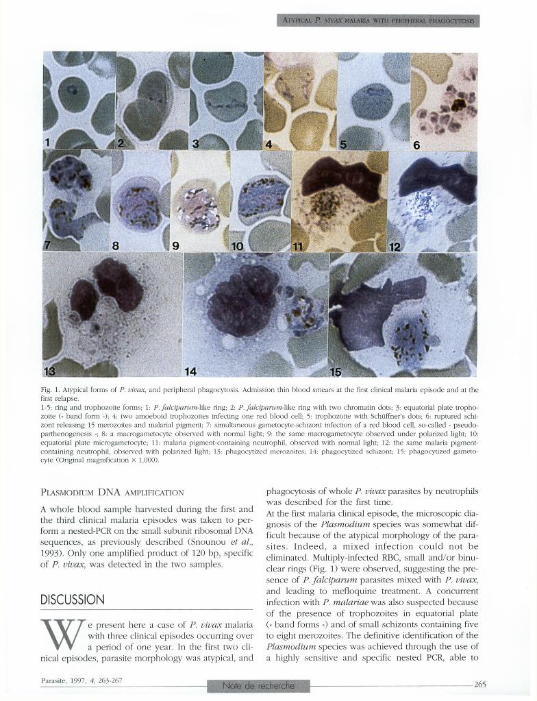
Blood samples were examined the January 8, 1995, the May 27, 1995 and the January 10, 1996. Five ml of whole blood were obtained by venepuncture into one tube containing EDTA as anticoagulant. Thin blood smears were stained with 10 % Giemsa for 20 min. In the thin blood smears of January 8, 1995, parasitaemia was very low (5,922 x 106 infected corpus-cules/L = 0.14 %). All parasite stages were observed: numerous trophozoites and gametocytes, and rare schi-zonts. Moreover, multiply-infected red blood cells (RBC), malaria pigment-containing leucocytes and some phagocytized parasites were observed (Fig. 1). Indeed fifty-five point six percent of infected RBC were invaded by trophozoites and measured between 6.5 and 9 µm (mean 7.5 µm). Fifty-five point six percent of infected RBC were invaded by one trophozoite,
1.5 % by two trophozoites, or rarely by three trophozoites. Trophozoites were polymorphous (Fig. 1): 0 their size ranged between 1.5 and 4 µm (mean 2.5 µm), ii) few of them showed an equatorial plate morphology, iii) others were binuclear trophozoites. Schuffner's dots were not observed in most of the RBC parasitized with trophozoites (Fig. 1). A few schizonts were observed in these smears: they were ruptured mature forms, containing five to sixteen merozoites with malaria pigment (Fig. 1). Thirty-nine percent of the infected RBC were invaded by gametocytes. Their size ranged between 7 and 12 µm (mean 9 µm) and gametocytes measured between 6 and 10 µm (mean 7.5 µm). Rare « pseudo-parthenogenesis » forms were also observed (Fig. 1); these forms correspond to simultaneous infections of a same RBC with an asexual form and a sexual one (Poirriez et al., 1991, 1996). Furthermore, numerous malarial pigment-containing neutrophils and monocytes were detected by microscopy, especially under polarized light (Lawrence & Olson, 1986) (Fig. 1). Phagocytized parasites were also observed (Fig. 1). Eight percent of the neutrophils (83 % of total leucocytes) contained haemozoin, parasites (trophozoites, schizonts, gametocytes or merozoites) (4 %), whereas only 3 % of the monocytes (6 % of total leucocytes) contained parasite material.
On May 27, 1995 (first relapse), parasitaemia was low (14,620 x 106 infected corpuscules/L = 0.34). Ninety point two percent of infected RBC were invaded by trophozoites, they measured between 6 and 9 µm (mean 8 µm). Eighty-four percent, 5.5 and 0.7 % of the infected RBC were invaded by one, two or three trophozoites respectively. The trophozoites measured between 2 and 4 pm (mean 3.5 µm). They were also polymorphous and a high proportion of equatorial forms (10 % of trophozoites) was observed (Fig. 1). The rare mature schizonts contained five to eight merozoites. Nine point six percent of infected RBC were invaded by gametocytes. Their diameter ranged between 6 and 10 µm (mean 8.5 µm). Gametocytes measured between 5 and 8 µm (mean 7 µm) (Fig. 1). As in the first clinical malaria episode, few malaria pigment-containing leucocytes and phagocytized parasites were also present. No « pseudo-parthenogenesis » forms were observed.
On January 10, 1996 (second relapse), parasitaemia was the lowest (0.8 x 10⁶ infected corpuscules/L = 0.017 %). In this third episode, 87.5 and 12.5 % of infected RBC were invaded respectively by trophozoites and gametocytes. No schizonts were observed in these blood smears. A typical P. vivax morphology was found: enlarged and pale parasitized RBC, amoeboid shape of trophozoites, relative late development of Schuffner's dots. Only rare RBC were invaded by two trophozoites or binuclear trophozoites.
264 Note de recherche Parasite, 1997, 4, 263-267
ATYPICAL P. VIVAX MALARIA WITH PERIPHERAL PHAGOCYTOSIS
Fig. 1. Atypical forms of P. vivax, and peripheral phagocytosis. Admission thin blood smears at the first clinical malaria episode and at the first relapse. 1-5: ring and trophozoite forms; 1: P. falciparum-like ring ; 2 : P. falciparum-like ring with two chromatin dots; 3: equatorial plate trophozoite (« band form ») ; 4 : two amoeboid trophozoites infecting one red blood cell ; 5 : trophozoite with Schüffner's dots; 6: ruptured schi-zont releasing 15 merozoites and malarial pigment; 7 ; simultaneous gametocyte-schizont infection of a red blood cell, so-called « pseudo-parthenogenesis » ; 8 : a macrogametocyte observed with normal light ; 9 : the same macrogametocyte observed under polarized light; 10: equatorial plate microgametocyte ; 11 : malaria pigment-containing neutrophil, observed with normal light ; 12 : the same malaria pigment-containing neutrophil, observed with polarized light; 13 : phagocytized merozoites; 14 : phagocytized schizont ; 15 ; phagocytized gameto-cyte (Original magnification x 1,000).
PLASMODIUM D N A AMPLIFICATION
A whole blood sample harvested during the first and the third clinical malaria episodes was taken to perform a nested-PCR on the small subunit ribosomal DNA sequences, as previously described (Snounou et al., 1993). Only one amplified product of 120 bp, specific of P. vivax, was detected in the two samples.
DISCUSSION
W e present here a case of P. vivax malaria with three clinical episodes occurring over a period of one year. In the first two cli
nical episodes, parasite morphology was atypical, and
phagocytosis of whole P. vivax parasites by neutrophils was described for the first time. At the first malaria clinical episode, the microscopic diagnosis of the Plasmodium species was somewhat difficult because of the atypical morphology of the parasites. Indeed, a mixed infection could not be eliminated. Multiply-infected RBC, small and/or binu-clear rings (Fig. 1) were observed, suggesting the presence of P. falciparum parasites mixed with P. vivax, and leading to mefloquine treatment. A concurrent infection with P. malariae was also suspected because of the presence of trophozoites in equatorial plate (« band forms ») and of small schizonts containing five to eight merozoites. The definitive identification of the Plasmodium species was achieved through the use of a highly sensitive and specific nested PCR, able to
Parasite, 1997, 4, 263-267 NOTE DE RECHERCHE 265
MAZARS E., LABERENNE J.E., SNOUNOU G., DEI-CAS E., CATTOEN C. & POIRRIEZ J.
detect less than ten parasites, to identify the four human Plasmodium species and therefore to detect mixed malaria infections (Snounou et al., 1993). The only detected Plasmodium species was P. vivax. The PCR assay has clearly allowed to identify the parasite species and to exclude unequivocally a mixed infection diagnosis, which was suspected by routine microscopy. At the second malaria episode (first relapse), the parasites found in the thin blood films were morphologically quite similar to those seen during the first attack. Unfortunately, a blood sample had not been taken for PCR assay. Although P. falciparum-like and P. mala-riae-like blood forms were observed, the diagnosis of P. falciparum and P. malariae was considered most unlikely: this second attack occurred eight months after the return of the patient to France, about five months after the first attack which was adequately treated with mefloquine and in which the PCR assay detected only P. vivax. The presence of P. ovale was also excluded because the morphological features of this species were absent (Poirriez et al., 1991). At the third episode (second relapse), the diagnosis of P. vivax infection was firmly established by microscopy on the basis of the typical blood parasite morphology, and confirmed by PCR.
A high degree of polymorphism has long been known in P. vivax (as multiply-infected RBC, crisis forms, « pseudo-parthenogenesis », or scarce Schüffner's dots) and has been reviewed recently (Prasad et al., 1990 ; Poirriez et al., 1991, 1995). In the present case, P. vivax was considered atypical partly because of the high gametocyte percentage detected at the first clinical malaria episode. Indeed, to our knowledge, so high a gametocyte percentage (39 %) has never been reported in P. vivax. It is interesting to note that the typical P. vivax morphology was only observed at the second relapse, one year after the first clinical malaria episode. Usually, atypical strains became typical throughout the course of a clinical attack or during a relapse (Poirriez et al., 1991).
Malaria chemoprophylaxis prevents neither malaria infection, nor a relapse of P. vivax or P. ovale, it is only aimed at preventing the first clinical manifestations. After the prophylaxis ceases, relapses can be observed at varying times depending on the parasite strain. In the present case, the P. vivax strain was chlo-roquine sensitive and could be considered as a II-III intermediate type, in regard to the WHO classification (Garnham et al., 1975). At the first clinical malaria episode, less so during the second attack, numerous leucocytes contained malaria pigment (haemozoin), which is produced by Plasmodium sp erythrocytic parasites as an end product of haemoglobin digestion. In addition, numerous neu
trophils and some monocytes were also found to have phagocytized various stage parasites (trophozoites, schizontes, gametocytes and merozoites). Pigment-containing leucocytes are more frequently observed in the peripheral blood of patients with severe malaria, where high percentage of neutrophils containing malarial pigment could reflect acute hyperparasitaemia (Metzger et al., 1995). In severe P. falciparum malaria, a count of peripheral neutrophils containing visible pigment > 5 % would predict a fatal outcome with higher sensitivity and specificity than parasitaemia (Hoan Phu et al., 1995). To our knowledge, the significance of the level of peripheral pigment-containing neutrophils has not been investigated in P. vivax infections. In our case, the high percentage of these cells recorded, especially at the first clinical episode, was not correlated with a bad prognosis.
One century ago, Golgi called attention to circulating leucocytes containing not only pigment, but also malarial organisms, either well preserved or in various stages of desintegration (Taliaferro & Mulligan, 1937). Nevertheless, he noticed that the extent of phagocytosis in the peripheral blood was incomparably less than that occurring in the spleen, the liver and the bone marrow. Several years later, Taliaferro et Mulligan (1937) reported that phagocytic neutrophils are rarely encou-tered in human malaria either in the peripheral blood or in the internal organs, except occasionally in cases of extremely acute pernicious malaria. More recently, a peripheral phagocytosis by only monocytes of whole parasites in P. falciparum malaria, was reported in three patients living in Gabon (Ventes et al., 1980). Our unusual observation indicates that leucocytes, especially neutrophils, are able to engulf not only P. falciparum parasites in cases of pernicious malaria but also whole P. vivax parasites without predicting a bad clinical evolution.
In conclusion, atypical strains of P. vivax are relatively frequent (Prasad et al., 1990; Poirriez et al., 1991, 1995). The most atypical features of the P. vivax strain described here are the following : i) a high gametocyte percentage detected at the first clinical malaria episode, ii) an important percentage of leucocytes containing malarial pigment at the first malaria episode, iii) a P. vivax marked peripheral phagocytosis at the first and the second malaria episodes.
ACKNOWLEDGEMENTS
We thank Richard Torka for his valuable technical assistance, and Gérard Vinchent for the excellent photography reproduction.
266 Note de recherche Parasite, 1997, 4, 263-267
ATYPICAL P. VIVAX MALARIA WITH PERIPHERAL PHAGOCYTOSIS
REFERENCES
AMOR D. & RICHARDS M. Mefloquine resistant P. vivax malaria in PNG. Medical Journal of Australy, 1992, 156, 883.
BAIRD J.K., BASRI H., SUBIANTO B., FRYAUFF D., MCELROY P., LEK-SANA B., RICHIE T., MASBAR S., WIGNALL S. & HOFFMAN S. Treatment of chloroquine-resistant Plasmodium vivax with chloroquine and primaquine or halofantrine. The Journal of Infectious Diseases, 1995, 171, 1678-1682.
BRUMPT E. The human parasites of the genus Plasmodium. In : Boyd MF. ed. Malariology. WB Saunders Company, Philadelphia and London, 1949.
GARNHAM P.C.C., BRAY R.S., BRUCE-CHWATT L.J., DRAPER C.C., KILLICK-KENDRICK R., SERGIEV P.G., TIBURSKAJA N.A., SHUTE P.G. & MARYON M. A strain of Plasmodium vivax characterized by prolonged incubation: morphological and biological characteristics. World Health Organization Bulletin, 1975, 52, 21-32.
HOAN PHU N., DAY N., THI DIEP P., FERGUSON D.J.P. & WHITE N.J. Intraleucocytic malaria pigment and prognosis in severe malaria. Transactions of the Royal Society of Tropical Medecine and Hygiene, 1995, 89, 200-204.
LAWRENCE C. & OLSON J.A. Birefringent hemozoin identifies malaria. American Journal of Clinical Pathology. 1989, 86, 360-363.
METZGER W.G, MORDMÜLLER B.G. & KREMSNER P.G. Malaria pigment in leucocytes. Transactions of the Royal Society of Tropical Medecine and Hygiene, 1995, 89, 637-638.
POIRRIEZ J., LANDAU I., VERHAEGHE A., SAVAGE A. & DEI-CAS E. Les formes atypiques de Plasmodium vivax. A propos d'une observation. Annales de Parasitologie Humaine et Comparée, 1991, 66, 149-154.
POIRRIEZ J., SNOUNOU G. & BLANCKAERT D. Multiple invasion of red blood cells by Plasmodium vivax in vivo. Transactions of the Royal Society of Tropical Medecine and Hygiene, 1995, 89, 509-510.
PRASAD R.N., PRASAD H., VIRK K.J. & SHARMA V.P. Detection of multiple invasion of erythrocytes by Plasmodium vivax. Tropical Medecine and Parasitology, 1990, 41, 437-438.
RIECKMANN K.H., DAVIS D.R. & HUTTON D.C. Plasmodium vivax resistance to chloroquine? The Lancet, 1989, ii, 1183-1184.
SNOUNOU G., VIRIYAKOSOL S., ZHU X.P., JARRA W., PINHEIRO L., DO ROSARIO V.E., THAITHONG S. & BROWN K.N. High sensitivity of detection of human malaria parasites by the use of nested polymerase chain reaction. Molecular and Biochemical Parasitology, 1993, 61, 315-20.
TALIAFERRO W.H. & MULLIGAN H.W. The histopathology of malaria with special refence to the function and origin of the macrophages in defence. Indian Medical Research Memoirs, 1937, 29, 138.
VERNES A. Phagocytosis of P. falciparum parasitized erythrocytes by peripheral monocytes. The Lancet, 1980, ii, 1297-1298.
Reçu le 27 mars 1997 Accepté le 12 juin 1997
Parasite, 1997, 4, 263-267 Note de recherche 267