Embed Size (px)
Citation preview

http://www.cabi.org/cabreviews
CAB Reviews 2021 16, No. 023
Red rot of sugarcane (Colletotrichum falcatum Went)
Rasappa Viswanathan*
Address: ICAR-Sugarcane Breeding Institute, Coimbatore, 641007, India.
ORCID information: Rasappa Viswanathan (orcid: 0000-0002-7274-8144)
*Correspondence: Rasappa Viswanathan. Email: [email protected]
Received: 23 December 2020Accepted: 09 February 2021
doi: 10.1079/PAVSNNR202116023
The electronic version of this article is the definitive one. It is located here: http://www.cabi.org/cabreviews
© CAB International 2021 (Online ISSN 1749-8848)
Abstract
Red rot of sugarcane was recorded more than 100 years before in Java, India, Argentina, USA and other countries, and it is one of the most devastating diseases of sugarcane. Since the cultivated sugarcane (Saccharum officinarum) has failed across the countries, systematic inter-specific hybridization between S. officinarum and the wild species S. spontaneum referred as ‘nobilization’ was done to develop resistant varieties and the disease was managed in most of the countries. However, in the countries especially in Asia, varietal breakdown to red rot caused severe epiphytotics, by which the resistant varieties failed in the field at regular intervals. New pathogenic strains of Colletotrichum falcatum with higher virulence were found responsible for varietal breakdown in sugarcane. Extensive cultivation of a single variety over large areas led to extensive crop damages due to ‘vertifolia’ effect in different decades in India. The current epiphytotic on the ruling variety Co 0238 has caused loss of more than one billion US dollars in the current season in the country. Detailed studies were done on pathogenic variation, epidemiology, screening methods, disease resistance mechanism, identifying effectors, pathogenicity determinants, antifungal genes and transgenics. Recently, complete genome and transcriptomes of C. falcatum were sequenced and pathogenicity hot spots and candidate secreted effector proteins were identified and this will further help to identify the candidate genes for further genetic manipulation. In spite of poor understanding on inheritance of resistance to C. falcatum in sugarcane, new varieties with red rot resistance were developed and deployed after each of the epiphytotic to save the crop. Further, other management practices including bioagents, chemicals and inducers were attempted and improved efficacy by mechanized sett treatment showed promising results to manage the disease under field conditions.
Keywords: sugarcane, red rot, Colletotrichum falcatum, life cycle, epiphytotics, varietal breakdown
Review Methodology: The review has been an update of classical and recent research works done on the important plant pathogen, affecting sugarcane production worldwide. The following web sources https://www.isosugar.org/sugarsector/sugar and https://indiansugar. com/Statics.aspx were referred for global scenario of sugarcane and its importance. Research works of Dr C.A. Barber and Dr E.J. Butler, who laid foundation for red rot work during the early part of the last century, were referred for historical perspectives on the area of research. CABI abstracts of the last 40 years were referred to collect recent research works on the area of review. The site of https://www.cabi.org/isc/datasheet/25361#toDistributionMaps was referred for disease distribution in the world. The following books, Diseases of Sugarcane: Major Diseases (Elsevier), Plant Disease: Red Rot of Sugarcane (Anmol Publications), Red rot of sugarcane (Technical Bulletin, USDA), Sugarcane Crop Management (SCI TECH Publishing) and Sugarcane Improvement through Breeding (Elsevier) were referred for disease symptoms, pathogenicity, disease cycle and disease management. Online papers from different sources were referred for detailed information on recent developments on pathogenic variation, host-pathogen interaction, sugarcane breeding, disease management, transgenic approaches etc.

2 CAB Reviews
http://www.cabi.org/cabreviews
Sugarcane and its importance
Sugarcane (Saccharum spp.) is a monocotyledon and member of the family Poaceae, tribe Andropogoneae. The genus Saccharum consists of 6 to 37 species depending on taxonomic interpretation and the members are of tall grasses, native to warm temperate to tropical regions of South and South East Asia. Sugarcane has thick, jointed and fibrous stalks of 2 to 6 metres tall that store sugar. Six Saccharum spp. viz. S. officinarum, S. sinense, S. barberi, S. edule (cultivated species), S. robustum and S. spontaneum (wild species) are well characterized and the cultivated sugarcane is an interspecific hybrid involving two or more species of Saccharum [1].
Sugar is extracted by evaporating the water from cane juice. Crystallized sugar production was reported 2500 years ago in India [1]. Arabs introduced sugar to the Mediterranean around the eighth century AD and Spain has started sugarcane cultivation by that time [1]. Sugarcane was among the early crops brought to the Americas by Spaniards. Although sugarcane was grown principally for sugar in the previous decades, now it is also grown for fibre and energy, primarily ethanol (biofuel), electricity from bagasse and bio-manure from filter cake. Since sugarcane is a C4 crop, it is viewed as one of the most capable biomass producer. The crop faces a wide range of issues from sugarcane production to sugar processing. Research institutions across the globe and industrial groups are pursuing ways and means to tackle the constraints associated to sugar manufacture, bioethanol production and sustaining cane farming (https://www.isosugar.org/sugarsector/sugar). Traditionally, sugarcane is propagated from stem cuttings (referred as setts) of one to three buds. The crop duration of sugarcane is 12–14 months and it can be harvested two to ten times; after each harvest, new stalks come from the stools, called ratoons; hence, the crop is cultivated like a plantation crop in many countries [2].
Sugarcane contributes approximately 80% of the global sugar requirement; sugar beet meets the remainder. Brazil, India, Thailand, China, the USA, Mexico, Pakistan, Australia and Guatemala are the major sugarcane-producing countries (http://www.fao.org/faostat/en). In India, sugarcane is grown in 5.2 M Ha area, which is approximately 3.0% of the total cultivable area in the country, and it contributes 7.5% gross value of agricultural production (https://indiansugar.com/Statics.aspx).
Disease incidence
Red rot of sugarcane was first reported as a disease in sugarcane in Java in 1893 [3]. Within a decade after Went’s description of the disease and its economic damages to sugarcane milling in Java, its occurrence was reported in several other parts of the world. All the reports indicated that the disease was widely spread and recognized as a new disease of sugarcane in the countries like Australia;
India; the USA, including Hawaii and mainland; West Indies; Brazil; Mauritius; Philippines etc. [4–6] (Fig. 1).
Red rot is one of the most serious diseases of sugarcane in many countries including India, Pakistan, Bangladesh, Thailand, Myanmar, Nepal, Vietnam and other countries. In Louisiana, red rot was described as one of the factors causing stubble deterioration of sugarcane [7, 8]. The disease severely affected sugarcane production in Thailand during 1991–1992 and 2004–2005 due to cultivation of susceptible varieties. During 2011–2012 also, a severe red rot epidemic occurred only in Lopburi province where the variety K93-236 was grown [9, 10]. In Myanmar, red rot is a constraint to cane cultivation in all the sugarcane-growing areas. The disease is managed in the country by releasing resistant varieties. They regularly screen new sugarcane clones for red rot resistance [11, 12].
Red rot has been reported in different countries from Asian, African and American continents like Malaysia [13], Nigeria with 5%–12% incidences [14], Sudan [15], Indonesia, [16], Philippines [17], China [18], Colombia [19], Peru [20], Taiwan [21], Australia [22] and South Africa [23]. In South Africa, red rot was first identified in 1941 and the disease caused considerable destructions in the varieties POJ 2725 and Co 290 [24]. Red rot was found as one of the diseases that is economically affecting cane production in Guatemala and Nicaragua [25]. In Thailand, red rot occurs along with wilt; hence, it is referred as ‘red rot wilt’ or sugarcane ‘root and stem rot’ or ‘red rot Fusarium stem rot’ [26]. Currently, the disease occurs in all the sugarcane-growing continents and it is reported in 77 countries [5].
Red rot in India
In India, Dr C.A. Barber did foundation work on the disease when it struck for the first time in the then Madras Presidency [27–29]. Butler [30], the Imperial mycologist, at the Imperial Agricultural Research Institute, Pusa, Bihar (India), studied extensively on the causal organism and its portals of entry into the cane stalk. Based on the most distinctive feature of rotting of the internal stalk tissues with reddish discolouration, he named the disease as ‘red rot’. Both Barber and Butler recognized the importance of the disease and devised management strategies of healthy seed and avoidance of waterlogging to reduce the crop damage in India. Severe red rot epiphytotics in the Godavari delta and North Indian plains caused extensive damages to sugarcane; however, this scenario resulted in the establishment of Sugarcane Breeding Institute (SBI) at Coimbatore, India, in 1912 by Dr Barber to develop red rot–resistant varieties through interspecific hybridization. Development of interspecific hybrids involving S. officinarum and S. spontaneum from Coimbatore started from Co 205 in 1918 and later hundreds of ‘Co’ varieties were released for commercial cultivation and adopted in India and in many other countries. Dr Barber was instrumental in developing many such interspecific hybrids; his contributions to

Rasappa Viswanathan 3
http://www.cabi.org/cabreviews
sugarcane have been recognized by naming one of the species of Saccharum native to India as S. barberi [31]. Similarly, sugarcane breeding centres in Java and Barbados also developed new varieties and these efforts have promoted growth of sugar industry in many countries [6].
Economic Impact
During the 1938–1939 season, a red rot epiphytotics of exceptional severity occurred in the subtropical region predominantly in Uttar Pradesh (UP) and Bihar, the major sugarcane region in India. This devastation resulted in failure of the major commercial variety Co 213, in which thousands of hectares were devastated. Due to the poor supply of canes, the sugar mills in the eastern UP crushed only one-third of their normal canes during 1938–1939 and half during 1939–1940 [32]. Severe infections of red rot can cause loss of nearly two-third of cane stalks produced in subtropical India [33, 34]. In Pakistan, 28.5% losses in cane weight was reported at initial infection by the red rot pathogen and it reached 82.7% when the disease intensity increased to 75% in the cvs L 116 and B 4360. Sucrose content, the main economic produce, was reduced in the range of 31%–75% at different infection levels [35,36]. Severe red rot epiphytotics in peninsular India during 1990s caused losses of 30%–50% in cane yield in the varieties like Co 6304, CoC 671, CoC 85061, CoC 86062, CoC 92061, CoSi 86071 etc. Yield losses of up to 100% were found in ratoon crops in different factory areas [37–40].
Red rot–affected canes show a decline of 29 to 83% in cane weight and 24 to 90% in juice extraction [6]. In the history of red rot epiphytotics in India, the country
currently faces one of the worst crop losses due to sudden failure of the most popular variety Co 0238 in the states of Uttar Pradesh (UP) and Bihar (Fig. 2). In UP state alone, nearly 0.5 M ha out of 2.6 M ha area has severe red rot during the 2020–2021 season (S. P. Singh, Unpublished). The crop losses due to red rot in the state have increased from few thousand ha during 2016–2017 season to this mammoth figure [41]. The total loss caused due to this disease outbreak works out to be 1.0 to 1.414 billion US$ during the current season alone. The previous year’s loss could be at least 40%–50% of the current loss. Overall, the disease has affected nearly 10% of the cane area in the country and it may further increase in the coming seasons. This is the economic repercussions caused by the disease in India; hence, it is called as ‘cancer of sugarcane’ [42]. Due to the disease, losses are encountered in different sectors such as farmers, small industries that manufacture gur/khandsari sugar, sugar mills etc. in the country (Fig. 3).
In South Africa, in the 1970s, widespread occurrence of the disease was found in the cooler, southern and inland parts, causing damage in many varieties including NCo 376 [43]. In Thailand, red rot caused cane yield losses from 34.6% to 73.7% in plant crop and up to 100% losses in the ratoons. The crop losses were estimated to approximately US$20 million during severe epiphytotics of red rot wilt in 1990s in the country [26].
C. falcatum infects nodal tissues and kills the buds; hence, it causes losses in bud germination and crop establishment in the field. This leaves gaps in the field and a poor crop stand leading to losses in cane production. When setts of latent infections were planted in spring crop season, a maximum reduction in germination of up to 73% was recorded, whereas in autumn season, only 19%–56% reduction was recorded [44]. In a set of 13 varieties, impact
Figure 1. Distribution of sugarcane red rot in the world (https://www.cabi.org/isc/datasheet/25361#toDistributionMaps).
4 CAB Reviews
http://www.cabi.org/cabreviews
of 9 isolates of C. falcatum inoculum in the soil at the time of planting of setts on bud germination and crop stand was studied for two seasons at Coimbatore under tropical conditions in India. In the first-year trial, the healthy controls recorded an overall germination of 74.0%, while the pathogen-inoculated plots recorded a mean germination of 43.6% only, showing a drastic reduction of 41.4% in bud germination. In the subsequent year, the same set of host varieties recorded an overall mean germination of 46.0% in the presence of the pathogen as compared to 75.3% in uninoculated control plots, showing a reduction
of 39.0% in sett germination [45]. Subsequently, it was found that the soil inoculum slowly builds up and causes disease in susceptible (S) and moderately susceptible (MS) varieties to varying extent. By 360 days, the nine C. falcatum isolates caused a mean reduction of 80.1% and 86.1% in cane population in the susceptible cvs CoC 671 and Co 94012, respectively, in a favourable season. Likewise, the MS cvs Co 06030, Co 06022, CoV 09356 and Co 06027 recorded a loss in cane population of 63.1%, 52.6%, 48.0% and 42.5%, respectively [45]. This study indicated that poor crop establishment due to red rot can cause a huge loss to
Figure 2. Complete destruction of sugarcane crops due to red rot in Uttar Pradesh, India.
Figure 3. Impact of red rot on sugar(cane) economy.

Rasappa Viswanathan 5
http://www.cabi.org/cabreviews
cane production in the plant crop in the disease-endemic regions. From such plant crops, ratoons cannot be taken up, which causes further losses in cane farming.
Red rot symptoms
The disease is recognized based on its characteristic rotting and reddening symptoms of stalk tissues. However, the pathogen infects the crop at all the stages of its growth. Earlier, the author has described detailed symptoms of the disease [6, 46]. Here, the disease symptoms are grouped into young crop, stalk and foliage symptoms.
Symptoms in young crop
During the germination phase, symptoms of pre-germination death of buds and drying of germinated sprouts/shoots are observed (Fig. 4). Infected canes or seed cane infections immediately after planting show progress of disease from the seed canes to new sprouts. In such cases, typical red rot symptoms can be seen inside the canes along with foliage discolouration in the sprouts (Fig 5). In case of infections of post-germination phase, the settlings show yellow to orange discolouration and this leads to drying of entire shoots (Fig. 5). During late germination phase of the crop, drying of whorl alone within healthy leaves is seen due to passage of infection from shoot base to the apex. Later, the affected plant may gradually die or the plant may recover from the disease with new side tillers.
During tillering phase or before cane formation, the symptoms of the disease are witnessed in the form of mostly orange-yellow discolouration of spindle leaves and drying of the tillers (Fig. 6). Sometimes, the leaves in the whorl may show typical midrib lesions of varying intensities. Similar to the symptoms in plant crop, ratoons also show
death of new sprouts from the stools and this leads to poor crop establishment. Death of the stools in the ratoons shows gaps of varying sizes, depending on the severity of stools’ infection.
Stalk symptoms—external
The disease-affected canes show drying of canes either singly or in clumps. The top two to three leaves in the spindle of the affected stalks show orange to yellow discolouration, which can be easily recognized from the background of green foliage (Fig. 7). The discoloured leaves dry soon in a few days. In severe cases, the entire field shows drying and complete crop failure occurs (Fig. 8). Similar to red rot, wilt or infestation of insect pests also manifests such as drying of canes in a clump. By critical examination of the affected stalks, red rot can be recognized based on rind discolouration of varying intensities and internal symptoms. Infection of internal core tissues leads to expression of characteristic rind discolouration, usually at varying intensities of brown or paleness of rind (Fig. 9). Other characteristic symptoms of red rot on the canes are typical nodal necrosis around the region of bud, growth ring and leaf scar; pinkish spore masses of the pathogen on nodal region, especially at root eyes, leaf scar or rind surface (Fig. 10); break-off of canes at nodes and de-topped canes in the field and appearance of fruiting bodies of the fungus (acervuli) on rind surface (Fig. 11). Rampant colonization of the fungus inside the canes severely impairs sap movement and translocation of photosynthates to the roots. Hence, the affected cane dries out in course of time.
Internal symptoms
Splitting of the affected canes longitudinally shows characteristic red rot symptoms inside the stalk as
Figure 4. Death of germinated sprouts/shoots due to red rot.

6 CAB Reviews
http://www.cabi.org/cabreviews
Figure 5. Primary infections of C. falcatum cause red rot inside the planted canes and progressive foliage discolouration in the sprouts.
Figure 6. Drying of the entire sugarcane with prominent midrib lesions (blackish) on the top leaves.

Rasappa Viswanathan 7
http://www.cabi.org/cabreviews
reddening of internal tissues with white spots that are usually elongated laterally covering the entire width of the cane (Fig. 12). Further, immediately after opening, a slightly acidic starchy odour emits from the affected cane tissue. The white spots may vary in size and number and occasionally they are so abundant as to give the tissue a mottled appearance (Fig. 13). Later, the affected canes show longitudinal pith cavities filled with greyish mycelia of C. falcatum (Fig. 14) and drying of the cane tissues. The red rot–affected canes also show other kinds of symptoms like dull brown discolouration of the ground tissue, characteristic serial spots in the internode tissues, reddish discolouration of the ground tissue without white spots, pits of varying sizes and numbers on pith region etc. (Fig. 15). Nature of varieties, stage of the crop, soil moisture, prevailing weather conditions etc. influence expression of these symptoms in the affected canes.
Foliar (midrib) symptoms
Usually, C. falcatum infects stalk tissues and foliar symptoms are observed as typical midrib lesions, occasionally, reddish areas on the sheaths and small reddish brown spots on the leaf lamina. On the midrib, the infection first appears as small brown to red spots on the upper and lower surface. These small spots expand rapidly in both directions and coalesce to form long lesions of few inches to entire length of the leaf or they may remain as a series of unconnected lesions. When favourable condition prevails, all the newly emerging leaves show midrib lesions (Fig. 16). During
Figure 7. Red rot–affected sugarcane shows typical orange yellow discolouration of spindle leaves.
Figure 8. Red rot–affected canes exhibit drying in large numbers in the entire field.

8 CAB Reviews
http://www.cabi.org/cabreviews
monsoon/ post-monsoon season, rarely top rot–like symptoms are noticed with rotting of entire undifferentiated leaves within the whorl with lesions (Fig. 17). Usually, a higher intensity of midrib lesions is observed after summer rains and during monsoon months in the field.
Combined infections of red rot and wilt
Since C. falcatum and Fusarium sacchari, the wilt pathogens, infect stalk tissue in sugarcane, combined infections of the pathogens are common in India and many other countries.
Figure 9. Red rot–affected canes show discolouration of greenish rind.
Figure 10. Diseased cane shows C. falcatum conidial mass on the nodal tissues.
Figure 11. C. falcatum fruiting bodies (acervuli) on nodal tissues and rind.

Rasappa Viswanathan 9
http://www.cabi.org/cabreviews
Usually, F. sacchari follows C. falcatum, and in such canes, progress of the latter inside the cane is ceased and the rotten tissues show drying with varying tinges of purple to pink or pinkish red in the affected tissues. After its entry, F. sacchari moves rapidly through the infected internodes from the base, reaches upper uninfected internodes and causes pithy cavities with pinkish discoloured patches that are characteristic to wilt (Fig. 18). Generally, combined infections of these two fungi cause quick drying of cane stalks than to their separate infections. Occasionally, red rot–affected canes show F. sacchari and Ceratocystis paradoxa, the pathogen associated with pineapple disease in standing canes (Fig. 19). Under field conditions, the stalk diseases of sugarcane like wilt, stalk rot and pineapple disease show certain overlapping symptoms with red rot [46, 47]. However, red rot–affected canes can be easily
identified with the characteristic reddening of internal tissues with white spots.
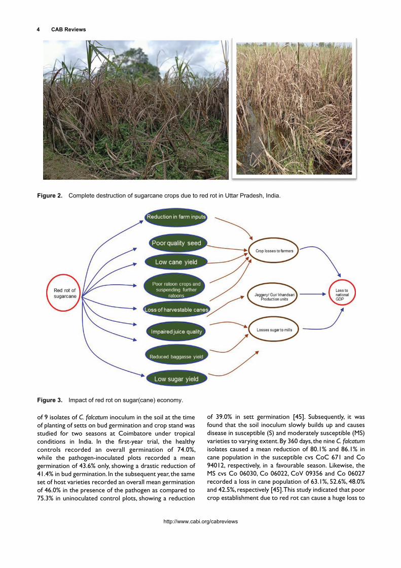
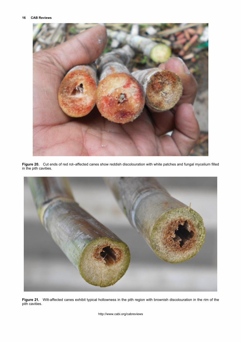
Cut ends of the affected canes show characteristic reddish patches interspersed with whitish or pale ground tissue or in some occasions reddish lesion accompanied with greyish mycelium in pith cavities (Fig. 20). This symptom is unique to red rot–affected canes, whereas wilt-affected canes exhibit typical hollowness in the pith region with brownish discolouration of the rim of the pith cavities (Fig. 21).
Pathogen
The Ascomycete fungus, Glomerella tucumanensis (Spegazzini) von Arx & Muller, is the associated pathogen. Under field conditions, only anamorphic stage of the pathogen Colletotrichum falcatum F.A. Went is recorded. The fungus comes under Phylum: Ascomycota, Subphylum: Pezizomycotina, Class: Sordariomycetes and Family: Glomerellaceae. After describing the disease in detail with the symptoms, Went [3] demonstrated the parasitism of the fungus he isolated from the diseased tissues, calling it Colletotrichum falcatum Went and further carried out life history studies.
Cultural characteristics
The fungal colony on oatmeal agar consists of abundant aerial mycelium, broadly spreading, sometimes zonate, densely woven into compact, velvety turf in some isolates, or a cottony, floccose one in others, referred as dark and light races, respectively [7]. Colour ranges from almost white through light ashy grey to dark grey, growing darker with age (pale olive grey to pearl grey) with no colour or pigmentation in reverse or in the medium. Hyphae are densely interwoven anastomosing in definite ropes. Conidia develop in a pink- or salmon-coloured, water soluble, mucilaginous mass, and when produced rapidly on the upper portion of the acervulus, it is covered with a shining droplet. The isolates show enormous phenotypic variation on the plates and broadly they are grouped as light and dark isolates; however, there are intermediate types with variation for sporulation and colony characters [6]. Several of the isolates studied at Coimbatore, India, were able to produce acervuli in culture with diameter ranging from 0.639 to 1.54 mm and setae are found in all the isolates, and in few cases, it was rare. The number of setae ranged from 3 to 20 per acervulus and length ranged from 90 to 220 μm. Conidiophore length ranged from 120 to 330 μm. In the past, several authors have reported emergence of new isolates with variation in morphological and cultural characters in C. falcatum. However, the dimensions of all the isolates may fall within the broad range for conidial, acervuli, conidiophore and setae.
Figure 12. Typical red rot symptoms of reddish discolou-ration of ground tissue with white spots.

10 CAB Reviews
http://www.cabi.org/cabreviews
Abbott [7] has observed that in artificial culture the dark-type isolates produce chlamydospores in much greater abundance than the light ones. Certain isolates of C. falcatum are reported to produce black hard structures, that is, stromatic bodies on culture media that consist of fertile hyphae, some of which perform true function as conidiophores and bear typical falcate conidia of C. falcatum at their tips. Earlier, a stroma-forming strain of C. falcatum was reported from infected sugarcane varieties from Karnal, India [48]. It is presumed that they may play an important role in perpetuation of C. falcatum; however, the conditions that favour development of stroma in the field are unknown.
Cultural instability of C. falcatum with the formation of sectors has been reported. Usually, the cultures grow less luxuriantly or become mycelial with low sporulation after a prolonged period of artificial cultivation. The successive
batches of cultures of an isolate obtained at different times varied in sporulation. It was reported that incubation in a dry atmosphere tends to reduce fructification, whereas incubation in a humid atmosphere favours it [7]. However, it was also reported that the isolates are morphologically stable without significant changes in virulence as a result of long continued cultivation on oatmeal medium [7]. The author has recorded instability of the fungal cultures in the long run and they have lost their virulence after repeated sub-cultures on oatmeal agar. Hence, many sugarcane pathologists pass the cultures through their respective host cultivars regularly, re-isolate and maintain in culture collections. Sometimes, the poorly defined ‘sectors’ or ‘patch’ variants appear in the plates, but transfer of these sectors to fresh plates produces colonies apparently identical with the original. Numerous reports on physiology of C. falcatum including media, nutrients, pH,
Figure 13. Red rot–affected stalks show a mottled appearance of white spots.

Rasappa Viswanathan 11
http://www.cabi.org/cabreviews
temperature, conidial germination etc. were reported earlier in detail [6, 49]; hence, more emphasis is given on pathogenicity, pathogenic and molecular variation in the sub-chapter.
Although asexual phase of the pathogen is commonly recovered from the infected tissues, Spegazzini [50] and Carvajal and Edgerton [51] described and characterized the perfect state of the pathogen. In India also, the occurrence of perfect state of the fungus was recorded both under field conditions and in culture [52–54]. Duttamajumder [49] made a detailed account of perfect state of the fungus in India. He felt that the red rot pathogen is basically a leaf parasite similar to Colletotrichum infecting sorghum and it completes its life cycle on sugarcane leaf. He also opined that so long as C. falcatum restricts itself on the midrib, it does not cause any significant harm to the cane crop. It was emphasised that most of the midrib
population survives on the midrib without causing much harm to sugarcane and only a few of them adopt to invade the stalk. However, studies conducted at Coimbatore revealed that most of the midrib isolates of C. falcatum from susceptible varieties are found to be more virulent than stalk isolates, which is in contrast to the previous observations [55].
Pathogenicity
The historic red rot epiphytotic on the popular cv Co 213 in the subtropical India led to the emergence of light-coloured cultures of C. falcatum with highly sporulating phenotype, which caused severe devastation on the variety during 1936–1939 [56]. For the first time in the country, the researchers were prompted to further study on the
Figure 14. Pith cavities filled with greyish mycelia of C. falcatum during later stages of disease development.
Figure 15. Red rot–affected canes show serial spots (left) and pits (right) in the internodes tissue.

12 CAB Reviews
http://www.cabi.org/cabreviews
pathogen side since the research till that time was focussed only on deploying high-quality and high-yielding varieties. Successive failures of the varieties such as Co 301, Co 312, Co 313, Co 385, Co 421, Co 453, Co 513, BO 11, CoS 443 etc. were attributed to the highly virulent strains of the pathogen [57, 58]. Due to these developments, 1940 onwards, the researchers have initiated varietal screening for red rot resistance including germplasm [33].
The pathogenic isolates were maintained in different names in different centres. Historic failures of the popular variety Co 1148 in 1970s and later prompted the scientists to characterize the new virulent pathogenic strains on a set of differentials. In 1980s, a clear report on occurrence of three distinct pathotypes of C. falcatum from the subtropical region was reported based on the pathogenicity on a set of host varieties [59]. Subsequently, to characterize the pathogenic variation in C. falcatum, detailed studies were conducted in tropical and subtropical locations to identify suitable host differentials from 48 Saccharum spp. and hybrid cultivars [60]. A set of 13 host differentials were identified and established occurrence six pathotypes in C. falcatum in the country [61]. Later, prevalence of a new pathotype from the cv 85A261 along with three identified pathotypes from the cvs Co 419, CoC 671 and Co 997
was reported in coastal Andhra Pradesh and Odisha [62]. The new isolates were phenotyped on the host differentials; about 11 of them were characterized as designated pathotypes from 4 agro-climatic zones in the country till 2010 and used for screening of sugarcane progenies in the respective zones [6]. Another pathotype CF12 was recently designated from the tropical region [63].
Molecular variation
Earlier RAPD was used to group C. falcatum isolates and it was reported that the isolates had a general agreement with their pathogenicity on different sugarcane varieties [64, 65]. ITS nucleotide sequence analysis of 15 C. falcatum isolates in Thailand showed 95.32–100% identity with each other and 96.30–97.74% related to C. falcatum [12]. Nine isolates from red rot–affected locations possessed a high degree of pathogenicity on the susceptible cvs E-Heaw and K93-236, whereas those from non-epidemic were nonpathogenic. Furthermore, in the same study, the isolates from endemic areas had a light type of colony character and it was concluded that in Thailand C. falcatum is differentiated into two distinct races (pathogenic and
Figure 16. Entire midrib in the spindle leaves show continuous lesions.

Rasappa Viswanathan 13
http://www.cabi.org/cabreviews
nonpathogenic) based on their pathological character on the stalks of sugarcane.
Nine major C. falcatum pathotypes used for disease screening were characterized based on sequencing of 5.8-internal spacer (ITS) region of rDNA into two phylogenetic groups. Further, by using nitrate non-utilizing (nit) mutants, heterokaryon formation was demonstrated and vegetative compatibility grouping (VCG) in C. falcatum was standardized. The VCG grouped the mutants into five categories and through this different isolates of the same pathotype were recognized [66]. Grouping of the pathotypes based on VCG, pathogenicity and ITS was similar with certain deviation; however, these tools distinguished the two contrasting pathotypes Cf1148 and Cf7717 as reported earlier by RAPD [61, 66]. Additionally, serological reactions between antisera of the pathotypes and the antigen in diffusion agar plates gave a clear relationship between the pathotypes [61, 67]. Subsequent molecular grouping of the C. falcatum isolates revealed that the Indian isolates fell into three separate assemblages as Group I, II and III. Among them, the latter had a distinct phenotype of dark coloured, least virulent and non-sporulating had a similarity to other country isolates, whereas the first two subgroups had overlapping phenotypic and pathogenic features [68]. Apart from ITS
spacer region sequencing, other conserved genes calmodulin, actin and GPDH were used to further refine molecular grouping, which revealed the presence of one major group of virulent isolates and a minor group of least virulent isolates, which was established by definite nucleotide variation and further confirmed by RAPD and ISSR. Some of the primers were able to differentiate the virulent isolates with specific markers with the least virulent isolates. In addition, C. falcatum proteomes were established between virulent and least virulent isolates and identified distinctive and differentially expressed proteins related to virulence. Pathogenicity-related genes identified from Colletotrichum spp. and other fungi like glutathione S-transferase, DJ-1/PfpI family protein and serine protease were identified from C. falcatum [69]. Recently, utilizing the NGS data, virulent strain–specific simple sequence repeat (SSR) marker from the genome of C. falcatum was identified [70].
Virulence diversity in C. falcatum
C. falcatum occurs in nature with enormous diversity for pathogenicity. It is dictated either by the varietal diversity or large spread of a ruling variety over large areas. The new
Figure 17. Red rot–affected plant shows death of young leaf with prominent midrib lesions on the leaves in the whorl.

14 CAB Reviews
http://www.cabi.org/cabreviews
pathogenic variants may be evolving from the prevailing pathotype(s) in the field with a significant gain in their pathogenicity and virulence due to adaptation to the new host varieties or to a new environment for the pathogen carried through seed canes. Diversity in the new C. falcatum isolates was carried out in the respective locations in India and no study was made to assess pathogenic diversity in huge collection of the isolates simultaneously in the country. The author has made an elaborate study to document pathogenic behaviour for nearly 117 isolates originated from tropical and subtropical states in two locations, Coimbatore (tropical) and Karnal (subtropical) on a highly susceptible cv CoC 671 for five seasons by following plug method of testing under field conditions [71]. The isolates displayed variation in their behaviour from season to season for red rot reactions at both the
locations. In the subtropical conditions, more number of less and least virulence reactions was recorded; however, more highly virulent categories also recorded there and it may probably be due to weather conditions prevalent after pathogen inoculation. Though both the locations showed similar behaviour for moderate virulence, at the subtropical location, it ranged from 5.22% to 21.15% during the five seasons with a mean of 11.94%, whereas in the tropical location, it ranged from 10.08% to 16.36% with a mean of 12.73%, indicating a stable behaviour in the latter than the former. Some of the isolates from the tropical region maintained a higher virulence in both the locations, whereas many of the subtropical isolates and pathotypes exhibited less virulence in both the locations. Virulence of the isolates was not static in the locations over the years for many of the isolates due to their stable and unstable
Figure 18. Sugarcane stalks exhibiting symptoms of combined infections of C. falcatum and F. sacchari.

Rasappa Viswanathan 15
http://www.cabi.org/cabreviews
behaviour and place of phenotyping, source of host variety and prevailing weather conditions. Assessing more than 100 C. falcatum isolates simultaneously brought out existence of vast diversity in C. falcatum isolates for their pathogenicity in India. Probably such a high pathogenic variation is responsible for frequent varietal breakdown in the country for the 100 years.
Pathogenicity factors
It is well established that C. falcatum pathotypes vary in their virulence and this specific trait decides the pathogenicity of a particular pathotype. The pathotypes produce secondary metabolites and hydrolytic enzymes,
considered as bioweapons that probably contribute aggressiveness/virulence during pathogen infection and colonization. Several workers studied production of hydrolytic enzymes, viz. pectolytic and cellulolytic by C. falcatum. It was reported that virulent isolates of the pathogen produce more hydrolytic enzymes than weakly pathogenic ones [72–74].
C. falcatum produces a toxic metabolite initially reported as anthraquinone compound, which may facilitate pathogen infection and spread in the stalk. The phytotoxin is soluble in water and in many organic solvents, capable of producing symptoms of red rot in the stalks barring white spot [75, 76]. Later studies established a correlation between disease expression and production of phytotoxins and hydrolytic enzymes by the pathogen. C. falcatum is a typical hemibiotroph, initiates infections as a biotroph and turns to necrotrophic phase for colonization and ramification inside the host tissue to complete life cycle. The phytotoxins and hydrolytic enzymes produced during necrotrophic phase of the pathogen determine the pathogen colonization and tissue damage. When pathogenicity is interfered by co-inoculating Trichoderma harzianum, production of these metabolites was reduced with no symptoms production on the host [77]. Further, less virulent pathotype produces low levels of phytotoxins and hydrolytic enzymes compared to a virulent pathotype CF06 [78].
When major C. falcatum pathotypes were studied for the relation between toxin production and symptom production on leaves, it was found that the virulent pathotypes caused more severe symptoms along with more loss of electrolyte leakage. Further, the virulent isolates produced higher levels of pectinolytic and cellulolytic enzymes especially exo-polygalacturonase and melanin [79]. All these biochemical studies clearly revealed a positive association on the production of secondary metabolites like toxins and melanin and hydrolytic enzymes in C. falcatum virulence and disease expression. Recently, molecular basis of the pathogenicity factors were studied using both genomic and proteomic tools. A study of 28 pathogenicity gene homologues in two distinct pathotypes that vary in their virulence revealed a specific role of some of these pathogenicity genes in C. falcatum pathogenesis with a clear differential expression [80]. Tricyclazole interferes in germination of the conidia and appressoria production and its melanisation in many fungal pathogens. Using tricyclazole, the melanin inhibitor, the role of melanin in C. falcatum pathogenesis has been proved. The role of melanin was further established by studying the expression of melanin biosynthesis genes such as PKS1, SCD1 and THR1 [81]. Subsequently, knockdown mutants of C. falcatum were developed through RNA silencing strategy and Agrobacterium-mediated transformation to functionally analyse polyketide synthase 1 (PKS1), the major gene that regulates dihydroxy naphthalene melanin production. The loss-of-function mutants for PKS1 showed reduced pathogenicity in leaf and stalk tissues and established a clear role for melanin in pathogenic virulence in C. falcatum [82].
Figure 19. Combined infections of F. sacchari and C. paradoxa in red rot–affected cane (right) against healthy canes (left).
16 CAB Reviews
http://www.cabi.org/cabreviews
Figure 20. Cut ends of red rot–affected canes show reddish discolouration with white patches and fungal mycelium filled in the pith cavities.
Figure 21. Wilt-affected canes exhibit typical hollowness in the pith region with brownish discolouration in the rim of the pith cavities.

Rasappa Viswanathan 17
http://www.cabi.org/cabreviews
C. falcatum effectors
Proteomics-based investigations lead to characterization of cellular and extracellular virulence and pathogenicity factors produced by pathogens as well as to identify changes in protein levels in host plant upon infection by pathogenic organisms and symbiotic counterparts [83]. In vitro secretome of C. falcatum cultured under light and dark conditions using 2DE coupled with MALDI TOF/TOF MS was analysed. The study has identified nine differentially expressed proteins and revealed a major portion of alterations occurred in low molecular weight (LMW) proteins of less than 30 kDa. In dark cultures, the LMW proteins were either less abundant or absent, except a ceratoplatanin protein called eliciting plant response like protein 1 (CfEPL1), very high abundant LMW protein. While in light cultures, a novel protein named as ‘plant defence inducing protein 1’ (CfPDIP1) was highly abundant [84]. Further studies functionally characterized distinct domains of CfEPL1 and CfPDIP1 by in vitro expression and purification, which indicated that CfEPL1∆N1-92 and CfPDIP1∆N1-21 induce hypersensitive reaction in tobacco and systemic resistance in sugarcane against C. falcatum. These studies have identified proteins that putatively contribute to C. falcatum virulence and demonstrated the potential role of PAMPs/effectors of C. falcatum inducing PAMP-triggered immunity (PTI)/effector-triggered immunity (ETI) in sugarcane [85].
Complete genome of C. falcatum
For the first time, the C. falcatum genome was sequenced to be of 48.16 MB in size with 12,270 genes [86]. Subsequently, the transcriptome of C. falcatum (in vitro) was reported to be 31 MB with 23,136 predicted CDS [87]. Mining of C. falcatum genome and transcriptome data yielded putative 768 and 884 small secreted proteins (SSPs), respectively. The predicted secretory proteins were further divided into classical and non-classical proteins and discovered that signal peptides have an apparent role during pathogenesis by stabilizing fungal secretory proteins in the host environment. The SSPs contained a large number of esterase, proteinase, CAZy families, cytochrome P450, peptidases, secondary metabolites, transporters and transcription factors. In in planta transcriptomic studies, these SSPs were recognised as major pathogenic determinants [88].
Recently, the C. falcatum pathotype Cf671 (MTCC accession number-12142) and CfROC (isolated from the sugarcane variety ROC), R-1 (virulent isolate of CfROC recovered from the cv CoC 671) maintained at the red rot culture collection facility of ICAR-SBI, Coimbatore, were used for whole genome and consecutive transcriptome sequencing and also for the phenotypic and genotypic studies. Phenotypic studies were carried out to identify the infection process, mating type and population structure using high mobility group (HMG) proteins. Further
characterization had been done using SEM analysis, which revealed the nature of C. falcatum isolates. This finding shows that C. falcatum is a definite stalk-intriguing pathogen that establishes itself as a precursor in attributing gene families for its virulence. During interaction with sugarcane, C. falcatum expresses 2/3 of CAZy genes during biotrophic and necrotrophic phases.
Diagnosis
Serology-based diagnostics especially ELISA and dot-blot assays were developed earlier with antisera developed against mycelial proteins or specific polypeptide of C. falcatum. These assays were sensitive to detect the pathogen colonization in the nodal tissues like buds, root eyes, leaf scars and pith and rind tissue of an infected cane [89–91]. However, they have not been put into use later. Chandra et al. [92] amplified C. falcatum DNA with high specificity, efficiency and rapidity under isothermal conditions in LAMP assay. In this assay, they demonstrated that visual judgement of colour change in 1 h without further post-amplification processing makes the LAMP method convenient, economical and useful in C. falcatum diagnosis. However, here also the assay could not be adopted under field conditions. Dot-blot–based assay with non-radioactive probes was standardized at the institute and was found efficient to detect the pathogen in the soil [93]. It has been applied to assess C. falcatum propagule load in the soil before taking up new crop in the endemic locations in Tamil Nadu, India.
Infection process and life cycle
C. falcatum pathogenesis and infection process in sugarcane have been well documented. Recently, using modern tools, the process has been explained in detail with more clarity. On contact, the conidia germinate with germ tubes, produce appressoria, attach firmly to the sugarcane tissue and endure stably for varying periods. This may support the fungal pathogen to perpetuate when the infective mycelium is unable to proliferate further. The main portals of entry for the pathogen are leaf scar, root primordia and buds in the nodal region (Fig. 22). After penetration, the fungus makes inter- and intracellular colonization. Later, acervuli produce profusely around the infected tissues. Using C. falcatum isolate expressing green fluorescent protein (GFP) markers and other sensitive histological assays, infection, colonization and fructification of the pathogen on the leaf and stalk tissues were clearly established. On sugarcane leaf tissues, by 12 h post-inoculation (hpi), C. falcatum conidia germinate and form appressorial structures; by 24 hpi, after formation of primary hyphae, the fungus enters into the host cell; by 48 hpi, it spreads to nearby cells through secondary hyphae and this phase marks the end of biotrophic phase, in which

18 CAB Reviews
http://www.cabi.org/cabreviews
the fungus does not kill the cells. Later, necrotrophic phase of pathogenesis begins, in which the secondary hyphae continue to damage the cell structure with widespread colonization and kill the colonized the cells to make rapid proliferation. The pathogen makes both intra- and intercellular colonization and emerges outside through the stomatal pores with sporulating acervuli structures with setae by 72 hpi (Fig. 23). On stalk tissue also, the pathogen shows similar infection cycle, unlike in the leaf tissue, and in stalks, the pathogen continues to make colonization due to availability of host tissue and make both upward and downward progress. In due course, it occupies the entire core tissues in the internodes and nodes. By 30–45 days, the pathogen macerates entire tissues and causes complete destruction of the stalk tissues. The pathogen also grows
inside and fills the pith cavities with greyish-black mycelia. The presence of acervuli throughout rind tissues and typical sporulation can also be seen on the root eyes, leaf scar and rind [6, 94–96]
In young sugarcane, pathogen infection occurs at the base of the shoot at the point of contact with old bud scales and scale-like leaves. The dormant mycelium/appressoria present in the buds and bud scales in the node and soil-borne inocula are the sources of infection for young shoots. This inoculum grows with the emerging bud and enters into the shoot, infecting the new leaves continuously and perhaps the growing point in a systemic way. Later in the season, the infection takes place through the nodal regions of the cane near ground level from conidia of the pathogen, carried through irrigation water or rainwater. The conidia of the fungus produced in great abundance on leaf midrib get washed down to soil by rain or dew into the cavity between the cane stalk and the leaf sheath and thus reach the nodes and cause infection mainly through the leaf scar region, root primordia and growth ring (Fig. 22).
The pathogen infection progresses considerably in the susceptible varieties in the same season or it may remain as dormant incipient infection. In highly susceptible varieties, the conidia landed in the whorl directly infect and penetrate the growing point to reach the stalk. Although C. falcatum may infect almost any part of the sugarcane plant, its importance is limited largely to its occurrence on the leaf midribs, the internal stalk tissues and the stubbles of the ratoons. Infection of the roots may occur; however, red rot is not important as a root disease.
Seed cane infection
Although in laboratory tests, infection of setts is possible through cut ends of the setts, and this mode of infection is of little practical importance. Agnihotri et al. [97] reported that diseased setts are the main sources of C. falcatum infection, and the disease spreads from the mother setts to the new shoots. Setts having both internal and external infections cause much more damage than the setts having either internal or external (nodal) infections. Nodal infections are mostly responsible for the spread of the disease in nature because of their very small size. They also reiterated that healthy setts can become infected if planted in soil that harbours red rot–affected cane debris. The inoculum in the debris readily infects nodal region, gradually proceeds to bud sprouts and causes death of young settlings (Figs. 4 and 5).
Leaf infection
In general, the pathogen rarely affects leaf lamina. Lesions produced on the leaf midribs are the characteristic symptoms on leaf tissues. In Louisiana, the first leaf infections are usually noted during May or June and
Figure 22. C. falcatum making progressive infection after entry through nodal tissues in sugarcane.

Rasappa Viswanathan 19
http://www.cabi.org/cabreviews
infections on the midrib arise from small punctures made by leafhoppers when ovipositing. However, in India, researchers confirmed that infection takes place through the apparently uninjured epidermis [98, 99]. Fructification begins in 10 to 14 days following inoculation, the lesions extend longitudinally along the midrib and the fruiting area likewise increases so that old lesions are usually covered with black masses of acervuli.
Chona and Bajaj [52] observed only the perithecial stage of red rot fungus on the blade. Edgerton [100] reported stray occurrence of small spots on the lamina. Prakasam and Appalanarasiah [101] reported dark brown spots with well-defined margins and mostly on matured leaves. In the advanced state, the area between the spots dried up presenting a straw-coloured appearance. In some of the top leaves, they found lesions on leaf lamina and sheath. The canes from such infected plants upon replanting exhibited disease and thereby confirmed the lesions caused by C. falcatum.
There is confusion regarding virulence associated with profuse sporulation in midrib isolates. Not all the highly sporulating isolates are found to cause stalk infections on the susceptible varieties [102, 103]. There are reports that most midrib isolates of C. falcatum are weak pathogens or less virulent and do not present any serious threat to cane cultivation. There is an opinion that the pathotypes that
cause midrib lesions are markedly different from those causing infection in stalk [104–106]. However, the author has found that midrib isolates are highly virulent on the stalks [55].
The author has studied development of laminar lesions under field as well as controlled conditions. On the leaf lamina, the pathogen causes reddish-brown lesions of varying sizes. The lesions are always linear in shape parallel to veins with irregular margin. Very often, the centre of the lesions will be broader with narrow ends. Yellow halos of 2–3 mm breadth surrounding the lesions are observed under high humidity conditions and lesion margin is not clear in such lesions. Usually, the centre of the lesions is lighter in colour than the margins where the colour intensity is more. Sometimes progressive lesions with yellow discolouration alone are noticed on the infected leaves. Such lesions may extend up to 60 cm in length. Later, such lesions turn pale and that particular region dry off. Adjoining lesions coalesce to cause extensive linear lesions with yellow discolouration covering most of the laminar region under favourable conditions. Sometimes the yellow halo at the ends extend on both directions as yellowish runners as observed in eye spot caused by Bipolaris sacchari on sugarcane leaves. Gradually, the runners extend on the sides and cause extensive lesions. In the large patch of discoloured lesions, laminar tissue shows
Figure 23. C. falcatum produces acervuli (with black paraphyses) with extensive conidial discharge on the surface of the cane tissue (Staining with fluorescence brightener shows conidia in white in the background).

20 CAB Reviews
http://www.cabi.org/cabreviews
shot holes on the primary lesions and subsequently leaf shredding (Fig. 24).
Stalk infection
The fungus may enter the cane stalk through various channels. Went [3] concluded that natural infection occurs chiefly through the holes made by boring insects. Various other workers have reported influence of borer holes in aggravating red rot incidence [6]. In Louisiana, 50% of the Diatraea saccharalis–infested plants were badly infected with red rot [107]. Abbott [7] established that red rot infection in the growing stalks occurs principally through
the tunnels of the moth borer (D. saccharalis) in Louisiana and Southern Florida and, in some varieties, through the root primordia. However, studies of Singh et al. [108] revealed that stalk borer infestation does not play a conspicuous role in accelerating C. falcatum infection but showed a possible role in secondary transmission of the pathogen. Under Indian conditions, both in the tropical and subtropical regions, no association was found between infestations of stalk borer (Chilo auricilius), internode borer (Chilo sacchariphagus indicus) or top borer (Scirpophaga excerptalis) and red rot severity in sugarcane varieties. The author has found no changes in the disease progress from the bottom internodes to the upper nodes due to borer injury or there is a mild change in the lesion spread (Fig. 25).
Figure 24. Laminar infection of C. falcatum in sugarcane. Left: sugarcane cv CoS 8436 exhibits lesions on lamina, leaf sheath and midrib under natural conditions in Haryana. Right: laminar lesions of varying sizes with yellow halo extending to several cm under artificial inoculation (cv CoC 671).

Rasappa Viswanathan 21
http://www.cabi.org/cabreviews
Hence, borer pests of canes do not favour stalk infection under Indian conditions.
Chona [109] reported that red rot infection in varieties like Co 233, Co 396, Co 548 and BO 4 took place rapidly through growth cracks. Edgerton [110] also reported similar findings with Co 290. Later, it was clarified that pathogen infection takes place only when the propagules land on the split before the sealing process starts [99]. Some of the varieties have tendency of producing growth crakes in the internodes, especially during high rainfall or waterlogging. The longitudinal splits in the cane receives fresh conidia washed from midrib lesions in the canopy,
rain splash or wind. When the split injury is fresh, infection occurs and the disease progress is witnessed on both directions (Fig. 26). The fungus continues to develop in the tissues of both midrib and the stalk until the cane is harvested for seed or for milling.
Nodal infection
The conidia lodge at the nodes of mature stalks and cause infection in various tissues of the node such as the bud scales, root primordia, growth ring and leaf scar. Among
Figure 25. C. falcatum–inoculated canes show limited or no impact to lesion development with internode borer infesta-tion. Yellow arrows indicate the pathogen-inoculated bore holes by plug method of inoculation. White arrows indicate borer tunnels.

22 CAB Reviews
http://www.cabi.org/cabreviews
them, leaf scar is the main portal of entry for red rot pathogen into the stalk [110–112]. Immediately after abscission, the ends of the vessels of the leaf scar are open and conidia may be sucked in or infection hyphae may affect entry. Srinivasan and Alexander [112] showed that dormant infections might occur in leaf scar tissue and on the bud scales, which could remain in that condition for long periods. Similar observations have been reported from Louisiana [113, 114]. It is probable that dormant infections under certain favourable conditions are activated and leads to the development of characteristic symptoms
of red rot. Hence, such infections are epidemiologically important.
Great differences were also observed in different sugarcane varieties for their susceptibility to nodal infection. The varieties that possess a comparatively greater resistance to nodal infections are likely to remain free from disease even if growing with plenty of C. falcatum inoculum in the field. The popular sugarcane cv Co 86032 of tropical India though susceptible to C. falcatum by plug method showed greater levels of nodal resistance to most of the prevailing pathotypes. Similar observations were made in many of the varieties tested under advanced varietal trials at Coimbatore, where attempted infections of the pathogen are noticed after nodal swab method of inoculation as reddish discolouration of leaf scar region to varying intensities in terms of spread and pigmentation (Fig. 27). The dark pigmentation due to accumulation of 3-deoxyanthocyanidin compounds restricts the pathogen progress through the leaf scar injury, whereas in the susceptible varieties, disease development occurs within few days after inoculation.
Further, certain C. falcatum isolates have tendency to cause infections on the nodal tissue while making progress inside the stalks, whereas other isolates skip the nodal region that is of fibrous nature to the next internode (Fig. 28). In case of the former, after completing infections on the nodal tissue, the fungus comes out through nodal tissues, mostly, root eyes and leaf scar, exposing the fungal propagules (Fig. 10). Through such events, the fungus spreads quickly in the field; however, in
Figure 26. Progress of red rot lesions through internodal splits. Arrows indicate the cracks or lesions below the split region.
Figure 27. Attempted penetration of C. falcatum in a resistant sugarcane variety shows dark reddish discoloura-tion on nodal tissues.

Rasappa Viswanathan 23
http://www.cabi.org/cabreviews
case of the latter, the infective propagules release to the environment occurs after the death of the canes, probably in the next season as cane debris inoculum.
During 1990s, the author has observed no red rot in the cv Co 86032 in many fields that had 60%–80% red rot with the susceptible varieties like CoC 671, CoC 92061, CoSi 96071 etc. in the previous season. Subsequent observations for the last 25 years in the tropical region revealed that the cv Co 86032 remained free from the disease due to its nodal resistance and existence of field tolerance may have been due to resistance to nodal infection [31, 115]. It was also reported that the power
of nodal infection is not possessed by all the isolates of C. falcatum [109]. Evidently, the predominance of such isolates that possess this trait could cause any appreciable secondary infection of the crop and bring about an epiphytotic. Probably, the variety like Co 86032 may survive in the field till emergence of such matching pathotype(s) with capability to penetrate through node. The author has recorded occurrence of red rot in this variety in different occasions; however, it was confined to few clumps in Tamil Nadu state and probably new isolates have not emerged with complete pathogenic weapons to cause knockdown effect in this variety.
Figure 28. Nodal infection in sugarcane stalks as a trait of virulence in C. falcatum pathotypes. Left: The virulent pathotype causes susceptible reaction with extensive nodal necrosis and abundant fungal growth; Right: Less virulent pathotype causes moderately susceptible reaction with poor nodal necrosis.

24 CAB Reviews
http://www.cabi.org/cabreviews
Pathogen spread
The rate of C. falcatum spread within the seed canes after planting depends on the degree of red rot susceptibility of sugarcane varieties. Soil temperature and moisture significantly influence disease spread; however, both have a significant bearing on the growth of the sugarcane itself. Under favourable conditions, the pathogen spreads rapidly through both the vascular bundles and the parenchyma, and the entire tissues of the stalk may be invaded within 2 or 3 months after infection.
Pathogen spread within cropMidrib lesions serve as the principal source of inoculum for stalk infections. Midrib lesions appear from two months after planting in the field and the frequent winds and dashing summer rains provide an excellent medium for spreading millions of propagules by splashing and blowing them to the stalks or to other leaves or by trickling over the leaf lesions and down to the stalk, where drops of water containing conidia may be held for several hours by the cup-like ligule. Detailed studies were carried out in the past on C. falcatum pathogenesis and disease spread under field conditions. Most of the Colletotrichum spp. infect foliar tissues or fruits in different plants, whereas C. falcatum infects stalk tissues in sugarcane. Foliar infection of the pathogen in sugarcane is rare and it never infects other plants under natural conditions. It has developed specific adaptation to infect sugarcane tissues at different stages. Most significant adaptation of C. falcatum is its capability to infect hard nodal tissues in the cane and cause midrib lesions. These two characters of C. falcatum make it a successful pathogen on sugarcane. After infection of nodal tissues, either it spreads both vertically and horizontally to cause damage to the cane stalks in the same season or the restricted infection as dormant infections on the nodal tissues help it to initiate fresh infection in the next crop
(Fig. 22). Midrib lesions directly help the pathogen to spread in the current season. Here also cane infection initiated from the midrib lesions in the whorl may cause dormant infections in the later stages of the crop. Detailed studies are required on the pathogenicity mechanisms developed by C. falcatum to understand more about its pathogenesis.
Pathogen spread from plant crop to ratoonDuring harvesting, all the infected canes are cut and removed along with the healthy canes for sugar extraction. Although above-ground portion of the infected canes are removed, there is residual inoculum in the stubbles (Fig. 29). The pathogen remaining in the stubbles spreads to new sprouts depending upon the prevailing conditions at the time of ratooning. Some sugarcane varieties may show more of upward movement of the pathogen in the stalks. However, in some, the pathogen moves rapidly towards bottom and colonize effectively in the stool. Such varieties favour disease spread substantially from plant to ratoon crops. If we consider the leftover inoculum, it will be less in the former when compared to the latter. Overall, the pathogen completes life cycle within few months of planting of the crop or takes one season depending upon the variety, source of inoculum, prevailing weather conditions and agronomic practices followed in the field (Fig. 30).
Adaptation of C. falcatum to sugarcane varieties
Continuous emergence of new variants of C. falcatum is the major concern in the field as they pose challenge to host resistance in the new varieties. However, only limited information is known on the adoption of the pathotypes to the newly released varieties under field conditions. Earlier, Srinivasan [116] studied in detail on the role of host varieties on emergence of new isolates. He has shown that some sugarcane varieties induce rapid development
Figure 29. Red rot in the harvested canes, which serves as primary source for new ratoon crop.

Rasappa Viswanathan 25
http://www.cabi.org/cabreviews
and dominance in infected tissues of the dark, avirulent type of variant, while others appear to favour the dominance of the virulent parental clone. Sometimes a more virulent isolate than its parental clone has also appeared. Srinivasan [117] also opined adaptive changes in cultivated C. falcatum in relation to the host varieties, with subsequent alterations in the virulence patterns of the fungus. He also observed that the pathogenic isolates are often unstable in their pathogenicity and have a tendency to pass irrevocably into an avirulent phase. Earlier studies of the author explained how a less virulent isolate gains virulence after several rounds of repeated inoculation and isolation on an incompatible host variety [118]. After repeated inoculations, the dark isolates at initial phases become light with increased sporulation on their adapted hosts. Development of light isolates and reduced latent period for symptom expression by repeated inoculations on incompatible host varieties indicated gain of virulence or pathogenicity of that pathotype for adaptation on a particular cultivar [119]. The adapted cultures were able to tolerate to the new cytoplasm as suggested by Srinivasan [116].
A detailed study was conducted by the author with 12 C. falcatum isolates and 20 varieties varying in disease resistance for 10 years to identify how the isolates change their behaviour on the host varieties under tropical conditions [118]. The isolates always exhibited their virulence on the susceptible varieties but not on the varieties with moderately susceptible or intermediate reactions when they were inoculated by plug method under field conditions. Also the isolates originated from susceptible or MS varieties had expressed their virulence
similarly, indicating that all the new pathotypes completely evolved with potential virulence to cause knockdown effect on the host. The study revealed that although the resistant varieties remained free from infections from the isolates, on few occasions, the isolates have broken barriers of incompatibility and exhibited disease severity. On susceptible host varieties, the expression of the virulence was in the range of 62.9%–97.9%, whereas on MS hosts, it was 21.3%–40%, clearly revealing that the pathogen has to evolve further to completely adapt on the latter group of varieties. Such evolution happens under field conditions after varietal introduction and the existing pathotype adapts to the host variety and slowly gain its virulence to cause knockdown effect. This study also demonstrated on the gain of virulence by the new pathotypes over the old pathotypes that were isolated 30 years ago, designated as CF04 (Cf419), CF05 (Cf997) and CF06 (Cf671) and used for the resistance screening in the tropical states. Meanwhile, in the last two to three decades, new varieties were deployed and correspondingly new pathotypes have evolved from the new varieties suggesting the gain of virulence from the new hosts.
In another interesting study, the author [63] has again demonstrated on the continuous evolution of C. falcatum in tune with the new varieties deployed in the field. He used the case study with C. falcatum pathotype CF06, the predominant pathotype of the tropical region in India used for varietal screening, and assessed how it has given way for new pathotype CF12 with 32 host varieties under field conditions for seven years. The then-popular cv CoC 671, which was cultivated extensively during 1980s and 1990s
Figure 30. Disease cycle of red rot in sugarcane.
26 CAB Reviews
http://www.cabi.org/cabreviews
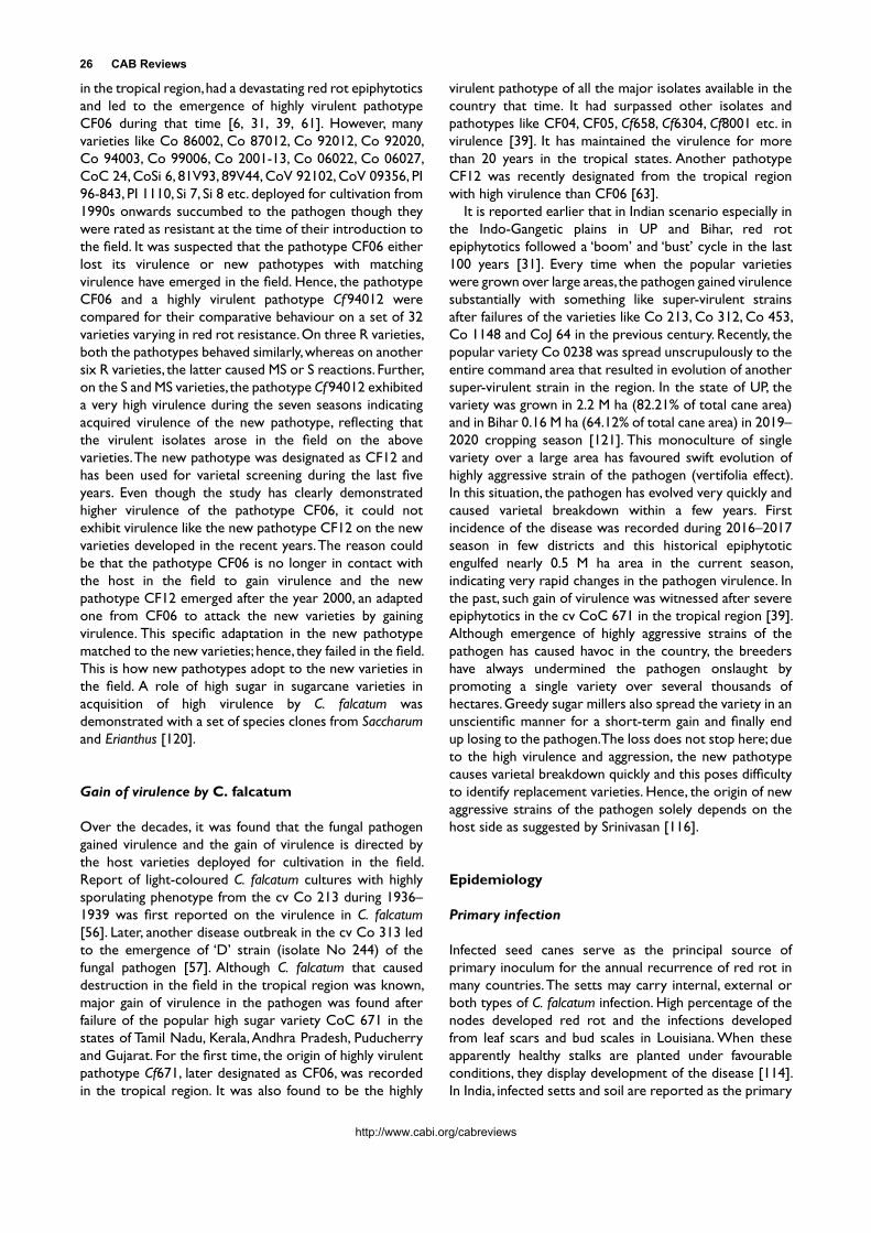
in the tropical region, had a devastating red rot epiphytotics and led to the emergence of highly virulent pathotype CF06 during that time [6, 31, 39, 61]. However, many varieties like Co 86002, Co 87012, Co 92012, Co 92020, Co 94003, Co 99006, Co 2001-13, Co 06022, Co 06027, CoC 24, CoSi 6, 81V93, 89V44, CoV 92102, CoV 09356, PI 96-843, PI 1110, Si 7, Si 8 etc. deployed for cultivation from 1990s onwards succumbed to the pathogen though they were rated as resistant at the time of their introduction to the field. It was suspected that the pathotype CF06 either lost its virulence or new pathotypes with matching virulence have emerged in the field. Hence, the pathotype CF06 and a highly virulent pathotype Cf 94012 were compared for their comparative behaviour on a set of 32 varieties varying in red rot resistance. On three R varieties, both the pathotypes behaved similarly, whereas on another six R varieties, the latter caused MS or S reactions. Further, on the S and MS varieties, the pathotype Cf 94012 exhibited a very high virulence during the seven seasons indicating acquired virulence of the new pathotype, reflecting that the virulent isolates arose in the field on the above varieties. The new pathotype was designated as CF12 and has been used for varietal screening during the last five years. Even though the study has clearly demonstrated higher virulence of the pathotype CF06, it could not exhibit virulence like the new pathotype CF12 on the new varieties developed in the recent years. The reason could be that the pathotype CF06 is no longer in contact with the host in the field to gain virulence and the new pathotype CF12 emerged after the year 2000, an adapted one from CF06 to attack the new varieties by gaining virulence. This specific adaptation in the new pathotype matched to the new varieties; hence, they failed in the field. This is how new pathotypes adopt to the new varieties in the field. A role of high sugar in sugarcane varieties in acquisition of high virulence by C. falcatum was demonstrated with a set of species clones from Saccharum and Erianthus [120].
Gain of virulence by C. falcatum
Over the decades, it was found that the fungal pathogen gained virulence and the gain of virulence is directed by the host varieties deployed for cultivation in the field. Report of light-coloured C. falcatum cultures with highly sporulating phenotype from the cv Co 213 during 1936–1939 was first reported on the virulence in C. falcatum [56]. Later, another disease outbreak in the cv Co 313 led to the emergence of ‘D’ strain (isolate No 244) of the fungal pathogen [57]. Although C. falcatum that caused destruction in the field in the tropical region was known, major gain of virulence in the pathogen was found after failure of the popular high sugar variety CoC 671 in the states of Tamil Nadu, Kerala, Andhra Pradesh, Puducherry and Gujarat. For the first time, the origin of highly virulent pathotype Cf671, later designated as CF06, was recorded in the tropical region. It was also found to be the highly
virulent pathotype of all the major isolates available in the country that time. It had surpassed other isolates and pathotypes like CF04, CF05, Cf658, Cf6304, Cf8001 etc. in virulence [39]. It has maintained the virulence for more than 20 years in the tropical states. Another pathotype CF12 was recently designated from the tropical region with high virulence than CF06 [63].
It is reported earlier that in Indian scenario especially in the Indo-Gangetic plains in UP and Bihar, red rot epiphytotics followed a ‘boom’ and ‘bust’ cycle in the last 100 years [31]. Every time when the popular varieties were grown over large areas, the pathogen gained virulence substantially with something like super-virulent strains after failures of the varieties like Co 213, Co 312, Co 453, Co 1148 and CoJ 64 in the previous century. Recently, the popular variety Co 0238 was spread unscrupulously to the entire command area that resulted in evolution of another super-virulent strain in the region. In the state of UP, the variety was grown in 2.2 M ha (82.21% of total cane area) and in Bihar 0.16 M ha (64.12% of total cane area) in 2019–2020 cropping season [121]. This monoculture of single variety over a large area has favoured swift evolution of highly aggressive strain of the pathogen (vertifolia effect). In this situation, the pathogen has evolved very quickly and caused varietal breakdown within a few years. First incidence of the disease was recorded during 2016–2017 season in few districts and this historical epiphytotic engulfed nearly 0.5 M ha area in the current season, indicating very rapid changes in the pathogen virulence. In the past, such gain of virulence was witnessed after severe epiphytotics in the cv CoC 671 in the tropical region [39]. Although emergence of highly aggressive strains of the pathogen has caused havoc in the country, the breeders have always undermined the pathogen onslaught by promoting a single variety over several thousands of hectares. Greedy sugar millers also spread the variety in an unscientific manner for a short-term gain and finally end up losing to the pathogen. The loss does not stop here; due to the high virulence and aggression, the new pathotype causes varietal breakdown quickly and this poses difficulty to identify replacement varieties. Hence, the origin of new aggressive strains of the pathogen solely depends on the host side as suggested by Srinivasan [116].
Epidemiology
Primary infection
Infected seed canes serve as the principal source of primary inoculum for the annual recurrence of red rot in many countries. The setts may carry internal, external or both types of C. falcatum infection. High percentage of the nodes developed red rot and the infections developed from leaf scars and bud scales in Louisiana. When these apparently healthy stalks are planted under favourable conditions, they display development of the disease [114]. In India, infected setts and soil are reported as the primary

Rasappa Viswanathan 27
http://www.cabi.org/cabreviews
sources of C. falcatum inoculum for the disease recurrence in endemic regions [97]. C. falcatum forms abundant acervuli in nodal tissues and rind (Fig. 11). In nature, such nodal infections are mainly responsible for spread of the disease since latent infections are difficult to be detected by naked eyes [4].
Along with sett-borne infections, soil-borne inocula play a major role in red rot perpetuation and initiate disease cycle in endemic locations. Detailed studies were conducted in different decades on the role of soil-borne inocula in initiating disease cycle, their survival and influence of weather conditions. It was reported that various factors influence survival of C. falcatum in soil under field conditions [109, 122]. When one-cm long pieces of red rot–affected stalks and midribs were used, the fungus survives in the soil for two months [123]. Later, it was reported that C. falcatum–infected nodal and internodal tissue bits on soil surface survive for nine months. After burial of the stalk pieces, the presence of different types of fungal structures viz. conidia, thick-walled hyphae, setae, appressoria and chlamydospores were found for two months [124].
Survival of C. falcatum in the soil is influenced by the factors such as soil depth, soil moisture, sterilized or natural soil, nature of affected canes like split or un-split etc. Further saprophytic survival of C. falcatum in soil is low [6]. Furthermore, the occurrence of midrib lesions in the field especially in the young crop is an indication of disease spread from the infected setts/soil or ratoons from affected plant crop (Fig. 5).
The application of certain organic and inorganic amendments increased red rot incidence especially in the susceptible cv Co 312 [125]. Studies conducted in Coimbatore revealed disease development from C. falcatum–infected cane debris or multiplied on sorghum grains. The susceptible varieties readily picked up infections from such inoculum, whereas MS varieties had shown pathogen infection less frequently. However, MR varieties had shown infections rarely and both MS and MR varieties showed a differential interaction when different isolates are used [45]. Under field conditions, C. falcatum survives in the soil or stubbles for more than one year, especially when the stubbles are left undisturbed after one or two ploughings with or without green manure crop [6, 126] However, Singh et al. [123] reported that the fungal pathogen survives in affected cane pieces in the soil up to 34 and 63 days in autumn and winter, respectively, under subtropical India. They found approximately 50 propagules per g of soil are sufficient to initiate infection and root primordia and leaf scars are the main portals of entry for the pathogen from soil.
The survival of red rot pathogen in the soil as thick-walled mycelium, appressoria, setae, chlamydospores and conidia that are liberated to soil during the process of decomposition of red rot–affected tissues are reported by many workers [124]. After two months of burial of infected tissues, all types of fungal structures, namely conidia, thick-
walled hyphae, hyaline hyphae setae, chlamydospores and appressoria, were present in about 30 to 50% of them. Five months later, they found setae, appressoria, chlamydospores and thick-walled mycelium and eight months later only setae and thick-walled mycelium were found [124].
Further, disease development of the soil-borne inocula of 11 C. falcatum isolates on 10 sugarcane varieties varying in disease resistance showed that the disease susceptible and MS varieties picked up infections and exhibited disease from germination phase onwards. Apart from a differential interaction of the C. falcatum isolates against the host varieties, the varieties showed a year-to-year variation in disease development from the isolates. For example, the cvs Co 403 and Co 06027 that remained disease-free against all the isolates during 2017–2018 season had red rot from four and eight isolates, respectively, during 2018–2019 season. Overall, the C. falcatum isolates from soil caused very severe red rot incidences of 71.4%–100%, in the two susceptible cvs CoC 671 and Co 94012. The differential interaction of the host varieties and the pathogenic isolates were clearly visible in the MS varieties. Among the MS varieties, the cv Co 06030 exhibited disease against 10 of 11 isolates with 83.3% mean disease incidence, whereas the cvs Co 06022, Co 06027 and CoV 09356 recorded red rot against 5–7 isolates with mean disease incidences of 23.4%, 47.5% and 27.1%, respectively [45]. Earlier, Singh [125] reported that the C. falcatum can survive in the rhizosphere of an MR variety without causing symptoms and the residual inoculum is able to infect a succeeding crop of a susceptible variety.
The general hypothesis is that high soil moisture favours red rot severity in sugarcane. However, other workers have reported that dry or drought period during growing season favours severe attack of the disease [127, 128]. Singh et al. [129] observed maximum red rot incidence in plant and ratoon crops in the fields, where irrigation after two months of cane germination was avoided as compared to other fields with normal irrigation or with waterlogging during monsoon in subtropical conditions. They explained that moisture stress in the summer predisposes the plant to attack of the pathogen and promotes formation of more dormant structures. Further, the presence of more amounts of dead parenchyma cells may enable the spread of the pathogen in stalk tissues. Due to water stress, more dead tissues occur; hence, the resistance offered by the host tissues against the pathogen spread appears to be reduced, which hastens the conquest of the pathogen [130]. The fungus surviving in dormant stages on young plants or infected sugarcane debris gets favourable environment upon commencement of rain at the end of June in subtropical regions to sporulate and produce abundant acervuli laden with conidia, which may initiate fresh infections. However, the formation of dormant infections in ratoon does not differ significantly with moisture levels [131]. Desiccation has been found to increase disease severity in the stalks of certain cultivars in Louisiana. It was found that red rot and desiccation have a

28 CAB Reviews
http://www.cabi.org/cabreviews
synergistic effect on spring shoot population reductions in all the tested cultivars, except one that was very sensitive to desiccation, suggesting that drought condition during the initial growth processes in sugarcane increases red rot severity [132].
In compost, the fungus was not found to survive for more than a month. This shows that the mycelium is unable to hold its own against microorganisms, which offer a keen competition [132]. Higher rhizosphere population of Trichoderma and other microbes was recorded in healthy sugarcane as compared to red rot–infected sugarcane in the fields [133]. There were also reports that rhizosphere soil of red rot–resistant cultivars was more lytic to C. falcatum than the rhizosphere soil of susceptible cultivars [125, 134].
Soil-borne inoculum and varietal breakdown
The experience of various workers indicates that sugarcane varieties easily succumb to C. falcatum under waterlogged conditions and even the MR varieties cannot withstand against the attack of the pathogen [6]. Various hypotheses have been put forth to explain varietal breakdown under waterlogged conditions. Several authors have opined that predisposition of the host to infection under such condition is the major factor [27, 30, 56, 135]. In Louisiana, the red rot is more severe in the heavy or black soils, which are usually poorly drained [7]. Such conditions favour the disease through the negative influence they have on the normal growth activities of the sugarcane plant, as well as by the direct favourable effect of high moisture on the fungus.
On stagnant water, aggregation of floating mycelia occurs with the production of conidia in acervuli. Further, the conidia germinate and fuse with each other, leading to aggregation of fused conidia. After fusion of conidia, some of the fused conidia germinate to produce conidia directly on them [136]. Waterlogged condition may favour exchange of genetic material between variable isolates through heterokaryosis and possibly new variant in the pathogen population may originate under field conditions. Further, it is possible that waterlogged conditions commonly prevail during the southwest monsoon periods in sub-tropical region and during northeast monsoon periods in east coastal region in India and may favour origin of new pathogenic variants capable of knocking down existing variety in the field.
Although such phenomenon occurred regularly, how the varieties that were hitherto resistant to the pathogen succumb to C. falcatum has not been comprehensively explained. Also longevity of resistance to red rot in sugarcane varieties has been unpredictable after their introduction to commercial cultivation. Recently, Viswanathan and Selvakumar [115] made a detailed field study on how varietal breakdown occurs in the sugarcane varieties to red rot using soil-borne inoculum sources and artificial inoculation by plug method. MR/MS varieties do
not harbour sett-borne infections, and debris-borne inoculum is the only available inoculum available under field conditions. Hence, about 11 C. falcatum isolates with different virulence potential were tested to assess behaviour on 10 varieties varying in red rot resistance from MR to HS under tropical conditions in the field. The varieties maintained their disease reactions in the plug method of inoculation, whereas they exhibited a differential behaviour against soil-borne inoculum of the respective isolates in the field trials. When the pathogenic behaviours in the two inoculation methods were compared, their phenotypic behaviour was similar in MR and HS varieties. However, four of the five MS varieties behaved differently to soil-borne inoculum of C. falcatum isolates, that is, they tend to show very high levels of disease incidences even up to 100% like susceptible varieties. Under field conditions, C. falcatum exhibits enormous variation, when a new variety is introduced although the interaction is incompatible initially, it makes continuous effort to adopt to the new host variety and make the incompatible to compatible in due course of time. Such a scenario was observed during the three years that the isolates with varied virulence were somehow able to cause disease in the MS varieties akin to their behaviour under endemic locations. Although the MS varieties succumbed to the pathogen under endemic locations, they were not totally knocked down as in the case of susceptible varieties. This observation also revealed that the study mimicked the natural adaptation of the pathogen to the new varieties and caused varietal breakdown under simulated conditions. This study also clearly established certainty of field tolerance possessed by the ruling variety of the tropical region in India Co 86032, which survived in the field for more than two decades in spite of its continuous exposure to abundant inoculum in endemic locations [31]. And, the same variety remained immune to soil-borne inoculum of all the isolates in the field trials, whereas it showed MS or S reactions by the plug method to different isolates. Additionally, it was found that due to its adaptation to withstand attack from the soil-borne inoculum, probably the variety remained in the endemic regions for considerably extended period in the field without perishing like other varieties as witnessed in the trials. This study also illustrated how a variety with field tolerance potential survived in the field and others even though they are MR or MS to red rot failed in the field conditions. This is the first study to explain and experimentally demonstrate varietal breakdown to red rot in sugarcane varieties.
By contrast, another popular variety Co 0238 cultivated in more than 2.5 million ha in the subtropical states in India succumbed to the pathogen after its adoption to large areas in Uttar Pradesh and Bihar states. The variety could not withstand the pathogen onslaught within two to three seasons especially in Central and Eastern Uttar Pradesh and Bihar. Currently, more than 0.4 M ha area is affected in the Central and Eastern Uttar Pradesh (Fig. 2), whereas in Western Uttar Pradesh, Haryana and Punjab regions, the
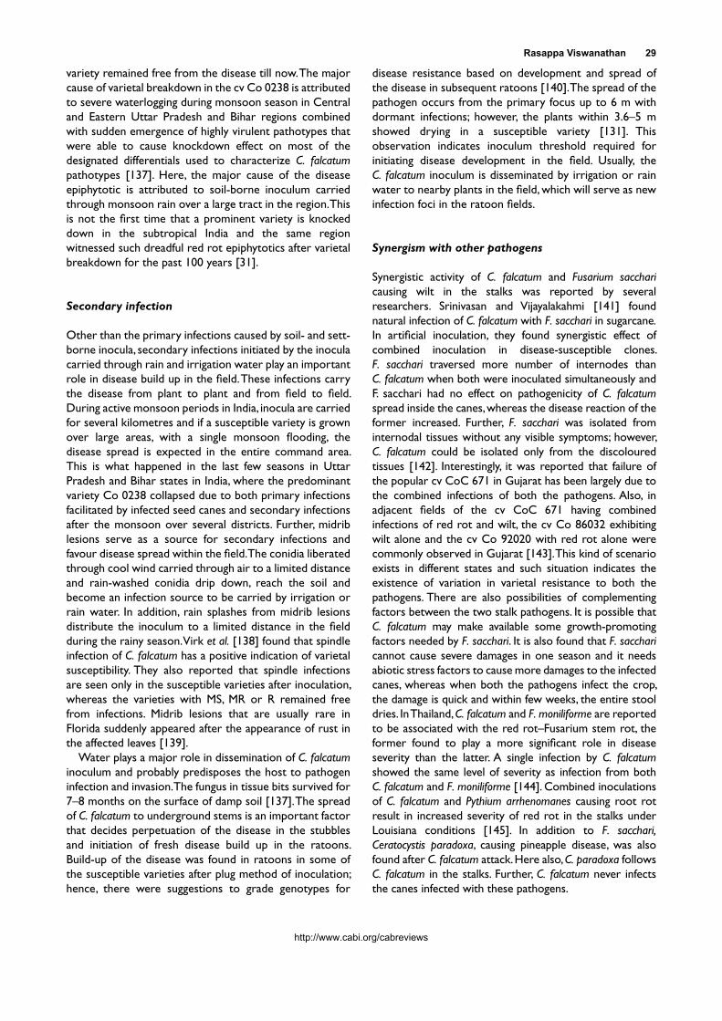
Rasappa Viswanathan 29
http://www.cabi.org/cabreviews
variety remained free from the disease till now. The major cause of varietal breakdown in the cv Co 0238 is attributed to severe waterlogging during monsoon season in Central and Eastern Uttar Pradesh and Bihar regions combined with sudden emergence of highly virulent pathotypes that were able to cause knockdown effect on most of the designated differentials used to characterize C. falcatum pathotypes [137]. Here, the major cause of the disease epiphytotic is attributed to soil-borne inoculum carried through monsoon rain over a large tract in the region. This is not the first time that a prominent variety is knocked down in the subtropical India and the same region witnessed such dreadful red rot epiphytotics after varietal breakdown for the past 100 years [31].
Secondary infection
Other than the primary infections caused by soil- and sett-borne inocula, secondary infections initiated by the inocula carried through rain and irrigation water play an important role in disease build up in the field. These infections carry the disease from plant to plant and from field to field. During active monsoon periods in India, inocula are carried for several kilometres and if a susceptible variety is grown over large areas, with a single monsoon flooding, the disease spread is expected in the entire command area. This is what happened in the last few seasons in Uttar Pradesh and Bihar states in India, where the predominant variety Co 0238 collapsed due to both primary infections facilitated by infected seed canes and secondary infections after the monsoon over several districts. Further, midrib lesions serve as a source for secondary infections and favour disease spread within the field. The conidia liberated through cool wind carried through air to a limited distance and rain-washed conidia drip down, reach the soil and become an infection source to be carried by irrigation or rain water. In addition, rain splashes from midrib lesions distribute the inoculum to a limited distance in the field during the rainy season. Virk et al. [138] found that spindle infection of C. falcatum has a positive indication of varietal susceptibility. They also reported that spindle infections are seen only in the susceptible varieties after inoculation, whereas the varieties with MS, MR or R remained free from infections. Midrib lesions that are usually rare in Florida suddenly appeared after the appearance of rust in the affected leaves [139].
Water plays a major role in dissemination of C. falcatum inoculum and probably predisposes the host to pathogen infection and invasion. The fungus in tissue bits survived for 7–8 months on the surface of damp soil [137]. The spread of C. falcatum to underground stems is an important factor that decides perpetuation of the disease in the stubbles and initiation of fresh disease build up in the ratoons. Build-up of the disease was found in ratoons in some of the susceptible varieties after plug method of inoculation; hence, there were suggestions to grade genotypes for
disease resistance based on development and spread of the disease in subsequent ratoons [140]. The spread of the pathogen occurs from the primary focus up to 6 m with dormant infections; however, the plants within 3.6–5 m showed drying in a susceptible variety [131]. This observation indicates inoculum threshold required for initiating disease development in the field. Usually, the C. falcatum inoculum is disseminated by irrigation or rain water to nearby plants in the field, which will serve as new infection foci in the ratoon fields.
Synergism with other pathogens
Synergistic activity of C. falcatum and Fusarium sacchari causing wilt in the stalks was reported by several researchers. Srinivasan and Vijayalakahmi [141] found natural infection of C. falcatum with F. sacchari in sugarcane. In artificial inoculation, they found synergistic effect of combined inoculation in disease-susceptible clones. F. sacchari traversed more number of internodes than C. falcatum when both were inoculated simultaneously and F. sacchari had no effect on pathogenicity of C. falcatum spread inside the canes, whereas the disease reaction of the former increased. Further, F. sacchari was isolated from internodal tissues without any visible symptoms; however, C. falcatum could be isolated only from the discoloured tissues [142]. Interestingly, it was reported that failure of the popular cv CoC 671 in Gujarat has been largely due to the combined infections of both the pathogens. Also, in adjacent fields of the cv CoC 671 having combined infections of red rot and wilt, the cv Co 86032 exhibiting wilt alone and the cv Co 92020 with red rot alone were commonly observed in Gujarat [143]. This kind of scenario exists in different states and such situation indicates the existence of variation in varietal resistance to both the pathogens. There are also possibilities of complementing factors between the two stalk pathogens. It is possible that C. falcatum may make available some growth-promoting factors needed by F. sacchari. It is also found that F. sacchari cannot cause severe damages in one season and it needs abiotic stress factors to cause more damages to the infected canes, whereas when both the pathogens infect the crop, the damage is quick and within few weeks, the entire stool dries. In Thailand, C. falcatum and F. moniliforme are reported to be associated with the red rot–Fusarium stem rot, the former found to play a more significant role in disease severity than the latter. A single infection by C. falcatum showed the same level of severity as infection from both C. falcatum and F. moniliforme [144]. Combined inoculations of C. falcatum and Pythium arrhenomanes causing root rot result in increased severity of red rot in the stalks under Louisiana conditions [145]. In addition to F. sacchari, Ceratocystis paradoxa, causing pineapple disease, was also found after C. falcatum attack. Here also, C. paradoxa follows C. falcatum in the stalks. Further, C. falcatum never infects the canes infected with these pathogens.

30 CAB Reviews
http://www.cabi.org/cabreviews
Sugarcane borer interaction
The incidence of red rot in South Africa was greater in the higher altitude areas, with long growing cycles, and also where canes are damaged by Sesamia or Eldana borers [23]. The disease severity in the country is aggravated when the incidence of stalk borers is high. Trenor and Bailey [146] found a direct relationship between the frequency of borer damage in the stalks and red rot incidence. Diatraea saccharalis is reported to transmit C. falcatum in Cuba [147]. Infestation of the cane stalks by sugarcane borers provides a main opportunity for C. falcatum infection in the stalks in any climatic conditions [148]. In the borer–red rot complex, nitrogen fertigation was found to aggravate the incidences of both the insect and red rot. The number of larval bore holes had a correlation with the incidence of red rot. Increase in the number of red rot–affected internodes proportionately reduced sugar percent in the canes. Though nitrogen doses did not affect the attractiveness of sugarcane, higher N doses increased internodal tissue damages by D. saccharalis larvae [149]. Although N fertilisation through irrigation gave higher returns in stalk and sugar yields as compared to rainfed crops in Brazil, it had favourable effects on borer-rot complex. However, the gains of stalk and sugar yields due to N application were found to compensate for the increase in borer-rot complex infestation [150].
Red rot pathogen is associated with injury of the sugarcane borer D. saccharalis in Brazil. The effect of C. falcatum infection in sugarcane and borer weight gain and the attractiveness of herbivore-induced plant volatiles to the parasitoid Cotesia flavipes were examined. The wasps preferred C. falcatum–infected canes when a choice was given between mock-inoculated and herbivore-damaged infected plants. When fed on healthy plants, D. saccharalis larvae were larger as compared to those fed on C. falcatum–infected sugarcane plants and this indicated that the parasitoids are likely to benefit from being attracted to volatiles of healthy than the infected/damaged sugarcane. Further, C. falcatum infection was not likely to disrupt the parasitoid host location; however, the disease attenuates the attractiveness of herbivore-induced sugarcane volatiles to Cotesia flavipes [151].
Weather factors
After planting of seed canes, soil temperatures have an important bearing on the development of red rot within the stalk. Contrary to what might be expected from the fact that the optimum temperature for C. falcatum is relatively high (30 to 32oC), the disease is more destructive to seed cane when soil temperatures following planting are low rather than when they are high in Louisiana [7]. In the field trials conducted in Coimbatore, a higher red rot severity was recorded from soil-borne inocula during 2018–2019 season than the previous season. It was found that the latter
was warmer as compared to the former with more variations between maximum and minimum temperatures due to scanty summer rains during April-June. However, the 2018–2019 season that was conducive for disease development had limited variation for temperature and continuous rain during March to September [45]. Due to the favourable weather, the soil inoculum survived in the soil and caused disease during different stages of crop growth in the trial, whereas higher day temperature with a climate of drought during germination and grand growth phase do not favour disease development. A low day temperature due to frequent rains during grand growth phase is ideal for red rot incidences from the soil-borne inoculum or expression of the disease from the dormant infections in the underground tissues. This applies to the ratoon crops also, where the residual inocula in the stools cause fresh infections in the newly emerging shoots, if cool environment prevails. In the young affected shoots, midrib lesions appear very frequently and readily serve as a source for secondary spread (Fig. 16 and 17). In warm conditions of 16.5°C–35°C prevailing in September under Lucknow conditions favoured pathogen infection sugarcane varieties and the pathogen reaction was seen within 15 days of inoculation, whereas at low temperature, the susceptible cultivars tested behaved as moderately susceptible and the MS ones became resistant. Further, the age of the plant and sugar content did not have an effect on disease development in the internodal tissues [152].
Cold weather may check the spread of the rot but does wipe out the progress it has already made. Steib [153] reported that when the fungus was exposed to subfreezing temperature (−19°C) for 2–5 months it was not destroyed. This indicated that the red rot fungus in the mycelial state in the soil or in sugarcane residues survived winter conditions in Louisiana, USA.
A multiple linear model was developed for predicting secondary spread of red rot by Kumar et al. [154] under Haryana conditions in India. Their model accounted for 94% of variation in red rot incidence at different periods within the cropping season. The results also revealed that rainfall and number of rainy days along with minimum temperature and morning relative humidity are the major factors that affect the spread of the disease.
Host-pathogen interaction
In the absence of clear-cut evidences on inheritance of red rot resistance, the exact mechanism governing red rot resistance in sugarcane has not been understood clearly. Earlier, different biochemical features that impart resistance in a particular genotype have been studied. In response to C. falcatum infection, disease-resistant varieties develop gum deposits that block the spread of the advancing hyphae by filling up the intercellular spaces. This process takes place in advance of infection and seals off further spread of the pathogen in adjoining tissues [100, 155].

Rasappa Viswanathan 31
http://www.cabi.org/cabreviews
Although the role of phenolic compounds in red rot resistance was reported, subsequently no correlation was found between total phenolic content and degree of resistance to red rot in sugarcane [156]. The role of enzymes involved in oxidative and phenyl-propanoid pathways was reported to be associated with resistance in many sugarcane varieties [156–158]. Also at cellular level, these enzyme activities varied in sugarcane varieties; calli from a resistant variety recorded very high enzyme activities than the susceptible variety. Similarly, the pathogen toxin also elicited these defence enzymes differentially in the calli. Further, calli from the susceptible varieties recorded a more pronounced loss of electrolytes upon toxin treatment than in the resistant variety [159, 160]. The pathogen toxin caused more damages in the susceptible variety and it is reflected in electrolyte release to the medium.
Phytoalexins
The red substance released in the tissues including intercellular spaces near the invading hyphae in the stalks is referred as red rot pigment (RRP). Detailed studies were conducted on the RRPs and their inhibition on conidial germination in vitro and mycelial growth was reported [161, 162]. Subsequently, the RRPs showed presence of several compounds after fractionation and a differential induction of the compounds was found among the resistant (R) and susceptible (S) varieties after pathogen inoculation [161, 162]. Fractionation of the pigments by high-performance liquid chromatography (HPLC) revealed the presence of 3-deoxyanthocyanidin compounds, which were identified as luteolinidin, apigeninidin and caffeic acid ester of 5-0-apigeninidin. In incompatible interaction, these compounds present at much higher concentration, whereas in the compatible reaction, they were completely absent or present in very low concentration [163, 164]. Another interesting finding is that the S variety accumulated reasonable amount of these phytoalexins when the fungal toxin was used for inoculation, but in response to pathogen inoculation, the same variety failed to synthesize the antifungal compounds due to their degradation by the pathogen [164]. These findings clearly demonstrated that the phytoalexins are specifically accumulated only in incompatible host-pathogen interactions and compatible interactions had no such phytoalexins or with trace quantities. These three compounds were identified as phytoalexins in sorghum and C. sublineolum interaction, a similar host-pathogen interaction [165].
Further studies with a set of host differentials proved that there is accumulation of anthocyanin compounds in incompatible interactions and such induction/accumulation in compatible interactions is limited [166, 167]. While screening sugarcane clones for red rot reactions, phenotypic expression of incompatible (resistant)
interactions was always characterized by restricted dark pigmentation at the site of infection in the nodal or internodal tissues as compared to diffused pigmentation with spreading lesions in the compatible (susceptible) interactions (Fig. 31). Such rapid induction of the phytoalexin compounds in large quantities confines the pathogen at the site of penetration in the resistant varieties.
Later studies of Malathi et al. [168] conclusively proved that 3-deoxyanthocyanidin compounds luteolinidin and apigeninidin act as sugarcane phytoalexins and govern resistance in sugarcane to red rot. Recent HPLC assays revealed that the differential induction of nine different 3-deoxyanthocyanidin compounds upon the pathogen infection in cane varieties varies in disease resistance. Four of them were identified as luteolinidin, 5-methoxy luteolinidin, apigeninidin and arabinosyl-5-O-apigeninidin. The resistant cvs Co 93009, BO 91 and Baragua recorded multi-fold induction of luteolinidin, apigeninidin and arabinosyl-5-O-apigeninidin, respectively, in the stalk tissues after C. falcatum inoculation as compared to the susceptible varieties [169]. This study established that the phytoalexins are not induced in similar pattern in disease-resistant varieties and probably the compounds’ induction varies based on genomic constitution of host varieties. Recently, it was established that phytoalexin fractions were found to exhibit a clear inhibition of C. falcatum on seeded agar [170].
Pathogenesis-related (PR) proteins
Detailed studies on pathogenesis-related (PR) proteins in red rot resistance showed higher activities of chitinase and β-1,3-glucanase in R varieties as compared to S varieties, constitutively and upon pathogen inoculation [171]. Further, Western blot assays revealed specific induction of chitinases and TLPs after pathogen inoculation in stalk and leaf tissue inoculation. The R variety showed more number of PR proteins with high intensity during early intervals than the S variety [172]. These studies gave a clear indication that the PR proteins may play an important defence mechanism operating in sugarcane against C. falcatum. This differential induction of chitinases was subsequently validated through transcript expression and real-time PCR assays [173, 174]. Sugarcane chitinase ScChiB1 was characterized further based on its full-length sequence, and it had structural resemblances with the classes I/IV chitinase beginning with a signal peptide and ending with a signature domain. The results concluded that the predicted structure of ScChiB1 belongs to class IV of family 19 glycosyl hydrolases. Wound-inducible protein in sugarcane SUGARWIN2, a homologue of a barley wound-inducible protein (BARWIN), was found to induce in response to stalk borer Diatraea saccharalis infestation but after pathogen infections. Recombinant SUGARWIN2 was found to exhibit anti-fungal activities on C. falcatum including alterations in morphological features like

32 CAB Reviews
http://www.cabi.org/cabreviews
increasing vacuolization, damages to fungal integrity and leak of intracellular material that led to conidial apoptosis [175]. Quantitative transcript analyses of mucoprotective genes SUGARWIN1 and SUGARWIN2 revealed maximum expression in the resistant genotype as compared to lowest in the susceptible genotype [176], and the study has suggested a probable antifungal activity of SUGARWIN proteins in sugarcane against C. falcatum.
Molecular basis of red rot resistance
To elucidate the mechanism underlying host-pathogen interaction, it is important to determine the host targets in sugarcane during its interaction with C. falcatum. To investigate the mechanism of red rot resistance, both genomic and proteomic tools were adopted and standardized for sugarcane [156, 177]. Since the pathogen
Figure 31. Nodal tissues exhibit a diffused pigmentation with spreading lesions in the internodes of compatible interactions (left); incompatible reactions show a restricted dark pigmentation at the site of pathogen entry in the nodes (right). Yellow circles indicate inoculated nodes by cotton swabbing method.

Rasappa Viswanathan 33
http://www.cabi.org/cabreviews
infects the stalk tissues, standardized protein extraction from the fibrous stalk tissues and MS analysis and developed a stalk specific proteome for the first time [178]. The 2D-based core proteome analysis classification was performed during the interaction in sugarcane with C. falcatum [179]. The study has identified proteins involved in several defence and cellular functions such as cyclic nucleotide–gated ion channel protein, R2R3-MYB transcription factor MYB6, WRKY transcription factor, flagellar-associated protein, class III HD-Zip protein HDZ34, calcium-dependent protein kinase, DNAJ heat shock protein, callose synthase, PISTILLATA-like protein, putative p-coumarate 3-hydroxylase, NB-ARC domain–containing protein, cytokinin oxidase 3, UDP glucose:flavonoid 3-O-glucosyltransferase, NADH dehydrogenase, ACC synthase, putative disease resistance protein etc.
Expression pattern of transcription factors (TFs) in sugarcane in incompatible interaction and systemic acquired resistance (SAR) inducer priming revealed that eight TFs are highly induced in both the cases. The TFs that are significantly induced early are expected to involve actively in triggering or co-ordinating defence against pathogen invasion [180]. Subsequently, temporal expression of 22 putative defence-related genes by reverse transcription (RT)-PCR in red rot–susceptible cv CoC 671 with response to priming using various SAR inducers, viz. benzothiadiazole (BTH), salicylic acid (SA) and C. falcatum elicitor, was studied. Upon SAR induction in response to pathogen challenge, differentially regulated phenylpropanoid pathway genes such as cinnamic acid 4-hydroxylase, 4-coumarate:coenzyme A ligase, chalcone synthase, and chalcone reductase and R genes such as NBS-LRR genes and basal layer antifungal peptide (BAF) genes were found upregulated. These upregulated genes are likely to play a potential role in SAR induction and might contribute to defence against C. falcatum [181].
Later, throughput genomic approaches like differential display, subtractive libraries and next-generation sequencing platforms were employed to unravel red rot resistance in sugarcane. In differential display, reverse transcription polymerase chain reaction (DD-RT-PCR) identified altered expression of genes in response to C. falcatum infection in red rot resistant cv Co 93009. Differentially expressed genes represented five categories, of which the defence/stress/signalling group was the largest, with clones homologous to genes known to be actively involved in various pathogenesis-related functions in plant species. The study reported overexpression of several transcripts related to ethylene-mediated and jasmonic acid pathway of plant defence mechanisms [182]. Later, an in vitro system of using sugarcane suspension cells and crude elicitor of C. falcatum was standardized for transcriptome analysis and identifying defence-related genes in sugarcane. The results revealed upregulation of many potential defence-related transcripts like putative chitinase, glycine-rich protein, 14-3-3-like protein, xylanase
inhibitor protein, calmodulin-related protein, Myb-related transcription factor CBM2-like, basal layer antifungal peptide etc. Further, by adopting RACE-PCR approach, complete gene sequences of 14-3-3-like protein and xylanase inhibitor were identified and the genes were characterized to domain level. It was reported that the transcript profile in in vitro system of sugarcane suspension cells and Cf-elicitor has been close to the cane tissue challenged with the pathogen and the approach was useful to identify defence-related traits in sugarcane against C. falcatum [183].
Suppression subtractive hybridization (SSH), a more efficient molecular tool, was employed to a series of SSH libraries, developed from sugarcane stalk samples to understand sugarcane defence responses during the initial phase of C. falcatum pathogenesis. A total of 139 ESTs were obtained, which were functionally categorized as belonging to recognition and signal transduction, oxidative stress, redox maintenance, membrane trafficking and transport, defence and programmed cell death, energy and photosynthesis, metabolism, secondary metabolite biosynthesis, cell/nuclear structure and unknown categories. For the first time, this study identified a network of early defence responses and related signals in C. falcatum–resistant sugarcane in response to the fungal pathogen infection. Also for the first time, a set of differentially expressed EST clusters in red rot–resistant sugarcane variety was identified in response to C. falcatum infection [184, 185].
Subsequently, the next-general sequencing (NGS) approach was combined with SSH and large transcriptomic data specific to red rot resistant and susceptible libraries were obtained. After sequencing, 10,038 and 4,022 high-quality transcripts were obtained from resistant and susceptible libraries, respectively. Based on mapping of the transcripts to KEGG-KASS database, the presence of a CEBiP receptor and the signals ROS, Ca2+, BR, jasmonic acid (JA) and ABA was identified in both the responses. However, MAPK, ethylene (ET), phosphoinositide (PI) signals and JA amino conjugation were found only in the resistant interaction and validated expression of the transcripts involved in these pathways through qRT-PCR assays. This study concluded that perception of PAMPs occurs in both compatible and incompatible systems; however, downstream signalling through MAPK, ET, PI and JA amino conjugation and activation of R genes happens only in the resistant interaction. Further, this is the first detailed transcriptomic analyses of compatible and incompatible interactions in a sugarcane variety exhibiting differential interaction with two C. falcatum pathotypes that vary in their virulence through SSH and the NGS platform [186]. When two varieties were used to generate SSH libraries, interference of their varied genetic background cannot be ruled out, but the use of a variety that behaves differentially to two pathotypes circumvented these cross reactions. This study has clearly brought out a library of expressed genes during resistant and susceptible

34 CAB Reviews
http://www.cabi.org/cabreviews
interactions with C. falcatum in sugarcane. Further studies are envisaged on applying genome-editing approaches to specifically manipulate susceptibility-associated genes to obtain genome-edited plants with tolerance to the dreaded pathogen. This strategy may bring more dividends to safeguard elite sugarcane varieties that failed in the field due to varietal breakdown.
Induced resistance
In line with induced systemic resistance (ISR) against C. falcatum in sugarcane, detailed studies were conducted on systemic acquired resistance (SAR) by synthetic signal molecules in sugarcane. Initially, acibenzolar S-methyl (ASM) was studied along with salicylic acid (SA) and ASM and was found to persist up to 30 days in the pre-treated cut canes. Increased phenolic content and accumulation of pathogenesis-related (PR) proteins, viz., chitinase, β-1,3-glucanase and thaumatin-like protein (TLP) (PR-5), were observed in sugarcane cvs CoC 671 and BO 91 [187]. Subsequently, the synthetic elicitors like benzothiadiazole (BTH), SA and isonicotinic acid were found to reduce C. falcatum colonization in cane stalks by triggering a range of defence-related compounds [188]. Later it was found that Cf-elicitor, a glycoprotein extracted and purified from the cell wall of red rot pathogen, exhibited induction of many defence-related compounds and PR proteins similar to BTH in susceptible sugarcane cvs CoC 671 and CoC 92061 [189].
Comprehensively, studies on induced resistance involving synthetic/biotic inducers have demonstrated the disease-suppressive effects of BTH and Cf-elicitor against red rot of sugarcane under field conditions. However, many factors like cost-effectiveness and lack of protein sequence information have deterred scaled-up applications of BTH and glycoproteinaceous Cf-elicitor, respectively, for field situations. Efficacy of the inducers BTH (125 μM) and Cf-elicitor (60 μg glucose equivalents ml−1) was studied for induced resistance in reducing red rot incidence during the early phases of crop growth against soil-borne inoculum and in that they suppressed disease intensity significantly. During grand growth phase also, the inducers exhibited induced resistance in the stalks upon pathogen challenge in the susceptible variety [190]. Further studies were continued to identify pathogen-associated molecular patterns (PAMPs) and C. falcatum effectors to improve induced resistance through pathogen-derived resistance approaches. In this regard, recently, PAMP/effector molecules, viz. CfEPL1 and CfPDIP1, are found to suppress C. falcatum colonization in the stalks by activating systemic resistance. Molecular basis of induced resistance revealed an upregulation of the master regulator NPR1 and PR genes, a similar phenomenon demonstrated earlier for BTH-mediated induced systemic resistance. The study further revealed involvement of SA-mediated defence pathway during priming of sugarcane with BTH, CfEPL1
and CfPDIP1 [95]. The recent studies on host-pathogen have opened opportunities to explore further to identify interacting partners of the characterized PAMPs/effectors to reveal the series of molecular signals and downstream activation of defence molecules that could lead to delineation of PTI/ETI mechanism in sugarcane.
Management
Developing resistant varieties to red rot has sustained sugarcane cultivation over a century. Hence, breeding in sugarcane revolves around red rot resistance in many Asian countries [31]. However, other management strategies were developed time to time to manage the disease in different countries to reduce the damages caused by the disease in the field. Moreover, in many occasions, the ruling varieties fail due to varietal breakdown to C. falcatum, and alternate strategies are required to moderate the disease severity and to save the crop.
Breeding for red rot resistance
Managing red rot through host resistance is the most sustainable way; hence, research efforts are being made to develop and deploy resistant varieties for the past 100 years in different countries. In India, cv Co 205, the first successful interspecific hybrid involving Saccharum officinarum and S. spontaneum, was introduced for cultivation [31]. Though the variety became popular in the subtropical India, it picked up red rot within few years after its introduction. Subsequently, many varieties were developed and deployed for cultivation; however, many of them succumbed to the disease. The adoption of improved cvs Co 210 and Co 213 in northern India immensely contributed in quantum jump in the cane yield and replaced all the S. barberi varieties under cultivation. However, during expansion of cane cultivation to larger areas with the new elite varieties, precautions of good quality and healthy seed were totally ignored. Hence, during 1938–1940, cv Co 213 perished in several thousands of hectares in that region in the severe red rot epiphytotics. Later, the disease gradually destroyed other varieties like Co 290, Co 312, Co 313, Co 357, Co 370, Co 393, Co 421, Co 453, CoS 5 etc. in the region. This made a policy change in sugarcane researchers in India to focussed breeding with red rot resistance as one of the major component [6,31]. In tropical India, replacement of S. officinarum cultivated in the early decades of the last century with interspecific hybrids reduced the disease severity. Similarly, S. officinarum clones were replaced with the hybrids in West Indies in the past and red rot as a major disease became a minor disease [6].
Experience of different researchers over the decades revealed only limited information is available on inheritance of resistance to major diseases. In case of smut and rust diseases, additive genetic variance is important, which

Rasappa Viswanathan 35
http://www.cabi.org/cabreviews
clearly indicates that substantial progress can be made by selecting resistant parents [191, 192], whereas little is known about the genetics of resistance to red rot [193]. Earlier, it was suggested that the dominant genes from S. officinarum might be involved in masking the effect of S. spontaneum resistance genes [194]. However, there were conflicting reports that either there is no association between the degree of resistance of the progenies and parents [195] or crosses involving resistant parents tend to have more resistant progenies than from crosses involving susceptible parents [196, 197].
Information available from a large number of crosses made in Coimbatore and elsewhere indicates that in crosses that involve resistant parents, a high percentage of the progenies tend to be resistant or moderately resistant. This was also true with respect to a directed breeding programme for red rot resistance initiated by Alexander et al. [198] at ICAR-SBI, Coimbatore. This was a directed breeding programme to evolve varieties with broad spectrum of resistance against two or more major pathotypes of red rot. Under this programme, by inter-crossing selected resistant parents and subsequently by inter-crossing and selecting resistant progenies, a number of clones that are resistant to 2 to 4 pathotypes were obtained. This study indicated that as in the case of other diseases, for red rot also, additive genetic component is important and the incorporation of parents with resistance in crosses will help in building up a source population. Alexander et al. [199] identified horizontal resistance in sugarcane progenies by testing them at various locations in India and screening them with the respective zonal pathotypes in Karnal in Haryana and Chakia in Bihar in subtropical region, and Tanuku in Andhra Pradesh and Nellikuppam in Tamil Nadu in the tropical region. Natarajan et al. [200] investigated the relative importance of vertical (race specific) and horizontal (essentially race non-specific) resistance in sugarcane to red rot biometrically. They pointed out a progressive shift against HR brings out the importance of gene complexes from S. spontaneum in imparting stable horizontal resistance against red rot in sugarcane.
Ram et al. [201] identified the parental clones Co 8347, ISH 021 and Co 86011 as good general combiners for imparting red rot resistance. Of the 30 parental combinations studied for their contribution to impart red rot resistance, Bora et al. [202] found only 12 combinations involving elite parents viz. Co 1148, Co 88039, Co 6806, C 79180, Co 356, CoS 70 and Co 6304 in developing agronomically superior progenies with resistance to red rot. Slow red rot development in sugarcane was described in sugarcane varieties based on drying of tops, nodal sporulation and spindle infection after pathogen inoculation by nodal, nodal injury and plug methods. The cvs CoH 110, CoH 119 and CoPant 84211 show slow red rot as they did not show any of the three symptoms and nodal sporulation was found to be positively correlated with spindle drying [203]. Babu [204] analysed association of red rot resistance
in sugarcane with major yield and quality traits for effective family selection. He reported a positive correlation between red rot resistance and tiller counts at 90 and 120 days. The author claimed the presence of S. spontaneum genomic complement in the progenies, which is characterized by high tillering and resistance to red rot. In the individual families, he found a positive correlation with sucrose per cent in two families Co 98006 × 987001 and CoJ 72 × Co 62198. Among the two families, the former produced the highest proportion of resistant progenies (58%) with moderate level of sucrose content (14.98%). This cross combination offers much scope for future exploitation in breeding programme targeting to combine red rot resistance and appreciable juice quality. However, the conclusion made by the author is that the association of red rot resistance with tiller count has no meaning. Scientists should not be carried away by the statistical analyses and they have to introspect genetic control of disease resistance with parental genome.
There were many attempts to find a relation between sucrose content and red rot resistance in the past, though it is clearly known that red rot resistance is a dynamic one and any such established relation will be subjective. Ram et al. [205] also made such an attempt in that they showed a significant positive correlation between disease reaction and juice quality during October and January with the clones with higher HR Brix during August. They suggested that the clones with high HR Brix during August (>10.6) are likely to be more susceptible to disease. However, the claim is not supported by robust data and analyses. Further, such relation is less likely to exist with a dynamic pathogen like C. falcatum. Shanthi and Alarmelu [206] reported that three crosses Co 7201 × Co 62198, Co 85002 × CoT 8201 and CP 49-50 × CoT 8201 gave more clones combining high sucrose and red rot resistance. To negate the earlier findings, Singh and Govindaraj [207] found 23.9% progenies of six crosses resistant at low to medium HR Brix; however, at high to very high HR Brix level, they also found the progenies are resistant. They concluded that HR Brix value in the progenies, an indicator for high quality, is not associated with the resistance.
Role of parental clones in red rot resistanceThe degree of resistance of the parents is reported to have little difference in the percentage of resistant seedlings [208]. In Louisiana, heritability studies indicated that red rot resistance can be augmented through careful cross-based selection [145]. Singh et al. [209] reported that R × R parental combination gave higher frequency of resistant progenies than in R × S parents. Their findings contradicted the earlier assumption that the degree of resistance of the parent made little difference in the frequency of resistant seedlings in the progenies [208, 210, 211]. They expressed that genes for red rot resistance and ratooning ability are incorporated from S. spontaneum. In juice analysis, they found most of the low-sugar clones are resistant to the

36 CAB Reviews
http://www.cabi.org/cabreviews
disease and vice versa. However, experience of the author suggests that not all the low-sugar genotypes are resistant to red rot. In a set of crosses involving parents that vary in red rot resistance, 57.1% of the clones were found to be resistant when either the female or the male parent was resistant and 20.5% clones were resistant when both the parents are susceptible [212]. However, the progeny behaviour is not uniform in R × R, R × S or S × S combinations, since in five S × S crosses, resistant population ranged from 66.7% to 2.5% indicating that there is no definite pattern of segregation. Similarly, in case either one of the parents is resistant, it varied from 100% to 33.3% among five crosses. However, Chaudhary et al. [213] observed different ratios (1:3, 3:1 and 1:5) for resistant to susceptible clones. They further suggested that a few genes with additive effect may govern the disease resistance. The number of loci without having dominant recessive alleles in a particular genotype and interaction thereof probably decides the proportion of R to HS categories in the progenies. Further, meiotic irregularities are also responsible for such a ratio in sugarcane. Their further studies revealed that specific cross combinations like Co 1148 × Co 775, CoH 7803 × CoS 510, CoH 7803 × Co 775, CoJ 83 × Co 62198 etc. give red rot–resistant progenies with desirable agronomic traits [214]. Sugarcane hybrids are highly complex polyploids with chromosome number ranging from 100 to 120 (2n), where basic chromosome number (x) is only 8. It indicates the representation of genes in 12 or more dosages. Due to this inherent complexity, loss of some genetic material is not always reflected in the phenotype. Mishra [215] reported that switching over from Co to BO varieties reduced red rot in Bihar where epiphytotics are common. Without much scientific proof, he concluded that crosses made in tropics and selections made in the subtropics in India will probably enhance the red rot problem and cultivation of BO varieties and raising of seedlings from the crosses made in subtropics will minimize the red rot menace.
The highly polyploid genome of the parental clones in these studies poses problems for objective conclusions on the significance of the segregative ratios observed on the progeny. The allelic interactions and the extreme variability expected in the gametes by the meiotic process involving clones with different chromosome numbers do affect the reliability of conclusions on the number of genes involved. The heritability of the resistant trait is evidenced from the different studies. The self-pollinated progeny of the susceptible cv CoC 671 were almost all susceptible and the hybrids between CoC 671 and the resistant cv BO 91 were segregated with 25%–30% resistant progeny.
Sources for red rot resistanceS. spontaneum clones appear to have built up resistant genes through adaptation in red rot–endemic conditions and so they are valuable sources of resistance. However, while many hybrids carrying ‘spontaneum’ genes possess a high degree of resistance, they also appear, for this reason,
to be potent source of origin of new virulent strains through natural selection of variants. Since new pathogenic strains are believed to arise through mutation and parasexual recombination in vivo in the host [116, 216], the appearance of strains with increased virulence and concomitant periodical losses through epiphytotics appears to be a price that has to be paid for increased productivity through the development of interspecific hybrid varieties [217]. Hence, the emphasis should be shifted towards safeguarding the level of resistance already available in hybrid sugarcane and then building it up rather than trying to infuse resistance against specific isolates of the fungus.
Srinivasan and Alexander [217] screened about 660 Saccharum spp. clones for red rot resistance and found poor resistance in the clones of S. officinarum, S. robustum and S. sinense, and a third of the entries in S. barberi and more than half in S. spontaneum were resistant. They opined that the origin of S. officinarum and S. robustum in a milieu where they were not subjected to the disease hazard probably contributed to their lack of resistance. In the germplasm utilization programme initiated during 1980s at ICAR-SBI, Coimbatore, many useful hybrids involving hitherto unutilized clones of S. officinarum, S. barberi, S. sinense, S. robustum, S. spontaneum and a few commercial varieties have been identified. Resistance to red rot in these clones was evaluated at Karnal and Motipur in subtropical India. Although there are many clones with higher yield levels and with other desirable traits, the level of resistance to red rot is low [218]. Recent screening of 419 sugarcane germplasm clones comprising 206 S. officinarum, 141 S. robustum, 30 S. sinense and 42 S. barberi for red rot resistance revealed 7 are R and 34 are MR against red rot and rest are MS to HS, indicating very low resistance in the germplasm clones. The S. officinarum clones Baragua, Koelz 11131 and Koelz 11132, S. robustum clones 28 NG 251 and 57 NG 238, S. barberi clones Chin, DhaurKalig, Kansar, Maneria IMP-1552, Mungo 254, Nargori, Kewali-14G, Manga (SIC) and S. sinense clones Reha, Ikhri and Kalkya consistently proved to be resistant in the plug method and controlled condition testing [219].
Natarajan et al. [200] suggested introduction of new variants of S. spontaneum in breeding programmes to enhance the probability of obtaining varieties with stable resistance expeditiously as a long-term measure. They reported creation of genetic variability by introducing gene sources from 10 clones of S. spontaneum besides 12 S. officinarum, which were hitherto unutilized in varietal development, and hence they suggested that the new gene pool offers a rich repository of variability for red rot resistance as well as productivity. This has become all the more important since it was observed that the cultivars cultivated earlier world over had a narrow genetic base with 19 clones of S. officinarum and 2 clones of S. spontaneum [220].
Overall, the frequency of red rot resistance is low among the breeding population and progenies in the USA [145]. Hence, there were suggestions to introgress genes from wild relatives of Saccharum spp. to enhance red rot

Rasappa Viswanathan 37
http://www.cabi.org/cabreviews
resistance in the parent population. Ram et al. [221] have introgressed sugarcane with Erianthus for cold tolerance and red rot under subtropical conditions in India. Singh et al. [222] reported that the cv CoS 8436 resistant to major pathotypes of C. falcatum contributed more resistance in the progenies in the range of 41.5%–58.3% when used as female or male parent in crossing with R/MS or S parents than the other resistant parents viz. Co 7201, Co 87267 and BO 91. However, the latter produced more resistant progenies when it was used as female parent and crossed with susceptible male parents. Hale et al. [223] detected significant differences among the species and accessions within species clones of sugarcane germplasm for all traits contributing to rot index. Among the Saccharum spp., S. spontaneum, S. barberi and S. robustum displayed the lowest red rot symptom severity means, whereas S. officinarum and S. sinense had the highest rot severity means. Clones of S. spontaneum showed a high level of variation with RI means among themselves in the range of 2-29 and S. barberi also exhibited a high level of resistance with the values in the range of 1-20 RI. Erianthus accessions were found to possess high resistance with all of them showing very less values of RI. They suggested the use of Erianthus and selected accessions of S. spontaneum and S. barberi to impart red rot resistance in sugarcane. Nair et al. [224] diversified the genetic base of the sugarcane varieties with Erianthus procerus and suggested this approach to obtain red rot resistance with cane yield and drought tolerance. Recently, interspecific hybrids (ISH) and intergeneric hybrids (IGH) involving Erianthus spp. with Saccharum spp. or other hybrids were screened and resistant genotypes were identified in Coimbatore. It was found that some of the cytoplasmic derivative clones and IGH clones in specific cross combinations displayed greater levels of red rot resistance. This study provided the status of red rot resistance in wild relatives of Saccharum spp. and their derivatives for further utilization to develop red rot–resistant progenies [225].
Molecular markersThere have been few attempts to study host resistance in sugarcane against C. falcatum through molecular markers especially using resistance gene analogues (RGAs). In different host-pathogen interactions, many resistant (R) genes were discovered and they are found to have conserved domain of NBS-LRR. The NBS-LRR genes were completely characterized and have ATP/GTP binding activity and kinase 2a, kinase 3, P-loop and GLPL-conserved motifs [226]. These R genes confer resistance against different pathogens, but they have similar motifs with conserved DNA sequences. These conserved DNA sequences are being exploited for the isolation of new RGAs. In this regard, conserved regions of R genes were targeted using degenerate oligonucleotide primers for RGAs through PCR [227]. Many attempts were made to study RGAs from sugarcane and their role in imparting red rot resistance. RGAs specific for R and S reactions in
sugarcane to red rot were identified and sequencing of resistance fragments showed 80%–99% similarity with other resistant gene sequences especially reported from rice, sugarcane and maize [228, 229]. From a resistant variety, Hameed et al. [230] identified 15 RGAs from conserved motifs of nucleotide-binding site (NBS) class and kinase class of RGAs and these RGAs were found to have a close similarity with RGAs from other plants. In addition, Singh et al. [231] reported close association of two microsatellite markers UGSM316850 and UGSM31660 with MR varieties and tight linkage of UGSM316400 marker with HS varieties. Parida et al. [232] characterized and genotyped single nucleotide polymorphisms (SNPs) and insertion or deletion (InDels) in disease resistance genes along with sugar pathway of Saccharum complex and sugarcane varieties using amplicon sequencing and CAPS assays. Five SNPs were found to have a strong genetic association with the pathways and evidenced an InDel marker in the promoter sequence of sucrose synthase-2 gene, with sugar content and red rot resistance. The functionally related SNPs and InDels, in disease resistance genes, and designed genic CAPS markers are expected to be of immense use in marker-assisted genetic improvement of sugarcane for red rot resistance. Earlier, ESTs were generated from tissue and red rot–specific cDNA libraries and identified EST clusters from Indian sugarcane varieties [233].
To identify marker-trait associations (MTAs) for resistance in sugarcane to red rot, Singh et al. [234] fingerprinted a set of 119 Indian sugarcane genotypes for 944 SSR alleles. Four MTAs were detected after mixed linear model containing population structure and kinship as co-factor, and among them, three were found to have homology with sorghum genome. They found many genes encoding plant defence–related proteins like cytochrome P450, MAP Kinase-4, serine/threonine-protein kinase, ring finger domain protein, glycerol-3-phosphate transporter-1 and other genes that present close to these MTAs. This is an important finding to identify reliable molecular marker towards marker-assisted breeding in sugarcane. In this direction, in the ongoing Indo-Australian research project between ICAR-SBI and Sugar Research Australia/CSIRO, Brisbane, genomic markers associated with red rot resistance trait were identified in sugarcane through genome-wide association study using an SNP marker array. These markers suggested the presence of a major effect QTL in sugarcane for red rot resistance. It is expected that these SNP markers can predict red rot resistance with good accuracy, and identification of these novel markers may aid in developing red rot–resistant varieties and provide further insight into the molecular mechanism of red rot resistance (Prakash Lakshmanan, personal communication).
Somaclonal variation
After standardization of tissue culture technique in 1980s in sugarcane, there were many attempts to exploit

38 CAB Reviews
http://www.cabi.org/cabreviews
somaclonal variation and select red rot–resistant types. Jalaja et al. [235] reported the release of the first somaclone Co 94012 for commercial cultivation with red rot resistance. However, it was never found to be resistant to red rot as reported by the author on several occasions [46, 63, 71, 156]. Kumar et al. [236] reported that somaclones from donor cv CoS 88230 were better in red rot resistance. Kumar et al. [237] also reported the development of red rot–resistant somaclone. In Bangladesh, Mahmud et al. [238] found varied red rot reactions in the somaclones from that of source varieties on MS tissue culture media containing 2, 4-D. Using RAPD and SSR markers, they verified the presence of variability in the red rot–resistant somaclones in comparison to the parent. In Pakistan, Ahmed et al. [239] reported that approximately 10% of the somaclones from S and MS varieties are found resistant to C. falcatum. In the resistant somaclones, four candidate genes such as catalase, sucrose phosphate synthase, gibberellin 2-oxidase 4 and tillering branched 1 showed no SNP changes in their exon regions with the parental clones. They claimed that somaclonal variation is a good source of genetic improvement in sugarcane for disease resistance with no SNP changes in candidate genes.
Screening for red rot resistance
Chona [211] had done detailed studies to standardize screening sugarcane clones for red rot resistance by plug method. He has taken average linear spread of infection in the inoculated canes as the sole criterion to judge the comparative resistance or susceptibility of the clones. However, the relative importance of drying of tops, lesion width, occurrence and nature of white spots and nodal transgression was ignored in this plug method; subsequently, Srinivasan and Bhat [155] have found only these four characters mentioned above are critical and dependable to rate resistance in sugarcane to C. falcatum. The 0–9 scale developed by them is used across the countries to rate the clones into different categories viz. resistant (R: 0.0–2.0), moderately resistant (MR: 2.1–4.0), moderately susceptible (MS: 4.1–6.0), susceptible (S: 6.1–8.0) and highly susceptible (HS: 8.1–9.0). There were attempts to modify the 0–9 scale and on delivery of inoculum, discriminant function equation by giving relative weightage to the three important characters viz. lesion width, occurrence of nature of white spots and nodal transgression by the pathogen [240, 241], insertion of a toothpick soaked with inoculum broth on a small hole drilled onto the internode [99, 240, 241], a modified hypodermic syringe for making puncture on the internodes and placing the inoculum [242] and a revised criteria to rate sugarcane clones after plug method by combining the lesion width and nodal transgression [243]. However, these methods did not add any advantages over the 0–9 rating system developed by Srinivasan and Bhat [155]; hence, they were rejected by the scientists due to complications in inoculation and evaluation.
The plug method of inoculation with 0–9 rating scale has been adopted in most of the research centres around the world to screen sugarcane varieties for red rot reaction. Since rind tissue is injured to create bore hole, there were apprehensions that it as rigorous and abnormal, breaking barriers of resistance. Hence, new inoculation methods were attempted to deliver the inoculum in a natural way by targeting it to reach nodal tissues that contain the portals of C. falcatum entry. Since the approach is to target nodal tissues for the pathogen entry, they are called as ‘nodal method’. Initially, C. falcatum conidial suspension dipped in cotton was wrapped around three clean nodes, 10 to 20 cm above the ground level and covered with a polythene tubing [111]. Subsequently, few drops of conidial suspension were placed between the leaf sheath and stalk [244]. After about three months, the inoculated nodes are scrapped to record the extent of infection and then the stalks are sliced longitudinally to measure the spread of pathogen. All these methods were not followed due to practical difficulties and failure to induce disease even in disease-susceptible varieties. However, the nodal swabbing method developed at Coimbatore has been found an ideal method to identify the clones with nodal resistance to C. falcatum. Here, cotton strips dipped in conidial suspension are swabbed around two nodes of 6–8th nodes from the top, immediately after removing the leaves. The cotton pads are tightly held by a wrapping with parafilm® and also to prevent loss of moisture. This method ensures ideal disease development from the inoculated nodes in the susceptible varieties (Fig. 32) and the clones are categorized into resistant and susceptible (Fig. 31) [245, 246].
In general, red rot development by nodal method is highly influenced by environmental factors. Temperature also plays a crucial role in disease development after artificial inoculation by plug method. The climatic conditions prevailed at Shahjahanpur in the subtropical India during 2nd to 3rd weeks of September with mean relative humidity (RH) of approximately 90% and the maximum and minimum temperatures of about 33°C and 24°C, respectively, are found most favourable for C. falcatum infection in artificial inoculations [244]. Rapid red rot development in the temperature range of 25°C–30°C under Gorakhpur location in Uttar Pradesh by nodal method was reported. An optimum temperature of 28°C–30°C was found ideal for disease development by spray inoculation method [247]. The environmental conditions with mean temperatures ranging from 29.4°C to 31.0°C prevail during second week of August to first week of September and are found to be optimum for red rot at Hisar in subtropical India [248]. The disease development is maximum, when canes are inoculated nodally during the month of July and later inoculations resulted in poor red rot development [249].
Apart from stalk inoculation of C. falcatum approaches, there were attempts to apply the inoculum in the soil to favour disease development without injury. One such method was applying pathogen-infected crop debris to

Rasappa Viswanathan 39
http://www.cabi.org/cabreviews
induce disease development [250]. Subsequently, the inoculum was multiplied on sorghum grains and optimized disease development in sugarcane varieties [251]. Recently, disease development from soil-borne inoculum sources was demonstrated in sugarcane varieties varying in disease resistance [45]. This method of inoculum application and selection of clones is expected to identify the clones with field tolerance, where susceptible clones pick up infections at regular intervals, whereas the tolerant clones remain free from the disease infections [45].
In Cuba, Yakutkin and Rodrigues [147] successfully assessed red rot resistance on a 1–5 scale after injecting conidial suspension of the pathogen into 8–9 months old stalks. Later, Alfonso et al. [252] reported screening of sugarcane varieties and progenies for red rot resistance in Cuba. In Louisiana, harvested stalks are inoculated with C. falcatum and incubated for 6 weeks. After split open, parameters of number of nodes crossed, number of nodes rotted and internode rot severity are recoded and based on the extent of node damage and internode rot, a rot index (RI) is developed [244]. Artificial inoculation by plug
method is followed to screen sugarcane varieties for red rot resistance in many countries, including Thailand, Argentina, Myanmar, Pakistan, Bangladesh and in other countries [6, 9, 253, 254].
Rapid/early screening for red rot resistanceIn sugarcane varietal improvement programmes, a rigorous selection is required in early stages because of practical difficulties in handling a large variable population. Further, the scientists are disappointed when a large number of agronomically superior clones are rejected after a number of years of evaluation due to their susceptibility to red rot. Hence, seedling blast technique was developed in which six weeks old seedlings are inoculated by spraying C. falcatum conidial suspension and later kept under a polythene shed to favour high humidity for infection. In this, more than 80% of red rot–susceptible seedlings are eliminated at an early stage of growth [255]. The method is even now found useful to screen out the susceptible population in a cross and forward the resistant types for further evaluation in varietal selection process (Fig. 33).
Figure 32. Cotton swab inoculation of C. falcatum on the top nodes of a sugarcane variety in a field trial at Coimbatore (left); the pathogen causes spreading lesions from the inoculated nodes. The lesions are seen as irregular dark patches of discolouration on the rind (right).

40 CAB Reviews
http://www.cabi.org/cabreviews
Whereas in mature seedling method, conidial suspension of the pathogen is sprayed on the five- to six-month-old seedlings on the nodes after removing two to three green leaves under field conditions [244, 256, 257]. In 1990s, a new controlled condition testing was developed, in which cane tops of 6–8 months old canes are inoculated on two nodes with a cotton pad dipped in C. falcatum conidial suspension. The inoculated canes are kept inside a temperature and humidity controlled chamber to favour disease development and disease severities are categorized from R to HS based on disease symptoms on leaf scar, bud, growth ring, root eyes and rind and the evaluation is completed in 10 days (Fig. 34a(a)–(c)) [93]. The clones behave similarly in their disease reactions in this method and plug method [258, 259]. By this method, thousands of clones comprising progenies of different crosses, somaclones, germplasm, inter-specific/inter-generic clones etc. are screened every year at this institute, which is the major sugarcane breeding centre in India. Unlike field screening methods that require one year to complete screening cycle, in this method, it is completed in 10 days. This rapid method enabled identification of red rot reactions from very early stages of varietal selection. In addition, it reduced resources significantly in terms of land and labour to screen such a large number of clones every year [31]. In Brazil, phytopathometry was used to assess the damage caused by Fusarium verticillioides and/or C. falcatum in sugarcane [260].
Biocontrol approaches
Detailed studies conducted earlier revealed effectiveness of plant growth–promoting rhizobacteria (PGPR) belonging to Pseudomonas fluorescens and Trichoderma isolates against red rot pathogen under in vitro and in vivo conditions [261–264] in sugarcane. PGPR-mediated induced systemic resistance (ISR) was demonstrated against C. falcatum in sugarcane. Additionally, the bacterial strains enhanced sett germination, tillering and growth of the cane under field conditions. The induced resistance has persisted up to 90 days in plants and significantly reduced disease incidence in red rot–susceptible varieties by effectively reducing the pathogen colonization [261, 265–267]. Mechanism of induced resistance by the fluorescent pseudomonads was investigated and found different PR proteins such as β-1, 3-glucanases, chitinases and thaumatin-like proteins (TLPs) and enzymes involved in phenyl-propanoid and oxidative pathways [268, 269]. Further, P. fluorescens efficacy was improved by integrating the PGPR strains with fungicides under field conditions governing PGPR - mediated ISR in sugarcane [270]. Additionally, the PGPR strains were found to suppress the soil-borne pathogen inoculum surviving in the rhizosphere [271]. The PGPR strains produced various antibiotics, plant growth–promoting hormones, siderophores, chitinases etc. to cause beneficial effects on sugarcane and deleterious effect on C. falcatum [272–274]. Senthil et al.
Figure 33. View of testing for red rot resistance in sugarcane seedlings inside the chamber at Coimbatore. The suscep-tible lines show death of seedlings.

Rasappa Viswanathan 41
http://www.cabi.org/cabreviews
[275] reported effective control of C. falcatum in sugarcane using liquid formulation of P. fluorescens Pf1 by applying the same @ 4 litres ha−1. The application of the bacterial culture through drip system showed establishment of
bacterial colonies in the rhizosphere region and enhanced plant growth and yield of sugarcane.
In Pakistan, six PGPR strains are identified with prolonged survival in the sugarcane rhizosphere with a
Figure 34. Continued

42 CAB Reviews
http://www.cabi.org/cabreviews
potential to suppress red rot as Bacillus subtilis, Pseudomonas putida, Ochrobactrum intermedium, B. subtilis, Bacillus sp. and Stenotrophomonas maltophilia by 16S rDNA. Three antagonistic Bacillus strains reduced red rot incidence by 45%–49% in susceptible sugarcane upon challenge inoculation of the pathogen in the stalks and by 48%–56% when inoculated in the soil near the roots. It was speculated that high performance of the antagonists was due to induced systemic resistance as well as direct suppression of the pathogen [276, 277]. Recently, characterization of antagonists from sugarcane rhizosphere grouped 20 bacterial strains into four groups, viz. Paenibacillus alvei, Alcaligenes faecalis, Pseudomonas fluorescens and uncultured bacterium [278]. Efficient endophytic PGPR strains were isolated from sugarcane and their antagonistic activities and biocontrol potential were established against C. falcatum [279, 280]. On the basis of the 16S rRNA gene sequencing, PGPR strains native to sugarcane that are antagonistic to C. falcatum were characterized. Among them, 13 of 26 were found to be proteobacteria, 10 Firmicutes and 3 Bacteroides. Some of the selected biocontrol strains protected the crop from red rot disease in a susceptible variety [281]. Inoculation of Bacillus xiamenensis strain PM 14 claimed to be multi-stress tolerant to sugarcane plants, suppressed red rot symptoms and enhanced plant growth under greenhouse experiments. Augmented production of antioxidative enzymes and proline content was reported to the ISR against C. falcatum in sugarcane [282]. Pressmud
(filter cake), a sugar industry by-product, is used as an organic substrate and is very often used as a career for bioinoculants in sugarcane. Hassan et al. [283] assessed survival of two antagonistic strains of Bacillus sp. and found that the formulation maintained 9.0 log CFU g−1 population till nine months. Further, introduced antagonists were found to be compatible to the indigenous bacteria in the organic substrate, which maintained a population at 7.8–8.0 log CFU g−1. Under field conditions, the bacterial bioformulation induced defence-related enzymes, reduced red rot and improved cane yield. The study revealed that pressmud is a viable career to formulate antagonists, and such formulation could be used as a potential biopesticide to manage red rot of sugarcane. In Thailand, the application of Bacillus subtilis strains on red rot wilt–affected setts at the time of planting reduced the disease incidence and improved sett germination. By the third month after planting approximately 36.17% disease reduction was observed in the treated plots [284].
Among the antagonistic fungi, the Trichoderma spp. were effective against red rot pathogen under in vitro and greenhouse conditions [263]. Efficacy of fungal antagonists belonging to Trichoderma has been reported in sugarcane by several authors from India and other countries. T. harzianum has been found to have the potential to suppress red rot in sugarcane [285, 286]. Healthy sugarcane setts were treated by applying T. harzianum strains grown in sterilized maize bran (2 kg) and later mixed with sterilized
Figure 34. (a). Two nodes with cotton pads in the cane tops were inoculated with C. falcatum and incubated inside the humidity-controlled red rot testing chamber at ICAR-SBI, Coimbatore. (b). Closer view of the inoculated canes show disease development in five days in the susceptible clones amidst resistant types. (c). Resistant clones show no disease develop-ment or restricted lesions on the nodes (left); the susceptible clones exhibit extensive lesions on the nodal and internodal tissues (right); the inoculated nodes are marked in yellow rectangles.

Rasappa Viswanathan 43
http://www.cabi.org/cabreviews
press mud or farm yard manure @ 20 kg ha−1. The treatments of T. harzianum spore suspension containing 106 conidia ml−1 and metabolites as 2.5% culture filtrate were also compared along with combinations to improve biocontrol efficacy. The results revealed protection of 45%–55% against C. falcatum infection in sugarcane. The protected plants had no pathogen infection as against 97%–100% infection in unprotected plants. In 20%–25% plants, the pathogen infection was considerably suppressed where severity of infection was reduced to 2–4 in 0–9 scale [285]. Similarly, 27 Trichoderma strains were grouped into four strains such as T. asperellum, T. harzianum, T. aureoviride and T. atroviride. Among them, the most effective strains against C. falcatum were found to be P. alvei BA2 and BA19 and T. harzianum T17 and T20 [278]. Studies of Joshi et al. [287] revealed that among the 29 sugarcane rhizopheric Trichoderma spp., 21 as T. harzianum and the remaining 8 as T. longibrachiatum, indicating that the former is the predominant species in sugarcane rhizosphere. Further, delivery of a combination of sett and soil treatment of T. longibrachiatum isolates exhibited maximum disease suppression accompanied by higher cane yield as compared to untreated control. However, their earlier studies on direct application of secondary metabolites of antagonistic Trichoderma isolates as sett treatment did not appreciably reduce red rot [288].
Antifungal proteins and metabolites produced by the antagonistsThe effectiveness of the antagonists is mainly attributed to mycoparasitism action, by the action of cell wall–degrading enzymes (CWDEs) such as chitinase, β-1,3-glucanase, β-1,4-glucanase, β-1,6-glucanase, protease and xylanases. Earlier CWDEs produced by T. harzianum inhibited the red rot pathogen [289] and P. fluorescens strains inactivated the pathogen toxin, which failed to elicit characteristic symptoms on the host [290]. Recently, PGPR strain BA2 has been found to produce higher cellulase, chitinase, β-1,3-glucanase and protease enzymes to suppress C. falcatum. Similarly, the fungal isolates, T. harzianum T20 was found more effective in producing CWDE cellulase, chitinase, β-1,3-glucanase and protease enzymes and it was confirmed through expression assays during its interaction with C. falcatum. Production of polygalacturonase (PG) by the pathogen was also significantly reduced during interaction [291]. Antagonistic microbes produce different types of biosurfactants based on their physicochemical properties as neutral lipids, phospholipids, glycolipids, lipopeptides, polymeric compounds and fatty acids. Several biosurfactants from microbes exhibit antibacterial, antifungal and antiviral activities and hence they become relevant molecules to combat many diseases and infections [292]. The rhamnolipid biosurfactant produced by the bacterial strain Pseudomonas aeruginosa DS9 had a strong antifungal activity against C. falcatum [293].
Secondary metabolites of organic compounds were not directly involved in the normal growth, development and reproduction of microorganisms. But the mechanism of secondary metabolites plays an important role to defend against diseases and interspecies competition. In this context, GC-MS analysis of secondary metabolites from C. falcatum, P. alvei and T. harzianum and their respective interaction samples revealed that 11 new volatile compounds during C. falcatum and P. alvei interaction and 20 new compounds during interaction of C. falcatum and T. harzianum were produced, in addition to constitutive compounds of antagonistic strains and correspondingly there were massive inhibition of C. falcatum metabolites. Volatile compounds of P. alvei (Phenol; Eicosane; Dibutyl phthalate; Pyrrolo etc.) and T. harzianum (Styrene; 1-Undecanol, Decane, Eicosane etc.) identified during interaction with C. falcatum were reported to be antifungal in nature. The crude metabolites obtained during interaction of C. falcatum with P. alvei/T. harzianum inhibited the C. falcatum growth in plates. Further, there was no symptom production by these metabolites on detached leaves as compared to well-developed symptom by C. falcatum metabolites. These results also confirm the suppression on production of C. falcatum metabolites [294].
Interactive proteomics with red rot pathogen and antagonistsThe antagonists P. alvei BA2 and T. harzianum T20 specifically expressed approximately 10 and 25 kDa and 22, 45 and 65 kDa proteins, respectively, on culture filtrate of C. falcatum and these purified proteins exhibited inhibition of conidial germination, germ tube elongation and appressorium formation in C. falcatum. The identified proteins from two-way interaction of C. falcatum with P. alvei or T. harzianum inhibited C. falcatum in vitro and they were characterized and identified through 2-D gel electrophoresis and MS [295]. Further, three-way interaction studies were conducted in sugarcane leaf bits, cut canes and standing canes with T. harzianum and C. falcatum to identify the antagonists’ antifungal proteins and their expression. Expression analysis identified upregulated proteins related to defence and stress-related transcripts induced by T. harzianum in sugarcane and the pathogenicity/virulence-related proteins of C. falcatum cytochrome p450 and Hsp20-like proteins were downregulated during interaction [294, 295]. These studies clearly demonstrated ISR-mediated suppression of C. falcatum by T. harzianum in sugarcane and weakening of pathogenicity mechanism of C. falcatum.
Heat treatment
Gupta et al. [296] reported failure of hot water treatment at 50°C for 2 h or 52°C for 1.5 h to eradicate C. falcatum from infected setts. Subsequently, sett treatment in hot water (50°C for 2 h) combined with fungicide treatment

44 CAB Reviews
http://www.cabi.org/cabreviews
was found to eliminate C. falcatum infections in the planting materials [297]. Singh et al. [298] reported MHAT for 2 h eliminated C. falcatum infection from seed cane; however, success rate varied from 68.75% to 100%, depending on the variety. Singh [299] reported killing of red rot pathogen deep-seated in the sett tissues or latent in the nodal region by applying hot air at 54°C for 8 h. However, hot air treatment is found to harm germinating buds. Jha et al. [300] also reported its ineffectiveness to control C. falcatum from the setts; in addition, it adversely affected sett germination. In the USA, aerated steam therapy (AST) at 51°C for 4 and 5 h killed C. falcatum in 61 and 75% of naturally infected buds, respectively, and similarly at 52°C for 4 and 5 h killed the fungus in 88 and 91% of buds, respectively. AST at 52°C for 4 and 5 h killed the pathogen in naturally infected leaf sheaths and leaf midribs, stalk borer–tunnelled internodes and inoculated internodes. AST of stalks at 52°C for 4 h did not affect bud germination in the cv CP 65-357; however, it significantly reduced in the cv Co 290 [301]. In Tamil Nadu, India, AST improved germination from 24% to 34% in the infected canes; however, complete control of the disease was not found [302, 303].
Chemical control
Several fungicides, both systemic and non-systemic in mode of action, were found effective in suppressing the pathogen under in vitro conditions, but their efficacy under field conditions was comparatively less. Various reports on the efficacy of fungicides against C. falcatum were reported earlier [6]. Poor efficacy of fungicides under field conditions is mainly due to impervious nature of rind and nodal tissues that prevent the entry of fungicides or its metabolites. Hence chemicals move slowly reach the site of pathogen colonization inside the stalks or concentration of the translocated toxic compound is insufficient to kill the fungus. Therefore, for effective control of red rot under field conditions, chemicals alone may not be effective, but they should be used in combination with other means of control like seed selection, sanitation, crop rotation and rouging of infected stools [6].
Before planting, two- or three-budded setts are immersed in fungicide suspension for 30 min under field conditions and this is a prophylactic measure to reduce disease caused from sett and soil-borne inocula. In this way, the primary infection from C. falcatum surviving in the soil/debris may be arrested. Sett treatment with fungicides carbendazim or thiophanate methyl was found to reduce pathogen entry from soil through cut ends [297]. There were also reports on incomplete control of the disease by fungicides by treatment of disease-affected setts with fungicides for varying time intervals and partial reduction in disease development with single-budded setts [304]. The fungicide efficacy was improved by combining fungicide treatment with hot water treatment. Hot water (50oC; 2h)
in combination with aretan treatment was found highly effective in eliminating the infection of C. falcatum in two-budded setts [297]. In Thailand, stool spray application at least twice during the one- to five-month stage with different fungicides is recommended to control red rot. Further, fungicides are applied on the whole stalks placed in the furrows before planting instead of dipping setts in the fungicides [305]. Recent studies revealed efficacy of azoxystrobin 18.2% + difenoconazole 11.4% w/w SC (Amistar Top 325 SC) at the rate of 1.00 ml per litre to manage red rot and smut and rust diseases in sugarcane and to increase cane yield in sugarcane [306].
Studies conducted at ICAR-SBI proved effectiveness of the systemic fungicide thiophanate methyl against C. falcatum [307]. Further, sett treatment with thiophanate methyl at 0.25% was found to be effective against debris-borne inoculum of the pathogen in the soil [308]. These studies showed reduction in the disease incidence by prophylactic application of the fungicide, improved sett germination and better plant survival in the field. Further, compatibility of thiophanate methyl with Pseudomonas fluorescens strains was found more effective against primary inoculum of C. falcatum surviving in the soil [270]. Subsequently, eight systemic fungicides of azoles and strobilurins were evaluated in comparison with thiophanate methyl and carbendazim for their efficacy against C. falcatum. Screening of fungicides against the pathogen under in vitro conditions at 1, 5 and 10 μg mL−1 concentrations indicated that the fungicides significantly differed in their efficacy in inhibiting the pathogen growth. Of all the fungicides, carbendazim required only 1 μg mL−1 for the complete inhibition of the pathogen followed by thiophanate methyl, which required 10 μg mL−1 [309]. The new-generation fungicides were less inhibitory, azole-strobin, strobin-metiram, baycor, flusilazole and propiconazole gave more than 80% inhibition. Although carbendazim and thiophanate methyl were more effective under in vitro conditions, combinations of azole, strobilurin and thiophanate methyl were more protective than their separate applications under field conditions. Protection of setts with effective fungicides individually or in combination resulted in improvement in cane yield and cane juice quality in sugarcane under field conditions [309].
Based on the studies conducted at ICAR-SBI revealed that thiophanate methyl is highly compatible with other fungicides and biocontrol agents; however, sett treatment for 30 min is ineffective and needs more than 12 h or overnight soaking is required for effective fungicide diffusion in the setts and significant reduction in red rot. Since handling seed canes of high volume for prolonged fungicide treatment has practical difficulties, further attempts were made to develop mechanized sett treatment system for easy, effective and rapid fungicide treatment. In this regard, sett treatment device employing high-pressure fungicide diffusion technique was developed. Improved fungicide delivery through the mechanized sett

Rasappa Viswanathan 45
http://www.cabi.org/cabreviews
treatment was validated under red rot–endemic locations in Cauvery delta in Tamil Nadu, India, with disease-susceptible varieties. The field trials clearly indicated that the mechanized sett treatment with the fungicide thiophanate methyl (TM) (1000ppm) protected the planted setts from soil-borne inoculum of red rot and significantly improved the plant survival in the field. Further, cane yield was improved by 1.4- and 1.2-fold increase in sett treatment + soil drenching with TM and sett treatment with TM + Pseudomonas fluorescens soil drenching, respectively, whereas cane yield doubled in case of sett treatment alone with TM as compared to pathogen-inoculated control treatment, which recorded only 35% of cane yield of uninoculated control plots. The efficacy of fungicide delivery was again confirmed in another season in disease-endemic area, revealing the potential of mechanized sett treatment to protect sugarcane from red rot and also against smut [310–312]. Based on its efficacy against red rot and smut, new sett treatment devices were installed in many states viz. Uttar Pradesh, Haryana, Tamil Nadu, Gujarat, Andhra Pradesh and Maharashtra in India and benefiting sugar mills to manage red rot from primary sources of the inoculum (Fig. 35). By this, young crops are protected from the disease and chances for disease build-up through secondary spread are reduced. In the history of disease management in sugarcane through fungicides, this is a success story benefiting the industry and saving the crops from red rot. Further, treating the single-budded setts with fungicide, insecticide and nutrients has largely benefitted commercial nurseries to raise vigorous and healthy settlings intended for direct planting in the field (Fig. 36). Earlier in the USA, an added air pressure of 10 lb/in2 for 15 min was devised for treating three-budded setts in fungicide suspension in water. The treated setts under air pressure recorded normal growth and the fungicides
exhibited more effective control of red rot, when applied under an added pressure of 10 psi than under normal atmospheric pressure. In bioassays, the zone of inhibition of the pathogen around fungicide-treated stalk-core sections from setts under pressure was much larger than that from setts treated at atmospheric pressure [313]. However, no further reports are available on the application of this approach to manage red rot in sugarcane.
There were claims from some sugar mills that distillery effluents are effective to contain red rot under field conditions. However, studies of Nallathambi et al. [314] clearly established that such efficacy is false under field conditions. Further, under in vitro conditions, the effluents promoted C. falcatum growth and no inhibition was found.
Transgenic approach
Nayyar et al. [315] reported the expression of β-1,3-glucanase gene from Trichoderma spp. in red rot–susceptible sugarcane cv CoJ 83 with CaMV 35S promoter through Agrobacterium mediated in planta genetic transformation. The transgenic lines have expressed resistant reaction to the C. falcatum pathotypes CF08 and CF09 under in vivo conditions. Inside the parenchymatous tissues, the expressed gene caused inhibition of fungal growth by lysis. They further reported that upregulation of the transgene after C. falcatum inoculation up to 2.0-fold in leaves and 5.0-fold in roots and the expressed protein cleaved β-1,3-glycosidic bonds, which leads to lysis of the invading fungus. In Pakistan, transgenic lines in sugarcane expressing barley chitinase class-II gene and HarChit and HarCho encoding chitinase and chitosanase proteins, respectively, were developed and the C. falcatum challenged canes were found to show strong resistance against the pathogen [316, 317].
Figure 35. Mechanized sett treatment device to treat sugarcane setts with fungicides and other agro inputs. Left: View of the sett treatment device installed at Vellode, Erode District, Tamil Nadu, India; Right: Inside view of the device with setts dipped in the treatment solution.

46 CAB Reviews
http://www.cabi.org/cabreviews
Conclusion
The disease caused enormous damage to sugarcane cultivation across the countries amounting to billions of dollars for more than 100 years. Though many countries such as the USA, Australia, Brazil, South Africa, Argentina, Indonesia, Cuba etc. suffered heavily during the first few decades of the last century, they are either free from the disease or the disease severity is at the minimum. It is no longer a threat to sugarcane cultivation in these countries, whereas many Asian countries especially those in the South and South East Asia encounter serious impact every year due to red rot. In many of the countries in the New World and Africa, the impact of C. falcatum is associated with stalk borer as ‘borer-rot complex’ or causing germination failure in the planted stalks as in Louisiana. These countries managed the severe epiphytotics of red rot through systematic varietal and seed replacements. Further, sugarcane being an introduced and industrial crop, replacement of the varieties is not challenging, whereas in the Asian countries, the crop is grown traditionally for sugar industry, cottage industries to manufacture different sweeteners and chewing; hence, complete replacements of the affected varieties have not been successful. In addition, growing of sugarcane as plantation in large holdings of thousands of hectares enabled them to effectively replace the seed and variety, as compared to Asian countries where cane is cultivated by thousands of growers in small holdings. Over the
decades, the islands of red rot–affected gardens were common in Asian countries that served as reservoir of inoculum for continuous attack in the new varieties. Apart from the nature of farm holdings, lack of domestic quarantine and ineffective government policies failed to remove disease-affected varieties and prevent red rot spread in the Asian countries.
In a country like India where sugarcane is grown in more than 5 M ha, the disease severely affects the productivity in many of the states. Varietal breakdown to red rot in sugarcane is witnessed over the decades and practically no variety withstood the attack of red rot for the past 100 years [31, 115, 318]. Though the disease was contained after each of the varietal breakdown and epiphytotics, the disease severity has not been reduced and pathogen gained virulence from one epiphytotic to another [31]. Though the impact of ‘boom and bust cycle’ enacted by the red rot pathogen is well known, the policy makers have not learnt from the past epiphytotics. For example, the current popular variety Co 0238 has occupied more than 80% area in the state of Uttar Pradesh in subtropical India, which is grown more than 50% of sugarcane in the country [121]. Due to ‘vertifolia’ effect, the pathogen became more aggressive on the variety and it resulted in the destruction of thousands of hectares in the ongoing season. The total loss caused is estimated to be 1.0 to 1.414 billion US$ due to this disease outbreak during the season. The unscrupulous spread of a single variety over large areas happened before the eyes of
Figure 36. Production of disease-free sugarcane settlings from single-node cuttings in a nursery at Vellode, Erode District, India.

Rasappa Viswanathan 47
http://www.cabi.org/cabreviews
scientific community and policy makers and it was partly due to sugar mills who wanted to make fast cash without anticipating such a catastrophe.
Sugarcane pathologists have developed effective screening techniques to identify resistance in the breeding progenies, parents and germplasm sources and identified several hundreds of resistant clones every year. Only red rot–resistant varieties are recommended for commercial cultivation; however, unscientific promotion of some varieties for the benefit of sugar millers and others has failed the scientific advances made for 14 years to develop and release a new variety.
C. falcatum genome sequencing and transcriptome profiling is a proven approach to bring insights into pathogenicity mechanisms to invade host tissues, strategies of acquiring nutrients, avoid plant defence and to provoke disease symptoms. These developments have identified gene families that attribute to virulence and other active enzymes similar to other hemi-biotrophic pathogens. It is envisaged that characterizing the genes involved in C. falcatum pathogenicity with the help of whole genome analytes as background information has identified molecular biomarkers and a breakthrough by identifying unique genes in sugarcane × C. falcatum interaction. Identifying effector molecules in the pathogen, interactive proteomics with biocontrol agent, identifying resistant and susceptible genes through subtractive libraries, characterizing red rot pathotypes and host adaptation of C. falcatum pathotypes have paved way for better understanding of host-pathogen interaction, pathogenic virulence, emergence of new isolates and varietal breakdown. The new knowledge on genomic and proteomic studies will further strengthen to manipulate host through genetic engineering and genome editing.
The new disease management through mechanized fungicide delivery in the setts has for the first time showed ways and means to efficiently manage the disease using fungicides; further, the system also enables delivery of biocontrol agents through setts. Additionally, micro-irrigation system needs to be optimized to deliver fungicides or bioagents during grand growth phase of the crop to arrest disease development from secondary infections. Also, the inefficient heat treatment systems should be discouraged for disease management.
An important area of work by the extension workers at sugar mills is to extend field life of elite and agronomically superior varieties by preventing varietal breakdown. This can be achieved by avoiding varietal mixtures, clean seed nursery programme, proper disease surveillance in the field, maintaining field hygiene by removing and destroying infected canes and, if needed, withdrawing the variety for a short time. Further, scientific varietal deployment in the disease-prone areas based on C. falcatum pathotype mapping will protect the new varieties from the contact of the pathotype with potential resistance breaking. It is also valuable to take advantages of recent developments on
GWAS using SNPs to identify red rot resistance in the parental clones and progeny population precisely, to develop disease-resistant varieties through marker-assisted selection.
Acknowledgements
Financial support received from funding agencies of ICAR, Department of Biotechnology and Sugar Development Fund, Government of India, New Delhi, in different projects is gratefully acknowledged. The author is thankful to the Directors of the Institute for providing necessary facilities to carry out the work over the decades in Coimbatore and sugar mill areas in India.
References
1. Julien MHR, Irvine JE, Benda GTA. Sugarcane anatomy, morphology and physiology. In: Ricaud C, Egan ET, Gillaspie Jr AG, Hughes CG, editors. Diseases of sugarcane: major diseases. Amsterdam: Elsevier; 1989. p. 1–20.
2. Daniels J, Roach BT. Taxonomy and evolution. In: Heinz DJ, editor. Sugarcane crop science. Amsterdam: Elsevier; 1987. p. 7–84.
3. Went FAFC. Het rood snot. Arch. Java-Suikerindus 1893;1:265–82.
4. Singh K. Singh RP. Red rot. In: Ricaud C, Egan ET, Gillaspie Jr AG, Hughes CG, editors. Diseases of sugarcane: major diseases. Amsterdam: Elsevier; 1989. p. 169–88.
5. Singh RP, Lal S. Red rot. In: Rott P, Bailey RA, Comstock JC, Croft BJ, Saumtally AS, editors. A guide to sugarcane diseases. Montpellier, France: CIRAD/ISSCT; 2000. p. 153–8.
6. Viswanathan R. Plant disease: red rot of sugarcane. New Delhi, India: Anmol Publications Pvt Ltd; 2010.
7. Abbott EV. Red rot of sugarcane. Technical Bulletin, USDA 1938;641:96.
8. Edgerton CW, Tims EC, Mills PJ. Stubble deterioration of sugarcane. Louisiana Agricultural Experiment Station Bulletin 1934;256:27.
9. Leksomboon C, Lertsrutaiyotin R. Evaluation of red rot disease resistance in sugarcane varieties Kamphaeng Saen. In: Proceedings of the 52nd Kasetsart University Annual Conference, 4-7 February 2014; Kasetsart University, Thailand, Vol. 1: Plants. 2014. p. 329–33.
10. Sangdit P, Leksomboon C, Lertsrutaiyotin R. Cultural, morphological and pathological characterization of Colletotrichum falcatum causing red rot disease of sugarcane in Thailand. Kasetsart Journal (Natural Science) 2014;48:880–92.
11. Myint MM, Naing TAA, Win KK. Screening red rot resistant sugarcane varieties by inoculation with four regional isolates of Colletotrichum falcatum. In: Proceedings of first Agricultural Research Conference of Yezin Agricultural University, Yezin, Myanmar: Yezin Agricultural University; 1999. p. 144–6.
12. Su T, Myint MM, Thein S. Screening sugarcane vareties resistant to red rot. Myanmar Journal of Agricultural Sciences 1991;3(2):56–66.

48 CAB Reviews
http://www.cabi.org/cabreviews
13. Hossain MI, Ahmad K, Siddiqui Y, Saad N, Rahman Z, Haruna AO, Bejo SK. Current and prospective strategies on detecting and managing Colletotrichum falcatum causing red rot of sugarcane. Agronomy 2020;10:1253. doi: 10.3390/agronomy10091253.
14. Olahan GS, Fatoba PO, Balogun OS. Incidence of smut and red rot diseases of sugarcane in Southern part of Niger State, North Central Nigeria. Science World Journal 2020;15(2):73–5.
15. Nasr IA. Plant pathology. In: Annual Report Gezira Research Station and Sub Stations (Sudan), 1975-1976, Gezira, Sudan. 1982. p. 18–196.
16. Handojo H. Resistance testing of Indonesian sugar cane varieties in Taiwan. Majalah Gula Indonesia (Indonesia) 1981;7(4):529–33.
17. Dela Cueva FM, Samaco MA. Disease screening and post-entry quarantine program for safe introduction of sugarcane varieties in the Philippines. Journal of ISSAAS 2017;23(1):33–43.
18. JinJu W, Zhan YD, Cheng HH, Wei HH, Mei XY, Hai BL, Hong QT. Preliminary investigation on the main fungal diseases of sugarcane in Guangxi Province of China. Plant Diseases and Pests 2013;4(1):28–32.
19. Giraldo-Arias J, Celis-Zapata S, Franco-Sierra ND, Arroyave-Toro JJ, Jaramillo-Mazo C, Alvarez JC. Identification of Fusarium cf. verticillioides as the causal agent of Pokka boeng disease in sugarcane in the Department of Antioquia, Colombia. Ingenieria y Ciencia 2018;14(28):113–34.
20. Cueva CM. Sugarcane diseases in Peru. Boletin Tecnico, Instituto Central de Investigaciones Azucareras 1979;8(1/2):25–44.
21. Anonymous. Annual Report 1979-80, Taiwan Sugar Research Institute. 1980. p. 58.
22. Magarey RC, Croft BJ, Bull JI, Greet AD, James A. The resistance of the Australian sugarcane germplasm to diseases in Queensland. Proceedings of Australian Society for Sugar Cane Technologists 2005;27:199–210.
23. McFarlane SA, Bailey RA. Screening of new sugarcane genotypes for resistance to red rot (Glomerella tucumanensis). Proceedings of South African Sugar Technologists Association 1996;70:7–10.
24. McMartin A. Red rot in sugarcane in Natal. Proceedings of South African Sugar Technologists Association 1943;17:51–2.
25. Ovalle W, Viswanathan R. Sustaining sugarcane production in Guatemala and Nicaragua through efficient disease management approaches. Sugar Tech 2020;22:361–6.
26. Pliansinchai U, Sophanodon T, Chansukkho P. Loss assessment of sugarcane caused by red rot wilt in Sing Buri region [Thailand]. Suphan Buri Field Crops Research Center (Thailand). [Research report in 1992: Sugarcane]. Raingan phonngan wichai pracham pi 2535: Oi. Suphan Buri (Thailand) 1994;739:1–8.
27. Barber CA. Sugarcane diseases in Godawari and Ganjam Districts. Madras Department Land Records and Agriculture Bulletin 1901;512(43):181–94.
28. Barber CA. The Samalkota sugarcane farm. Agriculture Journal of India 1906;1:44–8.
29. Viswanathan R. Changing scenario of sugarcane diseases in India since introduction of hybrid cane varieties: path travelled
for a century. Journal of Sugarcane Research 2018;8(1):1–35.
30. Butler EJ. Fungal diseases of sugarcane in Bengal. In: Memoirs of Department of Agriculture, India Botany Series. Vol. 1(3). Kolkata, India: 1906. p. 2–24.
31. Viswanathan R. Sustainable sugarcane cultivation in India through threats of red rot by varietal management. Sugar Tech 2021. doi: 10.1007/s12355-020-00882-3.
32. Chona BL. Annual report scheme for research on diseases of the sugarcane for the year 1939-40. New Delhi: Indian Council of Agricultural Research; 1941.
33. Chona BL. Red rot of sugarcane and sugar industry – a review. Indian Phytopathology 1980;33:191–207.
34. Viswanathan R, Alexander KC. Management of sugarcane diseases. Indian Journal of Sugarcane Technology 1997;12:37–48.
35. Munir A, Roshan A, Fasihi SD. Effect of different infection levels of red rot of sugarcane on cane weight and juice quality. Journal of Agricultural Research 1986;24:129–31.
36. Khan HMWA, Awais M, Raza W, Zia A. Identification of sugarcane lines with resistance to red rot. Pakistan Journal of Phytopathology 2011;23:98–102.
37. Alexander KC, Viswanathan R. Major diseases affecting sugarcane production in India and recent experiences in quarantine. In: Croft BJ, Piggin CM, Wallis ES, Hogarth DM, editors. Sugarcane germplasm conservation and exchange. Canberra: ACIAR Proceedings; 1996, No 67. p. 46–8.
38. Alexander KC, Viswanathan R. Diseases of sugarcane in India and its rapid diagnosis. In: Singh SB, Rao GP, Eswaramoorthy S, editors, Sugarcane crop management, Houston, Texas, USA: SCI TECH Publishing LLC; 2002. p. 10–51.
39. Viswanathan R, Padmanaban P, Mohanraj D. Growing virulence of red rot pathogen of sugarcane in Tamil Nadu. Indian Sugar 1997;47:23–30.
40. Viswanathan R, Sundar AR, Padmanaban P, Mohanraj D. Red rot disease of sugarcane and its management. In: Upadhyay RK, Mukherji KG. Dubey OP, editors. IPM systems in agriculture Vol. 8 key pathogens and diseases. New Delhi: Aditya Books; 2002. p. 277–301.
41. Viswanathan R, Jayakumar V, Selvakumar R. Technical Report for 2019–20, Plant Pathology, All India Coordinated Research Project on Sugarcane (Indian Council of Agricultural Research), ICAR-Sugarcane Breeding Institute, Coimbatore, India. 2020. p. 118.
42. Sharma R, Tamta S. A review on red rot: the cancer of sugarcane. Journal of Plant Pathology and Microbiology 2015;S1:003. doi: 10.4172/2157-7471.S1-003.
43. Bailey RA. An assessment of the status of sugarcane diseases in South Africa. Proceedings of South African Sugar Technologists Association 1979;53:120–8.
44. Singh N, Singh GB. Effect of red rot incipient infections in sugarcane seed setts on germination. Indian Journal of Sugarcane Technology 1994;9:149–51.
45. Viswanathan R, Selvakumar R, Manivannan K, Nithyanandam R, Kaverinathan K. Pathogenic behaviour of soil borne inoculum of Colletotrichum falcatum in causing red rot in sugarcane varieties with varying disease resistance. Sugar Tech 2020;22:485–97.

Rasappa Viswanathan 49
http://www.cabi.org/cabreviews
46. Viswanathan R. Sugarcane diseases and their management. Coimbatore, India: ICAR-Sugarcane Breeding Institute; 2012.
47. Viswanathan R, Premachandran MN, Balamuralikrishnan M, Jothi R. A new stalk rot disease of sugarcane caused by Phaeocytostroma sacchari in India. Sugar Tech 2003;5(1&2):61–4.
48. Chona BL, Bajaj BS, Sharma R. A new stoma forming strain of Colletotrichum falcatum Went. All India Conference of Sugar Cane Research & Development Workers 1961 4:620–2.
49. Duttamajumder SK. A century of red rot disease of sugarcane in India. In: Singh SB, Rao GP, Eswaramoorthy S, editors. Sugarcane Crop Management, Houston, Texas, USA: SCI TECH Publishing LLC; 2002. p. 52–108.
50. Spegazzini C. Hongas de la cana de azucar. Rev. Fac. Agron. Y Vet. Argentina 1896;2:227–58.
51. Carvajal F, Edgerton CW. The perfect stage if Colletotrichum falcatum. Phytopathology 1944;34:206–13.
52. Chona BL, Bajaj BS. Occurrence in nature of Physalospora tucumanensis Speg., the perfect stage of sugarcane red rot organism in India. Indian Phytopathology 1953;6:63–5.
53. Chona BL, Srivasatava DN. The perithecial stage of Colletotrichum falcatum Went in India. Indian Phytopathology 1952;5:158–60.
54. Chona BL, Bajaj BS, Agarwal HO, Sreenivasan TN. A note on the factors influencing perithecial production in Glomerella tucumanensis (Speg.) Arx and Muller. All India Conference of Sugar Cane Research & Development Workers 1961;4:624–26.
55. Viswanathan R, Padmanaban P, Mohanraj D, Jothi R. Midrib lesions in red rot epidemiology. Sugarcane Breeding Institute Newsletter 2003;22(1):3.
56. Chona BL, Padwick WG. More light on the red rot of epidemic. Indian Farming 1942;3:70–3.
57. Rafay SA. Another strain of Physalospora tucumanenesis. Current Science 1950;19:385–6.
58. Rafay SA, Singh VB. A new strain of Glomerella tucumanensis. Current Science 1957;26:19–20.
59. Beniwal MS, Satyavir, Virk KS. Pathogenic variability in Colletotrichum falcatum incitant of red rot of sugarcane. Indian Phytopathology 1989;42:95–8.
60. Padmanaban P, Mohanraj D, Viswanathan R, Rao MM, Prakasam N, Jothi R, et al. Differential interaction of sugarcane clones to pathotypes of Colletotrichum falcatum Went. Sugar Cane 1996;4:16–20.
61. Viswanathan R, Malathi P, Padmanaban P. Variation in sugarcane red rot pathogen Colletotrichum falcatum Went. In: Rao GP, Manoharachari C, Bhat DJ, Rajak RC, Lakhanpal TN, editors. Frontiers of Fungal Diversity in India. Lucknow, India: International Book Distributing Co; 2003. p. 639–67.
62. Rao GVN, Patro TSSK. Virulence pattern of seven isolates of Colletotrichum falcatum Went, the incitant of red rot of sugarcane. Indian Journal of Plant Protection 2005;33:301–2.
63. Viswanathan R. Pathogen virulence in sugarcane red rot pathogen versus varieties in cultivation: classical case of loss in virulence in the pathotype CF06 (Cf671). Sugar Tech 2017;19:293–9.
64. Mohanraj D, Kumaresan S, Sreenivasan TV. Molecular characterization of isolates of the sugarcane red rot pathogen. Indian Phytopathology 2002;55:147–51.
65. Suman A, Lal S, Shasany AK, Gaur A, Singh P. Molecular assessment of diversity among pathotypes of Colletotrichum falcatum prevalent in sub-tropical Indian sugarcane. World Journal of Microbiology and Biotechnology 2006;21:1135–40.
66. Malathi P, Viswanathan R, Sundar AR, Prakasam N, Padmanaban P, Jothi R, Renuka Devi SR, Poongothai M. Variability among Colletotrichum falcatum pathotypes used for screening red rot resistance in sugarcane. Sugar Cane International 2010;28(2):47–52.
67. Alexander KC, Jothi R. Current status of race flora of Colletotrichum falcatum Went. In: Singh GB, Shukla US, Agnihotri VP, Sinha OK, Singh RP, editors, Proceedings of National Seminar on strategies for research and management of red rot, Part II, Lucknow, India: ICAR-IISR; 1995. p. 273–84.
68. Malathi P, Viswanathan R, Sundar AR, Padmanaban P, Prakasam N, Mohanraj D, Jothi R. Phylogenetic analysis of Colletotrichum falcatum isolates causing red rot in sugarcane. Journal of Sugarcane Research 2011;1:69–74.
69. Malathi P, Kaverinathan K, Scindiya M, Elamathi E, Sundar AR, Viswanathan R. Colletotrichum falcatum causing red rot in sugarcane: genomic and proteomic approaches to characterize the pathogenic variation. Journal of Sugarcane Research 2019;9(2):164–75.
70. Prasanth NC, Viswanathan R, Malathi P, Sundar AR. Development and characterization of genomic SSR marker for virulent strain specific Colletotrichum falcatum infecting sugarcane. 3 Biotech 2020;11:20. doi: 10.1007/s13205-020- 02572-z.
71. Viswanathan R, Malathi P, Sundar AR, Kaverinathan K, Chhabra ML, Parameswari B, et al. Diversity of Colletotrichum falcatum population in India: comparative virulence at two different agro-climatic regions. International Sugar Journal 2017;119:966–77.
72. Manocha MS, Vasudeva RS. Factors affecting the pathogenicity of Colletotrichum falcatum Went causing red rot of sugarcane. Indian Journal of Agricultural Sciences 1964;34:264–72.
73. Singh GP, Husain A. Role of enzymes in red rot disease of sugarcane, Current Science 1963;32:81–3.
74. Singh GP, Husain A. Relation of hydrolytic enzyme activity with the virulence of strains of Colletotrichum falcatum. Phytopathology 1964;54:1100–1.
75. Olufolaji DB, Bamgboye AO. Production, partial characterization and bioassay of toxins from Physalospora tucumanensis Speg - sugarcane red rot fungus. Cryptogamie Mycologie 1986;7(4):331–4.
76. Mohanraj D, Padmanaban P, Jothi R, Alexander KC. Possible role of phytotoxin(s) in red rot disease of sugarcane. In: Singh GB, Shukla US, Agnihotri VP, Sinha OK, Singh RP, editors. Proceedings of National Seminar on Sugarcane Production Constraints and Strategies for Research and Management of Red Rot. Part II. Lucknow: IISR; 1995. p. 263–70.
77. Malathi P, Viswanathan R. Pathogenicity determinants in Colletotrichum falcatum. Annual Report of Sugarcane Breeding Institute, Coimbatore, India; 2004.
78. Malathi P, Viswanathan R. Pathogenicity factors in Colletotrichum falcatum. In: Annual Report 2006-07. Coimbatore: Sugarcane Breeding Institute; 2007.
79. Malathi P, Viswanathan R. Identification of pathogenicity determinants in Colletotrichum falcatum using wild and mutant cultures. Sugar Tech 2012;14:383–90.

50 CAB Reviews
http://www.cabi.org/cabreviews
80. Scindiya M, Malathi P, Kaverinathan K, Viswanathan R, Sundar AR. Molecular characterization of pathogenicity gene homologs in Colletotrichum falcatum causing red rot in sugarcane. Sugar Tech 2017;19:563–72.
81. Kaverinathan K, Scindiya M, Malathi P, Viswanathan R, Sundar AR. Role of melanin in Colletotrichum falcatum pathogenesis causing sugarcane red rot. Sugar Tech 2017;19:584–91.
82. Scindiya M, Malathi P, Kaverinathan K, Sundar AR, Viswanathan R. RNA-mediated silencing of PKS1 gene in Colletotrichum falcatum causing red rot in sugarcane. European Journal of Plant Pathology 2019;153:371–84.
83. Ashwin NMR, Barnabas L, Sundar AR, Malathi P, Viswanathan R, Masi A, et al. Advances in proteomic technologies and their scope of application in understanding plant–pathogen interactions. Journal of Plant Biochemistry and Biotechnology 2017;26:371–86.
84. Ashwin NMR, Barnabas L, Sundar AR, Malathi P, Viswanathan R, Masi A, et al. Comparative secretome analysis of Colletotrichum falcatum identifies a cerato-platanin protein (EPL1) as a potential pathogen-associated molecular pattern (PAMP) inducing systemic resistance in sugarcane. Journal of Proteomics 2017;169:2–20.
85. Ashwin NMR, Barnabas L, Sundar AR, Malathi P, Viswanathan R, Masi A, et al. CfPDIP1, a novel secreted protein of Colletotrichum falcatum, elicits defense responses in sugarcane and triggers hypersensitive response in tobacco. Applied Microbiology and Biotechnology 2018;102:6001–21.
86. Viswanathan R, Prasanth CN, Malathi P, Sundar AR. Draft genome sequence of Colletotrichum falcatum – a prelude on screening of red rot pathogen in sugarcane. Journal of Genomics 2016;4:1–3. doi: 10.7150/jgen.13585.
87. Prasanth NC, Viswanathan R, Krishna N, Malathi P, Sundar AR, Tiwari T. Unravelling the genetic complexities in gene set of sugarcane red rot pathogen Colletotrichum falcatum through transcriptomic approach. Sugar Tech 2017;19:604–15.
88. Prasanth NC, Viswanathan R, Malathi P, Sundar AR. Comparative transcriptome analysis of candidate secretory effector proteins from Colletotrichum falcatum infecting sugarcane. Agri Gene 2019;13:100089.
89. Viswanathan R, Samiyappan R, Padmanaban P. Specific detection of Colletotrichum falcatum Went causing red rot disease in sugarcane by serological techniques. Sugar Cane 1998;3:18–23.
90. Viswanathan R, Padmanaban P, Mohanraj D, Jothi R. Indirect ELISA technique for the detection of the red rot pathogen in sugarcane (Saccharum spp. hybrid) and resistance screening. Indian Journal Agricultural Sciences 2000;70:308–11.
91. Bukhari KAIM, Nithya K, Valluvaparidasan V, Paranidharan V, Velazhahan R. Detection of Colletotrichum falcatum causing red rot of sugarcane by enzyme-linked immunosorbent assay. Archives of Phytopathology and Plant Protection 2012;45:823–30.
92. Chandra A, Keizerweerd AT, Que YX, Grisham MP. Loop-mediated isothermal amplification (LAMP) based detection of Colletotrichum falcatum causing red rot in sugarcane. Molecular Biology Reports 2015;42:1309–16.
93. Abraham ND, Malathi P, Viswanathan R, Sundar AR. Validation of species specific primers for diagnosis of Colletotrichum Falcatum in sugarcane tissues and soil. In:
Proceedings of the International Symposium on New Paradigms in Sugarcane Research; October 15-18, 2012; Coimbatore, India. Coimbatore: ICAR-SBI. 2012. p. 239–41.
94. Mohanraj D, Padmanaban P, Viswanathan R, Alexander KC. Sugarcane screening for red rot resistance. Sugar Cane 1997;3:18–23.
95. Ashwin NMR, Barnabas L, Amalamol D, Lakshana KV, Sundar AR, Malathi P, Viswanathan R. Transcriptional reprogramming of major defense-signaling pathways during defense priming and sugarcane-Colletotrichum falcatum interaction. Molecular Biology Reports 2020. doi: 10.1007/s11033-020-05944-z.
96. Nandakumar M, Malathi P, Sundar AR, Viswanathan R. Use of green fluorescent protein expressing Colletotrichum falcatum for precise host-pathogen interaction studies in sugarcane. Sugar Tech 2020;22:112–21.
97. Agnihotri VP, Budharaja TR, Singh K. Role of diseased setts and soil in the annual recurrence of red rot in sugar cane. International Sugar Journal 1979;81:263–65.
98. Butler EJ, Khan AH. Red rot of sugarcane. In: Memoirs of Department of Agriculture, India Botany Series. Vol. 6. Kolkata, India: 1913. p. 151–78.
99. Agnihotri VP. Diseases of sugarcane. New Delhi, India: Oxford & IBH Publishing Co; 1983.
100. Edgerton CW. Sugarcane and its diseases. Louisiana, USA: Louisiana State University Press; 1955. 290p.
101. Prakasam P, Appalanarasiah P. An unusual leaf spot of sugarcane caused by Physalospora tucumanensis Speg. Sugarcane Pathologist’s Newsletter 1971;12.
102. Kirtikar, Rana OS, Gupta SC. An intermediate virulent isolate of Colletotrichum falcatum in Eastern tract of Uttar Pradesh. Indian Sugar 1965;15:161–3.
103. Lewin HD, Muthusamy S, Sanjeevi PS. Role of mid-rib lesion isolates of Glomerella tucumanensis in causing red rot in sugarcane. Sugarcane Pathologist’s Newsletter 1978;20:13–6.
104. Agnihotri VP, Budhraja TR. Pathogenic potentialities of mid isolates of Colletotrichum falcatum in inciting stalk rot of sugarcane. Sugarcane Pathologist’s Newsletter 1974;11(12):19–21.
105. Sandhu SS, Mohan VK, Singh K. Role of mid-rib lesions in the epidemiology of red rot caused by Colletotrichum falcatum Went in Punjab. Indian Sugar 1974;24:391–5.
106. Shukla RK, Pandey AK, Singh SP, Verma KP, Singh RR. Studies on comparative pathogenic behaviour of stalk and midrib isolates of red rot pathogen (Colletotrichum falcatum Went.). Indian Sugar 2001;51(3):175–80.
107. Edgerton CW. The red rot of sugarcane. A report of progress. Louisiana Agricultural Experiment Station Bulletin 1911;133:22.
108. Singh N, Tiwari NK, Varma A, Tiwari SD. Relationship of stalk borer (Chilo auricilius Dudgeon) with red rot diseases of sugarcane. In: Singh GB, Shukla US, Agnihotri VP, Sinha OK, Singh RP, editors. Proceedings of National Seminar on Sugarcane Production constraints and strategies for Research and Management of red rot Part II, Lucknow: ICAR-IISR; 1995. p. 369–73.
109. Chona BL. Studies on the diseases of sugarcane in India. III Sources and mode of red rot infection. Indian Journal of Agricultural Sciences 1950;20:363–85.

Rasappa Viswanathan 51
http://www.cabi.org/cabreviews
110. Edgerton CW. Sugarcane and its diseases. Louisiana, USA: Louisiana State University Press; 1959. p. 69–100.
111. Singh K, Budhraja TR. Method of inoculating sugarcane for red rot. Plant Disease Report 1964;48:991–93.
112. Srinivasan KV, Alexander KC. A study of mode of nodal infection and occurrence of dormant infection of red rot. Proceedings of All India Conference of Sugarcane Research & Development Workers 1966;5:676–84.
113. Steib RJ. Infection of sugarcane stalks by red rot [PhD thesis]. USA: Louisiana State University; 1947.
114. Steib RJ, Chilton SJP. Infection of sugarcane stalks by red rot fungus by Physalospora tucumanensis. Phytopathology 1951;41:522–26.
115. Viswanathan R, Selvakumar R. Varietal breakdown to red rot in sugarcane revealed by comparing two Colletotrichum falcatum inoculation methods. Sugar Tech 2020;22:1063–75.
116. Srinivasan KV. Some observations on variation in the red rot pathogen, Glomerella tucumanensis (Speg.) Arx and Muller. Proceedings of International Society Sugar Cane Technologists 1962a;11:795–802.
117. Srinivasan KV. Towards the ideals of red rot resistance - ends and means. Proceedings of International Society Sugar Cane Technologists 1965;12:1108–17.
118. Viswanathan R, Padmanaban P, Selvakumar R. Emergence of new pathogenic variants in Colletotrichum falcatum, stalk infecting ascomycete in sugarcane: role of host varieties. Sugar Tech 2020;22:473–84.
119. Malathi P, Viswanathan R, Jothi R. Specific adaptation of Colletotrichum falcatum pathotypes to sugarcane cultivars. Sugar Tech 2006;8(1):54–8.
120. Malathi P, Viswanathan R. Variation in Colletotrichum falcatum-red rot pathogen of sugarcane in relation to host resistance. Sugar Tech 2012;14:181–7.
121. Ram B. Co 0238 – its impact, management and what’s next? e-Proceedings of Annual Convention of Sugar Technologists Association of India 2020;78:167–75.
122. Singh N, Agnihotri VP. Some soil aspects of epidemiology of red rot disease. In: Singh GB, Shukla US, Agnihotri VP, Sinha OK, Singh RP, editors. Proceedings of National Seminar on Sugarcane Production constraints and strategies for Research and Management of red rot. Part II. Lucknow: ICAR-IISR; 1995. p. 294–312.
123. Singh K, Budhraja TR, Agnihotri VP. Survival of Colletotrichum falcatum in soil, its portals of entry and role of inoculum density in causing infection. International Sugar Journal 1977;79:43–4.
124. Singh N, Lal S, Singh K. Survival of red rot pathogen in sugarcane infected debris. Indian Journal of Sugarcane Technology 1986;3:125–8.
125. Singh N. Effect of amendment on red rot development in sugarcane. Indian Phytopathology 1985;38:382–4.
126. Sharma SL, Jha HC. Longevity of red rot fungus (Glomerella tucumanensis Arx. And Muller) in sugarcane debris in soil. Indian Journal of Sugar Research & Development 1957;2:50–1.
127. Belt AF. Annual Report of Bureau of Sugar Experiment Stations. Queensland: Bureau of Sugar Experiment Stations; 1928. p. 10–3.
128. Mungomery RW. Report of the division of the entomology and pathology. Report of Sugar Experiment Station, 1946-47. Brisbane, Australia: 1947. p. 35–45.
129. Singh N, Lal S, Agnihotri VP, Yadav DV. Role of moisture on the development and spread of red rot. Annual Convention of Sugar Technologists Association of India 1991;53:85–91.
130. Papelis AJ, Katsanos RA. An approach to the study of the physiology of senescence and parasitism in sugarcane. Phytopathology 1965;55:620–2.
131. Singh N. Spread of red rot pathogen and infection from the primary focus in standing sugarcane crop. Indian Phytopathology 1988;41:253–5.
132. Chona BL, Nariani TK. Investigations on the survival of the sugarcane red rot fungus in the compost. In: Proceedings of 2nd Biennial Conference of Sugarcane Research and Development Workers. Part II. Jullundur, India: Indian Central Sugarcane Committee; 1954. p. 333–8.
133. Singh K, Singh N, Mishra SR. Rhizosphere microflora associated with sugarcane plants affected by red rot. Indian Phytopathology 1982;35:310–3.
134. Singh N. Antagonistic activity of sugarcane rhizosphere soils against red rot pathogen. Indian Botany Reporter 1983;2:142–4.
135. Singh N, Lal RJ. Viability of red rot pathogen under waterlogged condition. Plant Disease Research 1987;2(2):104–5.
136. Duttamajumder SK, Singh N, Agnihotri VP. Behaviour of Colletotrichum falcatum under waterlogged conditions. Indian Phytopathology 1990;43:227–9.
137. Viswanathan R, Jayakumar V, Selvakumar R. Technical Report, Plant Pathology, All India Coordinated Research Project on Sugarcane, 2019-20. Coimbatore, India: ICAR-Sugarcane Breeding Institute; 2020.
138. Virk KS, Singh N, Satyavir. Red rot infection on spindle leaves and its relation to susceptibility. Indian Sugar 1996;46(7):525–8.
139. Dean JL. Red rot on rust-infected sugarcane leaf blades in Florida. Sugarcane Pathologists' Newsletter 1981;27:32–4.
140. Singh N, Lal RJ. Effect of red rot inoculation in sugarcane plant crop on build-up of disease in subsequent ratoon. Plant Disease Research 1994;9(1):53–5.
141. Srinivasan KV, Vijayalakshmi U. Note on a synergistic asscoation between Glomerella tucumanensis Speg. and strains of Fusarium moniliforme. Proceedings of All India Conference of Sugarcane Research & Development Workers, Waltair 1961;4:619–22.
142. Biswas A, Samajpati N. Study of ecological relationship of Colletotrichum falcatum with other fungi in sugarcane stalks. Journal of Mycopathological Research 1991;29(1):67–75.
143. Viswanathan R, Malathi P, Sundar AR, Poongothai M, Singh N. Current status of sugarcane wilt in India. Sugar Cane International 2006;24(4):2–7.
144. Ouvanich W, Long TB. Relationship between Colletotrichum falcatum and Fusarium subglutinans or F. moniliforme sugarcane red rot-fusarium stem rot pathogens. Thai Agricultural Research Journal (Thailand), Warasan Wichakan Kaset 1994;12(2):117–23.
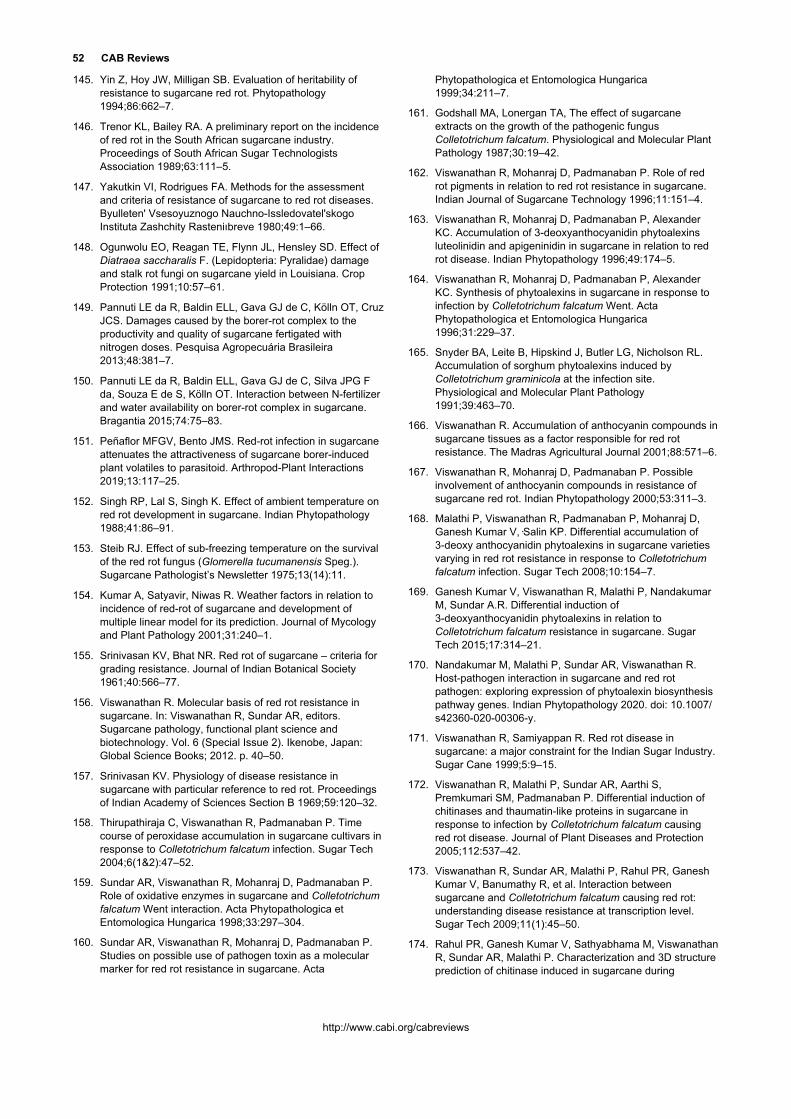
52 CAB Reviews
http://www.cabi.org/cabreviews
145. Yin Z, Hoy JW, Milligan SB. Evaluation of heritability of resistance to sugarcane red rot. Phytopathology 1994;86:662–7.
146. Trenor KL, Bailey RA. A preliminary report on the incidence of red rot in the South African sugarcane industry. Proceedings of South African Sugar Technologists Association 1989;63:111–5.
147. Yakutkin VI, Rodrigues FA. Methods for the assessment and criteria of resistance of sugarcane to red rot diseases. Byulleten' Vsesoyuznogo Nauchno-Issledovatel'skogo Instituta Zashchity Rasteniıbreve 1980;49:1–66.
148. Ogunwolu EO, Reagan TE, Flynn JL, Hensley SD. Effect of Diatraea saccharalis F. (Lepidopteria: Pyralidae) damage and stalk rot fungi on sugarcane yield in Louisiana. Crop Protection 1991;10:57–61.
149. Pannuti LE da R, Baldin ELL, Gava GJ de C, Kölln OT, Cruz JCS. Damages caused by the borer-rot complex to the productivity and quality of sugarcane fertigated with nitrogen doses. Pesquisa Agropecuária Brasileira 2013;48:381–7.
150. Pannuti LE da R, Baldin ELL, Gava GJ de C, Silva JPG F da, Souza E de S, Kölln OT. Interaction between N-fertilizer and water availability on borer-rot complex in sugarcane. Bragantia 2015;74:75–83.
151. Peñaflor MFGV, Bento JMS. Red-rot infection in sugarcane attenuates the attractiveness of sugarcane borer-induced plant volatiles to parasitoid. Arthropod-Plant Interactions 2019;13:117–25.
152. Singh RP, Lal S, Singh K. Effect of ambient temperature on red rot development in sugarcane. Indian Phytopathology 1988;41:86–91.
153. Steib RJ. Effect of sub-freezing temperature on the survival of the red rot fungus (Glomerella tucumanensis Speg.). Sugarcane Pathologist’s Newsletter 1975;13(14):11.
154. Kumar A, Satyavir, Niwas R. Weather factors in relation to incidence of red-rot of sugarcane and development of multiple linear model for its prediction. Journal of Mycology and Plant Pathology 2001;31:240–1.
155. Srinivasan KV, Bhat NR. Red rot of sugarcane – criteria for grading resistance. Journal of Indian Botanical Society 1961;40:566–77.
156. Viswanathan R. Molecular basis of red rot resistance in sugarcane. In: Viswanathan R, Sundar AR, editors. Sugarcane pathology, functional plant science and biotechnology. Vol. 6 (Special Issue 2). Ikenobe, Japan: Global Science Books; 2012. p. 40–50.
157. Srinivasan KV. Physiology of disease resistance in sugarcane with particular reference to red rot. Proceedings of Indian Academy of Sciences Section B 1969;59:120–32.
158. Thirupathiraja C, Viswanathan R, Padmanaban P. Time course of peroxidase accumulation in sugarcane cultivars in response to Colletotrichum falcatum infection. Sugar Tech 2004;6(1&2):47–52.
159. Sundar AR, Viswanathan R, Mohanraj D, Padmanaban P. Role of oxidative enzymes in sugarcane and Colletotrichum falcatum Went interaction. Acta Phytopathologica et Entomologica Hungarica 1998;33:297–304.
160. Sundar AR, Viswanathan R, Mohanraj D, Padmanaban P. Studies on possible use of pathogen toxin as a molecular marker for red rot resistance in sugarcane. Acta
Phytopathologica et Entomologica Hungarica 1999;34:211–7.
161. Godshall MA, Lonergan TA, The effect of sugarcane extracts on the growth of the pathogenic fungus Colletotrichum falcatum. Physiological and Molecular Plant Pathology 1987;30:19–42.
162. Viswanathan R, Mohanraj D, Padmanaban P. Role of red rot pigments in relation to red rot resistance in sugarcane. Indian Journal of Sugarcane Technology 1996;11:151–4.
163. Viswanathan R, Mohanraj D, Padmanaban P, Alexander KC. Accumulation of 3-deoxyanthocyanidin phytoalexins luteolinidin and apigeninidin in sugarcane in relation to red rot disease. Indian Phytopathology 1996;49:174–5.
164. Viswanathan R, Mohanraj D, Padmanaban P, Alexander KC. Synthesis of phytoalexins in sugarcane in response to infection by Colletotrichum falcatum Went. Acta Phytopathologica et Entomologica Hungarica 1996;31:229–37.
165. Snyder BA, Leite B, Hipskind J, Butler LG, Nicholson RL. Accumulation of sorghum phytoalexins induced by Colletotrichum graminicola at the infection site. Physiological and Molecular Plant Pathology 1991;39:463–70.
166. Viswanathan R. Accumulation of anthocyanin compounds in sugarcane tissues as a factor responsible for red rot resistance. The Madras Agricultural Journal 2001;88:571–6.
167. Viswanathan R, Mohanraj D, Padmanaban P. Possible involvement of anthocyanin compounds in resistance of sugarcane red rot. Indian Phytopathology 2000;53:311–3.
168. Malathi P, Viswanathan R, Padmanaban P, Mohanraj D, Ganesh Kumar V,·Salin KP. Differential accumulation of 3-deoxy anthocyanidin phytoalexins in sugarcane varieties varying in red rot resistance in response to Colletotrichum falcatum infection. Sugar Tech 2008;10:154–7.
169. Ganesh Kumar V, Viswanathan R, Malathi P, Nandakumar M, Sundar A.R. Differential induction of 3-deoxyanthocyanidin phytoalexins in relation to Colletotrichum falcatum resistance in sugarcane. Sugar Tech 2015;17:314–21.
170. Nandakumar M, Malathi P, Sundar AR, Viswanathan R. Host-pathogen interaction in sugarcane and red rot pathogen: exploring expression of phytoalexin biosynthesis pathway genes. Indian Phytopathology 2020. doi: 10.1007/s42360-020-00306-y.
171. Viswanathan R, Samiyappan R. Red rot disease in sugarcane: a major constraint for the Indian Sugar Industry. Sugar Cane 1999;5:9–15.
172. Viswanathan R, Malathi P, Sundar AR, Aarthi S, Premkumari SM, Padmanaban P. Differential induction of chitinases and thaumatin-like proteins in sugarcane in response to infection by Colletotrichum falcatum causing red rot disease. Journal of Plant Diseases and Protection 2005;112:537–42.
173. Viswanathan R, Sundar AR, Malathi P, Rahul PR, Ganesh Kumar V, Banumathy R, et al. Interaction between sugarcane and Colletotrichum falcatum causing red rot: understanding disease resistance at transcription level. Sugar Tech 2009;11(1):45–50.
174. Rahul PR, Ganesh Kumar V, Sathyabhama M, Viswanathan R, Sundar AR, Malathi P. Characterization and 3D structure prediction of chitinase induced in sugarcane during

Rasappa Viswanathan 53
http://www.cabi.org/cabreviews
pathogenesis of Colletotrichum falcatum. Journal of Plant Biochemistry and Biotechnology 2015;24:1–8.
175. Franco FP, Santiago AC, Henrique-Silva F, de Castro PA, Goldman GH, Moura DS, et al. The sugarcane defense protein SUGARWIN2 causes cell death in Colletotrichum falcatum but not in non-pathogenic fungi. PLoS One 2014;9:e91159.
176. Parvaiz A, Mustafa G, Khan HMWA, Joyia FA, Niazi AK, Anwar S, et al. Field evaluation ratified by transcript and computational analyses unveils myco-protective role of SUGARWIN proteins in sugarcane. 3 Biotech 2019;9:377. doi: 10.1007/s13205-019-1896-0.
177. Viswanathan R, Sundar AR. Malathi P, Ganesh Kumar V, Sathyabhama M, Muthumeena M, et al. Molecular understanding of red rot resistance in sugarcane through genomics and proteomics. In: Chakraborty B, Chakraborty U, editors. Review of plant pathology. Vol. 6. Jodhpur, India: Scientific Publishers; 2014. p. 189–214.
178. Sundar AR, Nagarathinam S, Ganesh Kumar V, Rahul PR, Raveendran M, Malathi P, et al. Sugarcane proteomics: Establishment of a protein extraction method for 2-DE in stalk tissues and initiation of sugarcane proteome reference map. Electrophoresis 2010;31:1959–74.
179. Ganesh Kumar V, Viswanathan R, Malathi P, Sundar AR, Prasanth CN, Nandakumar M. Identification of differential expressed proteins and establishing a defense proteome of sugarcane in response to Colletotrichum falcatum infection. Journal of Plant Pathology 2020. doi: 10.1007/s42161-020-00577-4.
180. Muthaiah M, Ashwin R, Sundar AR, Malathi P, Viswanathan R. Expression profiling of transcription factors (TFs) in sugarcane X Colletotrichum falcatum interaction. Journal of Plant Biochemistry and Biotechnology 2013;22:286–94.
181. Selvaraj N, Ashwin R, Sundar AR, Malathi P, Viswanathan R. Molecular profiling of systemic acquired resistance (SAR)-responsive transcripts in sugarcane challenged with Colletotrichum falcatum. Applied Biochemistry and Biotechnology 2014;174:2839–50.
182. Prathima PT, Raveendran M, Kumar KK, Rahul PR, Ganesh Kumar V, Viswanathan R et al. Differential regulation of defense-related gene expression in response to red rot pathogen Colletotrichum falcatum infection in sugarcane. Applied Biochemistry and Biotechnology 2013;171:488–503.
183. Rahul PR, Ganesh Kumar V, Viswanathan R, Sundar AR, Malathi P, Prasanth NC, et al. Defense transcriptome analysis of sugarcane and Colletotrichum falcatum interaction using host suspension cells and pathogen c. Sugar Tech 2016;18:16–28.
184. Sathyabhama M, Viswanathan R, Nandakumar M, Malathi P, Sundar RA. Understanding sugarcane defence responses during the initial phase of Colletotrichum falcatum pathogenesis by suppression subtractive hybridization (SSH). Physiological and Molecular Plant Pathology 2015;91:131–40.
185. Sathyabhama M, Viswanathan R, Malathi P, Ramesh Sundar A. Identification of differentially expressed genes in sugarcane during pathogenesis of Colletotrichum falcatum by suppression subtractive hybridization (SSH). Sugar Tech 2016;18:176–83.
186. Viswanathan R, Sathyabhama M, Malathi P, Sundar AR. Transcriptome analysis of host-pathogen interaction
between sugarcane and Colletotrichum falcatum by Suppression Subtractive Hybridization and Illumina sequencing. Proceedings of the International Society of Sugar Cane Technologists 2016;29:1639–44.
187. Sundar AR, Velazhahan R, Viswanathan R, Padmanaban P, Vidhyasekaran P. Induction of systemic resistance to Colletotrichum falcatum in sugarcane by a synthetic signal molecule, acibenzolar-S-methyl (CGA 245704). Phytoparasitica 2001;29:231–42.
188. Sundar AR, Viswanathan R, Malathi P, Padmanaban P. Mechanism of resistance induced by plant activators against Colletotrichum falcatum in sugarcane. Archives of Phytopathology and Plant Protection 2006;39:259–72.
189. Sundar AR, Viswanathan R, Nagarathinam S. Induction of systemic acquired resistance (SAR) using synthetic signal molecules against Colletotrichum falcatum in Saccharum officinarum. Sugar Tech 2009;11:274–81.
190. Ashwin NMR, Barnabas L, Sundar AR, Muthumeena M, Malathi P, Viswanathan R. Disease suppression effects of resistance inducing agents against red rot of sugarcane. European Journal of Plant Pathology 2017;149:285–97.
191. Tai PYP, Miller JD, Dean JL. Inheritance of resistance to rust in sugarcane. Field Crops Research 1981;4:261–8.
192. Wu KK, Heinz DJ, Meyer HK. Heritability of sugarcane smut resistance and correlation between smut grade and yield components. Crop Science 1983;23:54–6.
193. Hogarth DM. Genetics of sugarcane. In: Heinz DJ, editor. Sugarcane improvement through breeding. Amsterdam, The Netherland: Elsevier; 1987. p. 255–69.
194. Azab YE, Chilton SJP. Studies on the inheritance of red rot disease of sugarcane. Phytopathology 1952;42:282.
195. Chona BL. Opening address of the Chairman: pathology session. Proceedings of International Society Sugar Cane Technologists 1956;9:975–86.
196. Harnandez RG. Selection of sugarcane resistant to red rot. Proceedings of International Society Sugar Cane Technologists 1980;17:1397–401.
197. Benda GTA. Breeding for disease resistance. In: Copersucar International Sugarcane Breeding Workshop (Brazil) Sao Paulo, Brazil: Copersucar; 1987. p. 161–79.
198. Alexander KC, Kandasami PA, Rao TCR, Mohanraj D, Rao MM. A new approach to genetic defense against red rot disease of sugarcane. Indian Sugar 1979;28:695–6.
199. Alexander KC, Rao MM, Mohanraj D, Seshadri K, Padmanaban P, Prakasam N, et al. A new approach to improvement of red rot resistance in sugarcane. Indian Journal of Mycology and Plant Pathology 1986;16:108–12.
200. Natarajan US, Balasundaram N, Rao TCR, Padmanaban P, Mohanraj D, Karthigeyan S, et al. Role of Saccharum spontaneum in imparting stable resistance against sugarcane red rot. Sugar Cane International 2001;1:17–20.
201. Ram B, Singh N, Sahi BK. Combining ability and heterosis for disease index of red rot in sugarcane (Saccharum officinarum L.). Indian Journal of Genetics and Plant Breeding 2005;65:112–4.
202. Bora GC, Sarma PC, Chandra K. Progenies of sugarcane crosses with red rot resistance. Indian Sugar 2006;56(6):33–5.
54 CAB Reviews
http://www.cabi.org/cabreviews
203. Singh N, Kumar A, Raj K, Virk KS, Satyavir. Slow red rot resistance in sugarcane. Sugar Cane International 2009;27(3):125–30.
204. Babu C. Associations of red rot resistance with yield and quality of sugarcane and their utility in family selection. Indian Sugar 2009;59(7):23–8.
205. Ram B, Singh N, Sahi BK. Selection for juice quality and red rot disease index in sugarcane (Saccharum officinarum L.). Indian Journal of Genetics and Plant Breeding 2006;66:151–2.
206. Shanthi RM, Alarmelu S. Assessment of the first cycle hybrid progenies for early high sugar content in sugarcane. Indian Journal of Genetics and Plant Breeding 2007;67:384–7.
207. Singh N, Govindaraj P. Distribution of red rot resistance in progenies of sugarcane varietal crosses and its relation with sugar content. Indian Sugar 2009;59(1):41–8.
208. Azab YE, Chilton SJP. Inheritance of resistance and susceptibility of sugarcane to the red rot fungus (Physalospora tucumanensis Speg.). Proceedings of International Society Sugar Cane Technologists 1959;10:1127–9.
209. Singh K, Tripathi BK, Pandey S. Screening of sugarcane varietal crosses for resistance to red rot. Proceedings of International Society Sugar Cane Technologists 1983;18:765–70.
210. Dutt NL, Hussainy SA. A note on the breeding and testing of sugarcane varieties resistant to the three major diseases with particular reference to red rot. Proceedings of All India Conference of Sugar Cane Research & Development Workers (Coimbatore) 1951;1(3):46–55.
211. Chona BL. Relative resistance of sugarcane varieties to red rot. Indian Journal of Agricultural Sciences 1954;24:301–15.
212. Atchutharama Rao M, Sarma MN. Studies on parental influence on red rot resistance in sugarcane seedlings. Sugarcane Pathologist’s Newsletter 1982;29:22–6.
213. Chaudhary BS, Virk KS, Ram K. Inheritance of red rot resistance in sugarcane. Agricultural Science Digest 1988;8:210–2.
214. Chaudhary BS, Mehla AS, Virk KS, Pal R. Breeding for red rot resistance in sugarcane (Saccharum Hybrid complex): review, concept and theory. In: Singh GB, Shukla US, Agnihotri VP, Sinha OK, Singh RP, editors, Proceedings of National Seminar on strategies for research and management of red rot, Part II. Lucknow, India: ICAR-IISR; 1995. p. 221–32.
215. Mishra MM. Role of BO varieties in minimizing the red rot incidence in Bihar. In: Agnihotri VP, Sinha OK, Singh RP. editors. Proceedings of National Seminar on Sugarcane Production constraints and strategies for Research and Management of red rot, Part II. Lucknow, ICAR-IISR; 1995. p. 354–6.
216. De Carvalho PCT. Heterokaryosis in Colletotrichum falcatum. Proceedings of International Society of Sugar Cane Technologists 1968;13:1251–61.
217. Srinivasan KV, Alexander KC. Sources of resistance in the different species of Saccharum to red rot and smut disease of sugarcane. Sugarcane Pathologist's Newsletter 1971;6:6–7.
218. Sreenivasan TV. Research strategies for breeding for red rot resistance in sugarcane. In: Singh GB, Shukla US,
Agnihotri VP, Sinha OK, Singh RP, editors. Proceedings of National Seminar on Sugarcane Production constraints and strategies for Research and Management of red rot, Part II. Lucknow: ICAR-IISR; 1995. p. 233–9.
219. Viswanathan R, Malathi P, Chandran K, Gopi R. Screening of sugarcane germplasm for red rot resistance. Journal of Sugarcane Research 2017;7(2):100–11.
220. Price S. Interspecific hybridization in sugarcane breeding. Proceedings of International Society Sugar Cane Technologists 1967;12:1021–6.
221. Ram B, Sreenivasan TV, Sahi BK, Singh N. Introgression of low temperature tolerance and red rot resistance from Erianthus in sugarcane. Euphytica 2001;122:145–53.
222. Singh N, Govindaraj P, Singh BD. Parental influence of red rot resistance in progenies of varietal crosses in sugarcane. Indian Sugar 2008;58(1):37–40.
223. Hale AL, Hoy JW, Veremis JC. Identification of sources of resistance to sugarcane red rot. Proceedings of International Society of Sugar Cane Technologists 2010;27:1–8. Available from: URL: http://www.issct.org/proceedings/2010.html
224. Nair NV, Mohanraj K, Sunadaravelpandian K, Suganya A, Selvi A, Appunu C. Characterization of an intergeneric hybrid of Erianthus procerus × Saccharum officinarum and its backcross progenies. Euphytica 2017;213:267.
225. Viswanathan R, Padmanaban P, Mohanraj D, Prakasam N, Singh N, Selvakumar R, et al. Status of red rot resistance in wild relatives of sugarcane, Saccharum spontaneum, interspecific hybrids and intergeneric hybrids. Journal of Sugarcane Research 2018;8(2):169–84.
226. Martin GB, Bogdanove AJ, Sessa G. Understanding the function of plant disease resistance proteins. Annual Review of Plant Biology 2003;54:23–61.
227. McDowell JM, Simon SA. Recent insights into R gene evolution. Molecular Plant Pathology 2006;7:437–48.
228. Jayashree J, Selvi A, Nair NV. Characterization of resistance gene analog polymorphisms in sugarcane cultivars with varying levels of red rot resistance. Electronic Journal of Plant Breeding 2010;1(4):1191–9.
229. Sharma R, Tamta S. Red rot resistant gene characterization using RGAP markers among sugarcane cultivars resistant and susceptible to the red rot disease. 3 Biotech 2017;7:306. doi: 10.1007/s13205-017-0941-0.
230. Hameed U, Pan YB, Iqbal J. Genetic analysis of resistance gene analogues from a sugarcane cultivar resistant to red rot disease. Journal of Phytopathology 2015;163:755–63.
231. Singh RK, Singh RB, Singh SP, Sharma ML. Genes tagging and molecular diversity of red rot susceptible/tolerant sugarcane hybrids using c-DNA and unigene derived markers. World Journal of Microbiology and Biotechnology 2012;28:1669–79.
232. Parida SK, Kalia S, Pandit A, Nayak P, Singh RK, Gaikwad K, et al. Single nucleotide polymorphism in sugar pathway and disease resistance genes in sugarcane. Plant Cell Reports 2016;35:1629–53.
233. Gupta V, Raghuvanshi S, Gupta A et al. The water-deficit stress- and red-rot-related genes in sugarcane. Functional & Integrative Genomics 2010;10(2):207–14.

Rasappa Viswanathan 55
http://www.cabi.org/cabreviews
234. Singh RK, Banerjee N, Khan MS et al. Identification of putative candidate genes for red rot resistance in sugarcane (Saccharum species hybrid) using LD-based association mapping. Molecular Genetics and Genomics 2016;291:1363–77.
235. Jalaja NC, Sreenivasan TV, Pawar SM, Bhoi PG, Garker RM. Co 94012 - a new sugarcane variety through somaclonal variation. Sugar Tech 2006;8(2/3):132–6.
236. Kumar P, Agarwal A, Tiwari AK, Lal M, Jabri MRA. Possibilities of development of red rot resistance in sugarcane through somaclonal variation. Sugar Tech 2012;14:192–4.
237. Kumar A, Bharti YP, Sharma ML. Screening of somaclones against red rot (Colletotrichum falcatum Went) in sugarcane. Cooperative Sugar 2009;41(4):51–4.
238. Mahmud K, Nasiruddin KM, Hossain MA, Hassan L. Screening sugarcane somaclones and their parent varieties against red rot (Colletotrichum falcatum) and assessment of variability by RAPD and SSR markers. SAARC Journal of Agriculture 2015;13(2):173–82.
239. Ahmed MS, Gardezi DA, Batley J, Hayashi S, Zander M, Javid A, et al. Somaclonal variations for red rot and sugarcane mosaic virus resistance and candidate genes integrity assessment in somaclones of selected sugarcane varieties (Saccharum officinarum L.). Pakistan Journal of Agricultural Sciences 2019;56:(1)15–27.
240. Prasada Rao KK, Sarma MN, Satyanarayana V, Rao MA. Discriminant function as a reliable guide for assessing varietal reaction to red rot of sugarcane. Proceedings of International Society Sugar Cane Technologists 1978;16:395–400.
241. Wang ZN, Lee CS. Improvements on the methods of testing sugarcane varietal resistance to leaf blight and red rot. Report of Taiwan Sugar Experiment Station 1982;95:1–7.
242. Virk KS, Satyavir. Evaluation of screening methods for red rot resistance. Indian Sugar 1989;39:621–2.
243. Duttamajumder SK, Singh, RP. Revised criteria for grading resistance against red rot disease of sugarcane. Indian Phytopathology 1999;52:157–9.
244. Rana OS, Gupta SC. An easy method of screening out red rot susceptible varieties at initial stages of multiplication. Indian Sugar 1968;18:447–52.
245. Viswanathan R. Screening for diseases: red rot. In: Annual Report of Sugarcane Breeding Institute 2012-13, Coimbatore, India. Coimbatore: ICAR - Sugarcane Breeding Institute; 2013.
246. Viswanathan R, Sundar AR, Selvakumar R, Malathi P. Progress in understanding fungal diseases affecting sugarcane: red rot. In: Rott P, editor, Achieving sustainable cultivation of sugarcane Volume 2: breeding, pests and diseases. Cambridge, UK: Burleigh Dodds Science Publishing; 2018. p. 201–20.
247. Singh K, Alexander KC. Laminar infection of sugarcane leaves by red rot (Physalospora tucumanensis) organism in nature. Indian Phytopathology 1970;23:114–5.
248. Beniwal MS, Satyavir. Effect of atmospheric temperature on the development of red rot of sugarcane. Indian Phytopathology 1991;44:333–8.
249. Satyavir, Kumar A, Raj K, Virk KS. Red rot of sugarcane: the research scene in Haryana. In: Singh SB, Rao GP,
Eswaramoorthy S, editors. Sugarcane crop management. Houston, Texas, USA: SCI TECH Publishing LLC; 2002. p. 109–26.
250. Viswanathan R, Padmanaban P, Mohanraj D, Sundar AR. An alternate method for assessing field resistance to red rot disease. Sugarcane Breeding Institute Newsletter 2000;19:(3):4.
251. Mohanraj D, Padmanaban P, Viswanathan R. Screening for red rot resistance in sugarcane. In: Viswanathan R, Sundar AR. (Eds) Functional plant science and biotechnology. Vol. 6 (Special Issue 2). Ikenobe, Japan: Global Science Books; 2012. p. 51–62.
252. Alfonso F, Santiesteban R, Rodríguez J. Test for sugarcane clone and cultivar resistance to stalk red rot (Colletotrichum falcatum Went). Revista INICA 1987;2:1–8.
253. De Ramallo VNE, Ploper LD. Red rot in sugar cane: technique of testing for resistance. Revista Industrial y Agricola de Tucuman (Argentina) 1976;53(2):15–20.
254. Manjaroenchot A. A stem rot and root rot of sugarcane. Bangkok (Thailand): Office of the Permanent Secretary, Bangkok (Thailand). Office of the Cane and Sugar Board; Kasetsart University; 1986. p. 99.
255. Srinivasan KV. A technique for the elimination of red rot susceptible sugarcane seedlings at an early stage. Current Science 1962;31:112–3.
256. Kirtikar, Gupta SC, Kureel DG. Screening of sugarcane varities for red rot resistance. Proceedings of International Society Sugar Cane Technologists 1974;15:189–93.
257. Singh HN, Gupta SC, Singh SB, Singh MP. Improvement of the technique for screening against red rot at seedling stage. Sugarcane Pathologists’ Newsletter 1978;21:29–31.
258. Viswanathan R, Padmanaban P, Mohanraj D. Comparison of three testing methods for the evaluation of red rot (Colletotrichum falcatum Went) in sugarcane. Indian Journal of Agricultural Sciences 1998;68:226–30.
259. Viswanathan R, Rao GP. Developing disease resistant varieties and methods of disease screening in sugarcane. In: Singh SB, Rao GP, Solomon S, Gopalasundaram P, editors. Sugarcane: crop production and improvement. Houston: USA: Studium Press LLC; 2006. p. 759–800.
260. Giglioti EA, Canteri MG. Phytopathometry applied to the integrated management of borer-rot complex in sugarcane: (I) a standard diagram to assess disease severity. In: Rao GP, Filho AB, Autrey JC, Megarey R, editors. Sugarcane pathology. Enfield (NH), USA: Science Publishers; 1999. p. 183–94.
261. Viswanathan R, Samiyappan R. Induction of systemic resistance by plant growth promoting rhizobacteria against red rot disease in sugarcane. Sugar Tech 1999;1(3):67–76.
262. Viswanathan R, Samiyappan R. Antagonism of different Pseudomonas strains against red rot pathogen, Colletotrichum falcatum: Indian Journal of Sugarcane Technology 2001;16(1):44–9.
263. Malathi P, Padmanaban P, Viswanathan R, Mohanraj D. Interaction between Colletotrichum falcatum pathotypes and biocontrol agents Archives of Phytopathology and Plant Protection 2008;41:311–7.
264. Viswanathan R, Malathi P. Biocontrol strategies to manage fungal diseases in sugarcane. Sugar Tech 2019;21:202–12.

56 CAB Reviews
http://www.cabi.org/cabreviews
265. Viswanathan R, Samiyappan R. Induced systemic resistance by fluorescent pseudomonads against red rot disease of sugarcane caused by Colletotrichum falcatum. Crop Protection 2002;21:1–10.
266. Viswanathan R, Samiyappan R. Pseudomonas spp. colonization in sugarcane rhizosphere reduces titre of Colletotrichum falcatum Went-causing red rot disease of sugarcane. Archives of Phytopathology and Plant Protection 2006;39:39–44.
267. Viswanathan R, Samiyappan R. Bio-formulation of fluorescent Pseudomonas spp. induces systemic resistance against red rot disease and enhances commercial sugar yield in sugarcane. Archives of Phytopathology and Plant Protection 2008;41:377–88.
268. Viswanathan R, Samiyappan R. Role of oxidative enzymes in the plant growth promoting rhizobacteria (PGPR) mediated induced systemic resistance against Colletotrichum falcatum in sugarcane. Journal of Plant Diseases and Protection 2002;109:88–100.
269. Viswanathan R, Nandakumar R, Samiyappan R. Involvement of pathogenesis-related proteins in Pseudomonas spp. induced systemic resistance against Colletotrichum falcatum in sugarcane. Journal of Plant Diseases and Protection 2003;110:524–37.
270. Malathi P, Viswanathan R, Padmanaban P, Mohanraj D, Sundar, AR. Compatibility of biocontrol agents with fungicides against red rot disease of sugarcane. Sugar Tech 2002;4(3&4):131–6.
271. Viswanathan R, Samiyappan R. Efficacy of Pseudomonas spp. strains against soil borne and sett borne inoculum of Colletotrichum falcatum causing red rot disease in sugarcane. Sugar Tech 2000;2(3):26–9.
272. Viswanathan R, Samiyappan R. Antifungal activity of chitinases produced by some fluorescent pseudomonads against Colletotrichum falcatum Went causing red rot disease in sugarcane. Microbiological Research 2001;155:309–14.
273. Viswanathan R, Samiyappan R. Secondary metabolites production by Pseudomonas spp. strains antagonistic to Colletotrichum falcatum causing red rot disease in sugarcane. Acta Phytopathologica Entomologica et Hungarica,2004;39(1-3):28–37.
274. Viswanathan R, Samiyappan R. Siderophores and iron nutrition on the pseudomonas mediated antagonism against Colletotrichum falcatum in sugarcane. Sugar Tech 2007;9(1):57–60.
275. Senthil R, Selvaraj S, Anand T, Raguchander T, Samiyappan R. Efficacy of liquid Pseudomonas fluorescens (Pf1) against sugarcane red rot, caused by Colletotrichum falcatum, under field conditions. International Sugar Journal 2011;113:(1356)888–93.
276. Hassan MN, Afghan S, Hafeez FY. Suppression of red rot caused by Colletotrichum falcatum on sugarcane plants using plant growth-promoting rhizobacteria. BioControl 2010;55(4):531–42.
277. Hassan MN, Afghan S, Hafeez FY. Biological suppression of sugarcane red rot by Bacillus spp. under field conditions. Journal of Plant Pathology 2012;94(2):325–29.
278. Elamathi E, Malathi P, Viswanathan R, Sundar AR. Potential of Paenibacillus and Trichoderma isolates against Colletotrichum falcatum causing red rot disease in sugarcane. Journal of Sugarcane Research 2016;6(2):100–11.
279. Jayakumar V, Sundar AR, Viswanathan R. Biocontrol of Colletotrichum falcatum with volatile metabolites produced by endophytic bacteria and profiling VOCs by headspace SPME coupled with GC–MS. Sugar Tech 2021;23:94–107. doi: 10.1007/s12355-020-00891-2.
280. Viswanathan R, Rajitha R, Sundar AR, Ramamoorthy V. Isolation and characterization of endophytic bacterial strains from sugarcane stalks and their in vitro antagonism against the red rot pathogen. Sugar Tech 2003;5(1&2):25–9.
281. Patel P, Shah R, Joshi B, Ramar K, Natarajan A. Molecular identification and biocontrol activity of sugarcane rhizosphere bacteria against red rot pathogen Colletotrichum falcatum. Biotechnology Reports 2018. doi: 10.1016/j.btre.2019.e00317.
282. Amna XY, Farooq MA, Javed MT, Kamran MA, Mukhtar T, Ali J, et al. Multi-stress tolerant PGPR Bacillus xiamenensis PM14 activating sugarcane (Saccharum officinarum L.) red rot disease resistance. Plant Physiology and Biochemistry 2020;151:640–9.
283. Hassan MN, Shah SZ-U-H, Afghan S, Hafeez FY. Suppression of red rot disease by Bacillus sp. based biopesticide formulated in non-sterilized sugarcane filter cake. BioControl 2015;60:691–702.
284. Teerapensang A, Leksomboon C, Lersrutaiyotin R. Efficacy of rhizobacteria for controlling red rot wilt of sugarcane under greenhouse condition. In: Proceedings of the 48th Kasetsart University Annual Conference, Kasetsart, 3-5 March, 2010, Kasetsart University, Nakhon Pathom, Thailand.
285. Singh V, Joshi BB, Awasthi SK, Srivastava SN. Ecofriendly management of red rot disease of sugarcane with Trichoderma strains. Sugar Tech 2008;10:58–61.
286. Singh V, Lal RJ, Awasthi SK, Verma MR. Managing red rot of sugarcane by Trichoderma harzianum. Indian Sugar 2009;59:25–30.
287. Joshi D, Singh P, Holkar SK, Kumar S. Trichoderma-mediated suppression of red rot of sugarcane under field conditions in subtropical India. Sugar Tech 2019;21:496–504.
288. Joshi D, Singh P, Singh AK, Lal RJ, Tripathi N. Antifungal potential of metabolites from Trichoderma sp. Against Colletotrichum falcatum Went causing red rot of sugarcane. Sugar Tech 2016;18:529–36.
289. Viswanathan R, Sundar AR, Premkumari SM. Mycolytic effect of extracellular enzymes of antagonistic microbes to Colletotrichum falcatum, red rot pathogen of sugarcane. World Journal of Microbiology and Biotechnology 2003;19:953–9.
290. Malathi P, Viswanathan R, Padmanaban P, Mohanraj D, Sundar, AR. Microbial detoxification of Colletotrichum falcatum toxin. Current Science 2002;83:745–9.
291. Elamathi E, Malathi P, Viswanathan R, Sundar AR. Expression analysis on mycoparasitism related genes during antagonism of Trichoderma with Colletotrichum falcatum causing red rot in sugarcane. Journal of Plant Biochemistry and Biotechnology 2018;27:351–61.
292. Gudina EJ, Rocha V, Teixeira JA, Rodrigues LR. Antimicrobial and antiadhesive properties of a biosurfactant isolated from Lactobacillus paracasei ssp. paracasei A20. Letters in Applied Microbiology 2010;50:419–24.

Rasappa Viswanathan 57
http://www.cabi.org/cabreviews
293. Goswami D, Borah SN, Lahkar J, Handique PJ, Deka S. Antifungal properties of rhamnolipid produced by Pseudomonas aeruginosa DS9 against Colletotrichum falcatum. Journal of Basic Microbiology 2015;55:1–10.
294. Elamathi E, Malathi P, Viswanathan R, Sundar AR. Molecular analysis of the suppression of Colletrotrichum falcatum by Trichoderma harzianum in sugarcane. Journal of Plant Pathology 2017;99:211–8.
295. Elamathi E, Malathi P, Viswanathan R, Sundar AR. Identification and characterization of differentially expressed proteins from Trichoderma harzianum during interaction with Colletotrichum falcatum causing red rot in sugarcane. Sugar Tech 2019;21):765–72.
296. Gupta SC, Verma KP, Singh MP, Misra SC. Control of diseases by hot water treatment of sugarcane seed material. Indian Sugar Crops Journal 1978;5(2):28–9.
297. Pandey S, Agnihotri VP. Elimination of red rot infection in seed-pieces by chemo-thermotherapy: a new approach in disease control. Indian Phytopathology 1996;49:48–51.
298. Singh K, Misra SR, Shukla US, Singh RP. Moist hot air therapy [MHAT] of sugarcane control of sett-borne infections of GSD, smut and red rot. Sugar Journal 1980;43(5):26–8.
299. Singh K. Hot air therapy against red rot of sugarcane. Plant Disease Report 1973;57:220–2.
300. Jha A, Singh RK, Prasad RN, Jha CS. Attempt to control red-rot disease of sugarcane through heat therapy in Bihar. Indian Sugar 1976;26(6):441–6.
301. Yang S-M. Effect of aerated steam on the red-rot pathogen in sugarcane stalks. Phytopathology 1979;69:702–5.
302. Natarajan S, Muthusamy S. Effect of aerated steam treatment on the incidence of red rot (Colletotrichum falcatum Went) of sugar cane. International Sugar Journal 1981;83:300–1.
303. Padmanabhan D, Natarajan S, Kalaimani T, Rajasekaran S. Disease build up in sugarcane setts treated with aerated steam. SISSTA Sugar Journal 1990;16(2):31–4.
304. Rao MA, Satyanarayana Y. Chemical control of sett-borne infection of red rot pathogen. In: Singh GB, Shukla US, Agnihotri VP, Sinha OK, Singh RP, editors. Sugarcane production constraints and strategies for Research Management of Red Rot. Lucknow: ICAR-IISR; 1995. p. 323–30.
305. Pliansinchai U. Trends in sugarcane fungal disease control in Thailand. In: Rao GP, Filho AB, Magarey RC, Autrey LJC, editors. Sugarcane pathology Vol. I. Fungal diseases. New Delhi: Oxford & IBH Publishing Co; 1999. p. 209–37.
306. Shailbala, Kumar A. Field evaluation of Azoxystrobin 18.2% + Difenoconazole 11.4% w/w SC, a new fungicide for the
management of major sugarcane diseases. International Journal of Bio-resource and Stress Management 2016;7:732–7.
307. Malathi P, Padmanaban P, Viswanathan R, Mohanraj D, Sundar AR. Efficacy of thiophanate methyl against red rot of sugarcane. Acta Phytopathologica et Entamologica Humgarica 2004;39:39–47.
308. Padmanaban P, Mohanraj D, Viswanathan R, Malathi P. Use of fungicide in the integrated management of sugarcane red rot. Sugarcane Breeding Institute Newsletter 2007;26:(4)1–2.
309. Malathi P, Padmanaban P, Sundar AR, Viswanathan R. Efficacy of new systemic fungicides against sugarcane red rot. SBI News 2012;31(2):3–4.
310. Malathi P, Viswanathan R, Sundar AR. Mechanized means of sett treatment: an effective way of delivering fungicides for the management of red rot in sugarcane. Sugar Tech 2017;19:176–82.
311. Viswanathan R, Malathi P, Naik R. A sett treatment device for fungal disease management and healthy nursery programme in sugarcane. Proceedings of Annual Convention of Sugar Technologists of India 2016;77:236–53.
312. Viswanathan R, Malathi P, Prasanth CN, Naik R, Annamalai SJK. Sett way of growing sugarcane for fungal treatment. ICAR NEWS 2017;23(2):1–3.
313. Anzalone Jr L. Air pressure incorporation of fungicides for the control of red rot. Proceedings of the International Society of Sugar Cane Technologists 1971;14:1030–3.
314. Nallathambi P, Viswanathan R, Padmanaban P, Mohanraj D, Jothi R. Studies on sugar factory effluents against Colletotrichum falcatum Went causing red rot disease in sugarcane. Indian Sugar 1999;49:111–23.
315. Nayyar S, Sharma BK, Kaur A, Kalia A, Sanghera GS, Thind KS, et al. Red rot resistant transgenic sugarcane developed through expression of β-1,3-glucanase gene. PLoS One 2017;12(6):e0179723.
316. Ijaz S, Rana IA, Khan IA. Engineering sugarcane genome to develop resistance against Colletotrichum falcatum to control red rot diseases Academia Journal of Agricultural Research 2018;6(5):106–16. doi: 10.15413/ajar. 2018.0104.
317. Tariq M, Khan A, Tabassum B, Toufiq N, Bhatti MU, Riaz A, et al. Antifungal activity of chitinase II against Colletotrichum falcatum Went causing red rot disease in transgenic sugarcane. Turkish Journal of Biology 2018;42(1):45–53.
318. Viswanathan R, Rao GP. Disease scenario and management of major sugarcane diseases in India. Sugar Tech 2011;13:336–53.