Embed Size (px)
Citation preview

MOLECULAR AND CELLULAR BIOLOGY,0270-7306/01/$04.0010 DOI: 10.1128/MCB.21.12.3935–3946.2001
June 2001, p. 3935–3946 Vol. 21, No. 12
Copyright © 2001, American Society for Microbiology. All Rights Reserved.
RAG-1 Mutations Associated with B-Cell-Negative SCIDDissociate the Nicking and Transesterification Steps of
V(D)J RecombinationWENHUI LI,† FU-CHUNG CHANG, AND STEPHEN DESIDERIO*
Department of Molecular Biology and Genetics and Howard Hughes Medical Institute, The Johns Hopkins UniversitySchool of Medicine, Baltimore, Maryland 21205
Received 13 November 2000/Returned for modification 9 January 2001/Accepted 30 March 2001
Some patients with B-cell-negative severe combined immune deficiency (SCID) carry mutations in RAG-1 orRAG-2 that impair V(D)J recombination. Two recessive RAG-1 mutations responsible for B-cell-negativeSCID, R621H and E719K, impair V(D)J recombination without affecting formation of single-site recombina-tion signal sequence complexes, specific DNA contacts, or perturbation of DNA structure at the heptamer-coding junction. The E719K mutation impairs DNA cleavage by the RAG complex, with a greater effect onnicking than on transesterification; a conservative glutamine substitution exhibits a similar effect. Whencysteine is substituted for E719, RAG-1 activity is enhanced in Mn21 but remains impaired in Mg21, suggest-ing an interaction between this residue and an essential metal ion. The R621H mutation partially impairsnicking, with little effect on transesterification. The residual nicking activity of the R621H mutant is reducedat least 10-fold upon a change from pH 7.0 to pH 8.4. Site-specific nicking is severely impaired by an alaninesubstitution at R621 but is spared by substitution with lysine. These observations are consistent with involve-ment of a positively charged residue at position 621 in the nicking step of the RAG-mediated cleavage reaction.Our data provide a mechanistic explanation for one form of hereditary SCID. Moreover, while RAG-1 isdirectly involved in catalysis of both nicking and transesterification, our observations indicate that these twosteps have distinct catalytic requirements.
The central event in the generation of immunological diver-sity is the assembly of T-cell receptor and immunoglobulingenes from discrete DNA segments, V, D, and J, during lym-phocyte development. This process, termed V(D)J recombina-tion, is the only known form of site-specific DNA rearrange-ment in vertebrates. V(D)J recombination is mediated byheptamer and nonamer signal sequences, which are separatedby spacer regions of 12 or 23 bp (28), and is initiated by therecombination-activating proteins RAG-1 and RAG-2 (33,39), which act in concert to cleave DNA at the junctions be-tween antigen receptor coding segments and conserved recom-bination signals that specify sites of recombination (31, 48).
RAG-mediated DNA cleavage occurs in two steps (31).First, one DNA strand is nicked between the recombinationsignal sequence (RSS) heptamer and the coding sequence.This is followed by a transesterification reaction in which thefree hydroxyl group at the 39 end of the coding sequenceattacks a phosphodiester on the opposite strand (49, 50). As aresult, two DNA ends are produced: a signal end, terminatingin a blunt, 59-phosphorylated, double-strand break, and a cod-ing end, terminating in a hairpin.
RAG-1 and RAG-2 are both essential for initiation ofV(D)J recombination. RAG-2 alone has no detectable DNAbinding activity in vitro but collaborates with RAG-1 in RSS
recognition (2, 11, 20, 29, 44, 45). In the absence of RAG-2,RAG-1 interacts weakly with the RSS through the nonamerelement (11, 20, 42, 45). In contrast, assembly of a complexcontaining the RSS, RAG-1, and RAG-2 is strongly heptamerdependent (2, 11, 20, 45) and is accompanied by kinking orunwinding of substrate DNA near the scissile bond at theheptamer-coding boundary (45). Chemical interference andfootprinting have shown that in the presence of RAG-1 alone,DNA contacts are restricted to the vicinity of the nonamer(45). Occupancy of the heptamer requires RAG-2 (45), whichinduces RAG-1 to approach the scissile bond (44). The prox-imity of RAG-1 to the site of DNA cleavage in the presence ofRAG-2 has suggested a direct role for RAG-1 in DNA cleav-age. The relative roles of RAG-1 and RAG-2 in catalysis ofnicking and transesterification, however, remain incompletelyunderstood.
The chemistry of DNA cleavage by the RAG proteins isformally equivalent to that employed by classical transposasessuch as Mu A (37, 49, 50). Several additional lines of evidenceindicate that V(D)J recombination represents a specializedform of DNA transposition, including the formal equivalenceof hybrid joint formation to the retroviral disintegration reac-tion and the ability of the RAG proteins to integrate signalends into nonhomologous DNA (1, 21). The similarity betweenV(D)J rearrangement and other types of transposition sug-gests that the RAG catalytic core may resemble that of othertransposases. The transposases of retroviruses, retrotrans-posons, IS3 elements, and Mu, as well as the TnsB transposasecomponent of Tn7, all contain an array of acidic amino acids,the DDE motif (7, 35), which binds an essential divalent cationsuch as Mg21 at or near the active sites for DNA cleavage and
* Corresponding author. Mailing address: Department of MolecularBiology and Genetics and Howard Hughes Medical Institute, TheJohns Hopkins University School of Medicine, Baltimore, MD 21205.Phone: (410) 955-4735. Fax: (410) 955-9124. E-mail: [email protected].
† Present address: Division of Biology, California Institute of Tech-nology, Pasadena, CA 91125.
3935
on February 17, 2018 by guest
http://mcb.asm
.org/D
ownloaded from

strand transfer (3, 5, 12, 24, 36, 38). Direct participation of theDDE motif residues in metal binding has been demonstratedfor the human immunodeficiency virus and avian sarcoma virusintegrases by crystallographic analysis (5, 12) and for TnsB ofTn7 by the observation that cysteine substitution results inaltered metal specificity (38). Although the RAG proteins lacksignificant homology to the retroviral integrase superfamily,precluding identification of a putative active center by se-quence alignment, recent studies employing iron-induced hy-droxyl radical cleavage and site-directed mutagenesis haveidentified three acidic residues within RAG-1, D600, D708,and E962, that are essential for nicking and transesterificationactivity (14, 23, 27). Substitution of cysteine at two of theseresidues, D600 and D708, was associated with metal ion-spe-cific recovery of DNA cleavage activity, implicating theseamino acids in the binding of an essential divalent cation.
About one-third of all patients with severe combined immu-nodeficiency (SCID) lack detectable B cells. A number ofB-cell-negative SCID patients have been shown to carry debil-itating mutations in RAG-1, RAG-2, or both, resulting in im-pairment of V(D)J recombination (40). In this communicationwe have examined the biochemical properties of SCID-associ-ated RAG-1 alleles, with the goal of defining the precise mo-lecular defects arising from these mutations.
Two RAG-1 mutations associated with B-cell-negativeSCID, R621H and E719K, impair V(D)J recombination in vivowithout affecting formation of single-site RSS complexes invitro. The E719K mutation impaired both the nicking andtransesterification steps of DNA cleavage. In contrast, theR621H mutation partially impaired nicking, with little or noeffect on transesterification. Further analysis suggested thatRAG-mediated strand scission requires a positively chargedresidue at position 621 of RAG-1. These data provide a mech-anistic explanation for one form of hereditary SCID and indi-cate that RAG-dependent nicking and transesterification arelikely to have distinct catalytic requirements.
MATERIALS AND METHODS
DNA constructs and site-directed mutagenesis. Maltose-binding protein(MBP) fusions containing RAG-1 (residues 384 to 1008) or RAG-2 (residues1 to 387) cores, tagged at the carboxyl terminus with a myc epitope and poly-histidine, have been described previously (31, 48). DNA fragments encodingthese proteins (gifts of Dik van Gent and Martin Gellert) were cloned betweenthe BamHI and NotI sites of pcDNA-1 (Invitrogen) to create pcDNAR1 andpcDNAR2 (45).
RAG-1 cDNA, cloned into pBluescript SK(II), was the starting material formutagenesis. Mutations R621H, E719K, and Y935Stop were introduced by stan-dard PCR; E719C was introduced using divergent PCR (30). Products wereconfirmed by nucleotide sequencing. For expression in mammalian cells, DNAcassettes spanning the mutations were exchanged for the corresponding wild-type fragment of pcDNAR1.
Cell culture, transfection, and protein purification. The 293 cell line wasmaintained in Dulbecco’s modified Eagle’s medium supplemented with 10%fetal bovine serum. RAG expression constructs were cotransfected with pRSV-Tinto 293 cells by the calcium phosphate method.
For purification of RAG fusion proteins, pcDNAR1 or a corresponding mu-tant construct was cotransfected with pcDNAR2 into 293 cells (10 mg of eachplasmid per 10-cm-diameter culture plate). At 48 h after transfection, RAG-1and RAG-2 fusion proteins were copurified by amylose affinity chromatographyas described elsewhere (29, 45).
EMSAs. Electrophoretic mobility shift assays (EMSAs) were carried out asdescribed previously (45). Binding reaction mixtures (10 ml) contained 0.02 pmolof 32P-labeled substrate DNA, 100 nM nonspecific duplex oligonucleotideDAR81/82 (20), 20 ng of RAG-1, and 15 ng of RAG-2 in 60 mM potassium
acetate, 60 mM KCl, 25 mM morpholinepropanesulfonic acid-KOH (pH 7.0), 10mM Tris Cl (pH 7.6), 1 mM CaCl2, 0.8 mM dithiothreitol, 100 mg of bovineserum albumin/ml, 20% dimethyl sulfoxide, and 4% glycerol. Except whereindicated, reaction mixtures were incubated at 37°C for 20 min and chilled on icefor 5 min, after which time 4 ml of ice-cold 25% glycerol and 0.01% bromophenolblue was added. Samples were fractionated by 4% polyacrylamide gel electro-phoresis in 0.53 Tris-borate-EDTA (TBE). Gels and running buffer were equil-ibrated to 4°C before loading, and electrophoresis was carried out at 4°C. 32P wasdetected using a phosphorimager.
DNA cleavage assays. Except where indicated, DNA cleavage reactions wereperformed in Mg21 as described previously (19), using substrates DAR39/DAR40 (containing a 12-spacer RSS), DAR61/DAR62 (containing a 23-spacerRSS), or both, as indicated in Results. Reaction mixtures (10 ml) contained 0.02pmol of radiolabeled substrate, 20 ng of RAG-1, and 15 ng of RAG-2. In assaysinvolving the sulfur-substituted RAG-1 mutant E719C, reactions were conductedin 6 mM Mg21 or 1 mM Mn21. The prenicked substrate was constructed byannealing oligonucleotides SD2049 (59-GATCTGGCCTGTCTTA-39), SD2050(59-CACAGTGCTACAGACTGGAACAAAAACCCTGCAG-39), and SD2082(59-CTGCAGGGTTTTTGTTCCAGTCTGTAGCACTGTGTAAGACAGGCCAGATC-39). SD2050 was phosphorylated by T4 polynucleotide kinase at its 59end before annealing to simulate a physiologic nicking product. HMG-1 protein(8 mg/ml, final concentration) was added to some cleavage reaction mixtures asindicated in Results. Reaction products were detected with a phosphorimagerand quantitated using ImageQuant software.
Assays for V(D)J recombination and DNA cleavage in vivo. The wild-typeRAG-1 expression construct pcDNAR1 (5 mg) or corresponding mutant plas-mids were contransfected into 293 cells with 10 mg of the recombination sub-strate pJH200 (17) and 5 mg of pcRAG-2. Rearrangement of the recombinationsubstrate was assayed as described elsewhere (17). Signal ends at the 12-spacerRSS were assayed by ligation-mediated PCR using the oligonucleotide linkerFM25/11 (48); amplification was performed with primers 1233 and FM25 asdescribed previously (48). To control for recovery of the recombination sub-strate, primers 1233 and DR1 (48) were used to amplify a portion of the pJH200backbone.
Modification interference assays. Modification interference assays wereperformed as described previously (45). In brief, a 12-spacer substrate wasformed by annealing oligonucleotides SD2609 (59-CGTGATCTGGCCTGTCTTACACAGTGCTACAGACTGGAACAAAAACCCTGCAGTGTAACGTAG) and SD2610 (59-CTACGTTACACTGCAGGGTTTTTGTTCCAGTCTGTAGCACTGTGTAAGACAGGCCAGATCACG); heptamer and nonamer se-quences are underlined. 59-32P-labeled oligonucleotides were modified withdimethyl sulfate (DMS) or KMnO4 (41, 46), annealed to the unlabeled comple-mentary strand, and purified (29). Preparative EMSA was carried out as de-scribed above. DNA was electrophoretically transferred to DEAE cellulose pa-per (DE81; Whatman) in 0.53 TBE at 20 V and 4°C for 18 h. The paper waswashed in 50 mM NaCl, 10 mM Tris (pH 7.6), and 1 mM EDTA for 5 min at25°C, and DNA was eluted in 1 M NaCl, 10 mM Tris (pH 7.6), and 1 mM EDTAat 65°C for 30 min. The eluate was filtered through 0.22-mm-pore-size celluloseacetate (Spin-X; Costar), and DNA was recovered by ethanol precipitation.DNA was cleaved at modified positions in 10% piperidine. Cleavage productswere fractionated by denaturing gel electrophoresis and detected with a phos-phorimager.
RESULTS
SCID-associated RAG-1 mutations R621H and E719K im-pair accumulation of free signal ends in vivo. It has previouslybeen shown by photo-cross-linking that RAG-1 is positionednear the scissile bond in a precleavage complex containingRAG-2 and an RSS (13, 32, 44), suggesting that RAG-1 mightparticipate directly in DNA cleavage. This was borne out byevidence that residues D600, D708, and E962 of RAG-1 playan intimate role in catalysis of DNA cleavage (14, 23, 27). Ithas been estimated that about 10% of patients with SCID carrymutations in RAG-1 or RAG-2 that impair V(D)J recombina-tion (40). The SCID-associated RAG-1 point mutationsR624H and E722K profoundly impair both signal joint forma-tion and coding joint formation in an extrachromosomal V(D)Jrecombination assay (40). Both mutations occur at residues
3936 LI ET AL. MOL. CELL. BIOL.
on February 17, 2018 by guest
http://mcb.asm
.org/D
ownloaded from

that are phylogenetically invariant. Defective recombinationdoes not result from mislocalization of mutant proteins ordefects in protein accumulation (40).
To determine whether the R624H and E722K mutationsexert their effects before or after DNA cleavage, the corre-sponding amino acid substitutions, R621H and E719K, wereintroduced into a mouse RAG-1 core (amino acids 384 to1008), fused at the amino terminus to MBP and at the carboxylterminus to polyhistidine and a c-myc epitope (Fig. 1A) (31, 45,48). A similar fusion protein representing a SCID-associatedRAG-1 truncation at residue Y935 (corresponding to humanRAG-1 residue Y938 [40]) was also constructed. The extra-chromosomal V(D)J recombination substrate pJH200 was co-transfected into 293 cells with expression plasmids encodingwild-type or mutant RAG-1 and wild-type RAG-2 core (resi-
dues 1 to 387) fusion proteins. Recombination frequency wasscored at 48 h (17). Plasmid DNA was examined in parallel byligation-mediated PCR for the presence of signal breaks at the12-spacer RSS (48). As expected, each of the RAG-1 muta-tions impaired recombination in vivo (Fig. 1B, lanes 5 to 7).Moreover, recombination was not rescued by coexpression ofany two mutant alleles of RAG-1 (Fig. 1B, lanes 2 to 4),indicating a lack of interallelic complementation. Althoughsimilar amounts of pJH200 plasmid DNA were recovered fromall transfections, only wild-type RAG-1 supported detectableproduction of free signal ends (Fig. 1C, compare lane 1 tolanes 2 through 7). While RAG-1(Y935Stop) was poorly sol-uble (data not shown), RAG-1(R621H) and RAG-1(E719K)were expressed in soluble form at levels equivalent to that ofwild-type RAG-1 (Fig. 2 and data not shown). From theseresults we infer that the R621H and E719K mutations act at orprior to DNA cleavage.
FIG. 1. Impairment of V(D)J recombination and DNA cleavage invivo by RAG-1 mutations associated with B-cell-negative SCID. (A)Diagram of the RAG-1 fusion protein. The RAG-1 core (amino acids384 to 1008), MBP, polyhistidine tag (H), and c-Myc epitope (M) areindicated. Positions of SCID-associated point mutations are indicated.(B) Wild-type or mutant RAG-1 fusion protein was coexpressed withthe RAG-2 core in 293 cells and signal joint formation was quantitatedby using the extrachromosomal V(D)J recombination substratepJH200. Percent recombination was calculated as described by Hesseet al. (17); values represent the means of at least three independentexperiments. Lanes: 1, wild-type RAG-1; 2, RAG-1(R621H) andRAG-1(E719K); 3, RAG-1(R621H) and RAG-1(Y935Stop); 4, RAG-1(E719K) and RAG-1(Y935Stop); 5, RAG-1(R621H); 6, RAG-1(E719K); 7, RAG-1(Y935Stop). All assays included wild-type RAG-2core. (C) Signal ends (upper panel) were assayed by ligation-mediatedPCR. Products were detected by staining with ethidium bromide. TotalpJH200 was assayed by PCR amplification of a backbone sequence(lower panel), as described in Materials and Methods. Lanes are num-bered as for panel B.
FIG. 2. Impairment of RSS cleavage in vitro by SCID-associatedRAG-1 mutations. (A) Wild-type or mutant RAG-1 core fusion pro-teins, diagrammed in Fig. 1A, were coexpressed with an MBP-taggedRAG-2 core in 293 cells and purified by affinity chromatography asdescribed elsewhere (29, 45). Equal volumes of purified protein (25 ml)were fractionated by sodium dodecyl sulfate-polyacrylamide gel elec-trophoresis and detected by silver staining. Lane 1, wild-type RAG-1;lane 2, wild-type RAG-1 and RAG-2; lane 3, RAG-1(R621H) andRAG-2; lane 4, RAG-1(E719K) and RAG-2. Positions of RAG-1 andRAG-2 fusion proteins are indicated by arrows. (B) In vitro cleavageassay. Cleavage of 32P-labeled 12-spacer (lanes 1 to 5, 11 to 15, and 21to 25) or 32P-labeled 23-spacer (lanes 6 to 10, 16 to 20, and 26 to 30)substrate (0.02 pmol in a 10-ml reaction volume) was assayed in thepresence of Mg21 as described elsewhere (19). Lanes 2 to 5 and 7 to10, wild type RAG-1 and RAG-2; lanes 12 to 15 and 17 to 20, RAG-1(R621H) and RAG-2; lanes 22 to 25 and 27 to 30, RAG-1(E719K)and RAG-2; lanes 1, 6, 11, 16, 21, and 26, and RAG added. Lanes 4,5, 14, 15, 24, and 25, addition of equimolar unlabeled 23-spacer sub-strate (u). Lanes 3, 5, 8, 10, 13, 15, 18, 20, 23, 25, 28, and 30, additionof HMG-1 to 8mg/ml. Positions of nicked and hairpin products areindicated by arrows at left.
VOL. 21, 2001 SCID MUTATIONS AND RAG-1 CATALYSIS 3937
on February 17, 2018 by guest
http://mcb.asm
.org/D
ownloaded from

R621H and E719K mutations impair substrate DNA cleavagein vitro. To examine the effects of the R621H and E719K muta-tions on RAG-1 activity in vitro, the chimeric wild-type and mu-tant RAG-1 proteins were coexpressed with a chimeric RAG-2core (amino acids 1 to 387 [31, 45, 48]) in 293 cells and purified byamylose affinity chromatography as described previously (45). Allthree RAG-1 proteins were obtained in similar yield and in sim-ilar proportion to the RAG-2 fusion protein (Fig. 2A).
Purified RAG proteins were assayed in the presence ofMg21, which favors pairwise cleavage of 12-spacer and 23-spacer substrates in vitro. As expected, wild-type RAG-1, incombination with RAG-2, supported nicking and transesteri-fication of single 12-spacer and 23-spacer substrates (Fig. 2B,lanes 2, 3, 7, and 8). Paired cleavage of 12-spacer and 23-spacersubstrates was also observed, as evidenced by a significantincrease in the yield of hairpin product in the presence of bothsubstrates and HMG-1 protein (Fig. 2B, lanes 5 and 10). Con-sistent with published results, the efficiency of hairpin forma-tion from a radiolabeled 12-spacer substrate was reduced whenunlabeled 23-spacer substrate was present in the absence ofHMG-1 (Fig. 2B, lane 4), possibly by unproductive, competi-tive binding of RAG proteins. As expected, HMG-1 stimulatedcleavage of a radiolabeled 23-spacer substrate, even in theabsence of unlabeled 12-spacer oligonucleotide (Fig. 2B, lane8). DNA cleavage in Mg21 was most efficient, however, when12- and 23-spacer substrates were combined in the presence ofHMG-1 (Fig. 2B, lanes 5 and 10), consistent with previousobservations (47).
While DNA cleavage was impaired by both SCID-associatedpoint mutations, a more severe defect was seen with the E719Kmutant (Fig. 2B, lanes 11 to 30; Fig. 3). RAG-1(R621H) sup-ported some nicking of 12- and 23-spacer substrates in the ab-sence or presence of HMG-1 (Fig. 2B, lanes 12, 13, 17, and 18).Hairpin product was detected in paired cleavage reactions withRAG-1(R621H) in the presence of HMG-1 (Fig. 2B, lanes 15 and20), reflecting the stimulatory effect of paired RSS signals inreaction mixtures containing Mg21. In contrast to RAG-1(R621H), RAG-1(E719K) exhibited impairment of DNA cleav-age under all conditions tested (Fig. 2B, lanes 21 to 30).
Tests of nicking and transesterification by RAG-1 R621Hand E719K mutants. We next quantified the effects of theR621H and E719K mutations on the kinetics of nicking inMg21 by using a mutant 12-spacer substrate (C17A) in whichthe cytosine residue in the first heptamer position had beenmutated to adenosine. Because the C17A substrate undergoesnicking without subsequent transesterification (29), accumula-tion of nicked products is a direct indicator of the kinetics ofnicking. Substrate was incubated with RAG-2 and wild-typeRAG-1 (Fig. 3A, lanes 1 to 7), RAG-1(E719K) (Fig. 3A, lanes8 to 14), or RAG-1(R621H) (Fig. 3A, lanes 15 to 21) in thepresence of Mg21 under standard reaction conditions (seeMaterials and Methods), and nicked products were assayed atvarious times up to 2 h. The initial reaction rate, relative to thatof the wild type, was decreased more than 20-fold by theR621H mutation and more than 450-fold by the E719K muta-tion (Fig. 3D). Purified, chimeric mutant proteins were alsoassayed in the presence of RAG-2 and Mg21 for the ability toconvert a prenicked, 12-spacer substrate to a hairpin product.Transesterification under these conditions was severely im-paired by both mutations (Fig. 3B and F).
The same mutants were assayed for the ability to supportnicking and hairpin formation in Mn21, because under theseconditions transesterification of a single-site RSS substrate invitro is more efficient than in the presence of Mg21. As assayedwith the C17A substrate, the initial rate of nicking in Mn21 wasreduced about 10-fold by the R621H mutation and more than40-fold by the E719K mutation (Fig. 3E). Transesterificationwas less severely impaired in Mn21 than in Mg21, with de-creases in initial reaction rate of about four- to five fold forRAG-1(R621H) and seven- to nine fold for RAG-1(E719K)(Fig. 3C and G). Taken together, these results are consistentwith participation of residues E719 and R621 in DNA cleavageby the V(D)J recombinase, preferentially at the nicking step.
Wild-type RAG-1, RAG-1(R621H), and RAG-1(E719K)show similar patterns of binding to the RSS. The deleteriouseffects of the R621H and E719K mutations on formation offree signal ends in vivo and DNA cleavage in vitro could resultfrom defects in catalysis or at some prior step, such as RAG-2association or DNA binding. Wild-type or mutant RAG-1 fu-sion proteins were tested for binding to radiolabeled 12-spacersubstrates with or without wild-type RAG-2 in calcium, asdescribed previously (45). Under these conditions RAG-1,RAG-2, and substrate DNA form a stable preinitiation com-plex, termed M1/2, composed of a RAG-1 dimer, monomericRAG-2, and DNA (45). M1/2 represents a functional complexas assessed by its ability to support nicking and transesterifi-cation (20), as well as by the coordinate impairment of M1/2formation, in vitro DNA cleavage, and recombination activityby a subset of RAG-1 mutations (45). Binding reaction mix-tures containing separately coexpressed RAG proteins wereanalyzed in parallel with reaction mixtures containing coex-pressed RAG proteins (Fig. 4A). In the absence of RAG-2,wild-type RAG-1 forms a mobility shift complex termed M1(45) (Fig. 4A, lane 1). RAG-2 alone exhibits undetectablebinding activity (Fig. 4A, lane 2). When RAG-2 is combinedwith wild-type RAG-1, the slower migrating M1/2 complex isobserved in addition to the M1 species (45) (Fig. 4A, lane 3).
As expected from previous observations (20, 45), the yield ofthe M1/2 complex was enhanced when wild-type RAG-1 andRAG-2 were coexpressed and copurified (Fig. 4A, lane 4).RAG-1(R621H) (Fig. 4A, lane 5) and RAG-1(E719K) (Fig.4C, lane 11) also supported formation of M1/2 complexes,suggesting that these mutants retain the ability to dimerize,associate with RAG-2, and bind substrate. Consistent withprevious observations (19), a complex of slower mobility thanM1/2 was observed in reaction mixtures containing wild-typeRAG-1 (Fig. 4A, lane 4, and Fig. 4C, lane 12), the R621Hmutant (Fig. 4A, lane 5), or the E719K mutant (Fig. 4C, lane11).
To compare RSS binding by wild-type and mutant RAG-1proteins, we assessed the effects of guanine- and thymine-specific modification on formation of the M1/2 complex bybinding interference (41, 46), reasoning that differences in theinteractions between protein and DNA would be revealed bythis approach. Top- or bottom-strand oligonucleotides weretreated with DMS (Fig. 4D) or potassium permanganate (Fig.4E), which react with G or T residues to produce N7 methyl-guanine or a C5, C6 glycol of thymine, respectively. N7-meth-ylation of guanine affects major groove contacts (41). Glycol-ization displaces the modified thymine (26), potentially disrupting
3938 LI ET AL. MOL. CELL. BIOL.
on February 17, 2018 by guest
http://mcb.asm
.org/D
ownloaded from

major and minor groove contacts. After chemical modificationand annealing, M1/2 complexes were formed in the presence ofCa21. Bound and free DNA fragments were separated by gelelectrophoresis and cleaved by piperidine. Underrepresentedcleavages in the bound fraction indicated positions at whichmodification interfered with formation of the M1/2 complex.In combination with RAG-2, wild-type RAG-1, RAG-1(R621H), and RAG-1(E719K) yielded identical patterns,with prominent interferences occurring at residues 6 (hT6) and7 (hG7) of the heptamer, at residues 2 and 4 through 7 of thenonamer (nG2 and nT4 to nT7), and in the heptamer-proximal
half of the spacer region (sT2, sG4), as numbered in the senseorientation (Fig. 4D and E). (The faint band at the heptamer-coding junction seen in Fig. 4D, lane 4, probably represents asmall amount of nicked product formed in the presence ofCa21 and wild-type protein; migration of this species at aposition midway between piperidine cleavage products is con-sistent with this interpretation.)
These observations are consistent with our previous map-ping of DNA-protein contacts in a wild-type RAG-DNA com-plex (45) and strongly suggest that such interactions are notsignificantly disrupted by the R621H or E719K mutations.
FIG. 3. Effects of the R621H and E719K mutations on nicking and transesterification. (A) Kinetic analysis of RSS nicking by RAG-1(R621H)and RAG-1(E719K) in Mg21. Lanes 1 to 7, wild-type RAG-1 and RAG-2; lanes 8 to 14, RAG-1(E719K) and RAG-2; lanes 15 to 21,RAG-1(R621H) and RAG-2. Assays were carried out in Mg21 as described in Materials and Methods by using the mutant 12-spacer substrateC17A, which undergoes nicking in the absence of transesterification (29). Samples were withdrawn at times indicated. The position of nickedproduct is indicated by the shaded arrow at left. (B) Kinetic analysis of hairpin formation by RAG-1(R621H) and RAG-1(E719K) in Mg21. Lanes1 to 6, wild-type RAG-1 and RAG-2; lanes 7 to 12, RAG-2(E719K) and RAG-1; lanes 13 to 18, RAG-1(R621H) and RAG-2. Assays were carriedout in Mg21 using a prenicked substrate as described in Materials and Methods. Samples were withdrawn at times indicated. The positions ofprenicked substrate (shaded arrow) and hairpin product (filled arrow) are indicated at left. (C) Kinetic analysis of hairpin formation byRAG-1(R621H) and RAG-1(E719K) in Mn21. Lanes 1 to 8, wild-type RAG-1 and RAG-2; lanes 9 to 16, RAG-1(R621H) and RAG-2; lanes 17to 24, RAG-1(E719K) and RAG-2. Assays were carried out in Mn21 using a prenicked substrate as described in Materials and Methods. Sampleswere withdrawn at times indicated. The positions of prenicked substrate (shaded arrow) and hairpin product (filled arrow) are indicated at left.(D and F) The yield of nicked products in panel A and hairpin products in panel B was quantitated by phosphorimager and plotted as a functionof time. Filled squares, wild-type RAG-1; filled diamonds, RAG-1(E719K); dotted squares, RAG-1(R621H). (E and G) The kinetics of nicking(E) or hairpin formation (G) by RAG-1(R621H) and RAG-1(E719K) were assayed for panels D and F except that reactions were performed inthe presence of Mn21. Products were quantitated by phosphorimager and plotted as a function of time. Symbols are as defined for panel D.
VOL. 21, 2001 SCID MUTATIONS AND RAG-1 CATALYSIS 3939
on February 17, 2018 by guest
http://mcb.asm
.org/D
ownloaded from

FIG. 4. (A to C) RAG-1(R621H), RAG-1(E719K), and RAG-1(E719C) retain the ability to form RAG-2-dependent RSS complexes. Bindingto a 32P-labeled 12-spacer substrate and EMSAs were carried out as described in Materials and Methods. (A) EMSAs of wild-type RAG-1 andRAG-1(R621H). Lane 1, wild-type RAG-1 alone; lane 2, RAG-2 alone; lane 3, wild-type RAG-1 and RAG-2, expressed separately and combined;lane 4; wild-type RAG-1 and RAG-2, coexpressed and copurified; lane 5, RAG-1(R621H) and RAG-2, coexpressed and copurified. Positions ofthe M1 and M1/2 complexes are indicated by arrows at left. (B) Wild-type or mutant MBP–RAG-1 fusion proteins, coexpressed and copurifiedwith MBP–RAG-2, were fractionated by sodium dodecyl sulfate-polyacrylamide gel electrophoresis and detected by silver staining. Increasingamounts of purified complexes containing wild-type RAG-1 (lanes 1 to 3), RAG-1(E719K) (lanes 4 to 6), and RAG-1(E719C) (lanes 7 to 9) wereloaded. Positions of RAG-1 and RAG-2 are indicated at left. (C) EMSAs of wild-type RAG-1 (lanes 3, 6, 9, and 12), RAG-1(E719C) (lanes 1,4, 7, and 10), and RAG-1(E719K) (lanes 2, 5, 8, and 11) or in the absence of added protein (lane 13). Equal amounts of protein, copurified withRAG-2 and quantitated for panel B, were added. Reactions were carried out in the presence of Mn21 (lanes 1 to 6), Mg21 (lanes 7 to 9), or Ca21
(lanes 10 to 13) at 4°C for 30 min (lanes 1 to 3) or at 37°C for 20 min (lanes 4 to 13). (D and E) RAG-1(R621H) and RAG-1(E719K) yieldmodification interference patterns identical to that of wild-type RAG-1. (D) DMS modification interference. Cleavage products from free or boundDNA, radiolabeled on the top strand (left panel) or bottom strand (right panel), were fractionated by gel electrophoresis and detected with aphosphorimager. The RSS heptamer (7) and nonamer (9) are indicated by vertical bars. The arrowheads mark positions of strongest interference.Lanes 1 and 9, G-specific sequencing tracts; lanes 2 and 10, T-specific sequencing tracts; lanes 3, 5, 7, 11, 13, and 15, products from free DNA;
3940 LI ET AL. MOL. CELL. BIOL.
on February 17, 2018 by guest
http://mcb.asm
.org/D
ownloaded from

Moreover, the characteristic overrepresentation of thyminemodifications at the heptamer-coding junction and thenonamer-spacer junction also remains unaltered in the twomutant complexes (Fig. 4E), indicating that RAG-1(R621H)and RAG-1(E719K), like their wild-type counterpart, retainthe ability to perturb the DNA structure at these sites. Takentogether, these results indicate that the defects in single-siteDNA cleavage observed for RAG-1(R621H) and RAG-1(E719K) do not result from disruption of RSS contacts in theprenicking complex.
Evidence that E719 of RAG-1 interacts with a divalent cat-ion essential for catalysis of DNA cleavage. RAG-1(E719K)exhibits impairment of nicking and transesterification activityin Mg21, although its contacts with the DNA substrate aresimilar or identical to those of wild-type RAG-1. We consid-ered the possibility that the deleterious effect of the E719Kmutation results specifically from the nonconservative substi-tution of lysine for glutamate. To address this, we tested themore conservative mutants RAG-1(E719Q) and RAG-1(E719A) for V(D)J recombination and DNA cleavage in vivoby using the extrachromosomal assay and ligation-mediatedPCR as described above. Like E719K, the E719Q and E719Amutations profoundly impaired V(D)J recombination (Fig. 5A,compare lanes 2, 4, and 5 to lane 1). Although similar amountsof the pJH200 substrate were recovered from all transfections(Fig. 5B, lower panel), signal ends were undetectable withRAG-1(E719Q), RAG-1(E719A), or RAG-1(E719K) (Fig.5B, upper panel, lanes 3, 4, and 7 to 10). Thus, we excluded thepossibility that the E719K defect in vivo results specificallyfrom the introduction of a positive charge.
In classical transposases, the acidic residues aspartate andglutamate form a catalytic triad, the DDE motif, which pro-vides a binding site for an essential divalent cation at or nearthe active site for DNA hydrolysis and strand transfer (3, 5, 7,12, 24, 35, 36, 38). Residues D600 and D708 of RAG-1 havebeen implicated in the binding of a divalent cation essential forDNA cleavage in vivo (23, 27).
To address whether E719 of RAG-1 might also participatein metal binding, we pursued a strategy previously used todemonstrate direct interaction of an essential metal ion withthe substrate in RNA self-splicing (8, 34); with the DDE motifof TnsB, a subunit of the Tn7 transposase (38); or with otheracidic residues of RAG-1 (14, 22, 26). This approach exploitsthe differential chemistry of metal-sulfur and metal-oxygenbinding. In TnsB, for example, replacement of D114 with cys-teine altered the metal specificity of the 59 processing reactionfrom Mg21 to Mn21, implying that in the wild-type protein,D114 participates in metal binding (38). An analogous muta-tion, by which E719 was replaced with cysteine (E719C), wasintroduced into the chimeric RAG-1 core protein. UnlikeE719K, E719Q, and E719A, the E719C mutant supported de-
tectable, albeit greatly reduced, V(D)J recombination andDNA cleavage in vivo (Fig. 5A, column 3, and 5B, faint bandsin lanes 5 and 6).
RAG-1(E719C) and RAG-2 were coexpressed, copurified,and assayed for cleavage of the 12-signal substrate in vitro inthe presence of 6 mM Mg21 or 1 mM Mn21. Wild-type RAG-1and RAG-2 were assayed in parallel. The wild-type RAG pro-teins readily supported nicking in Mg21 (Fig. 6A, lanes 1 to 6);transesterification was inefficient under these conditions, con-
FIG. 5. Impairment of V(D)J recombination and DNA cleavage invivo by diverse substitutions at E719 of RAG-1. (A) Wild-type ormutant RAG-1 fusion protein was coexpressed with RAG-2 core in293 cells, and signal joint formation was quantitated using the extra-chromosomal V(D)J recombination substrate pJH200. Percent recom-bination was calculated; values represent the means of two indepen-dent experiments. Lanes: 1, wild-type RAG-1; 2, RAG-1(E719K); 3,RAG-1(E719C); 4, RAG-1(E719A); 5, RAG-1(E719Q). (B) Signalends were assayed by ligation-mediated PCR (upper panel); totalpJH200 was assayed by PCR amplification of a backbone sequence(lower panel), as described in Materials and Methods. Lanes 1 and 2,wild-type RAG-1; lanes 3 and 4, RAG-1(E719K); lanes 5 and 6, RAG-1(E719C); lanes 7 and 8, RAG-1(E719A); lanes 9 and 10, RAG-1(E719Q). All transfections included wild-type RAG-2 core.
lanes 4, 6, 8, 12, 14, and 16, products from bound fractions. Lanes 3, 4, 11, and 12, wild-type RAG-1 and RAG-2; lanes 5, 6, 13, and 14,RAG-1(R621H) and RAG-2; lanes 7, 8, 15, and 16, RAG-1(E719K) and RAG-2. (E) KMnO4 modification interference. Cleavage products fromfree or bound DNA, radiolabeled on the top strand (left panel) or bottom strand (right panel), were fractionated by gel electrophoresis anddetected by phosphorimager analysis. The heptamer (7) and nonamer (9) are indicated by vertical bars. Closed and open arrowheads markpositions at which modification is underrepresented or overrepresented, respectively, in the bound fraction. Lanes 1 and 8, G-specific sequencingtracts; lanes 2, 4, 6, 9, 11, and 13, products from free DNA; lanes 3, 5, 7, 10, 12, and 14, products from bound fractions. Lanes 2, 3, 9, and 10,wild-type RAG-1 and RAG-2; lanes 4, 5, 11, and 12, RAG-1(R621H) and RAG-2; lanes 6, 7, 13, and 14, RAG-1(E719K) and RAG-2.
VOL. 21, 2001 SCID MUTATIONS AND RAG-1 CATALYSIS 3941
on February 17, 2018 by guest
http://mcb.asm
.org/D
ownloaded from

sistent with published results (49, 50). In contrast, RAG-1(E719C) showed very little nicking activity (about 50-foldreduction in initial rate, relative to that of the wild type) in thepresence of RAG-2 and Mg21 (Fig. 6A, lanes 7 to 12; Fig. 6B,left panel). In the presence of Mn21, as expected, the combi-nation of wild-type RAG-1 and RAG-2 core proteins sup-ported nicking and hairpin formation (Fig. 6A, lanes 13 to 18).Strikingly, in the presence of Mn21, RAG-1(E719C) exhibiteda marked (approximately fivefold) enhancement of cleavageactivity relative to that of wild-type protein (Fig. 6A, lanes 18to 24; Fig. 6B, right panel).
We considered the possibility that the differential activitiesof RAG-1(E719C), RAG-1(E719K), and wild-type RAG-1 inMg21 and Mn21 might reflect differences in RSS binding.Wild-type RAG-1, RAG-1(E719K), or RAG-1(E719C) wascopurified with RAG-2. Following quantitation by gel electro-phoresis and silver staining (Fig. 4B), equivalent amounts ofprotein were assayed for binding to a radiolabeled 12-spacer
substrate in the presence of Ca21 (Fig. 4C, lanes 10 to 12),Mg21 (Fig. 4C, lanes 7 to 9), or Mn21 (Fig. 4C, lanes 1 to 6).For comparison, probe was incubated in the presence of Ca21
but in the absence of protein (Fig. 4C, lane 13). Binding reac-tions were carried out under standard conditions of time andtemperature (20 min at 37°C; Fig. 4C, lanes 4 to 13) or for 30min at 4°C (Fig. 4C, lanes 1 to 3). In the presence of eachdivalent cation, RAG-1(E719C), RAG-1(E719K), and wild-type RAG-1 supported formation of the M1/2 complex. Theyields of M1/2 complexes in reaction mixtures containingMn21 were substantially increased when binding was carriedout at 4°C for 30 min rather than at 37°C for 20 min (Fig. 4C,compare lanes 1 to 3 and lanes 4 to 6). Importantly, underconditions in which RAG-1(E719C) is significantly more activefor DNA cleavage than either RAG-1(E719K) or wild-typeRAG-1, all three proteins supported formation of the M1/2complex to a similar extent. Moreover, the ability of the threeproteins to form M1/2 complexes, relative to each other, was
FIG. 6. Substitution of RAG-1 E719 by cysteine, but not by glutamine or lysine, confers preferential cleavage activity in Mn21. (A) Kineticanalysis of RSS cleavage by purified wild-type RAG-1 or RAG-1(E719C) and wild-type RAG-2. Assays were carried out against a 12-spacersubstrate in 6 mM Mg21 (left panel) or 1 mM Mn21 (right panel) as described in Materials and Methods. Lanes 1 to 6 and 13 to 18, wild-typeRAG-1 and RAG-2; lanes 7 to 12 and 19 to 24, RAG-1(E719C) and RAG-2. Samples were withdrawn at times indicated. Positions of nicked andhairpin products are indicated at right. (B) The total yield of nicked and hairpin products shown in panel A, as quantitated by phosphorimager,is plotted as a function of time. Open squares, wild-type RAG-1; filled diamonds, RAG-1(E719C); left panel, cleavage activity in Mg21; right panel,cleavage activity in Mn21. (C) RAG-1(E719K) and RAG-1(E719Q) do not exhibit altered metal ion specificity. Kinetic analysis of RSS cleavagewas carried out by purified proteins against a 12-spacer substrate in 6 mM Mg21 or 1 mM Mn21 as in panel A. Lanes 1 to 6 and 19 to 24, wild-typeRAG-1 and RAG-2; lanes 7 to 12 and 25 to 30, RAG-1(E719K) and RAG-2; lanes 13 to 18 and 31 to 36, RAG-1(E719Q) and RAG-2. Positionsof nicked and hairpin products are indicated at right. (D) The total yield of nicked and hairpin products in panel C, as quantitated byphosphorimager, is plotted as a function of time. Open squares, wild-type RAG-1; filled diamonds, RAG-1(E719K); filled squares, RAG-1(E719Q); left panel, cleavage activity in Mg21; right panel, cleavage in Mn21.
3942 LI ET AL. MOL. CELL. BIOL.
on February 17, 2018 by guest
http://mcb.asm
.org/D
ownloaded from
not substantially altered by changes in the divalent cation in-cluded in the binding reaction mixture.
To confirm that the enhanced activity of RAG-1(E719C) inthe presence of Mn21 is a specific property of the cysteinemutant, RAG-1(E719K) and RAG-1(E719Q) were assayed inparallel in Mg21 or Mn21 (Fig. 6C and D). In Mg21, theconservative E719Q mutant exibited a reduction in activitysimilar to that seen for E719K (Fig. 6C; Fig. 6D, left) or E719C(Fig. 6B, left). In contrast to its effect on RAG-1(E719C),Mn21 failed to restore activity of either RAG-1(E719K) (Fig.6C, lanes 25 to 30) or RAG-1(E719Q) (Fig. 6C, lanes 31 to 36).We conclude that the rescue of RAG-1(E719C) activity byMn21 is a specific property of the thiol substitution at thatposition. Taken together, these results indicate that residueE719 is intimately involved in binding of an essential divalentmetal ion by RAG-1 and suggest that this residue lies at ornear the catalytic site for DNA nicking.
Differential dependence of nicking and transesterificationon presence of positive charge at residue 621 of RAG-1. Theretention of partial nicking activity by the RAG-1 R621H mu-tant prompted us to ask whether catalysis of this step wascorrelated with the presence of a positive charge at this posi-tion. Lysine or alanine was substituted for R621 and the re-sulting MBP fusion proteins were expressed and purified asdescribed above. RAG-1(R621K) and RAG-1(R621A), likeRAG-1(R621H), were similarly capable of associating withRAG-2 and binding to a single RSS substrate, as assessed byEMSA (data not shown). The ability of these mutants to sup-port nicking of the C17A substrate was assessed in the pres-ence of RAG-2 and Mn21 at standard pH (7.0). RAG-1(R621K) exhibited a two- to three fold diminution in theinitial rate of nicking, compared to that of the wild-type pro-tein, while in a reaction containing RAG-1(R621A) the initialrate was reduced 20- to 100-fold (Fig. 7A and B and Table 1).When assayed under the same conditions for transesterifica-tion, however, the R621K and R621A mutants retained sub-stantial activity, with decreases in initial reaction rates of onlyabout 1.5- to 4-fold (Fig. 7C and D and Table 1).
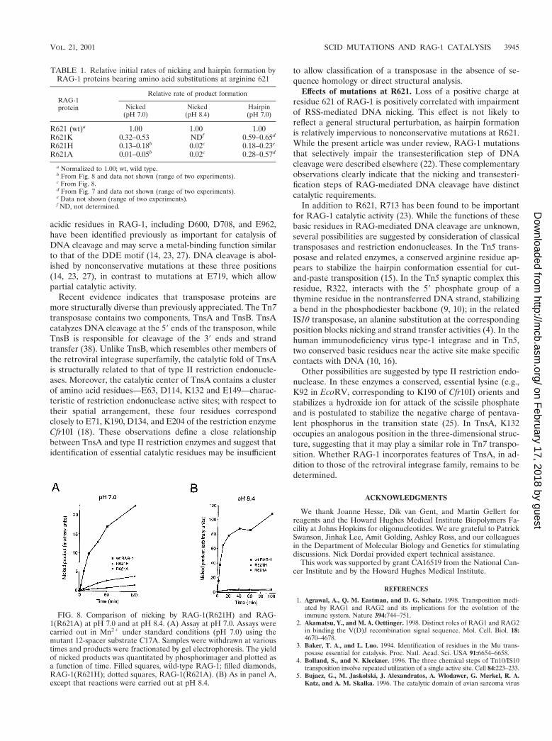
To obtain further support for the participation of a positivelycharged residue at position 621 in the nicking reaction, theactivities of RAG-1(R621H) and RAG-1(R621A) were com-pared at standard pH conditions (pH 7.0), relatively near thepKa of free histidine (' pH 6.5), and at pH 8.4, well above thepKa of free histidine but below that of arginine. At pH 7.0 (Fig.8A and Table 1), the initial reaction rate for RAG-1(R621H)was reduced to between 13 and 18% of wild type, while the ratefor RAG-1(R621A) was about 1 to 5% that of wild type. At pH8.4, however, the initial reaction rates for RAG-1(R621H) andRAG-1(R621A) were apparently identical, both being about2% that of wild-type RAG-1 (Fig. 8B and Table 1). Whileother interpretations are possible, these results suggest that apositively charged side chain at residue 621 participates pref-erentially in the nicking step of DNA cleavage.
DISCUSSION
Participation of RAG-1 in catalysis of nicking and transes-terification. DNA-protein photo-cross-linking had demon-strated that in the presence of RAG-2, RAG-1 approaches thescissile bond at the heptamer-coding junction (13, 32, 44). This
suggested that RAG-1 might play a direct catalytic role in oneor both steps of DNA cleavage, an idea that was borne out bymutational analysis (14, 23, 27). The B-cell-negative SCIDmutations analyzed here, R621H and E719K, also confer bio-chemical defects consistent with direct involvement of RAG-1in catalysis of DNA cleavage. Recombination, accumulation ofsignal ends in vivo, and DNA cleavage in vitro are impaireddespite the ability of the mutant proteins to form precleavagecomplexes that preserve the specific DNA-protein contactsobserved with wild-type protein. Mutations in RAG-1 are alsoassociated with Omenn syndrome, an immunodeficiency dis-order with less severe impairment of lymphocyte developmentthan classical B-cell-negative SCID (51). These mutations, indistinction to those occurring at R621 and E719, exert debili-tating effects on V(D)J recombination by impairing DNA bind-ing or RAG-1–RAG-2 association (51).
Evidence presented here suggests that residue E719 ofRAG-1 may interact with an essential metal ion. The E719Kmutation is associated with a decrease of more than 450-fold inthe initial rate of substrate nicking in vitro relative to that ofthe wild type. This effect is not a particular property of thenonconservative lysine substitution that occurs in SCID pa-tients, as the conservative E719Q mutation impaired RAG-1activity to a similar extent. When cysteine is substituted forE719, the resulting RAG-1 mutant exhibits enhanced activityin Mn21 but 50-fold reduced activity in Mg21, relative to thatof the wild type. The enhanced activity of RAG-1(E719C) inMn21 likely reflects an alteration in metal ion specificity con-ferred by the substitution of thiol for carboxylate, rather thana more general effect, because the activities of neither RAG-1(E719K) nor RAG-1(E719Q) were rescued by Mn21. A sim-ilar, Mn21-specific increase in enzymatic activity is associatedwith cysteine replacement mutations in the catalytic triad ofTnsB (38). Taken together, these results provide evidence thatresidue E719 of RAG-1 interacts with a metal ion essential forDNA cleavage by the V(D)J recombinase.
It is likely that two catalytic regions are present in a RAG-1dimer. These regions could be distributed between the indi-vidual subunits in one of two ways. In the first model, each sitecould be constructed from residues contributed by both sub-units, so that an intact catalytic region would straddle thedimer interface, as in the site-specific recombinase Flp (6). Inthe second model, each of the two catalytic regions wouldreside in a single subunit. Recent observations are consistentwith the latter model and indicate, moreover, that DNA cleav-age is catalyzed in trans by the RAG-1 subunit residing oppo-site the bound subunit (43). The present data do not eliminatethe possibility that RAG-2 also contributes a portion of thecatalytic site. Previous studies have indicated essential roles forRAG-2 in formation of stable RAG-RSS complexes and inpromoting contact between RAG-1 and the heptamer-codingjunction. Whether RAG-2 plays a more direct role in catalysisof DNA cleavage remains an open question.
Comparison to transposases. The initial steps of V(D)J re-combination and transposition are chemically identical, withthe exception that in V(D)J recombination transesterificationoccurs between neighboring DNA strands, while in transposi-tion it occurs between donor and target duplexes (see refer-ence 35 for review). This formal chemical identity is reinforcedby the ability of the RAG proteins to promote the transposi-
VOL. 21, 2001 SCID MUTATIONS AND RAG-1 CATALYSIS 3943
on February 17, 2018 by guest
http://mcb.asm
.org/D
ownloaded from

tion of a donor DNA segment flanked by RSSs into a nonspe-cific target DNA molecule in vitro (1, 21). Thus, RAG-1 andRAG-2 can be considered to represent the subunits of a spe-cialized, multicomponent transposase.
On this basis, the catalytic DDE motif characteristic of clas-
sical transposases, or its functional equivalent, might be ex-pected to occur within one or both RAG proteins. The DDEmotif cannot be reliably assigned on the basis of sequencehomology, however, because it contains only three highly con-served amino acid residues whose spacing is variable. Several
FIG. 7. Impairment of nicking but not transesterification by a nonconservative substitution at R621 of RAG-1. (A) Kinetic analysis of RSSnicking by RAG-1(R621K) and RAG-1(R621A). Lanes 1 to 9, wild-type RAG-1 and RAG-2; lanes 10 to 18, RAG-1(R621K) and RAG-2; lanes19 to 27, RAG-1(R621A) and RAG-2. Assays were carried out in Mn21 as described in Materials and Methods by using the mutant 12-spacersubstrate C17A, which undergoes nicking in the absence of transesterification (29). Samples were withdrawn at times indicated above. The positionof nicked product is indicated by the arrow at left. (B) The yield of nicked products in panel A was quantitated by phosphorimager and plottedas a function of time. Filled squares, wild-type RAG-1; filled diamonds, RAG-1(R621K); dotted squares, RAG-1(R621A). (C) Kinetic analysis ofhairpin formation by RAG-1(R621K) and RAG-1(R621A). Lanes 1 to 8, wild-type RAG-1 and RAG-2; lanes 9 to 16, RAG-1(R621K) and RAG-2;lanes 17 to 24, RAG-1(R621A) and RAG-2. Assays were carried out in Mn21 using a prenicked substrate as described in Materials and Methods.Samples were withdrawn at times indicated above. The positions of hairpin product are indicated at left. (D) The yield of hairpin products in panelC was quantitated by phosphorimager and plotted as a function of time. Symbols are as defined for panel B.
3944 LI ET AL. MOL. CELL. BIOL.
on February 17, 2018 by guest
http://mcb.asm
.org/D
ownloaded from
acidic residues in RAG-1, including D600, D708, and E962,have been identified previously as important for catalysis ofDNA cleavage and may serve a metal-binding function similarto that of the DDE motif (14, 23, 27). DNA cleavage is abol-ished by nonconservative mutations at these three positions(14, 23, 27), in contrast to mutations at E719, which allowpartial catalytic activity.
Recent evidence indicates that transposase proteins aremore structurally diverse than previously appreciated. The Tn7transposase contains two components, TnsA and TnsB. TnsAcatalyzes DNA cleavage at the 59 ends of the transposon, whileTnsB is responsible for cleavage of the 39 ends and strandtransfer (38). Unlike TnsB, which resembles other members ofthe retroviral integrase superfamily, the catalytic fold of TnsAis structurally related to that of type II restriction endonucle-ases. Moreover, the catalytic center of TnsA contains a clusterof amino acid residues—E63, D114, K132 and E149—charac-teristic of restriction endonuclease active sites; with respect totheir spatial arrangement, these four residues correspondclosely to E71, K190, D134, and E204 of the restriction enzymeCfr10I (18). These observations define a close relationshipbetween TnsA and type II restriction enzymes and suggest thatidentification of essential catalytic residues may be insufficient
to allow classification of a transposase in the absence of se-quence homology or direct structural analysis.
Effects of mutations at R621. Loss of a positive charge atresidue 621 of RAG-1 is positively correlated with impairmentof RSS-mediated DNA nicking. This effect is not likely toreflect a general structural perturbation, as hairpin formationis relatively impervious to nonconservative mutations at R621.While the present article was under review, RAG-1 mutationsthat selectively impair the transesterification step of DNAcleavage were described elsewhere (22). These complementaryobservations clearly indicate that the nicking and transesteri-fication steps of RAG-mediated DNA cleavage have distinctcatalytic requirements.
In addition to R621, R713 has been found to be importantfor RAG-1 catalytic activity (23). While the functions of thesebasic residues in RAG-mediated DNA cleavage are unknown,several possibilities are suggested by consideration of classicaltransposases and restriction endonucleases. In the Tn5 trans-posase and related enzymes, a conserved arginine residue ap-pears to stabilize the hairpin conformation essential for cut-and-paste transposition (15). In the Tn5 synaptic complex thisresidue, R322, interacts with the 59 phosphate group of athymine residue in the nontransferred DNA strand, stabilizinga bend in the phosphodiester backbone (9, 10); in the relatedIS10 transposase, an alanine substitution at the correspondingposition blocks nicking and strand transfer activities (4). In thehuman immunodeficiency virus type-1 integrase and in Tn5,two conserved basic residues near the active site make specificcontacts with DNA (10, 16).
Other possibilities are suggested by type II restriction endo-nuclease. In these enzymes a conserved, essential lysine (e.g.,K92 in EcoRV, corresponding to K190 of Cfr10I) orients andstabilizes a hydroxide ion for attack of the scissile phosphateand is postulated to stabilize the negative charge of pentava-lent phosphorus in the transition state (25). In TnsA, K132occupies an analogous position in the three-dimensional struc-ture, suggesting that it may play a similar role in Tn7 transpo-sition. Whether RAG-1 incorporates features of TnsA, in ad-dition to those of the retroviral integrase family, remains to bedetermined.
ACKNOWLEDGMENTS
We thank Joanne Hesse, Dik van Gent, and Martin Gellert forreagents and the Howard Hughes Medical Institute Biopolymers Fa-cility at Johns Hopkins for oligonucleotides. We are grateful to PatrickSwanson, Jinhak Lee, Amit Golding, Ashley Ross, and our colleaguesin the Department of Molecular Biology and Genetics for stimulatingdiscussions. Nick Dordai provided expert technical assistance.
This work was supported by grant CA16519 from the National Can-cer Institute and by the Howard Hughes Medical Institute.
REFERENCES
1. Agrawal, A., Q. M. Eastman, and D. G. Schatz. 1998. Transposition medi-ated by RAG1 and RAG2 and its implications for the evolution of theimmune system. Nature 394:744–751.
2. Akamatsu, Y., and M. A. Oettinger. 1998. Distinct roles of RAG1 and RAG2in binding the V(D)J recombination signal sequence. Mol. Cell. Biol. 18:4670–4678.
3. Baker, T. A., and L. Luo. 1994. Identification of residues in the Mu trans-posase essential for catalysis. Proc. Natl. Acad. Sci. USA 91:6654–6658.
4. Bolland, S., and N. Kleckner. 1996. The three chemical steps of Tn10/IS10transposition involve repeated utilization of a single active site. Cell 84:223–233.
5. Bujacz, G., M. Jaskolski, J. Alexandratos, A. Wlodawer, G. Merkel, R. A.Katz, and A. M. Skalka. 1996. The catalytic domain of avian sarcoma virus
FIG. 8. Comparison of nicking by RAG-1(R621H) and RAG-1(R621A) at pH 7.0 and at pH 8.4. (A) Assay at pH 7.0. Assays werecarried out in Mn21 under standard conditions (pH 7.0) using themutant 12-spacer substrate C17A. Samples were withdrawn at varioustimes and products were fractionated by gel electrophoresis. The yieldof nicked products was quantitated by phosphorimager and plotted asa function of time. Filled squares, wild-type RAG-1; filled diamonds,RAG-1(R621H); dotted squares, RAG-1(R621A). (B) As in panel A,except that reactions were carried out at pH 8.4.
TABLE 1. Relative initial rates of nicking and hairpin formation byRAG-1 proteins bearing amino acid substitutions at arginine 621
RAG-1protein
Relative rate of product formation
Nicked(pH 7.0)
Nicked(pH 8.4)
Hairpin(pH 7.0)
R621 (wt)a 1.00 1.00 1.00R621K 0.32–0.53 NDf 0.59–0.65d
R621H 0.13–0.18b 0.02c 0.18–0.23e
R621A 0.01–0.05b 0.02c 0.28–0.57d
a Normalized to 1.00; wt, wild type.b From Fig. 8 and data not shown (range of two experiments).c From Fig. 8.d From Fig. 7 and data not shown (range of two experiments).e Data not shown (range of two experiments).f ND, not determined.
VOL. 21, 2001 SCID MUTATIONS AND RAG-1 CATALYSIS 3945
on February 17, 2018 by guest
http://mcb.asm
.org/D
ownloaded from

integrase: conformation of the active-site residues in the presence of divalentcations. Structure 4:89–96.
6. Chen, J. W., J. Lee, and M. Jayaram. 1992. DNA cleavage in trans by theactive site tyrosine during Flp recombination: switching protein partnersbefore exchanging strands. Cell 69:647–658.
7. Craig, N. L. 1996. Transposon Tn7. Curr. Top. Microbiol. Immunol. 204:27–48.8. Dahm, S. C., and O. C. Uhlenbeck. 1991. Role of divalent metal ions in the
hammerhead RNA cleavage reaction. Biochemistry 30:9464–9469.9. Davies, D. R., L. M. Braam, W. S. Reznikoff, and I. Rayment. 1999. The
three-dimensional structure of a Tn5 transposase-related protein deter-mined to 2.9-A resolution. J. Biol. Chem. 274:11904–11913.
10. Davies, D. R., I. Y. Goryshin, W. S. Reznikoff, and I. Rayment. 2000. Three-dimensional structure of the Tn5 synaptic complex transposition intermedi-ate. Science 289:77–85.
11. Difilippantonio, M. J., C. J. McMahan, Q. M. Eastman, E. Spanopoulou,and D. G. Schatz. 1996. RAG1 mediates signal sequence recognition andrecruitment of RAG2 in V(D)J recombination. Cell 87:253–262.
12. Dyda, F., A. B. Hickman, T. M. Jenkins, A. Engelman, R. Craigie, and D. R.Davies. 1994. Crystal structure of the catalytic domain of HIV-1 integrase:similarity to other polynucleotidyl transferases. Science 266:1981–1986.
13. Eastman, Q. M., I. J. Villey, and D. G. Schatz. 1999. Detection of RAGprotein-V(D)J recombination signal interactions near the site of DNA cleav-age by UV cross-linking. Mol. Cell. Biol. 19:3788–3797.
14. Fugmann, S. D., I. J. Villey, L. M. Ptaszek, and D. G. Schatz. 2000. Identi-fication of two catalytic residues in RAG1 that define a single active sitewithin the RAG1/RAG2 protein complex. Mol. Cell 5:97–107.
15. Haren, L., B. Ton-Hoang, and M. Chandler. 1999. Integrating DNA: trans-posases and retroviral integrases. Annu. Rev. Microbiol. 53:245–281.
16. Hazuda, D. J., P. Felock, M. Witmer, A. Wolfe, K. Stillmock, J. A. Grobler,A. Espeseth, L. Gabryelski, W. Schleif, C. Blau, and M. D. Miller. 2000.Inhibitors of strand transfer that prevent integration and inhibit HIV-1replication in cells. Science 287:646–650.
17. Hesse, J., M. Lieber, M. Gellert, and K. Mizuuchi. 1987. ExtrachromosomalDNA substrates in pre-B cells undergo inversion or deletion at immuno-globulin V(D)J joining signals. Cell 49:775–783.
18. Hickman, A. B., Y. Li, S. V. Mathew, E. W. May, N. L. Craig, and F. Dyda.2000. Unexpected structural diversity in DNA recombination: the restrictionendonuclease connection. Mol. Cell 5:1025–1034.
19. Hiom, K., and M. Gellert. 1998. Assembly of a 12/23 paired signal complex:a critical control point in V(D)J recombination. Mol. Cell 1:1011–1019.
20. Hiom, K., and M. Gellert. 1997. A stable RAG1-RAG2-DNA complex thatis active in V(D)J cleavage. Cell 88:65–72.
21. Hiom, K., M. Melek, and M. Gellert. 1998. DNA transposition by the RAG1and RAG2 proteins: a possible source of oncogenic translocations. Cell94:463–470.
22. Kale, S. B., M. A. Landree, and D. B. Roth. 2001. Conditional RAG-1mutants block the hairpin step of V(D)J recombination. Mol. Cell. Biol.21:459–466.
23. Kim, D. R., Y. Dai, C. L. Mundy, W. Yang, and M. A. Oettinger. 1999.Mutations of acidic residues in RAG1 define the active site of the V(D)Jrecombinase. Genes Dev. 13:3070–3080.
24. Kim, K., S. Y. Namgoong, M. Jayaram, and R. M. Harshey. 1995. Step-arrestmutants of phage Mu transposase. Implications in DNA-protein assembly,Mu end cleavage, and strand transfer. J. Biol. Chem. 270:1472–1479.
25. Kovall, R. A., and B. W. Matthews. 1999. Type II restriction endonucleases:structural, functional and evolutionary relationships. Curr. Opin. Chem.Biol. 3:578–583.
26. Kung, H. C., and P. H. Bolton. 1997. Structure of a duplex DNA containinga thymine glycol residue in solution. J. Biol. Chem. 272:9227–9236.
27. Landree, M. A., J. A. Wibbenmeyer, and D. B. Roth. 1999. Mutationalanalysis of RAG1 and RAG2 identifies three catalytic amino acids in RAG1critical for both cleavage steps of V(D)J recombination. Genes Dev. 13:3059–3069.
28. Lewis, S. 1994. The mechanism of V(D)J joining: lessons from molecular,immunological and comparative analyses. Adv. Immunol. 56:27–150.
29. Li, W., P. Swanson, and S. Desiderio. 1997. RAG-1- and RAG-2-dependent
assembly of functional complexes with V(D)J recombination substrates insolution. Mol. Cell. Biol. 17:6932–6939.
30. Li, Z., D. I. Dordai, J. Lee, and S. Desiderio. 1996. A conserved degradationsignal regulates RAG-2 accumulation during cell division and links V(D)Jrecombination to the cell cycle. Immunity 5:575–589.
31. McBlane, J. F., D. C. van Gent, D. A. Ramsden, C. Romeo, C. A. Cuomo, M.Gellert, and M. A. Oettinger. 1995. Cleavage at a V(D)J recombinationsignal requires only RAG1 and RAG2 proteins and occurs in two steps. Cell83:387–395.
32. Mo, X., T. Bailin, and M. Sadofsky. 1999. RAG-1 and RAG-2 cooperate inspecific binding to the recombination signal sequence in vitro. J. Biol. Chem.274:7025–7031.
33. Oettinger, M. A., D. G. Schatz, C. Gorka, and D. Baltimore. 1990. RAG-1and RAG-2, adjacent genes that synergistically activate V(D)J recombina-tion. Science 248:1517–1523.
34. Piccirilli, J. A., J. S. Vyle, M. H. Caruthers, and T. R. Cech. 1993. Metal ioncatalysis in the Tetrahymena ribozyme reaction. Nature 361:85–88.
35. Polard, P., and M. Chandler. 1995. Bacterial transposases and retroviralintegrases. Mol. Microbiol. 15:13–23.
36. Rice, P., and K. Mizuuchi. 1995. Structure of the bacteriophage Mu trans-posase core: a common structural motif for DNA transposition and retroviralintegration. Cell 82:209–220.
37. Roth, D. B., and N. L. Craig. 1998. VDJ recombination: a transposase goesto work. Cell 94:411–414.
38. Sarnovsky, R. J., E. W. May, and N. L. Craig. 1996. The Tn7 transposase isa heteromeric complex in which DNA breakage and joining activities aredistributed between different gene products. EMBO J. 15:6348–6361.
39. Schatz, D. G., M. A. Oettinger, and D. Baltimore. 1989. The V(D)J recom-bination activating gene, RAG-1. Cell 59:1035–1048.
40. Schwarz, K., G. H. Gauss, L. Ludwig, U. Pannicke, Z. Li, D. Lindner, W.Friedrich, R. A. Seger, T. E. Hansen-Hagge, S. Desiderio, M. R. Lieber, andC. R. Bartram. 1996. RAG mutations in human B cell-negative SCID.Science 274:97–99.
41. Siebenlist, U., and W. Gilbert. 1980. Contacts between Escherichia coli RNApolymerase and an early promoter of phage T7. Proc. Natl. Acad. Sci. USA77:122–126.
42. Spanopoulou, E., F. Zaitseva, F.-H. Wang, S. Santagata, D. Baltimore, andG. Panayotou. 1996. The homeodomain region of Rag-1 reveals the parallelmechanisms of bacterial and V(D)J recombination. Cell 87:263–276.
43. Swanson, P. C. 2001. The DDE motif in RAG-1 is contributed in trans to asingle active site that catalyzes the nicking and transesterification steps ofV(D)J recombination. Mol. Cell. Biol. 21:449–458.
44. Swanson, P. C., and S. Desiderio. 1999. RAG-2 promotes heptamer occu-pancy by RAG-1 in the assembly of a V(D)J initiation complex. Mol. Cell.Biol. 19:3674–3683.
45. Swanson, P. C., and S. Desiderio. 1998. V(D)J recombination signal recog-nition: distinct, overlapping DNA-protein contacts in complexes containingRAG1 with and without RAG2. Immunity 9:115–125.
46. Truss, M., G. Chalepakis, and M. Beato. 1990. Contacts between steroidhormone receptors and thymines in DNA: an interference method. Proc.Natl. Acad. Sci. USA 87:7180–7184.
47. van Gent, D. C., K. Hiom, T. T. Paull, and M. Gellert. 1997. Stimulation ofV(D)J cleavage by high mobility group proteins. EMBO J. 16:2665–2670.
48. van Gent, D. C., J. F. McBlane, D. A. Ramsden, M. J. Sadofsky, J. E. Hesse,and M. Gellert. 1995. Initiation of V(D)J recombination in a cell-free system.Cell 81:925–934.
49. van Gent, D. C., K. Mizuuchi, and M. Gellert. 1996. Similarities betweeninitiation of V(D)J recombination and retroviral integration. Science 271:1592–1594.
50. van Gent, D. C., D. A. Ramsden, and M. Gellert. 1996. The RAG1 andRAG2 proteins establish the 12/23 rule in V(D)J recombination. Cell 85:107–113.
51. Villa, A., S. Santagata, F. Bozzi, S. Giliani, A. Frattini, L. Imberti, L. B.Gatta, H. D. Ochs, K. Schwarz, L. D. Notarangelo, P. Vezzoni, and E.Spanopoulou. 1998. Partial V(D)J recombination activity leads to Omennsyndrome. Cell 93:885–896.
3946 LI ET AL. MOL. CELL. BIOL.
on February 17, 2018 by guest
http://mcb.asm
.org/D
ownloaded from