Embed Size (px)
Citation preview

ILLEGITIMATE V(D)J REARRANGEMENT IN y-IRRADIATION INDUCED
T CELL LYMPHOMA IN NEWBORN scid MICE
Danny Vesprini
A thesis submitted in conformity with the requirements
for the degree of Master of Science
Graduate Department of ImmunoIogy
University of Toronto
O Copyright by Danny Vesprini (1997)

Acquisitions and Acquisitions et Bibliographic Services services bibliographiques
395 Wellington Streel 395, rue Wellington Ottawa ON K I A ON4 Ottawa ON KI A ON4 Canada Canada
Your file Votre rélaience
Our file Noira raldrerice
The author has granted a non- L'auteur a accordé une licence non exclusive licence allowing the exclusive pennettant à la National Library of Canada to Bibliothèque nationale du Canada de reproduce, loan, distribute or sel1 reproduire, prêter, distribuer ou copies of this thesis in microfom, vendre des copies de cette thèse sous paper or electronic formats. la forme de microfiche/film, de
reproduction sur papier ou sur format électronique.
The author retains ownership of the L'auteur conserve la propriété du copyright in this thesis. Neither the droit d'auteur qui protège cette thèse. thesis nor substantial extracts fiom it Ni la thèse ni des extraits substantiels may be printed or othenvise de celle-ci ne doivent être imprimés reproduced without the author's ou autrement reproduits sans son permission. autorisation.

1 would like to thank my supervisor, Jayne Danska, for her guidance,
understanding and friendship. 1 would also Iike to thank the rnembers of my
supervisory committee, Dr. Neil Berinstein, Dr. Susanna Lewis and Dr. Bob
PhilIips, who al1 provided excellent counsel throughout my graduate school
experience. Thanks to the mernbers of the Danska lab (Priscilla Chiu, Casey
Fox, Ildiko Grandal, Christine Williams, Kelly Williams), as well as Cynthia
Guidos, for their support and friendship. Finally, 1 would like to thank my
friends and family, who were always there when 1 needed them.
Dedication
This thesis is dedicated to the memory of grandrna Tilly Vesprini.
"Be good to the people."

No te:
The experiments presented in the first section of results, the RAG-2-/-scid
analysis, were done in collaboration with Christine Williams. The RAG2-/-
scid mice were bred by IIdiko Grandal at the HospitaI for Sick Children.
Fluorescent in situ hybridization was performed by the author under the
direction of Dr Barbara Beatty of the CGAT FISH mapping center, at the
Hospital for Sick Children.
Table of Contents
LIST OF TABLES
LIST OF FIGURES
ABSTRACT
BACKGROUND
V(D)J recombination and DNA repair
Role of V(D)J recombination in lymphocyte development
INTRODUCTION
The irradiated newborn scid mouse model
Mechanisms of lymphomagenesis
Tllegitimate V(D) J rearrangement in lymphoid tumours
The irradiated RAG-deficient mouse model
MATERIALS AND METHODS
DNA probes
Ce11 lines
Mice
Fluorescence activated ce11 sorting
Radiolabeling of DNA probes
RNA extraction
Northern analysis
Reverse transcriptase coupled polymerase c h a h reaction

DNA sequencing
Genomic DNA extraction
Southern blot analysis
Quantitative phophorimaging
Fluorescence in situ hybridization
Pulsed field gel electrophoresis
RESULTS
(A) Effects of low-dose ionizing radiation on RAG-2 deficient scid
mice
(3) Characterization of scid CD4' CD8' T ce11 lymphoma Iines
(C) Expression of TcRP genes in irradiated newborn scid DP T ce11
lines
(D) Rearrangement at the TcRrj locus in irradiated newborn scid
DP T ce11 lines
(E) TcR13 variable gene rearrangernent in irradiated newborn scid
DP T ce11 lines
(F) Physical integrity of the TcRP locus in irradiated newborn scid
DP T ce11 lines
DISCUSSION
(A) V(D)J rearrangement plays a role in rapid onset lymphoma in
irradiated newborn scid rnice
(B) Detection of abnormal rearrangement at the TcRCj locus in
irradiated newborn scid rnice
(Cl Expression of truncated TcRP chains: a role in lymphomagenesis?
(D) The TcRP locus is not involved in a gross reciprocal translocation
in the irradiated newborn scid T ce11 Iines 5 8
(E) TcR(3 locus disruption in irradiated newborn scid T ce11 lines 5 9
(F) Chrornosomal aberrations associated with abnorrnal TcRf3 locus
rearrangement
(G) Conclusions
TABLES 64
FIGURES 6 9
REFERENCES 119

LIST OF TABLES
Table 1 - Oligonucleotides used in this study 64
Table 2 - Effects of low-dose ionizing radiation on newborn RAG2 65
deficient scid mice.
Table 3 - Summary of FISH analysis of IRNB-SCID DP T ce11 lines, using 66
mouse chromosome 6 paint
Table 4 - Summary of PFGE analysis of IRNB-SCID DP T ce11 lines 67
TabIe 5 - Summary of restriction fragment mapping of constant region in 68
IRNB-SCID DP T ce11 lines

LlST OF FIGURES
Figure 1 - The constant region of the murine TcR(3 locus
Figure 2 - Thymocyte expansion and development in irradiated newborn
RAG2 deficient scid mice
Figure 3 - Phenotypic analysis of T lymphocyte development in
irradiated newborn RAG2 deficient scid mice
Figure 4 - Northern blot analysis of TcR expression in irradiated
newborn scid T ce11 lines
Figure 5 - RT-PCR strategy for amplification of TcRP mRNA
Figure 6 - TcRCj expression in irradiated newborn scid DP T ce11 lines
Figure 7 - DNA sequence analysis of TcRP transcripts in the scid DP
T ce11 line, SCTL2
Figure 8 - TcRP protein expression in the scid DP T ce11 line SCTL2
Figure 9 - Analysis of TcRCj gene rearrangments in the constant region
Figure 10 - Analysis of TcR CP1 cluster rearrangements
Figure 11 - Analysis of TcR JP2 cluster rearrangements
Figure 12 - Quantitative phosphorimaging
Figure 13 - Quantitative phosphorimaging of the TcR Jp2 gene cluster in
scid DP T ce11 lines
Figure 14 - Quantitative phosphorimaging of the TcR C(32 gene in
scid DP T ce11 lines
Figure 15 - Analysis of TcRCj locus rearrangement
Figure 16 - The murine TcRP chain locus
Figure 17 - Analysis of TcR VCj gene deletion
Figure 18 - Quantitative phosphorimaging of TcR V(3 genes in
scid DP T ce11 lines

Figure 19 - Fluorescent In Situ Hybridization
Figure 20 - Pulsed field gel electrophoresis strategy
Figure 21 - Pulsed field gel electrophoreçis anaiysis of the TcRP locus
in scid DP T ce11 lines
Figure 22 - Restriction maps of the TcR(3 constant region in scid DP
T ce11 lines

--- - O-----.--- . ,-,, *.-..----. "' , "'--'-"-'. ".-+.'-bv ' U L L * -,"'y"""'" "'
Newborn scid Mice, Master of Science, 1997, Danny Vesprini, Department of Imunology, University of Toronto
Abstract
The production of DNA doubie strand breaks during V(D)J recombination, makec
developing lymphocytes particularIy susceptible to neoplasia. Severe combined
immunodeficient (scid) mice inefficiently ligate free coding ends generated during
V(D)J recombination due to a mutation in the catalytic subunit of DNA-dependent
protein kinase (DNA-PK,,). Treatment of newborn scid mice with low dose (100
cGy) y-irradiation restores V(D)J rearrangement at multiple TcR loci and results in
the universal development of T ce11 lymphoma. In this study, we used a genetic
approach to determine that V(D)J rearrangement played an obligate role in
radiation-induced lymphoma in scid mice. We also present evidence of abnormal
V(D)J rearrangement in irradiated newborn scid (IRNB-SCIDJ T ce11 lines. Our
results collectively indicate that sequence specific breaks made during V(D)J
rearrangement are essential for radiation-induced lymphomagenesis in scid mice,
and that mutations accumulated during attempted TcRP locus rearrangement may
contribute to the invariant outgrowth of thymic Iymphoma.

Background
V(D1T RECOMBINATION AND DNA REPAIR
Clonally distinct T and B ce11 antigen receptors are generated from the joining
of dispersed variable (V), diversity (D), and lpining (J) gene segments, in a highly
ordered, multi-step process termed V(D)J recombination (reviewed in 1, 2).
Recombination signal Zquences (RSS], which flank recombinationally active gene -
segments, target the recombinase machinery to the antigen receptor Ioci. RSSs
consist of conserved heptamer and nonamer motifs separated by a non-conserved
spacer sequence of either 1 2 or 23 bp (3). Recognition of RSS is followed by the
introduction of double granded breaks (DSB) between the RSS and coding
sequences, generating two sets of DNA termini: coding ends and signal ends (2 ) . Co-
expression of the lymphoid specific, recombinase gtivating genes, rag-l and rag-2, is
required for V(D)J recombination, and RAG-l/RAG-2 protein expression in non-
lymphoid cells is sufficient to support recombination of extrachromosomal V[D)J
substrates (4). Moreover, targeted gene disruption of either rag-1 or rag-2 prevents
V(D)J recombination (5, 6). Consistent with these observations, the RAG-1 and
RAG-2 proteins have been determined to be both necessary and sufficient for
recognition of RSS, introduction of DSBs and formation of hairpin terminated
V(D)J coding ends (7, 8)
Ligation of free DNA termini leads to the creation of coding joints and signal
joints (2). Depending on the orientation of the RSSs, recombination results in the
inversion or deletion of the intervening DNA sequence. Prior to iigation, coding
ends are often subject to i~ucleotide deletion and/or addition (reviewed in 1, 9). T h e
addition of non-templated (N) nucleotides at coding junctions is catalyzed by the
lymphocyte-specific enzyme terminal deoxynucleotidyl &ansferase (TdT] (10-12).
Though not an obligate participant in the V(D)J recombination process, Tdî
contributes diversification to the germ-line (11, 12). Coding joins may also conta i r
palindromic [P) nucleotides, which are thought to arise during the resolution O
DNA-hairpin intermediates (13, 14). In contrast to coding ends, nucleotide additior
or loss at signal ends is rare (15). The asymmetry of these reaction products suggesti
that coding ends and signal ends are processed differently.
The crucial role of DSB repair in V(D)J recombination was revealed by studiei
of mice harbouring the zevere ombined immuno&ficiency (scid) mutation. Ir
these mice, failure to productively rearrange the antigen receptor genes results i r
the lack of mature T and B lymphocytes (16). The scid V(D)J recombination defec
results from a deficiency in DSB repair activity (17-20) which is required for tht
efficient joining of coding segments (15, 21, 22). In scid lymphocyte precursors, DSI
are introduced at RSS, (13) but coding ends are ligated inefficiently. Interestingly
though RSS ligation is essentially normal in scid mice (15, 23, 24), coding join
formation has been measured to occur at a frequency of approximately 10-100 time!
less than WT (25). The "leakiness" of the scid mutation is illustrated by the fact tha
rare normal V(D)J coding joints can be isolated from the lymphocytes of aged scil
mice (25-28). Moreover, coding joints analysed in scid rnice often displaj
characteristic structural abnormalities. For example, Iong P nucleotides are ofter
seen in the rare coding joints from "leaky" scid (29, 30). In spontaneous scid thymic
lymphomas and Abelson murine leukemia-virus transforrned scid bone marrov
cells, loss of DNA sequence due to nuclease digestion or illegitimate rearrangemen
has been observed (21, 22, 31, 32). Therefore, although the scid mutation confers i
that V(D)J rearrangement can occur in scil DSB repair defect, it appears
lymphocytes.
In addition to impairing
defective DSB repair in al1 ce11
V(D)J recombination, the scid mutation cause!
lineages, resulting in hypersensitivity to ionizinl
radiation and other agents that induce DSB (17-19). The scid mutation has been
mapped to the gene encoding the catalytic subunit of the DNA-dependent protein
kinase (DNA-PK, (33-35), which is a 460 kDa member of the phosphatidylinositol -
(PI)-3 serine/threonine kinase family (36). DNA-PK, is active when bound to DNA,
and this is mediated by the Ku nuclear cornplex, a heterodimer of 80 and 70 kDa (37).
The effects of the scid mutation implicate a crucial role for DNA-PK, in both DSB
repair and V(D)J recombination. Though the precise role of DNA-PK in DSB repair
is not defined, it has been suggested that DNA-PK phosphorylates downstream
target molecules in response to DSBs (38). Compatible with this suggestion, in vitro
studies have shown that several DNA binding proteins, including the transcription
factors SPI, c-JUN, c-FOS and OCT1, act as substrates for DNA-PK (39). Therefore,
the DSB repair defect in scid mice rnaybe due to the inability to effectively respond to
DNA lesions.
Until recently, the exact nature of the scid mutation was unknown. The
leakiness of the scid mutation suggested that the DSB repair defect was not due to a
nul1 mutation. Recent work dorte in Our lab, in collaboration with Cynthia Guidos,
supports the hypothesis that the scid phenotype is a result of abnormal co-
ordination or regulation of DSB repair (40), as opposed to the absence of DNA-PK
activity, which had been suggested by others (33, 35, 41). DNA sequence analysis of
the C-terminal kinase domain of DNA-PK,, identified a single base pair substitution,
which resulted in a premature ochre stop codon (40, 42). This mutation is predicted
to truncate DNA-PK, by approximately 8 kDa, but leaves intact the motifs thought
to be required for kinase activity (40). It was also discovered that expression of DNA-
PK, in scid cells was reduced 10-fold relative to that in WT thymocytes. Moreover,
the scid mutation apparently disturbs DNA-PK, nuclear localization, which is
presumably required for its enzymatic activity (40). Taken together, these data

suggest that the scid phenotype results from inefficient DSB repair, rather than the
absence of this activity.
ROLE OF VID)! RECOMBINATION IN LYMPHOCYTE DEVELOPMENT
The successful rearrangement of the immunoglobulin heavy chain (Igp) and
T ce11 ~eceptor beta chain (TcRB) play important roles in B and T ce11 development, - -
respectively. Bone marrow derived progenitor cells which migrate to the thymus
do not express CD4 or CD8 CO-receptors, and are referred to as CO-receptor double
negative (DN) cells. Within the DN cornpartment there is a maturational -
progression defined by the expression of the IL2 receptor alpha chain (CD25) and the
phagocytic glycoprotein 1 (CD44). Rearrangement at the TcR(3 locus occurs as the
CD25+ DN cells down-regulate CD44 expression (43, 44). Cells expressing a
successfully rearranged TcRP chain, probably paired with a non-rearranged pre-Ta
chain (45), proliferate extensively, downregulate CD25 expression, upregulate CD4
and CD8 CO-receptors to become double positive (DP), and initiate rearrangement at
the TcRa chain (46). DP thymocytes expressing low levels of the TcRaP heterodimer
undergo maturation into CD4+ or CD8+ single positive (SP) cells which express
high levels of the TcRaP heterodimer. Similarly, in B ce11 development, the
successful rearrangernent and expression of Igp chain, complexed with A5 and
VpreB proteins, mediates signals for developmental maturation of B220f CD43- pre
B cells from B220+ CD43+ large pro-B ce11 precursors (471, and promotes
rearrangement of the Ig light chain loci (48-50). Mature B cells are produced when
rearranged Ig light chains are paired with heavy chains and expressed at the ceIl
surface (51).
Rearrangement at antigen receptor loci is developmentally regulated. For
example, in T cells, rearrangernent at the TcRlj locus precedes that of TcRa in

normal developing cells (52, 53)., The TcRCj locus consists of at least 22 V gene
se,aments and a constant region that contains two tandernly arrayed clusters of
genes. The closely linked CP1 and CF2 are each preceded by a cluster of six
functional JP gene segments, one non-functional (pseudo, +) JP gene segment, and
one D(3 gene segment (Fig 1). At the TcR(3 locus, the rearrangement of these
originally separated gene segments occurs in a developmentally ordered fashion, as
joining of D to J is followed by that of V to DJ (54, 55). Consistent with this, DJ
rearrangements at the TcRP locus are seen in mice by day 14 of gestation (55, 56).
Moreover, during murine embryogenesis, an ordered program of TcR gene
expression takes place in the thymus; TcRy, 6 and fl gene expression occurs between
days 14 and 16 of gestation, whereas TcRa gene expression is delayed until day 17.
These observations demonstrated the tight coupling of V(D)J rearrangement with
development, and in particular, implicated a role for TcRCj locus rearrangement in T
ce11 developrnent.
As previously stated, mice bearing the scid mutation have a DSB repair defect
which affects al1 ce11 lineages (17-20). During V(D)J recombination in scid mice, RSS-
mediated DSB are initiated, but coding ends are unable to join efficiently (15). Mice
with a targeted disruption of either the rag-1 o r rag-2 genes are unable to initiate
DSB at RSS, and their antigen receptor genes remain in germ-line (5, 6).
Lymphocyte development in both scid and RAG-deficient mice is therefore arrested
at a similar stage: T ceIl maturation is arrested at the CD44- CD25' DN stage, while B
ce11 development is arrested at the B220f CD43" large pro B ce11 (5,6). Introducing a
functionatly rearranged TcR13 or lgp transgene into RAG-deficient or scid mice
promotes the development and expansion of DP thymocytes (53, 57, 58) or pre-B
cells (59, 60), confirming that successful rearrangement of these antigen receptor
genes provides essential developmental and proliferative signals in T and B ce11
development.

Introduction
THE IRRADIATED NEWBORN scid MOUSE MODEL
Our iab has studied the effects of DNA damage on T ce11 development and 1
ce11 lymphomagenesis in scid mice. Exposure of newborn scid mice to 100 cGy O
ionizing radiation relieved the block in thymocyte development [61, 62) anc
restored the production of highly diverse, in-frame TcRP chains (61). Surprisingly
the developmental block in B ce11 maturation was unaffected by radiation treatmeni
and productive Igp rearrangement was not detected. TcRrj coding joins frorr
irradiated aewbprn çcid mice (IRNB-SCID) displayed normal fine structure anc
lacked anomalies characteristic of "leaky" scid antigen receptors (25-28)
Furthermore, restoration of V(D)J rearrangement was not specific to the TcR(3 locus
as normal rearrangement at the TcRG (63) and TcRy locus (64) was also detected ir
IRNB-SCID mice. Collectively, these results revealed that irradiation of newborr
scid mice induces transient rescue of V[D)J rearrangement, leading to productive
TcR rearrangement and thymocyte maturation to the DP stage.
At four to five months post-irradiation, al1 IRNB-SCID mice displayed DF
thymic lymphoma. This invariant progression to lymphoma suggests that growtl-
promoting mutations occurred in some of the rescued DP cells following
irradiation. Interestingly, no tumours of other ce11 lineages have been detected ir
the IRNB-SCID mice even though the scid mutation affects DSB repair in al1 tissues
Taken together, these results suggest that irradiation induced rescue of V(D)]
recombination in T ce11 precursors which harbour a defect in DSB repair ma)
enhance the generation of chromosomal aberrations which promote the invarian!
development of thymic lymphoma. We hypothesize that y-irradiation inducec

restoration of V(D)J rearrangement leads to an increased frequency of illegitimat
rearrangement events at the TcRP locus, a common feature of lymphocyti
malignancies (65).
MECHANISMS OF LYMPHOMAGENESIS
Multiple doses of sub-lethal y-irradiation repcated at regular intervals (split
dose) has been shown to produce thymic lymphoma in susceptible mouse strain
(66). This irradiation treatment activates murine leukemia retro-viruses which ac
as insertional mutagens (66-70). Integrated retroviruses can activate flankinl
cellular genes through several mechanisms: 1) promoting transcription from th1
viral promoter, 2) affecting expression of adjacent cellular genes, 3) disrupting cis
acting elements, or, 4) integrating within a transcription unit, thus altering th1
structure and function of the encoded protein. Through searching for genomil
DNA rearrangements, novel murine leukemia virus integration events were no
detected in irradiation induced scid thymomas (Jolicoeur and Danska, unpublishec
observations). Activation of murine leukemja viruses therefore do not appear tc
account for the invariable development of T ce11 lymphoma in IRNB-SCID mice.
A recurring theme in the molecular pathogenesis of hematopoietil
malignancy is activation of cellular proto-oncogenes by chromosomal aberration:
the most frequent of which are chromosomaL translocations. Analyses O
chromosomal translocations in human Iymphoid neoplasias have shown that thest
aberrations fa11 into two categories. In the first, the chromosomal translocatioi
causes the formation of a novel transcriptional unit derived from the fusion O
genes at the breakpoint sites (fusion transcripts). A chimeric gene found in bot1
human chronic m_yelogeneous leukemia (CML) and acute lymphoblastic leukemi
(ALL) is the BCR-ABL fusion gene (71). The resultant fusion protein has enhance<
tyrosine kinase activity and tumourigenic properties (72). Another example of
chimeric gene creation involves a translocation present in B ce11 ALL, where the
basic-helix-loop-helix transactivation domain of the transcriptional regulator E2A, is
fused to the carboxy-terminal portion of PBXI, which contains a DNA-binding
homeodomain (73, 74). In translocations such as these, fusion transcript formation
leads to chimeric proteins displaying novel biochemical properties distinct from
those of the wild-type proteins.
In the second category of aberrations, reciprocal translocation juxtaposes TcR
or Ig loci to a gene from another chromosome. In T cells, this ectopic localization
causes deregulated expression of the translocated gene, driven by powerful
enhancers present in the T ce11 receptor loci (reviewed in 75). The same mechanism
occurs in B cells. For example, in Burkitt's lymphoma, c-myc is translocated to the
Ig heavy chain locus, resulting in the activation of this proto-oncogene (76-78). The
TcRP locus is also a frequent site for chromosomal translocations in lymphoid
cancers. One such translocation juxtaposes the gene encoding the protein tyrosine
kinase p561~k (lck) and the TcRP locus, which causes lck mRNA levels to become
significantly elevated (79). Therefore, in translocations involving antigen receptor
loci, the predominant biological consequence is transcriptional deregulation of the
juxtaposed gene, resulting in inappropriate levels of the gene product cornpared to
normal cells. Given the prevalence of chromosomal rearrangements at antigen
receptor loci in lymphoid mahgnancies, and Our detection of y-irradiation induced
rescue of V(D)J rearrangement in scid thymocytes, illegitimate rearrangernent of the
TcRCj locus could likely play a role in the universal development of T cell
lymphoma in IRNB-SCID mice.
ILLEGITIMATE V(D11 REARRANGEMENT IN LYMPHOID TUMOURS
Much evidence implicates illegitimate V(D)J recombination in transloca tionz
involving the antigen receptor loci. Evidence for V(D)J recombinase mediatec
translocations can be seen through examination of the translocation breakpoints. Ir
many cases, DNA sequences that resemble the heptamer and/or nonarner RSS arc
found on the non-antigen receptor locus partner of the translocation (80-85)
Though these 'cryptic' RSS differ from the consensus RSS, they may be able tc
mediate RAG dependent DSB, as a recent study showed that RSS elements that have
significant sequence or structural variation still serve as recognition sites for th^
M G proteins (86). The observation of cryptic RSS at translocation breakpoints
therefore supports the view that abnormal recombination results frorn mistakes i n
the process of recombinase mediated joining.
Some characteristics of V(D)J recombinase mediated translocation breakpoints
are non-templated 'Nt region nucleotide additions (80-82, 84, 85, 87, 88), palindromic
'Pt sequence additions (891, and evidence of exonuclease activity (80, 89, 90). In
multiple reported cases, one of the products of translocations involving illegitimatc
rearrangement is not modfied, much like a signal-join, while the reciprocal produci
manifests coding-join-like modifications (79, 82, 85). The breakpoint characteristics
mentioned above are not restricted to translocations involving Ig or TcR loci, bu1
have also been observed in a recurrent site-specific deletion in T-ALL ce11 lines, thai
fuses two genes, scl and si1 (83). Normally, the expression of scl in hematopoietic
tissues is heterogeneous: it is expressed in cells with stem ce11 attributes but i:
consistently absent in thymocytes and mature cells (91). scl is ectopically expressec
as a result of juxtaposition to the si1 gene, which is constitutively active ir
thymocytes (92). The recombination event that produces the scl-si1 fusion display:
features suggesting that it is mediated by the action of the V(D)J recombinasc

machinery. These observations clearly implicate illegitimate V(D)J rearrangement
as being a common contributor to the development of human lymphoid neoplasias.
THE IRRADIATED RAG-DEFICIENT MOUSE MODEL
The rescue of DP T ce11 development in irradiated scid mice was associated
with TcRP rearrangement and expression (61). It remained possible that the DSB
repair defect caused by the scid mutation also promoted T ce11 development, which
was independent of TcRP expression. To determine the effect of irradiation on the
development of T cells in the absence of TcRrj expression, Our lab in collaboration
with Cynthia Guidos, treated RAG-1 and RAG-2 deficient mice with y-irradiation. A
single, sublethal dose (750 cGy adults, 400 cGy newborns) of irradiation rescued the
development and expansion of DP thymocytes in irradiated RAG-1 and RAG-2
deficient mice in a TcRP-independent pathway (93, 94). In contrast to the IRNB-
SCID mice, T ce11 development in irradiated RAGdeficient mice occurred in the
absence of T ce11 lymphoma, consistent with the idea that the progression to
lymphoma in IRNB-SCID mice was related to restoration of V(D)J rearrangement.
Nevertheless, these studies could not rule out the possibility that the invariable
development of T ce11 lymphoma in IRNB-SCID mice was attributable to the
imposition of random DNA darnage in cells with a DSB repair defect, and thus was
independent of the restoration of V(D)J rearrangement.
In the current study we have investigated: 1) the role of RSS cleavage in the
development of lymphoma in irradiated newborn scid mice, and 2) the putative
role of V(D)J recombinase mediated transIocation of the TcR(3 locus in five IRNB-
SCID DP T ce11 lines; LK3, LK3C, LK6.2, LK8 and SCTL2. Our results support the
view that the restoration of V(D)J rearrangement plays an important role in the
universal development of lymphoma in IRNB-SCID mice. We also present

evidence of abnormal rearrangement in the IRNB-SCID DP T cell lines, which i~
one of the lines appears to have resulted in a chromosomal translocation at thi
TcRCj locus. Even though chromosomal transIocations involving illegitimate V(D)
rearrangement have been well characterized in humans, there is little evidence O
this mechanism in murine models of Iymphoma or leukemia. Given Our evidencc
that the restoration of V(D)J rearrangement appears to be involved in the invariablc
deveIopment of T ce11 lymphoma, we postulated that the IRNB-SCID mode1 woulc
prove to be a useful system in which to study illegitimate rearrangement.

Materials and Metho ds
DNA PROBES
The cDNA and genomic probes used in this study were: murine p-actin (0.59
PstI/PstI fragment;(95)); murine TcRCa (0.55 kb EcoH/PstI fragment; (96); murine
TcR C(3 (0.4 kb HindIII/EcoRI fragment; (97)); murine TcR 5'DPl (1.2 kb PstllAccl
fragment; (98)); murine TcR 3'J(32 (0.8 kb ClaI/EcoRI fragment; (98)); murine TcR
VP2 (0.23 kb RsaI/Rsal fragment; (97); murine TcR V(33 (0.27 kb EcoRI/Rsal
fragment; (97); murine TcR VP7 (0.3 kb EcoRIIPstI fragment; (97); murine TcR VP14
(0.3 kb EcoRIIBglI fragment; (97); murine Thy-1 (TM8) (0.7 kb Pstl/Pstl fragment;
(99)); and murine pTa (0.66 kb EcoRI/EcoRI fragment) which was generated by RT-
PCR based upon the published sequence (45).
CELL LINES
LK3, LK3C, LK6.2, LK8 and SCTL2 are CD4/CD8 CO-receptor double positive
(DP) T ce11 lines derived from irradiated newborn scid (IRNB-SCID) mice (61).
LK6.2, LK8 and SCTL2 were isolated from individual mice. LK3 and LK3C are
distinct clonal T ce11 lines derived frorn one mouse. VL3-3M2 (100) is a subclone of
BL/VL3 (101), a radiation leukemia virus-induced C57BL6/Ka thymic lymphoma.
Al1 ce11 lines were maintained in RPMI 1640 (Antibiotics, Toronto, ON)
supplemented with 5% fetal calf serum (FCS; Gibco BRL, Gaithersburg, MD], 5 X 10⁻⁵
M (3-mercaptoethanol (Sigma, St. Louis, MO), 10 mM L-glutamine (Gibco BRL,
Gaithersburg MD) and 10 mM N-2-hydroxyethylpiperazine-NI-2-ethanesulfonic acid
bu ffered saline (HEPES).

Animals carrying a targeted disruption of the RAG-2 gene coding region
[RAG-2-1- mice; (6)] were obtained from GenPharm (Mountain View, CA) and bred
at the Hospital for Sick Children. C.B.-17 SCID (16) referred to in the text as scid
mice, and BALB/c mice were bred and housed at the Hospital for Sick Children. To
generate R .4~2 -1 - scid double mutant mice, (RAG-2-/- X scid) FI offsspring were
backcrossed to scid mice. scid homozygotes were identified by flow cytometric
screening of peripheral blood, for the presence of T ceils (anti-CD~E, 145-2C11, [102j)
and B ceIls (anti-IgM, F 9259, Sigma, St. Louis, MO). Mice deficient in T and B cells
were scid homozygotes. The MG-2 mutant and wildtype alleles were identified by
PCR amplification of tail DNA using the primer trio of RAG2-3, Neo-3, and RAG2-1
(the sequences of these primers are found in Table 1; (103)). Thermal cycling
conditions for this primer trio were 30 cycles of 30 seconds at 94OC, 90 seconds at
60°C, and 120 seconds at 72OC. Under these conditions, RAG2-3 and Ne03 amplified
a 937 bp mutant fragment, and the RAG2-3 and RAG2-1 amplified a 973 bp W T
fragment, which were resolved by electrophoresis through a 2% agarose gel. Mice
found to be RAG-2+/- scid/scid were intercrossed to derive MG-2-1- scid
homozygous double mutants, which were bred and housed at the Hospital for Sick
Children.
Newborn RAG-~-/- scid mice received 100 cGy of irradiation from a 1 3 7 ~ s
source within 24 hours of birth. For adoptive transfer experiments, 1x10⁶ cells
isolated from the thymus of a 16 week old irradiated newborn RAG-2-/- scid rnouse,
were resuspended in 200 pl of 1X Hanks Buffered Salt Solution (HBSS, Gibco BRL,
Gaithersburg, MD; 10X HBSS is 1.4 g/L calcium chloride, 4.00 g/L potassium
chloride, 0.60 g/L potassium phosphate, 1.00 g/L rnagnesium chloride, 1.00 g/L
magnesium sulfate, 80.00 g/L sodium chloride, 0.90 g/L dibasic sodium phosphate
and 10.00 g/L D-glucose) and injected (intravenously) into the tail vein of RAG-2 -1.
recipients. Twelve weeks post transfer, thymocytes were harvested and analysed bj
flow cytometry for expression of CD4 and CD8 co-receptors.
FLUORESCENCE ACTIVATED CELL SORTING lFACSl AND ANALYSE
(i) Preparation of Antibodies
Serum free concentrates of monoclonal antibodies (MAbs) were prepared bj
affinity purification on a Econo-column (Bio-Rad, Mississauga, Ont.) containing
Protein A Sepharose (Pharmacia, Baie d'Urfe, QUE). MAbs were eluted first with
two volumes of 0.2 M sodium acetate/l mM NaN3 (azide) pH 4.1, followed by one
volume of 0.1 M acetic acid. The pooled MAb elutent was dialyzed againsi
phosphate buffered saline (PBS; 150 mM sodium chloride, 2 mM potassium
chloride, 10 rnM Sodium phosphate, 2 mM potassium phosphate, pH 7.4). MAbc
were stored at 1 mg/mI at -70°C. MAbs were biotinylated by adding 100 pL of a 1
mg/ml solution of biotin-XX-succinimydyl ester, P-1606 (Molecular Probes Inc.,
Eugene, OR) in dimethyl suifoxide, per ml of MAb solution. Prior to biotin
addition, the pH of the MAb solution was adjusted to between 8.2 and 8.6 with 1 M
sodium carbonate/bicarbonate buffer, pH 9.2. The biotin/MAb solution was then
incubated with mixing for four hours at room temperature (RT), dialyzed againsl
two changes of one L of PBS, and stored at 4OC. MAbs were conjugated tc
fluorescein isothiocyanate (FITC) by adding 100 CIL of a 1 mg/ml solution of FITC
(Molecular Probes Inc., Eugene, OR) in 1 M sodium carbonate/bicarbonate buffer, pH
9.2, per ml of MAb solution. This FITC/MAb solution was mixed for two hours ai
RT with the tube wrapped in tin foi1 to protect i t from the light. The conjugatec
MAb was then diaIyzed against two changes of one liter of PBS, stabilized by a d d i n ~
a 1:10 dilution of 50 mg/ml BSA in 0.1 M azide, and stored at 4OC. Each MAb wac

titrated to derive the concentration at which it was to be used in the flow cytometric
analysis.
(ii) FACS Analysis
For flow cytometric analysis, T ce11 lines and freshly isolated lymph node cells
or thymocytes were stained with: (i) biotinylated antibody to CD4 (anti-CD4; YTS
191.1; ref 104) and FITC conjugated anti-CD8 (YTS 169-4(104), (ii) biotinylated anti-
TCRP (£357-597; (105)) and FITC-conjugated anti-IL-2Ra (7D4;(106)) or (iii)
biotinylated anti-TcR(3 and FITC-conjugated anti-TcRVP5 (MR9-4; (107)) as described
(1081. Briefly, one to two million cells suspended in PBS were transferred to four m 1
conical tubes (Bio-Rad, Mississauga, Ont.), underlayed with 0.3 ml of bovine calf
serum (CS), pelleted by centrifugation 1500 x g, 4"C, five minutes), resuspended in 50
pL of the first step MAb reagents diluted appropriated in staining media [lx HBSS,
2% FCS], and allowed to incubate under aluminum foil, on ice for 30 minutes. The
cells were washed by adding 0.5 ml of staining media and underlaying with 0.3 ml of
CS. The cells were then pelleted by centrifugation (500 x g, 4"C, five minutes), and
the supernatant was removed by aspiration. The cells were resuspended in 50 pL of
the second step reagent [phycoerythrin (PE)-conjugated streptavidin (Caltag,
Cedarlane Lab. Ltd, Hornby ON)], and allowed to incubate under aluminum foil, on
ice for 30 minutes. The cells were then washed as described above, resuspended in
0.5 ml of staining media containing 1-2 pg/ml propidium iodide (Sigma, St. Louis,
MO). The ce11 suspension was then filtered through nytex into four ml round
bottom tubes (Falcon). Fluorescence was analyzed on either a FACScan or a
FACSCalibur flow cytometer (Becton Dickinson, Mississauga, ON). Dead cells and
debris were excluded by gating on cells with high forward scatter and low propidium
iodide fluorescence. Lysis II and CELLQuest software was used to analyze
fluorescence data (Becton Dickinson, Mississauga, ON).

RADIOLABELING OF DNA PROBES
DNA probes were prepared by adding 11.4 pl of labeling solution [5 mM each
dATP, dGTP, dTTP; 62.5 mM Tris pH 8.0; 6.25 mM MgC12; 5 mM P-mercaptoethanol;
250 mM HEPES pH 6.6; 70 pmol ethylenediamine tetra acetic acid (EDTA); 70 pmol
Tris pH 7.5; and 7x10⁻⁵ units of random hexamer oligonucleotides (p(dN)6;
Pharrnacia, Baie dtUrfe, QUE)], 10 pg BSA, 50pCuries (pCi) 3 2 ~ - d ~ ~ ~ (Amers ham,
Oakville, ON) and ten units Klenow DNA polymerase (Promega, Madison, WI), to
100-200 ng of heat denatured double stranded (ds) DNA probe. Reactions were
incubated for a minimum of two hours at RT, purified on NucTrap pushcoIumns
(Stratagene, La Jolla, CA), and stored at -20°C.
RNA EXTRACTION
RNA was isolated from mouse tissue and ce11 lines using TRIzol (Gibco BRL,
Gaithersburg, MD) as per the manufacturers instructions. Briefly, cells were pelleted
by centrifugation (500 x g, 4OC, five minutes) and lysed using one ml of TRIzol
reagent per 5x706 cells. After five minutes at RT, 0.2 ml of chloroform was added
per ml of TRIzol reagent, the samples were agitated for 15 seconds, and allowed to sit
at RT for five minutes. The aqueous and organic phases were separated by
centrifugation (3000 x g, 4"C, 20 minutes). The RNA was precipitated from the
aqueous phase by mixing with one half volume of isopropanol, allowed to sit at RT
for ten minutes, and then subjected to centrifugation (3000 x g, 4"C, 20 minutes).
The RNA pellet was washed once with ice-cold 75% EtOH, and once with ice-cold
100% EtOH, allowed to air dry for 20 minutes and resuspended in RNase free ddH20
[diethyl pyrocarbonate (DEPC) treated ddH201.

NORTHERN ANALYSIS
Total RNA was separated according to size by the Northern method (109).
RNA sarnples were prepared as a solution of ten pg of RNA, 1 X -[N-
morphoIin]propanesuIfonic acid buffer (MOPS), 50% formamide (Boehringer
Mannheim Corp., Laval, QUE) and 2.2 M formaldehyde (Mallinckrodt Canada 1nc.j.
Prior to loading the samples were heated at 65°C for 5-10 minutes, and immediately
placed on ice. Two pl of DEPC-treated gel loading buffer (50% glycerol, 1 mM EDTA,
0.25% bromophenol blue, 0.25% xylene cyanol) was added to each sample while the
1.5% agarose-formaldehyde gel was prerun in 1 X MOPS and 2.2 M formaldehyde
running buffer, at 100 V for approximately five minutes. The samples were then
loaded and electrophoresed for 6OOVH. The gel was washed in several changes of
DEPC-treated water before the RNA was transferred to nylon membrane (Micron
Separations Inc., Westborough, MA) by capillary transfer, overnight in 10 X SSC (20X
SSC is 3 M NaCl, 0.3 M Nacitrate). RNA was crosslinked to the filter by exposing it
to 1200 pjoules/cm2 of UV irradiation in a Stratalinker W crosslinker (Stratagene,
La Jolla, CA). Filters were prehybridized for a minimum of two hours in a solution
of 2X Denhardt's [1% Ficoll, 1% polyvinylpyrrolidine, and 1% molecular grade
bovine serum albumin (BSA; Sigma, St. Louis, MU)], 5 X SSPE (20X SSPE is 3 M
NaCl, 0.2 M Sodium Phosphate monohydrate, and 0.02 M EDTA), 0.1% sodium
dodecyl sulfate (SDS), and 50% formamide at 42°C. Filters were hybridized for 12-24
hrs in the sarne solution at 42OC with the addition of heat denatured 3 2 ~ labeled
DNA probe at a final concentration of 1 X 10⁶ counts per minute per ml of
hybridization solution. Filters were washed for 20 minutes at RT in 1 X SSC,
O.l%SDS, followed by three washes of 20 minutes each at 55OC in 0.2 X SSC,
O.l%SDS. Damp filters were enveloped in plastic wrap, exposed to a phosphorçcreen
for 12-18 hours, and the radiographic image was digitized by a phosphorscanner

(Molecular Dynamics). Filters were stripped of hybridized probe by incubating in 200
ml of 0.01% SDS, 0.01 X SSC at 100°C, while shaking, for 2-3 minutes. This
procedure was repeated three times. After the final wash, the filter was blotted,
enveloped in plastic wrap, exposed to a phosphorscreen, and the radiographic image
was analysed to ensure stripping was efficient.
REVERSE TRANSCRIPTASE COUPLED POLYMERASE CHAIN REACTION (RT-
PCRl
(il cDNA Synthesis
The sequences of the oligonucleotide primers used in this study are shown in
Table 1. Generation of cDNA was accomplished using established methods (110). A
20 pL reaction contained 10.5 pL of reaction mixture (50 mM Tris, pH 8.3; 40 m M
KCl; 6 mM MgCl2; 1 mM DTT, 0.1 mg/ml BSA), 0.5 mM of each deoxynucleotide
triphosphate (dATP, dCTP, dGW, dTTP; Promega, Madison, WI); 1 &ml oligo
(dT)15 primer (Promega, Madison, WI); 12 units of ribonuclease inhibitor [RNasin;
Promega, Madison, WI); 2.5 units avian myeloblastosis virus reverse transcriptase
(AMV-RT; Promega, Madison, WI]]; and two pg of RNA in a volume of 9.5 yL
DEPC-treated ddH20. RNA/ddH20 samples were heated at 70°C for five minutes
before addition of 10.5 pL of the reaction mixture. cDNA reactions were incubated at
42°C for two hours, after which the cDNA mixture was diluted by the addition of 20
pL of ddH20. Mock reactions without AMV-RT were included with each series of
reactions to provide negative controls.
(ii) Polymerase Chain Reaction
Two and a half pL of each cDNA sample was subjected to the reverse
transcriptase coupled polymerase chain reaction (RT-PCR) in a DNA thermal cycler
(Perkin-Elmer 460, Perkin-Elmer, Rexdale, Ontario). PCR reactions included 50 m M

KCI; 1.5 mM MgCI2; 10 mM Tris pH 9.0; 0.1% Triton-X 100; 0.2 mM each of dATP,
dCTP, dGTP and dTTP; 0.5 FM of each primer; and 1.65 units of Taq polymerase
(Perkin-EImer, Rexdale, Ontario). cDNA quality was confirmed by the detection of a
RT-PCR amplification product using (3-actin primers. TcR(3 cDNA was amplified
with a degenerate, consensus V(3 primer specific for sequences in the V(31, 2, 5, 6, 8,
10, 12, 13, 15 and 16 genes (61) or primers specific for V(3 3, 11 and 14, (61, 97) together
with a C(3 antisense primer (Cpuni) designed from a conserved sequence in the CF1
and CF2 genes (61). cDNA containing a TcR C(3 gene segment (TcR CF cDNA) was
amplified with primers (CP sense, C(3 antisense) which were designed from
conserved sequences in both Crjl and CF2 such that cDNA containing both Cpl and
C(32 were ampIified. As a negative ctlntrol for each PCR reaction, mock cDNA
template (synthesized without reverse transcriptase) was amplified with the same
PCR reaction mix. PCR cycling conditions for V(3 specific/Cpuni, the CP sense/Cj3
antisense, and the (3-actin primers were 30 cycles at 94OC for 30 seconds, 55°C for 60
seconds, and 72°C for 60 seconds. For the V(3 consensus primer, the annealing
temperature for the first 3 cycles was 37*C, followed by 30 cycles at 55OC. A final ten
minute extension was performed at 72OC for al1 primer reactions. PCR products
were resoIved on 1% agarose gels, stained with ethidium bromide, photographed
under ultraviolet Iight, denatured in 1.5 M NaCI/O.S M NaOH, neutralized in 1 M
Tris/l.5 M NaCl, and transferred to nylon membrane in 10XSSC. After overnight
transfer, the DNA was crosslinked to nylon membrane and the filters were
prehybridized, hybridized and analysed as was described in the Northern Analysis
section, with the following exceptions. The hybridization solution was 5X
Denhardt's, 6 X SSC, 1% SDS, 50% formamide and 100pg/ml denatured herring
sperm DNA. FiIters were stripped of hybridized probe by gentle agitation in 0.4N
NaOH for 40 minutes, followed by 30 minutes in 0.1X SSC, 0.1% SDS and 0.2 M Tris
p n 7.5.

DNA SEOUENCING
PCR products were agarose gel-purified using a QIAEX II gel extraction kit
(QIAGEN, Chatsworth, CA) and ligated directIy into the vector pCR-2 (Invitrogen) as
per the manufacturers instructions. The ligation products were transformed into
OneshotTM competent E. coli (Invitrogen, San Diego, CA) as per the manufacturers
instructions, and plated on Luria Broth (LB) agar containing ampicillin at 50 &ml.
Individual colonies were selected, inoculated in cultures of three ml of LB
containing ampicillin at 50 pg/mI, and incubated overnight in a 37°C bacterial
incubator and shaker. The plasmid DNA was then extracted by the alkaline lysis
method (109). Clones containing the PCR product were identified by djgesting the
DNA isolated from each clone with the restriction enzyme EcoRI (Promega,
Madison, WI) and resolving them by gel electrophoresis to determine insert size
(PCR products are ligated into the pCR-2 vector such that they are flanked by EcoRl
sites, allowing easy restriction digest analysis). The DNA sequence of positive clones
was determined on a 7% urea-polyacrylamide gel by the dideoxy chain termination
method (111) using a T7 sequencing kit (Sequenase, U.S. Biochemicals, Cleveland,
OH) as per the manufacturer's instructions. DNA sequence analysis was performed
with MacMolly software (Soft Gene, Koln, Germany). Sequences were compared to
those in the GenBank database using the NCBl Blast E-mail server (112).
GENOMIC DNA EXTRACTION
DNA was extracted from rnouse tissue (kidney, thymus) or ce11 lines (LK3,
LK3C, LK6.2, LK8, SCTL2, VL3-3M2) by resuspending 1 x 1 0 ~ cells in 400 yL of solution
A (10 mM Tris pH 7.5,10 mM EDTA, 10 rnM NaCl), to which an equivalent volume
of solution B (solution A with 2% SDS) was added. Proteinase K (Promega,

Madison, WI} was added to a final concentration of 100 &ml and incubated at 50°C
for 2-16 hours. The DNA was then extracted sequentially with equal voIumes O
phenol , phenol/chloroforrn/isoamyl alcohol (25:24:1), and chloroform. Prior tc
use, the phenol was equilibrated to a pH greater than 7.8 by washing with Tris pH 8.(
(109) because DNA partitions into the organic phase at acidic pH. At each extractior
step, the aqueous phase was transferred to a new tube. The DNA was ther
precipitated by adding NaCl to a concentration of 200 mM and two volumes of 100%
EtOH. DNA was spooled onto a heat sealed micropipette and washed twice witl-
75% EtOH, followed by one wash with 100% EtOH. The DNA was allowed to air ciq
for five minutes, and resuspended in ddH20 by gentle rocking for 12-36 hours.
SOUTHERN BLOT ANALYSE
Heat denatured dsDNA probes were hybridized to genomic DNA using the
protocol described by Southern (109,113). Briefly, 15 pg of DNA was digested with 3(
units of the appropriate restriction enzyme and electrophoresed through a 1%
agarose gel for approximately 600VH. The DNA in the gel was denatured
neutralized, transferred and crosslinked to nylon membrane, and the filters werc
prehybridized, hybridized and analysed as was described in the RT-PCR section.
QUANTITATIVE PHOSPHORIMAGING
Southern blots were exposed to a phosphorscreen for 12-18 hours anc
digitized by a phosphorscanner [Molecular Dynamics). AbsoIute pixel number i r
each sample was quantified by volume integration using ImageQuan t softwarf
(Molecular Dynamics). Pixel nurnber is directly correlated to signal intensity. Tc
determine the copy number of the gene of interest, the ratios of the pixel value:
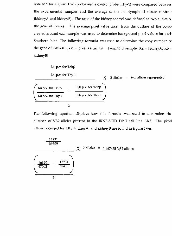
obtained for a given TcRB probe and a control probe (Thy-1) were compared betweer
the experimental samples and the average of the non-lymphoid tissue controk
(kidneyA and kidneyB]. The ratio of the kidney control was defined as two alleles O
the gene of interest. The average pixel value taken from the outline of the objec:
created around each sample was used to determine background pixel values for eacl-
Southern blot. The following formula was used to determine the copy number O:
the gene of interest: tp.v. = pixel value; 1.s. = lymphoid sample; Ka = kidneyA; Kb =
kidneyB)
1,s. p.v. for TcRP
1,s. p.v. for Thy-1 )( 2 alleles = # of alleles represented
/ Ka p.v. for TcRp Kb p.v. for TcRp \ Ka p.v. for Thy-1 Kb p.v. for Thy-1 J
The following equation displays how this formula was used to determine the
number of V(32 alleles present in the IRNB-SCID DP T ce11 line LK3. The pixel
values obtained for LK3, kidneyA, and kidneyB are found in figure 17-A.

FLUORESCENCE in situ HYBRIDIZATION lFISHl
A - PREPARATION OF METHAN0L:ACETIC ACID FIXED CELLS
(i)Spleen Cell Preparation:
Mouse spleen was minced into a culture dish with three drops of RPMI 164i
supplemented with 20% fetal calf serum (FCS; Gibco BRL, Gaithersburg, MD), 5 X 10
5 M P-mercaptoethanol (Sigma, St. Louis, MO), 10 mM glutamine [Gibco BRL
Gaithersburg, MD), 10 mM Penicillin/Streptomycin (Gibco BRL, Gaithersburg, MD
and 10 mM HEPES, until no large lumps were visible. The ceIl suspension wai
transferred to a 15 ml conical tube. The cells were pelleted by centrifugation (500 x g
4"C, five minutes], washed two times with PBS, and resuspended in ten ml of medii
containing three pg/ml concanavalin A (Calbiochem, Behring Diagnostics, La Jolli
California). From this suspension, two sets of dilutions were made, one at 1 x 10'
cells per ml, and the other at 0.5 x 10⁶ cells per ml. The cells were allowed t(
incubate at 3 7 T in a 5% CO2 incubator for 48 hours.
( i i j Cultured Cell Line Preparation:
The irradiated newborn scid (IRNB-SCID) DP T ceIl lines (LK3, LK3C, LK6.2
LK8, and SCTL2) were cultured starting at a density of 0.5~106 per ml in ten ml O
RPMI 1640 supplemented with 10% FCS, 5 X 10⁻⁵ M P-mercaptoethanol, 10 mM L
glutamine, and 10 mM HEPES. The cells were allowed to incubate at 37OC in a 5%
CO2 incubator for 24 hours.
(ii i) Cell Harvesting:
Both cultured cells and spleen cells were pelleted by centrifugation (500 x g
4"C, five minutes), and resuspended in ten ml of RPMI 1640 suppIemented witk
20% FCS, 5 X 10⁻⁵ M (3-rnercaptoethanol, 10 mM glutamine, 10 m k
Penicillin/Streptomycin, and 10 rnM HEPES, at a density of 2x10⁶ per ml. The cell:
were then treated with colcemid (Gibco BRL, Gaithersburg, MD) at 0.1 pg/ml

incubated at 37°C in a 5% CO2 incubator for 30 minutes, transferred to 15 ml conical
tubes, washed with ten ml of PBS, and pelleted by centrifugation (500 x gr 4"C, five
minutes). The supernatant was decanted, Ieaving approximately 0.5 ml of fluid i n
which the cells were resuspended.
(iv) Hypotonic Treatment:
Ten ml of 37°C hypotonic medium (seven mI ddH20 plus five ml RPMI 1640
media supplemented with 5 X 1 0 - ~ M p-mercaptoethanol and 10 mM HEPES) was
added to the cells. The first ml of hypotonic solution was added two drops at a time,
with mixing between additions. After the first ml was added, hypotonic solution
was added in increasing amounts between episodes of mixing. The cells were then
incubated in a 37" incubator for ten minutes.
(v) Fixation:
One mI of 3:l methanol/acetic acid fixative solution was added to each tube
and the cells were pelleted by centrifugation (500 x gr 4"Cr five minutes). The
supernatant was decanted, leaving approximately 0.5 ml of fluid in which the pellet
was resuspended by gently flicking the tube. Ten ml of the 3:l fixative was slowly
added to the tubes. The first ml of fixative solution was added two drops at a time,
with mixing in between additions. After the first ml was added, fixative solution
was added in increasing amounts between episodes of mixing. The ce11 suspensions
were placed at RT for 20 minutes and then pelleted by centrifugation (500 x gr 4"C,
five minutes). The supernatant was decanted, leaving approximately 0.5 ml of
fixative solution in which the pellet was resuspended by gently flicking the tube.
The cells were then washed two times in the fixative solution. After each wash the
cells were pelleted by centrifugation (500 x g, 4"C, five minutes). After the final wash
the cells were resuspended in one ml of fresh fixative solution. Fixed celIs were
either used immediately, or stored at 4OC.

B - SLIDE PREPARATION
(i l Cleaning:
Slides were cleaned by immersing first in acetone, then in HC1:ethanol 1:1, for
five minutes each, and then placing under running tap water. The slides were then
stored in distilled water at 4°C for a minimum of 30 minutes.
(ii) Metaphase preparation:
The following procedure was performed in a fume hood.
Using a Pasteur pipette, one clrop of the fixed ce11 suspension was dropped on
to a slide (removed from cold, 4"C, distilled water) from a height of 1-3 cm. The
slide was placed at an angle of 45' so that the drop spread out producing an elliptical
area. Excess fluid was removed and the slide was placed ont0 a hot plate until dry.
Remaining large drops of fluid were removed by tapping the slide on gauze.
Ambient humidity was critical for this procedure: too dry an atmosphere impaired
the quality of metaphase chromosomes, too humid an atmosphere and slow drying
time resulted in "ghost" chromosomes, which appeared empty with a dark outline.
To increase the humidity, a humidifier was placed into the fume hood during the
procedure. After the slides had dried, they were checked under a phase contrast
microscope (10X objective) in order to evaluate the number of cells undergoing
mitosis (mitotic index) and metaphase quality. Two to three metaphases with no
overlapping chromosomes, little cytoplasm, and 50 nuclei per 1 0 X power field was
determined to be suitable for analysis. In optimal preparations, the chromosomes
appeared dark grey in colour. Chromosomes which were light grey or very black
and refractile did not hybridize well. If the mitotic index was sub-optimal, the
original ce11 suspension was either diluted with fixative, or concentrated by
centrifugation (500 x g, 4"C, five minutes). The slides were allowed to rest for two
days at RT before being hybridized with chromosome "paint" probes (114). The
slides were stored at -20°C.

C - HYBRIDIZATION AND DETECTION
(i) Target Slide Denaturation
Cold slides were equilibrated to RT before use to prevent condensation from
forming on the slide. The chromosome spreads were treated with 100 pl of 100
,ug/ml ribonuclease A (RNase) in 2X SSC for one hour at 37°C in a box humidified
with Millipore filtered water soaked Whatman paper. Fifty ml of freshly prepared
70% formamide/2X SSC, was placed in a coplin jar and warmed to 75°C in a water
bath. While the formamide solution was warming, the RNase treated chromosome
spreads were taken from the incubator and washed three times, for three minutes,
in 2X SSC at RT. The chromosome spreads were then dehydrated by washing in an
ethanol series of 70% ethanol, 90% ethanol, and 100% ethanol (al1 a t -20°C) for five
minutes each, then allowed to air dry for five minutes. The chromosomes were
denatured at 75'C for two minutes in the 70% formamide/2X SSC solution. The
fina1 desired temperature was 70°C. The temperature of the formamide solution
decreased one OC per slide added, thus to denature the chromosomes on five slides,
the bath was set to 75°C. If the temperature was <70°C, denaturation was
incomplete. After denaturation, the chromosome spreads were placed in cold 70%
ethanol for five minutes, dehydrated in an ethanol series as describe above, and
allowed to air dry for ten minutes.
( i i ) Probe Denaturation
Mouse chromosome paints were obtained from Pamela H. Rabbitts of the
Medical Research Council in Cambridge, England (114). Each paint is a complex
mixture of DNA probes prepared by PCR amplification of individually sorted
chromosomes using partially degenerate primers (degenerate oligonucleotide-
primed-polyrnerase chain reaction, DOP-PCR, (114, 115). The 'paints' are labeled
with biotin by a secondary PCR reaction in which biotin dUTP is incorporated into

the probes. This allows subsequent detection with fluorescent antibodies followinl
hybridization of the paint to metaphasic chromosomes. For an in depth explanatior
on how to produce mouse chromosome specific paints see (114). For each slide
seven pl of the chromosome paint probe [50% formamide, 5% Dextran Sulphate
0.1% SDS, 1X Denhardts, 100 pg/ml salmon sperm DNA, 0.1 pg/ml mouse repetitivt
DNA (CoT1 fraction), and 15 ng/pl of chromosome probe DNA] was added to eigh
p1 of hybrisol VI (65% formamide/2X SSC; Oncor), and the mixture was warmed in 2
37°C incubator. One hundred ng of probe per slide was optimal for hybridization
The chromosome paints were denatured at 75°C for ten minutes, then allowed tc
prehybridize to mouse CoTl DNA for 90 minutes in a 37°C incubator.
(iii) Hybridization
Cover slips and chromosome spreads were prewarmed in a 37°C incubator foi
ten minutes prior to use. The denatured, CoT1-prehybridized, chromosome paint:
were applied to the chromosome spreads and then covered by a 24x40 mm coverslip
The coverslips were then sealed ont0 the slides with rubber cernent, and incubatec
overnipht at 37°C in a box humidified with moist filter paper.
(iv) Washing and Detection
Using tweezers, the rubber cernent was removed from the coverslips, and thc
coverslips were removed from the slides by rinsing in 2X SSC. To remove non.
hybridized probe, slides were washed three times in 50% formamide, 1 X SSC at 42°C
for five minutes each and then three times in 2X SSC at 42°C for five minutes each
The chromosome spreads were then treated with 3% BSA, 4XSSC, 0.1% Tween 2(
(BDH, Toronto, ON), to block non-specific hybridization. Forty pL of the blocking
solution was pipetted ont0 each sIide, covered by a 24x30 mm coverslip and tht
chromosome spreads were then placed at 37OC for 20 minutes in a humidified box.
The following steps were done in a dark room as fluorescent-antibodj
complexes are light sensitive. After treatment with the blocking solution, coverslip!

were removed by gently tapping the slides on the edge of a plastic beaker. Forty pl of
avidin conjugated to fluoroscein isothiocyanate (FITC-Avidin; Oncor) was added to
each slide, covered by a 24x30 mm coverslip and incubated for 30 minutes at 37°C.
Chromosome spreads were then washed three times in 4X SSC, 0.1% Tween 20 at
42°C for 5 minutes each. Forty pl of biotinyIated goat anti-avidin antibody (Oncor)
was then added to each slide, covered with a 24x40 mm coverslip and incubated for
20 minutes at 37°C. The chromosome spreads were then washed three tirnes in 4X
SSC, 0.1% Tween 20 at 42OC for five minutes each. Forty pl of FITC-Avidin was then
added to each slide, covered with a 24x40 mm coverslip, and incubated for 20
minutes at 3792 The chromosome spreads were then washed three times in 4X
SSC, 0.1% Tween 20 at 42OC for five minutes each, counterstained with 4',6-
diamino-2 phenylindole (DAPI) and propidium iodide (PI) by adding 1 2 pl of a
mixture (8:4 v/v) of DAPI/antifade (Oncor; 0.1 ,ug/ml DAPI, antifade contains 70%
glycerol] and PI/antifade (Oncor; 0.6 yg/ml PI) to each slide, covered with a 24x30
mm coverslip, and stored in a dry, Iight proof container covered with aluminurn
foi1 at 4°C until analysed. Fluorescent signals were detected using a confocal laser
scanning microscope (Nikon Microphot-FXA) and images were collected and
composite images were generated using Adobe Photoshop (Adobe Systems Inc.)
PULSED FIELD GEL ELECTROPHORESIS IPFGEl
( i ) Preparation of DNA in agarose blocks:
Cultured cells (LK3, LK3C, LK6.2, LK8, and SCTL2) were washed three tirnes
in ice-cold HBSS, resuspended at a concentration of 5x10⁷ cells per ml in buffer (10
mM Tris, pH 7.2, 20 mM NaCl, 100 mM EDTA; TNE), and incubated at 42°C for five
minutes. An equal volume of 2% low melt preparative grade agarose (LMP; Bio-
Rad, Mississauga, Ont.) in TNE buffer containing 100 pg/ml Proteinase K (Promega,

Madison, WI], was prepared and equilibrated to 42OC. The 42°C molten agarose was
mixed with the ce11 suspension using a sealed pasteur pipette, and transferred to
plug molds (Bio-Rad, Mississauga, Ont.) using a micro-pipette fitted with a tip with
the end cut off with a razor blade to create a wider opening. The agarose was
allowed to solidify at 4OC for 30 minutes, then transferred to 50 ml conical tubes
containing Proteinase K Reaction buffer [TNE containing 1 mg/ml proteinase K and
1% N-Lauroyl-Sarcosine (Sigma, St. Louis, MO)], and incubated at 50°C for 24 hours.
The digestion mixture was replenished and the plugs were incubated for a further 24
hours at 50°C. The plugs were then incubated for an hour at 50°C in 50 volumes of
10 mM Tris, 1 mM EDTA (TE) pH 7.6. After one hour the TE, pH 7.6 was replaced
with fresh buffer and allowed to incubate for another hour. The plugs were then
washed one final time in TE, pH 7.6 and stored at 4OC in 0.5 M EDTA pH 8.0.
Genomic DNA is these plugs rernained stable for approximately two months at 4°C.
(ii) Restriction enzyme digestion of DNA in agarose blocks:
Agarose plugs (stored in 0.5 M EDTA) were incubated in d m 2 0 for 30
minutes at 4°C in a 1.5 ml microcentrifuge tube. After 30 minutes the plugs were
incubated in fresh ddH20 for 30 minutes, then incubated in one ml of the
appropriate restriction enzyme buffer for one hour at 4OC. After incubation, the
buffer was removed and 200 pL of fresh restriction enzyme buffer was added along
with 30 units of the restriction enzyme Nae 1 (Boehringer Mannheim), and the
tubes were incubated at 4OC for 30 min in order to aallow the enzyme to diffuse
throughout the agarose plugs. The restriction enzyme reaction was activated by
placing the tubes at 37OC and the DNA was digested for 12-16 hours. After digestion
was complete, the blocks were incubated in one ml of TE pH 7.6 at 4OC for one hour.
( i i i ) Gel Electrophoresis
Following digestion, the agarose plugs were transferred into the wells of a 1%
agarose gel in 0.5X Tris-borate/EDTA electrophoresis buffer (TBE; 1 X TBE is 90 m M

Tris/90 mM boric acid/2.5 mM EDTA), and molten LMP agarose was used to seal the
plugs into the sample welIs. The gel was electrophoresed in a Chef-DR III (Bio-Rad,
Mississauga, Ont.) at 14OC with the following parameters: ramped switch time from
10-45 seconds, voltage at six V/cm, field angle of 120 degrees, and a run-time of 19
hours. These parameters allowed good separation of DNA fragments between 50 kb
and 1000 kb. h phage DNA concatamers embedded in 0.75% LMP agarose (Bio-Rad,
Mississauga, Ont.) were applied to the gel as DNA size standards.
(iv) Southern Blot transfer of PFGE gels
Transfer of DNA was performed as described for genomic Southern blots with
the following exceptions. After electrophoresis, the gel was depurinated with 0.25 M
HC1 [prepared from I N HydrochIoric acid (HC1) stock; Fisher Scientific, Don Mills,
ON] for ten minutes before transfer to nylon (Magna nylon transfer membrane;
Micron Separations Inc., Westborough, MA) in 10 X SSC for 48 hours. The filter was
prehybridized, hybridized and analysed as described for standard Southern blots
with the following exception. The filter was exposed to the phosphorscreen for 48-
96 hours before the radiographic image was digitized by a phosphorscanner.

(A) Effects of low-dose ionizing radiation on RAG-2 deficient scid mice
Irradiation of MG-2 deficient scid mice induces developmen t of DP thymocytes
wifhout rapid developmenf of thymic lyrnphomn
In order to determine if RAG activity was crucial to the invariable
progression to thymic lymphoma in irradiated newborn scid (IRNB-SCID) mice, we
analysed the effects of irradiation on RAG-2-deficient scid mice (RAG-~J- scid mice).
These animals bear the scid DSB repair defect, but are unable to introduce DSB at
RSS adjacent to V, D and J coding ends. By uncoupling the ability to cut at RSS from
the generalized DSB repair defect, we tested whether abrogating RAG mediated
cleavage at RSS would impact susceptibility to thymic lymphoma in irradiated
newborn RAG-2-/- scid rnice. If RAG-deficiency had a protective effect, a role would
be implicated for illegitimate V(D)J rearrangement in the invariable progression to
T ce11 lymphoma in IRNB-SCID mice.
We found that a single dose of y-irradiation was able to rescue DP T ce11
development and expansion in the RAG-2-/- scid rnice. By three weeks post-
irradiation, ~ ~ c - 2 - i - scid thymocytes were 20-60% DP (Table 2). Thymus cellularity
in three week old irradiated double mutant mice was 10-100 times greater than in
non-irradiated, age-matched controls. The loss of CD25 expression, which occurs
during the DN to DP transition (521, was also observed in RAG-2-/- scid rnice by
three weeks post irradiation, concomitant with the appearance of DP cells in the
thymus. By six weeks of age, thymus cellularity and frequency of DP thymocytes
declined [Fig 2) as had been previously observed in irradiated RAG-1 and RAG-2
deficient mice (93). Therefore, the kinetics of irradiation induced T ce11 development

in RAG-2-1- scid mice were similar to those observed in irradiated RAG-1 or RAG-2
deficient mice (93).
Evidence for the development of T ce11 lyrnphoma was not observed in the
irradiated newborn RAG-~-/- scid mice analyzed at later time points post-treatment.
By 10-16 weeks post-irradiation, two phenotypes ('A' and, 'B'), were observed in the
irradiated RAG-2-/- scid animals. 'Af mice resembled the non-irradiated, three
week old, RAG-2-/- scid controls, with low thymus cellularity and few DP
thymocytes (Table 2, Fig 3). 'B' mice displayed greater than 50% DP thymocytes, and
thymus celullarity was 10-60 fold greater than non-irradiated controls (Table 2, Fig
3). None of the analysed IWG-2-i- scid mice displayed thymus cellularity or
development that was comparable to that observed in IRNB-SCID animals 10-16
weeks post irradiation (Fig 2, and ref 61). We therefore found no evidence of
development of DP T ce11 lymphoma in irradiated RAG-2-1- scid mice.
It is possible that the DP thymocytes detected in 'Br mice were preneoplastic
cells with the potential to develop into a thymic lymphoma. Previous studies have
shown that preneoplastic lymphocytes can develop into thymic tumours after
explantation to a compatible host (116-118). To determine if 'B' mice had
preneoplastic cells, one million thymocytes were isolated from a 16 week old ' B '
mouse and transferred intravenously into two RAG-2 deficient recipients. Twelve
weeks post-transfer, no DP cells were observed in the thymus of the recipient mice
(data not shown). The thymus cellularity of these recipients was identical to non-
treated RAG-2 deficient, or RAG-~-/- scid mice, as they had less than 1 x 10⁶ cells in
the thymus. We cannot exclude the possibility that the absence of donor cells was
due to their failure to reach a suitable microenvironment and expand.
Alternatively, the DP thymocytes taken from the irradiated RAG-~-/- scid donor
may have reached the thymus but were unable to develop into a thymic tumour.
This possibility is supported by a failure of DP thymocytes derived from ' B '

irradiated RAG-~-/- scid mice to adapt to tissue culture, whereas thymic turnourç
explanted from IRNB-SCID mice were readily adapted in this manner (61). This
analysis supported the view that the DP thymocytes isolated from the 'B' irradiated
RAG-2-/- scid mouse were not preneoplastic.
These results suggest that RAG-2 deficiency protectç irradiated scid mice from
T ce11 lymphoma presumably by abrogating cleavage at RSS. In thymocytes of IRNB-
SCID mice, oligoclonal TcRB rearrangements were detected as early as 6 weeks post
irradiation (61). Therefore, thymocytes harbouring oligoclonal TcRP
rearrangements expanded from an initially polyclonal pool, suggesting preferential
survival and/or proliferation of pre-lymphoma cells. Taken together, these results
implicate a role for rescued V(DM rearrangement at the TcRP locus in
lymphomagenesis in IRNB-SCID rnice. We therefore decided to determine i f
aberrant V(D)J rearrangement had occurred at the TcR(3 locus in five IRNB-SCID DP
T ce11 lines that were established from primary radiation-induced scid thymic
lymphomas (61).
(B) Characterization of scid CD4TD8' T ce11 lyrnphoma lines
The IRNB-SCID DP T ce11 lines are blocked nt an enrly stage of T cell developmen t
Five ce11 lines (LK3, LK3C, LK6.2, LK8 and SCTLZ) were established from
primary irradiation induced tumours frorn individual irradiated newborn scid
(IRNB-SCID) mice (61). Previous characterization for T ce11 developmental rnarkers
found each line to be CD4+CD8'CD25- (61; Guidos and Danska, unpublished
observations). If illegitimate TcRB locus rearrangemen t played a role in IRNB-SCID
lymphomagenesis, we would have expected each cell line to have a common
pattern of gene expression reflecting their developmental level of clona1 expansion
and maturation arrest.
As discussed in the background of this thesis, cells expressing a successfully
rearranged TcRB chain, paired with the invariant pre-Ta chain (451, initiate
rearrangement at the TcRa chain locus (46). The pTa gene is expressed at varying
amounts a t successive stages of T ce11 development (451. In immature, DP T cells,
pTa expression is high, but as DP thymocytes mature into single positive (SP;
CD4+CD8- or CD4-CD8+) thymocytes, pTa mRNA expression is downregulated.
TcRa expression is also coupled to T ce11 development, as exemplified by the finding
that DP thymocytes which lack expression of the TcRa gene do not progress to the
SP stage of development (57). Rearrangement and expression of the TcRa locus
therefore plays a role in the transition from the DP stage to the SP stage of T ce11
development. In Our study, the detection of pTa message, coupled with the lack of
TcR Ca expression would have indicated that the IRNB-SCID lines were blocked at
the DP stage of development just after TcR(3 rearrangement. This result would
support the view that illegitimate rearrangement at the TcRP locus played a role in
the universal development of lymphoma in IRNB-SCID mice.

Through Northern blot analysis, we found that al1 of the IRNB-SCID lines
expressed high levels of pTa message, as compared to the BALB/c thymus control
(Fig 4-A). Low expression of pTa in normal thymus has been previously shown and
is thought to be due to the fact that the majority of cells in a wildtype thymus are
small DP cells which have presumably downregulated pTa expression (45). In
contrast, the IRNB-SCID lines each represent a clona1 population of DP cells that
retain pTa expression, but lack expression of the TcR C a gene (Fig 4-B). Based o n
the fact that TcRa rearrangement normally initiates after productive TcRf3
rearrangement (44), we propose that the IRNB-SCID lines were blocked at a stage of
development where TcRP rearrangement had occurred, but TcRa rearrangement
had either not been initiated, or rearrangement at this locus was non-functional
(out-of-frame). Consistent with this interpretation, hairpin-terminated TcRa coding
end molecules were detected in scid DP thymocytes, 1 week post-irradiation (63),
indicating that RSS specific DSB had been introduced at the TcRa locus. These
results support the view that illegitimate TcR(3 locus rearrangement played a role in
the rapid development of DP lymphoma in IRNB-SCID mice.

(C) Expression of TcRP genes in irradiated newborn scid DP T ce11 lines
Defecfion of TcR Ci3 expression in irradiated newborn scid DP T ce11 lines
In human lymphoid neoplasms, translocations involving antigen receptoi
loci can lead to the production of chimeric mRNA species [fusion transcripts) thai
encode portions of the TcR or Ig loci (81, 90, 119-122). Chromosoma1 translocation ai
the TcRP locus in the IRNB-SCID DP T ce11 lines may have resulted in abnormallj
sized mRNA species containing either of the TcR13 constant genes (CF1 or C(32). Tc
address this possibility, we examined TcR CP containing messages in the IRNB-SCIE
lines by both Northern blot and RT-PCR analysis.
In wildtype thymocytes, two TcR CP containing mRNA species of 1.3 kb and
1.0 kb have been defined [54). The 1.3 kb species is the full length V-D-J-C TcRP
transcript, while the 1.0 kb message represents an incomplete D to J rearrangement,
This 1.0 kb message appears early in T ce11 development, and may precede full V(D)]
rearrangement (123, 124). Our Northern blot analysis revealed that one of the five
IRNB-SCID lines, LK3C, expressed a 1.0 kb TcR C(J containing message (Fig 4-C). The
presence of this TcR CP mRNA species was confirmed by RT-PCR, using a stratem
(Fig 5; 5' CF-sense and 3' Cp-antisense primers) that detected CF containing
transcripts (Fig 6-A), Although we cannot rule out that the 1.0 kb transcripl
represented a fusion transcript, the size suggested it was the result of incomplete D-]
rearrangement ((54)). SCTL2 contained an apparently full length 1.3 kb TcRP
message (Fig 4-C). A RT-PCR strategy which detected rearranged TcRCj V-D-J-C
cDNA templates (Fig 5; 5' specific VPs or VP consensus, 3' CCj uni prirners:
supported the interpretation that this transcript was a full length TcRF chain [Fig 6.
B). Therefore, by both Northern blot and RT-PCR analysis, we did not deteci
abnormally sized TcRCP containing transcripts, suggesting that a fusion transcripi

containing a TcRP constant gene segment was not present in any of the IRNB-SCID
DP T ce11 lines.
TcRP chain expression in SCTL-2
We then examined whether the 1.3 kb TcR C(3 transcript in SCTL2 was the
result of a productive (in-frame) rearrangement. If the TcRp message was the
product of normal V(D)J recombination, we expected to observe a DNA sequence
with normal modifications (small deletions and few N nucleotide additions) at the
coding ends. We therefore RT-PCR amplified, cloned and sequenced the TcR C(3
chain from SCTL2. Our sequence analysis showed that the RT-PCR fragment
amplified in SCTL2 corresponded to an in-frame TcRP transcript, of the VP5.1, J(32.5
and CB2 gene segments (Fig 7). The V(D)J joining sequence notably did not contain
P nucleotides, or evidence of extensive exonuclease degradation as have been
observed in TcR genes from scid thymocytes (21). To confirm that the TcRlj
transcript detected in SCTL2 was translated into protein, we analysed intracellular
expression of TcR(3 protein using a MAb specific for a TcR VP5 determinant (MR9-4;
(107)l). We found that SCTL2 cells expressed TcR VP5 protein (Fig 8).
Taken together, these results show that none of the IRNB-SCID lines
expressed abnormally sized TcRCj mRNA species, and one line displayed a
productive TcR(3 rearrangement presumably produced during the irradiation-
induced transient restoration of TcR(3 locus rearrangement. The lack of TcRP
expression in the other IRNB-SCID lines could be explained if rearrangement had
either not occurred, or was out-of-frame. Alternatively, illegitimate V(D)J
rearrangement may have resulted in a chromosomal translocation that disrupted
the TcR(3 locus. We therefore decided to look for abnormal rearrangement at the
TcR(3 locus in the IRNB-SCID lines through Southern blot analysis.

(D) Rearxangement at the TcRP locus in irradiated newborn scid DP T ce11 lines
TcRP locz~~ rearrangement in the irradia ted newborn scid DP T ce11 lines.
Was there a chromosomaI translocation at the TcR(3 locus in any of tht
IRNB-SCID lines? To explore this possibility, we first sought to confirm that tht
TcR(3 locus had undergone rearrangement in al1 of the scid lines through Southerr
blot analysis of PvuII digested genomic DNA using a TcR C(3 probe specific for bot1
C(31 and CP2 genes (TcRCP; (97)). Because the PvtrII sites located on the 5' side o.
C(3l and Crj2 lie within the J(31 and JP2 gene clusters respectively (Fig 9-BI, non.
germ-line restriction fragments hybridizing to the TcR C(3 probe representec
rearrangement events occurring 5' of Crjl or CP2.
When PvuII digested BALB/c kidney and thymus DNA was analysed, twc
closely migrating germ-line fragments (6.2 kb and 6.5 kb; C(31 and CP2 respectively)
were observed (Fig 9-A). The reduced intensity of the germ-line bands in tht
BALB/c thymus DNA , relative to the kidney DNA, was indicative of polyclona
TcR(3 locus rearrangements. Al1 of the IRNB-SCID DP T ce11 lines were found tc
have rearranged both TcRP locus alleles, as indicated by the absence of a germ-lint
restriction fragment hybridizing to the TcR CP probe (Fig 9-A).
The analysis described above did not resolve CF1 from C(32 rearrangements
In order to detect rearrangements invoiving the CF1 cluster (ie. D(31, one of JPl.1-1.6
and C(31; depicted in Fig 1) the same TcR Crj specific probe used in the previou:
analysis was hybridized to HindIII digested genomic DNA. This mapping strategj
took advantage of the position of the HindIII sites which flank the CF1 and C(3:
genes (Fig 10-B). The 3.1 kb HindIII restriction fragment that contains the C(32 gen(
is not altered during normal V[D)J rearrangement because the 5'HindIII site ii
located 3' of the J(32 cluster. In contrast, rearrangements involving the CF1 cluste.
alter the size of the C(31 containing restriction fragment because the 5'HindIII site ir

located upstream of the J(j1 cluster. Therefore, any restriction fragments which werc
not 3.1 kb in size corresponded to rearrangements involving the Cpl cluster.
The germ-line (kidney) configuration of the Cf31 cluster produced a 9.6 kt:
HindIIl fragment (Fig 10-A). Most rearrangements to the CF2 cluster involve
deletion of the CF1 cluster. Therefore, in lines that deleted both CF1 alleles, the TcR
Cf3 probe detected only the 3.1 kb, Cf32 containing restriction fragment. This analysic
showed that LK3, LK3C and LK6.2, had deleted both Cf31 alleles, while LK8 and
SCTL2 had restriction fragments which represented rearrangements invoIving the
Cf31 cluster (Fig 10-A).
These results showed that rearrangement had occurred on both alleles of the
TcRP locus in a11 of the IRNB-SCID lines. As SCTL2 was the only ceIl line that
expressed full length TcRf3 mRNA, we wanted to determine why the
rearrangements detected in the other IRNB-SCID lines did not result in
transcription of TcRP message. One possibility was that abnormal rearrangement
had occurred, resulting in a chromosomal translocation. This possibility was
supported by our detection of CP1 gene segment deletion. Although this deletion
could have been the result of rearrangement to the Cr32 cluster, CF1 gene deletion
resulting frorn abnormal rearrangement has been detected in scid thymocytes (211,
and T cells harbouring a chromosomal aberration involving the TcRP locus (79, 84,
87). We therefore wanted to determine if abnormal rearrangement had occurred af
the TcRf3 locus in any of the IRNB-SCID DP T ce11 lines.
Abnormal rearrangement a t fhe TcRP locus
We wanted to determine if abnormal V(D)J rearrangement had deleted the
CPl gene segments in the IRNB-SCID T ce11 lines. The DNA sequence found 3' oi
the JP2 gene segments is not deleted during normal V(D)J rearrangement because
there are no recombinationally active gene segments situated between the Jf32

cluster and C(32 (Fig 1). Previous work showed that abnormal CF region
rearrangement in scid lymphocytes often resulted in deletions that covered the
entire JP2 region and extended to the flanking sequence (21, 125). We therefore
examined the Jp2 gene segments by Southern blot analysis using a probe situated 3'
of the JC32 gene segments (3'J(32; (98)). Loss of the sequence 3' of JP2 indicated that
abnormal rearrangement had occurred at the TcRP locus.
When PvuII digested DNA from the IRNB-SCID T ce11 lines was hybridized
with a 3'JP2 probe, one or more new restriction fragments, corresponding to C(32
cluster rearrangements, were observed fFig Il-A). In LK3C, no fragment hybridizing
to the 3'Jp2 probe was detected (Fig Il-A), suggesting that an abnorrnal
rearrangement had occurred in this ce11 line. 3'Jp2 probe hybridization of genornic
DNA digested with other restriction enzymes similarily failed to detect a restriction
fragment in LK3C (data not shown). The possibility that the entire TcR constant
region was deleted in LK3C was excluded by the fact that we detected two distinct
TcR C(3 containing PvuII fragments (Fig 9). These restriction fragments likely
represented C(32 cluster rearrangements, as LK3C had deleted both CF1 genes (Fig
10). Collectively, these results revealed that LK3C harboured abnormaI deletion of
the sequence 3' of the JP2 cluster on both TcRP alleles.
In both LK6.2 and LK8, analysis of the CP2 gene cluster with a 3'J(32 probe
resolved a single PvuII restriction fragment (Fig Il-A). This observation may have
been due to deletion of the J(32 gene cluster on one TcRP allele in each ce11 line. In
order to rule out the possibility that these single PvuII fragments represented two
3'J(32 cluster alleles, we performed quantitative phosphorimaging. Calibration of the
phospl-iorimaging (Materials and Methods), validated this means of detecting one
versus two alleles of a given gene (Fig 12). Our analysis revealed that the single
bands detected for both LK6.2 and LK8 represented one JC32 allele (Fig 13), suggesting
that sequences 3' of J(32 were deleted on one TcRrj allele in both of these ce11 lines.

In order to determine if the Jp2 cluster deletion detected in LK6.2 and LK8
included the CF2 gene segment, quantitative phosphorimaging was performed on
the 3.1 kb, CF2 containing, HindIIi restriction fragment seen in Fig 10-A. We found
that one copy of CF2 had been deleted in both LK6.2 and LK8 (Fig 14). Taken
together, these results show that LK6.2 and LK8 had abnormal deletions of the
sequence found 3' of the JP2 cluster, which included the Cp2 gene segment.
Southern blot analysis revealed abnormal rearrangement in 3 of the 5 IRNB-
SCID DP T ce11 lines. In LKSC, LK6.2 and LK8, sequences 3' of Jp2 were deleted on
either one (LK6.2 and LK8) or both (LK3C) alleles, as has been documented for
several chromosomal translocations involving antigen receptor loci (79-81, 84, 87).
Our results are also consistent with abnormal rearrangements resulting from
inefficient V to DJ rearrangement at the TcRP locus in scid thymocytes, (21). W e
therefore wanted to determine if the rearrangements in the IRNB-SCID T ce11 lines
were the result of V to DJ rearrangement, or chromosomal translocation.

(E) TcRP variable gene rearrangement in irradiated newbom scid DP T ce11 lines
Attempted VP fo DJP rearrangement on both TcRp alleles in al1 of the irradiafe~
newborn scid DP T cell lines.
Could abnormal V to DJ rearrangement account for the TcR(3 deletions wi
observed in the IRNB-SCID T ce11 lines? To address this question, we sought tc
determine if VP to DJP rearrangernent had been attempted in any of these ce11 line5
by examining a DNA sequence which is lost during deletional rearrangemeni
(reviewed in 2). We performed Southern blot analysis on PuuIl digested genomic
DNA, using a probe (5'DfJ; (98)) that is situated 5' of the DCjl gene (Fig 15-B), and i?
therefore lost during V to DJ rearrangement, Loss of a restriction fragmenl
hybridizing to this probe was indicative of attempted VP to DP rearrangement.
We observed deletion of the 5' DPl region on both TcRrj alleles in al1 of tht
IRNB-SCID lines by probing genomic DNA digested with PvuII (Fig 15-A) and othei
restriction enzymes (data not shown). In the germ-line DNA configuration, the 5
DCjl sequence was represented by a 5.7 kb germ-line PvuII fragment (Fig 15-A). Thc
absence of this restriction fragment in the IRNB-SCID lines could have been due tc
exonuclease digestion of exposed DNA double strand breaks, which has beer
postulated to play a role in the abnormal deletions observed in scid thyrnocytes (21)
The 5'DPl region could also have been lost if V to DJ rearrangement had beer
attempted on both TcR(3 alleles in al1 of the IRNB-SCID lines. Alternatively, 2
chromosomal translocation could have deleted the 5' DPl region, as exemplified bj
a chromosomal translocation involving the TcRP locus in a human neoplasm (79)
In this aberrant rearrangement, a non-TcR gene (fck) rearranged upstream of tht
TcRP constant region, and the sequence found between the V and D genes wai
deleted. Therefore, instead of resulting from V to DJ deletional rearrangement, th(
5' Drjl region may have been deleted in the IRNB-SCID lines due to chromosoma

translocation upstream of the constant region. We therefore wanted to determine i l
V to DJ rearrangement had deleted the 5' D(31 sequence in the IRNB-SCID lines
if@ gene deletion in irraciiated newborn çcid DP T cell Iines.
To determine if V to DJ rearrangernent had deleted the 5' D(31 region, WC
looked at VP gene deletion in the IRNB-SCID ce11 lines. The VP genes situated
proximal to the TcR(3 constant region are often deleted as a result of V to D]
rearrangement. Nineteen of the twenty-one known rnurine VP genes that
rearrange by deletion are found upstream of V(37 (Fig 161, so VP7 has a high
likelihood of being deleted in T cells that have rearrangements at both TcR(3 alleles.
The absence of V(37 in any or al1 of the IRNB-SCID T ceIl lines would therefore have
suggested that V to DJ rearrangement had occurred on both TcRP alleles.
PvuII digested DNA contains two germ-line fragment which hybridize to a
probe specific for V(37, sized 1.0 kb and 2.4 kb (only the 1.0 kb band is shown in Fig
17-A). The wildtype DP T ce11 line, VL3-3M2, was negative for a VP7 fragment,
suggesting that V(D)J rearrangement had deleted both copies of V(37. Al1 of the
IRNB-SCID T ce11 lines had at least one copy of VP7 in germ-line configuration (Fig
17-A). Quantitative phosphorimaging showed that in LK3, LK3C and SCTL2, one
copy of VP7 had been deleted, while in LK6.2 and LK8 both VP7 alleles were i n
germ-line configuration (Fig 18-A). V137 was therefore present on seven of ten TcR(3
alleles that, having deleted the sequence 5' of DP1, appeared to have undergone V tc
DJ deletional rearrangement.
To ensure that the deletion of V(37 in LK3, and LK3C was due to attempted V
to DJ rearrangement, rather than deletion of the entire TcR V(3 gene region, WC
examined another segment of the large V(3 gene cluster. Deletion of V132 is
indicative of loss of the entire V(3 gene region because it is the V gene most distal tc
the constant region (Fig 16), and therefore is not deleted during normal V to D:

rearrangement. Through quantitative Southern blot analysis of Vp2, we found that
al1 of the IRNB-SCID ce11 lines retained two V82 alleles (Fig 17-B; Fig 18-B),
indicating that the Vp7 deletion observed in LK3 and LK3C did not reflect deletion
of the entire TcR Vp locus.
To explore the possibility that V to DJ rearrangement had occurred on those
TcRB alleles that retained Vp7 in germ-line configuration, we performed Southern
blot analysis on PuuII digested genomic DNA, using a Vp14 probe. Vp14 is the only
known V p gene that rearranges by inversion (126), and unlike deletional
rearrangement, no DNA is lost during inversional rearrangernent (reviewed in 2).
Our analysis suggested that Vp14 had not been involved in a rearrangement in any
of the IRNB-SCID lines, as both V(314 alleles were in germ-line configuration (Fig
17-C; Fig 18-C). These results indicated that inversional rearrangement had not
occurred at the TcR(3 locus in any of the çcid lines.
The results presented in this section reveal that in the IRNB-SCID DP T ce11
lines, either one (LK3, LK3C, SCTL2) or two (LK6.2, LK8) V(37 alleles remained in
germ-line configuration, but deletions of the locus 5' to DJ had occurred. There are
several possible explanations for these observations. One is that Vp18, the VB gene
which is situated most proximal to the 5' end of the TcRB constant region, was
involved in the apparent V to DJ rearrangements, leaving V(37 in germ-line
configuration (Fig 16). Although we did not exclude this possibility, it is unlikely
that Vp18 was involved in rearrangement of seven of ten TcR(3 alleles in the five
IRNB-SCID ce11 lines.
Deletion of the sequence just 5' of the TcR(3 constant region could have
resulted from excessive exonuclease activity (21), which could explain why it
appeared as if V to DJ rearrangement had occurred on both alleles of the TcRp locus
without deletion of the proximal Vp7 gene. Another possibility, consistent with the
possibility of illegitimate TcRB rearrangement is that a translocation occurred 5' of

the TcRF constant region, and the V(3 genes (inciuding VP7J were translocated tc
another chromosome. We therefore decided to determine if a chromosoma
translocation had disrupted the physicaI integrity of the TcRP locus in the I R N B
SCID ceIl lines.

(F) Physical integrity of the TcRF locus in irradiated newborn scid DP T ce11 Iines
Fltrorescent in situ hybridization analysis of metaphase chromosomes
Having estabIished that abnormal rearrangement had occurred in LK3C
LK6.2 and LK8, we sought to determine if a chromosomal translocation hac
occurred at the TcRCj locus on chromosome 6 in any of the IRNB-SCID DP T ce1
lines. We performed fluorescent in situ hybridization [FISH) using moust
chromosome paint probes (114). Chromosome specific "paints" are a comple~
mixture of 200-2000 bp DNA probes (127), spaced approximately four kb apart (115)
which allow the visualization of entire chromosomes. These probes enablt
detection of gross abnormalities such as translocations or large insertions (115). Tc
detect chromosome 6 abnormalities, we examined metaphase spreads of the IRNB
SCID ce11 lines, in cornparison to BALB/c splenocyte controls.
Metaphase spreads of each ce11 line were prepared and analysed thret
independent tirnes. Approximately 50 spreads were scored per cell line on eacl
occasion. We found no detectable abnormalities involving chromosome 6, as al1 O
the metaphasic spreads analysed clearly displayed two chromosomes that hybridizec
strongly to the chromosome 6 paint probes (Table 3 and Fig 19). We did detec
several other chromosomal abnormalities in the scid lines. Mouse chromosome!
are acrocentric. The presence of metacentric chromosomes is therefore indicative O
chromosomaI instability. There were two metacentric chromosomes in LK3, anc
one metacentric chromosome in LK6.2 (Fig 19, white arrows). We also notec
abnormal chromosome number (aneuploidy) in LK3 and SCTL2 [Table 3). Thus
although chromosomal abnormalities were present in several of the IRNB-SC11
lines, those we detected did not appear to involve chromosome 6.
It has been shown that irradiation of newborn scid mice also transientl:
rescues V(D)J rearrangement at the TcRG (63) and TcRy (64) loci. Having access tc

paint probes specific for mouse chromosome 14, on which the TcRcr/S locus is
situated, we explored the possibility that the TcRalS locus was irivolved in a
chromosomal translocation. As observed in Our chromosome 6 analysis, al1 of the
IRNB-SCID lines had two copies of chromosome 14 (data not shown). As an added
control, chromosome 11, which does not harbour an antigen receptor locus, was
similarly analysed and reveaIed no evidence of chromosomal translocation (data
not shown). We therefore concluded that none of the IRNB-SCID T ce11 lines
harboured a gross abnormality involving chromosome 6, 14 or 11.
Pldsed field gel electrophoresis analysis of the TcR13 locus
We considered the possibility that chromosomal abnormalities involving the
TcRrj locus existed in the IRNB-SCID T ce11 lines, but were not detected in our FISH
analysis due to the limitations of using whole mouse chromosome paint probes.
These probes hybridize along the length of the whole chromosome, and
abnormalities that do not alter the hybridization pattern are not detectable.
Therefore, our analysis would have missed most deletions and inversions
involving chromosome 6. Although the limits of resolution for murine whole
chromosome paints have not been carefully analysed, small insertions might have
been obscured by the contiguous hybridization signals emitted by the probes (Fig 19).
In order to address the possibility that Our FISH analysis failed to detect smal1 TcRP
locus abnormalities, we performed gulsed field gel electrophoresis [PFGE), to
examine potential chromosomal translocations (128), deletions (129, 1301, and
inversions (1311.
Illegitimate rearrangement at the TcRP locus in human lymphoid turnours
can result in a chromosomal aberration that dislocates the V(3 genes from the
constant region (79). We therefore used PFGE to determine if the VCJ gene cIuster
remained linked to the Crj region in the IRNB-SCID T ce11 lines. The enzyme NaeI
has been useful in mapping the TcRP locus (132, 133). Due to variable restriction
site methylation, and the fact that Nue1 uses a two site binding mechanism to cIeave
substrate DNA (1341, NaeI generates a defined pattern of partial digestion products
that allow linkage of genes located far apart. The V(37 and Cp gene segments reside
on a 360 kb NaeI restriction fragment (Fig 20). Due to Nue1 site methylation within
the TcRP constant region, VC37 and Vp14 may also remain on a single 430 kb
restriction fragment (Fig 20). Demonstration of linkage between Vp7 and CF, or VCj7
and V(314, would therefore suggest that a chromosomal translocation had not
occurred between these gene segments.
Using this PFGE strategy, we determined that Vt37 remained linked to the C(3
and VCJ14 genes in SCTL2, LK8, LK6.2 and LK3C (Fig 21 A-D, Table 4). In each of
these ce11 lines, a 430 kb restriction fragment CO-hybridized to a V(37 and Vp14-
specific probe. The linkage of VCj7 and VCj14 indicated that VP7 was also linked to
the CF region (Fig 16). In SCTL2, LK8 and LK3C, linkage was confirmed by a 430 kb
fragment that also hybridized to a TcR CCj probe (Fig 21-A,-B and -D, respectively).
The TcR C(3 probe did not hybridize to the 430 kb fragment in LK6.2 (Fig 21-Cl,
probably because LK6.2 had deleted three of four C(3 gene segments, reducing the
TcR CF hybridization signal below detectable levels.
These PFGE data suggest that both TcRCj alleles were intact in LK6.2 and LK8.
In LK3C, which had deleted one copy of V(37 (Figl81, our results are limited to the
interpretation that the TcR(3 locus was physically intact on the allele that retained
V137 in germ-line configuration. We cannot resohe whether the allele lacking V(37
(600 kb NaeI fragment; Fig 21-D), was involved in an aberrant rearrangement. In
SCTL2, which had lost one copy of V(37 as a result of a functional Vp5.1
rearrangement, the allele lacking VCj7 was not detected. It is possible that the
compression zone (CZ, zone where DNA migrates independently of electrophoresis
parameters), which migrated as a diffuse band (Fig 21 A-E) obscured the detection of

this allele. Consistent with this possibility, a faint restriction fragment was visible
just below the CZ in NrreI digested SCTL2 DNA (Fig 21).
Unlike what was observed for LK3C, LK6.2, LK8 and SCTL2, PFGE analysis of
LK3 could not link VP7 with either the CP region or Vp14. When Nue1 digested
genomic DNA from LK3 was hybridized with the V137 probe, a 360 kb restriction
fragment was revealed (Fig 21-E). This 360 kb restriction fragment was not revealed
by either the TcR CF or VP14 probe, suggesting that VP7 was no longer linked to the
TcRP constant region. One piece of evidence to support this interpretation is that
the VC37 probe did not hybridize to the 430 kb fragment representing partial digestion
of the Nue1 sites within the CP region (Fig 20), which was detected in al1 of the other
analysed IRNB-SCID T ce11 lines (Fig 21 A-D). If VP7 had remained linked to the
constant region, the VP7, Cf3 and VP14 probes wouId have CO-hybridized to the 430
kb fragment. Alternatively, it is possible that the NaeI sites within the C(3 region
were digested to completion in LK3. In order to exclude this possibility, further
pulsed field gel electrophoresis using a different restriction enzyme would have to
be done to ensure VP7 was no longer linked to the CP region. NevertheIess, we
showed that VP7 could no longer be linked to the same NaeI restriction fragment as
the constant region in the IRNB-SCID T ce11 line, LK3.
Our analysis of the physical integrity of the TcRP locus revealed no gross
abnormalities involving chromosome 6, but in at least one of the IRNB-SCID T celI
lines, VP7 was discontiguous with the TcR CP region and Vp14. These results
suggest a chromosomal aberration caused the V(3 genes in LK3 to dissociated from
the constant region. In previous sections we presented evidence that V(D)J
rearrangement played a role in the invariable progression to rapid onset lymphoma
in IRNB-SCID mice, and that abnormal rearrangement at the TcR(3 locus was a
common feature of the IRNB-SCID DP T ce11 lines. Collectively these results suggest
that in LK3, an illegitirnate rearrangement at the TcRP locus may have occurred and

contributed to a proliferative/survival advantage, which eventualIy lead to the
development of lymphoma.

Discussion
This study investigates the putative role of illegitimate V(û)J rearrangemen
in the universal development of T ce11 lymphoma induced by irradiation O
newborn scid mice (61). As described previously, treating newborn scid mice with 2
single dose (100cGy) of y-irradiation, resulted in the appearance of CD4+CD8+ (DP) 1
cells in the thymus (61, 62): as well as the restoration of V(DJJ rearrangement at th(
TcRCj [61), TcRG (63) and TcRy loci (64). In addition, the universal development of 1
ce11 lymphoma was observed in irradiated newborn scid (IRNB-SCID) rnice by 16-2C
weeks post irradiation. Similar treatment of newborn RAG-1 and RAG-2 deficien,
mice rescued the block in T ce11 development, in the absence of restoration of V(D),
rearrangement. Strikingly, none of the irradiated RAG deficient animals developec
lymphoma (93). Activation of murine leukemia virus was investigated anc
appeared not to contribute to lymphomagenesis in the IRNB-SCID mice, as nc
novel viral integration sites were detected in several IRNB-SCID T ce11 lines (P
Joliceour and J. Danska, unpublished observations). Collectively, these result:
suggested that irradiation of lymphocyte precursors with a DSB repair defec.
enhances the generation of oncogenic events which iead to the universal, rapic
development of T ceil lymphoma. We therefore attempted to determine the role O
V(D)J rearrangement in this invariable progression to lymphoma in IRNB-SCIE
mice.
(A) V(D)J rearrangement plays a role in the rapid onset T ceIl lyrnphoma in
irradiated newborn scid mice
In order to determine the contribution of the scid DSB repair defect to the
development of lymphoma in the absence of V(D)J rearrangement, we bred RAG-2
deficient scid (RAG-2-1- scid) rnice. These double mutant mice were unable to
generate DSB at RSS, but retained the DSB repair defect conferred by the scid
mutation. We found that treatrnent of newborn RAG-2-1- scid mice with a single
dose (100 cGy) of y-irradiation did not lead to the universal development of rapid
onset lymphoma (Figs 2 and 3), as was the case for IRNB-SCID anirnals (61). This
result suggests that disabling RSS-specific DNA cleavage is protective against
irradiation induced lymphoma, even in the context of the scid DSB repair defect.
Since introduction of the RAG-2 mutation reduced the susceptibility to
irradiation-promoted thymic lymphoma, we favour the interpretation that V (D) J
recombinase activity plays a critical role in lymphomagenesis in IRNB-SCID mice.
However, it is possible that the protective effect was not dependent on the RAG-2
deficiency, but conferred by some other gene(s) derived from the M G - 2 deficient
background that was inadvertently crossed ont0 the scid background. Consistent
with this possibility, there is one report that the response to DNA-damaging agents
is heterogeneous between mouse strains (1351. In that study, differences in the rate
of repair in a given gene were observed in different inbred strains of rnice. For
example, UV-induced DNA darnage in c-myc is rapidly repaired in B lymphoblasts
from DBA/2N mice, but is poorly repaired in B cells from BALB/c mice (136).
However, if the protective effect on lymphornagenesis was conferred by another
unlinked gene from the RAG-2 deficient background, random segregation would
have occurred. If such a protective genetic element functioned in a dominant
fashion, and was not linked to the rag-2 gene, the frequency at which it would co-
segregate with the disrupted rag-2 gene would have been approximately 20%.

Therefore, we would have expected four of five irradiated RAG~-/- scid mice to
develop rapid onset lymphoma. By 10-16 weeks post-irradiation in IRNB-SCID
mice, thymus cellularity was 50-150 ~ 1 0 6 , while none of the 16 analysed 10-16 week
old RAG-2-/- scid mice had a comparable number of cells in the thymus [Table 2;
Fig2). The absence of irradiation induced T ce11 lymphoma in RAG-2-/- scid mice
therefore appears to have been the result of the inability to initiate V[D)J
rearrangement.
(B) Detection of abnormal rearrangement at the TcRCj locus in irradiated
newborn scid T cell lines
This study revealed that rearrangement had occurred on both alleles of the
TcRrj locus in al1 of the IRNB-SCID lines (LK3, LK3C, LK6.2, LK8 and SCTL2;
summarized in Table 5). Results obtained from Southern, and pulsed field gel
analyses were used to generate physical maps of both alleles of the TcR(3 constant
locus in these IRNB-SCID DP T ce11 lines (Figure 22 A-E). The rnajority of the
rearrangements were non-productive, as only SCTL2 and LK3C expressed TcR(3
mRNA (Figs 4 and 6) . Attempts to determine if the lack of expression of TcRJ3 in the
remaining scid lines was due to abnormal V(D)J rearrangement revealed that three
of the five IRNB-SCID lines analysed had deleted the JC32 gene cluster. In LK3C, both
alleles had been deleted while one allele was missing in LK6.2 and LK8 [Fig II).
During normal V(D)J rearrangement the deletion of al1 of the J(32 gene segments
does not occur, as there are no gene segments 3' of this cluster that are involved in
rearrangement [Fig 1). Jp2 gene cluster deletion is thus uncommon in wildtype T
cells, as reported in a study of T ce11 lines and lymphomas (125). Of the 34 T ceIl
lines tested in that study, only three had deleted the Jp2 cluster on one allele.
Moreover, the data could not exclude that the loss of the Jp2 cluster was actually due
to the loss of chromosome 6, on which the TcRP locus is located. Therefore, we

suggest that abnorrnal rearrangement had deleted this Jp2 gene cluster in the LK3C,
LK6.2 and LK8 lines.
Our observations are similar to those previously reported in spontaneous
thymic lymphomas from scid mice (21), where 8 of 12 lymphomas examined had
deleted the J(32 gene cluster. Of these eight lines, five had also deleted the JP1 cluster
and the CF1 gene segment. The fact that the CF2 gene was immediately downstream
of the D(3l gene in these five ce11 lines suggested that the deletions detected in the
spontaneous scid lymphomas probably reflected imprecise ligation of the DP1
element to a J(32 gene segment (21). These results thus implicate the scid defect in
mediating abnormal TcRB rearrangements which result in the deletion of sequences
within the constant region.
The abnormal rearrangements in the IRNB-SCID lines do not appear to have
been caused by the imprecise ligation mechanism stated above because the sequence
5' of D(31 had been deleted on both TcR(3 alleles in al1 of the ce11 lines (Fig 15). The
loss of this sequence suggested that attempted V to DJ rearrangement had occurred
on both alleles in every line. Although this is true, we found VP7, one of the VP
genes most proximal to the constant region (Fig 16), to be in germ-line configuration
on either one (LK3, LK3C, SCTL2) or both alleles (LK6.2, LK8) (Figs 17 and 18). One
unlikely explanation for this observation is that Vp18, the only VrJ gene situated
between V(37 and the constant region (Fig 16), was involved in the V to DJ
rearrangement in al1 of the alleles that had not deleted VP7. Thus Vp18 to DJB
rearrangement does not delete V(37. A more likely scenario is that V to DJ
rearrangement had not occurred in the IRNB-SCID ce11 lines, but that loss of the
sequence 5' of Dg1 was the due to excessive exonuclease activity. Suc11 an analysis
would fit with the idea that normal rearrangement between D to J had been
transiently restored following irradiation, but V to DJ rearrangement manifested the
inefficiency the scid mutation, including excessive exonucleolytic degradation of

coding ends. As most studies examining V(D)J rearrangernent in scid mice have
focused on D to J rearrangements (21, 25, 31, 32), the IRNB-SCID ceil lines would
allow a more detailed examination of V to DJ rearrangement.
The putative exonuclease activity invoked above does not preclude the
possibility of an illegitimate rearrangement at the TcRCj locus in the IRNB-SCID
lines. In fact, such modification of free signal and coding ends may contribute to the
infidelity of the V(D)J recombinase activity. Evidence has been reported that the
DNA sequences that flank signal sequences (86), as well as the sequence of the
coding elements thernselves (137), influence V(D)J rearrangement. Therefore,
modification of the DNA sequence flanking the DSBs at the TcR(3 locus rnay have
altered the efficiency of V(D)J rearrangement in the IRNB-SCID thymocytes, and
increased the frequency of illegitirnate rearrangement events. This possibility
among others, is consistent with the roIe of abnormal rearrangement at the TcRP
constant locus, a common characteristic of Our IRNB-SCID T ceIl lines, in the
invariable progression to lyrnphoma in irradiated scid mice.
(C) Expression of truncated TcRlj chahs: a role in lymphomagenesis?
As described above, our TcR locus mapping analysis showed that deIetions,
including Cljl, D132 and the J(32 cluster, had occurred in both TcRP alleles in LK3C
(depicted in Fig 22-B). In this regard, i t is interesting that LK3C expressed a 1.0 kb
TcR C(3 containing mRNA species (Figs 4 and 6) which may have resulted from
translocation of a foreign enhancer into the TcRrj locus. Such an event would
support a role for V(D)J rearrangement in the development of lymphorna in IRNB-
SCID mice.
Alternatively, the 1.0 kb CF containing transcript detected in LK3C may have
been a germ-line transcript, as it has been proposed that transcription at the TcRP
locus does not require D to J rearrangement (138, 139). This transcript may have also

represented a Dpl-JCj1-CF2 transcript, resulting from deletion of the CF1 gene
segment and the Jrj2 cluster. D(31-J(31-C/32 transcripts can arise due to V(D)J
rearrangement, and have been documented in New Zealand White (NZW) mice,
which carry an 8.8 kb germ-Iine deletion which includes Crjl, D(32 and the Jp2 cluster
(140). Although the T ce11 repertoire of NZW mice is more limited, they express
functional TcRp protein (140, 141)) representing functional rearrangernents despite
the large deletion.
Transcripts resulting from DP1 to JP rearrangement are thought to result
from a promoter 5' of DQ1 (54,125). In LK3CI we showed that the region 5' of D/31
had been deleted (Fig 15), making it unlikely that the 1.0 kb mRNA species was
initiated from this 5' DF1 promoter. Further characterization of LK3C would be
required to determine which of the mechanisms presented above is responsible for
the 1.0 kb transcript in LK3C. If non-TcRP DNA sequence was found upstream of
the constant region, illegitimate V(D)J rearrangement would be implicated as
having occurred in this T ce11 line.
In addition to the possibility that the LK3C 1.0 kb transcript was the result of a
chromosomal abnormality at the TcRCj locus, production of an abnormal protein
from this transcript may have contributed to oncogenesis. A recent study showed
that expression of a TcRrj protein lacking a VCj domain (TcRrjAV) elicits DP T ce11
lymphomas in transgenic mice (142). In this study, it was found that the TcRBAV
protein mediated allelic exclusion of the endogenous TcR(3 loci and thymocyte
development was btocked ai the DP CD25 stage. Interestingly, TcR(3AV transgenic
mice had very low thymus cellularity compared to normal TcR(3 transgenic mice.
The possibility that ongoing recombinase activity rnay have been contributing to the
high occurrence of T ce11 lymphoma was excluded when it was found that crossing
the TcRpAV transgene ont0 a RAG-1-" background djd not prevent tumour
development. I t is possible that the absence of a wildtype thyrnic

microenvironment may be created in which TcRfiAV expressing thymocytes can
accumulate mutations leading to lyrnphoma (142). For example, TcRfiAV
thymocytes which develop into DP T ceIIs (142, 143) may not receive signals to cause
them to die due to their lack of a variable domain. In this scenario the DP TcRrjAV
T cells could persist in the thymus long enough to attain oncogenic mutations. In
the context of a DSB repair defect, the number of oncogenic mutations accumulated
by the TcRfiAV T ce11 would be exaggerated. The investigation of such an oncogenic
mechanism in a non-transgenic animal would be valuable in understanding how
similar mutant TcR or Ig chains contribute to T or B ce11 neoplasia. Therefore,
chromosomal aberrations invoIving the TcR(3 locus may have contributed to
lymphomagenesis by creating a truncated TcRCj protein. Such an oncogenic
mechanism would clearly implicate illegitimate V(D)J rearrangement in the
transformation of this ceIl line.
(D) The TcRF locus is not involved in a gross reciprocal translocation i n the
irradiated newborn scid T celi lines
As was discussed above, the abnormal rearrangements detected in the IRNB-
SCID DP T ce11 lines may have been the result of inefficient ligation of coding ends
associated with the scid DSB repair defect (1, 21, 31, 32), opposed to some other
mechanism such as a translocation event occurring 5' of the constant region. Our
fluorescent in situ hybridization (FISH) analysis using mouse chromosome 6 paint
(114) supported this hypothesis. In this analysis we found no evidence of gross
translocation involving chromosome 6 in any of the IRNB-SCID lines [Fig 19). A
similar chromosome paint probe analysis of chromosome 14, containing the TcRa/G
locus, did not. reveal evidence of a translocation (data not shown).
This analysis could have failed to resolve several types of TcR tocus
chromosomal abnormalities. The sensitivity of detection using whole chromosome

paints has not been systematically examined, but evidence from studies on hu mar
chromosomes suggests the limitations of this type of anaIysis. Although wholt
human chromosome paints have been shown to be useful in identifying certair
chromosomal abnormali ties (144, 145), a deletion, or small translocation may not bi
detectable (146). In Our study, deletion of a portion of chromosome 6 might no.
have been detectable. If an insertion of a segment from another chromosome hac
occurred we might have observed the translocated DNA on chromosome 6 becausc
the whole chromosome paint would not have hybridized to this ectopic DNA
Detection of such an event would depend upon the size of the inserted DNP
segment. In analyses of human chromosomal abnormalities, translocations thai
occur close to the centromeric regions are difficult to detect using wholc
chromosome paints (147), and telomeric regions are often under-represented ir
human chromosome paint probes (148). Therefore, our FISH analysis using wholc
chromosome painting may have failed to detect subtle chromosomal aberration:
involving the TcR(3 locus. NevertheIess, gross translocations of chromosome
were not observed in any of the IRNB-SCID T ce11 lines.
(E) TcRrj locus disruption in irradiated newborn scid T ce11 lines
We performed pulsed field gel electrophoresis (PFGE) to further resolve thc
physicai integrity of the TcRlJ locus. PFGE analysis is useful in mapping syntenic
genes that are separated by distances which are too great to be linked by traditiona
agarose gel electrophoresis(l28). PFGE has been instrumental in establishing thi
organization of the TcR loci in both humans and mice (132, 133, 149), as well as thc
detection of deletions in other genetic loci (150-153). We reasoned that PFGI
analysis might enable the detection of chromosomal abnormalities not visible bj
FISH analysis.

PFGE analysis revealed that the TcR(3 locus rnay have been disrupted in o n e
of the IRNB-SCID T ce11 lines, LK3. Here we observed discontiguity between the V
gene segments and the constant region of the TcRP locus, as VCj7 was not linked to
either the constant region, or V(314 (Fig 21-E), an element downstream of the CP2
region (Fig 16). It may be possible that abnormal rearrangement at the TcR(3 locus
lead to alterations in the methylation patterns of the NaeI sites in LK3. Consistent
with this possibility, changes in DNA methylation patterns have been documented
at the bcr-ab2 chromosomal translocation locus (154). Additionally, excessive
methylation of CpG islands has been observed in cells grown in culture for many
generations (155). Similarily, it has been shown that alteration of the sequences
flanking the NaeI restriction sequences modifies cleavage by NaeI (156). Therefore,
alterations within the TcRlj locus could have resulted in complete digestion of t he
NaeI restriction sites, thus explaining Our inability to link V(37 to the constant
region. Alternatively, the disruption of the TcRrj locus in LK3 codd have resulted
from an insertion or inversion that separated the Vlj gene segment region from the
constant region. Further pulsed field analysis, using other restriction enzymes will
have to be done to determine if the TcR(3 locus is disrupted in LK3. To determine
the nature of such a disruption, future studies would have to examine the DNA
sequence found upstream of the constant region. For example, an LK3 genomic
library could be generated, TcR CF containing clones isolated, and analysed to
determine the sequence upstream of the constant region. If this DNA was not from
the TcRP locus, it would confirm the view that an illegitimate V(D]J rearrangement
event contributed to f i e transformation of this cell.

(F) Chromosomal aberrations associated with abnormal TcRB locus
rearrangement
As has been suggested throughout this thesis, we favour the view that
chromosornal translocations explain the abnormal TcR(3 rearrangements observed
in the IRNB-SCID DP T ce11 lines. Translocations involving antigen receptor loci
often display abnormal deletions. For example, analysis of T-ALL cells showed that
a TcRB locus translocation involved a deletion from 5' of Dp1 to 3' of the Jp2 cluster
(85). Similarly, in the T-ALL cells of a ataxia-telangiectasia patient, a 17kb deletion
was found 3' of the translocation breakpoint within the TcRa locus (80). The
abnormalities we observed within the TcRB constant region in the IRNB-SCID lines
are very similar to those cited above, supporting the hypothesis that y-irradiation of
scid lymphocyte precursors increases the frequency of illegitimate rearrangement at
the TcRP locus, and contributes to lymphomagenesis in IRNB-SCID mice.
(G) Conclusions
We used a genetic approach to examine the role of illegitimate V(D)J
rearrangement in lymphomagenesis in IRNB-SCID mice. Our data revealed that
introducing the RAG-2 deficiency ont0 scid mice protects irradiated newborn RAG-
2-1- scid rnice from the universal development of rapid onset lymphoma. There are
two possible explanations for Our data. The first, which has been discussed in detail,
is that the ability to introduce DSB at RSS has no role in lyrnphomagenesis, rather
the protective effect of crossing the RAG-2 deficiency ont0 scid mice is due to an
unknown genetic factor contributed by the RAG-2 mouse background. To
determine if this statement iç true, RAG-2+/- scid mice should be irradiated and
assayed for T ce11 tumour development. Developrnent of lymphoma will verify
that initiation of V(D]J rearrangement plays an obligate role in IRNB-SCID
lymphomagenesis.

The second possibility is that aberrant V(D)J rearrangements are promoted by
irradiation of scid lymphocytes. In this view, aberrant V(D)J rearrangement
contributes to an oncogenic mutation which promotes progression to T ce11
neoplasia in the IRNB-SCID mice. For example, the RAG proteins create DSB at the
5' RSS of the DP gene segment and the 3' end of the VIJ gene segment, but the scid
recombinase machinery fail to unite the free coding ends. As DNA-PK is involved
in both V(D)J rearrangement and general DSB repair, a free DNA end generated by y-
irradiation could be ligated 5' of the DP element. If a growth promoting gene were
found adjacent to the non-TcR free DNA end, the powerful enhancers found in the
TcRlJ locus could then ectopically drive expression of the translocated gene.
This type of mutation rnay not be restricted to the TcRP locus, as V(D)J
rearrangement is transiently restored at multiple TcR loci in IRNB-SCID mice (63,
64). In fact, the oncogenic event may not involve a TcR locus at all, as aberrant
V(D)J rearrangement involving cryptic RSS elements can occur between two non-
antigen receptor loci (83). Regardless of the loci involved in illegitimate V(D)J
rearrangements, this illegitirnate joining mode1 explains why irradiated RAG-1- scid
mice are protected from irradiation induced lymphoma, owing to a failure to
initiate either bonafide or cryptic RSS cleavage.
The second major aspect of Our work, the characterization of the TcR(3 locus
in five IRNB-SCID lines, supports the illegitimate joining mode]. The abnormal
TcR(3 rearrangements in these lines share characteristics observed in chromosomal
translocations involving antigen receptor loci. For example, deletions of antigen
receptor constant regions such as those we observed in the TcR C(3 region have been
observed in human neoplasias (80, 157). More support for the illegitimate joining
mode1 is revealed when the abnormal rearrangements found in the IRNB-SCID
lines are compared to those anaiysed in spontaneous scid thymic lymphomas and
Abelson murine leukemia virus transformed scid bone marrow cells (21). For

exampfe, although JP2 cluster deletion was observed in both Our study and that ol
Schuler et al., CF2 gene deletion was only observed in the IRNB-SCID T ce11 lines
The 5'D(31 region was not deleted in spontaneous/Abelson transformed thymomas
with JP2 cluster deletion, as was observed in the IRNB-SCID line. These difference:
suggest that the abnormal rearrangements detected in the IRNB-SCID lines were noi
the same as those observed in spontaneous scid thyrnomas (21). The illegitimatc
rearrangement model is further strengthened by our results that suggest disruption
of the physical integrity of the TcRP locus may have occurred in one of the IRNB-
SCID lines. This study thus implicates a role for illegitimate V[D)J rearrangement in
the invariable development of rapid onset lymphoma in IRNB-SCID mice.
The study of chromosomal translocations in human Iymphoid malignancies
has reveaIed an enormous number of novel proto-oncogenes (reviewed in 65, 158).
If the model of illegitimate V(D)J recombinase mediated deveIopment of lymphoma
proves to be correct, the IRNB-SCID system could provide numerous ce11 lines thai
harbour rearrangements involving known proto-oncogenes, thus allowing for t h e i ~
analysis. IRNB-SCID mice could also prove to be a rich source for the identificatior
of unknown proto-oncogenes. This possibility is true if the translocation involvec
one partner that was generated by RSS specific cutting, while the other partner was
generated by an irradiation induced DSB. If the irradiation induced DSB occurs
adjacent to a proto-oncogene, and the free broken end is then ligated next to the RSC
mediated breakpoint, the IRNB-SCID system could theoretically allow for the
identification of virtually every proto-oncogene in the mouse genome. Using this
"proto-oncogene engine", one could study genes that are integral to norma
lymphocyte development as well as to neoplasia.

Table 1 Oligonucleotides used in this study
Oligonucleotides Sequencea
~ ~ ~ 2 - 3 b R 4 ~ 2 - l b ~ e o - 3 b S'P-actinc 3'j3-actinc Vp consensusd
GCC TGC TTA TTG TCT CCT GGT ATG TTA ATT CAA CCA GGC TTC TCA CTT CCA ACG CTA TGT CCT GAT AGC GGT TGG GTC AGA AGC ACT CCT ATG CAG GCA GCT CAT AGC TCT TCT TAA GCG GCC GCA TGS LYT GGT AYW XXC
[S=G or T; L=A, G, or T; Y=C or T; W = A or C; X=A or G] CAG CTC AGC TCC ACG TGG T AAG ATA TCT GGT GAA AGG GC GCT GGT GTC ATC CAA ACA CC TGG CCA GTT GCC GAG ATC AA ATA GAG GAT GGT CGC AGA CAA GAC GCT GAG CTG GTG GGT GAA TG
aAII primer sequences are written in the 5'-3' orientation b - rt-ference 103
C - reference 95 - refcrence 61
- reference 97

Table 2 Effects of low dose ionizing irradiation on newborn RAG-2-/- scid mice
*ge Cellularitya % c D ~ + c D ~ + ~ %CD25-C Post-Irradiation
3 wks (n=8)
6 wks (n=3)
10 wks 'A' [n=l) 1.0 x 10⁶ 40 54
'B1(n=4) 6.3X10⁶'4.8X10⁶ 76' 21 93' 11
16 wks 'A' (n=6) 7.3 x 10⁵ ' 2.2 x 10⁵ 0.5 +/- 0.8 25 ' 4 [N=4)
'B' (n=5) 2.3 x 107" 1.6 x 10⁷ 90' 5 98, 3
-awhole thymocyte suspensions were stained with trypan blue, and viable cells were countcd under t h microscope -bthymocytes were analysed for expression of CD4 [YTS-191.1) and CD8 (YTS 169-4) by multiparamtc flow cytometry as described in Materials and Methods
-Ctliymocytes were analysed for expression of CD25 (7D4) by multiparameter flow cytometry a described in Materials and Methods -niimbers shown are means +/- standard deviation of rcsults obtained from the nimber (n) of mice per tim point (d=days, wks=weeks) -when only one or two mice were analysed, the numbers for those mice are shown -'A', and 'B' are distinct thymocyte devclopmental phenotypes

Table 3 Fluorescent In Situ Hybridization analysis of irradiated newborn scid
DP T ce11 lines, using mouse chromosome 6 paint probes
chromosome 6 total # of # of cell type equivalentsa chromsomes at abnormal
at metaphase metaphaseb chromosom~
B ALB / c splenocyte 2
W(3 2
LK3C 2
LK6.2 2
LK8 2
SCTL2 2
aTwo chromosome 6 equivalents per ce11 means two cliromosomes strongly hybridizing to the murine chromosornc 6 paint at metaphase bincluding tnetacentnc chromosomcs, cach metacentric chromosome was counted as being one chromosomc CMetacentric cliromosomcs visually detccted by DAPI staining of entire chromsomes

Table 4 - Pulsed field gel electrophoxesis analysis of irradiated newborn
scid DP T ce11 iines
IRNB-SCID
ce11 line
SCTL2
LK8
LK6.2
LK3C
LK3
size (kb) I c(3
+ + + +
-
V(314
?
?
+
4-
Interpretation
+ -
+ +
-
+ ?
V(37 linked to C(S, V(S14
V(37 linked to Cp, V(314
C(3 linked to Vp14
VF7 linked to C(3, VCJ14
Nael sites within constant region are
deleted
+ -
+
4-
+ -
VP7 linked to C(3, V(314
V137 linked to Cp, V(314
C(S linked to Vp14
600 kb fragment represents allele with
VB7 deleted
+ -
-
V(S7 linked to CCJ V(314
+ -
+
C(3 linked to Vp14
- V137 is not linked to C(3
or V(314
+ C(S Iinked to V(S14
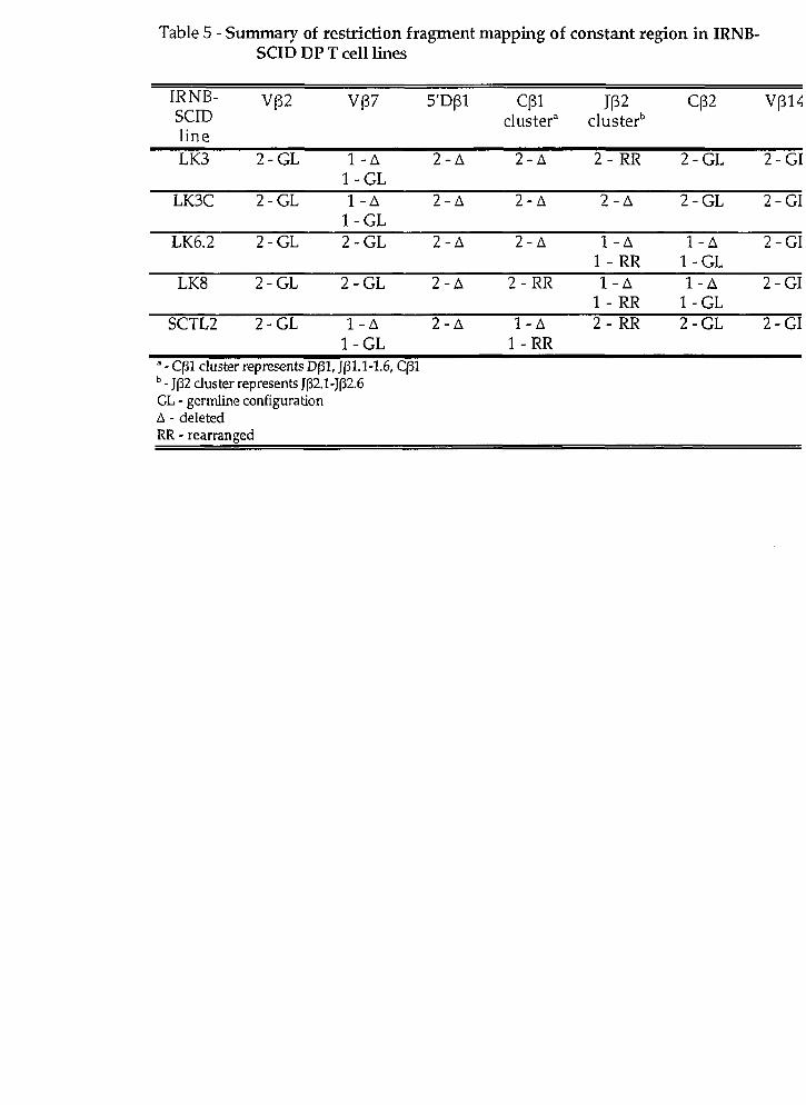
Table 5 - Summary of restriction fragment mapping of constant region in IRNB- SCID DP T ce11 lines
IRNB- v ~ 2 V137 s ' ~ p l CPl JP2 CP2 vp14 SClD cluster" clusterb line LK3 2 - G L 1 - A 2 -A 2 - A 2 - R R 2 - G L 2 - G I
1 -GL LK3C 2 - G L 1 - A 2 -A 2 - A 2 - A 2 - G L 2 - GI
1 -GL LK6.2 2 - G L 2 - G L 2 -A 2 - A 1 - A 1 - A 2 -CI
1 - RR 1 -GL LK8 2 - G L 2 - G L 2-11 2 - RR 1 - A 1 - A 2 - GI
1 - R R 1 -GL SCTL2 2-GL 1 - A 2 - A 1 - A 2 - RR 2 - G L 2 - GI
1 - G L 1 - R R a - CF1 cluster represents D(31, J(31.1-1.6, C(31 - J82 cluster represents Jp2.1-J(32.6 CL - gerdine configuration A - deleted RR - rearranned

Figure 1
The constant region of the murine TcRP locus
Genomic organization of the constant region of the murine TcR(3 locus, u
located on chromosome 6. The two diversity (D) segments are indicated by vertical
lines. Each D segment is associated with a cluster of seven joining (J(31.1-1.7 and
J(32.1-2.7) segments (indicated by the filled boxes), and one constant (CF1 and C(32)
gene segment [indicated by the open boxed). Each J cluster has six functional, and
one pseudo-gene (denoted by $). C(3l and Cf32 cluster designations cited in this work
are indicated.

Fig 1
Cpl cluster CB2 cluster

Figure 2
Th yrnocyte expansion and development in irradiated RAG2 def icient s cid mice
Thymocyte suspensions were stained with trypan blue, and viable cells were
counted under a microscope. Thymocytes were obtained at various time points aftei
irradiation (100cGyJ of newborn RAGZ-1- scid mice and analysed for expression of
CD4 (YTS-191.1) and CD8 (YTS 169-4) by multiparameter flow cytometry as described
in the Materials and Methods. Data is presented as thymus celldarity as a function
of time [top graph], and frequency of CD4/CD8 double positive (DP) thymocytes as a
function of time (top graph). Each circle represents one irradiated RAG~- / - scid
mouse, At 10 and 16 weeks post irradiation open circles (O) represent mice of
phenotype A, filled circles (el represent mice of phenotype B. The number of mice
(n] analysed for each time point is indicated on the X axis. Mean values obtained
from irradiated newborn (100cGy) scid mice are displayed as hatched Iine. Each
triangle represents 4-24 rnice (611, and standard deviation is represented as error bars
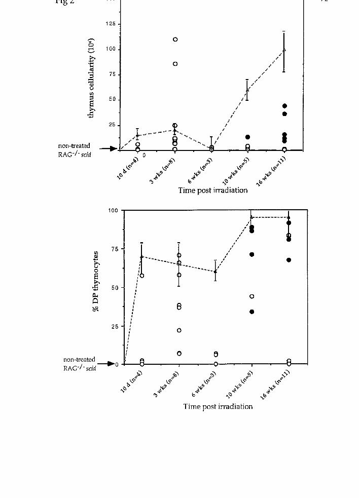
RAG-/- scid
rn 8
25 - ,' /
..* %, ,"" .+.+ -"-"'
/
1 8 '--& f
non-aeated + , O î
Q O
n T Y
f i
4@ &" 9 %' + b* w k? &
2 M
xQ 4 8 b dq
9 di5
& 4%"
Time post irradiation
non-trcated ~ ~ ~ - / - s c i d -0
Time post irradiation

Figure 3
Phenotypic analysis of T lymphocyte development in irradiated newborn RAGZ-1-
scid mice
Thymocytes were obtained at 10 or 16 weeks after irradiation (100cGy) of newborn
RAG-2-1- scid mice and analysed for forward scatter (FSC) and expression of CD4
(YTS-191.1), CD8 (YTS 169-4), or CD25 (7D4) by multiparameter flow cytometry as
describedin Materials and Methods. Two-pararneter contour plots (7% probabiIity)
show CD4 versus CD8 expression (far left). Single-parameter histograms show the
expression of CD25 (middle), compared to staining with isotype-matched control
antibodies (shaded histograms), and FSC (far right). Two distinct phenotypes are
labeled 'A' and 'B' (see also Table 2). Staining of BALB/c, irradiated newborn RAG-
2-/-, irradiated newborn scid and untreated contml RAG-2 -1- scid thymocytes are
shown for cornparison.

Fig 3
16 wk IRNB RAG2" scid Pbeqotype 'A'
1 O0
10
16 wk W B RAG-2" scid Phenotype 'B'
A 10 IO0 1000 10 100 1000 100 200

Figure 4
Northern blot analysis of TcR expression in irradiated newborn scid T ce11 Lines
(A) Ten pg of total RNA from the IRNB-SCID DP T ceil lines LK3, LK3C, LK6.2, LK8
and SCTL2, as well as from BALB/c thymus, BALB/c bone marrow and the C57B1/6
DP T ce11 line VL3-3M2 were electrophoresed through a 1.5% agarose-formaldehyde
geI and transferred to nylon membrane. Hybridization was performed with a 3 2 ~
labeled pTa DNA probe. The same filter was hybridjzed sequentially with (B) a 3 2 ~
labeled TcR C a probe; (C) a 3%' labeled TcR C(3 probe; and (D) a S-actin probe.
Hybridized filters were exposed to a phosphorscreen for 12-24 hours, and digitized
on a Molecular Dynamics Phosphorimager. The position and size of detected
mRNA species are indicated to the Ieft edge of each panel.

Fig 4 $
VI i E IRNB-SCID DP T ce11 Iines
g CD
s" c O P
Figure 5
RT-PCR strategy for amplification of TcR(3 mRNA
Schematic representation of the RT-PCR strategy utilized to detect TcR(3 chain cDNA
prepared from DP T ce11 lines. OIigonucleotides used as PCR primers are
represented by arrows. Oligonucleotide sequences are found in Table 1, and their
use are described in the Methods and MateriaIs.

Fig 5
specific VP
3, 11, 14
Cl3 sense Cp antisense
\ CP uni
Vp consensus

Figure 6
TcR(3 expression in scid DP T ce11 lines
RNA from IRNB-SCID lines LK3, LK3C, LK6.2, LK8, and SCTL2, BALB/c thymocytes
and the C57B1/6 line VL3-3M2 was isolated using TRIzol as directed by the
manufacturer (Gibco). cDNA was prepared by reverse transcription and supplied as
template for PCR reactions. For evaluation of CF-region expression, cDNA was
amplified with the primers CF antisense and C(3 sense, which were designed to
amplify an interna1 sequence in both CF1 and CP2 [panel A). For evaluation of TcR
V(3-region expression, cDNA was amplified with either the V(3 consensus primer
(panel B) or V-region specific primers (panel C-VCj3; panel D-VCj11; panel E-V(314) in
conjunction with the CB uni primer. (3-actin primers (panel E) were used as a
positive control for the quality of cDNA. The PCR products were resolved on
agarose gels, transferred to nylon membranes, hybridized to 32~-labeled TcR C(3 or
(3-actin probed, exposed to a phosphorscreen, and the signal was digitized on a
Molecular Dynamics Phosphorimager. The position and size of the RT-PCR
amplification products are indicated on the left edge of each panel.

Fig 6
I vo consensus I I

Figure 7
DNA sequence analysis of TcRp transcripts i n scid DP T ce11 line, SCTL2
The TcR(3 transcript expressed by the IRNB-SCID DP T ce11 line SCTL2 was amplifie^
by RT-PCR using the consensus Vf3 primer in conjunction with the C(3 uni primer.
The product was cloned into the pCR-2 vector (Invitrogen), and was used as a
template in a dideoxy-sequencing reaction (Sequenase, U.S. Biochemicals). The
nucleotide sequence is compared with germ-line V(35.1 and Jp2.5 gene segments
(underlined). The predicted amino acid sequence is also shown (bold).

Fig 7
VP5.1 TGTGCCAGCTCTCTC
SCTL2 TGTGCCAGCTCTCTC GCTGTG
protein C A S S L A V
JB2.5 AACCAAGACACC
AACCAAGACACC
N Q D T..

Figure 8
TcR(3 protein expression in the scid DP T ce11 line, SCTL2
Freshly isolated BALB/c thymocytes, the scid DP T ce11 lines LK3C andSCTL2 as well
as the WT control VL3-3M2 were analysed for expression of TcR(3 using biotin-
conjugated H57-597 (anti-CF] and FITC-conjugated MR9-4 (anti-V(35) by
multiparameter flow cytometry as described in the materials and methods. Two-
parameter contour plots (7% probability) show TcR(3 versus V(35 expression (far Ieft).
Single-parameter histograms show the expression of TcR(3 (middle, clear
histograms) and VP5 (far right, clear histograms) compared to staining with isotype-
rnatched control antibodies (shaded histograms).

Fig 8
Figure 9
Analysis of TcRP gene xeaxrangernents in the constant region
(A) DNA (15pg) from IRNB-SCTD DP T ce11 Iines LK3, LK3C, LK6.2, LK8 and SCTL2,
BALB/c kidney, BALB/c thymus and VL3-3M2 was digested with PvuII,
electrophoresed through a 1% agarose gel, transferred to nylon membrane, and
hybridized to a 3%' labeled TcRCB DNA probe. Phosphorimages were obtained afteer
a 24 hour exposure with the use of a Molecular Dynamics imaging system. The
position and size of unrearranged germ-line fragments are indicated on the Ieft edge
of the panel. The two closely migrating germ-line fragments are not resohable in
this image. (B) A PvuII restriction digest map (not to scale) and the positions of the
C(3 probe are included to illustrate the germ-Iine configuration of the CP region.
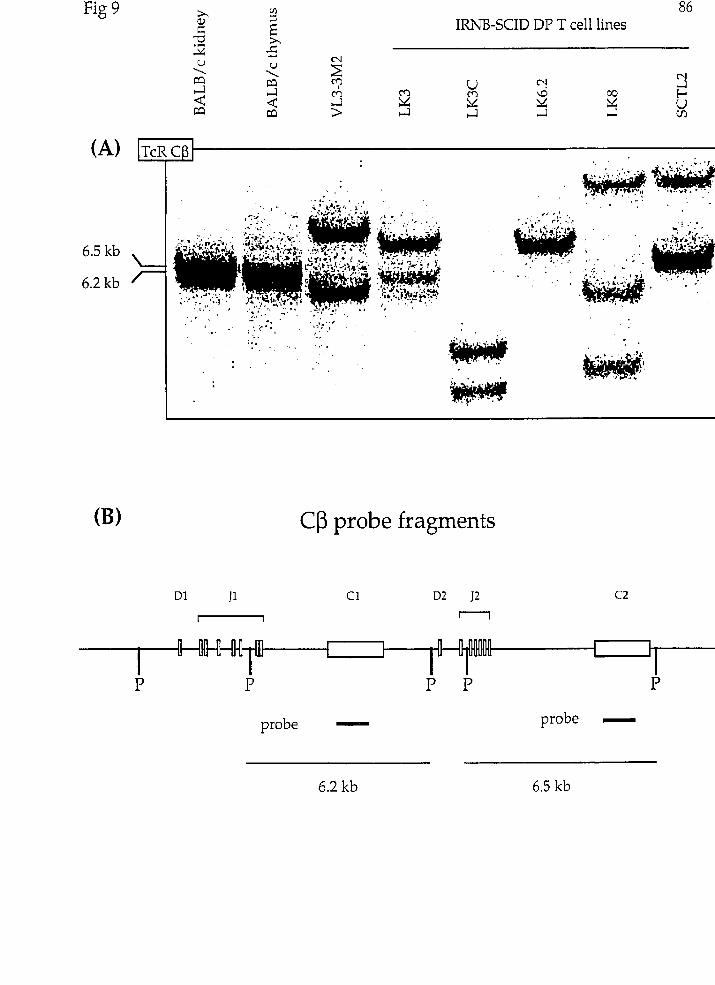
Fig 9
Cp probe fragments
probe - probe -

Figure 10
Analysis of TcR CF1 cluster rearrangements
(A) DNA (15pg) from IRNB-SCID DP T ce11 Iines LK3, LK3C, LK6.2, LK8 and SCTL2,
BALB/c kidney, BALB/c thymus and VL3-3M2 was digested with HindIII,
electrophoresed through a 1% agarose gel, transferred to nylon membrane, and
assayed for hybridization to a 3 2 ~ labeled TcR CF probe. Phosphorimages were
obtained after a 24 hour exposure with the use of a Molecular Dynamics irnaging
system. The position and size of the unrearranged germ-line fragments are
indicated on the left edge of the panel. (B) A HindlII restriction digest map (not to
scale), and positions of the CrJ probe are included to illustrate the germ-line
configuration of the CC3 region.

rig l u
Cp probe fragments
probe - 9.6 kb
H H
probe -

Figure I l
Analysis of TcR Jfi2 cluster rearrangements
(A) DNA (15pg) from IRNB-SCID DP T ce11 lines LK3, LK3Cr LK6.2, LK8 and SCTL2,
BALB/c kidney, BALB/c thymus and VL3-3M2 was digested with PvuII,
electrophoresed through a 1% agarose geI, transferred to nylon membrane, and
assayed for hybridization to a 3 2 ~ labeled 3'JFZ probe. (B) The same Southern blot
was stripped of 3IJP2 probe and hybridized sequentially with a 3 2 ~ labeled Thy-1
probe to provide a single copy gene control. Phosphorimages were obtained after a
24 hour exposure with the use of a Molecular Dynamics imaging system. The
position and size of the unrearranged germ-Iine fragments are indicated on the left
edge of the panel. (C) A PvuII restriction digest map (not to scale), and position of
the 3'JCj2 probe are included to illustrate the germ-line configuration of the Cfi
region.

1IZNB-SCID DP T ce11 lines YU
3'J(32 probe fragment
P P
probe ,

Figure 12
Quantitative phosphorimaging
Varying amounts of BALB/c thymus DNA was digested with PvuII, electrophoresed
through an agarose gel, transferred to nylon membrane, and assayed for
hybridization to a 32~-labeled Thy-1 DNA probe. The different amounts of DNA (15
pg, 7.5pg, 5pg, lpg and Opg) were used to represent different allele copy numbers (2, 1,
1/3,1/15, and 0, respectively). The hybridized filter was exposed to a
phosphorscreen, digitized by a Molecular Dynamics phosphorscanner, and absolute
pixel number in each sample was quantified by volume integration using
ImageQuant software (Molecular Dynamics). The expected nurnber of alleles
represented, pixel value, and calculated number of alleles represented, are dispIayed
under each band, To determine the calculated number of alleles, the pixel value for
the sample was compared to the pixel value obtained for the 15pg sample (which
represented two alleles of the fhy-1 gene). See the Materials and Methods for a
detailed description.

Fig 12
Figure 13
Quantitative phosphorirnaging of the TcR Jp2 gene cluster in scid DP T ce11 lines
(A) DNA (15pg) from IRNB-SCID DP T ce11 lines LK3, LK3C, LK6.2, LK8 and SCTL2,
BALB/c kidney, BALB/c thymus and VL3-3M2 was digested with PvuII,
electrophoresed through a 1% agarose geI, transferred to nylon membrane, and
assayed for hybridization to a 3%' labeled Thy-1 probe. (B) The same Southern blot
was hybridized sequentially with a 3% labeled 3'JFZ probe. Phosphorimages were
obtained after a 24 hour exposure with the use of a Molecular Dynamics imaging
system. Densitometry was performed on the restriction fragments hybridizing to
either probe. Absolute pixel number in each sample was quantifjed by volume
integration using ImageQuant software (Molecular Dynamics). In order to
determine the copy number of the 3'JP2 sequence, the ratios of the pixel values
obtained for 3'JF2 and Thy-1 were compared between the experimental samples
(IRNB-SCID T ce11 lines) and the average of non-rearranging tissue controls
(BALB/c kidney). The predicted number of alleles is represented above each band
analysed. (C) The pixel values for each sample is shown in the bottom panel, along
with the ratios used to calculate the predicted number of alleles. Samples denoted
by an asterix '*' had two different sized bands hybridizing to the 3'JP2 probe, of
which one was analysed. The method of calculating the predicted number of alleles
is explained fully in the Methods and Materials section.

nle n.v. Thv-1 n.v. 3'TW # ofalleles
kidney A 67003 kidney B 86419 thymus 74093 VL3* 81788 LK3* 69509 LK3C 58184 LK6.2 77893 LK8 118041 SCTL2* 62369

Figure 14
Quantitative phosphorimaging of the TcR CF2 gene in scid DP T ce11 lines
(A) DNA (15pg) from IRNB-SCID DP T ce11 lines LK3, LK3C, LK6.2, LK8 and SCTL2,
BALB/c kidney, BALB/c thymus and VL3-3M2 was digested with HindIII,
electrophoresed through a 1% agarose gel, transferred to nylon membrane, and
assayed for hybridization to a 3 2 ~ labeled Thy-1 probe. (B) The same Southern blot
was hybridized sequentially with a 3%' Iabeled TcRCfl probe. Phosphorimages were
obtained after a 24 hour exposure with the use of a Molecular Dynamics imaging
system. Derisitometry was performed on the restriction fragments hybridizing to
either probe. Absolute pixel number in each sample was quantified by volume
integration using ImageQuant software (Molecular Dynamics). In order to
determine the copy number of the C(32 gene, the ratios of the pixel values obtained
for C132 (3.1 kb fragment, Fig 10) and Thy-1 were compared between the experimenta:
samples (IRNB-SCID T ce11 lines) and the average of non-rearranging tissue controls
[BALB/c kidney). The predicted number of alleles is represented above each band
analysed. (C) The pixel values for each sampIe is shown in the bottom panel, along
with the ratios used to calculate the predicted number of alleles. The method of
calculating the predicted number of alleles is explained fully in the Methods and
Materials section.

kidney A 12820 kidney B 20078 thymus 15829 VL3 13613 LK.3 10612 LK3C 15061 LK6.2 17196 LK8 25604 SCTL2 19841

Figure 15
Analysis of TcRrj locus remangement
(A) DNA (15pg) from IRNB-SCID DP T ce11 lines ,K3C, LK6.2, LK8 and SCTL
BALB/c kidney, BALB/c thymus and VL3-3M2 was digested with PvuII,
electrop horesed through a 1% agarose gel, transferred to nylon membrane, and
assayed for hybridization to a 3 2 ~ labeled 5'Dpl probe. (B) The same Southern blot
was stripped of hybridized probe and hybridized with a 3 2 ~ labeled Thy-1 probe to
provide a single copy gene control. Phosphorimages were obtained after a 24 hour
exposure with the use of a MolecuIar Dynamics imaging system. The position and
size of the unrearranged germ-line fragments are indicated on the left edge of each
panel. (C) A PvuII restriction digest map (not to scale), and position of the SfDP1
probe are included to illustrate the germ-line configuration of the C(3 region.

(A) I 5 ' ~ p l l
5' Dpl probe fragment
probe -

Figure 16
The murine TcRP h a i n locus
Physicai map of the (3-chain gene locus of the murine T ce11 receptor, which is
situated on chromosome 6. The approximate positions of variable regions and the
kilobase scaie in this figure are based on the data presented in Lindsten et al., 1987,
and Lee and Davis, 1988. Vj3 and Dj3 genes are represented as vertical lines [l). The
Jrjl and JP2 gene clusters are represented as open boxes. The CC3 genes are
represented as fiIled boxes. The CC3 region (including the VP14 gene) has been
expanded for cIearer representation.

Fig 16
Figure 17
Analysis of TcR V(3 gene deletion
(A) DNA (15pg) from IRNB-SCID DP T ce11 lines LK3, LK3C, LK6.2, LK8 and SCTL2,
BALB/c kidney, BALB/c thymus and VL3-3M2 was digested with PvuII,
electrophoresed through a 1% agarose gel, transferred to nylon membrane, and
assayed for hybridization to a 3 2 ~ labeled Vp7 probe. Although two PvuII fragments
(2.4 kb and 1.0 kb) were detected with the Vp7 probe, only the 1.0 kb fragment is
shown. (B) The same DNA biot was stripped of hybridized probe and hybridized
sequentially with a 3%' labeled V82 probe. (C) The same DNA blot was stripped of
hybridized probe and hybridized sequentially with a 3%' labeled V(314 probe.
Phosphorimages were obtained after a 24 hour exposure with the use of a Molecular
Dynamics imaging system. The position and size of the unrearranged germ-line
fragments are indicated on the left edge of each panel.


Figure 18
Quantitative phosphorimaging of TcR VrJ genes in scid DP T ce11 lines
A: (i) DNA (15yg) from LK3, LK3Cr LK6.2, LK8 and SCTL2, BALB/c kidney, BALB/c
thymus and VL3-3M2 was digested with PuuII, electrophoresed through a 1%
agarose gel, transferred to nylon membrane, and assayed for hybridization to a 3 2 ~
labeled Thy-1 probe. (ii) the same filter was sequentially hybridized with a 3 2 ~
labeled VP2 probe. (iii) densitometry/volume integration results B: (i) The same
DNA samples described above were digested with PvuII, eIectrophoresed through a
1% agarose gel, transferred to nylon membrane, and assayed for hybridization to a
3 2 ~ labeled Thy-1 probe. (ii) the same filter was sequentially hybridized with a 3 2 ~
labeled VP14 probe. (iii) densitometry/volume integration results C: (i) The same
DNA samples described above were digested with PvuII, electrophoresed through a
1% agarose gel, transferred to nylon membrane, and assayed for hybridization to a
32P labeled Thy-1 probe. (ii) the same filter was sequentially hybridized with a 3%
labeled VP7 probe. (iii) densitornetry/volume integration results. Densitometry
was performed on the restriction fragments hybridizing to the Thy-1 and VIJ probes.
Absolute pixel number in each sample was quantified by volume integration using
ImageQuant software (Molecular Dynamics]. In order to determine the copy
number of the V(3 gene of interest, the ratios of the pixel values obtained for that V(3
and Thy-1 were compared between the experimental samples (IRNB-SCID T ce11
lines) and the average of non-rearranging tissue controls (BALB/c kidney). The
predicted number of alleles is represented above each band analysed. The pixel
values for each sample is shown in the bottom panel, along with the ratios used to
calculate the predicted number of alleles. The method of calculating the predicted
number of alleles is explained fully in the Methods and Materials section.
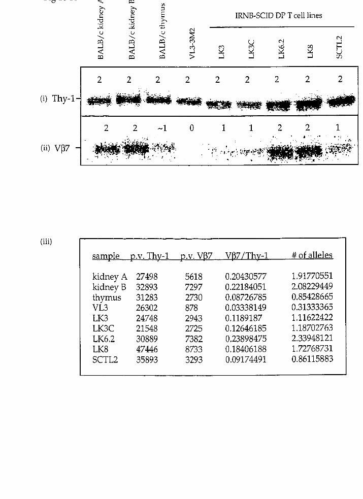
(i) Thy-1
(ii) vp7
(iii)
2i X a,
C C h
IRNB-SCID DP T ce11 lines 2 2 24 24 5
.CI
samde V.V. Thv-1 U.V. VB7 VB71Thv-1 # of alleles
kidney A 27498 kidney B 32893 thymus 31283 VL3 26302 LK3 24748 LK3C 21548 LK6.2 30889 LK8 47446 ÇCTLZ 35893

Fig 18-B
(iii) samde D.V. Thv-1 D.V. VP2 VP2IThv-1 # of alleles
kidney A kidney B thymus VL3 LK3 LK3C LK6.2 LK8 SCTL2

Fig 18-C
IRNB-SCID DP T ce11 lines
(iii) samde -1 D.V. Vp14 VBl41Thv-1 # of alleles
kidney A kidney B thymus VL3 LK3 LK3C LK6.2 LK8 SCTL2

Figure 19
Fluorescent In Situ Hybridization
Murine chromosome 6 paint was hybridized to metaphasic spreads prepared from
BALB/c spleen cells (BALB/c) and the IRNB-SCID DP T ce11 lines LK3, LK3C, LK6.2,
LK8 and SCTL2. Metaphasic spreads were prepared and assayed for hybridization to
the murine paint as described in the Materials and Methods. Chromosomes that
hybridized to the chromosome 6 paints are yellow. White arrows indicate
metacentric chromosomes.
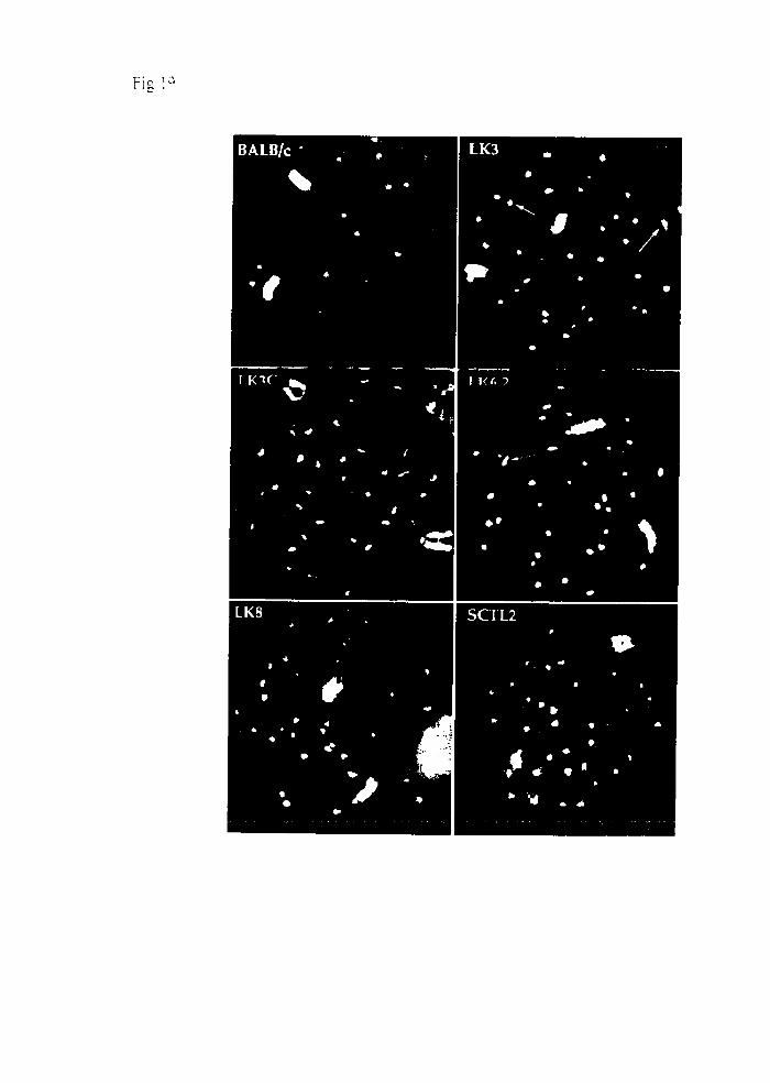
Fis i C '

Figure 20
Pulsed Field Gel Electrophoresis Strategy
NaeI restriction map of the murine TcRP locus (not to scale). The horizontal lines
below the map indicate NaeI restriction fragments of germ-line DNA that contain
VP7, Cp1, C(32 or V(314. The hatched lines represent possible products resulting
from partial digestion of the NaeI sites within the constant region. Numbers to the
right of the horizontal brackets indicate the size of each particular DNA fragment.

Fig 20

Figure 21
Pulsed Field Gel Electrophoresis of the TcRP locus in scid DP T ce11 lines
DNA from IRNB-SCID DP T ce11 lines SCTL2, LK8, LK6.2, LK3C and LK3 [panels A-
Er respectively) was prepared in agarose blocks as described in the Materials and
Methods section. The DNA was digested in the agarose blocks with the enzyme
NneI, separated by pulsed field gel electrophoresis, transferred to nylon membrane,
and assayed for hybridization to TcR VP7, TcR CCJ and TcR Vp14 DNA probes. The
approximate size of each fragment is indicated on the left edge of each row of panels.
Detectable fragments are indicated by an arrow on the left edge of each panel. Size
approximation was done by comparison with h phage DNA concatamer size
standards. The compression zone (C.Z., zone where DNA migrates independently
of electrophoresis parameters) is indicated by a '*' on the left edge of each panel.
Phosphorimages were obtained after a 48-72 hour exposure with the use of a
Molecular Dynamics imaging system.

C.Z. 4
C.Z. 4
(D) LK3C (E) LK3 VB7 CP VP14 VP7 CB Vp14
Figure 22
Restriction maps of the TcRB constant region in scid DP T ce11 lines
Physical maps of both alleles of the TcRP constant region in LK3 (A), LK3C (B), LK6.:
(C), LK8 (D), and SCTL2 (E). The approximate positions of restriction enzyme sites
in relation to constant region genes are based on the data presented in this study.
The size of the restriction fragments hybridizing to the TcR CF probe used
throughout this study are found beneath each TcRp allele. TcRF gene segments are
represented by open boxes. Bolded ? and hatched bars signify that the represented
DNA sequence is unknown. H = HindIII, P = PvuII restriction sites.

allele 1 (rearranged)
germline (wearranged)
allele 2 (reananged) rearrangemen
C2 VI4

germ lj ne (unrearranged)
allele 1 (rearranged) rearrangernent
V7 ,++Mi -m VI4
P H HP
germline (unrearranged)
alleIe 2 (rearranged)
pi- - - 1 P 4.0-4.5 kb

\\ .
rearrangement
aIlete 1 (rearmnged)
V7 C2 VI4
gennline (unreanangedl
allele 2 (rearranged)
\ rearrangement /

germline (unrearranged)
gerrnline (unrearranged)
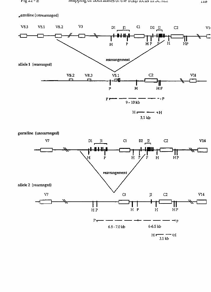
allele 1 (rearranged)
germiine (unreananged)
V7
1
allele 2 (reananged)
V7 Cl C2 VI4

References
1.
2.
3.
4.
5.
6.
7.
8.
9.
10.
I l .
12.
Lieber, M. R. 1991. Site-specific recombination in the immune system. FASEB J. 52934.
Lewis, S. M. 1994. The mechanism of V(D)J joining Iessons from molecular, immunological, an(
comparative analyses. A h . Immunol. 5327.
Tonegawa, S. 1983. Somatic generation of antibody diversity. Nature 302575.
Oettinger, M. A., D. G. Schatz, C. Gorka, and D. Baltimore. 1990. RAG-1 and RAG-2 adjacen
genes that synergistically activate V(D)J recombination. Science 248:1517.
Mombaerts, P., J. Iacomini, R. S. Johnson, K. Herrup, S. Tonegawa, and V. E. Papaioannou. 1992
RAG-1-deficient mice have no mature B and T lymphocytes. Cell 68:869.
Shinkai, Y., G. Rathbun, K.-P. Lam, E. Oltz, V. Stewart, M. Mendelsohn, J. Charron, M. Datta
F. Young, A. Stall, and F. Alt. 1992. RAG2 -deficient mice lack mature lymphocytes owing ti
inability to initiate V(D)J rearrangement. Ce11 68:855.
van Gent, D. C., J. F. McBlane, D. A. Ramsden, M. J. Sadofsky, J. E. Hesse, and M. Gellert. 1995
Initiation of V(D)J recombination in a cell-free system. Cell81:925.
van Gent, D. C., D. A. Ramsden, and M. Gellert 1996. The RAGl and RAG2 proteins establis1
the 12/23 rule in V(D)J recombination. Ce11 85107.
Schatz, D. G., M. A. Octtinger, and M. S. Schlissel. 1992, V(D)J recombination: molecula
biology and regulation. Ann. Rrv. iiilinirnoi. 10:359.
Deçidcrio, S. V., G. D. YancopouIos, M. Paskind, E. Thosma, M. A. Boss, N. Landau, F. W. Alt
and D. Baltimore. 1984. Insertion of N regions into heavy-chain gcnes is correlated witl
expression of termina1 deoxytransferase in B cells. Nntirrr 31 l:ZZ.
Gilfillan, S., A. Dierich, M. Lemeur, C. Benoist, and D. Mathis. 1993. Mice lacking TdT: Matur,
animals with an immature lymphocyte rcpertoire. Science 261:1175.
Komori, T., A. Okada, V. Stewart, and F. W. AIt. 1993. Lack of N regions in antigen recepto
variable region genes of TdT-deficient lymphocytes. Scirncr 261:1171.
Roth, D. B., J. P. Menetski, P. Nakajima, M. Bosma, and M. Gcllcrt. 1992. V(D)J recombination:
Broken DNA moleciilcs with covalently sealed (hairpin) coding ends in scid mouse tliymocytes.
Cr11 70:983.
Lewis, S. M. 1994b. P nucleotide insertions and the rcsolution of hairpin DNA structures in
mammalian cells. P m . Nntl. Acad. Sci. USA 91:1332.
Lieber, M. R., J. E. Hesse, S. Lewis, G. C. Bosma, N. Rosenber, K. Mizuuchi, M. J. Bosma, and M.
GeIlert. 1988. The defect in murine severe combined immune deficiency: joining of signal
sequences but not coding segments in V(D)J recombination. Ce11 55:7.
Bosma, G. C., R. P. Custer, and M. J. Bosma. 1983. A severe combined immunodeficiency mutation
in the mouse. Nafrire 301:527.
Fulop, G. M., and R. A. Phillips. 1990. The scid mutation in mice causes a general defect in DNA
repair. Na tiire 347:479.
Biedermann, K. A., J. Sun, A. J. Giaccia, L. M. Tosto, and J. M. Brown. 1991. scid mutation in mice
confers hypersensitivity to ionizing radiation and a deficicncy in DNA double-strand break
repair. Proc. Nalt. Acad Sci. USA 88:1394.
Hendrickson, E. A., X.-Q. Qin, B. E. A., D. G. Schatz, M. Oettinger, and D. T. Weavcr. 1991. A
link bctwccn double-strand break-related repair and V(D)J rccombination: The scid mutation.
Proc. Nad. Acad Sci. USA 88:4061.
Disney, J., A. Barth, and L. Shultz. 1992. Dcfcctive repair of radiation-induced chromosomal
damage in scid/scid mice. Cytogenet. C d . Genet. 59-39.
SchuIer, W., 1. J. Wciler, A. Schulcr, R. A. Phillips, N. Roscnbcrg, T. W. Mak, J. F. Kearney, R.
P. Perry, and M. J. Bosma. 1986. Rcarrangcnicnt of antigcn rcccptor gcncs is dcfcctivc in mice
with severe combined immune deficiency. CeIl 46963.
Malynn, B. A., T. K. Blackwell, G. M. Fulop, G. A. Rathbun, A. J. Furley, P. Ferrier, L. B.
Hcinkc, R. A. Phillips, G. D. Yancopoulos, and F. W. Ah. 1988. The scid dcfect affects the final
step of the immunoglobulin VDJ recombinase mechanism. Ce11 54.

23. Blackwell, T. K., B. A. MaIynn, R. 11. PolIock, P. Ferrier, L. R. Covey, G. M. Fiilop, R. A
Phillips, G. D. Yancopoulos, and F. W. Alt. 1989. Isolation of scid pre-B cells that rearrangi
kappa light chain genes: formation of normal signal but abnormal coding joins. EMBO J. 8.735.
24. Ait, F. W., E. M. Oltz, F. Young, J. Gorman, G. Taccioli, and J. Chen. 1992. VDJ recornbination
Itnrnimol. Today 13:306.
25. Penneycook, J. L., Y. Chang, J. Celler, R. A. Phillips, and G. E. Wu. 1993. High frequency O
normal DJH joints in B ce11 progenitors in severe combined immune deficiency mice. j. Exp. Med
1 78:lOOZ
26. Ferrier, P., L. R. Covey, S. C. Li, H. Suh, B. A. Malynn, T. K. Blackwell, M. A. Morrow, and F
W. Alt 1990. Normal recombination substrate Vh to DJh rearrangements in pre-B ce11 lines fron
scid mice. J. Exp. Med l7l:l9O9.
27. Petrini, J. H., A. M. Carroll, and M. J. Bosma. 1990. T-cell receptor gene rearrangements ir
functional T-ce11 clones from sud mice: reversion of the scid phenotype in individua
lymphocyte progenitors. Proc. Nad Acad. Sci. USA 87:3450.
28. Kotloff, D. B., M. J. Bosma, and N. R. Ruetsch. 1993. V(D)J recombination in peritoneal B cells O
leaky scid mice. J. Exp. Med. li'8:198l.
29. Kienker, L. J., W. A. Kuziel, B. A. Garni-Wagner, W. Kumar, and P. W. Tucker. 1991. T ce1
receptor y and 6 gene rearrangements in scid thymocytes: similarity to ffiose in norma
thymocytes. j. Itnrnunol. 147:4351.
30. Schuler, W., N. R. Ruetsch, M. Amster, and M. J. Bosma. 1991. Cocling joint formation O
endogenous T ce11 receptor genes in lymphoid cells from scid mice: unusual P-nucleotidi
additions in VJ-coding joints. Etlr. J. Itntniinol. 21:589.
31. Okazaki, K., S. 1. Nishikawa, and H. Sakano. 1988. Aberrant irnmunoglobulin genc
rearrangement in scid mouse marrow tells.]. bnmunol. 141:1348.
32. Kim, M., W. Schuler, M. J. Bosma, and K. B. Marcu. 1988. Abnormal recombination of Igh D and
gene segments in transformed pre-B cells of scid mice.]. Inlilitlnol. 141:1341.

Blunt, T., N. J. Finnic, G. E. Taccioli, G. C. M. Smith, J. Demengeot, T. M. Gottleib, R. Mizuta, A.
J. Varghese, F. W. Alt, P. A. Jeggo, and S. P. Jackson. 1995. Defectivc DNA-dependent proteîn
kinase activity is linked to V[D]J rccombination and DNA repair dcfccts associated with the
murine scid mutation. Cd1 80:813.
Kirchgessner, C. U., C. K. Patil, J. W. Evans, C. A. Cuomo, L. M. Fried, T. Carter, M. A.
Oettinger, and J. M. Brown 1995. DNA-dependent kinase (p350) as a candidate gene for the
murinc SCID defect. Science 267:1178.
Peterson, S . R., A. Kurimasa, M. Oshimura, W. S. Dynan, E. M. Bradbury, and D. J. Chen. 1995.
Loss of catalytic subunit of the DNA-dependent protein kinase in DNA double-strand break
repair mutant mammalian cells. Proc. Natl. Acad Sci U S A 92:3171.
Hartley, K. O., D. Gcll, G. C. M. Smith, H. Zhang, N. Diveclia, M. A. Connelly, A. Admon, S.
P. Lees-Miller, C. W. Anderson, and S. P. Jackson. 1995. DNA-dependent protein kinasc
catalytic subunit A reIativc of phospliatidylinositol 3-kinase and the ataxia telangiectasia
gene product. C d 82:849.
Gottlieb, T. M., and S. P. Jackson. 1993. The DNA-dependent protein k i n a s e :
requirement for DNA ends and association with Ku antigen. Ce11 72:131.
Gottlieb, T. M., and S. P. Jackson. 1994. Protein kinases and DNA damage. Trends in Biocliem.
Sci. 19:SOO.
Anderson, C. W. 1993. DNA damagc and thc DNA-activatcd protein kinase. T r m Biochtrn.
Sci. 18:433.
Danska, J. S., D. P. HoIland, S. Mariathasan, K. M. Williams, and C. J. Guidos. 1996.
Biochemical and gcnctic dcfects in the DNA-dependent protein kinasc in munne scid
lymphocytes. Mol. Ceil Bio. 165507.
Boubnov, N. V., and D. T. Weaver. 1995. scid cells are deficient in Ku and replication protein A
pliosphorylation by DNA-dependent protein kinase. Mol. Cd1 Bio. 15:5700.

Blunt, T., D. Gcll, M. Fox, G. E. Taccioli, A. R. Lehmann, S. P. Jacton, and P. A. Jeggo. 1996.
Identification of a nonsense mutation in the carboxy-terminal region of DNA-dependent protein
kinase catalytic subunit in the scid mouse. Proc. Nntl. Acnd Sci. USA 93:10285.
Godfrey, D. I., J. Kennedy, P. Mombaerts, S. Tonegawa, and A. Zlomik. 1994. Onset of TCRp
rearrangement and role of TCR(3 expression during CD3-CD4-CD8- thymocyte differentiation. 1.
Immztnol. 152:4783.
Dudley, E. C., H. T. Petrie, L. M. Shah, M. J. Owen, and A. C. Hayday. 1994. T ce11 receptor beta
chain gene rearrangement and selection during thymocyte development Imtniinity 1:83.
Saint-Ruf, C., K. Ungewiss, M. Groettrup, L. Bruno, H. J. Fehling, and H. von Boehmer, 1994.
Analysis and expression of a cloned pre-T ce11 receptor gene. Science 266:1208.
Groettrup, M., and H. von Boehmer. 1993. A role for a pre-T-ce11 receptor in T-ce11 development.
Itmiinol. Today 14r6101.
Ehlich, A., S. Çchaal, H. Gu, D. Kitamura, W. Muller, and Rajewsky. 1993. Immunoglobulin
heavy and light chain gens rearrange independently at early stages of B ce11 development.
Ce11 72:695.
Reth, M. G., E. Petrac, P. Wiese, L. Lobel, and F. W. Ah. 1987. Activation of Vx gene
rearrangement in pre-B cells follows the expression of membranebound immunoglobulin heavy
chains. EMBO 1. 6:3299.
Iglesias, A., M. Kopf, G. S. Williams, B. Buhler, and G. Kohler. 1991. Molecular requiremcnts
for the p-induced light chain gene rearrangement in pre-B cells. EMBO]. 10:2147.
Tsubata, T., R. Tsubata, and M. Reth. 1992. Crosslinking of the ceIl surface irnmunoglobriIin (p-
siirrogate light chains complcx) on prc-B cells induces activation of V gene rearrangements a t
thc immunoglobulin .x locus. Int Immunril. 4.637.
Mclchers, F., H. Karaçuyama, D. Haasncr, S. Baucr, A. Kudo, N. Sakaguchi, B. Janieson, and A.
Rolink. 1993. The çurrogatc light chain in B-ceIl development. Inrrniinol. Tudny 14:60.

Pearsc, M., L. Wu., M. Egerton, A. Wilson, K. Shortman, and R. Scollay. 1989. A murine carly
thymocytc developmental sequence is marked by transient expression of the interlerikin-2-
receptor. Proc. Nafl. Acad Sci. USA 86:1614.
Kishi, H., P. Borgulya, 3. Scott, K. Karjalainen, A. Traunecker, J. Kaufman, and H. Von
Boehmer. 1991. Surface expression of the P T ce11 receptor (TCR) chain in the absencc of othcr
TCR or CD3 proteins on immature T cells. EM130 J. 10.
Siu, G., M. Kronenberg, E. Strauss, R. Haars, T. W. Mak, and L. Hood. 1984. The structure,
rcarrangement and expression of D(3 gene segments of the murine T-cell antigen receptor. Natilre
31 l:34V.
Born, W., J. Yague, E. Palmer, J. Kappler, and P. Marrack. 1985. Rearragement of the T-cell
receptor 0-chain genes during T ce11 development. Proc. Natl. Acad. Sci. USA 82:2925.
Born, W., M. McDuffie, N. Roelim, E. Kushnir, J. White, D. Thorpe, J. Stefano, J. Kappler, and
P. Marrack 1987. Expression and role of the T cell receptor in early thymocyte differentiation
in vitro. j. Immunol. 138:999.
Mombaerts, P., A. Clarke, M. Rudnicki, J. Iacomini, S. Itohara, J. Lafaille, L. Wang, Y.
Ichikawa, i7. Jaenisch, M. Hooper, and S. Tonegawa. 1992b. Mutations in T-ce11 antigen receptor
genes a and (3 block thymocyte development at different stages. Natrrre 360225.
Shinkai, Y. , S. Koyasu, K.-1. Nakayama, K. Murphy, D. Loh, E. Reinherz, and F. Alt. 1993.
Restoration of T ce11 development in RAC-2-deficient mice by functional TCR transgenes.
Science 259:822.
Spanopoulou, E., C. Roman, L. Corcoran, M. Sclilissel, D. Silver, D. Nemazee, M. Nussenzweig,
S. Shinton, R. Hardy, and D. Baltimore. 1994. Functional immunoglobulin transgcncs guide
ordered B ce11 differentiation in RAC-1 -deficient mice. Genes Dev. 8:1030.
Karasuyama, H., A. Rolink, Y. Sliinkai, F. Young, F. W. Alt, and F. Melchers. 1994. The
expression of Vpre-B/h5 surrogate light chain in early bone marrow precursor B cells of normal
and B cell-deficient mutant mice. Cell 77:133.

Danska, J. S., F. Pflumio, C. Williams, O. Hiiner, 1. E. Dick, and C. J. Guidos. 1994. Rescue of T
ceIl-specific V(D)J recombination in SCID mice by DNA-damaging agents. Science 266450.
Murphy, W. J., S. K. Durum, M. R. Anver, D. K. Ferris, D. W. McVicar, J. J. O'Shea, S. K .
Ruscetti, M. R. Smith, H. A. Young, and D. L. Longo. 1994. Induction of T ce11 differentiation and
lymphomagenesis in the thymus of mice with severe cornbined immune deficiency (SCID). 1.
In~rnimol. 1 S3:lOO4.
Livac, F., S. C. Welsh, G. C.J., 1. N. Crispe, J. S. Danska, and D. G. Schatz. 1996. Transienl
restoration of gene rearrangement at multiple T cell receptor loci in y-irradiated scid mice. J.
Exp. Med l 8Ml9 .
Bogue, M. A., C. Zhu, E. Aguilar-Cordova, L. A. Donehower, and D. B. Roth. 1996. p53 is
required for both radiation-induced differentiation and mscue of V(D)J rearrangement in scid
mouse thymocytes. G e n s Dev. 10:553.
Rabbitts, T. H. 1994. Chromosomal translocations in human cancer. Natiire 372:143.
Kaplan, H. S. 1967. On the natural history of the murine leukemias: Presidential address.
Cancer Res. 27:1325.
Cuypers, H., G. Seltcn, W. Quint, M. Zijlçtra, E. Maandag, W. Boelens, P. van Wezenbeek, C.
Melief, and A. Berns. 1984. Murine leukemia virus-induced T-ce11 lymphomagenesis:
Intcgration of proviriises in a distinct chrornosomal region. Cell 37:141.
Selten, G., H. Cuypers, and A. Berns. 1985. Proviral activation of the putative oncogene p h - 1
in MuLV-induced T ce11 lymphomas. EMBO]. 4:l793.
Voronova, A. F., and B. M. Sefton. 1986. Expression of a new tyrosine protcin kinase ic
stimulated by retrovirus promoter insertion. Nntiirc. 31 9:682.
Lieberman, M., G. A. Hansteen, J. M. McCune, M. L. Scott, J. H. White, and 1. L. Wcissman. 1987.
Indirect induction of radiation lymphomas in mice. j. Exp. Med. 166:1883.
Hermans, A., N . Heisterkamp, M. von Lindern, S. van Baal, D. Meijer, D. van der Plas, L. M.
Wiedemann, J. Groffen, D. Bootsma, and G. Grosveld. 1987. Unique fusion of ber and c-alil gene:
in philadelphia chromosome positive acutc lymphoblastic leukemia. Cell 51:33.

Konopka, J. B., S. M. Watanabe, and W. O.N. 1984. An alteration of the human c-ab1 protein in
K562 leukemia cells unmasks associated tyrosine-kinase activity. Crll51:33.
Kamps, M. P., C. Murre, X. Sun, and D. Baltimore. 1990. A new homeobox gene conb-ibutes the
DNA binding domain of the t[1;19) translocation protein in Pre-B ALL. Cr11 60547.
Nourse, J., J. D. Mellentin, N. Galili, J. Wilkinson, E. Stanbridge, S. D. Smith, and M. L. CIeary.
1990. Chromosomal translocation t[1;19] results in synthesis of a homeobox fusion mRNA tha t
codes for a potential chimeric transcription factor. Cr11 60:535.
Leiden, 1. M. 1993. Transcriptional regulation of T ce11 receptor genes. Annir. Rrv. Jnrmirnol.
11:539.
Zech, L., U. Haglund, K. NiIsson, and G. Klein. 1976. Characteristic chromosomal
abnormalities in biopsies and lymphoid-ce11 lines from patients with Burkitt and non-Burkitt
lymphomas. Int. J. Cancer 17:47.
Dalla-Favera, R., M. Bregni, J. Erikson, D. Patterson, R. C. Gallo, and C. M. Croce. 1982. Hurnan
c-myc onc gene is located on the region of chromosome 8 that is translocated in Burkitts
lymphoma cells. Proc. Natl. Acad. Sci. USA 79:7824.
Erikson, J. , J. Finan, P. C. Nowell, and C. M. Crocc. 1982. Translocation of immunoglobulin V H
genes in Burkitt lymphoma. Proc, Natl. Acad. Sci. U.S.A. 79:5611.
Tycko, B., S. Smith, and J. Sklar. 1991. Chromosomal translocations joining LCK and TCRB loci
in human T ce11 leukemia. J. Exp. Md. 1 74.967.
Davcy, M., V. Bermess, K. Nakahara, J. Johnson, 0. McBride, T. Waldmann, and 1. L. Kirsch.
1988. Juxtaposition of theT ce11 receptor a-chah locus (14ql1) and a region 114q32) of potential
importance in leukernogensis by a 14;14 translocation in a patient with T-cell chronic lymphocytic
leukernia and ataxia-telangiectasis. Proc. Nafl. Acad. Sci. USA 85:9287.
Begtey, C . G., A. P.D., M. P. Davey, K. Nakahara, K. Tchorz, J. Kurkberg, M. S. Hershfield, B .
F. Haynes, D. I. Cohen, T. A. Waldmann, and 1. R. Kirsch. 1989b. Chromosomal translocation in
a human Icukernia stem-ce11 line disrupts the T-ceIl antigen receptor d-cliain diversity region
and results in a previously unreported fusion transcript. Proc. Natl. Acad. Sci. USA 86:2031.

Finger, L. R., J. Kagan, G. Christopher, J. Kurtzberg, M. S. Hershfield, P. C. Nowell, and C. M.
Croce. 1989. Involvement of the TCL5 gene on liuman chromosome 1 in T-ce11 leukemia and
nielanoma. Proc. Natl. Acad. Sci. USA 86:5039.
Aplan, P. D., D. P. Lomardi, A. M. Ginsberg, J. Cossman, V. L. Bertness, and 1. R. Kirsch. 1990.
Disruption of the Hurnan SCL Locus by "Illegitimate" V-(D)-J Recombinase Activity. Science
250:1426.
Elisen, L. W., J. Bird, D. C. West, A. L. Soreng, T. C. Reynolds, S. D. Smith, and J. Sklar. 1991.
TAN-1, the human homolog of the drosophila notch gene, is broken by chromosomal
translocatioris in T lymphoblastic neoplasms. Cd1 66:649.
Fitzgerald, T. J., N. G.A.M., S. C. Raimondi, and R. M. Goorha. 1991. c- tal , a helix-loop-helix
protein, is juxtaposed to the T-ce11 receptor-(3 chain gene by a reciprocal translocation:
t(1;7)(p32;q35). BIood 78:2686.
Cuomo, C. A., C. L. Mundy, and M. A. Oettinger. 1996. DNA sequence and stnicture requirements
for cleavage of V(D)J recombination signal sequences. Mol. Cell Bio. 165683.
Reynolds, T. C., S. D. Smith, and J. Sklar. 1987. Analysis of DNA surrounding the breakpoints of
clirornosomal translocations involving the (3 T cell receptor gene in human lymplioblastic
neoplasms. Cell 50:107.
Cleary, M., J. Mellentin, J. Spies, and S. Smith. 1988. Chromosomal translocation involving the
p T ce11 receptor gene in acute 1eukemia.j. Exp. Med 167:682.
Bernard, O., N. Lecointe, P. Jonveaux, M. Souyri, M. Mauchauffe, R. Berger, C. J. Larsen, and M.-
M. D. 1991. Two Site-Specific Deletions and t(1;14) Translocation Restrictcd to Hurnan T-Cc11
Acute Leukemias Disrupt the 5' Part of thc tal-1 Genc. Oncogene 6:1477.
Ye, B. H., S. Chaganti, C. C. Chang, H. Niu, P. Corradini, R. S. K. Chaganti, and R. Dalla-
Favera. 1995. Chromosomal translocations cause deregulated BCLG expression by promoter
substitution in B cell lymphoma. EMRO J. 14:6209.

Begley, C. G., P. D. Aplan, S. M. Denning, B. F. Haynes, T. A, Waldmann, and 1. R. Kirsch. 1989.
The gcne SCL is expressed during early hernatopoiesis and encodes a differentiation-related
DNA binding motif. Proc. Natl. Acad. Sci. USA 86:10128.
Aplan, P. D., D. P. Lombardi, and 1. R. Kirsch. 1991. Structural Characterization of SIL, a Gene
Frcquently Disrupted in T-Ce11 Acute Lymphoblastic Leukemia. Mol. Cell. Bio. 1 l:5462.
Guidos, C. J., C. J. Williams, G. E. Wu, C. J. Paige, and J. S. Danska. 1995. Development of
CD4+CD8+ thymocytes in RAG-deficient rnice Through a T ce11 receptor P chain-independent
pathway. J. Exp. Mrd. 181:1187.
Zuniga-Pflucker, J. C., D. Jiang, P. L. Schwartzberg, and M. J. Lenardo. 1994. Sublethal y-
irradiation induces differentiation of CD4-/CD& into CD4+/CD8+ thymocytes without T ce11
receptor (3 rearrangement in recombinase activation gene 2-/- mice. J. Exp. Med. 180:1517.
Fox, C. J., and J. S. Danska. 1997. IL-4 expression at the onset of islet inflammation predicts
nondestructive insulitis in nonobese diabetic mice. J. Itntntinol. 158:2414.
Chien, Y., D. M. Becker, T. Lindsten, M. Okamura, D. 1. Cohen, and M. M. Davis. 1984. A third
type of murine T-ce11 receptor gene. Nature 312:31.
Galley, K. A., and J. S. Danska. 1995. Peri-islet infiltrates of young non-obese diabetic mice
display restricted TCR p-chain diversity. J. Itnrntinol. 154:2969.
Hurwitz, J., J . Samaridis, and J. Pelkonen. 1989. Immature and advanced patterns of T ce11
receptor gene rearrangement among lymphocytes in splenic culture. J. Imrnunol. 142:2533.
Hedrick, S. M., D. 1. Cohen, E. A. Nielsen, and M. M. Davis. 1984. Isolation of cDNA clones
encoding T cell-specific membrane-associated proteins. Natzirr 308:149.
Groves, T., P. Katis, Z. Madden, K. Manickam, D. Ramsden, G. Wu, and C. J. Guidos. 1995. In
vitro maturation of clonal CD4+CD8+ cell lines in response to TCR engagement. J I ~ r m r r l u l .
l54:5Oll.
Licberman, M., G. A. Hansteen, J. M. McCune, M. L. Scott, J. H. White, and 1. L. Weissman. 1987.
Indirect induction of radiation lymphomas in mice. Evidence for a novel, transmissible
leukemogen. J. Exp. Med. 166:1883.

Leo, O., M. Foo, D. H. Sachs, L. E. Samelson, and J. A. Bluestone. 1987. Identification of a
monoclonal antibody specific for a murine CD3 polypeptide. Proc. Nnfl. Acnd. Sci. U S A
84:1374.
Guidos, C. J., C. J. Williams, 1. Grandal, G. Knowles, M. T. F. Huang, and J. S. Danska. 1996.
V(D)J recombination activates a p53-dependent DNA damage checkpoint in scid lymphocyte
precursors. Gencs Dev. 10:2038.
Cobbold, S. P., A. Jayasuriya, A. Nash, T. D. Prospero, and H. Waldmann. 1984. Therapy with
monoclonal antibodies by dimination of T-ce11 subsets in vivo. Nature 312:548.
Kubo, R. T., W. Born, J. W. Kappler, P. Marrack, and M. Pigeon. 1989. Characterization of a
monclonal antibody which dekcts al1 murine a(3 T ce11 receptors. J. Immunol. 14.22736.
Ortega, G., R. J. Robb, E. M. Shevach, and T. R. Malek. 1984. The murine IL 2 receptor. 1.
Monoclonal antibodies that define distinct functional epitopes on activated T cclls and react
with activated B cells. J. I~nmirnol. 133:1970.
Kanagawa, O., Y. Utsunomiya, J. Bill, E. Palmer, M. W. Moore, and F. R. Carbone. 1991.
Conformational difference of a T cell antigen receptor revealed by monoclonal antibodies to
mouse V135 T ceIl receptor antigen determinants. J. I~winrnol. 147:1307.
Guidos, C. J., J. S. Danska, C. G. Fathman, and 1. L. Weissman. 1990. T cefl receptor-mediated
negative selection of autoreactive T lymphocyte precursors occm after cornmitment to the CD4
or CD8 lineages. J. Exp. Md. 172:835.
Maniatis, T., E. F. Fritsch, and J. Sambrook. 1982. Molecidar cloning, A laboratorj manual. Cold
Spring Harbor Laboratory Press, Cold Spring Harbor, NY.
Gubler, U., and B. J. Hoffrnan. 1983. A simple and vcry efficient method for gencrating cDNA
libraries. Gcne (Arrtst.) 25:263.
Sanger, F., S. Nicklen, and A. R. Coulson. 1977. DNA sequencing witli chain-tcrminating
inhibitors. Proc. Natl. Acud. Sci. U.S.A. 74:5463.
Altschul, S. F., W. Gish, W. Miller, E. W. Myers, and D. J. Liprnan. 1990. Basic local alignnient
search tool. J. Mol. Biol. 215:403.
Soutiiern, E. 1975. Detection of specific sequences among DNA fragements separated by gel
electrophoresis. J. Mol. Rio. 98:503.
Rabbitts, P., H. Impey, A. Heppell-Parton, C. Langford, C. Tease, N. Lowe, W. Bailey, M.
Ferguson-Smith, and N. Carter. 1995. Chromosome specific paints from a high resolution fiow
karyotype of the mouse. Natrire Genetics 9:369.
Teknius, H., A. H. Pelmear, A. Tunnacliffe, N. P. Carter, A. Behmel, M. A. Ferguson-Smith, M.
Nordenskjold, R. Pfragner, and B. A. J. Ponder. 1992. Cytogenetic analysis by chromosome
painting using DOP-PCR amplified flow-sorted chromosomes. Genes Clirom. and Cancer 4:257.
Muto, M., E. Kubo, and T. Sado. 1987. Development of prelymphoma cells committed to thymic
lymphomas during radiation-induced thymic lymphomagenesis in BI0 mice. Cancer Rrs.
47.-3469.
Kubo, E., M. Muto, T. Sado, S. Takeshita, T. Shimizu, and H. Yamagishi. 1992. Novel TCR gene
rearrangements and expression in radiation-induced thymic lymphomas. J. of Rad R u . 33:227.
Shimizu, T., M. Muto, E. Kubo, T. Sado, and H. Yamagishi. 1993. Mutiple pre-neoplastic events
and clonal selection of radiation induced mouse thymic lymphomas shown by TCR gene
rearrangcments. Lertk. Res. 17:959.
Baer, R., K.-C. Chen, S. Smith, and T. Rabbitts. 1985. Fusion of an immunoglobulin variable
region and a T cell receptor constant gene in the chromosome 14 inversion associated with T ce11
tu mors. Ce11 43:705.
Dcnny, C., Y. Yoshikai, T. W. Mak, S. Smith, G. Hollis, and 1. R. Kirsch. 1986. A chromosome 14
inversion in a T cell lymphoma is caused by site-specific recombination between
immunoglubuling and T ce11 receptor loci. N n t w 320549.
Cleary, M. L., S. D. Smith, and J. Sklar. 1986. Cloning and structural analysis of cDNAs for bcI-2
and a hybrid bd-2/immunoglobulin transcript resulting from the t(14;18) translocation.. Crl i
47:lg.
Lipkowitz, S., M.-H, Stern, and 1. R. Kirsch. 1990. Hybrid T ce11 receptor genes formed bj
interlocus recombination in normal and Ataxia-telangiectasia lymphocytes. 1. Exp. M r d
1721409.
Raulet, D. H., R. D. Garman, H. Saito, and S. Tonegawa. 1985. Developmental regulation of T
ce11 receptor gene expression. Natirre 314:103.
Snodgrass, H . R., Z. Dembic, M. Steinmek, and H. von Boehmer. 1985. Expression of T-ce1
antigen rcceptor genes during fetal development in the thymus. Nature 315.232.
Kronenberg, M., J. Goverman, R. Haars, M. Malissen, E. Kraig, L. Phillips, T. Delovitch, N
Suciu-Foca, and L. Hood. 1985. Rearrangement and transcription of the 0-chain genes of the T.
ce11 antigen receptor in d ifferent types of murine lymphocytes. Nature 313.~547.
Malissen, M., C. McCoy, D. Blanc, J. Trucy, C. Devaux, A. M. Schmitt-Verhulst, F. Fitch, L
Hood, and B. Malissen. 1986. Direct evidence for chromosomal inversion during T-ce11 recepto~
beta- gene rearrangements. Nafiire 319:28.
Carter, N . P. 1994. Cytogenetic analysis by chromosome painting. Cytornetry 18:2.
Smith, C. L., P. W. Warburton, A. Gaal, and C. R. Cantor. 1986. Analysis of genomt
organization and rearrangements by pulsed field gel electrophoresis. Genetic Engineering 8:45.
van Ornnen, G. J. B., J. M. H. Verkerk, M. H. Hofker, A. P. Monaco, L. M. Kunkel, P. Ray, R
Worton, B. Wieringa, E. Bakker, and P. L. Pearson. 1986. A physical map of 4 billion bp arounc
the Duchenne muscular dystrophy gene on the human X-chromosomc. Cd! 47:499.
Compton, D. A., M. W. Wcil, C. Jones, V. M. Riccardi, L. C. Strong, and G. F. Saundcrs. 1988
Long range physical map of the Wilm's tumour-Aniridia region on human chromosome 11. Cr1
55:827.
Hcrmann, B. G., D. P. Barlow, and H. Lehrach. 1987. A large invertcd duplication allow:
homologous recombination berneen cIiromosomcs hcterozygous for the proximal t complc:
inversion. Cdl 48:813.
Lindsten, T., N. E. Lee, and M. M. Davis. 1987. Organization of the T-cell antigen-receptor B
locus in mice. Proc. NafI. Acad Sci. USA 84:7639.

Chou, H. S., C. A. Nelson, S. A. Godarnbe, D. D. Chaplin, and D. Y. Loli. 1987. Germline
organization of the mtirine T ce11 receptor F-chain genes. Science 238-545.
Yang, C. C., and M. D. Topal. 1992. Nonidentical DNA-binding sites of endonuclease Nael
recognize different families of sequences flanking the recognition site. Biocireinisfry 31:9657.
Beecham, E. J., G. M. Jones, C. Link, K. Huppi, M. Potter, J. F. Mushinski, and V. A. Bohr. 1994.
DNA repair defects associated with chromosomal translocation breaksite regions. Mol. Cell.
Biol.Mol CelI Biol 143204.
Beecham, E. J., J. F. Mushinsski, E, Shacter, M. Potter, and V. A. Bohr. 1991. DNA repair in the
c-myc proto-oncogene; possible involvement in a susceptibilty or resistance to plasmacytoma
induction in BALB/c mice. Mol. Cell Bio. 11:3095.
NadeI, B., and A. J. Feeney. 1995. Influence of codingend sequence on coding-end processing in
V(D]J recombination. J. Itnmunol. 155:4322.
Schuler, W., A. Schuler, G. G. Lcmon, C. C. Bosma, and M. J. Bosma. 1988. Transcription of
unrearranged antigen receptor genes in scid rnice. EMBO J. 7:2019.
Furley, A. J., S. Mizutani, K. Weilbaecher, H. S. Dhaliwal, A. M. Ford, L. C. Chan, H. V.
Molgaard, B. Toyonaga, T. Mak, P. van den Elsen, D. Gold, C. Terhorst, and M. F. Greaves. 1986.
DevelopmentaIly regulated rearrangement and expression of genes encoding the T ceIl receptor-
T3 complex. Ce11 4695.
Woodland, D. L., B. L. Kotzin, and E. Palmer. 1990. Functional consequences of a T cc11 receptor
D(32 and J(32 gene segement deletion. J. Irnniunol. 144379.
Kotzin, B. L., V. L. Barr, and E. Palmer. 1985. A largc deletion within the T-ce11 receptor beta-
chain gcnc complex in New Zcaland whitc mice. Science 229:167.
Jacobs, H., F. Ossendorp, E. de Vries, K. Ungewiss, H. von Boehmcr, J. Borst, and A. Berns.
1996a. Oncogenic potential of a pre-T ce11 receptor lacking the TCRB variable domain. Oncogerie
1 Z:ZO89.
Jacobs, H., J. Iacornini, M. van de Ven, S. Tonegawa, and A. Berns. 1996b. Domains of the TcRb-
chain requircd for early thymocyte developnicnt. J. Exp. Med 184:1833.

Kraker, W. J., T. J. Borell, C. R. Schad, M. J. Pemington, P. S. Karnes, G. W. Dewald, and R. B.
Jenkins. 1992. Fluorescent in situ hybridization: use of whole chromosome paint probes to
identify unbalanced chromosome translocations. Mayo Clin. Proc. 67:658.
Jenkins, R. B., M. M. Le Beau, W. J. Kraker, T. J. Borell, P. G. Stalboerger, E. M. Davis, L.
Penland, A. Fernald, R. Espinosa, D. J. Schaid, and e. al. 1992. FIuorescence in situ
hybridization: a sensitive method for trisomy 8 detection in bone marrow specimens. Blood
79:3307.
Poire!, H., H. Rack, E. Delabesse, 1. Radford-Weiss, X. Troussard, C. Debert, D. Leboeuf, C.
Bastard, F. Picard, A. Veil-Buzyn, G. FIandrin, O. Bernard, and E. Macintyre. 1996. Incidence
and characterization of MLL gene (llq23) rearrangements in acute myeloid Ieukemia Ml and
M5. Blood 87:2496.
Los, F. J., D. Van Opstal, M. P. Schol, J. L. Gaillard, H. Brandenburg, A. M. Van Den Ouweland,
and P. A. Veld. 1995. Prenatal diagnosis of mosaic tetrasomy 12p/trisomy 12p by fluorescent in
situ hybridization in amniotic fiuid cells; a case report of Pallister-Killian syndrome Prenatal
Diagnosis 15:1155.
Collins, C., W . L. Kuo, R. Segraves, J. Fuscoe, D. Pinkel, and G. J.W. 1991. Consûuction and
characterization of plasmid libraries enriched in sequenccs from single human chromosomes.
Geno~~zics 1 l:99i'.
Ibberson, M. R., J. P. Copier, and A. K. So. 1995. Genomic organization of the human T-ceIl
receptor variable alpha (TCRAV) gene cluster. Genornics 28:lX.
Tirnmerman, V., A. Lofgren, E. Le Cuem, P. Liang, P. De Jonghe, J. J. Martin, D. Verhalle, W.
Robberecht, R. Gouider, A, Brice, and C. Van Broeckhoven. 1996. Molccular genetic analysis of
the 17p11.2 region in patients with hereditary neuropathy with liability to pressure palsies
(HNPP). flzim. Genet. 97:26.
Lippert, M. J., J. A. Nicklas, T. C. Hunter, and R. ). Albertini. 1995. Pulsed field analysis of l iprt
T-cc11 large deletions: telomeric region breakpoint spectrum. Mtrf. Rrs. 32651.

152. Olsson, P. G., H. Rabbani, L. Harnrnarstrom, and C. 1. Smith. 1993. Novel human
immunoglobulin heavy chain constant region gene deletion haplotypes characterizcd by pulsed
field elcctrophoresis. Clin. Exp. I~nrn. 94.234.
153. Barr, F. G., R. J. Davis, L. Eichenfield, and B. S. Emanuel. 1992. Structural analysis of a
carcinogen-induced genomic rearrangement event. Proc. Natl. Acad. Sci. USA 89:942.
154. Zion, M., D. Ben-Yehuda, A. Avraham, O. Cohen, M. Wetzler, D. Melloul, and Y. Ben-Neriah.
1994. Progressive de novo DNA methylation ai the bcr-ab1 locus in the course of chronic
myelogenous ieukemia. Proc. Na tt. Acad Sci. U.S.A. 9l:lOïî2.
155. Antequera, F., J. Boyes, and A. Bird. 1990. High levels of de novo methylation and al tered
chromatin structure ai CpG islands in ce11 lines. C d 62:503.
156. Conrad, M., and M. D. Topal. 1992. Modified DNA fragments activate NaeI cleavage of
refractory DNA sites. Nrlcleic Acids Res. 205127.
157. Mengle-Gaw, L., D. Albertson, P. Sherrington, and T. Rabbitts. 1988. Analysis of a tumor-
specific breakpoint cluster at human chromsome 14q32. Proc. Natl. Acad Sci USA 85:9Z7l.
158. Korsmeyer, S. J. 1992. Chromosomal translocations in lymphoid malignancies reveal novel
proto-oncogenes. Annu. Rev. Imrnunol. 1 W85.

APPLIEO A IMAGE. lnc 1653 East Main Street - -. , Rochesier. NY 14609 USA -- -- - , Phone: 716/482-0300 -- -- - - Fax: 7161258-5989
O 1993, Applied Image. Inc.. All Righls R e s e ~ e d