Embed Size (px)
Citation preview

Protocol
Apatinib for Advanced Osteosarcoma after Failure of Standard
Multimodal Therapy: an open label phase 2
clinical trial

1.0 Abstract
The prognosis of patients with recurrent or refractory osteosarcoma remains poor,
with 5-year overall survival rates around 20%. Given the continued poor prognosis in
this group of patients, novel treatment strategies are needed. There are no standard
chemotherapeutic agents or targeted therapies proven to prolong survival in recurrent
osteosarcoma. Apatinib, also known as YN968D1, is a tyrosine kinase inhibitor that
selectively inhibits the vascular endothelial growth factor receptor-2 (VEGFR2, also
known as KDR). It is an orally bioavailable, small molecule agent which is thought to
inhibit angiogenesis in cancer cells; specifically, apatinib inhibits VEGF-mediated
endothelial cell migration and proliferation thus blocking new blood vessel formation
in tumor tissue. This agent also mildly inhibits c-Kit and c-SRC tyrosine kinases.
Pre-clinical data suggest that apatinib may have anti-tumor activity in osteosarcoma.
In this Phase 2 study, eligible patients between the ages of more than 16 years with
unresected, recurrent or refractory osteosarcoma will receive apaptinib tablets
administered daily. Progression free survival and response to therapy will be assessed.
In addition, the tolerability and quality of life will be evaluated.

2.0 Experimental Design Schema
3.0 Goals and Objectives (Scientific Aims)
3.1 Primary Aims
1.1.1 To estimate the objective response rate (ORR) at 3 month and progression
free survival rate (PFS) at 4 month in patients with recurrent osteosarcoma who are
administered apatinib therapy daily.
3.2 Secondary Aims
3.2.1 To estimate the overall survival rate (OS), clinical benefit rate (CBR) at 6
month, duration of response(DOR) in patients with recurrent osteosarcoma who are
administered apatinib therapy daily.

3.2.2 To assess the feasibility and toxicity profile of apatinib in patients with
recurrent or refractory osteosarcoma, pain improvement and life quality score.
4.0 Background
4.1 Introduction/Rationale for Development
The outcome of patients with newly diagnosed, localized osteosarcoma
improved following the addition of multi-agent chemotherapy to complete surgical
resection [1,2]. Current results in contemporary cooperative group studies reveal
5-year event-free survival (EFS) ranging between 50-75%. [1,2] Cisplatin and
doxorubicin are the most active agents, and standard chemotherapy includes the use
of these two agents alone or in combination with high-dose methotrexate and/or
ifosfamide [2,3,6]. The prognosis is poor for the 30-40% of patients who develop
recurrent disease [3]. as well as for those with clinically detectable metastases at the
time of initial diagnosis with 2-year survival rates of 20-30% [6,10].
The aggregate EFS for patients with recurrent osteosarcoma enrolled on seven
closed Phase 2 studies from the Children’s Oncology Group (COG) or its predecessor
groups is poor, with an overall 12% EFS at 4 months [5,7,8,9,10]. Due to the lack of
progress seen in treatment of osteosarcoma over the past 25 years and overall poor
prognosis for children, adolescents and young adults with recurrent or refractory
osteosarcoma, novel treatment strategies are needed.
The study of oncogenesis and pathobiological behavior of osteosarcoma told us
that new blood vessel formation (angiogenesis) is fundamental to tumor growth,
invasion, and metastatic dissemination [23]. Several groups have evaluated tumor
microvessel density and outcome in osteosarcoma. Expression of VEGF has been
suggested as a means of evaluating the prognostic importance of angiogenesis in
osteosarcoma [17,18,19]. Monotherapy with second-generation broad-spectrum
VEGF receptor tyrosine kinase inhibitors (TKIs) in sarcoma has now become an area
of active research and application beyond gastrointestinal stromal tumors (GISTs)
[4,14]. Within all of those preclinical experiments and clinical trials [16,18,20], the

milestone of the treatment on advanced osteosarcoma should count on the application
of anti-angiogenesis TKIs sorafenib on refractory cases from the Italian Sarcoma
Group [20,22], which officially raised the 6-month progression-free survival (PFS)
from <30-46% for the first time. However, things had seemed not to change as
dramatically as was expected since then.
Apatinib, also known as YN968D1, is a tyrosine kinase inhibitor that selectively
inhibits the vascular endothelial growth factor receptor-2 (VEGFR2, also known as
KDR)(Fig. 4.1.1). It is an orally bioavailable, small molecule agent which is thought
to inhibit angiogenesis in cancer cells; specifically apatinib inhibits VEGF-mediated
endothelial cell migration and proliferation thus blocking new blood vessel formation
in tumor tissue. This agent also mildly inhibits c-Kit and c-SRC tyrosine kinases.
Fig.4.1.1 Schematic illustration of the possible mechanism of apatinib as the
inhibitor of VEGFR-2.
Apatinib was approved and launched in People’s Republic of China in 2014 as a
subsequent-line treatment for patients with advanced gastric cancer (AGC). In
addition, it is also currently undergoing Phase II/III clinical trials in People’s Republic
of China for the treatment of many cancer types, such as non-small-cell lung cancer
(NSCLC) [24], breast cancer, and hepatocellular carcinoma. These clinical trials
demonstrate that apatinib has potential antitumor activity across a broad range of
advanced solid tumors. Compared with other TKIs focused on anti-angiogenesis,
Apatinib is more effective on VEGFR2 (Table.4.1.1).

Table 4.1.1 Comparation of IC 50 in different TKIs
target IC50(nM)
Apatinib Sorafenib Sunitinib Pazopanib
VEGFR-1 70 - 2 10
VEGFR-2 2 90 10 30
VEGFR-3 - - 17 47
PDGFR-b 537 - 8 84
c-kit 420 68 - 74
FGFR-1 >10000 580 - -
FLT-3 - 58 - -
4.2 Preclinical Studies
Several basic studies have been dedicated to the antitumor activity of apatinib in
vitro and in vivo. In vitro, apatinib potently suppressed the kinase activities of
VEGFR-2, c-Kit, and c-Src, and inhibited cellular phosphorylation of VEGFR-2,
c-Kit, and PDGFRb. Apatinib could also effectively inhibit proliferation, migration,
and tube formation of human umbilical vein endothelial cells induced by fetal bovine
serum, and block the budding of rat aortic ring. What’s more, apatinib has also been
demonstrated effective in several kinds of human cancers in vivo(Table 4.2.1).
Encouraged by the remarkable inhibitory activity against VEGFR-2 tyrosine kinase in
vitro, they further performed the investigation of the potential antitumor effect of
apatinib in vivo. The results demonstrated that apatinib showed antitumor efficacy in
vivo when administrated alone or in combination with chemotherapy against a variety
of established tumor xenografts with good tolerance[24]. To date, the outcome of
cancer chemotherapy still encounters two major challenges: nonspecific targets and
multidrug resistance (MDR). In fact, more than 90% of patients with malignant
tumors die of MDR. Apatinib could also reverse cancer MDR mediated by MDR
protein 1 (ABCB1), MDR-associated protein 1 (MARP1), and breast cancer resistant
protein (BCRP) through inhibiting their transport function as well[24]. Thus, apatinib
may be useful in overcoming MDR to other conventional antineoplastic drugs.
Table 4.2.1 Apatinib is effective in several kinds human cancers in mice.

pharmacodynamic model Dose(mg/kg) inhibitory effect(%)
Gastric cancer SCG-7901 50-200 39.3-80.7
Colon cancer Ls174t 50-200 20.3-83.7
Colon cancer HCT-116 50-200 41-81.2
Colon cancer HT-29 50-200 37.2-74.5
NSCLC A549 50-200 29.2-72.8
NSCLC NCI-H460 100, 200 43.1-78.8
Hepatic cancer H22 50-200 43.3-84.7
Sarcoma S180 50-200 60.3-69.9
4.3 Apatinib in Published Clinical Trials
Apatinib has been administered to a total of 338 patients with gastric cancer,
non-small cell lung cancer or breast cancer in 3 clinical trials.
In a randomized, placebo-controlled, parallel-arm Phase III trial (NCT00970138),
Li et al evaluated apatinib as a subsequent-line therapy for patients with histologically
confirmed gastric cancer. In the trial, 144 enrolled patients were divided into three
groups: placebo (group A), 850 mg of apatinib qd (group B), and apatinib 425 mg bid
(group C). Progression-free survival (PFS) was the primary end point. Secondary end
points included DCR, objective response rate, overall survival, and quality of life. The
outcomes demonstrated that the median progression-free survival for groups A, B,
and C was 1.40 months, 3.67 months, and 3.20 months, respectively. OS for groups A,
B, and C was 2.50 months, 4.83 months, and 4.27 months, respectively. There were
statistically significant differences between the apatinib groups and the placebo group
in PFS and overall survival (P<0.001). But, there was no significant difference
between apatinib 425 mg bid vs 850 mg qd (hazard ratio [HR]:1.22, P<0.551). These
results illustrated that apatinib showed improved PFS and overall survival in heavily
pretreated patients who had experienced treatment failure with two or more
chemotherapy regimens.
A prospective, open-label, Phase II trial by Hu et al aimed to evaluate the
efficacy and safety of apatinib in heavily pretreated patients with metastatic
non-triple-negative breast cancer (TNBC) (NCT01176669). In the trial, after the

optimum dose level of 500 mg/day was recommended by Phase IIa, a Phase IIb study
of 59 patients with metastatic TNBC was activated, with the endpoint PFS. The
outcomes were reported that mPFS and mOS were 3.3 months and 10.6 months,
respectively. In the 56 evaluable patients, overall response and clinical benefit rates
were 10.7% and 25.0%, respectively. As for adverse events, the most common grade
3/4 hematologic toxicities were thrombocytopenia (13.6%), leukopenia (6.8%),
neutropenia (3.4%), and anemia (1.7%). The most frequent grade 3/4 nonhematologic
toxicities were hand–foot syndrome, proteinuria, hypertension, and increased alanine
aminotransferase.
A multicenter, randomized, placebo-controlled, Phase II trial by Zhang et al was
to determine whether apatinib could improve PFS compared with placebo in patients
with advanced nonsquamous NSCLC who failed two lines of treatment
(NCT01270386). In the study, 135 patients were randomized in a 2:1 fashion to
apatinib 750 mg qd vs placebo until disease progression or unacceptable toxicity. As
expected, mPFS was 4.7 months for apatinib group vs that of 1.9 months for placebo
group with an HR of 0.278 (P<0.0001). The response rate and DCR were also
significantly better in apatinib arm (12.2% and 68.9%) than in placebo arm (0% and
24.4%) (P=0.0158 and P<0.0001). Although hypertension, proteinuria, and hand–foot
syndrome were the most frequent adverse events, they were generally mild or
moderate in severity and were manageable.
4.4 Dose Rationale
Apatinib will be administered orally at a dose of 750mg/d for patients with body
surface area (BSA) of more than 1.5m2, and 500mg/d for patients with BSA of less
than 1.5m2. Pharmacokinetic analysis showed that the time to maximum plasma
concentration level was ~4 hours after dose and the mean half-life was 9 hours. In
three separate dosing groups, 500 mg (n=8), 750 mg (n=12), and 800 mg (n=8), of 28
patients enrolled into a single dose arm, the Cmax and AUC24 values were
dose-dependent and demonstrated variability among patients with Cmax of 1,521,
2,379, and 2,833 ng/mL and AUC24 of 11,295, 18,172, and 21,975 ng·h/mL,
respectively. Steady-state conditions analysis of eleven patients enrolled in the

multidose cohort suggested no accumulation during 56 days once a day administration
of 750 mg apatinib.
5.0 Study Enrollment Procedures and
Patient Eligibility
5.1 Study Enrollment Criteria
Patients must be enrolled before treatment begins. The date protocol therapy is
projected to start must be no later than 7 business days after the date of study
enrollment.
The eligibility criteria listed below are interpreted literally and cannot be
waived. All clinical and laboratory data required for determining eligibility of a
patient enrolled on this trial must be available in the patient's medical/research record
which will serve as the source document for verification at the time of audit.
All clinical and laboratory studies to determine eligibility must be performed
within 14 days prior to enrollment unless otherwise indicated. Laboratory values used
to assess eligibility must be no older than 14 days at the start of therapy. Laboratory
tests need not be repeated if therapy starts within 14 days of obtaining labs to assess
eligibility. If a post-enrollment lab value is outside the limits of eligibility, or
laboratory values are > 14 days old, then the following laboratory evaluations must be
re-checked within 48 hours prior to initiating therapy: complete blood count (CBC)
with differential, bilirubin, ALT and serum creatinine. If the recheck is outside the
limits of eligibility, the patient may not receive protocol therapy and will be
considered off protocol therapy. Imaging studies, if applicable, must be obtained
within 2 weeks prior to start of protocol therapy (repeat the tumor imaging if
necessary).

5.2 Patient Eligibility Criteria
5.2.1 Age
Patients must be equal to or greater than 16 years of age at the time of
enrollment.
5.2.2 Diagnosis
5.2.2.1 Patients must have had histologic verification of high grade osteosarcoma
at original diagnosis or relapse.
5.2.2.2 Patients must have measurable disease according to RECIST 1.1 (see
Appendix 1), and have relapsed or become refractory to conventional therapy,
unresectable.
5.2.3 Performance Level
Patients must have a performance status corresponding to ECOG scores
(Appendix 2) of 0, 1 or 2, with a life expectancy >3 months.
5.2.4 Prior Therapy
Patients must have fully recovered from the acute toxic effects of all prior
chemotherapy, immunotherapy, or radiotherapy prior to entering this study.
5.2.4.1 Myelosuppressive chemotherapy: Must not have received within 2 weeks
of entry onto this study (4 weeks if prior nitrosourea).
5.2.4.2 Biologic (anti-neoplastic agent): At least 7 days since the completion of
therapy with a biologic agent.
5.2.4.3 Radiation therapy (RT): ≥ 2 weeks for local palliative RT (small port); ≥
6 months must have elapsed if prior craniospinal RT or if ≥ 50% radiation of pelvis; ≥
6 weeks must have elapsed if other substantial BM radiation.

5.2.4.4 Monoclonal antibodies: Must not have received any monoclonal based
therapies within 4 weeks, and all other immunotherapy (tumor vaccine, cytokine, or
growth factor given to control the cancer) within 2 weeks, prior to study enrollment.
5.2.4.5 surgery completion at least 4 weeks before enrollment.
5.2.4.6 Prior treatment (completed >2 weeks before trial entry) should include
but not limited to standard high-grade osteosarcoma chemotherapy agents including
doxorubicin, cisplatin, high-dose methotrexate, and ifosfamide.
5.2.5 Organ Function Requirements
5.2.5.1 Adequate Bone Marrow Function Defined As:
- Peripheral absolute neutrophil count (ANC) ≥ 1500/μL
- Platelet count ≥ 80,000/μL (transfusion independent)
- Hemoglobin ≥ 8.0 g/dL (may receive RBC transfusions)
5.2.5.2 Adequate Renal Function Defined As:
- Creatinine clearance or radioisotope GFR ≥ 70 mL/min/1.73 m2 or
- A serum creatinine based on age/gender as follows (Table 5.2.6.2.1):
Table 5.2.6.2.1 Serum creatinine requirements
5.2.5.3 Adequate Liver Function Defined As:
- Total bilirubin ≤ 2 x upper limit of normal (ULN) for age, and
- Aspartate Aminotransferase (AST) or Alanine Aminotransferase
(ALT) < 135 U/L. For the purposes of this study the ULN is defined
as 45 U/L.
5.2.5.4 Adequate Cardiac Function Defined As:
- Shortening fraction of ≥ 27% by echocardiogram, or
- Ejection fraction of ≥ 50% by radionuclide angiogram.

5.2.6 Exclusion Criteria:
5.2.6.1 Previously exposed to other TKIs;
5.2.6.2 Central nervous system metastasis;
5.2.6.3 Have had other kinds of malignant tumors at the same time;
5.2.6.4 Cardiac insufficiency or arrhythmia;
5.2.6.5 Uncontrolled complications, such as hypertension, diabetes mellitus
and so on;
5.2.6.6 Coagulation disorders;
5.2.6.7 Urine protein≥ ++;
5.2.6.8 Pleural or peritoneal effusion that needs to be handled by surgical
treatment;
5.2.6.9 Combined with other infections or wounds.
5.2.6.10 A history of allergic reactions attributed to compounds of similar
composition to apatinib.
5.2.6.11 Pregnancy and Breast Feeding
5.2.6.11.1 Female patients who are pregnant are ineligible since there is
yet no available information regarding human fetal or
teratogenic toxicities.
5.2.6.11.2 Lactating females are not eligible unless they have agreed not
to breastfeed their infants.
5.2.6.11.3 Female patients of childbearing potential are not eligible
unless a negative pregnancy test result has been obtained.
5.2.6.11.4 Sexually active patients of reproductive potential are not
eligible unless they have agreed to use an effective
contraceptive method for the duration of their study participation.
6.0 Treatment Program
Timing of protocol therapy administration, response assessment studies, and
surgical interventions are based on schedules derived from the experimental design or

on established standards of care. Minor unavoidable departures (up to 72 hours) from
protocol directed therapy and/or disease evaluations (and up to 1 week for surgery)
for valid clinical, patient and family logistical, or facility, procedure and/or anesthesia
scheduling issues are acceptable (except where explicitly prohibited within the
protocol).
6.1 Overview of Treatment Plan
This is a single arm Phase 2 study to evaluate the use of apatinib, a small
molecule TKI against VEGFR2. All subjects will receive apatinib 750 mg
(BSA>1.5m2) or 500mg (BSA<1.5m2) orally once daily, given on the same time at
half an hour after meal.
Treatment will be discontinued if there is evidence of progressive disease or drug
related toxicity that requires removal from therapy, as defined in Section 7.0. Therapy
may otherwise continue. Radiographic imaging assessments of disease status obtained
every 2 months in the first 6 months, and then every 3 months, will be compared to
imaging done just prior to initiating therapy.
Other therapy: Although not encouraged, subjects who achieve a partial response
after 2 months that is confirmed after 6 months will be allowed to undergo resection
of sites of disease or radiation and remain on protocol therapy. Apatinib therapy will
be held 1 week before surgery and resumed when recovered from surgery but at least
2 weeks after surgery considering the time of wound healing. Apatinib therapy will be
continued during radiation. If the patient has not recovered from surgery within 1.5
months the patient will go off protocol therapy. Surgery or radiation performed on
any site of measurable disease before the end of the 6 months will render the patient
inevaluable for disease control assessment and they will be removed from protocol
therapy.

6.2 Concomitant Therapy
6.2.1 CYP3A4 inducers and inhibitors
The use of the following medications (Appendix 3) should be discontinued prior
to initiation of protocol therapy and should be avoided during protocol therapy if
reasonable alternatives exist. This is not an inclusive list; please refer to other
resources such as http://medicine.iupui.edu/clinpharm/ddis/table.aspx or other
frequently updated medical reference for additional information.
6.2.2 Allowed Therapy
6.2.2.1 No other antineoplastic agents may be given while the patient is receiving
protocol therapy.
6.2.2.2 Permitted therapy during this study: Allow the use of diuretics
(hydrochlorothiazide or furosemide), ACEI (captopril or enalapril) or ARB (Losartan
or valsartan) and other drugs alone or in combination for the treatment of
hypertension. Allow the use of calamine, antibiotic cream (e.g. mupirocin ointment),
hormone cream (e.g. Hydrocortisone Butyrate or Pevisone Cream) and other drug for
external use of skin rash, desquamation. Allow the use of Granulocyte colony
stimulating factor (granulocyte colony, stimulating factor, G-CSF), erythropoietin
(EPO), thrombopoietin (TPO) and interleukin -11 (IL-11) to treat leukopenia, anemia
or thrombocytopenia for salvage therapy while prophylaxis is forbidden.
All drugs used concurrently with apatinib must be recorded in the CRF section,
including blood transfusion, stem cell growth factors, antibiotics, analgesics and
antidiarrheal drugs.
6.2.3 Criteria to Continue Apatinib Therapy
Patients are evaluated every 2 months in the first 6 months and then every 3
months for the ability to continue apatinib therapy. They must recover from toxicities

at least possibly related to apatinib therapy to ≤ Grade 1 severity or baseline prior to
beginning next cycle, including:
• ANC ≥ 1500/μL
• Platelets ≥ 80,000/μL (transfusion independent)
• Hemoglobin ≥ 8.0 g/dL (may receive RBC transfusions)
• Creatinine clearance or radioisotope GFR ≥ 70 mL/min/1.73 m2 or a serum
creatinine based on age/gender as follows
• Total bilirubin ≤ 2 x upper limit of normal (ULN) for age, and
• AST or ALT < 135 U/L for the purposes of this study the ULN is defined as 45
U/L.
7.0 Dose Modifications for Toxicities
7.1 Definition of Toxicities
NOTE: Any suspected or confirmed dose-limiting toxicity should be reported to
the Study Chair within 24 hours of site knowledge of its occurrence. Dose
modifications are carried out according to table 7.1.1.
Table 7.1.1 Dose modification criteria
Level of Dose Group A (BSA>1.5m2) Group B (BSA<1.5m2)
Initial Level 750mg 500mg
Level-1 500mg 250mg
Level-2 250mg* 0* *The dose for each patient can be reduced no more than 2 times. If more dose reduction is needed,
the patient should be dropped off from the study. Apatinib therapy will continue until disease
progression is confirmed, or unacceptable toxicity occurs.

7.1.1 Definition of Dose-Limiting Toxicity (DLT)
DLT will be defined as any of the following events that are possibly, probably or
definitely attributable to protocol therapy. Dose limiting hematological and
non-hematological toxicities are defined differently.
7.1.1.1 Hematological Dose Limiting Toxicity
- Grade 4 neutropenia for > 7 days
- Grade 4 febrile neutropenia
- Platelet count < 25,000/μL
- Myelosuppression that causes a delay of > 14 days
7.1.1.2 Non-Hematological Dose-Limiting Toxicity
Any Grade 3 or greater non-hematological toxicity, except for the following:
- Grade3 nausea and/or vomiting of<3days duration
- Grade3 or 4 fever <5days duration
- Grade3 infection <5days duration
- Grade3 rash <5days duration
- Grade3 pruritis <5days duration
- Grade3 fatigue <5days duration
- Grade 3 non-hematologic laboratory abnormalities that resolve within 14
days to Grade 1, or to initial eligibility criteria, or to baseline (if the patient
entered the study with existing toxicity).
- Grade 3 hypophosphatemia, hypokalemia, hypocalcemia, or
hypomagnesemia responsive to oral supplementation.
Any Grade 2 non-hematological toxicity that persists for ≥ 7 days and is considered
sufficiently medically significant or sufficiently intolerable by patients that it requires
treatment interruption will also be considered a DLT.

7.2 Dose Modifications for Adverse Events
The Study Chair must be notified of any use of myeloid growth factor for
hematologic toxicity and of any other dosage modification described below.
7.2.1 Dose Modifications for Hematological Toxicity
Factor Hematologic Toxicity Action
ANCa
<500/uL or <1000/uL with infection
or fever for more than 5 days
Reduce to a lower levelc
PLTb < 75,000/μL Reduce to a lower levelc a G-CSF may be used before dose modification. b IL-11 or TPO may be used before dose modification. c Reduce to a lower level until removed from protocol therapy if dose-limiting
toxicity recurs.
7.2.2 Dose Modifications for Non-hematological Toxicity
Toxity Grade Dose modification
Nausea and/or vomittinga ≥3 Reduce to a lower level
Elevations of
serum AST
and/or ALT
≤2.5ULN ≥3 Reduce to a lower level
>2.5ULNb ≥3 Reduce to a lower level if benefit from apatinib;
otherwise removed from protocol therapy
Hypertension ≤140/90mmHg ≥3 Continue at former dose
>140/90mmHgc ≥3 Reduce to a lower level if benefit from apatinib;
otherwise removed from protocol therapy
Total bilirubin>ULN at any time ≥1 Reduce to a lower level
Others ≥3 Reduce to a lower level
a use antiemetic drugs, including serotonin/dexamethasone/lorazepam before reduction.
b after the use of medications to protect the liver.
c with the use of hypotensor.

8.0 Drug Information
8.1 Source and Pharmacology
Apatinib, also known as YN968D1, is a tyrosine kinase inhibitor that selectively
inhibits VEGFR2. It is an orally bioavailable, small molecule agent which is thought
to inhibit angiogenesis in cancer cells; specifically apatinib inhibits VEGF-mediated
endothelial cell migration and proliferation thus blocking new blood vessel formation
in tumor tissue. Apatinib was first synthesized by Advenchen Laboratories in
California, USA and is being developed by Jiangsu Hengrui Medicine (China).
Apatinib was approved by CFDA in December, 2014 for patients with late-stage
gastric carcinoma in China.A phase IV study on safety of Apatinib started in April,
2015. The study aims to recruit 2,000 patients
8.2 Pharmacokinetics
Pharmacokinetic analysis showed that the time to maximum plasma
concentration level was ~4 hours after dose and the mean half-life was 9 hours. In
three separate dosing groups, 500 mg (n=8), 750 mg (n=12), and 800 mg (n=8), of 28
patients enrolled into a single dose arm, the Cmax and AUC24 values were
dose-dependent and demonstrated variability among patients with Cmax of 1,521,
2,379, and 2,833 ng/mL and AUC24 of 11,295, 18,172, and 21,975 ng·h/mL,
respectively. Steady-state conditions analysis of eleven patients enrolled in the
multidose cohort suggested no accumulation during 56 days once a day administration
of 750 mg apatinib. The primary routes of apatinib biotransformation include E- and
Z-cyclopentyl-3-hydroxylation, N-dealkylation, pyridyl-25-N-oxidation,
16-hydroxylation, dioxygenation, and O-glucuronidation after 3-hydroxylation. Nine
major metabolites were confirmed, of which E-3-hydroxy-apatinib-O-glucuronide
(M9-2) was the major circulating metabolite. The total recovery of the administered
dose was 76.8% within 96 hours postdose, with 69.8% and 7.02% of the administered
dose excreted in feces and urine, respectively.

8.3 Adverse Effect
The incidence of all treatment-related AE in Phase III clinical trial in gastric
cancer was listed in table 8.3.1.
Table 8.3.1 Incidence of Treatment-related Adverse Events
8.4 Pregnancy and Lactation
Fertility and teratology studies have not been conducted with apatinib. It is not
known if apatinib or its metabolites is excreted in milk or can cross the placenta. For
this reason, pregnant and nursing women should not receive apatnib. Women of
childbearing potential and their partners who are admitted to the clinical study must
take adequate contraceptive measures.

8.5 Drug-drug Interactions
The effect of Apatnib on the absorption, metabolism, or excretion of other drugs
has not been studied. To date, there have been no unexpected interactions observed
between apatiniband other drugs.
In vitro data indicate that cytochrome P450 (CYP) 3A4 is the primary CYP
isoenzyme involved in the metabolism of apatinib. The use of the following
medications (Appendix 3) should be discontinued prior to initiation of protocol
therapy and should be avoided during protocol therapy if reasonable alternatives exist.
This is not an inclusive list; please refer to other resources such as
http://medicine.iupui.edu/clinpharm/ddis/table.aspx or other frequently updated
medical reference for additional information.
8.6 Guidelines for Administration
All patients will receive apatinib 750 mg (BSA>1.5m2) or 500mg (BSA<1.5m2)
orally once daily, given on the same time at half an hour after meal.
9.0 Criteria for Removal from Protocol
Therapy and off Study Criteria
9.1 Criteria for Removal from Protocol Therapy
- Progressive disease.
- Unacceptable toxicity due to protocol therapy (see Section 7.0).
- Refusal of further protocol therapy by patient/parent/guardian.
- Completion of planned therapy.
- Physician determines it is in patient’s best interest.
- Development of a second malignancy.

- Repeat eligibility studies are outside the parameters required for eligibility
- Surgery or radiation performed on any site of measurable disease before the
end of the 6th months.
- Failure to recover from surgery within 6 weeks.
- Pregnancy.
Patients who are off protocol therapy are to be followed until they meet the
criteria for Off Study (see below). Follow-up data will be required unless patient is
taken off study.
9.2 Off Study Criteria
- Death.
- Lost to follow-up.
- Patient enrollment onto another study with tumor therapeutic intent.
- Withdrawal of consent for any further data submission.
- The fifth anniversary of the date the patient was enrolled on this study.
10.0 Statistical Considerations
10.1 Sample Size and Study Design
There is a surprising lack of published data for PFS of patients with high-grade
osteosarcoma that have relapsed after standard treatments. At the same time, reported
objective responses to chemotherapy after MAPI vary widely, making this endpoint
unsuitable in the context of patients with advanced and unresectable high-grade
osteosarcoma. Preclinical study on osteosarcoma specimens demonstrated that
apatinib targets were expressed and effectively inhibited by this drug in xenografts,
providing a rationale to explore apatinib in progressing osteosarcoma patients.
Therefore, we chose ORR at 3 month and PFS at 4 month as a primary endpoint
rather than objective response recorded in previous trial as a point of reference. Thus,
the trial was designed to discard a PFS at 4 month of 10% (null hypothesis) aiming to

reach a PFS at 4 month of 30% or higher (alternative hypothesis). Using Simon’s
optimum two-stage design and setting α-error at 0·05 and β-error at 0·10, the presence
of at least six successes in the 17 patients enrolled in the first stage allowed the trial to
proceed to the second stage in which 20 more patients were needed to be enrolled for
the minimum total of 37 patients.
10.2 Statistical Analysis
The intention-to-treat analysis included all patients who received at least one
dose of apatinib. The population assessable for treatment activity comprised all
patients for whom at least one disease assessment (either clinical or radiological) was
done. The primary endpoint was analysed in the intention-to-treat population. We
estimated survival endpoints according to the Kaplan- Meier method, with 95% CIs.
RECIST overall responses and disease control were calculated and reported with 95%
CIs. We investigated the effect of P-ERK1/2 and P-RPS6 expression by comparing
survival outcomes with the two-sided Mantle-Cox log-rank test, Fisher’s exact test,
and the Mantel-Haenszel odds ratio (OR) estimate. We compared baseline versus
on-treatment PAS and BPI scores with a paired student’s t test. We computed all
statistics with IBM SPSS Statistics (version 20) and GraphPad Prism (version 5).
10.2.1 Evaluability for Disease Control and Response
10.2.1.1 Which Patients will be Considered Evaluable for RECIST Response:
Any eligible patient who receives at least one dose of apatinib will be considered
evaluable for response with the following exception: if a patient receives non-protocol
anti-cancer therapy during the response evaluation period after the patient is
considered as having a partial or complete response but prior to confirmation of this
status by tumor imaging and before progressive disease is noted, the individual will
be considered inevaluable for the response endpoint. Further, patients who stop
apatinib after the 1st evaluation because of toxicities or death will be considered
evaluable for the response evaluation and will be counted as non-responders for the
response endpoint.

10.2.2.2 Which Patients Will Be Considered Evaluable for Disease Control
Success:
Any eligible patient who receives at least one dose of apatinib will be considered
evaluable for response with the following exception: if a patient receives non-protocol
anti- cancer therapy during the first two months of therapy is considered as having a
partial or complete response but prior to confirmation of this status by tumor imaging
and before progressive disease is noted, the individual will be considered inevaluable
for the disease control success endpoint.
10.2.2.3 Which Patients Will be Considered a Disease Control Success:
Any patient who is evaluated free of all detectable disease (complete response)
or is considered as having a partial response after two months of therapy or is
considered as having stable disease (‘at least stable disease’) after three months of
therapy.
10.2.2.4 Which Patients Will be Considered Not a Disease Control Success:
Any evaluable patient who does not meet the criteria for disease control success
(complete response, partial response or stable disease) will be considered to not have
experienced disease control success.
In particular, any patient who dies because of treatment-related toxicity during
the first three months since starting treatment will be considered not to have
experienced disease control success. Also, any patient who is eligible, receives one
dose of apatinib and is lost to follow-up at (for example) the end of the second
(complete response, partial response) or third months will be considered not a disease
control success (stable disease).
Patients who are not evaluable for both disease control and response evaluation
may be replaced for the purposes of the statistical rule.
10.2.2 Evaluability for Toxicity
Tolerability of apatinib - An eligible patient will be considered for toxicity
monitoring if one of the following occurs: (1) complete two months of apatinib prior
to receiving non-protocol anticancer therapy; (2) die on protocol therapy for a reason
considered possibly, probably or likely related to apatinib; or (3) are removed from

protocol therapy because of an adverse experience possibly, probably or likely related
to apatinib. A toxicity- evaluable patient will be considered in the analysis during the
interval from study enrollment until the termination of protocol therapy. A
toxicity-evaluable patient will be considered to have experienced an excessive
toxicity event if: (1) the patient dies on protocol therapy for a reason considered
possibly, probably or likely related to; or (2) experiences a dose-limiting toxicity
(DLT). DLTs will be as defined in Section 7.1.1 of the protocol:
The analytic unit for monitoring for excessive toxicity will be the patient-month:
Each 2 months where the patient receives apatinib and does not receive non-protocol
anticancer therapy will be considered in the analysis. If there is overwhelming
evidence that the dose selected for this trial has a per-2 months-DLT probability of
more than 30%, we will identify the regimen to a toxicity profile that may require
modification of the regimen. We will use a Bayesian rule to monitor for excessive
toxicity. We will assume a beta prior to distribution with α=0.6 and β=1.4. If this
posterior probability of the chance of DLT is at least 30% exceeds 80%, we will
identify the regimen to a toxicity profile that may require modification of the regimen.
11.0 Evaluation Criteria
11.1 Common Terminology Criteria for Adverse Events
(CTCAE)
This study will utilize version 4.0 of the CTCAE of the National Cancer Institute
(NCI) for toxicity and performance reporting. A copy of the CTCAE version 4.0 can
be downloaded from the CTEP website
(http://ctep.cancer.gov/protocolDevelopment/electronic_applications/ctc.htm) or
Appendix 4. Additionally, toxicities are to be reported on the appropriate case
report forms.

Please note: ‘CTCAE v4.0’ is understood to represent the most current version of
CTCAE v4.0 as referenced on the CTEP website (ie, v4.02 and all subsequent
iterations prior to version 5.0).
11.2 Response Criteria for Patients with Solid Tumors
For the purposes of this study, patients should be evaluated for response
following Months 2, 4 and 6 and following every 3rd month thereafter. Patients who
have a RECIST response (CR or PR) at Month 6 will have confirmatory imaging after
Month 8.
Response and progression will be evaluated in this study using the new
international criteria proposed by the revised Response Evaluation Criteria in Solid
Tumors (RECIST) guideline (version 1.1) (Appendix 1). Changes in the largest
diameter (unidimensional measurement) of the tumor lesions and the shortest
diameter in the case of malignant lymph nodes are used in the RECIST criteria.
11.2.1 Disease Parameters
11.2.1.1 Measurable disease: Measurable lesions are defined as those that can be
accurately measured in at least one dimension (longest diameter to be recorded) as ≥
20 mm by chest x-ray, as ≥ 10 mm with CT scan, or ≥ 10 mm with calipers by
clinical exam. All tumor measurements must be recorded in millimeters (or decimal
fractions of centimeters).
11.2.1.2 Malignant lymph nodes: To be considered pathologically enlarged and
measurable, a lymph node must be ≥ 15 mm in short axis when assessed by CT
scan (CT scan slice thickness recommended to be no greater than 5 mm). At baseline
and in follow-up, only the short axis will be measured and followed.
11.2.1.3 Non-measurable disease: All other lesions (or sites of disease),
including small lesions (longest diameter <10 mm or pathological lymph nodes with
≥10 to <15mm short axis), are considered non-measurable disease. Bone lesions,
leptomeningeal disease, ascites, pleural/pericardial effusions, lymphangitis

cutis/pulmonitis, inflammatory breast disease, and abdominal masses (not followed by
CT or MRI), are considered as non-measurable.
Note: Cystic lesions that meet the criteria for radiographically defined simple
cysts should not be considered as malignant lesions (neither measurable nor
non-measurable) since they are, by definition, simple cysts.
‘Cystic lesions’ thought to represent cystic metastases can be considered as
measurable lesions, if they meet the definition of measurability described above.
However, if non-cystic lesions are present in the same patient, these are preferred for
selection as target lesions.
11.2.1.4 Target lesions: All measurable lesions up to a maximum of 2 lesions per
organ and 5 lesions in total, representative of all involved organs, should be identified
as target lesions and recorded and measured at baseline. Target lesions should be
selected on the basis of their size (lesions with the longest diameter), be representative
of all involved organs, but in addition should be those that lend themselves to
reproducible repeated measurements. It may be the case that, on occasion, the largest
lesion does not lend itself to reproducible measurement in which circumstance the
next largest lesion which can be measured reproducibly should be selected. A sum of
the diameters (longest for non-nodal lesions, short axis for nodal lesions) for all target
lesions will be calculated and reported as the baseline sum diameters. If lymph nodes
are to be included in the sum, then only the short axis is added into the sum. The
baseline sum diameters will be used as reference to further characterize any objective
tumor regression in the measurable dimension of the disease.
11.2.1.5 Non-target lesions: All other lesions (or sites of disease) including any
measurable lesions over and above the 5 target lesions should be identified as
non-target lesions and should also be recorded at baseline. Measurements of these
lesions are not required, but the presence, absence, or in rare cases unequivocal
progression of each should be noted throughout follow-up.
11.2.2 Methods for Evaluation of Measurable Disease
All measurements should be taken and recorded in metric notation using a ruler
or calipers. All baseline evaluations should be performed as closely as possible to the

beginning of treatment and never more than 2 weeks before the beginning of the
treatment.
The same method of assessment and the same technique should be used to
characterize each identified and reported lesion at baseline and during follow-up.
Imaging-based evaluation is preferred to evaluation by clinical examination unless the
lesion(s) being followed cannot be imaged but are assessable by clinical exam.
11.2.2.1 Clinical lesions: Clinical lesions will only be considered measurable
when they are superficial (eg, skin nodules and palpable lymph nodes) and ≥ 10
mm diameter as assessed using calipers (eg, skin nodules). In the case of skin lesions,
documentation by color photography, including a ruler to estimate the size of the
lesion, is recommended.
11.2.2.2 Chest x-ray: Lesions on chest x-ray are acceptable as measurable lesions
when they are clearly defined and surrounded by aerated lung. However, CT is
preferable.
11.2.2.3 Conventional CT and MRI: This guideline has defined measurability of
lesions on CT scan based on the assumption that CT slice thickness is 5 mm or less. If
CT scans have slice thickness greater than 5 mm, the minimum size for a measurable
lesion should be twice the slice thickness. MRI is also acceptable in certain situations
(eg, for body scans).
Use of MRI remains a complex issue. MRI has excellent contrast, spatial, and
temporal resolution; however, there are many image acquisition variables involved in
MRI, which greatly impact image quality, lesion conspicuity, and measurement.
Furthermore, the availability of MRI is variable globally. As with CT, if an MRI is
performed, the technical specifications of the scanning sequences used should be
optimized for the evaluation of the type and site of disease. Furthermore, as with CT,
the modality used at follow-up should be the same as was used at baseline and the
lesions should be measured/assessed on the same pulse sequence. It is beyond the
scope of the RECIST guidelines to prescribe specific MRI pulse sequence parameters
for all scanners, body parts, and diseases. Ideally, the same type of scanner should be
used and the image acquisition protocol should be followed as closely as possible to
prior scans. Body scans should be performed with breath-hold scanning techniques, if
possible.

11.2.2.4 Ultrasound: Ultrasound is not useful in assessment of lesion size and
should not be used as a method of measurement. Ultrasound examinations cannot be
reproduced in their entirety for independent review at a later date and, because they
are operator dependent, it cannot be guaranteed that the same technique and
measurements will be taken from one assessment to the next. If new lesions are
identified by ultrasound in the course of the study, confirmation by CT or MRI is
advised. If there is concern about radiation exposure at CT, MRI may be used instead
of CT in selected instances.
11.2.2.5 Endoscopy, Laparoscopy: The utilization of these techniques for
objective tumor evaluation is not advised. However, such techniques may be useful to
confirm complete pathological response when biopsies are obtained or to determine
relapse in trials where recurrence following complete response (CR) or surgical
resection is an endpoint.
11.2.2.6 Cytology, Histology: These techniques can be used to differentiate
between PR and CR in rare cases (eg, residual calcification in tumor types, where
known residual benign calcifications can remain).
The cytological confirmation of the neoplastic origin of any effusion that appears
or worsens during treatment when the measurable tumor has met criteria for response
or stable disease is mandatory to differentiate between response or stable disease (an
effusion may be a side effect of the treatment) and progressive disease.
11.2.3 Response Criteria
11.2.3.1 Evaluation of Target Lesions
Complete Response (CR): Disappearance of all target lesions. Any pathological
lymph nodes (whether target or non-target) must have reduction in short axis to <10
mm.
Partial Response (PR): At least a 30% decrease in the sum of the diameters of
target lesions, taking as reference the baseline sum diameters.
Progressive Disease: At least a 20% increase in the sum of the diameters of
target lesions, taking as reference the smallest sum on study (this includes the baseline
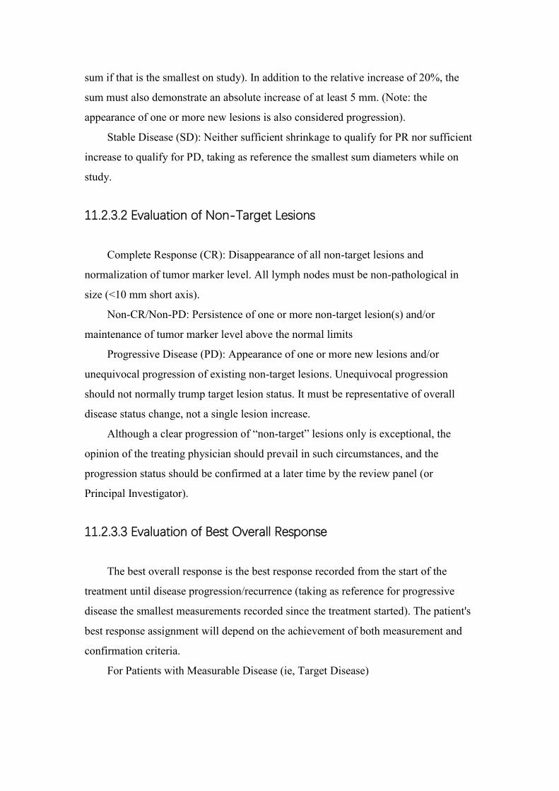
sum if that is the smallest on study). In addition to the relative increase of 20%, the
sum must also demonstrate an absolute increase of at least 5 mm. (Note: the
appearance of one or more new lesions is also considered progression).
Stable Disease (SD): Neither sufficient shrinkage to qualify for PR nor sufficient
increase to qualify for PD, taking as reference the smallest sum diameters while on
study.
11.2.3.2 Evaluation of Non-Target Lesions
Complete Response (CR): Disappearance of all non-target lesions and
normalization of tumor marker level. All lymph nodes must be non-pathological in
size (<10 mm short axis).
Non-CR/Non-PD: Persistence of one or more non-target lesion(s) and/or
maintenance of tumor marker level above the normal limits
Progressive Disease (PD): Appearance of one or more new lesions and/or
unequivocal progression of existing non-target lesions. Unequivocal progression
should not normally trump target lesion status. It must be representative of overall
disease status change, not a single lesion increase.
Although a clear progression of “non-target” lesions only is exceptional, the
opinion of the treating physician should prevail in such circumstances, and the
progression status should be confirmed at a later time by the review panel (or
Principal Investigator).
11.2.3.3 Evaluation of Best Overall Response
The best overall response is the best response recorded from the start of the
treatment until disease progression/recurrence (taking as reference for progressive
disease the smallest measurements recorded since the treatment started). The patient's
best response assignment will depend on the achievement of both measurement and
confirmation criteria.
For Patients with Measurable Disease (ie, Target Disease)

11.2.4 Duration of Response
11.2.4.1 Duration of overall response: The duration of overall response is
measured from the time measurement criteria are met for CR or PR (whichever is first
recorded) until the first date that recurrent or progressive disease is objectively
documented (taking as reference for progressive disease the smallest measurements
recorded since the treatment started).
The duration of overall CR is measured from the time measurement criteria are
first met for CR until the first date that progressive disease is objectively documented.
11.2.4.2 Duration of stable disease: Stable disease is measured from the start of
the treatment until the criteria for progression are met, taking as reference the smallest
measurements recorded since the treatment started, including the baseline
measurements.
12.0 Appendix
Appendix 1 RECIST 1.1
Appendix 2 ECOG scores

Appendix 3 CYP3A4 inducers and inhibitors
Appendix 4 CTCAE version 4.0
Appendix 5 EORTC QLQ C-30
Adverse Effect of apatinib from company
13.0 References
1. Bernthal, N.M., et al., Long-term results (>25 years) of a randomized, prospective
clinical trial evaluating chemotherapy in patients with high-grade, operable
osteosarcoma. Cancer, 2012. 118(23): p. 5888-93.
2. Bielack, S., et al., Osteosarcoma: the COSS experience. Cancer Treat Res, 2009.
152: p. 289-308.
3. Kempf-Bielack, B., et al., Osteosarcoma relapse after combined modality therapy:
an analysis of unselected patients in the Cooperative Osteosarcoma Study Group
(COSS). J Clin Oncol, 2005. 23(3): p. 559-68.
4. Cheong, J.H., et al., Surgical management and outcome of metachronous
Krukenberg tumors from gastric cancer. J Surg Oncol, 2004. 87(1): p. 39-45.
5. van Rijk-Zwikker, G.L., et al., Pulmonary metastasectomy in patients with
osteosarcoma. Eur J Cardiothorac Surg, 1991. 5(8): p. 406-9.
6. Thompson, R.C., Jr., et al., Results of treatment for metastatic osteosarcoma with
neoadjuvant chemotherapy and surgery. Clin Orthop Relat Res, 2002(397): p. 240-7.
7. Navid, F., et al., Combination of gemcitabine and docetaxel in the treatment of
children and young adults with refractory bone sarcoma. Cancer, 2008. 113(2): p.
419-25.
8. Berger, M., et al., Phase 2 trial of two courses of cyclophosphamide and
etoposide for relapsed high-risk osteosarcoma patients. Cancer, 2009. 115(13): p.
2980-7.
9. Saylors, R.L., 3rd, et al., Cyclophosphamide plus topotecan in children with
recurrent or refractory solid tumors: a Pediatric Oncology Group phase II study. J
Clin Oncol, 2001. 19(15): p. 3463-9.

10. Miser, J.S., et al., Ifosfamide with mesna uroprotection and etoposide: an
effective regimen in the treatment of recurrent sarcomas and other tumors of children
and young adults. J Clin Oncol, 1987. 5(8): p. 1191-8.
11. McNall-Knapp, R.Y., et al., Extended phase I evaluation of vincristine, irinotecan,
temozolomide, and antibiotic in children with refractory solid tumors. Pediatr Blood
Cancer, 2010. 54(7): p. 909-15.
12. Bomgaars, L.R., et al., Phase II trial of irinotecan in children with refractory solid
tumors: a Children's Oncology Group Study. J Clin Oncol, 2007. 25(29): p. 4622-7.
13. Cosetti, M., et al., Irinotecan for pediatric solid tumors: the Memorial
Sloan-Kettering experience. J Pediatr Hematol Oncol, 2002. 24(2): p. 101-5.
14. Debiec-Rychter, M., et al., KIT mutations and dose selection for imatinib in
patients with advanced gastrointestinal stromal tumours. Eur J Cancer, 2006. 42(8): p.
1093-103.
15. Shepherd, F.A., et al., Erlotinib in previously treated non-small-cell lung cancer.
N Engl J Med, 2005. 353(2): p. 123-32.
16. Sleijfer, S., et al., Pazopanib, a multikinase angiogenesis inhibitor, in patients
with relapsed or refractory advanced soft tissue sarcoma: a phase II study from the
European organisation for research and treatment of cancer-soft tissue and bone
sarcoma group (EORTC study 62043). J Clin Oncol, 2009. 27(19): p. 3126-32.
17. Linch, M., et al., Systemic treatment of soft-tissue sarcoma-gold standard and
novel therapies. Nat Rev Clin Oncol, 2014. 11(4): p. 187-202.
18. Schoffski, P., et al., Soft tissue sarcoma: an update on systemic treatment options
for patients with advanced disease. Oncol Res Treat, 2014. 37(6): p. 355-62.
19. Xu, M., et al., Effects of endostar combined multidrug chemotherapy in
osteosarcoma. Bone, 2013. 57(1): p. 111-5.
20. Grignani, G., et al., A phase II trial of sorafenib in relapsed and unresectable
high-grade osteosarcoma after failure of standard multimodal therapy: an Italian
Sarcoma Group study. Ann Oncol, 2012. 23(2): p. 508-16.
21. Yoo, C., et al., Multicenter phase II study of everolimus in patients with
metastatic or recurrent bone and soft-tissue sarcomas after failure of anthracycline and
ifosfamide. Invest New Drugs, 2013. 31(6): p. 1602-8.

22. Grignani, G., et al., Sorafenib and everolimus for patients with unresectable
high-grade osteosarcoma progressing after standard treatment: a non-randomised
phase 2 clinical trial. Lancet Oncol, 2015. 16(1): p. 98-107.
23. Lammli, J., et al., Expression of Vascular Endothelial Growth Factor correlates
with the advance of clinical osteosarcoma. Int Orthop, 2012. 36(11): p. 2307-13.
24. Li, J., et al., Apatinib for chemotherapy-refractory advanced metastatic gastric
cancer: results from a randomized, placebo-controlled, parallel-arm, phase II trial. J
Clin Oncol, 2013. 31(26): p. 3219-25.
25. Aras, M., et al., Comparison of WHO, RECIST 1.1, EORTC, and PERCIST
criteria in the evaluation of treatment response in malignant solid tumors. Nucl Med
Commun, 2016. 37(1): p. 9-15.
26. Choi, H.C., et al., Comparison of the RECIST 1.0 and RECIST 1.1 in Non-Small
Cell Lung Cancer Treated with Cytotoxic Chemotherapy. J Cancer, 2015. 6(7): p.
652-7.
27. Semiglazov, V., RECIST for Response (Clinical and Imaging) in Neoadjuvant
Clinical Trials in Operable Breast Cancer. J Natl Cancer Inst Monogr, 2015. 2015(51):
p. 21-3.
28. Young, J., et al., Comparison of ECOG/WHO performance status and ASA score
as a measure of functional status. J Pain Symptom Manage, 2015. 49(2): p. 258-64.
29. Broderick, J.M., et al., Patients over 65 years are assigned lower ECOG PS scores
than younger patients, although objectively measured physical activity is no different.
J Geriatr Oncol, 2014. 5(1): p. 49-56.
30. de Kock, I., et al., Conversion of Karnofsky Performance Status (KPS) and
Eastern Cooperative Oncology Group Performance Status (ECOG) to Palliative
Performance Scale (PPS), and the interchangeability of PPS and KPS in prognostic
tools. J Palliat Care, 2013. 29(3): p. 163-9.
31. Simon, R., Optimal two-stage designs for phase II clinical trials. Control Clin
Trials, 1989. 10(1): p. 1-10.