Embed Size (px)
Citation preview

BioMed CentralProteome Science
ss
Open AcceResearchBacillus anthracis secretome time course under host-simulated conditions and identification of immunogenic proteinsAlexander Walz*1, Cesar V Mujer1,4, Joseph P Connolly1, Tim Alefantis1, Ryan Chafin1, Clarissa Dake1, Jessica Whittington1, Srikanta P Kumar2, Akbar S Khan3 and Vito G DelVecchio1Address: 1Vital Probes, Inc., 1300 Old Plank Road, Mayfield, PA 18433, USA, 2BAE Systems Inc., 164 Totowa Road, Wayne, NJ 07474, USA, 3Chemical and Biological Defense Directorate, Defense Threat Reduction Agency, 6801 Telegraph Road, Alexandria, VA, USA and 4Calvert Laboratories, Inc., Scott Technology Park, 100 Discovery Drive, Olyphant, PA 18447, USA
Email: Alexander Walz* - [email protected]; Cesar V Mujer - [email protected]; Joseph P Connolly - [email protected]; Tim Alefantis - [email protected]; Ryan Chafin - [email protected]; Clarissa Dake - [email protected]; Jessica Whittington - [email protected]; Srikanta P Kumar - [email protected]; Akbar S Khan - [email protected]; Vito G DelVecchio - [email protected]
* Corresponding author
AbstractBackground: The secretion time course of Bacillus anthracis strain RA3R (pXO1+/pXO2-) during early, mid, andlate log phase were investigated under conditions that simulate those encountered in the host. All of the identifiedproteins were analyzed by different software algorithms to characterize their predicted mode of secretion andcellular localization. In addition, immunogenic proteins were identified using sera from humans with cutaneousanthrax.
Results: A total of 275 extracellular proteins were identified by a combination of LC MS/MS and MALDI-TOFMS. All of the identified proteins were analyzed by SignalP, SecretomeP, PSORT, LipoP, TMHMM, and PROSITEto characterize their predicted mode of secretion, cellular localization, and protein domains. Fifty-three proteinswere predicted by SignalP to harbor the cleavable N-terminal signal peptides and were therefore secreted via theclassical Sec pathway. Twenty-three proteins were predicted by SecretomeP for secretion by the alternative Secpathway characterized by the lack of typical export signal. In contrast to SignalP and SecretomeP predictions,PSORT predicted 171 extracellular proteins, 7 cell wall-associated proteins, and 6 cytoplasmic proteins.Moreover, 51 proteins were predicted by LipoP to contain putative Sec signal peptides (38 have SpI sites),lipoprotein signal peptides (13 have SpII sites), and N-terminal membrane helices (9 have transmembrane helices).The TMHMM algorithm predicted 25 membrane-associated proteins with one to ten transmembrane helices.
Immunogenic proteins were also identified using sera from patients who have recovered from anthrax. Thecharge variants (83 and 63 kDa) of protective antigen (PA) were the most immunodominant secreted antigens,followed by charge variants of enolase and transketolase.
Conclusion: This is the first description of the time course of protein secretion for the pathogen Bacillusanthracis. Time course studies of protein secretion and accumulation may be relevant in elucidation of theprogression of pathogenicity, identification of therapeutics and diagnostic markers, and vaccine development. Thisstudy also adds to the continuously growing list of identified Bacillus anthracis secretome proteins.
Published: 27 July 2007
Proteome Science 2007, 5:11 doi:10.1186/1477-5956-5-11
Received: 9 April 2007Accepted: 27 July 2007
This article is available from: http://www.proteomesci.com/content/5/1/11
© 2007 Walz et al; licensee BioMed Central Ltd. This is an Open Access article distributed under the terms of the Creative Commons Attribution License (http://creativecommons.org/licenses/by/2.0), which permits unrestricted use, distribution, and reproduction in any medium, provided the original work is properly cited.
Page 1 of 10(page number not for citation purposes)
Proteome Science 2007, 5:11 http://www.proteomesci.com/content/5/1/11
BackgroundBacillus anthracis is a Gram-positive spore-forming bacte-rium that is the etiologic agent of anthrax [1]. During thecourse of infection, the virulent spores germinate tobecome vegetative cells. The bacterium secretes two majorvirulence factors, the bipartite edema and lethal toxinsthat are encoded by plasmid pXO1 [1-3]. The commoncomponent of both toxins is the protective antigen (PA)which is not toxic by itself. PA binds a specific receptor onthe host cells and translocates the edema factor (EF) andlethal factor (LF) inside the cells where they exert theirdamaging action. Transcription of the genes coding forthese virulence factors has been shown to be coordinatelyinduced by bicarbonate-CO2. High CO2 tension isbelieved to simulate conditions encountered within thehost [4]. In addition, the effect of temperature has beenshown to be important for toxin production but not forproduction of antiphagocytic capsule, whose synthesis isencoded by genes on plasmid pXO2 [5]. Thus, when B.anthracis is cultured at 37°C in a bicarbonate-containingminimal medium, toxin production is enhanced.
The composition and identification of B. anthracis extra-cellular proteins (secretome) has been the subject ofrecent proteomic studies [6-9]. In B. cereus, a close relativeof B. anthracis, most of these secreted proteins include col-lagenases, phospholipases, hemolysins, proteases, andenterotoxins that are positively regulated by plcR [10].However, a non-sense mutation inactivates a homolog ofthis gene in B. anthracis resulting in a significant reductionof these secreted proteins [1,11,12]. Using 2-DE andMALDI-TOF MS, Chitlaru et al. [6] have identified 64extracellular proteins in a virulent B. anthracis strain, ofwhich 50 exhibit export signal peptides. Thirty-one ofthese secreted proteins harbor features that are character-istic of virulence determinants suggesting that in additionto the "classic" lethal and edema toxins, a large number ofproteins may be essential for B. anthracis virulence [6].Additionally, in minimal medium under high CO2 ten-sion, the presence of the plasmids led to the enhancedsecretion of 12 chromosome-encoded and 5 pXO1encoded proteins. Ten of the chromosome-encoded pro-teins could not be detected in the absence of the plasmids.These results suggest distinct plasmid and chromosomeCO2-dependent crosstalk mechanisms that modulateextracellular proteolytic activities. Likewise, Antelmann etal. [7] have identified 64 extracellular proteins in the non-virulent B. anthracis strain UM23C1-2 (pXO1-/pXO2-), 29of which were predicted to be secreted. The remainder ofthe extracellular proteins were predicted to be associatedwith the cell wall or the cytosol. Based on the nature of B.anthracis secretome, it was suggested that this organism isadapted to life in a protein-rich environment due to thepresence of a variety of proteases, peptidases, peptide-binding proteins, as well as enzymes required for the
metabolism of amino acids. It was also proposed thatthese secreted proteases and peptidases could be usefultargets for the development of improved vaccines.
In another study, Gohar et al. [8] compared the fully curedB. anthracis extracellular proteomes to two other membersof the B. cereus group, B. cereus and B. thuringiensis, thatwere also cured of their plasmids. The secretomes of thethree species included both cell wall and cytosolic pro-teins that are similar in all three species. However,whereas the extracellular proteins of B. cereus and B. thur-ingiensis contained a large number of secreted proteinssuch as degradative enzymes and toxins, those of B.anthracis contained only one secreted protein, a metallo-protease InhA1. These results are in contrast to thosereported by Chitlaru et al. [6] and Antelmann et al. [7]where 31 and 29 secreted proteins were identified in theculture filtrate of fully cured B. anthracis cells, respectively.Using minimal medium under high CO2 tension, a com-parative analysis of the extracellular proteomes of threeisogenic strains of B. anthracis that differed solely in theirplasmid content was conducted by Lamonica et al. [9]. Inthis study, the use of SignalP for the prediction of cellularfunction and location of each protein indicated that mostof the identified proteins were either cell wall-associatedor cytoplasmic in the fully cured strain, RA3:00.
The present study characterized the time course of proteinsecretion and accumulation in the culture of B. anthracisstrain RA3R (pXO1+/pXO2-). Secretion patterns at the dif-ferent time points are due not only to differentiallyexpressed proteins at various phases of exponentialgrowth, but also to the accumulation of proteins in theculture and their stability therein. Particular focus wasplaced on the analysis of protein secretion during theexponential growth phase to preclude the identification ofproteins that may have been caused by bacterial lysis. Inaddition, computer assisted comparative analysis using2D Phoretix of the cytosol with the secretome at16 hrunder induced conditions was performed. LC MS/MS wasused as a complementary tool in identifying proteins thatwere not amenable to MALDI-TOF MS, significantlyincreasing the number of identified secreted proteins. Thedata from this analysis add to the growing list of identifiedproteins in the secretome database of B. anthracis and helpin further identifying key pathways associated with viru-lence. Furthermore, immunogenic proteins, or the immu-nome, of the secretome were identified using sera fromhuman patients infected with cutaneous anthrax. Identifi-cation of these immunodominant antigens is essential fordevelopment of more efficacious and safer vaccinesagainst anthrax.
Page 2 of 10(page number not for citation purposes)
Proteome Science 2007, 5:11 http://www.proteomesci.com/content/5/1/11
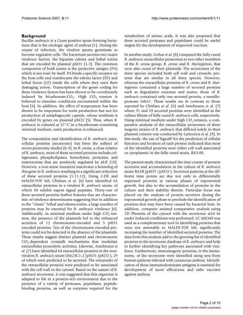
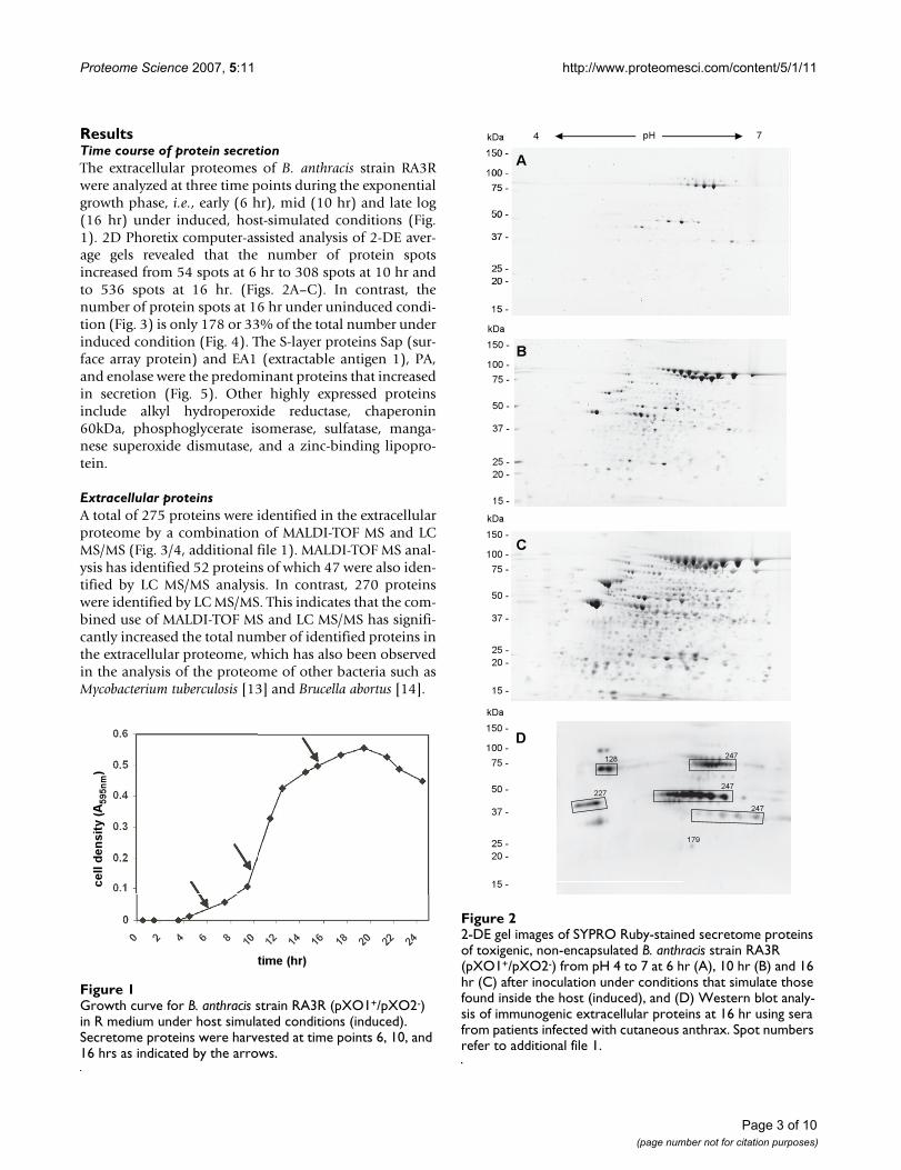
ResultsTime course of protein secretionThe extracellular proteomes of B. anthracis strain RA3Rwere analyzed at three time points during the exponentialgrowth phase, i.e., early (6 hr), mid (10 hr) and late log(16 hr) under induced, host-simulated conditions (Fig.1). 2D Phoretix computer-assisted analysis of 2-DE aver-age gels revealed that the number of protein spotsincreased from 54 spots at 6 hr to 308 spots at 10 hr andto 536 spots at 16 hr. (Figs. 2A–C). In contrast, thenumber of protein spots at 16 hr under uninduced condi-tion (Fig. 3) is only 178 or 33% of the total number underinduced condition (Fig. 4). The S-layer proteins Sap (sur-face array protein) and EA1 (extractable antigen 1), PA,and enolase were the predominant proteins that increasedin secretion (Fig. 5). Other highly expressed proteinsinclude alkyl hydroperoxide reductase, chaperonin60kDa, phosphoglycerate isomerase, sulfatase, manga-nese superoxide dismutase, and a zinc-binding lipopro-tein.
Extracellular proteinsA total of 275 proteins were identified in the extracellularproteome by a combination of MALDI-TOF MS and LCMS/MS (Fig. 3/4, additional file 1). MALDI-TOF MS anal-ysis has identified 52 proteins of which 47 were also iden-tified by LC MS/MS analysis. In contrast, 270 proteinswere identified by LC MS/MS. This indicates that the com-bined use of MALDI-TOF MS and LC MS/MS has signifi-cantly increased the total number of identified proteins inthe extracellular proteome, which has also been observedin the analysis of the proteome of other bacteria such asMycobacterium tuberculosis [13] and Brucella abortus [14].
2-DE gel images of SYPRO Ruby-stained secretome proteins of toxigenic, non-encapsulated B. anthracis strain RA3R (pXO1+/pXO2-) from pH 4 to 7 at 6 hr (A), 10 hr (B) and 16 hr (C) after inoculation under conditions that simulate those found inside the host (induced), and (D) Western blot analy-sis of immunogenic extracellular proteins at 16 hr using sera from patients infected with cutaneous anthraxFigure 22-DE gel images of SYPRO Ruby-stained secretome proteins of toxigenic, non-encapsulated B. anthracis strain RA3R (pXO1+/pXO2-) from pH 4 to 7 at 6 hr (A), 10 hr (B) and 16 hr (C) after inoculation under conditions that simulate those found inside the host (induced), and (D) Western blot analy-sis of immunogenic extracellular proteins at 16 hr using sera from patients infected with cutaneous anthrax. Spot numbers refer to additional file 1.
Growth curve for B. anthracis strain RA3R (pXO1+/pXO2-) in R medium under host simulated conditions (induced)Figure 1Growth curve for B. anthracis strain RA3R (pXO1+/pXO2-) in R medium under host simulated conditions (induced). Secretome proteins were harvested at time points 6, 10, and 16 hrs as indicated by the arrows.
Page 3 of 10(page number not for citation purposes)
Proteome Science 2007, 5:11 http://www.proteomesci.com/content/5/1/11
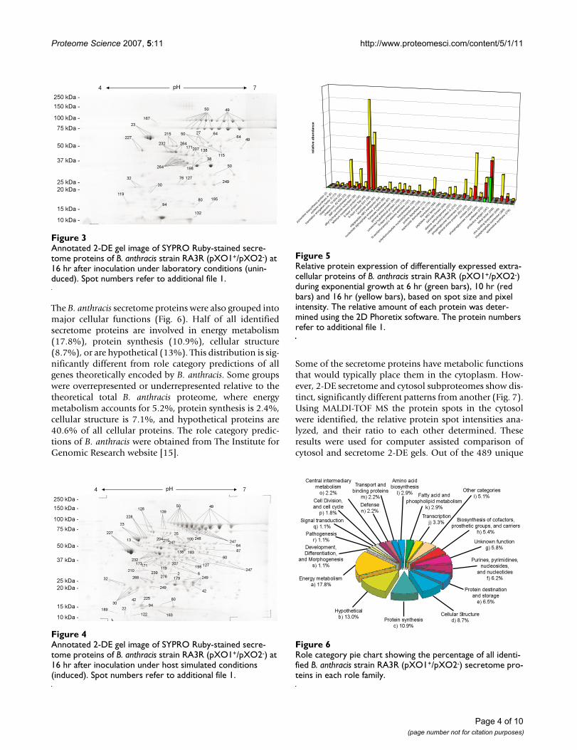
The B. anthracis secretome proteins were also grouped intomajor cellular functions (Fig. 6). Half of all identifiedsecretome proteins are involved in energy metabolism(17.8%), protein synthesis (10.9%), cellular structure(8.7%), or are hypothetical (13%). This distribution is sig-nificantly different from role category predictions of allgenes theoretically encoded by B. anthracis. Some groupswere overrepresented or underrepresented relative to thetheoretical total B. anthracis proteome, where energymetabolism accounts for 5.2%, protein synthesis is 2.4%,cellular structure is 7.1%, and hypothetical proteins are40.6% of all cellular proteins. The role category predic-tions of B. anthracis were obtained from The Institute forGenomic Research website [15].
Some of the secretome proteins have metabolic functionsthat would typically place them in the cytoplasm. How-ever, 2-DE secretome and cytosol subproteomes show dis-tinct, significantly different patterns from another (Fig. 7).Using MALDI-TOF MS the protein spots in the cytosolwere identified, the relative protein spot intensities ana-lyzed, and their ratio to each other determined. Theseresults were used for computer assisted comparison ofcytosol and secretome 2-DE gels. Out of the 489 unique
Relative protein expression of differentially expressed extra-cellular proteins of B. anthracis strain RA3R (pXO1+/pXO2-) during exponential growth at 6 hr (green bars), 10 hr (red bars) and 16 hr (yellow bars), based on spot size and pixel intensityFigure 5Relative protein expression of differentially expressed extra-cellular proteins of B. anthracis strain RA3R (pXO1+/pXO2-) during exponential growth at 6 hr (green bars), 10 hr (red bars) and 16 hr (yellow bars), based on spot size and pixel intensity. The relative amount of each protein was deter-mined using the 2D Phoretix software. The protein numbers refer to additional file 1.
pyro
xidine
bios
ynth
esis
prot
ein (2
)
cyste
ine sy
ntha
seA
(6)
trans
lation
elong
ation
facto
r G (1
3)
chap
eron
in,10
kDa (
22)
chap
eron
in,60
kDa (
23)
IMP
cyclo
hydr
olase
(25)
alkyl
hydr
oper
oxide
redu
ctase
(30)
telle
rium re
sista
nce p
rotei
n (32
)
S-laye
r pro
tein S
ap(4
9)
S-laye
r pro
tein E
A1 (50
)
isocit
rate
lyase
(60)
oligo
pepti
deABC tr
ansp
orte
r (64
)
nucle
oside
dipho
spha
te ki
nase
, puta
tive
(80)
fumar
ate h
ydra
tase (
87)
gene
ral s
tress
prot
ein(9
4)
cons
erve
dhy
pothe
tical
prot
ein(1
00)
S-laye
r pro
tein,
puta
tive
(119
)
N-ace
tylmur
amoy
l-L-a
lanine
amida
se(1
27)
trans
ketol
ase (
128)
polyr
ibonu
cleot
ide n
ucleo
tidylt
rans
fera
se (1
39)
proli
nedip
eptid
ase (
173)
supe
roxid
e dism
utas
e, M
n (17
9)
thior
edox
in (1
89)
pepti
dase
, M42
family
(198
)
pyru
vate
kina
se (2
04)
alanin
e deh
ydro
gena
se(2
07)
cata
bolite
cont
rol p
rotei
n A (2
10)
phos
phog
lucos
e iso
meras
e (21
5)
gene
ral s
tress
prot
ein20
U(2
25)
enola
se (2
27)
phos
phog
lycer
ate m
utase
(228
)
sulfa
tase
(232
)
prot
ectiv
e an
tigen
(247
)
letha
l facto
r (24
8)
zinc-b
inding
lipop
rote
in(2
49)
trios
epho
spha
te iso
meras
e (26
8)
sper
midi
ne sy
ntha
se(2
76)
rela
tive
ab
un
dan
ce
Role category pie chart showing the percentage of all identi-fied B. anthracis strain RA3R (pXO1+/pXO2-) secretome pro-teins in each role familyFigure 6Role category pie chart showing the percentage of all identi-fied B. anthracis strain RA3R (pXO1+/pXO2-) secretome pro-teins in each role family.
Annotated 2-DE gel image of SYPRO Ruby-stained secre-tome proteins of B. anthracis strain RA3R (pXO1+/pXO2-) at 16 hr after inoculation under laboratory conditions (unin-duced)Figure 3Annotated 2-DE gel image of SYPRO Ruby-stained secre-tome proteins of B. anthracis strain RA3R (pXO1+/pXO2-) at 16 hr after inoculation under laboratory conditions (unin-duced). Spot numbers refer to additional file 1.
Annotated 2-DE gel image of SYPRO Ruby-stained secre-tome proteins of B. anthracis strain RA3R (pXO1+/pXO2-) at 16 hr after inoculation under host simulated conditions (induced)Figure 4Annotated 2-DE gel image of SYPRO Ruby-stained secre-tome proteins of B. anthracis strain RA3R (pXO1+/pXO2-) at 16 hr after inoculation under host simulated conditions (induced). Spot numbers refer to additional file 1.
Page 4 of 10(page number not for citation purposes)

Proteome Science 2007, 5:11 http://www.proteomesci.com/content/5/1/11
protein spots in the cytosol, 56 are more than two-fold upregulated, 84 are more than two-fold down regulated, and177 proteins spots have no corresponding spot in thesecretome (Fig. 7B). If protein spots in the secretome weresimply the result of cell lysis, not only should their relativeposition on the gel be the same between the cytosol andthe secretome, but the ratio of the proteins to each otherin each subproteome should be the same in both. Theresults show that two thirds of all protein spot positionsas well as relative abundances are distinctively differentbetween the cytosol and secretome. Therefore, cell lysiscan be excluded as the major contributor to the proteinaccumulation in the secretome. The extracellular proteinswere further analyzed using various bioinformatics soft-ware programs, such as SignalP, SecretomeP, PSORT,LipoP, TMHMM, and PROSITE for predicting proteinsecretion and localization.
Classical Sec pathwayA total of 53 proteins were predicted by SignalP to besecreted in the classical Sec pathway, which is character-ized by the presence of a signal peptide [16,17] (addi-tional file 1). Of the 53 proteins containing the signalpeptides, 38 proteins have the cleavage site for signalpeptidase I (SpI). These proteins are predicted to besecreted into the external environment, because they lackadditional retention signals. However, 10 proteins havethe cleavage site for signal peptidase II (SpII) and theretention signals for lipid anchors while three proteinswere predicted to have transmembrane helices (TMH).These proteins are predicted to have an extracytoplasmicbut cell-associated location. LipoP predictions discrimi-nate between lipoprotein signal peptides, other signalpeptides, and N-terminal membrane helices with 93%accuracy in Gram positive bacteria [18]. In contrast toLipoP, TMHMM predicted 25 integral membrane proteinswith one to ten transmembrane helices [19] (additionalfile 1). TMHMM predicts transmembrane protein topol-ogy with a hidden Markov model with a 98% accuracy.Additional predictions using PROSITE identified severalsecreted proteins that are also know to be associated withthe bacterial cell wall by S-layer homology domains orlipoprotein lipid attachment sites [7,20]. No cell wallassociated proteins with LPXTG-motifs were found in thesecretome. Despite its name, the LPXTG-motif cell wallanchor domain protein does not contain such a domain.In fact, it contains a NEAT domain that might be involvedin the transport of iron.
Alternative Sec pathwayTwenty-three proteins were predicted by SecretomeP to besecreted by the non-classical Sec Pathway characterized bythe lack of typical export signals [21]. In contrast to thepredictions of SignalP and SecretomeP, PSORT [22] pre-dicted 171 extracellular proteins, 7 cell wall-associated
proteins, and 6 cytoplasmic proteins. Using the wholegenome of B. anthracis, Binnewies et al. have calculatedthat the Bacillus anthracis Ames strain has 6% secreted pro-teins predicted using SecretomeP, 3% using LipoP, and6% using SignalP [23].
Immunogenic extracellular proteinsThe main immunogenic proteins detected by 2-DE West-ern blot analysis using sera from humans infected withcutaneous anthrax include the 83 and 63 kDa charge var-iants of protective antigen (PA), followed by charge vari-ants of enolase and transketolase (Fig. 2D). The protectiveantigen produced in vivo has a molecular mass of 83 kDa
(A) 2-DE gel image of SYPRO Ruby-stained cytosol proteins of toxigenic, non-encapsulated B. anthracis strain RA3R (pXO1+/pXO2-) from pH 4 to 7 at 16 hr under induced con-ditionsFigure 7(A) 2-DE gel image of SYPRO Ruby-stained cytosol proteins of toxigenic, non-encapsulated B. anthracis strain RA3R (pXO1+/pXO2-) from pH 4 to 7 at 16 hr under induced con-ditions. (B) 2D Phoretix comparative analysis of the cytosol with the secretome at 16 hr under induced conditions. Pro-teins encircled in red are more than two fold up-regulated, proteins encircled in yellow are more than two fold down-regulated, proteins encircled in green are between these lim-its, and proteins encircled in blue are unmatched between the two sub proteomes.
Page 5 of 10(page number not for citation purposes)
Proteome Science 2007, 5:11 http://www.proteomesci.com/content/5/1/11
and is subsequently cleaved by cell-associated proteaseactivity resulting in a 63 kDa protein that binds the lethalfactor to form lethal toxin [24]. Notably, 17 charge andmass variants of PA were detected. These include fivecharge variants of the 83 kDa PA isoforms, seven chargevariants of the 63 kDa PA isoforms and five charge vari-ants of the 37 kDa PA isoforms. Charge variants of PAhave been reported previously. Their generation was dueto the spontaneous deamidation of asparagine residueswhich is dependent on pH, temperature, primarysequence ("nearest neighbor" effect) and protein confor-mation [25]. Charge variants of enolase and transketolasewere also noted. Enolase has also been reported previ-ously as component of the B. anthracis spore [26] and asone of the immunodominant spore antigens [27]. Otherminor immunogenic proteins were also detected by 2-DEWestern blot analysis but were not identified.
DiscussionThe secretome of B. anthracis has been the subject ofrecent proteomic studies [6-9]. Interest in its study comesfrom the fact that several secreted proteins of B. anthracisare known virulence factors (e.g., PA, EF, LF). Othersecreted proteins may potentially be involved in theadherence of the bacteria to host cells while some may berequired for the suppression of the host's defense mecha-nisms. While several of the virulence proteins have beenidentified by recent proteomic studies [6,7], the timecourse of protein secretion by a toxigenic but non-encap-sulated strain RA3R (pXO1+/pXO2-) under host simulatedconditions has not been reported. In this study, thedynamics of secretion of several proteins were investi-gated at different time points during the exponentialgrowth phase of B. anthracis. The same collection pointshave been used previously by Lamonica et al. [9] for theisolation of secretome proteins. Chitlaru et al. [6] andAntelmann et al. [7] isolated secretome proteins at analo-gous growth stages as well. As shown in Fig. 5, the rate ofprotein secretion varies for each protein. It should benoted that at 6 hours, PA has the highest rate of secretionof all secreted proteins. Since PA is a major component ofthe anthrax toxin, its high secretion rate at this early timepoint confirms the critical role this protein plays duringthe onset of anthrax pathogenesis. Two other proteins thatwere detected in high abundance in the culture superna-tant are the S-layer proteins, EA1 and Sap, both of whichare components of the cell wall. Both proteins contain asignal peptide followed by three SLH (S-layer homology)anchoring domains and are considered major surfaceantigens. The amount of these two proteins was highest atthe 16-hour time point compared to the other identifiedextracellular proteins. It has been previously describedthat Sap is sequentially replaced by EA1 [28]. This rapid S-layer turnover results in the release and spillover of theseproteins into the secretome.
Several of the secretome proteins have metabolic func-tions that would typically place them in the cytoplasm.This is similar to the results by Antelmann et al. [7] inwhich more than half of the identified secretome proteinswere associated with the cell wall or cytosol. We also iden-tified 25 proteins that were predicted to have transmem-brane helices which would typically associate them to thecell wall. All but one of these proteins were predicted to besecreted by SignalP or SecretomeP and half of these pro-teins have also been identified before as a natural compo-nent of the secretome (see additional file 1).
Extracellular accumulation of B. anthracis proteinsCombined analysis using SignalP and SecretomeP of theB. anthracis secreted proteins indicated that 28% of thedetected proteins are extracellular whereas 62% are pre-dicted to be extracellular using PSORT. The remainingproteins were predicted to be cytoplasmic since theylacked known export signals or are cell-associated becausethey have membrane-anchoring or cell wall retention sig-nals. Using LipoP, some of the proteins are predicted to bebound at the trans surface of the cytoplasmic membrane.Contrary to their predicted location, a number of proteinswith retention signals for covalent or non-covalent attach-ment to the cell walls were also found in the extracellularenvironment.
While bioinformatics tools are useful for predicting cellu-lar function and localization, considerable variation existsin the number of proteins that were predicted to be extra-cellular, cytoplasmic, or membrane bound. This isexpected because each of these bioinformatics tools usesdifferent algorithms and assumptions in their predictions.Further empirical studies are therefore required to verifythe precise location of proteins for which conflicting pre-dictions are noted.
Immunogenic extracellular proteinsThe use of sera from human patients infected with cutane-ous anthrax confirmed the high immunogenicity of PA inthe secreted proteins of the toxigenic but non-encapsu-lated B. anthracis strain RA3R (pXO1+/pXO2-). Althoughseveral mass and charge variants of PA were detected, themost immunogenic are the 63 and 83 kDa charge variants.In addition to PA, the major component of AVA (anthraxvaccine adsorbed), enolase and transketolase were foundto be highly immunogenic. AVA is an alum precipitateprepared from B. anthracis culture filtrates and is alicensed vaccine currently used to protect humans againstanthrax. Enolase was also described previously as part ofthe AVA culture filtrate [29]. AVA preparations containcontaminating proteins whose benefits are not known,but the presence of the highly immunogenic enolase inthis formulation might contribute to its protective immu-nity over a vaccine containing just PA. The membrane pro-
Page 6 of 10(page number not for citation purposes)

Proteome Science 2007, 5:11 http://www.proteomesci.com/content/5/1/11
teins Sap and EA1 were also found in the secretome byChitlaru et al. [6] and Antelmann et al. [7] as the mostabundant extracellular cell wall proteins. Both proteinswere reported as major surface antigens and potential vac-cine carriers in vivo [30,31]. EA1 and Sap are also majorcomponents of AVA [29] and are associated with a varietyof functions ranging from evasion of host recognition, celladhesion and resistance, and phagocytosis [1,32,33].Interestingly no reactivity to these proteins was observedusing sera from patients with subcutaneous anthrax infec-tion. Overall fewer immunogenic proteins were detectedusing human sera from patients infected with cutaneousanthrax than in a recent study by Chitlaru et al. [30] whoused experimentally challenged rabbit and guinea pigsera. There is a quantitative and qualitative differencebetween sera from recovering humans and sera from mul-tiple challenged animals. Since with human sera enolaseand transketolase were major immunogens beside PA,they might be promising candidates for next generationanthrax subunit vaccines besides the newly identified pro-teins using animal sera [30].
ConclusionThe work presented here is the first description of the timecourse of protein secretion for B. anthracis grown underhost-simulated conditions. The combined use of twotypes of mass spectroscopy led to the identification of 275proteins and their secretion patterns during the exponen-tial growth phase of B. anthracis. While the secretome con-tained several predictable proteins (e.g. PA, LF, Sap, andEA1) many proteins were identified which would not beexpected in the secretome such as proteins involved inenergy metabolism and protein translation. The discoveryof proteins in the secretome that are traditionally thoughtto be strictly cytosolic has often been assumed to resultfrom contamination. However, this may not be true sincecytosolic proteins, such as aldolase, enolase, elongationfactor G, and various dehydrogenases, have also beendetected in the secretome of group A streptococci [34],mycobacteria [35,36], and B. subtilis [37]. Some proteinsmay be cytoplasmic at one point of the cell cycle andsecreted via pathways which are yet to be understood dur-ing other stages of the cell cycle. While prediction softwareare a good tool to characterize a large group of proteins,different algorithms give diverging results. It is far moredifficult to accurately predict a precise location within acell of non-classical secretory proteins than to recognizeproteins which are secreted by a signal peptide. The ulti-mate proof is to empirically validate their results. Thisstudy helps to bridge the gap between pure in silico predic-tion and in vivo observation.
This study also identified the major immunoreactive pro-teins of the B. anthracis secretome using 2-DE Westernblot analysis from humans infected with the pathogen.
These proteins included the expected PA as well as enolaseand transkelolase. The immunoreactive secretome pro-teins contribute to the list of other B. anthracis immuno-genic proteins that were idenfied in other subproteomesor life-stages of the infectious agent. This knowledge willultimately lead to the development of a more-specific,safer, and highly efficacious vaccine against B. anthracis. Inaddition, the identification of early high abundance secre-tome proteins may aid in the development of detectionand diagnostic kits for those cases, where the direct cap-ture of the pathogen is not possible.
MethodsBacterial strain and culture conditionsAn attenuated derivative strain of B. anthracis, RA3R(pXO1+/pXO2-), was used. A loop was streaked on BHIagar overnight (16 hr) at 37°C. A single colony was trans-ferred to 2 ml R medium [4] and approximately 1 ml ofthe resulting suspension was immediately transferred in100 ml R medium supplemented with 0.25% [wt/vol]glucose. The flask was mixed gently and fitted with a Bug-Stopper (Whatman Inc., Clifton, NJ) sterile venting clo-sure. The culture was incubated at 37°C with shaking at120 rpm for 5 to 6 hr. Following this growth period, 5 mlof the culture was transferred to a 250-ml sterile, vented,canted Falcon tissue culture flask containing 70 ml Rmedium with 0.25% [wt/vol] glucose and 0.85% [wt/vol]sodium bicarbonate. The composition of the R-mediumin mg/l is: L-tryptophan, 35; glycine, 65; L-cystine, 25; L-tyrosine, 144; L-lysine, 230; L-valine, 173; L-leucine, 230;L-isoleucine, 170; L-threonine, 120; L-methionine, 73; L-aspartic acid, 184; sodium L-glutamate, 612; L-proline,43; L-histidine-hydrochloride, 55; L-arginine-hydrochlo-ride, 125; L-phenylalanine, 125; L-serine, 235; thiamine-hydrochloride, 1.0; CaCl2 2H20, 7.4; MgSO4 H20, 9.9;MnSO4 H20, 0.9; K2HPO4, 3,000; uracil, 1.4; and adeninesulfate, 2.1, pH 8.0. The culture was grown for 6 to 16 hrat 37°C under 5% CO2 in a humid incubator according toRistroph et al. [4] to simulate conditions encountered inthe host. The pH at the end of the incubation period risesto pH 8.15. During this time the cells are in the exponen-tial growth phase, between lag- and stationary- phase.Uninduced cultures were grown in R medium without theaddition of sodium bicarbonate and without CO2 supple-mentation.
Preparation of extracellular protein fractionCulture filtrates (secretomes) were collected at 6 hr (earlylog phase), 10 hr (mid log phase), and 16 hr (late logphase) time points based on the growth curve for B.anthracis in R medium, as determined by OD595 readings.The culture filtrates were centrifuged for 10 min and thesupernatant containing the extracellular proteins waspassed through a 0.45-µm filter to remove any suspendedvegetative cells. The culture filtrates were concentrated
Page 7 of 10(page number not for citation purposes)
Proteome Science 2007, 5:11 http://www.proteomesci.com/content/5/1/11
using Jumbosep, Macrosep, and Microsep filters with a 10kDa cutoff (Pall Inc., East Hills, NY). Ice-cold trichloroace-tic acid (TCA; Sigma Chemical Co., St. Louis, MO) wasthen added to a final concentration of 10% TCA (vol/vol),chilled on ice for 45 min, and then centrifuged for 45 min.The resultant pellet was washed with 3 ml acetone andcentrifuged. The pellet containing the secretome proteinswas resuspended in 100 µl of 7 M urea, 2 M thiourea, 1%3-(4-heptyl)phenyl-3-hydroxypropyl-dimethylammonio-propanesulfonate, and 40 mM Tris prior to IEF. The pro-tein concentration of the extracellular protein extract wasdetermined using the Bio-Rad Protein Assay kit (Bio-RadLaboratories, Hercules, CA). Bovine serum albumin wasused as the standard for determination of protein concen-tration.
2-DE and Western blot analysis2-DE was carried out with the ElectrophoretIQ3 systemand following the manufacturer's protocols (ProteomeSystems, Woburn, MA). One-hundred µg of proteins wereseparated by IEF on 11 cm (pH 4 to 7) linear IPG strips.After 12 hr rehydration, the following focusing parame-ters were applied: 50 µA per strip, linear voltage increaseover 8 hr from 100 V to 10,000 V, and then held at 10,000V for 10 hr. After IEF, IPG strips were equilibrated in equi-libration buffer and applied onto a 6–15% gradient SDS-PAGE. Gels were electrophoresed for 1.5 hr at 500 V andstained with Sypro Ruby (Sigma-Aldrich, St. Louis, MO)for gel analysis or with ProteomIQ Blue (Proteome Sys-tems) for MALDI-TOF MS analysis. Four replicate 2-DEgels of each sample were used for computer analysis usingPhoretix 2D Expression software (Nonlinear Dynamics)and MALDI-TOF MS. Following automatic spot detectionon quadruple gels using Phoretix 2D Expression's detec-tion algorithm, gels were manually warped and their com-mon spots were matched to generate average gels.
Immunoblotting was conducted according to Towbin etal. [38]. Proteins on 2-DE gels were transferred to PVDFmembranes using Towbin buffer (0.025 M trisma base in0.192 M glycine) with 20% methanol at 100 V for 30 min.After transfer, the PVDF membrane was washed twice for5 min each in 0.01% Tween-20 in PBS (PBST) andblocked with 0.2% I-Block (Tropix, Bedford, MA) for 1 hr.To identify immunogenic proteins, the PVDF membraneswere washed three times with PBST for 5 min and thenprobed with a 1:1000 dilution of sera pooled frompatients who recovered from cutaneous anthrax. ThePVDF membrane was washed three times with PBST andincubated with 1:5000 dilution of appropriate secondaryantibody. Chemiluminescent signals were visualizedusing the Western Lightning reagents (Perkin-Elmer,Wellesley, MA). Corresponding sera from uninfectedhumans were used as controls (Cambrex, Charles City,IA). Three replicate blots were used for computer analysis
using the Phoretix 2D Expression software to identify theimmunogenic proteins.
In-gel trypsin digestion and MALDI-TOF MSProtein spots were excised, washed, and trypsin digestedfrom 2-DE gels according to manufacturer's instructionsusing the Xcise robotic workstation (Shimadzu Biotech,Columbia, MD). Briefly, gel plugs were washed with 50mM ammonium bicarbonate and 50% acetonitrile(ACN), dried, and treated with 1.6 µg/ml of trypsin in 50mM ammonium bicarbonate at 37°C overnight. Trypticpeptides were applied to a MALDI-TOF MS plate in a solu-tion of 10 mg/ml alpha-cyano-4-hydroxycinnamic acid in0.1% trifluoroacetic acid and 50% ACN. MS spectra wereobtained using an Axima-CFR plus (Shimadzu Biotech) ina positive ion reflectron mode and analyzed against thetheoretical spectra of B. anthracis strain Ames, using theMascot Daemon software package (Matrix Science, Bos-ton, MA). The search parameters were: maximum of onemissed cleavage by trypsin, fixed modification of oxida-tion, charged state of + 1, and mass tolerance of ± 0.5 Da.
Protein identification by LC MS/MSAll LC MS/MS analyses were performed using an Agilent1100 nanopump system coupled with a Vydac C18reverse phase column (75 µM) (Agilent, Santa Clara, CA)and a QTRAP 2000 LC MS/MS system (Applied Biosys-tems, Foster City, CA) outfitted with a nanospray sourceand controlled with Analyst 1.4.1 software. Proteins wereprepared for digestion using heat denaturation and amodified organic-aqueous digestion method as describedbefore [14]. Each digested and desalted sample was re-sus-pended in 10 µl of Buffer A (95% water, 5% ACN, 0.1%formic acid). Each sample (7 µl) was loaded onto the LCMS/MS system and analyzed using an independent dataacquisition method with the following parameters: singleenhanced mass spectra (EMS, 500–1500 m/z) from whichthe three most intense peaks were subjected to anenhanced resolution, from which ions with a charge stateof + 2 to + 4 were subjected to an enhanced product ion[EPI (MS/MS)] scan. Once the three most intense peakswere subjected to downstream analysis, they were ignoredfor a period of 60 sec. LC gradient was 5–60% Buffer B(95% ACN, 5% water, 0.1% formic acid) over 35 min.General parameter settings are as follows (curtain gas:15.00, collision gas: High, ion spray: 2200 v, interfaceheater: on, declustering potential: 30, entrance potential:10, collision energy: 10, rolling collision energy). All MS/MS data were searched against the theoretical spectra of B.anthracis Ames using the MASCOT software (Matrix Sci-ence). The search parameters used were: maximum of onemissed cleavage by trypsin, fixed modification of oxidizedmethionine, charge state of + 2 and + 3, an MS toleranceof ± 1.2 Da and an MS/MS tolerance of ± 0.8 Da. Only
Page 8 of 10(page number not for citation purposes)
Proteome Science 2007, 5:11 http://www.proteomesci.com/content/5/1/11
protein identifications that met or exceeded the minimalMOWSE score of 22 were included in additional file 1.
Abbreviations2-DE: two-dimension gel electrophoresis
ACN: acetonitrile
EF: edema factor
IEF: isoelectric focusing
LC: liquid chromatography
LF: lethal factor
MALDI-TOF: matrix assisted laser desorption/ionizationtime of flight
MS: mass spectrometry
MS/MS: tandem mass spectrometry
PA: protective antigen
SLH S-layer homology
Competing interestsThe author(s) declare that they have no competing inter-ests.
Authors' contributionsAW carried out the secretome preparation, data analysis,conceived the study, and helped drafting the manuscript.CVM drafted the manuscript and participated in design ofthe study. JPC and TA performed the MALDI-TOF and LCMS/MS analysis. RC performed the 2D Phoretix compara-tive analysis. CD and JW performed the 2D gel electro-phoresis and Western blotting. SPK and ASK participatedin the design of the study, and VGD participated in thedesign of the study and helped to draft the manuscript. Allauthors read and approved the final manuscript.
Additional material
AcknowledgementsWe thank Dr. Xudong Liang of the University of Minnesota for the gift of the sera from patients infected with cutaneous anthrax. Thisresearch was supported bya contract with the US Army RDECOM ACQ CTR (#W911NF-05-C-0047) as part of the BioSPICE initiative.
References1. Mock M, Fouet A: Anthrax. Annu Rev Microbiol 2001, 55:647-671.2. Makino S, Uchida I, Terakado N, Sasakawa C, Yoshikawa M: Molec-
ular characterization and protein analysis of the cap region,which is essential for encapsulation in Bacillus anthracis. J Bac-teriol 1989, 171:722-730.
3. Uchida I, Makino S, Sasakawa C, Yoshikawa M, Sugimoto C, TerakadoN: Identification of a novel gene, dep, associated with depo-lymerization of the capsular polymer in Bacillus anthracis.Mol Microbiol 1993, 9:487-496.
4. Ristroph JD, Ivins BE: Elaboration of Bacillus anthracis antigensin a new, defined culture medium. Infect Immun 1983,39:483-486.
5. Sirard JC, Mock M, Fouet A: The three Bacillus anthracis toxingenes are coordinately regulated by bicarbonate and tem-perature. J Bacteriol 1994, 176:5188-5192.
6. Chitlaru T, Gat O, Gozlan Y, Ariel N, Shafferman A: Differentialproteomic analysis of the Bacillus anthracis secretome: dis-tinct plasmid and chromosome CO2-dependent cross talkmechanisms modulate extracellular proteolytic activities. JBacteriol 2006, 188:3551-3571.
7. Antelmann H, Williams RC, Miethke M, Wipat A, Albrecht D, Har-wood CR, Hecker M: The extracellular and cytoplasmic pro-teomes of the non-virulent Bacillus anthracis strain UM23C1-2. Proteomics 2005, 5:3684-3695.
8. Gohar M, Gilois N, Graveline R, Garreau C, Sanchis V, Lereclus D: Acomparative study of Bacillus cereus, Bacillus thuringiensis andBacillus anthracis extracellular proteomes. Proteomics 2005,5:3696-3711.
9. Lamonica JM, Wagner M, Eschenbrenner M, Williams LE, Miller TL,Patra G, Delvecchio VG: Comparative secretome analyses ofthree Bacillus anthracis strains with variant plasmid contents.Infect Immun 2005, 73:3646-3658.
10. Gohar M, Okstad OA, Gilois N, Sanchis V, Kolsto AB, Lereclus D:Two-dimensional electrophoresis analysis of the extracellu-lar proteome of Bacillus cereus reveals the importance of thePlcR regulon. Proteomics 2002, 2:784-791.
11. Agaisse H, Gominet M, Okstad OA, Kolsto AB, Lereclus D: PlcR isa pleiotropic regulator of extracellular virulence factor geneexpression in Bacillus thuringiensis. Mol Microbiol 1999,32:1043-1053.
12. Mignot T, Mock M, Robichon D, Landier A, Lereclus D, Fouet A: Theincompatibility between the PlcR- and AtxA-controlled reg-ulons may have selected a nonsense mutation in Bacillusanthracis. Mol Microbiol 2001, 42:1189-1198.
13. Schmidt F, Donahoe S, Hagens K, Mattow J, Schaible UE, KaufmannSH, Aebersold R, Jungblut PR: Complementary analysis of theMycobacterium tuberculosis proteome by two-dimensionalelectrophoresis and isotope-coded affinity tag technology.Mol Cell Proteomics 2004, 3:24-42.
14. Connolly JP, Comerci D, Alefantis TG, Walz A, Quan M, Chafin R,Grewal P, Mujer CV, Ugalde RA, Delvecchio VG: Proteomic anal-ysis of Brucella abortus cell envelope and identification ofimmunogenic candidate proteins for vaccine development.Proteomics 2006, 6:3767-3780.
15. The Institute for Genomic Research 2007 [http://www.tigr.org].16. Bendtsen JD, Nielsen H, Von HG, Brunak S: Improved prediction
of signal peptides: SignalP 3.0. J Mol Biol 2004, 340:783-795.17. Nielsen H, Engelbrecht J, Brunak S, Von HG: Identification of
prokaryotic and eukaryotic signal peptides and prediction oftheir cleavage sites. Protein Eng 1997, 10:1-6.
18. Juncker AS, Willenbrock H, Von HG, Brunak S, Nielsen H, Krogh A:Prediction of lipoprotein signal peptides in Gram-negativebacteria. Protein Sci 2003, 12:1652-1662.
19. Krogh A, Larsson B, Von HG, Sonnhammer EL: Predicting trans-membrane protein topology with a hidden Markov model:application to complete genomes. J Mol Biol 2001, 305:567-580.
Additional file 1Secretome proteins of B. anthracis strain RA3R (pXO1+/pXO2-) at 16 hr, as identified by MALDI-TOF and LC MS/MSClick here for file[http://www.biomedcentral.com/content/supplementary/1477-5956-5-11-S1.xls]
Page 9 of 10(page number not for citation purposes)
Proteome Science 2007, 5:11 http://www.proteomesci.com/content/5/1/11
Publish with BioMed Central and every scientist can read your work free of charge
"BioMed Central will be the most significant development for disseminating the results of biomedical research in our lifetime."
Sir Paul Nurse, Cancer Research UK
Your research papers will be:
available free of charge to the entire biomedical community
peer reviewed and published immediately upon acceptance
cited in PubMed and archived on PubMed Central
yours — you keep the copyright
Submit your manuscript here:http://www.biomedcentral.com/info/publishing_adv.asp
BioMedcentral
20. Hulo N, Bairoch A, Bulliard V, Cerutti L, de CE, Langendijk-GenevauxPS, Pagni M, Sigrist CJ: The PROSITE database. Nucleic Acids Res2006, 34:D227-D230.
21. Bendtsen JD, Jensen LJ, Blom N, Von HG, Brunak S: Feature-basedprediction of non-classical and leaderless protein secretion.Protein Eng Des Sel 2004, 17:349-356.
22. Nakai K, Horton P: PSORT: a program for detecting sortingsignals in proteins and predicting their subcellular localiza-tion. Trends Biochem Sci 1999, 24:34-36.
23. Binnewies TT, Bendtsen JD, Hallin PF, Nielsen N, Wassenaar TM,Pedersen MB, Klemm P, Ussery DW: Genome Update: Proteinsecretion systems in 225 bacterial genomes. Microbiology 2005,151:1013-1016.
24. Ezzell JW Jr., Abshire TG: Serum protease cleavage of Bacillusanthracis protective antigen. J Gen Microbiol 1992, 138:543-549.
25. Zomber G, Reuveny S, Garti N, Shafferman A, Elhanany E: Effects ofspontaneous deamidation on the cytotoxic activity of theBacillus anthracis protective antigen. J Biol Chem 2005,280:39897-39906.
26. Liu H, Bergman NH, Thomason B, Shallom S, Hazen A, Crossno J,Rasko DA, Ravel J, Read TD, Peterson SN, Yates J III, Hanna PC: For-mation and composition of the Bacillus anthracis endospore.J Bacteriol 2004, 186:164-178.
27. Delvecchio VG, Connolly JP, Alefantis TG, Walz A, Quan MA, PatraG, Ashton JM, Whittington JT, Chafin RD, Liang X, Grewal P, KhanAS, Mujer CV: Proteomic profiling and identification of immu-nodominant spore antigens of Bacillus anthracis, Bacilluscereus, and Bacillus thuringiensis. Appl Environ Microbiol 2006,72:6355-6363.
28. Mignot T, Mesnage S, Couture-Tosi E, Mock M, Fouet A: Develop-mental switch of S-layer protein synthesis in Bacillus anthra-cis. Mol Microbiol 2002, 43:1615-1627.
29. Whiting GC, Rijpkema S, Adams T, Corbel MJ: Characterisation ofadsorbed anthrax vaccine by two-dimensional gel electro-phoresis. Vaccine 2004, 22:4245-4251.
30. Chitlaru T, Gat O, Grosfeld H, Inbar I, Gozlan Y, Shafferman A: Iden-tification of In Vivo-Expressed Immunogenic Proteins bySerological Proteome Analysis of the Bacillus anthracis Secre-tome. Infect Immun 2007, 75:2841-2852.
31. Chitlaru T, Ariel N, Zvi A, Lion M, Velan B, Shafferman A, Elhanany E:Identification of chromosomally encoded membranalpolypeptides of Bacillus anthracis by a proteomic analysis:prevalence of proteins containing S-layer homologydomains. Proteomics 2004, 4:677-691.
32. Mesnage S, Tosi-Couture E, Mock M, Fouet A: The S-layer homol-ogy domain as a means for anchoring heterologous proteinson the cell surface of Bacillus anthracis. J Appl Microbiol 1999,87:256-260.
33. Sara M, Sleytr UB: S-Layer proteins. J Bacteriol 2000, 182:859-868.34. Lei B, Mackie S, Lukomski S, Musser JM: Identification and immu-
nogenicity of group A Streptococcus culture supernatantproteins. Infect Immun 2000, 68:6807-6818.
35. Jungblut PR, Schaible UE, Mollenkopf HJ, Zimny-Arndt U, Raupach B,Mattow J, Halada P, Lamer S, Hagens K, Kaufmann SH: Comparativeproteome analysis of Mycobacterium tuberculosis and Myco-bacterium bovis BCG strains: towards functional genomicsof microbial pathogens. Mol Microbiol 1999, 33:1103-1117.
36. Rosenkrands I, Weldingh K, Jacobsen S, Hansen CV, Florio W, Gian-etri I, Andersen P: Mapping and identification of Mycobacteriumtuberculosis proteins by two-dimensional gel electrophoresis,microsequencing and immunodetection. Electrophoresis 2000,21:935-948.
37. Antelmann H, Tjalsma H, Voigt B, Ohlmeier S, Bron S, van Dijl JM,Hecker M: A proteomic view on genome-based signal peptidepredictions. Genome Res 2001, 11:1484-1502.
38. Towbin H, Staehelin T, Gordon J: Electrophoretic transfer ofproteins from polyacrylamide gels to nitrocellulose sheets:procedure and some applications. Proc Natl Acad Sci U S A 1979,76:4350-4354.
Page 10 of 10(page number not for citation purposes)