Embed Size (px)
Citation preview
Proteinuria and Nephrotic syndrome

Case 1: Idiopathic Nephrotic Syndrome/ MCNS
– CC/HPI: 8 Year old Female with » Fever , sore throat x 4 days» Puffiness around eyes and swelling of feet x 2 days» Reduced urine output x 2 days
• H/o similar symptoms at 8 mths of age- diagnosed as Nephrotic Syndrome and treated with Prednisolone.
• H/o multiple relapses – responding to steroids• Renal Biopsy at 5 yrs – Minimal Change
Nephrotic Syndrome• 6 yrs – 6 weeks of cyclophosphamide therapy• Remained in remission till present symptoms onset

Examination /Laboratory:• HR = 97 RR = 28 BP = 98/62• Temp = 100.8• Periorbital and pedal edema• HEENT : normal • No other significant findings
• Na = 132 K = 4.2 Cl = 100 HCO3=23• BUN = 38 Creat = 0.7 • Protein = 5.3(6-8) Albumin = 1.6 (3.6–5)• HB = 13 WBC = 18.6 Plt = 450• UA = Protein-500mg SG-1.015, Urine protein/creat = 14.4 (<0.1)• Cholesterol = 206• Blood Cultures = Negative
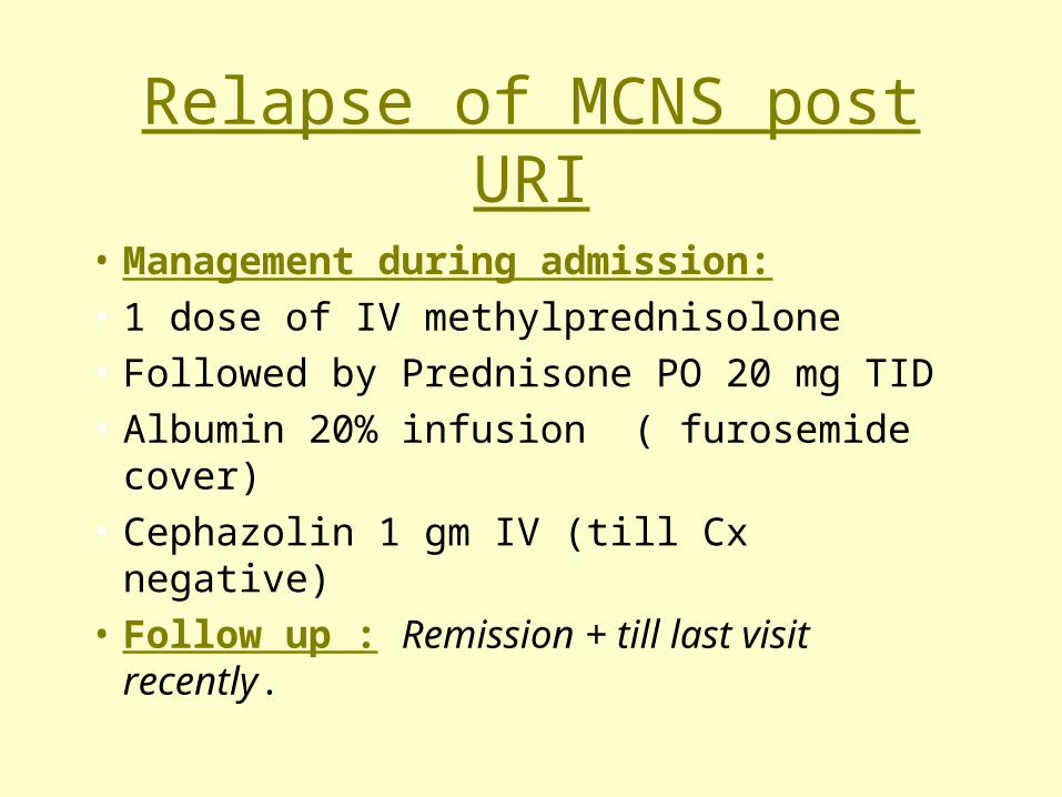
Relapse of MCNS post URI
• Management during admission:
• 1 dose of IV methylprednisolone
• Followed by Prednisone PO 20 mg TID
• Albumin 20% infusion ( furosemide cover)
• Cephazolin 1 gm IV (till Cx negative)
• Follow up : Remission + till last visit recently.

Case 2: Congenital Nephrotic Syndrome
• CC/HPI: 21 month old female admitted for renal transplant
• Born at 37 weeks, NVD.No abnormalities detected on prenatal US
• 5 wks : repeated vomiting .US showed pyloric stenosis – Sx correction
• B/L enlarged kidneys also noted on US
• Workup – nephrotic range proteinuria and chronic renal failure
• 6 wks – 9 mths : failure to thrive and 3 episodes of sepsis (CMV and H.influenza)
• 12 mths : B/L pre-emptive nephrectomy followed by peritoneal dialysis, grow and development improved
• 17 mths : viral myocarditis, transient CHF treated with digoxin
• 21 mths : admitted for LRRT (father donor)
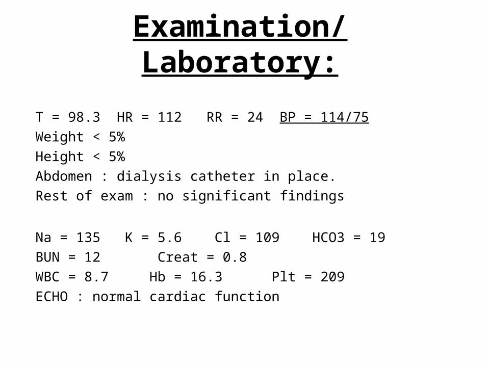
Examination/Laboratory:
• T = 98.3 HR = 112 RR = 24 BP = 114/75
• Weight < 5%
• Height < 5%
• Abdomen : dialysis catheter in place.
• Rest of exam : no significant findings
• Na = 135 K = 5.6 Cl = 109 HCO3 = 19
• BUN = 12 Creat = 0.8
• WBC = 8.7 Hb = 16.3 Plt = 209
• ECHO : normal cardiac function
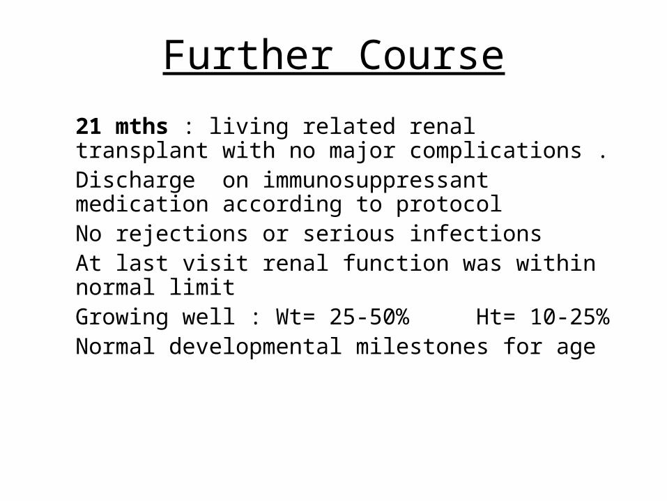
Further Course
• 21 mths : living related renal transplant with no major complications .
• Discharge on immunosuppressant medication according to protocol
• No rejections or serious infections • At last visit renal function was within normal
limit• Growing well : Wt= 25-50% Ht= 10-25%• Normal developmental milestones for age

Case 3: Idiopathic Multicenter Osteolysis with nephropathy
• CC/HPI: 17 year old female who underwent renal transplant at the age of 11 years .
• Presents with N&V for 5 days and acute deterioration of renal function• HPI: H/o carpal-tarsal bones osteolysis since early childhood. At 10 y/o : Persistent Proteinuria +
(incidental finding – pt was asymptomatic). She was also found to have HTN• 2 mths later : Renal Biopsy – FSGS (steroid resistant nephrotic syndrome).She was put on Enalapril
for HTN . Lost for follow up for 8 months.• 11y/o : pt had seizure (HTN encephalopathy). Found to have chronic renal failure with End Stage
Renal Disease. Put on Peritoneal Dialysis and later Hemodialysis• One month later : Cadaveric Renal Transplant• 5 years post Transplant : Uneventful course with good renal function.She has been on Prednisone 15
mgQOD, Tacrolimus, Azathioprine

Examination/Laboratory: • BP= 114/85 T=98.2 HR=108 RR=20• Wt >>95% Ht – 25-50%• Hirsuitism + Striae + • No signs of dehydration• Abdomen – normal bowel sounds• Rest of exam – no significant findings
• Na=137 K=4.7 Cl=107 CO2=16 pH=7.28• BUN=69 Creat=13.5 (baseline-1.0)• Ca = 7.0 Phosphorus = 7.6• CBC : normal• Blood Cx : negative

Further Course
• Renal Biopsy : acute rejection• Treated with : IV methylprednisone followed by
Prednisone PO 10mg QD• Renal Function gradually improved as shown by
renal scan and Creat=6.0 on D/c
• #3 weeks post D/C : Renal Function again deteriorated (creat=10.0).
• Hemodialysis catheter placed in case urgent dialysis needed

Proteinuria: Definition/diagnosis• Normal renal function is associated with urinary protein excretion of
~150 mg/day or less. It is heterogenous is type and source, compromising plasma proteins, renal tissue enzymes and antigens and renal secretions.
• Significant proteinuria can be defined:– Qualitative: 1+ on dipstick examination of 2 out of 3 random urine specimens
collected one week apart if urine sp <= 1015 or 2+ in similarly collected urine if USG >1015
– Semiquantitative: urine Pr/Cr ratio > 0.2 on early morning specimen ( r = 0.97 with 24 hs collection) in older children, >0.5 in infants. > 3 at any age is suspicious of nephrotic range.
– Quantitative: Normal: <4 mg/m2/hour in 12-24 hs collection, Abnormal: 4-40 mg/m2/hour, Nephrotic range >40 mg/meter/hour

Urinary protein excretion in healthy children
Relationship of age to significant proteinuria( based on weight at the 50th for median age for each age cathegory,
values are given by 2 SD above the mean) ( Mitenyi et al, 1979)
Age Upper limit of normal ( mg)
2-12 mo 155
3-4 yr 140
4-10 yr 190
10-16 yr 250

Proteinuria:Urine Dispstick • It is the easiest method, it typically reflects a range of
concentrations: negative <15 mg/dl, trace 15-30 mg/dl, 1+ 30-100 mg/dl, 2+ 100-300 mg/dl, 3+ 300-1000 mg/dl, 4+ >1000 mg/dl
• The reagent area is more sensitive to albumin than to globulins, hemoglobin, Bence-Jones protein and mucoprotein. A negative result does not r/o the presence of these other proteins.
• False positives can occur in highly alkaline urine( pH>8), gross hematuria, pyuria, bacteriuria, quaternary ammoniun cleaners ( antiseptics)
• False negatives can occur in very dilute urine or acidic urine ( pH 4.5) and non albumin proteinuria.
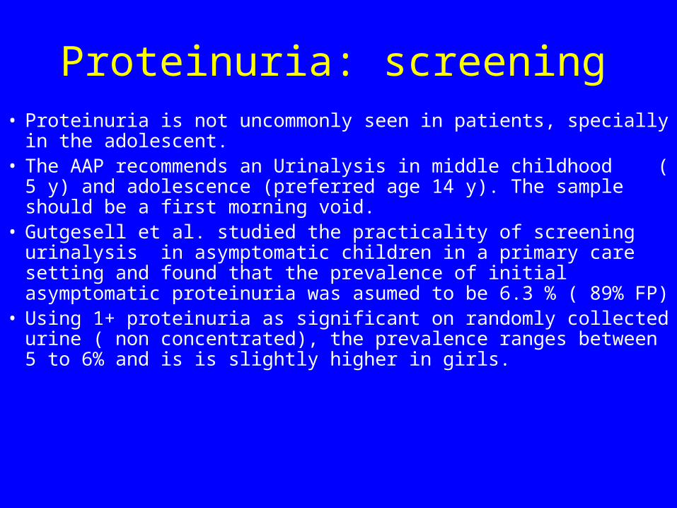
Proteinuria: screening• Proteinuria is not uncommonly seen in patients, specially in the
adolescent.• The AAP recommends an Urinalysis in middle childhood ( 5 y) and
adolescence (preferred age 14 y). The sample should be a first morning void.
• Gutgesell et al. studied the practicality of screening urinalysis in asymptomatic children in a primary care setting and found that the prevalence of initial asymptomatic proteinuria was asumed to be 6.3 % ( 89% FP)
• Using 1+ proteinuria as significant on randomly collected urine ( non concentrated), the prevalence ranges between 5 to 6% and is is slightly higher in girls.
Proteinuria: screening
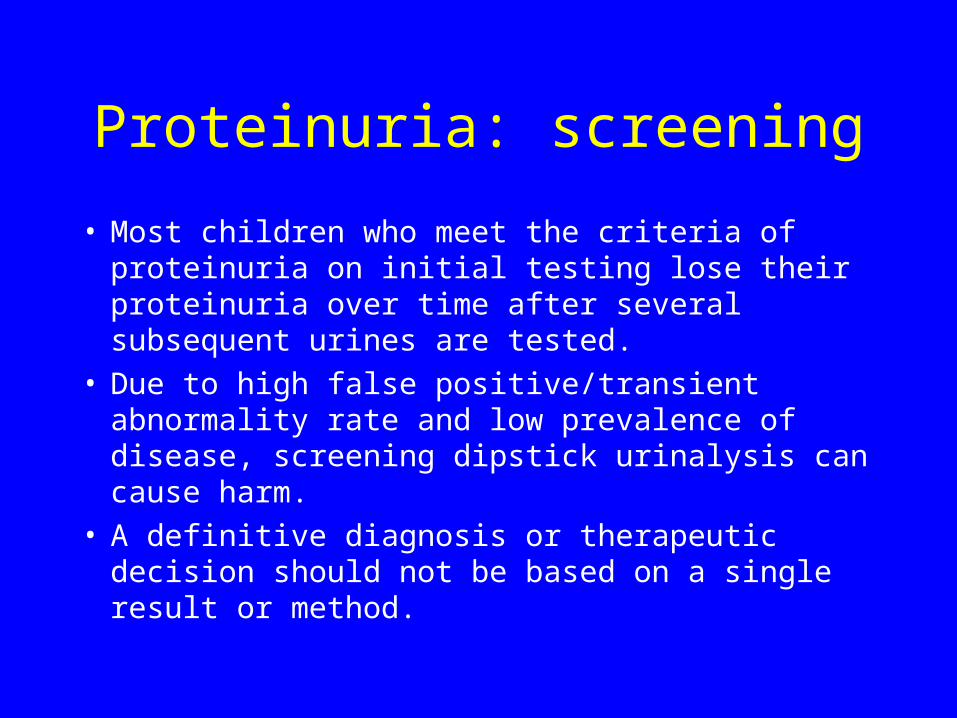
• Most children who meet the criteria of proteinuria on initial testing lose their proteinuria over time after several subsequent urines are tested.
• Due to high false positive/transient abnormality rate and low prevalence of disease, screening dipstick urinalysis can cause harm.
• A definitive diagnosis or therapeutic decision should not be based on a single result or method.

Proteinuria: Type and pathogenesis• 2 basic mechanisms :1. Glomerular: increased filtered load or protein or altered
glomerular permeability2. Tubular: impaired tubular reabsorption of filtered
protein3. Mixed
• 2/3 of the normally excreted protein in children is albumin, 1/3 is a combination of globulins and mucoprotein of tubular origin ( Tamm-Horsfall). The ratio is reversed in adults.

Classification of Proteinuria: Non-pathological
• Postural ( orthostatic)• Febrile/histuria• Exercise-induced

Classification of Proteinuria:Pathological
Tubular:
a. Inherited: cystinosis, Wilson disease, Lowe syndrome
b. Acquired: antibiotic-induced, interstitial nephritis, ATN, heavy metal poisoning ( lead, mercury, gold), reflux nephropathy
Glomerular:
a. Nephrotic syndrome: MCNS, mesangial proliferation, FSGS, CNG/Infantile
b. Glomerulonephritis: Idiopathic( membranous, membranoproliferative), Systemic diseases ( SLE)
c. HTN
d. DM
e. HUS
f. Hyperfiltration secondary to nephron loss ( with or without FS) due to chronic pyelonephritis or other renal diseases
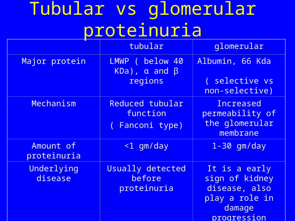
Tubular vs glomerular proteinuria
tubular glomerular
Major protein LMWP ( below 40 KDa), α and β regions
Albumin, 66 Kda ( selective vs non-
selective)
Mechanism Reduced tubular function
( Fanconi type)
Increased permeability of the glomerular membrane
Amount of proteinuria <1 gm/day 1-30 gm/day
Underlying disease Usually detected before proteinuria
It is a early sign of kidney disease, also play a role in
damage progression
edema no Yes, if in nephrotic range

Work-up of a child with proteinuria
Pediatrician’s work up:• early morning urinalysis to
include examination of sediment
• Ambulatory and recumbent urinalysis for Dipstick protein
• Blood electrolytes, BUN, Cr, serum proteins, cholesterol
• ASO titer, C3 complement, ANA, hepatitis B serology
• Timed 12-hour urine collections, recumbent and ambulatory
• Renal US, VCUG, renal scan
When to refer:Persistent nonorthostatic proteinuriaFamily history of GN, CRF, kidney
transplantationSystemic complaints ( such as fever,
arthritis and skin)HTN, edema, cutaneous vasculitis,
purpuraCoexistent hematuria with or without castsElevated BUN/CrIncreased parental anxiety
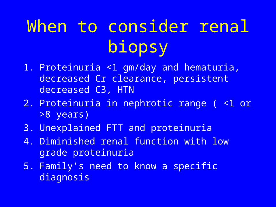
When to consider renal biopsy
1. Proteinuria <1 gm/day and hematuria, decreased Cr clearance, persistent decreased C3, HTN
2. Proteinuria in nephrotic range ( <1 or >8 years)
3. Unexplained FTT and proteinuria
4. Diminished renal function with low grade proteinuria
5. Family’s need to know a specific diagnosis
Nephrotic syndrome• The term nephrotic syndrome is applicable to any
condition with heavy proteinuria, hypoproteinemia, and edema.
• It is a disorder of glomerular permselectivity that may be primary or secondary to an overt systemic disease.
• In children the most common variety is MCNS with a characteristic response to corticosteroid therapy
• But a few patients with MCNS do not respond to steroids , and a few steroid responders have histology other than MCNS.

Nephrotic syndrome: Definitions Nephrotic syndrome: edema, plasma albumin less 2.5 gm/dl, proteinuria >40 mg/m2/hRemission: urinary protein excretion < 4 mg/m2/h or dipstick neg/trace for 3 consecutive daysSteroid response: remission achieved with steroid therapy aloneLate responder: remission occurring after 4 weeks prednisolone 60 mg/m2/day without other
drugsRelapse: urinary protein excresion >40 mg/m2/h or dispstick 2+ for 3 consecutive days having
previously been in remissionFrequent relapses: two or more relapses within 6 months of initial response or 4 or more in 12
monthsSteroid dependence: two consecutived relapses occurring during corticosteroid treatment or
within 14 days of cessationSteroid resistance: failure to achieve response in spite 8 weeks of prednisolone 60 mg/m2/dayEarly nonresponder: steroid resistance in the initial episode
Late nonresponder: steroid resistance developing in a patient who had previously been steroid
responsive

Idiopathic nephrotic syndromeChildren with steroid-resistant NS may have one of several
different histologic appearances in the glomeruli, being most common FSGS.
It is not clear is MCNS and FSGS should be consider two different entities or different ends of a single spectrum of disease.
Current opinion is to include both conditions under the single label of Idiopathic Nephrotic Syndrome, dividing into steroid-sensitive (SSNS) and steroid-resistant (SRNS). Patients with SRNS have higher risk for extrarenal complications as well as porgression to CRF and ESRD, with high recurrence in the graft after transplantation.
INS: Epidemiology• Prevalence is ~16/100000 children, annual incidence of 2-
7/100000 children
• Ratio M/F is 2/1, in younger children. This male predilection disappears in teenagers and adults
• 3/4 less than 6 years
• Median ages at time of presentation varies according to histopathological diagnosis: 3 years for MCNS, 6 years for FSGS and 10 years for MPGN
• Majority of patients that have SSNS has MCNS. The risk of progressing to CRF and ESRD for SRNS is 40% in 5 years.
INS: treatment• Immunosuppressants: corticosteroids, alkylating
agents ( or combination), CSA, Mycophenolate Mofetil, azathioprine
• Plasmaferesis
• ACE inhibitors
• Treatment of complications: edema, HTN, infections, thromboembolic events, hyperlipidemia, malnutrition, anemia, endocrine abnormalities

Inheritable forms of SRNSDisease Histopathologic
studyInheritance
patternGene locus Gene product
CNF ( FinnishType)
MCNS AR 19q13.1 Nephrin( NPHS1)
Familial FSGS FSGS AR 1q25-31 Podocin( NPHS2)
Familial FSGS FSGS AD 19q13 -actinin 4( ACTN4)
Familial FSGS FSGS AD 11q 21-22 unknown
Denys- Drashsyndrome
Diffusemesangialhypercellularity
sporadic 11p13 Wilms tumor(WT-1)
Nail-PatellaSyndrome( NPS)
AD Chrom 1 LMX1B(transcriptionfactor)