Embed Size (px)
Citation preview

Nephrotic Syndrome in
Children

Objectives
• Define Nephrotic Syndrome
• Pathogenesis of Oedema
• Causes of Nephrotic syndrome in Children
• Minimal Change disease

Nephrotic Syndrome
Histopathological process
OR
Clinical Entity

Nephrotic Syndrome
Histopathological process
OR
Clinical Entity☻
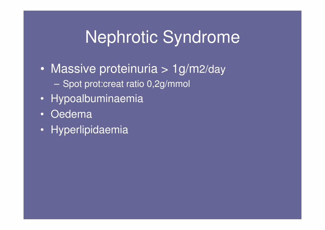
Nephrotic Syndrome
• Massive proteinuria > 1g/m2/day
– Spot prot:creat ratio 0,2g/mmol
• Hypoalbuminaemia
• Oedema
• Hyperlipidaemia

Nephrotic syndrome
(clinical entity)
oedema proteinuria hypoalbuminemia
Primary glomerulopathies AI post infective vasculitides
Biopsy: glomerulonephritis
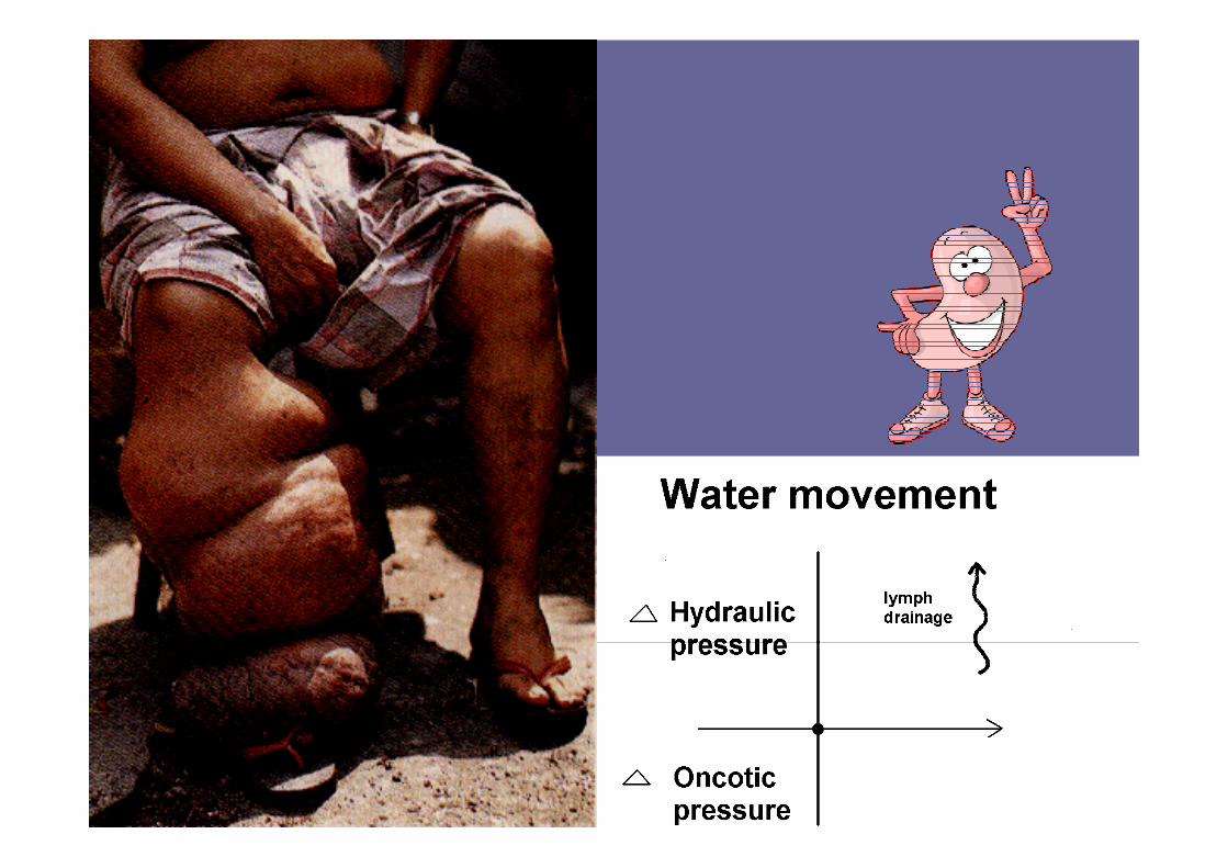
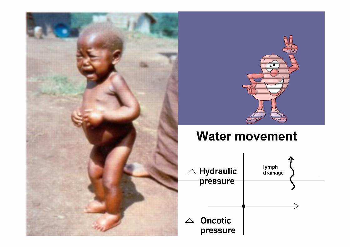
Oedema: Pathogenesis

Pathophysiology of
oedema
Two basic steps:
•Alteration of capillary
haemodynamics that favours
movement of fluid from
vascular space to interstitium
•+/-Retention of Na and H2O by
the kidneys

Macroscopic Haematuria
Hypertension
Generalized oedema
Creat and Urea increased

•Not sick looking
5yr M
•1week onset
-puffy eyes
-swelling legs
•Urine +++ protein
•Lab
-alb 16
-cholesterol 8
-U+E Normal
•


Causes of Nephrotic Syndrome
@ RXH• Minimal change 43%
• Mesangioproliferative 17%
• Membranous 16%
• APSGN 6%
• MCGN 2%
• HIV ?

Causes of Nephrotic Syndrome
in black children(Gauteng)
• FSGS 31%
• Minimal change 24%
• Membranous 13%
• Mesangial proliferative 13%
• Mesangiocapillary 4%
Thomson et al

Pathogenesis of Minimal change
Disease
• T cell dysfunction with release of lymphokines
– Podocyte dysfunction
– Loss of electronegative charge of basement
membrane
• Light microscopy no changes
• Immunofluorescence negative
• Electron microscopy fusion foot processes

Light microscopy : Minimal change
disease

Electron Microscopy
Normal Fusion of foot processes

Clincal presentation
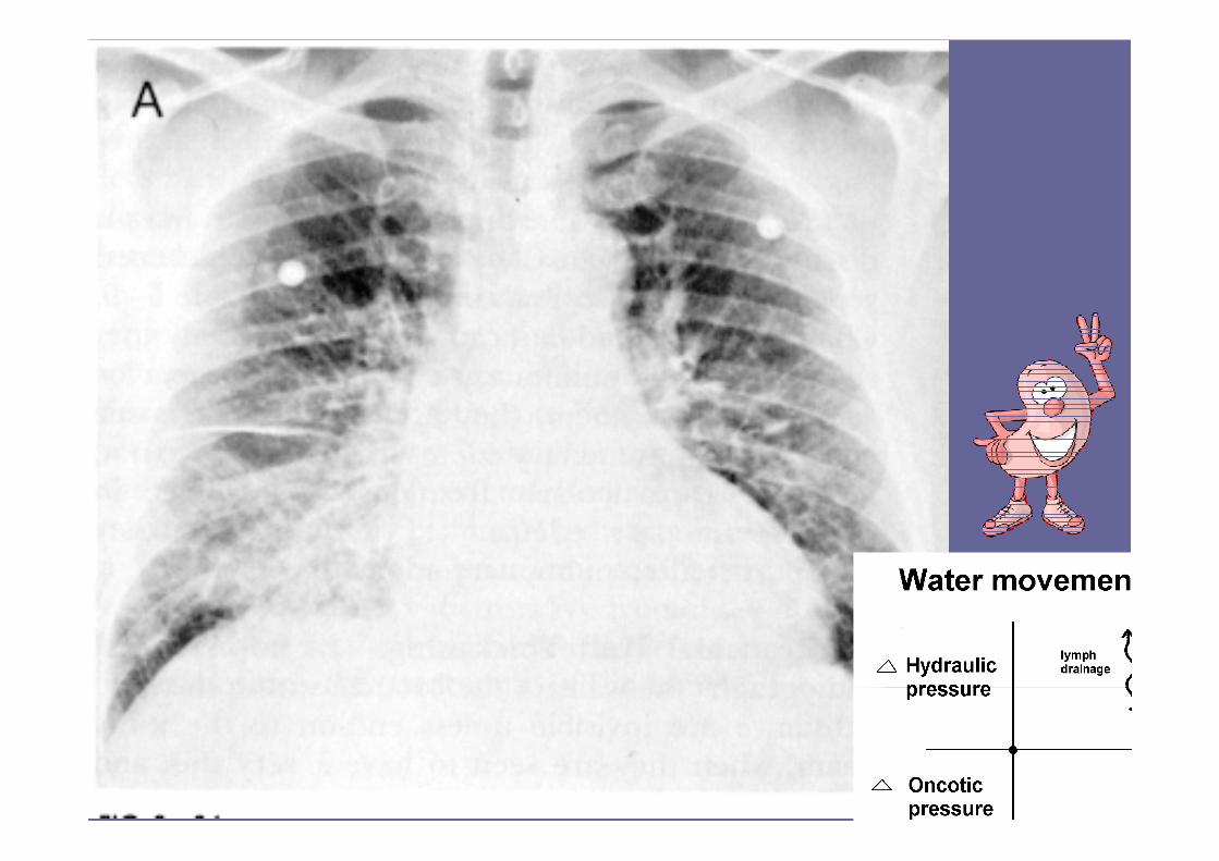
• Rapid onset of oedema progresses rapidly
• Transudation into body cavities– Pleural effussion
– Ascites
• Eyelids swell totally closed
• Severe scrotal/vulval oedema
• Urine may become frothy

Nephrotic Syndrome
Complications• Hypovolaemic crisis
• Pre –renal failure
• Thrombosis
• Ascites– Hernia umbilical inguinal
• Malnutriton
• Infection• Peritonitis, pneumonia, cellulitis
• Hyperlipidaemia • Cardiovascular risk
• Hypothyroidism

Nephrotic Syndrome
Investigations• Quantify how severe nephrotic syndrome?
• quantify proteinuria
• Serum alb cholesterol
• Are there any underlying diseases that could be causing this?• Urine microscopy: (bland urine in MCNS)
• Hepatitis B, VDRL, ASOT/ANtiDNAse B, ANA, C3
• Is pt hyovolaemic• Clinical signs of shock
• Urine Na
• Is renal function normal (some diseases may cause renal failure)• Creat Urea
• Probably going to use immunosuppressive treatment :• Exclude infection
• Exclude TB

Indications for biopsy
Indications that it might not be Minimal Change disease:
• Age < 1 year
• Age > 10 years
• Renal Failure
• Persistant hypertension
• Macroscopic haematuria
• Microscopic haematuria(persistant)
• Evidence of other disease e.g. SLE, HSP Hep B
• Failed trial of steroids
• Black kids

Nephrotic Syndrome
Management(1)• Daily urine dipsticks
• Salt restriction
• No fluid restriction unless hypertensive or in renal failure
• Treat oedema if severe :– ivi albumin and diuretics
– Furosemide(beware of powerful diuretics on their own in acute setting)
– Furosemide +amiloride

Manage complications:
– Treat shock 20% Alb, plasma
– Prophylactic Aspirin
– Aggressive treatment of infection
– ACE for proteinuria (long term)
– Lipid lowering agents (long term)

Management(2): Treat
Underlying cause• If Minimal change suspected:
– Trial of Steroids
– 2mg/kg/day for 4 weeks
• If NO response → Biopsy

Nephrotic Syndrome(minimal
Change)
Management
• If response with 1st episode:
– Continue for total of 3 months steroids
– 6 weeks daily 2mg/kg/day then
– 6 weeks alternate day weaning over last 2 weeks
• Subsequent episodes:
– 2mg/kg/day daily until urine clear for 3 days in row
– then alt days for 1 month and then rapid wean

Outcome
• 10% never relapse
• 60% infrequent relapsers
• 30% frequent relapsers or steroid dependant
• 90% permanent remission at puberty
• Minimal risk of chronic renal failure

FSGS
• More difficult
• High dose steroids
• Cyclophosphamide
• Cyclosporin