Embed Size (px)
Citation preview

lable at ScienceDirect
Progress in Retinal and Eye Research xxx (2016) 1e24
Contents lists avai
Progress in Retinal and Eye Research
journal homepage: www.elsevier .com/locate/prer
The role of dinucleoside polyphosphates on the ocular surface andother eye structures
Gonzalo Carracedo a, 1, Almudena Crooke b, 1, Ana Guzman-Aranguez b, 1,Maria J. P�erez de Lara b, 1, Alba Martin-Gil b, 1, Jesús Pintor b, *, 1
a Department of Optics II (Optometry and Vision), Faculty of Optics and Optometry, Universidad Complutense de Madrid, Madrid, Spainb Department of Biochemistry and Molecular Biology IV, Faculty of Optics and Optometry, Universidad Complutense de Madrid, Madrid, Spain
a r t i c l e i n f o
Article history:Received 28 February 2016Received in revised form30 June 2016Accepted 5 July 2016Available online xxx
Keywords:DinucleotidesPurinergic receptorsDiadenosine tetraphosphateDiquafosol
* Corresponding author. Dep. Biochemistry and MoOptics and Optometry, Universidad Complutense de M28037 Madrid, Spain.
E-mail address: [email protected] (J. Pintor).1 Percentage of work contributed by each autho
manuscript is as follows: Gonzalo Carracedo 35%;Guzman-Aranguez 10%; M.J. Perez de Lara 10%; Alba25%.
http://dx.doi.org/10.1016/j.preteyeres.2016.07.0011350-9462/© 2016 Elsevier Ltd. All rights reserved.
Please cite this article in press as: CarracedoProgress in Retinal and Eye Research (2016)
a b s t r a c t
Dinucleoside polyphosphates comprises a group of dinucleotides formed by two nucleosides linked by avariable number of phosphates, abbreviated NpnN (where n represents the number of phosphates).These compounds are naturally occurring substances present in tears, aqueous humour and in the retina.As the consequence of their presence, these dinucleotides contribute to many ocular physiologicalprocesses. On the ocular surface, dinucleoside polyphosphates can stimulate tear secretion, mucinrelease from goblet cells and they help epithelial wound healing by accelerating cell migration rate.These dinucleotides can also stimulate the presence of proteins known to protect the ocular surfaceagainst microorganisms, such as lysozyme and lactoferrin. One of the latest discoveries is the ability ofsome dinucleotides to facilitate the paracellular way on the cornea, therefore allowing the delivery ofcompounds, such as antiglaucomatous ones, more easily within the eye. The compound Ap4A has beendescribed being abnormally elevated in patient's tears suffering of dry eye, Sjogren syndrome, congenitalaniridia, or after refractive surgery, suggesting this molecule as biomarker for dry eye condition. At theintraocular level, some diadenosine polyphosphates are abnormally elevated in glaucoma patients, andthis can be related to the stimulation of a P2Y2 receptor that increases the chloride efflux and watermovement in the ciliary epithelium. In the retina, the dinucleotide dCp4U, has been proven to be useful tohelp in the recovery of retinal detachments. Altogether, dinucleoside polyphosphates are a group ofcompounds which present relevant physiological actions but which also can perform promising thera-peutic benefits.
© 2016 Elsevier Ltd. All rights reserved.
Contents
1. Introduction . . . . . . . . . . . . . . . . . . . . . . . . . . . . . . . . . . . . . . . . . . . . . . . . . . . . . . . . . . . . . . . . . . . . . . . . . . . . . . . . . . . . . . . . . . . . . . . . . . . . . . . . . . . . . . . . . . . . . . . 001.1. Dinucleoside polyphosphates: structure and features . . . . . . . . . . . . . . . . . . . . . . . . . . . . . . . . . . . . . . . . . . . . . . . . . . . . . . . . . . . . . . . . . . . . . . . . . . . . 001.2. Presence and actions of dinucleoside polyphosphates . . . . . . . . . . . . . . . . . . . . . . . . . . . . . . . . . . . . . . . . . . . . . . . . . . . . . . . . . . . . . . . . . . . . . . . . . . . . 00
1.2.1. Presence and intracellular actions of dinucleoside polyphosphates . . . . . . . . . . . . . . . . . . . . . . . . . . . . . . . . . . . . . . . . . . . . . . . . . . . . . . . . . 001.2.2. Presence and extracellular actions of dinucleoside polyphosphates . . . . . . . . . . . . . . . . . . . . . . . . . . . . . . . . . . . . . . . . . . . . . . . . . . . . . . . . . 00
1.3. Purinergic receptors and dinucleoside polyphosphates . . . . . . . . . . . . . . . . . . . . . . . . . . . . . . . . . . . . . . . . . . . . . . . . . . . . . . . . . . . . . . . . . . . . . . . . . . . . 001.4. Dinucleoside polyphosphate degradation . . . . . . . . . . . . . . . . . . . . . . . . . . . . . . . . . . . . . . . . . . . . . . . . . . . . . . . . . . . . . . . . . . . . . . . . . . . . . . . . . . . . . . . 00
2. Dinucleoside polyphosphates in the eye . . . . . . . . . . . . . . . . . . . . . . . . . . . . . . . . . . . . . . . . . . . . . . . . . . . . . . . . . . . . . . . . . . . . . . . . . . . . . . . . . . . . . . . . . . . . . . 00
lecular Biology IV, Faculty ofadrid, C/Arcos del Jalon 118,
r in the production of theAlmudena Crooke 10%; AnaMartin-Gil 10%; Jesus Pintor
, G., et al., The role of dinucle, http://dx.doi.org/10.1016/j.p
oside polyphosphates on the ocular surface and other eye structures,reteyeres.2016.07.001
G. Carracedo et al. / Progress in Retinal and Eye Research xxx (2016) 1e242
2.1. Ocular surface . . . . . . . . . . . . . . . . . . . . . . . . . . . . . . . . . . . . . . . . . . . . . . . . . . . . . . . . . . . . . . . . . . . . . . . . . . . . . . . . . . . . . . . . . . . . . . . . . . . . . . . . . . . . . . . 002.1.1. Purinergic receptors in the ocular surface . . . . . . . . . . . . . . . . . . . . . . . . . . . . . . . . . . . . . . . . . . . . . . . . . . . . . . . . . . . . . . . . . . . . . . . . . . . . . . . 002.1.2. Action of ectodinucleotidases in the ocular surface . . . . . . . . . . . . . . . . . . . . . . . . . . . . . . . . . . . . . . . . . . . . . . . . . . . . . . . . . . . . . . . . . . . . . . . 002.1.3. Presence in the tear film of healthy patients . . . . . . . . . . . . . . . . . . . . . . . . . . . . . . . . . . . . . . . . . . . . . . . . . . . . . . . . . . . . . . . . . . . . . . . . . . . . 002.1.4. Presence in ocular surface pathology . . . . . . . . . . . . . . . . . . . . . . . . . . . . . . . . . . . . . . . . . . . . . . . . . . . . . . . . . . . . . . . . . . . . . . . . . . . . . . . . . . . 002.1.5. Presence in contact lens wearers . . . . . . . . . . . . . . . . . . . . . . . . . . . . . . . . . . . . . . . . . . . . . . . . . . . . . . . . . . . . . . . . . . . . . . . . . . . . . . . . . . . . . . . 002.1.6. Presence after ocular surface surgery . . . . . . . . . . . . . . . . . . . . . . . . . . . . . . . . . . . . . . . . . . . . . . . . . . . . . . . . . . . . . . . . . . . . . . . . . . . . . . . . . . . 002.1.7. Effect on tear secretion . . . . . . . . . . . . . . . . . . . . . . . . . . . . . . . . . . . . . . . . . . . . . . . . . . . . . . . . . . . . . . . . . . . . . . . . . . . . . . . . . . . . . . . . . . . . . . . 002.1.8. Effect on tear components . . . . . . . . . . . . . . . . . . . . . . . . . . . . . . . . . . . . . . . . . . . . . . . . . . . . . . . . . . . . . . . . . . . . . . . . . . . . . . . . . . . . . . . . . . . . . 002.1.9. Contact lens delivery of dinucleoside polyphosphates . . . . . . . . . . . . . . . . . . . . . . . . . . . . . . . . . . . . . . . . . . . . . . . . . . . . . . . . . . . . . . . . . . . . . 002.1.10. Effect on corneal wound healing . . . . . . . . . . . . . . . . . . . . . . . . . . . . . . . . . . . . . . . . . . . . . . . . . . . . . . . . . . . . . . . . . . . . . . . . . . . . . . . . . . . . . . 002.1.11. Effect on epithelial permeability . . . . . . . . . . . . . . . . . . . . . . . . . . . . . . . . . . . . . . . . . . . . . . . . . . . . . . . . . . . . . . . . . . . . . . . . . . . . . . . . . . . . . . . 00
2.2. Dinucleoside polyphosphates in the anterior pole . . . . . . . . . . . . . . . . . . . . . . . . . . . . . . . . . . . . . . . . . . . . . . . . . . . . . . . . . . . . . . . . . . . . . . . . . . . . . . . . 002.2.1. Presence in the aqueous humour . . . . . . . . . . . . . . . . . . . . . . . . . . . . . . . . . . . . . . . . . . . . . . . . . . . . . . . . . . . . . . . . . . . . . . . . . . . . . . . . . . . . . . . 002.2.2. Effect on intraocular pressure . . . . . . . . . . . . . . . . . . . . . . . . . . . . . . . . . . . . . . . . . . . . . . . . . . . . . . . . . . . . . . . . . . . . . . . . . . . . . . . . . . . . . . . . . 002.2.3. Putative actions of dinucleotides in the lens . . . . . . . . . . . . . . . . . . . . . . . . . . . . . . . . . . . . . . . . . . . . . . . . . . . . . . . . . . . . . . . . . . . . . . . . . . . . 002.2.4. Neuroprotective role in the ciliary body . . . . . . . . . . . . . . . . . . . . . . . . . . . . . . . . . . . . . . . . . . . . . . . . . . . . . . . . . . . . . . . . . . . . . . . . . . . . . . . . 00
2.3. Dinucleoside polyphosphates in the posterior pole . . . . . . . . . . . . . . . . . . . . . . . . . . . . . . . . . . . . . . . . . . . . . . . . . . . . . . . . . . . . . . . . . . . . . . . . . . . . . . 002.3.1. Dinucleotide effect on retinal detachment . . . . . . . . . . . . . . . . . . . . . . . . . . . . . . . . . . . . . . . . . . . . . . . . . . . . . . . . . . . . . . . . . . . . . . . . . . . . . . 002.3.2. Presence in other posterior structures . . . . . . . . . . . . . . . . . . . . . . . . . . . . . . . . . . . . . . . . . . . . . . . . . . . . . . . . . . . . . . . . . . . . . . . . . . . . . . . . . . 00
3. Conclusions and future directions . . . . . . . . . . . . . . . . . . . . . . . . . . . . . . . . . . . . . . . . . . . . . . . . . . . . . . . . . . . . . . . . . . . . . . . . . . . . . . . . . . . . . . . . . . . . . . . . . . . 00Disclosure . . . . . . . . . . . . . . . . . . . . . . . . . . . . . . . . . . . . . . . . . . . . . . . . . . . . . . . . . . . . . . . . . . . . . . . . . . . . . . . . . . . . . . . . . . . . . . . . . . . . . . . . . . . . . . . . . . . . . . . . . 00Acknowledgments . . . . . . . . . . . . . . . . . . . . . . . . . . . . . . . . . . . . . . . . . . . . . . . . . . . . . . . . . . . . . . . . . . . . . . . . . . . . . . . . . . . . . . . . . . . . . . . . . . . . . . . . . . . . . . . . . 00References . . . . . . . . . . . . . . . . . . . . . . . . . . . . . . . . . . . . . . . . . . . . . . . . . . . . . . . . . . . . . . . . . . . . . . . . . . . . . . . . . . . . . . . . . . . . . . . . . . . . . . . . . . . . . . . . . . . . . . . . 00
NN
O
OO
N
N N
N
O
OO
N
NN
N
P
O
OOO O
P
O
OO
P
O
OO
P
O
O
O
OO
O
OO
P
O
OOO O
P
O
OO
P
O
OO
P
O
O
N
N
O
O
N
N
O
O
Ap4A
Up4U(Diquafosol)
dCp4U(Denufosol)
O
OO
OO
P
O
OOO O
P
O
OO
P
O
OO
P
O
O
N
N
O
O
N
N
N
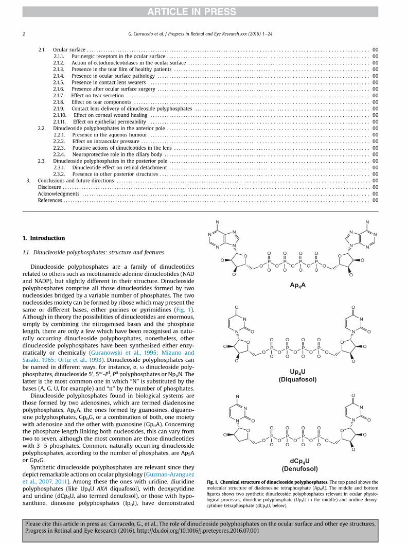
O
Fig. 1. Chemical structure of dinucleoside polyphosphates. The top panel shows themolecular structure of diadenosine tetraphosphate (Ap4A). The middle and bottomfigures shows two synthetic dinucleoside polyphosphates relevant in ocular physio-logical processes, diuridine polyphosphate (Up4U in the middle) and uridine deoxy-cytidine tetraphosphate (dCp4U, below).
1. Introduction
1.1. Dinucleoside polyphosphates: structure and features
Dinucleoside polyphosphates are a family of dinucleotidesrelated to others such as nicotinamide adenine dinucleotides (NADand NADP), but slightly different in their structure. Dinucleosidepolyphosphates comprise all those dinucleotides formed by twonucleosides bridged by a variable number of phosphates. The twonucleosides moiety can be formed by ribose which may present thesame or different bases, either purines or pyrimidines (Fig. 1).Although in theory the possibilities of dinucleotides are enormous,simply by combining the nitrogenised bases and the phosphatelength, there are only a few which have been recognised as natu-rally occurring dinucleoside polyphosphates, nonetheless, otherdinucleoside polyphosphates have been synthesised either enzy-matically or chemically (Guranowski et al., 1995; Mizuno andSasaki, 1965; Ortiz et al., 1993). Dinucleoside polyphosphates canbe named in different ways, for instance, a, u dinucleoside poly-phosphates, dinucleoside 50, 5000-P1, Pn polyphosphates or NpnN. Thelatter is the most common one in which “N” is substituted by thebases (A, G, U, for example) and “n” by the number of phosphates.
Dinucleoside polyphosphates found in biological systems arethose formed by two adenosines, which are termed diadenosinepolyphosphates, ApnA, the ones formed by guanosines, diguano-sine polyphosphates, GpnG, or a combination of both, one moietywith adenosine and the other with guanosine (GpnA). Concerningthe phosphate length linking both nucleosides, this can vary fromtwo to seven, although the most common are those dinucleotideswith 3e5 phosphates. Common, naturally occurring dinucleosidepolyphosphates, according to the number of phosphates, are Ap5Aor Gp4G.
Synthetic dinucleoside polyphosphates are relevant since theydepict remarkable actions on ocular physiology (Guzman-Aranguezet al., 2007, 2011). Among these the ones with uridine, diuridinepolyphosphates (like Up4U AKA diquafosol), with deoxycytidineand uridine (dCp4U, also termed denufosol), or those with hypo-xanthine, diinosine polyphosphates (IpnI), have demonstrated
Please cite this article in press as: Carracedo, G., et al., The role of dinucleoside polyphosphates on the ocular surface and other eye structures,Progress in Retinal and Eye Research (2016), http://dx.doi.org/10.1016/j.preteyeres.2016.07.001
G. Carracedo et al. / Progress in Retinal and Eye Research xxx (2016) 1e24 3
interesting therapeutic properties in the eye as it will later bedescribed (Fujihara et al., 2001; Guzman-Aranguez et al., 2012).
The history of dinucleoside polyphosphates started far awayfrom the future physiological and pharmacological applicationsfound in the eye. Indeed, the first news on these moleculesoccurred in the 1960's. Dinucleoside polyphosphates were discov-ered in the eggs of brine shrimps, mostly diguanosine poly-phosphates (Finamore and Warner, 1963; Warner, 1964). Thesedinucleotides are the basis for the development of the eggs intoproper shrimps since they will be used for DNA synthesis.
It was not until a few years later when it was possible to identifya biochemical reaction showing the synthesis of a dinucleosidepolyphosphate. Scientists, when investigating the reaction be-tween aminoacids and their corresponding tRNAs during the pro-tein synthesis process discovered the presence of somediadenosine polyphosphates (Randerath et al., 1966; Zamecniket al., 1966). Over the following years, most of the efforts werededicated to describing their structural and chemical properties,mainly by spectrometric methods (Bush and Tinoco, 1967; Cheng,1968; Warshaw and Tinoco, 1965). Changes in the molecules pre-sent were also studied when environmental properties (Lee et al.,1979) such as temperature or pH was modified (Chan and Nelson,1969; Davis and Tinoco, 1968; Glaubiger et al., 1968; Hruska andDanyluk, 1968; Ikehara et al., 1970; Rabczenko and Shugar, 1971;Ts'o et al., 1969). The structural changes are especially relevantwhen the pH medium is altered. The structure of a NpnN, forexample, Ap4A, under physiological pH condition (pHz 7), depictsa stacked conformation inwhich the adenine rings are piled up, thisbeing possible due to the phosphate chain folding. This is caused bythe negatively charged phosphate groups and the partially posi-tively charged adenine rings and the consequent electrostatic in-teractions. A different panorama is observed when the pH lowers(pH z 4). Under this condition the phosphates are folded but theadenine rings are pointing to opposite directions. The maximalopen possible conformation is reached at a pH below 3. Under thiscondition the protonation of the phosphates occurs and the in-teractions among the adenines and phosphates do not exist(Guzman-Aranguez et al., 2011).
The conformational changes observed at different pH, mayexplain why these dinucleotides can interact with different puri-nergic receptors, mainly when in some environmental conditions,such as acidic synapses (glutamatergic) the conditions can be farfrom neutral conditions (Gualix et al., 2003).
Concerning temperature, it seems that dinucleoside poly-phosphates at low temperatures present their bases in a stackingconformation being so close to each other that there is no room forwater between them. On the contrary, when temperature is high,even at denaturing conditions (above 40 �C), the bases are notstacked and they do not interact with each other and therefore caninteract with the solvent and other molecules. In this case, the onlylimitation of the aromatic bases would be their covalent bonds tothe respective riboses (Davis and Tinoco, 1968; Glaubiger et al.,1968).
1.2. Presence and actions of dinucleoside polyphosphates
1.2.1. Presence and intracellular actions of dinucleosidepolyphosphates
It took quite a long time from the moment these dinucleotideswere identified until some biochemical and physiological actionsfor these compoundswere detected. One of the first actions was theeffect of diadenosine tetraphosphate, Ap4A, on DNA polymerasetriggering DNA replication (Grummt, 1978, 1979; Grummt et al.,1979). Indeed, it has been possible to correlate changes (in-creases) in Ap4A concentrations during the cell cycle and specially
Please cite this article in press as: Carracedo, G., et al., The role of dinucleProgress in Retinal and Eye Research (2016), http://dx.doi.org/10.1016/j.p
during cell proliferation and DNA synthesis (Bambara et al., 1985).These experiments were quite controversial, although other au-thors could, in part, confirm the relationship between Ap4A andDNA synthesis, it seemed that apparently the dinucleotide wouldprime DNA synthesis rather than activating the polymerase(Zamecnik et al., 1982). Also, some other studies suggested a closerelation between the amino acid activation process and DNAreplication in mammalian cells (Grummt, 1983; Rapaport et al.,1981).
Another intracellular action suggested for dinucleoside poly-phosphates has been its possible role of “alarmone” (Lee et al.,1983; Varshavsky, 1983). An alarmone is a molecule that reflectsand indicates a stress situation within a cell. A classical situation isthat in which an elevation of temperature, apart from increasingthe levels of heat shock proteins (HSPs), also produces a rise indinucleoside polyphosphates such as Ap3A, Ap4A, Gp4A and Gp3A(Lee et al., 1983). The same dinucleotides are increased when cellsare submitted to oxidative stress in a process independent from thedinucleotide synthesis by aminoacyl-tRNA synthetases.
1.2.2. Presence and extracellular actions of dinucleosidepolyphosphates
Although the presence and actions of dinucleoside poly-phosphates have been reported in several tissues as indicated(Andersson,1989; Rapaport and Zamecnik, 1976; Ripoll et al., 1996),two sources are relevant concerning the eye physiology: the bloodvessels and the nervous system.
Apart from the presence of dinucleoside polyphosphates incrustaceans as previously indicated, these molecules were found asputative active vasoactive compounds when they were discoveredin platelet secretory granules (Flodgaard and Klenow, 1982; Luthjeand Ogilvie, 1983). From the moment they were discovered themain interest of the scientific community was to identify thoseactions triggered by the dinucleotides in the blood stream. In thissense, one of the first roles was their involvement in platelet ag-gregation. Interestingly, the two dinucleotides contained in theplatelets (in their dense granules), Ap3A and Ap4A, depictedopposite actions; while the first was inducing platelet aggregation,the second, showed disaggregating properties (Luthje et al., 1985).Nonetheless, the existence of dinucleoside polyphosphates in theplasma and blood (Luthje and Ogilvie, 1988), also suggested aneffect on vasoconstriction or/and vasodilation. When analysing theeffect of the vascular tone, dinucleoside polyphosphates such asAp4U produces a biphasic effect although the predominant one atlow concentrations is vasoconstriction, infusions of this dinucleo-tide elicit hypotension and electrolyte retention (Flores et al., 1999;Hansen et al., 2010).
The identification of dinucleoside polyphosphates and mainlydiadenosine polyphosphates in neural secretory vesicles showedthe storage of these dinucleotides with classical transmitters suchas noradrenalin (Rodriguez del Castillo et al., 1988). This co-localization was further extended to cholinergic and mono-aminergic synaptic vesicles (Miras-Portugal et al., 1998, 1999;Pintor et al., 1992). Moreover, it was possible to demonstrate thatthese dinucleotides were releasable in a Ca2þ dependent mannerfrom brain synaptic terminals (Pintor et al., 1991, 1992) and also bythe effect of drugs such as amphetamines (Pintor et al., 1993). Therole of diadenosine polyphosphates, mainly Ap4A and Ap5A in thenervous system has been the modulation of the release of mono-amines (Giraldez et al., 2001), glutamate (Gualix et al., 2003), or theinhibition of synaptic transmission in the hippocampus (Klishinet al., 1994).
oside polyphosphates on the ocular surface and other eye structures,reteyeres.2016.07.001
G. Carracedo et al. / Progress in Retinal and Eye Research xxx (2016) 1e244
1.3. Purinergic receptors and dinucleoside polyphosphates
Extracellular actions of dinucleoside polyphosphates occurredby means of receptors on the cell surface termed P2 purinergicreceptors. These types of receptors are divided into two families,the P2X ionotropic (Habermacher et al., 2015) and the P2Ymetabotropic receptors (von Kugelgen and Harden, 2011).
P2X receptors are activated by nucleotides and dinucleotideswhich mediate the rapid and non-selective cation transients(mainly Naþ and Ca2þ) from the extracellular space (Samways et al.,2014), causing depolarization of the membrane ion channels andsubsequent activation of voltage dependent calcium channels.Seven members of P2X receptors in mammalians have been cloned(P2X1-P2X7) and they combined various forms of these subunits(Saul et al., 2013). Cloning of P2X receptors has revealed that all thesubunits have large extracellular loop with ten cysteine residues,two transmembrane domains and short intracellular C and N ter-minals (Roberts et al., 2006). Three subunits are necessary to form afunctional native channel, and these channels are organized ashomotrimers or heterotrimers (Coddou et al., 2011).
P2Y receptors are metabotropic receptors, members of thefamily of G protein-coupled receptors containing seven hydro-phobic transmembrane domains connected by three extracellularloops and three intracellular loops, with an extracellular N-termi-nal end and another C-terminal oriented intracellularly (Jacobsonand Boeynaems, 2010). These receptors are coupled, via G pro-teins to others, such as phospholipase C (PLC), adenylate cyclase orto ion channels (von Kugelgen and Hoffmann, 2015). To date, theP2Y family comprises eight members (P2Y1, P2Y2, P2Y4, P2Y6, P2Y11,P2Y12, P2Y13 and P2Y14) encoded by different genes. They can be
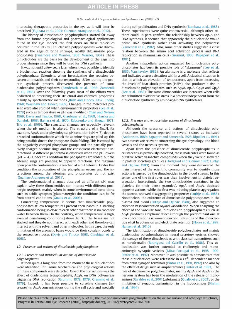
Ciliary process Physiological processesAgonists of P2Y6 Decrease of IOP
Antagonists of P2Y2 Decrease of IOP
Cornea Physiological processesAgonists of P2Y2 Accelerate corneal re-epithelization
CoAg
Trabecular meshworkAgonists of P2X2 Decrease of IOP
Agonists of P2Y1 Decrease of IOP
Re
A
AP
A
P
A
G
A
M
A
A
R
Aa
Fig. 2. Physiological processes mediated by purinergic receptors in the eye. Picture showsby activation or blockage of purinergic receptors and their therapeutic potential in certain
Please cite this article in press as: Carracedo, G., et al., The role of dinucleProgress in Retinal and Eye Research (2016), http://dx.doi.org/10.1016/j.p
organized into two groups based on their specific G proteincoupling. In one group, that includes the P2Y1, P2Y2, P2Y4, P2Y6 andP2Y11 receptors, are all coupled to Gq and the further activation ofphospholipase C b, with the consequent formation of inositoltriphosphate (IP3) and the mobilization of intracellular Ca2þ. Theother group includes the P2Y12, P2Y13 and P2Y14 receptors, asso-ciated with Gi proteins and inhibiting adenylate cyclase. The P2Y11is the only thing that can be coupled to both Gs and Gq proteins(Jacobson et al., 2015).
Concerning the action of dinucleoside polyphosphates and theactivation of P2 receptors, most of the research has been carried outfor ApnA compounds (Hoyle et al., 2001). In this sense, diadenosinepolyphosphates can activate all the P2X receptors (Wildman et al.,1999), including the heteromeric P2X2/3. It is noteworthy to indi-cate that the most active dinucleotide has been Ap4A in most cases.(Pintor and Miras-Portugal, 1995b). Also, the P2X7 was only sen-sitive to these dinucleotides at concentrations above 1 mM, thiseffect being far from the physiological concentrations of thesecompounds.
Regarding the P2Y receptors, P2Y1 and P2Y2 are the ones thatare stimulated by diadenosine polyphosphates (Patel et al., 2001).The tandem Ap4A-P2Y2 is of special interest since it participates inmany physiological processes in many ocular structures (Fig. 2 seebelow).
1.4. Dinucleoside polyphosphate degradation
The concentration of dinucleoside polyphosphates is tightlyregulated by a variety of enzymes, mainly hydrolases that arepresent in all organisms and generally act on the polyphosphates
njunctiva Physiological processesonists of P2Y2 Stimulate tear secretion
etinal pigment pithelium (RPE) Physiological processes
gonists of P2Y2Stimulate subretinal fluid reabsorption in retinal detachment
gonists of P2Y2 and 2Y6
Repair breakdown of RPE in retinal inflammation
ntagonists of P2X7 Prevention of apoptosis in AMD and diabetic retinopathy
hotoreceptors
ntagonists of P2X7 Prevention of apoptosis in inherited retinal diseases (retinitis pigmentosa)
anglion cells
ntagonists of P2X7 Prevention of apoptosis in retinal pathologies (glaucoma)
üller cells
ntagonists of P2X7 Reduce proliferation in PVR
gonists of P2Y2Prevention of osmotic swelling in ischemic or hypoxic retinopathies
etinal perycites
ntagonists of P2X7 nd agonists of P2Y4
Prevention of apoptosis in diabetic retinopathy
a schematic representation of the eye and the main physiological processes performedeye pathologies.
oside polyphosphates on the ocular surface and other eye structures,reteyeres.2016.07.001
G. Carracedo et al. / Progress in Retinal and Eye Research xxx (2016) 1e24 5
chain of these molecules. The enzymes involved in the catabolismof dinucleoside polyphosphates have been reviewed by Gur-anowski (Guranowski, 2000).
Extracellular dinucleotides, which regulate physiological pro-cesses via P2 receptors activation, are degraded principally by ecto-nucleotide pyrophosphatase/phosphodiesterases (NPPs) (Asensioet al., 2007; Vollmayer et al., 2003). This family of enzymes arehydrolases located on cell surface and are a type of the so calledecto-nucleotidases (Zimmermann et al., 2012). In vertebrates, NPPshave seven members (NPP1eNPP7) but only three (NPP1eNPP3)can degrade nucleotides (dinucleotides but also nucleoside tri-phosphates and diphosphates) (Goding et al., 1998; Jin-Hua et al.,1997; Stefan et al., 1999). Dinucleotides degradation by NPPs pro-duce nucleoside monophosphates (NMP) plus nucleoside poly-phosphates (NPn�1). Therefore, NPPs can generate metabolitesbiologically active such as ADP and ATP that are obtained from thenaturally occurring diadenosine polyphosphates (Vollmayer et al.,2003; Zimmermann, 2000). These nucleotides can also bedegraded by NPPs and essentially through ecto-nucleosidetriphosphate diphosphohydrolases (NTPDases), other family ofecto-enzymes that can degrade nucleoside triphosphates and di-phosphates (Zimmermann, 1999).
On the other hand, the degradation of diadenosine poly-phosphates and their metabolites ATP as well as ADP produce AMP.This nucleoside monophosphate can also be catabolized throughecto-50-nucleotidase (eN) as well as alkaline phosphatase enzymes(this last ecto-nucleotidasemay also act on ATP and ADPmolecules)(Zimmermann et al., 2012). Consequently, AMP is converted intothe nucleoside adenosine, which mediates purinergic effects via P1receptors interaction (Jacobson and Gao, 2006).
Finally, extracellular adenosine can be eliminated from thesynaptic cleft through cell surface adenosine deaminases thatconvert adenosine to inosine and/or nucleoside transporters(Bonan, 2012; Franco et al., 1997; Pastor-Anglada and Perez-Torras,2015). These transporters, equilibrative and concentrative nucleo-side transporters, are integral membrane proteins implicated in thecellular uptake of adenosine (Bonan, 2012; Pastor-Anglada andPerez-Torras, 2015).
2. Dinucleoside polyphosphates in the eye
2.1. Ocular surface
The ocular surface is the outer zone of the eye. It is a functionalunit composed of different structures such as corneal andconjunctival epithelium, lacrymal glands, eyelids and tear film. Allcomponents of this functional unit are linked through the inner-vation and vascular immunologic and endocrine systems. The mainfunctions of the ocular surface are to maintain the transparency ofthe cornea (optic quality issues) and to hydrate and protect thecorneal and conjunctival epithelium to the external agents. The tearfilm is mainly in charge of these functions. It is critical to have acorrect volume and stability of the tear film to maintain theintegrity of the ocular surface. The loss of the ocular homeostasisprovokes serious clinical manifestations as a dry eye, infections ofepithelial defects (Gipson, 2007; Knop and Knop, 2007; Paulsen,2008).
The ocular structure that has probably been investigated inmore depth regarding dinucleoside polyphosphates is the ocularsurface and specially the tear film. The presence of diadenosinepolyphosphates in the tear film has been noted in different con-ditions and also the different purinergic receptors in the ocularsurface that can be activated by the dinucleotides (Guzman-Aranguez and Pintor, 2012). Moreover, the effects of the ocularsurface of different dinucleotides when these compounds are
Please cite this article in press as: Carracedo, G., et al., The role of dinucleProgress in Retinal and Eye Research (2016), http://dx.doi.org/10.1016/j.p
instilled on the ocular surface have also been previously described(Crooke et al., 2008). In the following sections, the presence andeffect of dinucleoside polyphosphates will be reviewed.
2.1.1. Purinergic receptors in the ocular surfaceP2X and P2Y receptors are expressed in different zones of the
ocular surface, the P2Y2 subtype being the most abundant, identi-fied in the corneal and conjunctival epithelium, corneal endothe-lium and main lacrimal gland (Pintor et al., 2004b). All P2purinergic receptors described in the ocular surface and theirfunctions are summarized in Table 1. The expression of P2 receptorsin different ocular surface locations suggests the involvement ofthese receptors in the physiology of the eye (Guzman-Aranguezand Pintor, 2012). In fact, during the last decade, the role ofseveral nucleotides and their receptors in ocular processes,including tear secretion (through P2Y2 receptor) (Jumblatt andJumblatt, 1998; Murakami et al., 2000), corneal wound healing(P2Y2 is able to accelerate the corneal re-epithelization and pro-liferation, meanwhile P2Y6 inhibits the process) (Byun et al., 2016;Crooke et al., 2009; Mediero et al., 2006), and corneal epithelialmigration and stromal organization (mediated through P2X7 re-ceptor) have been described (Mankus et al., 2012; Mayo et al.,2008).
2.1.2. Action of ectodinucleotidases in the ocular surfaceAs we mentioned above, the dinucleotides effects on ocular
surface are due mainly to Ap4A and Ap5A but also their metabolitessuch as ATP and adenosine and the ecto-nucleotidases, mainlyNPPs, control the availability of these extracellular dinucleotidesand their metabolites. In this context, the expression of some ecto-enzymes has been detected, including ENPP1 as well as ENPP3isoforms, NTPD1 isoform and 5NTE in human conjunctival cellsobtained by impression cytology (see Fig. 3). Regarding adenosine,ocular surface also present ecto-adenosine deaminases and nucle-oside transporters that can control its possible effects (Majumdaret al., 2003; O'Brien et al., 1987).
The existence of conjunctival NPP isoenzymes (Fig. 3) withdifferent substrate specificity might explain, together with the non-neuronal origin of Ap4A and Ap5A, the different concentration ofthese dinucleotides in tear. Nevertheless, Vollmayer and colleagueshave proved that NPP1 and NPP3 hydrolyze Ap4A as well as Ap5Awith similar rates (Vollmayer et al., 2003).
2.1.3. Presence in the tear film of healthy patientsThe presence of diadenosine polyphosphates in the tear film
was studied due to their properties to enhance both tear secretionand to increase the wound healing rate (Guzman-Aranguez et al.,2007, 2011; Pintor et al., 2004a; Pintor et al., 2002b). The exoge-nous addition of these dinucleotides suggested their presence asnatural constituents of tears.
When investigating dinucleotides in tears, this fluid wasextracted by using paper strips to perform the Schrimer test (fortear volume measurement), placed in the inferior lid margin of theeye for 5min (van Bijsterveld,1969). The strips were then processedto be evaluated by High Pressure Liquid Chromatography (HPLC),following the procedure described by Pintor et al. (Pintor et al.,1992). The first adenine dinucleotides described in tear wereAp4A and Ap5A, which were found in New Zealand rabbit tears(Pintor et al., 2002b). Diadenosine tetraphosphate concentrationwas 2.92 ± 0.28 mM and diadenosine pentaphosphate concentra-tion was 0.587 ± 11 mM. These concentrations would be enough toactivate the ocular surface P2 receptors.
Initially, the hypothesis of the release of these dinucleotides inrabbit tear filmwas thought to occur via the exocytotic release fromthe nerve terminals present on the ocular surface, since this
oside polyphosphates on the ocular surface and other eye structures,reteyeres.2016.07.001
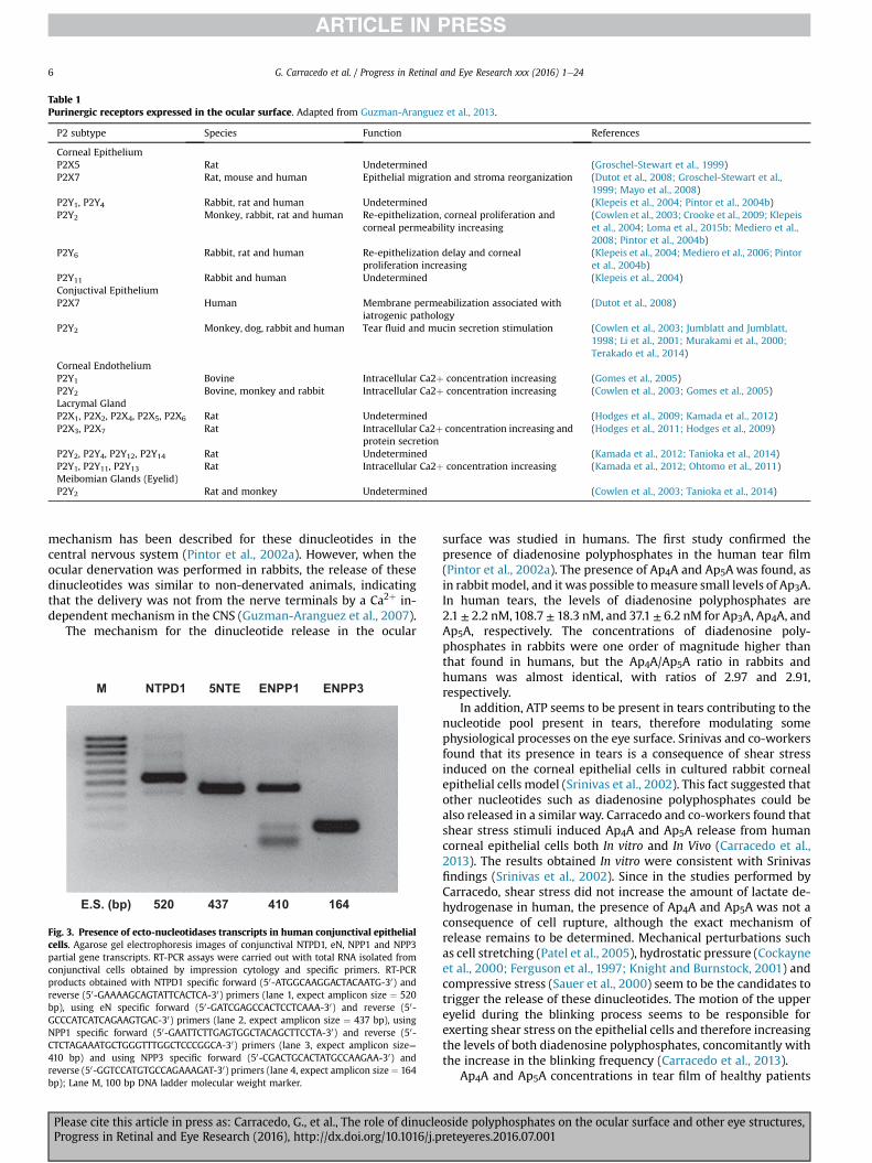
Table 1Purinergic receptors expressed in the ocular surface. Adapted from Guzman-Aranguez et al., 2013.
P2 subtype Species Function References
Corneal EpitheliumP2X5 Rat Undetermined (Groschel-Stewart et al., 1999)P2X7 Rat, mouse and human Epithelial migration and stroma reorganization (Dutot et al., 2008; Groschel-Stewart et al.,
1999; Mayo et al., 2008)P2Y1, P2Y4 Rabbit, rat and human Undetermined (Klepeis et al., 2004; Pintor et al., 2004b)P2Y2 Monkey, rabbit, rat and human Re-epithelization, corneal proliferation and
corneal permeability increasing(Cowlen et al., 2003; Crooke et al., 2009; Klepeiset al., 2004; Loma et al., 2015b; Mediero et al.,2008; Pintor et al., 2004b)
P2Y6 Rabbit, rat and human Re-epithelization delay and cornealproliferation increasing
(Klepeis et al., 2004; Mediero et al., 2006; Pintoret al., 2004b)
P2Y11 Rabbit and human Undetermined (Klepeis et al., 2004)Conjuctival EpitheliumP2X7 Human Membrane permeabilization associated with
iatrogenic pathology(Dutot et al., 2008)
P2Y2 Monkey, dog, rabbit and human Tear fluid and mucin secretion stimulation (Cowlen et al., 2003; Jumblatt and Jumblatt,1998; Li et al., 2001; Murakami et al., 2000;Terakado et al., 2014)
Corneal EndotheliumP2Y1 Bovine Intracellular Ca2þ concentration increasing (Gomes et al., 2005)P2Y2 Bovine, monkey and rabbit Intracellular Ca2þ concentration increasing (Cowlen et al., 2003; Gomes et al., 2005)Lacrymal GlandP2X1, P2X2, P2X4, P2X5, P2X6 Rat Undetermined (Hodges et al., 2009; Kamada et al., 2012)P2X3, P2X7 Rat Intracellular Ca2þ concentration increasing and
protein secretion(Hodges et al., 2011; Hodges et al., 2009)
P2Y2, P2Y4, P2Y12, P2Y14 Rat Undetermined (Kamada et al., 2012; Tanioka et al., 2014)P2Y1, P2Y11, P2Y13 Rat Intracellular Ca2þ concentration increasing (Kamada et al., 2012; Ohtomo et al., 2011)Meibomian Glands (Eyelid)P2Y2 Rat and monkey Undetermined (Cowlen et al., 2003; Tanioka et al., 2014)
G. Carracedo et al. / Progress in Retinal and Eye Research xxx (2016) 1e246
mechanism has been described for these dinucleotides in thecentral nervous system (Pintor et al., 2002a). However, when theocular denervation was performed in rabbits, the release of thesedinucleotides was similar to non-denervated animals, indicatingthat the delivery was not from the nerve terminals by a Ca2þ in-dependent mechanism in the CNS (Guzman-Aranguez et al., 2007).
The mechanism for the dinucleotide release in the ocular
M NTPD1 5NTE ENPP1 ENPP3
E.S. (bp) 520 437 410 164
Fig. 3. Presence of ecto-nucleotidases transcripts in human conjunctival epithelialcells. Agarose gel electrophoresis images of conjunctival NTPD1, eN, NPP1 and NPP3partial gene transcripts. RT-PCR assays were carried out with total RNA isolated fromconjunctival cells obtained by impression cytology and specific primers. RT-PCRproducts obtained with NTPD1 specific forward (50-ATGGCAAGGACTACAATG-30) andreverse (50-GAAAAGCAGTATTCACTCA-30) primers (lane 1, expect amplicon size ¼ 520bp), using eN specific forward (50-GATCGAGCCACTCCTCAAA-30) and reverse (50-GCCCATCATCAGAAGTGAC-30) primers (lane 2, expect amplicon size ¼ 437 bp), usingNPP1 specific forward (50-GAATTCTTGAGTGGCTACAGCTTCCTA-30) and reverse (50-CTCTAGAAATGCTGGGTTTGGCTCCCGGCA-30) primers (lane 3, expect amplicon size¼410 bp) and using NPP3 specific forward (50-CGACTGCACTATGCCAAGAA-30) andreverse (50-GGTCCATGTGCCAGAAAGAT-30) primers (lane 4, expect amplicon size ¼ 164bp); Lane M, 100 bp DNA ladder molecular weight marker.
Please cite this article in press as: Carracedo, G., et al., The role of dinucleProgress in Retinal and Eye Research (2016), http://dx.doi.org/10.1016/j.p
surface was studied in humans. The first study confirmed thepresence of diadenosine polyphosphates in the human tear film(Pintor et al., 2002a). The presence of Ap4A and Ap5A was found, asin rabbit model, and it was possible tomeasure small levels of Ap3A.In human tears, the levels of diadenosine polyphosphates are2.1 ± 2.2 nM,108.7 ± 18.3 nM, and 37.1 ± 6.2 nM for Ap3A, Ap4A, andAp5A, respectively. The concentrations of diadenosine poly-phosphates in rabbits were one order of magnitude higher thanthat found in humans, but the Ap4A/Ap5A ratio in rabbits andhumans was almost identical, with ratios of 2.97 and 2.91,respectively.
In addition, ATP seems to be present in tears contributing to thenucleotide pool present in tears, therefore modulating somephysiological processes on the eye surface. Srinivas and co-workersfound that its presence in tears is a consequence of shear stressinduced on the corneal epithelial cells in cultured rabbit cornealepithelial cells model (Srinivas et al., 2002). This fact suggested thatother nucleotides such as diadenosine polyphosphates could bealso released in a similar way. Carracedo and co-workers found thatshear stress stimuli induced Ap4A and Ap5A release from humancorneal epithelial cells both In vitro and In Vivo (Carracedo et al.,2013). The results obtained In vitro were consistent with Srinivasfindings (Srinivas et al., 2002). Since in the studies performed byCarracedo, shear stress did not increase the amount of lactate de-hydrogenase in human, the presence of Ap4A and Ap5A was not aconsequence of cell rupture, although the exact mechanism ofrelease remains to be determined. Mechanical perturbations suchas cell stretching (Patel et al., 2005), hydrostatic pressure (Cockayneet al., 2000; Ferguson et al., 1997; Knight and Burnstock, 2001) andcompressive stress (Sauer et al., 2000) seem to be the candidates totrigger the release of these dinucleotides. The motion of the uppereyelid during the blinking process seems to be responsible forexerting shear stress on the epithelial cells and therefore increasingthe levels of both diadenosine polyphosphates, concomitantly withthe increase in the blinking frequency (Carracedo et al., 2013).
Ap4A and Ap5A concentrations in tear film of healthy patients
oside polyphosphates on the ocular surface and other eye structures,reteyeres.2016.07.001

0.0
0.1
0.2
0.3
0.4
0.5
0.6
0.7
0.8Male Female
Ap 4
A c
once
ntra
tion
(μM
)0.0
0.1
0.2
0.3
0.4
Total < 50 y/o > 50 y/oA
p 5A
con
cent
ratio
n (μ
M)
Fig. 5. Sex-related differences in Ap4A and Ap5A concentrations in tear. Upper panelshows Ap4A concentrations in tear in the whole group and the subgroups of older andyounger than 50 years old. Lower panel shows Ap5A concentrations in tear in thewhole group and the subgroups of older and younger than 50 years old. There are nodifferences in dinucleotides levels between genders in any groups (p > 0.05, t-Studenttest for independent samples), even so, there is a significant increase in Ap4A and Ap5Aconcentration groups in subjects older than 50 years in males and females (p < 0.01 inAp4A levels and p < 0.05 in Ap5A levels, t-Student test for independent samples).Values represent means ± SEM.
G. Carracedo et al. / Progress in Retinal and Eye Research xxx (2016) 1e24 7
described above were collected in subjects from 18 to 38 years old(Pintor et al., 2002a). The age is considered one the most relevantfactors to suffer dry eye, this may be due to the reduction in thelacrimal production or due to the increase in the evaporation rateassociated with aging (Mathers et al., 1996). There are many studiescorroborating this assumption, in spite of the percentage of prev-alence which is very variable, changing from 5% to 35% at 50 yearsold, or 20%e40% at 80 years of age (Moss et al., 2000; Schein et al.,1997) (Chia et al., 2003; Lee et al., 2002; Lin et al., 2003;Schaumberg et al., 2003, 2009; Viso et al., 2009), although thereis consensus that the prevalence increases significantly after 50years of age. The other main factor to develop dry eye is gender, itbeing more common in women than men. The Beaver Dam studyshowed a prevalence of dry eye of 18% in women and only 11% inmen, and some other studies described even higher differencesbetween women and men (Paulsen et al., 2014). The principalreason for these differences would be the hormonal factor (Guillonand Maissa, 2010).
Due to the effect of some dinucleotides on the tear secretion (seesection 2.1.7.), our research group considered it interesting toevaluate the variations in diadenosine polyphosphates concentra-tions as a function of aging and gender. Ninety-three healthy sub-jects were recruited to participate in the study. The age range wasfrom 7 to 90 years old. Of the total 93 subjects, 47 were men and 46werewomen, all of themwithout any ocular surface pathology. Thelevels of Ap4A and Ap5A were analyzed with and without anes-thetic, discarding Ap3A due to the low concentration found in apreviously study (Pintor et al., 2002a).
The obtained results showed that Ap4A concentration remainedstable until 50 years old, increasing its concentration significantlyfrom this age (p value < 0.01; one way ANOVA). However, no sig-nificant changes were found for Ap5A concentration throughout theage range (p ¼ 0.245, one way ANOVA, Fig. 4). When comparingAp4A concentrations in function of gender, it was found that therewasmore diadenosine tetraphosphate in men tears than inwomen,but without a statistically significant difference (p value ¼ 0.078; t-Student test for independent samples). For Ap5A, the concentra-tions were similar in both groups of subjects (p value ¼ 0.761; t-Student test for independent samples, Fig. 5). As we will see in thefollowing section, a correlation between the age (dry eye diseasemain factor), and Ap4A concentration appears to exist.
2.1.4. Presence in ocular surface pathologyThe relationship between dry eye and dinucleotides has been
0.0
0.1
0.2
0.3
0.4
0.5
0.6
0.7
0.8
0.9
1.0
5-20 20-30 30-40 40-50 50-60 60-70 > 70
Din
ucle
otid
e C
once
ntra
tion
(μM
)
Ap4A Ap5A
Age
*
Fig. 4. Age-related variations in Ap4A and Ap5A concentrations in tear. Graph showsthe significant increase on Ap4A levels throughout age (* p < 0.01, one way ANOVA),while there are no differences on Ap5A levels (p ¼ 0.245, one way ANOVA). Valuesrepresent means ± SEM (n ¼ 93).
Please cite this article in press as: Carracedo, G., et al., The role of dinucleProgress in Retinal and Eye Research (2016), http://dx.doi.org/10.1016/j.p
proposed in two different ways: as molecular biomarker and astreatment for the pathology. Peral and co-workers were the first toevaluate the levels of diadenosine polyphosphates in tear secre-tions in two groups of dry eye symptomatic subjects, one with lowtear secretion volume and another with normal tear volume (Peralet al., 2006). They found that Ap4A and Ap5Awere increased 5- and1.5-fold in dry eye subjects with normal tear volume, respectively,compared with healthy subjects in the same age range. The dinu-cleotide concentrations were 0.590 ± 0.330 mM for Ap4A and0.058 ± 0.017 mM for Ap5A. For symptomatic patients with low tearproduction the increase was higher, being 100- and 345-fold morethan the value obtained in the healthy group. The concentrationswere 10.68 ± 1.87 mM for Ap4A and 12.45 ± 0.20 mM for Ap5A. Fromthese results, the authors proposed these molecules as an objectivebiomarker for scoring the severity of dry eye.
In 2010, a research paper was published about the diadenosinepolyphosphate concentrations in one of the most severe types ofdry eye, Sj€ogren Syndrome (Carracedo et al., 2010). This autoim-mune disorder is produced when the immune system attacks itsown exocrine glands by mistake, especially the salivary andlacrimal ones (Srinivasan and Slomovic, 2007). The mean concen-trations of Ap4A, and Ap5A in the Sj€ogren syndrome group were2.54 ± 1.02 mM and 26.13 ± 6.95 mM, respectively. When this groupwas divided into two subgroups, normal and low tear secretion, theconcentrations in normal tear secretion group were 0.47 ± 0.20 mMfor Ap4A and 8.30 ± 3.27 mM for Ap5A. The subgroup with low tearsecretion showed dinucleotide concentrations of 4.09 ± 1.36 mMand 39.51 ± 8.45 mM for Ap4A and Ap5A, respectively. It has beenreported that the upregulated presence of P2Y2 in the salivary
oside polyphosphates on the ocular surface and other eye structures,reteyeres.2016.07.001
G. Carracedo et al. / Progress in Retinal and Eye Research xxx (2016) 1e248
glands increases the epidermal growth factor receptor (EGFR) e
dependent expression of vascular cell adhesionmolecule (VCAM)-1and provokes the lymphocyte infiltration associated with theinflammation (Baker et al., 2008; Schrader et al., 2005). Meanwhilethis receptor has not yet been located in the lacrimal gland, asimilar effect could occur in Sj€ogren Syndrome (Tanioka et al.,2014). Therefore, the high concentrations of diadenosine poly-phosphates in Sj€ogren syndrome patients would not be able tostimulate more lacrimation; they may even increase lymphocyteinfiltration.
Another condition related with dinucleoside concentration intears is congenital Aniridia. This inherited pathology is a rare dis-order due to the mutation of the PAX6 gene (Glaser et al., 1992,1994), located in the short arm of the chromosome 11. The mainfeature of aniridia is the partial or total absence of the iris, but theocular surface is also affected with limbal stem cell deficiency, tearfilm disorders, corneal keratopathy and dry eye (Mayer et al., 2003).It seems that dinucleoside play an important role in the progressionof the pathology, since the Ap4A and Ap5A concentration increaseswith age, being higher in patients over 40 years of age than inyounger aniridia patients. It has been demonstrated that P2 puri-nergic receptors are associated with differentiation, proliferationand neurogenesis in neonatal and adult mouse olfactory epithelium(Jia and Hegg, 2012). In the corneal epithelium the cell proliferationprocess is initiated by P2Y2 receptor activation via the ERK1/2pathway (Muscella et al., 2004). Therefore, the increased levels ofdiadenosine polyphosphates have been proposed as a compensa-torymechanism to stimulate the proliferation and differentiation oflimbal stem cells that are deficient in aniridia disorder.
Dinucleotide presence in the tear film has been related to dryeye disorders, cell migration and proliferation disorders. Inflam-mation is another biological process where the purinergic receptorsand dinucleotides are involved (Guzman-Aranguez et al., 2014). Themain ocular surface condition, with dry eye, which is probablyassociated with inflammation is keratoconus. It is a corneal disor-der characterized by progressive corneal stromal thinning, a cone-shaped protrusion of the corneal surface (ectasia) (Davidson et al.,2014; Romero-Jimenez et al., 2010), that creates alterations of thecorneal tissue surface that result in visual distortion and reducedtear film quality. Tissue degradation by keratoconus involves theexpression of inflammatory mediators, and therefore patientsexhibit higher levels of pro-inflammatory cytokines, cell adhesionmolecules, and matrix metalloproteinases (MMPs) compared with
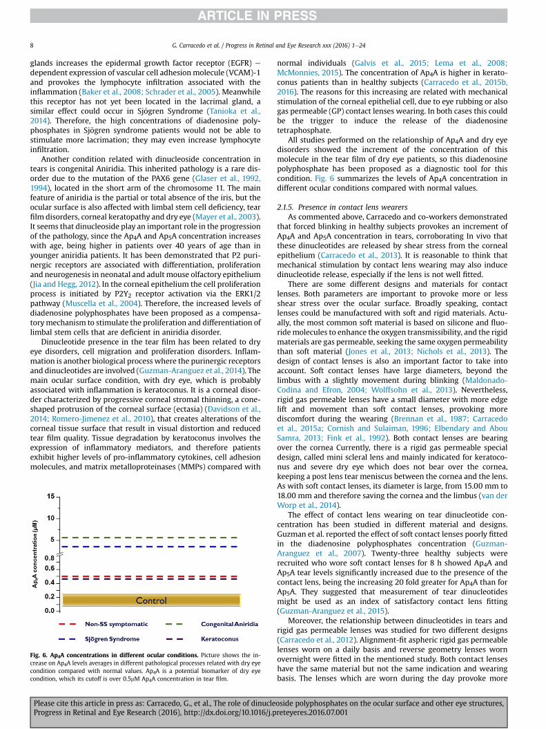
Fig. 6. Ap4A concentrations in different ocular conditions. Picture shows the in-crease on Ap4A levels averages in different pathological processes related with dry eyecondition compared with normal values. Ap4A is a potential biomarker of dry eyecondition, which its cutoff is over 0.5mM Ap4A concentration in tear film.
Please cite this article in press as: Carracedo, G., et al., The role of dinucleProgress in Retinal and Eye Research (2016), http://dx.doi.org/10.1016/j.p
normal individuals (Galvis et al., 2015; Lema et al., 2008;McMonnies, 2015). The concentration of Ap4A is higher in kerato-conus patients than in healthy subjects (Carracedo et al., 2015b,2016). The reasons for this increasing are related with mechanicalstimulation of the corneal epithelial cell, due to eye rubbing or alsogas permeable (GP) contact lenses wearing. In both cases this couldbe the trigger to induce the release of the diadenosinetetraphosphate.
All studies performed on the relationship of Ap4A and dry eyedisorders showed the increment of the concentration of thismolecule in the tear film of dry eye patients, so this diadenosinepolyphosphate has been proposed as a diagnostic tool for thiscondition. Fig. 6 summarizes the levels of Ap4A concentration indifferent ocular conditions compared with normal values.
2.1.5. Presence in contact lens wearersAs commented above, Carracedo and co-workers demonstrated
that forced blinking in healthy subjects provokes an increment ofAp4A and Ap5A concentration in tears, corroborating In vivo thatthese dinucleotides are released by shear stress from the cornealepithelium (Carracedo et al., 2013). It is reasonable to think thatmechanical stimulation by contact lens wearing may also inducedinucleotide release, especially if the lens is not well fitted.
There are some different designs and materials for contactlenses. Both parameters are important to provoke more or lessshear stress over the ocular surface. Broadly speaking, contactlenses could be manufactured with soft and rigid materials. Actu-ally, the most common soft material is based on silicone and fluo-ridemolecules to enhance the oxygen transmissibility, and the rigidmaterials are gas permeable, seeking the same oxygen permeabilitythan soft material (Jones et al., 2013; Nichols et al., 2013). Thedesign of contact lenses is also an important factor to take intoaccount. Soft contact lenses have large diameters, beyond thelimbus with a slightly movement during blinking (Maldonado-Codina and Efron, 2004; Wolffsohn et al., 2013). Nevertheless,rigid gas permeable lenses have a small diameter with more edgelift and movement than soft contact lenses, provoking morediscomfort during the wearing (Brennan et al., 1987; Carracedoet al., 2015a; Cornish and Sulaiman, 1996; Elbendary and AbouSamra, 2013; Fink et al., 1992). Both contact lenses are bearingover the cornea Currently, there is a rigid gas permeable specialdesign, called mini scleral lens and mainly indicated for keratoco-nus and severe dry eye which does not bear over the cornea,keeping a post lens tear meniscus between the cornea and the lens.As with soft contact lenses, its diameter is large, from 15.00 mm to18.00 mm and therefore saving the cornea and the limbus (van derWorp et al., 2014).
The effect of contact lens wearing on tear dinucleotide con-centration has been studied in different material and designs.Guzman et al. reported the effect of soft contact lenses poorly fittedin the diadenosine polyphosphates concentration (Guzman-Aranguez et al., 2007). Twenty-three healthy subjects wererecruited who wore soft contact lenses for 8 h showed Ap4A andAp5A tear levels significantly increased due to the presence of thecontact lens, being the increasing 20 fold greater for Ap4A than forAp5A. They suggested that measurement of tear dinucleotidesmight be used as an index of satisfactory contact lens fitting(Guzman-Aranguez et al., 2015).
Moreover, the relationship between dinucleotides in tears andrigid gas permeable lenses was studied for two different designs(Carracedo et al., 2012). Alignment-fit aspheric rigid gas permeablelenses worn on a daily basis and reverse geometry lenses wornovernight were fitted in the mentioned study. Both contact lenseshave the same material but not the same indication and wearingbasis. The lenses which are worn during the day provoke more
oside polyphosphates on the ocular surface and other eye structures,reteyeres.2016.07.001
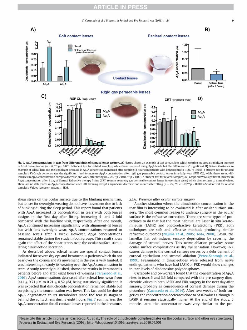
Fig. 7. Ap4A concentrations in tear from different kinds of contact lenses wearers. A) Picture shows an example of soft contact lens which wearing induces a significant increasein Ap4A concentration (n ¼ 6, *** p < 0.001, t-Student test for related samples), while there is a trend rising Ap5A levels but the difference isn’t significant. B) Picture illustrates anexample of scleral lens and the significant decrease in Ap4A concentration induced after wearing (8 hours) in patients with keratoconus (n ¼ 26, *p < 0.05, t-Student test for relatedsamples). C) Graph demonstrates the significant trend to increase Ap4A concentration after rigid gas permeable contact lenses in a daily wear (RGP CL), while there are no dif-ferences in Ap5A concentration except a decrease one week after fitting (n ¼ 22, **p < 0.01, ***p < 0.001, t-Student test for related samples). D) Graph shows a significant increase inAp4A concentration after 1 day of Corneal Refractive therapy fitting (CRT: reverse geometry gas permeable contact lenses in overnight wear) which then returns to normal values.There are no differences in Ap5A concentration after CRT wearing except a significant decrease one month after fitting (n ¼ 22, **p < 0.01,***p < 0.001, t-Student test for relatedsamples). Values represent means ± SEM.
G. Carracedo et al. / Progress in Retinal and Eye Research xxx (2016) 1e24 9
shear stress on the ocular surface due to the blinking mechanism,but lenses for overnight wearing do not havemovement due to lackof blinking during the sleep period. This report found that patientswith Ap4A increased its concentration in tears with both lensesdesigns in the first day after fitting, increasing 4- and 2-foldcompared with the baseline visit, respectively. After one month,Ap4A continued increasing significantly with alignment-fit lensesbut with lens overnight wear, Ap4A concentrations returned tobaseline levels after 1 week. However, Ap5A concentrationsremained stable during the study in both groups. This result showsagain the effect of the shear stress over the ocular surface stimu-lating dinucleotide secretion.
As described above, scleral lenses are special contact lensesindicated for severe dry eye and keratoconus patients which do notbear over the cornea and its movement in the eye is very limited. Itwas interesting to study its wearing over the Ap4A concentration intears. A study recently published, shows the results in keratoconuspatients before and after eight hours of wearing (Carracedo et al.,2016). Ap4A concentrations decreased after scleral lens wear from0.41 ± 0.71 mM to 0.21 ± 0.52 mM, being statistically significant. Itwas expected that dinucleotide concentration remained stable butsurprisingly the concentration was lower. This fact could be due toAp4A degradation to its metabolites since the tear is stagnantbehind the contact lens during eight hours. Fig. 7 summarizes theAp4A concentration for all contact lenses reported in the literature.
Please cite this article in press as: Carracedo, G., et al., The role of dinucleProgress in Retinal and Eye Research (2016), http://dx.doi.org/10.1016/j.p
2.1.6. Presence after ocular surface surgeryAnother situation where the dinucleotide concentration in the
tear film is interesting to be evaluated is after ocular surface sur-gery. The most common reason to undergo surgery in the ocularsurface is the refractive correction. There are some types of pro-cedures to do that but the most habitual are Laser in situ kerato-mileusis (LASIK) and photorefractive keratectomy (PRK). Bothtechniques are safe and effective methods producing similarrefractive outcomes (Nejima et al., 2005; Toda, 2008). LASIK, thelamellar flat cut induces sensory deprivation by severing thedamage of stromal nerves. This nerve ablation provokes someocular surface complications as dry eye sensation. However, PRKcauses damage to the corneal sensory nerves by a debridement ofcorneal epithelium and stromal ablation (Perez-Santonja et al.,1999). Presumably, if dinucleotides were released from nerveendings, patients who have had LASIK or PRK would show changesin tear levels of diadenosine polyphosphates.
Carracedo and co-workers found that the concentration of Ap4Arises in tears 5 and 3.5 fold compared with the pre-surgery dinu-cleotide values in both LASIK and PRK surgery in the next day aftersurgery, probably as consequence of corneal damage during theprocedure (Carracedo et al., 2014). After two weeks of both sur-geries, the concentration decreases close to basal values although inLASIK it remains statistically higher. At the end of the study, 3months later, the concentration was very similar to the pre-
oside polyphosphates on the ocular surface and other eye structures,reteyeres.2016.07.001
G. Carracedo et al. / Progress in Retinal and Eye Research xxx (2016) 1e2410
operative visit for both techniques. Guzman et al. have found thatAp4A trend to be higher than baseline, but not statistically signifi-cant, oneweek after LASIK surgery (Guzman-Aranguez et al., 2007).The results from both studies matched, although it is important tonotice that the lack of statistical significance in the study by Guz-man et al., is probably due to the small size of the sample. For Ap5A,the concentration remained stable in both studies during all theperiod evaluated.
In a non-published study performed by our research group,dinucleotide concentration in tears was also measured in patientsafter Intraocular collamer Lens (ICL) surgery. This refractive surgerytechnique consists of introducing an intraocular contact lens in theanterior chamber through a small incision in the corneal periphery,close to the limbus (Kamiya et al., 2010; Uusitalo et al., 2002). Withthis procedure the corneal neural network is preserved (Alfonsoet al., 2013; Ali et al., 2014). Our preliminary results show thatboth Ap4A and Ap5A do not change their concentrations in the tearfilm throughout the study. This fact suggests that diadenosinepolyphosphates present in tears are not released by the nerve ter-minals present at the ocular surface, but it rather confirms that thedinucleotides delivered from ocular surface epithelium is inducedby shear stress.
There are some studies describing how Ap4A has the ability tostimulate epithelial cell migration. In contrast, Ap5A has theopposite effect, inhibiting the cell migration (Crooke et al., 2009;Mediero et al., 2009, 2008, 2006). So, the high concentration ofAp4A on the day after LASIK and PRK surgery could be used toaccelerate the wound healing process.
2.1.7. Effect on tear secretionTear secretion is a physiological process because the tear forms
the interface between the air and the ocular tissues. To lubricateand to provide nutrients to the ocular surface tissues and to protectagainst infections are the more critical features of the tear film. Fora correct tear volume and stability, it is very important to maintaina compensated tear composition. A failure in the tear secretionmayprovoke several negative effects in the ocular surface such asdiscomfort, dryness, irritation or corneal and conjunctival staining(Herrero-Vanrell and Peral, 2007). Dinucleotides participate in thesecretion process of some compounds of tear film like aqueouscomponent, mucins and proteins.
The presence of diadenosine polyphosphates in tears of bothanimals and humans suggests that they maymodify the physiologyof the ocular surface. Diadenosine tetraphosphate have demon-strated their capability to increase the tear volume in the ocularsurface (Pintor et al., 2002b). In the case of Ap4A the increment is up60% compared to baseline values in experiments performed withNew Zealand white rabbits. Ap5A and Ap6A also significantlyincreased tear production by about 20% but, Ap2A and Ap3A arecompletely ineffective. However, it has not been fully clarified if theeffect of these dinucleotides is to induce tear production at thelacrymal gland or in other secretory structures. The studies per-formed with animal models, suppressing the effect with RB-2,suggest that diadenosine polyphosphates act as agonist of P2Y2
receptors for increasing the tear production (Murakami et al., 2000;Pintor et al., 2002b). Curiously, the combination of melatonin withAp4A enhances the effect of the diadenosine over the tear secretionin an animal model, but melatonin does not have any effect when itis applied alone. The authors blocked the effect of this combinationapplying luzindole, an antagonist of melatonin receptors (Hoyleet al., 2006). It has not been described how melatonin and its re-ceptors increase the effect of Ap4A but a possible mechanism is theheterodimerization of both dinucleotides and melatonin receptors.Both families receptor are coupled to G-protein and it has beensuggested that actually these receptors could be organized as
Please cite this article in press as: Carracedo, G., et al., The role of dinucleProgress in Retinal and Eye Research (2016), http://dx.doi.org/10.1016/j.p
heterodimers between different G-protein coupled families(Levoye et al., 2006).
Other dinucleotides which demonstrate their secretagoguepropierties is Gp4G and Up4U. Recently in our lab, it has beenproved that diguanine tetraphosphate (Gp4G) increases tearsecretion in around 40% above normal tear values in New ZealandWhite rabbits. As diadenosine tetraphosphate, it is expected thatthis action is mediated by P2Y2 (Guzman-Aranguez et al., 2015). Butthe dinucleotide probably most studied is Up4U, also called INS-365. First studies in animal models showed its properties toenhance tear secretion (Fujihara et al., 2001; Murakami et al., 2000,2002; Yerxa et al., 2002a). These results, as a secretagogue, were theprelude of Diquafosol, an ophthalmic formulation based on thiscompound, developed as a new treatment for dry eye and wasrecently approved for marketing in Japan and South Korea(Jacobson et al., 2012; Nakamura et al., 2012).
Diquafosol has shown its capability to increase the aqueous andmucin portion of tears (Murakami et al., 2004; Nakamura et al.,2012; Takamura et al., 2012; Tauber et al., 2004). In recent years,diquafosol has been used for treating different conditions relatedwith dry eye (Table 2). This dinucleotide improves the tear secre-tion and symptomatology in Sj€ogren syndrome (Bremond-Gignacet al., 2014; Yokoi et al., 2015), aqueous deficient dry eye (Kohet al., 2013a, 2014), contact lens wearers (Nagahara et al., 2015),Meibomian gland dysfunction (Arita et al., 2013; Yamaguchi et al.,2015), dry eye related with cataract surgery (Miyake et al., 2014;Park et al., 2015), even in normal eyes (Gendaszewska-Darmachand Kucharska, 2011; Nam et al., 2015; Yokoi et al., 2014).Regarding the safe use of diquafosol, the majority of adverse re-actions, found in different clinical trials, were of mild severity, andno serious treatment-related adverse events were reported.Adverse reactions were observed in 6.3% of patients, and the majoradverse reactions were eye irritation, and eye pain. It seems thatgood tolerance shown by diquafosol ophthalmic solution 3%remained in the longer term (Takamura et al., 2012;Wu et al., 2015;Yamaguchi et al., 2014).
2.1.8. Effect on tear componentsThe tear film is a complex solution composed of proteins, lipids,
carbohydrates and electrolytes. Two main features of the tear filmare to protect the ocular surface against potential pathogens and tolubricate and wet the corneal and conjunctival epithelia. So, thetear film has some antimicrobial components that are able to killthe pathogens or at least to inhibit their replication. The mainproteins in the tear film performing this task are lysozyme, lacto-ferrin, immunoglobulin A (IgA) and tear lipocaline (Farris, 1986;Kuizenga et al., 1996; Cabrera et al., 1997).
Lysozime presents a bactericide action against gram-positivebacteria, such as Micrococus lisodeikticus, catalyzing the hydrolysisof 1,4-b junctions between N-acetylmuramic acid and N-acetyl-D-glucosamine in the main chain of peptidoglycans of cell membrane(Burman et al., 1991; Qasba and Kumar, 1997). This protein issecreted by the main and accessories lacrymal glands, representing25% of total proteins in the tear film. Peral and co-workers inves-tigated the effect of dinucleotide instillation on the lysozymeconcentration in the tear film (Peral et al., 2008). Their hypothesiswas based on several studies that have shown a decrease in theconcentration of lysozyme in patients with dry eye (Caffery et al.,2008; Mackie and Seal, 1984; van Bijsterveld, 1969). The topicalapplication of 100 mM of Ap4A and Up4U (aka diquafosol) increasedthe lysozyme levels after one hour of instillation, remaining higherthan baseline during four hours. The maximum value was reachedat two hours for both dinucleotides, being the increase in lysozymeconcentration of 93% for Ap4A, 119% for Up4U.
Another interesting molecule with bacteriostatic properties is
oside polyphosphates on the ocular surface and other eye structures,reteyeres.2016.07.001
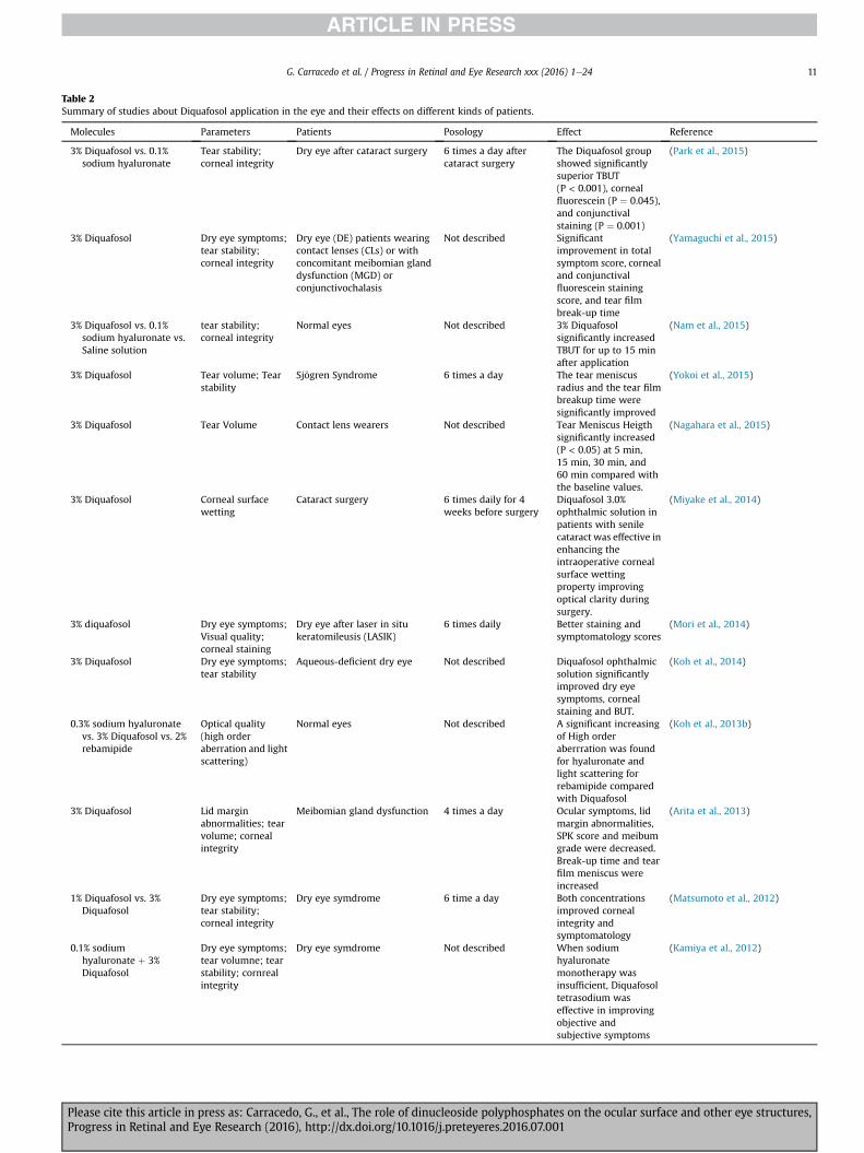
Table 2Summary of studies about Diquafosol application in the eye and their effects on different kinds of patients.
Molecules Parameters Patients Posology Effect Reference
3% Diquafosol vs. 0.1%sodium hyaluronate
Tear stability;corneal integrity
Dry eye after cataract surgery 6 times a day aftercataract surgery
The Diquafosol groupshowed significantlysuperior TBUT(P < 0.001), cornealfluorescein (P ¼ 0.045),and conjunctivalstaining (P ¼ 0.001)
(Park et al., 2015)
3% Diquafosol Dry eye symptoms;tear stability;corneal integrity
Dry eye (DE) patients wearingcontact lenses (CLs) or withconcomitant meibomian glanddysfunction (MGD) orconjunctivochalasis
Not described Significantimprovement in totalsymptom score, cornealand conjunctivalfluorescein stainingscore, and tear filmbreak-up time
(Yamaguchi et al., 2015)
3% Diquafosol vs. 0.1%sodium hyaluronate vs.Saline solution
tear stability;corneal integrity
Normal eyes Not described 3% Diquafosolsignificantly increasedTBUT for up to 15 minafter application
(Nam et al., 2015)
3% Diquafosol Tear volume; Tearstability
Sj€ogren Syndrome 6 times a day The tear meniscusradius and the tear filmbreakup time weresignificantly improved
(Yokoi et al., 2015)
3% Diquafosol Tear Volume Contact lens wearers Not described Tear Meniscus Heigthsignificantly increased(P < 0.05) at 5 min,15 min, 30 min, and60 min compared withthe baseline values.
(Nagahara et al., 2015)
3% Diquafosol Corneal surfacewetting
Cataract surgery 6 times daily for 4weeks before surgery
Diquafosol 3.0%ophthalmic solution inpatients with senilecataract was effective inenhancing theintraoperative cornealsurface wettingproperty improvingoptical clarity duringsurgery.
(Miyake et al., 2014)
3% diquafosol Dry eye symptoms;Visual quality;corneal staining
Dry eye after laser in situkeratomileusis (LASIK)
6 times daily Better staining andsymptomatology scores
(Mori et al., 2014)
3% Diquafosol Dry eye symptoms;tear stability
Aqueous-deficient dry eye Not described Diquafosol ophthalmicsolution significantlyimproved dry eyesymptoms, cornealstaining and BUT.
(Koh et al., 2014)
0.3% sodium hyaluronatevs. 3% Diquafosol vs. 2%rebamipide
Optical quality(high orderaberration and lightscattering)
Normal eyes Not described A significant increasingof High orderaberrration was foundfor hyaluronate andlight scattering forrebamipide comparedwith Diquafosol
(Koh et al., 2013b)
3% Diquafosol Lid marginabnormalities; tearvolume; cornealintegrity
Meibomian gland dysfunction 4 times a day Ocular symptoms, lidmargin abnormalities,SPK score and meibumgrade were decreased.Break-up time and tearfilm meniscus wereincreased
(Arita et al., 2013)
1% Diquafosol vs. 3%Diquafosol
Dry eye symptoms;tear stability;corneal integrity
Dry eye symdrome 6 time a day Both concentrationsimproved cornealintegrity andsymptomatology
(Matsumoto et al., 2012)
0.1% sodiumhyaluronate þ 3%Diquafosol
Dry eye symptoms;tear volumne; tearstability; cornrealintegrity
Dry eye symdrome Not described When sodiumhyaluronatemonotherapy wasinsufficient, Diquafosoltetrasodium waseffective in improvingobjective andsubjective symptoms
(Kamiya et al., 2012)
G. Carracedo et al. / Progress in Retinal and Eye Research xxx (2016) 1e24 11
Please cite this article in press as: Carracedo, G., et al., The role of dinucleoside polyphosphates on the ocular surface and other eye structures,Progress in Retinal and Eye Research (2016), http://dx.doi.org/10.1016/j.preteyeres.2016.07.001
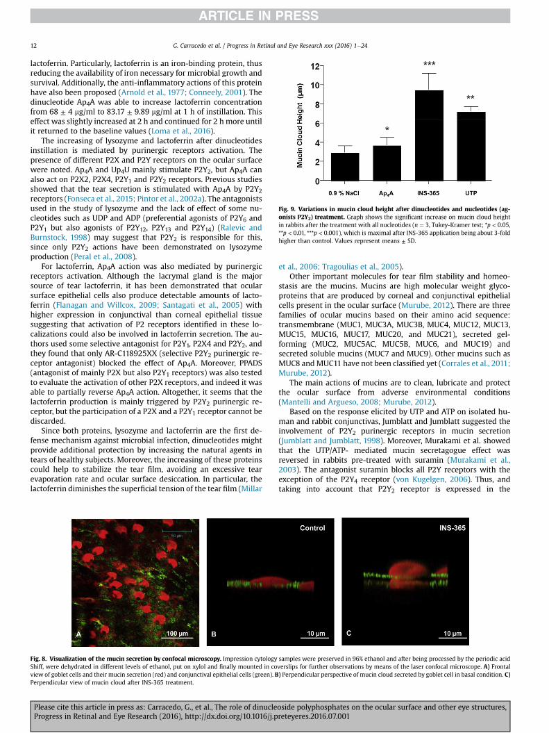
Fig. 9. Variations in mucin cloud height after dinucleotides and nucleotides (ag-onists P2Y2) treatment. Graph shows the significant increase on mucin cloud heightin rabbits after the treatment with all nucleotides (n ¼ 3, Tukey-Kramer test; *p < 0.05,**p < 0.01, ***p < 0.001), which is maximal after INS-365 application being about 3-foldhigher than control. Values represent means ± SD.
G. Carracedo et al. / Progress in Retinal and Eye Research xxx (2016) 1e2412
lactoferrin. Particularly, lactoferrin is an iron-binding protein, thusreducing the availability of iron necessary for microbial growth andsurvival. Additionally, the anti-inflammatory actions of this proteinhave also been proposed (Arnold et al., 1977; Conneely, 2001). Thedinucleotide Ap4A was able to increase lactoferrin concentrationfrom 68 ± 4 mg/ml to 83.17 ± 9.89 mg/ml at 1 h of instillation. Thiseffect was slightly increased at 2 h and continued for 2 h more untilit returned to the baseline values (Loma et al., 2016).
The increasing of lysozyme and lactoferrin after dinucleotidesinstillation is mediated by purinergic receptors activation. Thepresence of different P2X and P2Y receptors on the ocular surfacewere noted. Ap4A and Up4U mainly stimulate P2Y2, but Ap4A canalso act on P2X2, P2X4, P2Y1 and P2Y2 receptors. Previous studiesshowed that the tear secretion is stimulated with Ap4A by P2Y2receptors (Fonseca et al., 2015; Pintor et al., 2002a). The antagonistsused in the study of lysozyme and the lack of effect of some nu-cleotides such as UDP and ADP (preferential agonists of P2Y6 andP2Y1 but also agonists of P2Y12, P2Y13 and P2Y14) (Ralevic andBurnstock, 1998) may suggest that P2Y2 is responsible for this,since only P2Y2 actions have been demonstrated on lysozymeproduction (Peral et al., 2008).
For lactoferrin, Ap4A action was also mediated by purinergicreceptors activation. Although the lacrymal gland is the majorsource of tear lactoferrin, it has been demonstrated that ocularsurface epithelial cells also produce detectable amounts of lacto-ferrin (Flanagan and Willcox, 2009; Santagati et al., 2005) withhigher expression in conjunctival than corneal epithelial tissuesuggesting that activation of P2 receptors identified in these lo-calizations could also be involved in lactoferrin secretion. The au-thors used some selective antagonist for P2Y1, P2X4 and P2Y2, andthey found that only AR-C118925XX (selective P2Y2 purinergic re-ceptor antagonist) blocked the effect of Ap4A. Moreover, PPADS(antagonist of mainly P2X but also P2Y1 receptors) was also testedto evaluate the activation of other P2X receptors, and indeed it wasable to partially reverse Ap4A action. Altogether, it seems that thelactoferrin production is mainly triggered by P2Y2 purinergic re-ceptor, but the participation of a P2X and a P2Y1 receptor cannot bediscarded.
Since both proteins, lysozyme and lactoferrin are the first de-fense mechanism against microbial infection, dinucleotides mightprovide additional protection by increasing the natural agents intears of healthy subjects. Moreover, the increasing of these proteinscould help to stabilize the tear film, avoiding an excessive tearevaporation rate and ocular surface desiccation. In particular, thelactoferrin diminishes the superficial tension of the tear film (Millar
Fig. 8. Visualization of the mucin secretion by confocal microscopy. Impression cytologyShiff, were dehydrated in different levels of ethanol, put on xylol and finally mounted in coview of goblet cells and their mucin secretion (red) and conjunctival epithelial cells (green). BPerpendicular view of mucin cloud after INS-365 treatment.
Please cite this article in press as: Carracedo, G., et al., The role of dinucleProgress in Retinal and Eye Research (2016), http://dx.doi.org/10.1016/j.p
et al., 2006; Tragoulias et al., 2005).Other important molecules for tear film stability and homeo-
stasis are the mucins. Mucins are high molecular weight glyco-proteins that are produced by corneal and conjunctival epithelialcells present in the ocular surface (Murube, 2012). There are threefamilies of ocular mucins based on their amino acid sequence:transmembrane (MUC1, MUC3A, MUC3B, MUC4, MUC12, MUC13,MUC15, MUC16, MUC17, MUC20, and MUC21), secreted gel-forming (MUC2, MUC5AC, MUC5B, MUC6, and MUC19) andsecreted soluble mucins (MUC7 and MUC9). Other mucins such asMUC8 andMUC11 have not been classified yet (Corrales et al., 2011;Murube, 2012).
The main actions of mucins are to clean, lubricate and protectthe ocular surface from adverse environmental conditions(Mantelli and Argueso, 2008; Murube, 2012).
Based on the response elicited by UTP and ATP on isolated hu-man and rabbit conjunctivas, Jumblatt and Jumblatt suggested theinvolvement of P2Y2 purinergic receptors in mucin secretion(Jumblatt and Jumblatt, 1998). Moreover, Murakami et al. showedthat the UTP/ATP- mediated mucin secretagogue effect wasreversed in rabbits pre-treated with suramin (Murakami et al.,2003). The antagonist suramin blocks all P2Y receptors with theexception of the P2Y4 receptor (von Kugelgen, 2006). Thus, andtaking into account that P2Y2 receptor is expressed in the
samples were preserved in 96% ethanol and after being processed by the periodic acidverslips for further observations by means of the laser confocal microscope. A) Frontal) Perpendicular perspective of mucin cloud secreted by goblet cell in basal condition. C)
oside polyphosphates on the ocular surface and other eye structures,reteyeres.2016.07.001

G. Carracedo et al. / Progress in Retinal and Eye Research xxx (2016) 1e24 13
conjunctival epithelium of rabbits and monkeys (Cowlen et al.,2003), it has been considered that the P2Y2 receptor governs theconjunctival mucin secretion. Both Ap4A and Up4U are agonists ofP2Y2 receptor so they should promote the mucin secretion fromconjunctival Goblet cells.
The first study on dinucleotides and mucin secretion in tearswas published in 2001 (Fujihara et al., 2001). They found that INS-365, aka Up4U and also termed diquafosol (see section 2.1.7), hasthe ability to evoke mucin secretion on In vivo conjunctivas of a ratdry eye model. In 2004 they corroborated their results in the NewZealand white rabbit model (Pintor et al., 2004a). They concludedthat INS365 restored moisture and hydration of the ocular surfaceof rats by its effects on tear andmucin secretion. Other authors havefound the same effect of INS-365 in different animal models,increasing Goblet cell Density andmucin production (Fujihara et al.,2002; Kojima et al., 2014; Terakado et al., 2014).
Our research group performed a study to evaluate the possibleinvolvement of other P2Y receptors, not only P2Y2, such as P2Y4 andP2Y6 in rabbit conjunctival mucin secretion, testing the mucinsecretagogue effect of the dinucleotide Ap4A and INS-365. Thetechnique used for ocular mucin visualization was confocal laserscanningmicroscopy (Fig. 8). This technique, following the protocolstablished by Peral and Pintor, allows us to measure mucin cloudheight (MCH) over the goblet cell. In this sense, all the dinucleotidestested evoked a significant increase of the MCH parameter incomparisonwith control animals. The rank order of effect on mucinsecretion was INS-365 (MCH ¼ 9.50 ± 1.72 mm) greater than UTP(MCH ¼ 6.50 ± 0.49 mm) and greater than Ap4A(MCH ¼ 3.75 ± 0.79 mm), Fig. 9.
It was also observed that the secretory effect of INS-365 and UTPwas more robust than that obtained by Ap4A. This fact suggests theinvolvement not only of P2Y2 receptors but also of other uracilnucleotide-sensitive receptors. To determine the possible purino-ceptors involved in mucin secretion RB-2, suramin and PPADS an-tagonists were applied on rabbit conjunctiva in the presence orabsence of INS-365. RB-2 was the most effective purinoceptorantagonist, reducing INS365-evoked MCH by 3.5-fold (MCH RB-2 þ INS365 ¼ 3.18 ± 0.83 mm versus MCH INS-365 ¼ 11.20 ± 0.37 mm). Suramin and PPADS also reduced the MCHvalue in comparison with rabbits treated with INS365 alone, butthey were less effective. Taking into account that RB-2 is a veryweak antagonist of P2Y2 receptors, a modest antagonist of P2Y4receptors and a potent antagonist of P2Y6 receptors (Abbracchioand Ceruti, 2006; von Kugelgen, 2006), it seems that P2Y6, and toa lesser extent P2Y4 receptors, play an important role in rabbitconjunctival mucin secretion.
There is only one study regarding INS-365 (Diquafosol) andmucin production in human conjunctiva epithelium. Sigheyasuet al. related a significant increase of sialic acid concentration, agood marker for evaluating mucins in biological samples(Shigeyasu et al., 2015a, 2015b), in tears after Diquafosol instillationin healthy human eyes. This finding corroborates the data describedin animal model by other authors (Fujihara et al., 2002; Kojimaet al., 2014; Terakado et al., 2014).
2.1.9. Contact lens delivery of dinucleoside polyphosphatesThe most common system to treat ocular conditions is by
applying eye drops (Kojima et al., 2008; McCann et al., 2012).Sometimes, the use of punctal plugs are necessary to increase theresident time of the active molecule on the ocular surface(Karcioglu and Fleming, 2008). Probably the major challenge totreating ocular pathologies, including surface disease such as dryeye, is to enhance the bioavailability, reduced by the complexstructure of the ocular surface. In this context, contact lenses havebeen proposed as new vehicle for drug delivery. Contact lenses are
Please cite this article in press as: Carracedo, G., et al., The role of dinucleProgress in Retinal and Eye Research (2016), http://dx.doi.org/10.1016/j.p
separated from the cornea by a thin fluid layer called post-lens tearfilm. The clearance time of the post-lens tear film in soft contactlenses is about 30 min (McNamara et al., 1999). Thus, ophthalmicdrugs released from the contact lenses will have a residence time infront of the cornea for at least 30 min compared to 2 min for eyedrops. This method is currently being investigated to deliver mol-ecules as anti-inflammatory, antibiotics or lubricants molecules onthe ocular surface. In the case of dinucleotides, contact lens deliverysystem has been used with Ap4A to improve the secretagoguefunction of this molecule.
For experiments, two materials of hydrophilic contact lenses(Omafilcon A and Ocufilcon A) and another two materials ofhydrogel-silicone contact lenses (Comfilcon A and Balafilcon A)were used. All lenses were uploaded with 1 mM Ap4A solution.In vitro experiments showed that dinucleotide delivery from con-tact lens was more prolonged with non-ionic material (Omaficon Aand Comfilcon A) than ionic material Ocufilcon A and Balafilcon A).Moreover, the delivery rate was better in hydrogel-silicone lensesthan hydrophilic lenses. In vivo experiments reported that Ap4Adelivery from non-ionic hydrogel-silicone contact lens was able toinduce tear secretion higher than baseline values during 360 min(Dominguez-Godinez et al., 2013). It seems that the ionic charge ofthe contact lens materials could be the major factor in Ap4A de-livery. In this sense, it is important to note that Ap4A is a dinucle-otide presenting four phosphates that in neutral pH present fournegative charges (Patane et al., 2011). This could be the reason whythose lenses with ionic charge, which is also negative, may repelAp4A and the molecule delivery is faster.
2.1.10. Effect on corneal wound healingThe corneal epithelium are in permanent renovation, being
complete every 7e10 days (Liu and Kao, 2015). The proliferationand differentiation of limbal cells are responsible for the epitheliumrenovation. The new epithelial cells come to the mitotic activity oflimbal basal cells which replace the old epithelial cells following acentripetal and superficial movement (Lemp and Mathers, 1989).When the epithelium is damaged, the reepithelization process orwound healing begins, following three phases: latency phase,migration phase and cell mitosis (Suzuki et al., 2003).
There is cell reorganization during the latency phase. The syn-thesis cytoskeletal proteins, such as actine, vinculin and taline, andocular surface glycoproteins begins in this phase to act in themigration phase. Moreover, the wound area increases in this phasedue to the action of some leucocytes from the tear film, eliminatingnecrotic cells (Panjwani et al., 1990; Suzuki et al., 2003). Themigration phase mainly depends on actin filaments activation andother proteins to promote the movement of the cells from the pe-riphery to the wound centre (Kurpakus et al., 1999). Finally, the celldivision phase completes the wound healing process (Lehrer et al.,1998).
It has been described that some molecules released from thecorneal nerves stimulate the wound healing process after ocularsurface injuries (Muller et al., 2003). Among natural substances,nucleotides and dinucleotides present in tears have been shown tobe very active to modify the rate of healing (Bowman and Green,1981; Pintor et al., 2004a). Experiments performed in our labora-tory on an animal model, demonstrated that the levels of Ap4A andAp5A are significantly reduced after the damage, recovering after10 h. The same ratio of Ap4A/Ap5A concentration in tears remainedstable. This means that diadenosine polyphosphates remain lowduring the latency phase and recover during the migration phase,suggesting their involvement in the migration process (Medieroet al., 2006).
Nucleotides influence the corneal epithelial cells healing rate byeither accelerating or decelerating the healing process. Pintor et al.
oside polyphosphates on the ocular surface and other eye structures,reteyeres.2016.07.001
G. Carracedo et al. / Progress in Retinal and Eye Research xxx (2016) 1e2414
found that the estimated healing time was for saline, Ap3A, Ap4Aand Ap5Awere 32.2 ± 2.0 h, 35 ± 2.6 h, 26.0 ± 0.6 h and 29.2 ± 2.2 h,respectively. It means that only diadenosine tetraphosphate pro-vokes an increase in the wound healing rate (Pintor et al., 2004a).Based on these results, Mediero et al. evaluated the role of dia-denosine polyphosphate in the epithelial cell migration, performedin primary corneal epithelial cell culture (Mediero et al., 2006).Ap4A accelerate 5 h the wound healing whereas Ap3A and Ap5Adelayed it 6.5 h and 10 h respectively. The opposite effects may bejustified by the activation of different P2Y receptors subtypes pre-sent in the ocular surface (Klepeis et al., 2004;Weinger et al., 2005).Both Ap3A and Ap5A are agonist of P2Y6 and Ap4A of P2Y2. So, itseems that P2Y2 receptor stimulates the wound healing processand P2Y6 inhibits it.
Due to there not being a selective agonist for P2Y2 receptor, itsimplication in epithelial wound healing has been studied bysilencing it with small interference RNA (siRNA) against the rabbitP2Y2 receptor. The authors performed In vitro and In vivomigrationanalysis showed that the silencing process reduced the corneal cellsmigration when Ap4A was instilled, even showing less woundhealing than control group (without Ap4A instillation). It seems todemonstrate the effect on wound healing of the natural Ap4A intears, mediated by P2Y2 (Crooke et al., 2009). Moreover theinvolvement of the P2Y2 receptor has been demonstrated by otherauthors (Boucher et al., 2010; Sanderson et al., 2014). Mediero et al.demonstrated that the activation of the P2Y2 receptor with Ap4A,accelerates corneal epithelial cell migration during wound healingwith the activation of mitogen-activated protein kinase (MAPK)and cytoskeleton pathways, whereas activation of the Ap3A/P2Y6receptor signals only the MAPK pathway (Mediero et al., 2008).Moreover, phospholipase C/Protein Kinase C (PLC/PKC) pathway inthe activation of ERK1/2 and ROCK-I is involved (Mediero et al.,2011).
2.1.11. Effect on epithelial permeabilityCorneal permeability has also been studied under the di-
nucleotides application. Eye diseases are often treated by topicallyadministered drugs. However, as commented in section 2.1.9,ocular drug delivery is strongly limited by the barriers of the eyeresulting in poor drug bioavailability (Kompella et al., 2010). Thecornea is a chemical and mechanical barrier that stops the entry ofany foreign body, included drugs, to the anterior chamber (Leongand Tong, 2015). The corneal epithelium is the layer with a stron-ger barrier function (Gobbels et al., 1990; McNamara et al., 1997;Yokoi and Kinoshita, 1995), meanwhile the stroma and endothe-lium give less resistance to transcorneal permeability (Huang et al.,1983, 1989).
Fujihara et al. evaluated the corneal barrier function with amodified anterior fluorometer measuring fluorescein penetrance(Fujihara et al., 2001). They found that INS-365 as low as 1% de-creases the dye penetrance approximately by 50%. They suggestedthat this action was mediated by P2Y2 but they did not test theimplication of this metabotropic receptor.
The involvement and the effect on corneal permeability medi-ated by P2Y2 receptors have been quite different when studied byother authors. By means of the transepithelial electrical resistance(TEER) it has been possible to demonstrate that the dinucleotideAp4A is able to open the paracellular corneal epithelial barrier beingthe effect transitory and maximal 2 h after the application of thedinucleotide (Loma et al., 2015b). This is an interesting function ofAp4A because its topical application could “open the door” to othercompounds to treat internal ocular pathologies such as glaucoma orintraocular infections.
The possibility of facilitating the paracellular way for the de-livery of compounds within the eye presents two main advantages:
Please cite this article in press as: Carracedo, G., et al., The role of dinucleProgress in Retinal and Eye Research (2016), http://dx.doi.org/10.1016/j.p
first, to increase the effect with less concentration of the drug andconcomitantly and second less adverse events In this way, Ap4Aadministration showed that, when it was topically applied twohours before the application of a classical adrenergic compoundsfor glaucoma treatment, the concentrations of brimonidine andtimolol in the aqueous humour increased from 64.3 ± 5.3 nM to240.6 ± 8.6 nM and from 58.9 ± 9.2 nM to 183.7 ± 6.8 nM respec-tively, and this also produced a more robust reduction in IOP thanwhen they are applied alone (Loma et al., 2015a).
Diadenosine tetraphosphate effect is produced by the tempo-rary disruption of the tight junctions present in the cornealepithelium (Loma et al., 2015b). These tight-junctions form anepithelial junctional complex with the adherens junctions anddesmosomes to avoid the entry of substances within the eye (Banet al., 2003). Tight junctions are multifunctional and dynamicprotein complexes that connect adjacent cells by sealing theintercellular space between cells preventing the paracellularmovement of solutes, ions and water. The main proteins forming atight junction in the cornea are occludin (Langbein et al., 2002),claudins (Ban et al., 2003) and zonula occludens-1 (ZO-1) (Banet al., 2003; Sosnova-Netukova et al., 2007; Yoshida et al., 2009).It was possible to measure the effect of Ap4A on tight junctionproteins bywestern blot and the threementioned proteins, claudin,occludin-7 and ZO-1, showed a clear reduction in its expression 2 hafter the dinucleotide application, the fall of expression being 59.3%for ZO-1, 57.9% for occludin-7 and 58.8% for claudin (Loma et al.,2015b).
The implication of P2Y2 in Ap4A in thementioned effect was alsodemonstrated. An elegant approach to demonstrate the importanceof this receptor on the Ap4A effect on corneal barrier was per-formed by using P2Y2 receptor siRNAs. Silencing the P2Y2 receptorcompletely abolished the effect of drug delivery carried out byAp4A. Moreover, the proteins that form the commented tight-juntions remained unaltered in the presence of the dinucleotide.As happens in the Ap4A wound healing stimulation, the effect ofAp4A was mediated by P2Y2 activation coupled with ERK1/2pathway (Loma et al., 2015b).
2.2. Dinucleoside polyphosphates in the anterior pole
2.2.1. Presence in the aqueous humourAqueous humour, the secretion that nourishes the avascular
structures of the eye such as cornea and lens, contains dinucleosidepolyphosphates at low micromolar concentration (Castany et al.,2011; Pintor et al., 2003). Among the different dinucleotides, Pin-tor and colleagues have identified and quantified the presence ofAp4A (0.34 mM) and Ap5A (0.08 mM) in the aqueous humour ofrabbits (Pintor et al., 2003). These compounds are also present inthe aqueous humour of human, with levels of 0.020 mM (for Ap4A)and 0.021 mM (for Ap5A) (Castany et al., 2011). Moreover, in glau-coma patients, Ap4A concentration is 15 times higher than innormal individuals (Castany et al., 2011).
The origin of the dinucleotides in aqueous humour has onlyrecently been elucidated as a mechanism dependent of the ciliarybody epithelium although some clues about their extracellularorigin were suggested some years ago (Pintor et al., 2003). In thecase of nucleotides such as ATP, Mitchell and colleagues havedemonstrated its direct release from ciliary epithelial cells in cul-ture (Mitchell et al., 1998).
A key point in the release of nucleotides to the aqueous humouris the TRPV4 channel described as ciliary body epithelial cells whichindeed mediates in the release of different substances to theaqueous humour (Alkozi and Pintor, 2015). In this sense, either byproducing osmotic changes, which is one of the main features ofthis TRPV channel, or by using selective agonists such as
oside polyphosphates on the ocular surface and other eye structures,reteyeres.2016.07.001
G. Carracedo et al. / Progress in Retinal and Eye Research xxx (2016) 1e24 15
GSK1016790a, it has been seen that the amount of diadenosinetetraphosphate almost triplicates its extracellular concentration(Pintor et al., 2011). The mechanism of such release is mostly car-ried out by pannexins (pannexin 1) in a percentage of 40%, the samepercentage with connexins and finally 20% by exocytosis (Pintoret al., 2011). The molecular mechanisms that connect TRPV4 withpannexin and connexons are still under investigation, since theexocytosis may be explained by the typical Ca2þ entry producedwhen the TRPV4 channel is activated.
In general, the activation of a TRPV4 channel is relevant sincechanges in the hydrostatic and osmotic pressure, typical in manytypes of glaucoma, may respond to such variations in the aqueoushumour dinucleotide concentration. Nevertheless, it is important tonote that another important structure which may contribute to therelease of nucleotides and dinucleotides to the aqueous humour, isthe lens. Several studies have reported that in this structure therelease of nucleotides such as ATP is also performed by means of aTRPV4 and via mechanisms that involve the participation of hem-ichannels (Shahidullah et al., 2012a, 2012b).
The presence of the compounds Ap3A (Szwergold and Lal, 2005)and Ap4A as well as the release of the latter, has been demonstratedinmice. Interestingly, the release of thementioned substance variesdepending on the mice strain used. The strains FVB and C57, releaseequivalent amounts of Ap4A providing concentrations of about1.3 nM, while in the glaucomatous mice, DBA/2J, the concentrationof Ap4A is 5.9 nM. Indeed, the changes in the aqueous humour,making it more hypoosmotic, despite affecting the three micestrains, was particularly remarkable in the DBA/2J glaucomatousanimal when the levels increased to 61 nM. It seems, therefore, thatthe lens presents a very relevant role in the release of nucleotides tothe aqueous humour and particularly in models of glaucoma asindicated. The ability of the lens to release nucleotides is probablydue to its enormous concentrations of nucleotides, being the tissuewith higher nucleotide levels in the whole body (Pintor, 2011). It isinteresting to notice that nucleotides, apart from developing thetasks of keeping the pumps and transporters, operative, nucleo-tides and dinucleotides develop a physical action avoiding the entryof some UV wavelengths to the retina by absorbing this damaginglight (Pintor, 2011).
The concentration, and thereby function, of Ap4A and Ap5A inthe aqueous humour can also be controlled by ecto-nucleotidases.Corneal endothelial cells express NTPDase1 isoform as well as eNenzymes (Gomes et al., 2005). Additionally, non-pigmented ciliaryepithelial cells and ciliary stromal cells (endothelial and interstitialcells) express ENPP1, NTPD1 and eN/5NTE (Braun, 2010;Farahbakhsh, 2003). On the other hand, trabecular meshworkcells express ENPP1-3, NTPD2 as well as NTPD8 and 5NTE (Li et al.,2011). Since all these structures bathed by aqueous humour alsopresent purinergic receptors, dinucleotides and their metabolites(i.e. ATP, ADP and adenosine) can regulate their physiologicalfunctions including corneal endothelial fluid transport, aqueousfluid production and drainage (Crooke et al., 2008).
2.2.2. Effect on intraocular pressure2.2.2.1. General effects. The intraocular pressure (IOP), whichmaintains the spherical shape of the eye, depends on the balancebetween the aqueous humour production and its drainage (Goelet al., 2010). Aqueous humour is secreted by the ciliary processesof the ciliary body and is drained, mainly, through the trabecularmeshwork into the canal of Schlemm (Goel et al., 2010). Since bothstructures express nucleotide receptors (Crooke et al., 2008;Guzman-Aranguez et al., 2013), dinucleotides but also their me-tabolites could regulate aqueous humour secretion/drainage andthereby modify IOP. Some dinucleotides such as the diinosinepolyphosphates can also regulate IOP. Guzman-Aranguez and
Please cite this article in press as: Carracedo, G., et al., The role of dinucleProgress in Retinal and Eye Research (2016), http://dx.doi.org/10.1016/j.p
colleagues have described the ability of diinosine pentaphosphate(Ip5I) to increase IOP in normotensive rabbits (Guzman-Aranguezet al., 2012). Conversely, diinosine triphosphate (Ip3I) and diino-sine tetraphosphate (Ip4I) reduce IOP, the latter being the morepotent hypotensive compound (Guzman-Aranguez et al., 2012).
Indeed, the topical application of Ap4A in normotensive rabbitsreduces IOP in a concentration dependent manner (Pintor et al.,2003). Conversely, Ap5A (also present in aqueous humour), Ap2Aand Ap3A increase the IOP of these animals (Pintor et al., 2003).Although the topical application of diadenosine tetraphosphate onanimal models is the reduction of IOP, studying the effects on themain ocular structures responsible for the aqueous humour dy-namics, trabecular meshwork and ciliary body, the effects are theopposite. While in the trabecular meshwork Ap4A produces facili-tation in the aqueous humour drainage, in the ciliary body thisdinucleotide produces an increase in aqueous humour production.
2.2.2.2. Effect on the trabecular meshwork. Trabecular meshworkpresents many P2Y receptors, mainly P2Y1, P2Y2, and P2Y4 inbovine TM cells (Soto et al., 2005) and P2Y11 in a cellular line ofhuman trabecular meshwork (Crosson et al., 2004). Diadenosinepolyphosphates do not activate all these receptors in the same way.For instance, Ap3A and Ap4A can mainly activate P2Y1 (Schachteret al., 1996) receptors, nevertheless it is important to note thatAp4A is also able to trigger P2Y2 and P2Y4 receptors (Lazarowskiet al., 1995). Indeed, the application of Ap3A and Ap4A increasedoutflow facility at low micromolar concentrations (Soto et al.,2005), thereby contributing to the reduction in IOP previouslydetected in other animal models (Pintor, 2005). The application ofdiadenosine polyphosphates in trabecular meshwork cells pro-duces an increment in the intracellular Ca2þ, but only in pharma-cological studies, carried out by means of different mono anddinucleotide concentrations, as well as the use antagonists,permitted to identify which is responsible for the facilitation ofoutflow and Ca2þ mobilization. In this sense, the P2Y1 receptoragonist 2-MeS-ATP, together with Ap3A and Ap4A, suggests thatthis is the main P2Y receptor involved in the mentioned effects.Moreover, the blockade obtained by the P2Y1 selective antagonistMRS-2179 on the drainage facilitation induced by Ap4A, and thesharp inhibition in the Ca2þ increases, as previously commented,confirms that this is themain receptor responsible for the reductionof IOP at the trabecular meshwork (Pintor, 2005).
2.2.2.3. Effects on the ciliary body. Some of the first actions wherethe effects of nucleotides and dinucleotides were detected in theciliary body demonstrated the presence of P2X2 receptors in theciliary muscle (Peral et al., 2009). In this location and by means ofthe same ionotropic receptor, diadenosine tetraphosphate exertsthe same actions other mononucleotides do. Interestingly, the ac-tion of Ap4A, seems to occur on the cholinergic terminals inner-vating the ciliary muscle. These terminals after the activation of thedinucleotide promote the release of acetylcholine that contracts themuscle via muscarinic receptors. This contraction pulls the scleralspur opening the irido-corneal angle and therefore facilitating theevacuation of the aqueous humour by the trabecular meshwork(Pintor, 2005).
In a clear difference to what was described previously, otheractions of diadenosine polyphosphates, mainly Ap4A are mediatedby P2Y2 receptors present in the ciliary epithelium (Martin-Gilet al., 2012). The presence of this receptor and its activation pro-duces an effect which is opposite to the one observed in the ciliarymuscle and in the trabeculum. In the ciliary body the activation ofthis receptor with dinucleotides or mononucleotides (selective forthe P2Y2 receptor) produces a rise in IOP. This effect can bedemonstrated when the P2Y2 receptor is silenced by means of a
oside polyphosphates on the ocular surface and other eye structures,reteyeres.2016.07.001
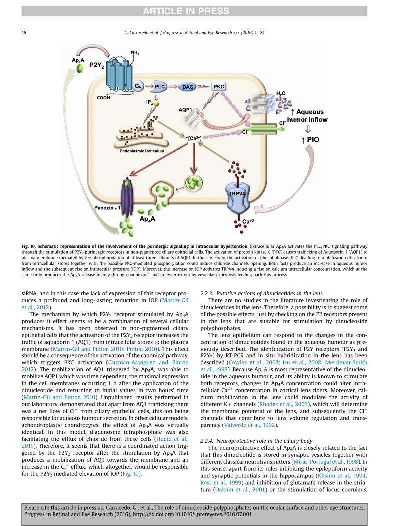
Fig. 10. Schematic representation of the involvement of the purinergic signaling in intraocular hypertension. Extracellular Ap4A activates the PLC/PKC signaling pathwaythrough the stimulation of P2Y2 purinergic receptors in non-pigmented ciliary epithelial cells. The activation of protein kinase C (PKC) causes trafficking of Aquoporin 1 (AQP1) toplasma membrane mediated by the phosphorylation of at least three subunits of AQP1. In the same way, the activation of phospholipase (PLC) leading to mobilization of calciumfrom intracellular stores together with the possible PKC-mediated phosphorylation could induce chloride channels opening. Both facts produce an increase in aqueous humorinflow and the subsequent rise on intraocular pressure (IOP). Moreover, the increase on IOP activates TRPV4 inducing a rise on calcium intracellular concentration, which at thesame time produces the Ap4A release mainly through pannexin 1 and in lesser extent by vesicular exocytosis feeding back this process.
G. Carracedo et al. / Progress in Retinal and Eye Research xxx (2016) 1e2416
siRNA, and in this case the lack of expression of this receptor pro-duces a profound and long-lasting reduction in IOP (Martin-Gilet al., 2012).
The mechanism by which P2Y2 receptor stimulated by Ap4Aproduces it effect seems to be a combination of several cellularmechanisms. It has been observed in non-pigmented ciliaryepithelial cells that the activation of the P2Y2 receptor increases thetraffic of aquaporin 1 (AQ1) from intracellular stores to the plasmamembrane (Martin-Gil and Pintor, 2010; Pintor, 2010). This effectshould be a consequence of the activation of the canonical pathway,which triggers PKC activation (Guzman-Aranguez and Pintor,2012). The mobilization of AQ1 triggered by Ap4A, was able tomobilize AQP1 which was time dependent, themaximal expressionin the cell membranes occurring 1 h after the application of thedinucleotide and returning to initial values in two hours' time(Martin-Gil and Pintor, 2010). Unpublished results performed inour laboratory, demonstrated that apart from AQ1 trafficking therewas a net flow of Cl� from ciliary epithelial cells, this ion beingresponsible for aqueous humour secretion. In other cellular models,achondroplastic chondrocytes, the effect of Ap4A was virtuallyidentical. In this model, diadenosine tetraphosphate was alsofacilitating the efflux of chloride from these cells (Huete et al.,2011). Therefore, it seems that there is a coordinated action trig-gered by the P2Y2 receptor after the stimulation by Ap4A thatproduces a mobilization of AQ1 towards the membrane and anincrease in the Cl� efflux, which altogether, would be responsiblefor the P2Y2 mediated elevation of IOP (Fig. 10).
Please cite this article in press as: Carracedo, G., et al., The role of dinucleProgress in Retinal and Eye Research (2016), http://dx.doi.org/10.1016/j.p
2.2.3. Putative actions of dinucleotides in the lensThere are no studies in the literature investigating the role of
dinucleotides in the lens. Therefore, a possibility is to suggest someof the possible effects, just by checking on the P2 receptors presentin the lens that are suitable for stimulation by dinucleosidepolyphosphates.
The lens epithelium can respond to the changes in the con-centration of dinucleotides found in the aqueous humour as pre-viously described. The identification of P2Y receptors (P2Y1 andP2Y2) by RT-PCR and in situ hybridization in the lens has beendescribed (Cowlen et al., 2003; Hu et al., 2008; Merriman-Smithet al., 1998). Because Ap4A is most representative of the dinucleo-tide in the aqueous humour, and its ability is known to stimulateboth receptors, changes in Ap4A concentration could alter intra-cellular Ca2þ concentration in cortical lens fibers. Moreover, cal-cium mobilization in the lens could modulate the activity ofdifferent Kþ channels (Rhodes et al., 2003), which will determinethe membrane potential of the lens, and subsequently the Cl�
channels that contribute to lens volume regulation and trans-parency (Valverde et al., 1992).
2.2.4. Neuroprotective role in the ciliary bodyThe neuroprotective effect of Ap4A is closely related to the fact
that this dinucleotide is stored in synaptic vesicles together withdifferent classical neurotransmitters (Miras-Portugal et al., 1998). Inthis sense, apart from its roles inhibiting the epileptiform activityand synaptic potentials in the hippocampus (Klishin et al., 1994;Ross et al., 1999) and inhibition of glutamate release in the stria-tum (Oaknin et al., 2001) or the stimulation of locus coeruleus,
oside polyphosphates on the ocular surface and other eye structures,reteyeres.2016.07.001
G. Carracedo et al. / Progress in Retinal and Eye Research xxx (2016) 1e24 17
producing excitation (Frohlich et al., 1996), this dinucleotide canpresent interesting properties protecting the nervous system fromdamage.
Studies performed at the CNS have demonstrated that Ap4A cangive protection from the neurodegeneration induced by methyl-amphetamine either in In vitro or in In vivo experiments (Harveyet al., 2009). This protective effect also exists when cerebralischemia exists or when sympathetic terminals are chemicallydamaged by means of 6-OH-dopamine (Wang et al., 2003).
Neurodegenerative diseases like Alzheimer's disease or Parkin-son's, can affect, of course the CNS, but they also alter the auto-nomic nervous system causing dysautonomia, this being importantin the eye since it can raise IOP augmenting the prevalence ofglaucoma in patients suffering from these two pathologies whichare significantly higher than in the normal population (Bayer et al.,2002). Many patients with Parkinson's disease have a significantloss of their visual field which is typical of the development ofglaucoma (Yenice et al., 2008).
Since the presence of diadenosine tetraphosphate has beendemonstrated in the aqueous humour both in humans and animalmodels (Castany et al., 2011; Pintor et al., 2003), the possibility ofthis dinucleotide acting in the sameway as in the CNS is a matter ofinterest. In this sense, it has been demonstrated in New Zealandwhite rabbits that the chemical destruction by means of 6-OH-dopamine or reserpine, clearly eliminates sympathetic terminalsaltering the control of IOP among other processes. In this sense,subconjunctival injections of 6-OHDA cause sympatholysis with asharp fall in IOP. Also a clear miosis has been observed as aconsequence of the chemical denervation together with a loss ofocular sympathetic reflexes and a sharp reduction in the labelling ofintraocular sympathetic neurons (Hoyle and Pintor, 2010).
On a preliminary study on the possible effect of diadenosinetetraphosphate protecting the nervous system, the application of100 mMAp4A three days before the treatment with 6-OH dopamineor reserpine and 2 more days after the chemical denervation,preserved the sympathetic terminals present in the ciliary bodyfrom chemical denervation and therefore rabbits IOP did not fall ashappens in the absence of the dinucleotide (Hoyle and Pintor,2010). This effect is quite unusual, since the nucleotide ATP andthe nucleoside adenosine were totally unable to mimic Ap4A pro-tective effect. Although adenosine acts via its own receptors, ATPand Ap4A should use common P2 receptors (Pintor and Miras-Portugal, 1995b). P2Y2 receptors have been described in theciliary body in New Zealand white rabbits (Martin-Gil et al., 2012).Nevertheless, and according to its pharmacology, both ATP andAp4A should activate this receptor the final effect being the same,which is not the case. It could be the case that a selective receptorfor diadenosine polyphosphates and not activated by ATP could bethe one producing this protective effect (Pintor and Miras-Portugal,1995a, 2000). To date this is the only evidence for a selectivedinucleotide receptor in the eye and more research is needed toconfirm its real existence.
The way the dinucleotide Ap4A is protecting the ciliary bodyagainst 6-OH dopamine has not been clarified to date. One possi-bility is that the P2 receptor activated by Ap4A may induce thedown-regulation (internalization) of the monomine transporter.This has been described for other transporters such as the dopa-mine transporter in the CNS (Xie and Miller, 2009). The involve-ment of P2Y receptors inhibiting transporters has already beendescribed and moreover, this process has been shown to occur viaprotein kinase C-dependent mechanism (Sen et al., 1993). Analternative possibility is that Ap4A stimulates a receptor connectedto protection by limiting the negative effect of excessive reactiveoxygen species. It could be the case that Ap4A may have an indirecteffect protecting neurons against free radical damage. Melatonin
Please cite this article in press as: Carracedo, G., et al., The role of dinucleProgress in Retinal and Eye Research (2016), http://dx.doi.org/10.1016/j.p
has been shown to protect against 6-OHDA (Dabbeni-Sala et al.,2001), and it is an intriguing possibility that Ap4A can stimulatethe release of melatonin from intraocular epithelium where it issynthesised (Zmijewski et al., 2009).
2.3. Dinucleoside polyphosphates in the posterior pole
2.3.1. Dinucleotide effect on retinal detachmentOne of the eye parts in which the research is weaker concerning
dinucleoside polyphosphates is the retina. The presence of P2 re-ceptors that might be stimulated by dinucleotides is broad inretinal cells, nevertheless, apart from the dinucleotide effect onretinal detachments, it is only possible to establish speculationsabout the actions of these compounds by means of such receptors.
Some therapeutic actions involve dinucleoside polyphosphatesin relevant pathological states of the retina such as retinaldetachment. Receptors for nucleotides in the retinal pigmentedepithelium were described in the late 90's (Peterson et al., 1997;Sullivan et al., 1997), mostly P2Y2 suitable for dinucleotide activa-tion (Guzman-Aranguez et al., 2011; Guzman-Aranguez and Pintor,2012). This fact made some researchers develop studies on animalmodels which were submitted to retinal detachment. In thesemodels it was possible to demonstrate that some dinucleosidepolyphosphates such as deoxycytidine tetraphospho uridine, alsoknown as INS37217, or denufosol, was able to re-absorb the blebbetween RPE and the neural retina in a very efficient way (Yerxaet al., 2002b). This dinucleotide (INS37217), which is a P2Y2
agonist, significantly enhanced subretinal fluid reabsorption whencompared with vehicle controls (Maminishkis et al., 2002; Meyeret al., 2002; Nour et al., 2003). One positive aspect is thatINS37217 had no adverse effects on electroretinograms. In thissense, both scotopic and photopic ERG amplitudes and latencyparameters remained stable when INS37217 was applied, this beinga beneficial aspect to take into account (Meyer et al., 2002). Otherauthors, on the contrary, indicate that subretinal injections of salinecontaining INS37217 significantly increased rod and cone ERG ofnormal and rds (±) mice at 1 and 10 days post-injections, whencompared with injection of saline alone (Nour et al., 2003). Theseexperiments indicate that the dinucleotide INS37217 enhances ERGrecovery probably by reducing retinal folding permitting re-attachment.
It is important to note that the experiments performed with thisdinucleotide demonstrated an increase in fluid absorption not onlyin animal models but also in freshly isolated bovine and fetal humanRPE monolayers (Maminishkis et al., 2002), and therefore the mol-ecules would be a good candidate for the treatment of retinal de-tachments. Nevertheless, it is not all good news. There are two mainhandicaps in these studies; first, denufosol in some cases, to developits action, needs to be used at high concentrations (around 1 mM)(Meyer et al., 2002), which may produce the re-absorption processbut presumably also the activation ofmany other P2 receptors due tothe high concentrations of the dinucleotide. Second, in order to act itis necessary to be applied by means of intravitreal injections(Maminishkis et al., 2002; Meyer et al., 2002; Nour et al., 2003).
2.3.2. Presence in other posterior structuresAs discussed above, diadenosine polyphosphates and other
nucleotides could activate metabotropic and ionotropic receptorsdescribed in mammalian retina (Cowlen et al., 2003; Franke et al.,2005; Fries et al., 2004; Greenwood et al., 1997; Kaneda et al.,2008; Notomi et al., 2011; Peterson et al., 1997; Pintor et al.,2004b; Puthussery and Fletcher, 2004, 2007; Shigematsu et al.,2007; Ward et al., 2008; Wheeler-Schilling et al., 2000, 2001).Numerous studies indicate a wide distribution of purinergic re-ceptors throughout the different layers of retina. It can be suggested
oside polyphosphates on the ocular surface and other eye structures,reteyeres.2016.07.001
G. Carracedo et al. / Progress in Retinal and Eye Research xxx (2016) 1e2418
the possible effects of dinucleotides on P2 receptors and their po-tential physiological roles.
The activation of purinergic receptors through nucleotides anddinucleotides, such as diadenosine polyphosphates, may be impli-cated in embryo development and retinal precursor cell prolifera-tion due to the implication of P2 receptors described in early neuralretina (Sugioka et al., 1999). Transcriptional regulation changes ofthese receptors during the development clearly indicate the rele-vant endogenous nucleotides participation.
Although both P2X and P2Y receptors have been observed inseveral neuronal populations including photoreceptors, amacrineand ganglion cells, recent studies suggest the implication of P2Xreceptors in the modulation of visual neurotransmission activity(Kaneda et al., 2008; Puthussery and Fletcher, 2004, 2007;Puthussery et al., 2006; Vessey and Fletcher, 2012) and the pre-sumable involvement of diadenosine polyphosphates. Moreover, itcould be contributing to the shedding of photoreceptor outer seg-ments via P2Y receptors.
Furthermore, nucleotides serve as vasoactive signals in thepericyte-containing microvasculature of the retina. Differentstudies suggest nucleotides-induced increase in calcium levels andcellular contraction (Kawamura et al., 2003; Sugiyama et al., 2006).In this sense, dinucleotides polyphosphates may contribute toregulate capillary function in retina. Moreover, P2X7 receptoractivation triggered pericyte apoptosis that may be improved byP2Y4 inhibition (Sugiyama et al., 2006). For that action, ATP, dia-denosine polyphosphates and other nucleotides balance haveparticular relevance in the environmental extracellular modulationand the concomitant activation of P2 receptors. Apart from peri-cytes it is also important to take into consideration the contributionof choroid in the retinal circulation. In this sense, it is important toconsider the role of nucleotides and dinucleotides due to theirrelevant participation in vasoactive signals in the retinal vascula-ture including choriocapillaris and choroidal vasculature. Severalauthors have observed ATP action modifying ionic currents leadingcontractions of pericytes as previously commented (Kawamuraet al., 2003) and apoptosis processes via P2X7 receptor activation.Moreover the dinucleotide concentration in blood should alsomodify endothelial cells by means of P2Y2 receptors (Flores et al.,1999) facilitating vasodilation and therefore changing the retinalphysiology.
Some of these functional implicationsmay possibly bemediatedby dinucleoside polyphosphates co-stored with ATP and classicalneurotransmitters. In fact, the presence of ATP and Ap4A levelsobserved in stimulated retinas indicate the importance of thesemolecules in neurotransmission process (Dr. Perez de Lara, per-sonal communication).
Finally, the presence of nucleotides in retinal glial cells could beresponsible for the maintenance of homeostasis regulation forsuitable functioning of the neural retina, among others. The mo-lecular cloning and functional characterization of different familiesof ectoenzymes capable of hydrolyzing ATP, diadenosine poly-phosphates and other nucleotides point to the relevance of thiscomplex balance maintenance.
Extracellular concentration of dinucleotides or their metabolitesat retinal level are regulated by ecto-nucleotidases, nucleosidetransporters and enzymes of adenosine metabolism. Neural retina,but also retinal pigment epithelium, expresses ecto-enzymesincluding NPP1 (described in Müller cells, the major glial compo-nent of the retina), NTPDase 2 (also expressed in Müller cells) andeN/5NTE (located on photoreceptors, Müller cells and retinalpigment epithelium) (Iandiev et al., 2007; Reigada et al., 2006;Takizawa, 1998; Wurm et al., 2010).
Again, retinal structures possess several purinergic receptorsand thus, the response of their cells depends on dinucleotides and
Please cite this article in press as: Carracedo, G., et al., The role of dinucleProgress in Retinal and Eye Research (2016), http://dx.doi.org/10.1016/j.p
also on their metabolites, mainly ATP and adenosine (Crooke et al.,2008). Furthermore, adenosine effects in retina may be regulatedby equilibrative (ENT1 and ENT2) and concentrative nucleosidetransporters (CNT2 and CNT3) as well as adenosine deaminase 2(ADA2) (Akanuma et al., 2013; dos Santos-Rodrigues et al., 2011;Elsherbiny et al., 2013; Nagase et al., 2006). In fact, patients withdiabetes express a high level of ADA2, which impaired the anti-inflammatory activity of adenosine at retina level (Elsherbinyet al., 2013).
Other relevant aspect that needs to be taken into considerationis the possible role of dinucleoside polyphosphates amelioratingretinal neuropathy. On the one hand, it has been described that theactivation of some P2 receptors such as P2Y2 (Nour et al., 2003;Maminishkis et al., 2002) and P2Y13 (Perez-Sen et al., 2015) maybe involved in neuroprotection processes mediated by MAPKpathway in the retina. Moreover, the exacerbated activity of ecto-enzymes (NPPs) in pathological conditions may also contribute tothis process, but more work is necessary to confirm the implicationof ectonucleotidases on retinal impairment.
3. Conclusions and future directions
The participation of dinucleoside polyphosphates in ocularphysiology opens the possibility of using these compounds for thetreatment of relevant eye pathologies. Beginning with the ocularsurface to the retina, the application of these dinucleotides mayhelp to improve several processes, which may be altered in thepathology.
Dry eye is treated in some countries such as Japan and SouthKorea by means of the dinucleotides Up4U. This nucleotide canclearly increase tear secretion, both the aqueous and the mucincomponents. This is the only dinucleoside polyphosphate that iscommercially available as a pharmaceutical compound. None of theothers have moved to a clinical study as a previous step tocommercialization. Nonetheless, many of the actions, mostly dia-denosine polyphosphates do present very promising aspects from atherapeutic point of view as has been commented.
The advance in research needs some changes in order to bemore successful in the search for dinucleotides as therapeuticagents. On the one hand, most of the compounds tested in theirability to counteract some of the pathological situations alreadydescribed, have the characteristics of being fully degradable byectonucleotidases (NPPs) therefore limiting their real effects(Asensio et al., 2007). In this sense it would be of interest to developsome compounds resistant to the hydrolysis. The best way to pro-tect these compounds from cleavage consists of substituting theester bonds between the phosphates by methylene or aminogroups. This has been done by some researchers (Biriukov et al.,1987) although it has not been tested if they improve the efficacyof the dinucleotides already tested in the eye (Chan et al., 1997).This is an area of interest that deserves work because it may indi-cate that lower concentrations can produce the same or even bettereffects. On the other hand, it is very probable that by introducingsuch changes it may be possible to synthesize dinucleoside poly-phosphates to becomemore selective to some receptors. Selectivitywould permit us to reach specific targets without affecting otherprocesses. It is important to be aware that, for example, a dinu-cleotide such as Ap4A can act on several P2 receptors (Pintor et al.,1996; Pintor and Miras-Portugal, 1995b, 2000), and therefore itwould be able to stimulate all of them while it could be the casethat only a particular subtype is needed to be activated. In thissense, hydrolysis resistant and selective dinucleotides are neces-sary to produce better, more selective and long-term effects.
Another important aspect that deserves attention is the deliveryof the dinucleotides to internal parts of the eye. It is clear that
oside polyphosphates on the ocular surface and other eye structures,reteyeres.2016.07.001

G. Carracedo et al. / Progress in Retinal and Eye Research xxx (2016) 1e24 19
actions on the ocular surface are obtained by topical applications ofthe given dinucleotides. Nonetheless, it is important to note thatintraocular actions implies the delivery through the cornea, andthis may reduce the availability of the dinucleotides in the aqueoushumour and in the inner structures. The use of carriers such as li-posomes is a possibility, although to our knowledge, no one hastested dinucleotides with vehicles like those. Interestingly, it hasbeen indicated that some dinucleotides such as Ap4A, when appliedon the ocular surface can temporarily open corneal epithelial tight-junctions (Loma et al., 2015b) facilitating the entry of compoundswithin the eye (Loma et al., 2015a). This means that those actionscarried out by dinucleoside polyphosphates in the inner eye may beenhanced if the application of the dinucleotide, Ap4A, is appliedtwo hours previously. It is necessary to investigate this since, if theentry of the dinucleotide is improved it is possible that it may reachvery deep areas of the eye.
In conclusion, dinucleoside polyphosphates are involved in amultitude of physiological processes which are relevant for thecorrect functioning of the eye. There are many promising per-spectives using some of these compounds from a therapeutic pointof view, however it is clear that new synthetic analogues of thesedinucleotides are necessary to fully develop a battery of pharma-ceutical compounds suitable for ocular therapy.
Disclosure
All authors have no financial interest in the subject matter ormaterials discussed in this manuscript. The authors alone areresponsible for the content and writing of the paper.
Acknowledgments
This work was supported by grants fromMinisterio de Ciencia eInnovaci�on (SAF2010-16024 and SAF2013-44416-R) and RETICS(RD12/0034/0003).
References
Abbracchio, M.P., Ceruti, S., 2006. Roles of P2 receptors in glial cells: focus on as-trocytes. Purinergic Signal 2, 595e604.
Akanuma, S., Soutome, T., Hisada, E., Tachikawa, M., Kubo, Y., Hosoya, K., 2013. Naþ-independent nucleoside transporters regulate adenosine and hypoxanthinelevels in Muller cells and the inner blood-retinal barrier. Invest.Investig. Oph-thalmol. Vis. Sci. 54, 1469e1477.
Alfonso, J.F., Lisa, C., Fernandez-Vega Cueto, L., Belda-Salmeron, L., Madrid-Costa, D.,Montes-Mico, R., 2013. Clinical outcomes after implantation of a posteriorchamber collagen copolymer phakic intraocular lens with a central hole formyopic correction. J. Cataract. Refract Surg. 39, 915e921.
Ali, M., Kamiya, K., Shimizu, K., Igarashi, A., Ishii, R., 2014. Clinical evaluation ofcorneal biomechanical parameters after posterior chamber phakic intraocularlens implantation. Cornea 33, 470e474.
Alkozi, H.A., Pintor, J., 2015. TRPV4 activation triggers the release of melatonin fromhuman non-pigmented ciliary epithelial cells. Exp. Eye Res. 136, 34e37.
Andersson, M., 1989. Diadenosine tetraphosphate (Ap4A): its presence and func-tions in biological systems. Int. J. Biochem. 21, 707e714.
Arita, R., Suehiro, J., Haraguchi, T., Maeda, S., Maeda, K., Tokoro, H., Amano, S., 2013.Topical diquafosol for patients with obstructive meibomian gland dysfunction.Br. J. Ophthalmol. 97, 725e729.
Arnold, R.R., Cole, M.F., McGhee, J.R., 1977. A bactericidal effect for human lacto-ferrin. Science 197, 263e265.
Asensio, A.C., Rodriguez-Ferrer, C.R., Castaneyra-Perdomo, A., Oaknin, S., Rotllan, P.,2007. Biochemical analysis of ecto-nucleotide pyrophosphatase phosphodies-terase activity in brain membranes indicates involvement of NPP1 isoenzyme inextracellular hydrolysis of diadenosine polyphosphates in central nervoussystem. Neurochem. Int. 50, 581e590.
Baker, O.J., Camden, J.M., Rome, D.E., Seye, C.I., Weisman, G.A., 2008. P2Y2 nucle-otide receptor activation up-regulates vascular cell adhesion molecule-1 [cor-rected] expression and enhances lymphocyte adherence to a humansubmandibular gland cell line. Mol. Immunol. 45, 65e75.
Bambara, R.A., Crute, J.J., Wahl, A.F., 1985. Is Ap4A an activator of eukaryotic DNAreplication? Cancer Invest.Investig. 3, 473e479.
Ban, Y., Cooper, L.J., Fullwood, N.J., Nakamura, T., Tsuzuki, M., Koizumi, N., Dota, A.,Mochida, C., Kinoshita, S., 2003. Comparison of ultrastructure, tight junction-
Please cite this article in press as: Carracedo, G., et al., The role of dinucleProgress in Retinal and Eye Research (2016), http://dx.doi.org/10.1016/j.p
related protein expression and barrier function of human corneal epithelialcells cultivated on amniotic membrane with and without air-lifting. Exp. EyeRes. 76, 735e743.
Bayer, A.U., Keller, O.N., Ferrari, F., Maag, K.P., 2002. Association of glaucoma withneurodegenerative diseases with apoptotic cell death: Alzheimer's disease andParkinson's disease. Am. J. Ophthalmol. 133, 135e137.
Biriukov, A.P., Zakharova, O.D., Lavrik, O.I., 1987. [Effect of diadenosine oligophos-phates (Ap4A and Ap3A) and their phosphonate analogs on catalytic propertiesof phenylalanyl-tRNA synthetase from E. coli]. Bioorg Khim 13, 1164e1169.
Bonan, C.D., 2012. Ectonucleotidases and nucleotide/nucleoside transporters aspharmacological targets for neurological disorders. CNS Neurol. Disord. DrugTargets 11, 739e750.
Boucher, I., Rich, C., Lee, A., Marcincin, M., Trinkaus-Randall, V., 2010. The P2Y2receptor mediates the epithelial injury response and cell migration. Am. J.Physiol. Cell Physiol. 299, C411eC421.
Bowman, K.A., Green, K., 1981. Corneal epithelial healing rates after topical nucle-otides. Curr. Eye Res. 1, 619e622.
Braun, J.S., 2010. Ecto-50-nucleotidase-positive cells in the choroid and ciliary bodyof the rat eye. Anat. Rec. Hob. 293, 379e382.
Bremond-Gignac, D., Gicquel, J.J., Chiambaretta, F., 2014. Pharmacokinetic evalua-tion of diquafosol tetrasodium for the treatment of Sjogren's syndrome. ExpertOpin. Drug Metab. Toxicol. 10, 905e913.
Brennan, N.A., Efron, N., Holden, B.A., Fatt, I., 1987. A review of the theoreticalconcepts, measurement systems and application of contact lens oxygenpermeability. Ophthalmic & Physiol. Opt. J. Br. Coll. Ophthalmic Opt. 7,485e490.
Burman, L.G., Lundblad, G., Camner, P., Fange, R., Lundborg, M., Soder, P., 1991.Lysozymeean enzyme of both historical and current interest as a therapeuticalagent. Lakartidningen 88, 3665e3668.
Bush, C.A., Tinoco Jr., I., 1967. Calculation of the optical rotatory dispersion ofdinucleoside phosphates. J. Mol. Biol. 23, 601e604.
Byun, Y.S., Yoo, Y.S., Kwon, J.Y., Joo, J.S., Lim, S.A., Whang, W.J., Mok, J.W., Choi, J.S.,Joo, C.K., 2016. Diquafosol promotes corneal epithelial healing via intracellularcalcium-mediated ERK activation. Exp. Eye Res. 143, 89e97.
Cabrera, M.J. Velasco, Sanchez, J. Garcia, Rodriguez, F.J. Bermudez, 1997. Lactoferrinin tears in contact lens wearers. CLAO J. Off. Publ. Contact Lens Assoc. Oph-thalmol. Inc 23, 127e129.
Caffery, B., Joyce, E., Boone, A., Slomovic, A., Simpson, T., Jones, L., Senchyna, M.,2008. Tear lipocalin and lysozyme in Sjogren and non-Sjogren dry eye. Optom.Vis. Sci. 85, 661e667.
Carracedo, G., Peral, A., Pintor, J., 2010. Diadenosine polyphosphates in tears ofSjogren syndrome patients. Invest.Investig. Ophthalmol. Vis. Sci. 51,5452e5459.
Carracedo, G., Gonzalez-Meijome, J.M., Pintor, J., 2012. Changes in diadenosinepolyphosphates during alignment-fit and orthokeratology rigid gas permeablelens wear. Invest.Investig. Ophthalmol. Vis. Sci. 53, 4426e4432.
Carracedo, G., Guzman-Aranguez, A., Loma, P., Pintor, J., 2013. Diadenosine poly-phosphates release by human corneal epithelium. Exp. Eye Res. 113, 156e161.
Carracedo, G., Cacho, I., Sanchez-Naves, J., Pintor, J., 2014. Diadenosine poly-phosphates after laser in situ keratomileusis and photorefractive keratectomyrefractive techniques. Acta Ophthalmol. 92, e5ee11.
Carracedo, G., Martin-Gil, A., Peixoto-de-Matos, S.C., Abejon-Gil, P., Macedo-de-Araujo, R., Gonzalez-Meijome, J.M., 2015a. Symptoms and signs in rigid gaspermeable lens wearers during adaptation period. Eye Contact Lens 42 (2),108e114.
Carracedo, G., Recchioni, A., Alejandre-Alba, N., Martin-Gil, A., Crooke, A.,Morote, I.J., Pintor, J., 2015b. Signs and symptoms of dry eye in Keratoconuspatients: a pilot study. Curr. Eye Res. 40, 1088e1094.
Carracedo, G., Blanco, M.S., Martin-Gil, A., Zicheng, W., Alvarez, J.C., Pintor, J., 2016.Short-term effect of Scleral lens on the dry eye biomarkers in Keratoconus.Optom. Vis. Sci. 93, 150e157.
Castany, M., Jordi, I., Catala, J., Gual, A., Morales, M., Gasull, X., Pintor, J., 2011.Glaucoma patients present increased levels of diadenosine tetraphosphate,Ap(4)A, in the aqueous humour. Exp. Eye Res. 92, 221e226.
Chan, S.I., Nelson, J.H., 1969. Proton magnetic resonance studies of ribose dinu-cleoside monophoshates in aqueous solution. I. The nature of the base-stackinginteraction in adenylyl 30e50)adenosine. J. Am. Chem. Soc. 91, 168e183.
Chan, S.W., Gallo, S.J., Kim, B.K., Guo, M.J., Blackburn, G.M., Zamecnik, P.C., 1997.P1,P4-dithio-P2,P3-monochloromethylene diadenosine 50 ,500-P1,P4-tetraphosphate: a novel antiplatelet agent. Proc. Natl. Acad. Sci. U. S. A. 94,4034e4039.
Cheng, P.Y., 1968. Characteristic optical rotatory dispersion spectra of thirteendinucleoside phosphates with mercurials. Biochem. Biophys. Res. Commun. 33,746e751.
Chia, E.M., Mitchell, P., Rochtchina, E., Lee, A.J., Maroun, R., Wang, J.J., 2003. Prev-alence and associations of dry eye syndrome in an older population: the BlueMountains eye study. Clin. Exp. Ophthalmol. 31, 229e232.
Cockayne, D.A., Hamilton, S.G., Zhu, Q.M., Dunn, P.M., Zhong, Y., Novakovic, S.,Malmberg, A.B., Cain, G., Berson, A., Kassotakis, L., Hedley, L., Lachnit, W.G.,Burnstock, G., McMahon, S.B., Ford, A.P., 2000. Urinary bladder hyporeflexia andreduced pain-related behaviour in P2X3-deficient mice. Nature 407, 1011e1015.
Coddou, C., Yan, Z., Obsil, T., Huidobro-Toro, J.P., Stojilkovic, S.S., 2011. Activation andregulation of purinergic P2X receptor channels. Pharmacol. Rev. 63, 641e683.
Conneely, O.M., 2001. Antiinflammatory activities of lactoferrin. J. Am. Coll. Nutr. 20,389Se395S discussion 396S-397S.
oside polyphosphates on the ocular surface and other eye structures,reteyeres.2016.07.001
G. Carracedo et al. / Progress in Retinal and Eye Research xxx (2016) 1e2420
Cornish, R., Sulaiman, S., 1996. Do thinner rigid gas permeable contact lenses pro-vide superior initial comfort? optometry and vision science. Off. Publ. Am. Acad.Optom. 73, 139e143.
Corrales, R.M., Narayanan, S., Fernandez, I., Mayo, A., Galarreta, D.J., Fuentes-Paez, G., Chaves, F.J., Herreras, J.M., Calonge, M., 2011. Ocular mucin geneexpression levels as biomarkers for the diagnosis of dry eye syndrome. Inves-t.Investig. Ophthalmol. Vis. Sci. 52, 8363e8369.
Cowlen, M.S., Zhang, V.Z., Warnock, L., Moyer, C.F., Peterson, W.M., Yerxa, B.R., 2003.Localization of ocular P2Y2 receptor gene expression by in situ hybridization.Exp. Eye Res. 77, 77e84.
Crooke, A., Guzman-Aranguez, A., Peral, A., Abdurrahman, M.K., Pintor, J., 2008.Nucleotides in ocular secretions: their role in ocular physiology. Pharmacol.Ther. 119, 55e73.
Crooke, A., Mediero, A., Guzman-Aranguez, A., Pintor, J., 2009. Silencing of P2Y2receptor delays Ap4A-corneal re-epithelialization process. Mol. Vis. 15,1169e1178.
Crosson, C.E., Yates, P.W., Bhat, A.N., Mukhin, Y.V., Husain, S., 2004. Evidence formultiple P2Y receptors in trabecular meshwork cells. J. Pharmacol. Exp. Ther.309, 484e489.
Dabbeni-Sala, F., Di Santo, S., Franceschini, D., Skaper, S.D., Giusti, P., 2001. Mela-tonin protects against 6-OHDA-induced neurotoxicity in rats: a role for mito-chondrial complex I activity. FASEB J. 15, 164e170.
Davidson, A.E., Hayes, S., Hardcastle, A.J., Tuft, S.J., 2014. The pathogenesis of ker-atoconus. Eye (Lond.) 28, 189e195.
Davis, R.C., Tinoco Jr., I., 1968. Temperature-dependent properties of dinucleosidephosphates. Biopolymers 6, 223e242.
Dominguez-Godinez, C.O., Martin-Gil, A., Carracedo, G., Guzman-Aranguez, A.,Gonzalez-Meijome, J., Pintor, J., 2013. In Vitro and in Vivo delivery of thesecretagogue diadenosine tetraphosphate from conventional and siliconehydrogel soft contact lenses. J Optom. 06, 205e211.
dos Santos-Rodrigues, A., Ferreira, J.M., Paes-de-Carvalho, R., 2011. Differentialadenosine uptake in mixed neuronal/glial or purified glial cultures of avianretinal cells: modulation by adenosine metabolism and the ERK cascade. Bio-chem. Biophys. Res. Commun. 414, 175e180.
Dutot, M., Liang, H., Pauloin, T., Brignole-Baudouin, F., Baudouin, C., Warnet, J.M.,Rat, P., 2008. Effects of toxic cellular stresses and divalent cations on the humanP2X7 cell death receptor. Mol. Vis. 14, 889e897.
Elbendary, A.M., Abou Samra, W., 2013. Evaluation of rigid gas permeable lensfitting in keratoconic patients with optical coherence tomography. GraefesArch. Clin. Exp. Ophthalmol. 251, 1565e1570.
Elsherbiny, N.M., Naime, M., Ahmad, S., Elsherbini, A.M., Mohammad, S., Fulzele, S.,El-Remessy, A.B., Al-Gayyar, M.M., Eissa, L.A., El-Shishtawy, M.M., Han, G.,White, R., Haroldo, T.F., Liou, G.I., 2013. Potential roles of adenosine deaminase-2 in diabetic retinopathy. Biochem. Biophys. Res. Commun. 436, 355e361.
Farahbakhsh, N.A., 2003. Ectonucleotidases of the rabbit ciliary body nonpigmentedepithelium. Invest.Investig. Ophthalmol. Vis. Sci. 44, 3952e3960.
Farris, R.L., 1986. Tear analysis in contact lens wearers. CLAO J. 12, 106e111.Ferguson, D.R., Kennedy, I., Burton, T.J., 1997. ATP is released from rabbit urinary
bladder epithelial cells by hydrostatic pressure changesea possible sensorymechanism? J. Physiol. 505 (Pt 2), 503e511.
Finamore, F.J., Warner, A.H., 1963. The occurrence of P1, P4-diguanosine 50-tetra-phosphate in brine shrimp eggs. J. Biol. Chem. 238, 344e348.
Fink, B.A., Hill, R.M., Carney, L.G., 1992. Influence of rigid contact lens base curveradius on tear pump efficiency. Optom. Vis. Sci. Off. Publ. Am. Acad. Optom. 69,60e65.
Flanagan, J.L., Willcox, M.D., 2009. Role of lactoferrin in the tear film. Biochimie 91,35e43.
Flodgaard, H., Klenow, H., 1982. Abundant amounts of diadenosine 50,5000-P1,P4-tetraphosphate are present and releasable, but metabolically inactive, in hu-man platelets. Biochem. J. 208, 737e742.
Flores, N.A., Stavrou, B.M., Sheridan, D.J., 1999. The effects of diadenosine poly-phosphates on the cardiovascular system. Cardiovasc Res. 42, 15e26.
Fonseca, B., Martinez-Aguila, A., Diaz-Hernandez, M., Pintor, J., 2015. Diadenosinetetraphosphate contributes to carbachol-induced tear secretion. PurinergicSignal 11, 87e93.
Franco, R., Casado, V., Ciruela, F., Saura, C., Mallol, J., Canela, E.I., Lluis, C., 1997. Cellsurface adenosine deaminase: much more than an ectoenzyme. Prog. Neuro-biol. 52, 283e294.
Franke, H., Klimke, K., Brinckmann, U., Grosche, J., Francke, M., Sperlagh, B.,Reichenbach, A., Liebert, U.G., Illes, P., 2005. P2X(7) receptor-mRNA and -proteinin the mouse retina; changes during retinal degeneration in BALBCrds mice.Neurochem. Int. 47, 235e242.
Fries, J.E., Wheeler-Schilling, T.H., Guenther, E., Kohler, K., 2004. Expression of P2Y1,P2Y2, P2Y4, and P2Y6 receptor subtypes in the rat retina. Invest.Investig.Ophthalmol. Vis. Sci. 45, 3410e3417.
Frohlich, R., Boehm, S., Illes, P., 1996. Pharmacological characterization of P2 puri-noceptor types in rat locus coeruleus neurons. Eur. J. Pharmacol. 315, 255e261.
Fujihara, T., Murakami, T., Fujita, H., Nakamura, M., Nakata, K., 2001. Improvementof corneal barrier function by the P2Y(2) agonist INS365 in a rat dry eye model.Invest.Investig. Ophthalmol. Vis. Sci. 42, 96e100.
Fujihara, T., Murakami, T., Nagano, T., Nakamura, M., Nakata, K., 2002. INS365suppresses loss of corneal epithelial integrity by secretion of mucin-likeglycoprotein in a rabbit short-term dry eye model. J. Ocul. Pharmacol. Ther.18, 363e370.
Galvis, V., Tello, A., Barrera, R., Nino, C.A., 2015. Inflammation in Keratoconus.
Please cite this article in press as: Carracedo, G., et al., The role of dinucleProgress in Retinal and Eye Research (2016), http://dx.doi.org/10.1016/j.p
Cornea 34, e22e23.Gendaszewska-Darmach, E., Kucharska, M., 2011. Nucleotide receptors as targets in
the pharmacological enhancement of dermal wound healing. Purinergic Signal7, 193e206.
Gipson, I.K., 2007. The ocular surface: the challenge to enable and protect vision:the Friedenwald lecture. Invest.Investig. Ophthalmol. Vis. Sci. 48 (4390),4391e4398.
Giraldez, L., Diaz-Hernandez, M., Gomez-Villafuertes, R., Pintor, J., Castro, E., Miras-Portugal, M.T., 2001. Adenosine triphosphate and diadenosine pentaphosphateinduce [Ca(2þ)](i) increase in rat basal ganglia aminergic terminals. J. Neurosci.Res. 64, 174e182.
Glaser, T., Walton, D.S., Maas, R.L., 1992. Genomic structure, evolutionary conser-vation and aniridia mutations in the human PAX6 gene. Nat. Genet. 2, 232e239.
Glaser, T., Ton, C.C., Mueller, R., Petzl-Erler, M.L., Oliver, C., Nevin, N.C.,Housman, D.E., Maas, R.L., 1994. Absence of PAX6 gene mutations in Gillespiesyndrome (partial aniridia, cerebellar ataxia, and mental retardation). Geno-mics 19, 145e148.
Glaubiger, D., Lloyd, D.A., Tinoco Jr., I., 1968. Temperature-dependent opticalproperties of a torsional oscillator model for dinucleoside phosphates. Bio-polymers 6, 409e414.
Gobbels, M., Breitbach, R., Spitznas, M., 1990. Barrier function of the cornealepithelium of contact lens patients. Fortschr. Ophthalmol. Z. Dtsch. Ophthalmol.Ges. 87, 646e648.
Goding, J.W., Terkeltaub, R., Maurice, M., Deterre, P., Sali, A., Belli, S.I., 1998. Ecto-phosphodiesterase/pyrophosphatase of lymphocytes and non-lymphoid cells:structure and function of the PC-1 family. Immunol. Rev. 161, 11e26.
Goel, M., Picciani, R.G., Lee, R.K., Bhattacharya, S.K., 2010. Aqueous humor dynamics:a review. Open Ophthalmol. J. 4, 52e59.
Gomes, P., Srinivas, S.P., Vereecke, J., Himpens, B., 2005. ATP-dependent paracrineintercellular communication in cultured bovine corneal endothelial cells.Invest.Investig. Ophthalmol. Vis. Sci. 46, 104e113.
Greenwood, D., Yao, W.P., Housley, G.D., 1997. Expression of the P2X2 receptorsubunit of the ATP-gated ion channel in the retina. Neuroreport 8, 1083e1088.
Groschel-Stewart, U., Bardini, M., Robson, T., Burnstock, G., 1999. Localisation ofP2X5 and P2X7 receptors by immunohistochemistry in rat stratified squamousepithelia. Cell Tissue Res. 296, 599e605.
Grummt, F., 1978. Diadenosine 50 ,500-P1,P4-tetraphosphate triggers initiation ofin vitro DNA replication in baby hamster kidney cells. Proc. Natl. Acad. Sci. U. S.A. 75, 371e375.
Grummt, F., 1979. Diadenosine tetraphosphate triggers in vitro DNA replication.Cold Spring Harb. Symp. Quant. Biol. 43 (Pt 1), 649e653.
Grummt, F., 1983. Diadenosine tetraphosphate (Ap4A): a putative chemicalmessenger of cell proliferation control and inducer of DNA replication: reviewpaper. Plant Mol. Biol. 2, 41e44.
Grummt, F., Waltl, G., Jantzen, H.M., Hamprecht, K., Huebscher, U., Kuenzle, C.C.,1979. Diadenosine 50 ,500-P1,P4-tetraphosphate, a ligand of the 57-kilodaltonsubunit of DNA polymerase alpha. Proc. Natl. Acad. Sci. U. S. A. 76, 6081e6085.
Gualix, J., Gomez-Villafuertes, R., Diaz-Hernandez, M., Miras-Portugal, M.T., 2003.Presence of functional ATP and dinucleotide receptors in glutamatergic synapticterminals from rat midbrain. J. Neurochem. 87, 160e171.
Guillon, M., Maissa, C., 2010. Tear film evaporationeeffect of age and gender. Cont.Lens Anterior Eye 33, 171e175.
Guranowski, A., 2000. Specific and nonspecific enzymes involved in the catabolismof mononucleoside and dinucleoside polyphosphates. Pharmacol. Ther. 87,117e139.
Guranowski, A., Starzynska, E., Gunther Sillero, M.A., Sillero, A., 1995. Conversion ofadenosine(50) oligophospho(50) adenosines into inosine(50) oligophospho(50)inosines by non-specific adenylate deaminase from the snail Helix pomatia.Biochim. Biophys. Acta 1243, 78e84.
Guzman-Aranguez, A., Pintor, J., 2012. Focus on molecules: purinergic P2Y(2) re-ceptor. Exp. Eye Res. 105, 83e84.
Guzman-Aranguez, A., Crooke, A., Peral, A., Hoyle, C.H., Pintor, J., 2007. Dinucleosidepolyphosphates in the eye: from physiology to therapeutics. Prog. Retin Eye Res.26, 674e687.
Guzman-Aranguez, A., Loma, P., Pintor, J., 2011. Focus on molecules: diadenosinetetraphosphate. Exp. Eye Res. 92, 96e97.
Guzman-Aranguez, A., Diez, L.M., Martin-Gil, A., Gualix, J., Miras-Portugal, M.T.,Pintor, J., 2012. Effect of diinosine polyphosphates on intraocular pressure innormotensive rabbits. Exp. Eye Res. 101, 49e55.
Guzman-Aranguez, A., Santano, C., Martin-Gil, A., Fonseca, B., Pintor, J., 2013. Nu-cleotides in the eye: focus on functional aspects and therapeutic perspectives.J. Pharmacol. Exp. Ther. 345, 331e341.
Guzman-Aranguez, A., Gasull, X., Diebold, Y., Pintor, J., 2014. Purinergic receptors inocular inflammation. Mediat. Inflamm. 2014, 320906.
Guzman-Aranguez, A., Fonseca, B., Carracedo, G., Martin-Gil, A., Martinez-Aguila, A.,Pintor, J., 2015. Dry eye treatment based on contact lens drug delivery: a review.Eye Contact Lens [Epub ahead of print].
Habermacher, C., Dunning, K., Chataigneau, T., Grutter, T., 2015. Molecular structureand function of P2X receptors. Neuropharmacology 104, 18e30.
Hansen, P.B., Hristovska, A., Wolff, H., Vanhoutte, P., Jensen, B.L., Bie, P., 2010. Uridineadenosine tetraphosphate affects contractility of mouse aorta and decreasesblood pressure in conscious rats and mice. Acta Physiol. (Oxf) 200, 171e179.
Harvey, B.K., Chou, J., Shen, H., Hoffer, B.J., Wang, Y., 2009. Diadenosine tetraphos-phate reduces toxicity caused by high-dose methamphetamine administration.Neurotoxicology 30, 436e444.
oside polyphosphates on the ocular surface and other eye structures,reteyeres.2016.07.001
G. Carracedo et al. / Progress in Retinal and Eye Research xxx (2016) 1e24 21
Herrero-Vanrell, R., Peral, A., 2007. International dry eye workshop (DEWS). Updateof the disease. Arch. Soc. Esp. Oftalmol. 82, 733e734.
Hodges, R.R., Vrouvlianis, J., Shatos, M.A., Dartt, D.A., 2009. Characterization of P2X7purinergic receptors and their function in rat lacrimal gland. Invest.Investig.Ophthalmol. Vis. Sci. 50, 5681e5689.
Hodges, M.D., Kelly, J.G., Bentley, A.J., Fogarty, S., Patel II, Martin, F.L., Fullwood, N.J.,2011. Combining immunolabeling and surface-enhanced Raman spectroscopyon cell membranes. ACS Nano 5, 9535e9541.
Hoyle, C.H., Pintor, J.J., 2010. Diadenosine tetraphosphate protects sympatheticterminals from 6-hydroxydopamine-induced degeneration in the eye. ActaPhysiol. (Oxf) 199, 205e210.
Hoyle, C.H.V., Hilderman, R., Pintor, J.J., Schlüter, H., King, B.F., 2001. Diadenosinepolyphosphates as extracellular signal molecules. Drug Dev. Res. 52 (2001),260e273.
Hoyle, C.H., Peral, A., Pintor, J., 2006. Melatonin potentiates tear secretion inducedby diadenosine tetraphosphate in the rabbit. Eur. J. Pharmacol. 552, 159e161.
Hruska, F.E., Danyluk, S.S., 1968. Conformational changes of the ribose group indinucleoside mono- and diphosphates. Temperature dependence. J. Am. Chem.Soc. 90, 3266e3267.
Hu, R.G., Suzuki-Kerr, H., Webb, K.F., Rhodes, J.D., Collison, D.J., Duncan, G.,Donaldson, P.J., 2008. Molecular and functional mapping of regional differencesin P2Y receptor expression in the rat lens. Exp. Eye Res. 87, 137e146.
Huang, H.S., Schoenwald, R.D., Lach, J.L., 1983. Corneal penetration behavior of beta-blocking agents III: in vitro-in vivo correlations. J. Pharm. Sci. 72, 1279e1281.
Huang, A.J., Tseng, S.C., Kenyon, K.R., 1989. Paracellular permeability of corneal andconjunctival epithelia. Invest.Investig. Ophthalmol. Vis. Sci. 30, 684e689.
Huete, F., Guzman-Aranguez, A., Ortin, J., Hoyle, C.H., Pintor, J., 2011. Effects ofdiadenosine tetraphosphate on FGF9-induced chloride flux changes in achon-droplastic chondrocytes. Purinergic Signal 7, 243e249.
Iandiev, I., Wurm, A., Pannicke, T., Wiedemann, P., Reichenbach, A., Robson, S.C.,Zimmermann, H., Bringmann, A., 2007. Ectonucleotidases in Muller glial cells ofthe rodent retina: involvement in inhibition of osmotic cell swelling. PurinergicSignal 3, 423e433.
Ikehara, M., Uesugi, S., Yasumoto, M., 1970. A highly stacked dinucleoside mono-phosphate derived from adenine 8-cyclonucleosides. J. Am. Chem. Soc. 92,4735e4736.
Jacobson, K.A., Boeynaems, J.M., 2010. P2Y nucleotide receptors: promise of ther-apeutic applications. Drug Discov. Today 15, 570e578.
Jacobson, K.A., Gao, Z.G., 2006. Adenosine receptors as therapeutic targets. Nat. Rev.Drug Discov. 5, 247e264.
Jacobson, K.A., Balasubramanian, R., Deflorian, F., Gao, Z.G., 2012. G protein-coupledadenosine (P1) and P2Y receptors: ligand design and receptor interactions.Purinergic Signal 8, 419e436.
Jacobson, K.A., Paoletta, S., Katritch, V., Wu, B., Gao, Z.G., Zhao, Q., Stevens, R.C.,Kiselev, E., 2015. Nucleotides acting at P2Y receptors: connecting structure andfunction. Mol. Pharmacol. 88, 220e230.
Jia, C., Hegg, C.C., 2012. Neuropeptide Y and extracellular signal-regulated kinasemediate injury-induced neuroregeneration in mouse olfactory epithelium. Mol.Cell Neurosci. 49, 158e170.
Jin-Hua, P., Goding, J.W., Nakamura, H., Sano, K., 1997. Molecular cloning andchromosomal localization of PD-Ibeta (PDNP3), a new member of the humanphosphodiesterase I genes. Genomics 45, 412e415.
Jones, L., Brennan, N.A., Gonzalez-Meijome, J., Lally, J., Maldonado-Codina, C.,Schmidt, T.A., Subbaraman, L., Young, G., Nichols, J.J., members of the,T.I.W.o.C.L.D, 2013. The TFOS international workshop on contact lens discom-fort: report of the contact lens materials, design, and care subcommittee.Invest.Investig. Ophthalmol. Vis. Sci. 54, 37e70.
Jumblatt, J.E., Jumblatt, M.M., 1998. Regulation of ocular mucin secretion by P2Y2nucleotide receptors in rabbit and human conjunctiva. Exp. Eye Res. 67,341e346.
Kamada, Y., Saino, T., Oikawa, M., Kurosaka, D., Satoh, Y., 2012. P2Y purinoceptorsinduce changes in intracellular calcium in acinar cells of rat lacrimal glands.Histochem Cell Biol. 137, 97e106.
Kamiya, K., Shimizu, K., Komatsu, M., 2010. Implantable Collamer Lens implantationand limbal relaxing incisions for the correction of hyperopic astigmatism afterlaser in situ keratomileusis. Cornea 29, 99e101.
Kamiya, K., Nakanishi, M., Ishii, R., Kobashi, H., Igarashi, A., Sato, N., Shimizu, K.,2012. Clinical evaluation of the additive effect of diquafosol tetrasodium onsodium hyaluronate monotherapy in patients with dry eye syndrome: a pro-spective, randomized, multicenter study. Eye (Lond.) 26, 1363e1368.
Kaneda, M., Ishii, T., Hosoya, T., 2008. Pathway-dependent modulation by P2-purinoceptors in the mouse retina. Eur. J. Neurosci. 28, 128e136.
Karcioglu, Z.A., Fleming, J.C., 2008. Long-term retention rates and complications ofsilicone punctal plugs in dry eye. Am. J. Ophthalmol. 145, 586 author reply586e587.
Kawamura, H., Sugiyama, T., Wu, D.M., Kobayashi, M., Yamanishi, S., Katsumura, K.,Puro, D.G., 2003. ATP: a vasoactive signal in the pericyte-containing micro-vasculature of the rat retina. J. Physiol. 551, 787e799.
Klepeis, V.E., Weinger, I., Kaczmarek, E., Trinkaus-Randall, V., 2004. P2Y receptorsplay a critical role in epithelial cell communication and migration. J. Cell Bio-chem. 93, 1115e1133.
Klishin, A., Lozovaya, N., Pintor, J., Miras-Portugal, M.T., Krishtal, O., 1994. Possiblefunctional role of diadenosine polyphosphates: negative feedback for excitationin hippocampus. Neuroscience 58, 235e236.
Knight, G.E., Burnstock, G., 2001. Identification of P1 and P2 purinoceptors in the
Please cite this article in press as: Carracedo, G., et al., The role of dinucleProgress in Retinal and Eye Research (2016), http://dx.doi.org/10.1016/j.p
aorta of the lizard (Agama sp.). Comp. Biochem. Physiol. C Toxicol. Pharmacol.128, 413e423.
Knop, E., Knop, N., 2007. Anatomy and immunology of the ocular surface. Chem.Immunol. Allergy 92, 36e49.
Koh, S., Ikeda, C., Takai, Y., Watanabe, H., Maeda, N., Nishida, K., 2013a. Long-termresults of treatment with diquafosol ophthalmic solution for aqueous-deficientdry eye. Jpn. J. Ophthalmol. 57, 440e446.
Koh, S., Maeda, N., Ikeda, C., Takai, Y., Fujimoto, H., Oie, Y., Soma, T., Tsujikawa, M.,Nishida, K., 2013b. Effect of instillation of eyedrops for dry eye on opticalquality. Invest.Investig. Ophthalmol. Vis. Sci. 54, 4927e4933.
Koh, S., Maeda, N., Ikeda, C., Oie, Y., Soma, T., Tsujikawa, M., Watanabe, H.,Nishida, K., 2014. Effect of diquafosol ophthalmic solution on the optical qualityof the eyes in patients with aqueous-deficient dry eye. Acta Ophthalmol. 92,671e675.
Kojima, T., Higuchi, A., Goto, E., Matsumoto, Y., Dogru, M., Tsubota, K., 2008.Autologous serum eye drops for the treatment of dry eye diseases. Cornea 27(Suppl. 1), S25eS30.
Kojima, T., Dogru, M., Ibrahim, O.M., Nagata, T., Higa, K., Shimizu, T., Shirasawa, T.,Satake, Y., Shimazaki, S., Shimazaki, J., Tsubota, K., 2014. The effects of 3%diquafosol sodium application on the tear functions and ocular surface of theCu,Zn-superoxide dismutase-1 (Sod1)-knockout mice. Mol. Vis. 20, 929e938.
Kompella, U.B., Kadam, R.S., Lee, V.H., 2010. Recent advances in ophthalmic drugdelivery. Ther. Deliv. 1, 435e456.
Kuizenga, A., van Haeringen, N.J., Meijer, F., Kijlstra, A., 1996. Analysis of human tearfluid components, inhibiting protein adhesion to plastic surfaces. Exp. Eye Res.63, 319e328.
Kurpakus, M.A., Daneshvar, C., Davenport, J., Kim, A., 1999. Human corneal epithelialcell adhesion to laminins. Curr. Eye Res. 19, 106e114.
Langbein, L., Grund, C., Kuhn, C., Praetzel, S., Kartenbeck, J., Brandner, J.M., Moll, I.,Franke, W.W., 2002. Tight junctions and compositionally related junctionalstructures in mammalian stratified epithelia and cell cultures derived there-from. Eur. J. Cell Biol. 81, 419e435.
Lazarowski, E.R., Watt, W.C., Stutts, M.J., Boucher, R.C., Harden, T.K., 1995. Phar-macological selectivity of the cloned human P2U-purinoceptor: potent activa-tion by diadenosine tetraphosphate. Br. J. Pharmacol. 116, 1619e1627.
Lee, C.H., Charney, E., Tinoco Jr., I., 1979. Conformations of dinucleoside phosphatesin aqueous solution. Biochemistry 18, 5636e5641.
Lee, P.C., Bochner, B.R., Ames, B.N., 1983. AppppA, heat-shock stress, and celloxidation. Proc. Natl. Acad. Sci. U. S. A. 80, 7496e7500.
Lee, A.J., Lee, J., Saw, S.M., Gazzard, G., Koh, D., Widjaja, D., Tan, D.T., 2002. Preva-lence and risk factors associated with dry eye symptoms: a population basedstudy in Indonesia. Br. J. Ophthalmol. 86, 1347e1351.
Lehrer, M.S., Sun, T.T., Lavker, R.M., 1998. Strategies of epithelial repair: modulationof stem cell and transit amplifying cell proliferation. J. Cell Sci. 111 (Pt 19),2867e2875.
Lema, I., Duran, J.A., Ruiz, C., Diez-Feijoo, E., Acera, A., Merayo, J., 2008. Inflamma-tory response to contact lenses in patients with keratoconus compared withmyopic subjects. Cornea 27, 758e763.
Lemp, M.A., Mathers, W.D., 1989. Corneal epithelial cell movement in humans. Eye(Lond.) 3 (Pt 4), 438e445.
Leong, Y.Y., Tong, L., 2015. Barrier function in the ocular surface: from conventionalparadigms to new opportunities. Ocul. Surf. 13, 103e109.
Levoye, A., Jockers, R., Ayoub, M.A., Delagrange, P., Savaskan, E., Guillaume, J.L.,2006. Are G protein-coupled receptor heterodimers of physiological rele-vance?efocus on melatonin receptors. Chronobiol Int. 23, 419e426.
Li, Y., Kuang, K., Yerxa, B., Wen, Q., Rosskothen, H., Fischbarg, J., 2001. Rabbitconjunctival epithelium transports fluid, and P2Y2(2) receptor agonists stim-ulate Cl(-) and fluid secretion. Am. J. Physiol. Cell Physiol. 281, C595eC602.
Li, A., Leung, C.T., Peterson-Yantorno, K., Stamer, W.D., Civan, M.M., 2011. Cyto-skeletal dependence of adenosine triphosphate release by human trabecularmeshwork cells. Invest.Investig. Ophthalmol. Vis. Sci. 52, 7996e8005.
Lin, P.Y., Tsai, S.Y., Cheng, C.Y., Liu, J.H., Chou, P., Hsu, W.M., 2003. Prevalence of dryeye among an elderly Chinese population in Taiwan: the Shihpai eye study.Ophthalmology 110, 1096e1101.
Liu, C.Y., Kao, W.W., 2015. Corneal epithelial wound healing. Prog. Mol. Biol. Transl.Sci. 134, 61e71.
Loma, P., Guzman-Aranguez, A., Perez de Lara, M.J., Pintor, J., 2015a. Diadenosinetetraphosphate improves adrenergic anti-glaucomatous drug delivery and ef-ficiency. Exp. Eye Res. 134, 141e147.
Loma, P., Guzman-Aranguez, A., Perez de Lara, M.J., Pintor, J., 2015b. Diadenosinetetraphosphate induces tight junction disassembly thus increasing cornealepithelial permeability. Br. J. Pharmacol. 172, 1045e1058.
Loma, P., Guzman-Aranguez, A., Perez de Lara, M.J., Pintor, J., 2016. Lactoferrin levelsin tears are increased by the topical application of diadenosine tetraphosphate.Curr. Eye Res. 1e3.
Luthje, J., Ogilvie, A., 1983. The presence of diadenosine 50 ,500-P1,P3-triphosphate(Ap3A) in human platelets. Biochem. Biophys. Res. Commun. 115, 253e260.
Luthje, J., Ogilvie, A., 1988. Catabolism of Ap4A and Ap3A in whole blood. The di-nucleotides are long-lived signal molecules in the blood ending up as intra-cellular ATP in the erythrocytes. Eur. J. Biochem. 173, 241e245.
Luthje, J., Baringer, J., Ogilvie, A., 1985. Effects of diadenosine triphosphate (Ap3A)and diadenosine tetraphosphate (Ap4A) on platelet aggregation in unfractio-nated human blood. Blut 51, 405e413.
Mackie, I.A., Seal, D.V., 1984. Diagnostic implications of tear protein profiles. Br. J.Ophthalmol. 68, 321e324.
oside polyphosphates on the ocular surface and other eye structures,reteyeres.2016.07.001
G. Carracedo et al. / Progress in Retinal and Eye Research xxx (2016) 1e2422
Majumdar, S., Tirucherai, G.S., Pal, D., Mitra, A.K., 2003. Functional differences innucleoside and nucleobase transporters expressed on the rabbit cornealepithelial cell line (SIRC) and isolated rabbit cornea. AAPS PharmSci 5, E15.
Maldonado-Codina, C., Efron, N., 2004. Impact of manufacturing technology andmaterial composition on the clinical performance of hydrogel lenses. Optom.Vis. Sci. Off. Publ. Am. Acad. Optom. 81, 442e454.
Maminishkis, A., Jalickee, S., Blaug, S.A., Rymer, J., Yerxa, B.R., Peterson, W.M.,Miller, S.S., 2002. The P2Y(2) receptor agonist INS37217 stimulates RPE fluidtransport in vitro and retinal reattachment in rat. Invest.Investig. Ophthalmol.Vis. Sci. 43, 3555e3566.
Mankus, C., Chi, C., Rich, C., Ren, R., Trinkaus-Randall, V., 2012. The P2X(7) receptorregulates proteoglycan expression in the corneal stroma. Mol. Vis. 18, 128e138.
Mantelli, F., Argueso, P., 2008. Functions of ocular surface mucins in health anddisease. Curr. Opin. Allergy Clin. Immunol. 8, 477e483.
Martin-Gil, A., Pintor, J., 2010. P2Y2 nucleotide receptors increase the presence ofaquaporin-1 in rabbit non pigmented ciliary epithelial cells. Purinergic Signal. 6,S155.
Martin-Gil, A., de Lara, M.J., Crooke, A., Santano, C., Peral, A., Pintor, J., 2012.Silencing of P2Y(2) receptors reduces intraocular pressure in New Zealandrabbits. Br. J. Pharmacol. 165, 1163e1172.
Mathers, W.D., Lane, J.A., Sutphin, J.E., Zimmerman, M.B., 1996. Model for ocular tearfilm function. Cornea 15, 110e119.
Matsumoto, Y., Ohashi, Y., Watanabe, H., Tsubota, K., Diquafosol Ophthalmic Solu-tion Phase 2 Study, G, 2012. Efficacy and safety of diquafosol ophthalmic so-lution in patients with dry eye syndrome: a Japanese phase 2 clinical trial.Ophthalmology 119, 1954e1960.
Mayer, K.L., Nordlund, M.L., Schwartz, G.S., Holland, E.J., 2003. Keratopathy incongenital aniridia. Ocul. Surf. 1, 74e79.
Mayo, C., Ren, R., Rich, C., Stepp, M.A., Trinkaus-Randall, V., 2008. Regulation byP2X7: epithelial migration and stromal organization in the cornea. Inves-t.Investig. Ophthalmol. Vis. Sci. 49, 4384e4391.
McCann, L.C., Tomlinson, A., Pearce, E.I., Papa, V., 2012. Effectiveness of artificialtears in the management of evaporative dry eye. Cornea 31, 1e5.
McMonnies, C.W., 2015. Inflammation and keratoconus. Optom. Vis. Sci. 92, e35e41.McNamara, N.A., Fusaro, R.E., Brand, R.J., Polse, K.A., Srinivas, S.P., 1997. Measure-
ment of corneal epithelial permeability to fluorescein. A repeatability study.Investig. Ophthalmol. Vis. Sci. 38, 1830e1839.
McNamara, N.A., Polse, K.A., Brand, R.J., Graham, A.D., Chan, J.S., McKenney, C.D.,1999. Tear mixing under a soft contact lens: effects of lens diameter. Am. J.Ophthalmol. 127, 659e665.
Mediero, A., Peral, A., Pintor, J., 2006. Dual roles of diadenosine polyphosphates incorneal epithelial cell migration. Invest.Investig. Ophthalmol. Vis. Sci. 47,4500e4506.
Mediero, A., Guzman-Aranguez, A., Crooke, A., Peral, A., Pintor, J., 2008. Corneal re-epithelialization stimulated by diadenosine polyphosphates recruits RhoA/ROCK and ERK1/2 pathways. Invest.Investig. Ophthalmol. Vis. Sci. 49,4982e4992.
Mediero, A., Alarma-Estrany, P., Pintor, J., 2009. New treatments for ocular hyper-tension. Auton. Neurosci. 147, 14e19.
Mediero, A., Crooke, A., Guzman-Aranguez, A., Pintor, J., 2011. Phospholipase C/Protein Kinase C pathway is essential for corneal re-epithelialization induced byAp(4)A. Curr. Eye Res. 36, 1108e1115.
Merriman-Smith, R., Tunstall, M., Kistler, J., Donaldson, P., Housley, G., Eckert, R.,1998. Expression profiles of P2-receptor isoforms P2Y1 and P2Y2 in the rat lens.Invest.Investig. Ophthalmol. Vis. Sci. 39, 2791e2796.
Meyer, C.H., Hotta, K., Peterson, W.M., Toth, C.A., Jaffe, G.J., 2002. Effect of INS37217,a P2Y(2) receptor agonist, on experimental retinal detachment and electro-retinogram in adult rabbits. Invest.Investig. Ophthalmol. Vis. Sci. 43,3567e3574.
Millar, T.J., Tragoulias, S.T., Anderton, P.J., Ball, M.S., Miano, F., Dennis, G.R.,Mudgil, P., 2006. The surface activity of purified ocular mucin at the air-liquidinterface and interactions with meibomian lipids. Cornea 25, 91e100.
Miras-Portugal, M.T., Gualix, J., Pintor, J., 1998. The neurotransmitter role of dia-denosine polyphosphates. FEBS Lett. 430, 78e82.
Miras-Portugal, M.T., Gualix, J., Mateo, J., Diaz-Hernandez, M., Gomez-Villafuertes, R., Castro, E., Pintor, J., 1999. Diadenosine polyphosphates, extra-cellular function and catabolism. Prog. Brain Res. 120, 397e409.
Mitchell, C.H., Carre, D.A., McGlinn, A.M., Stone, R.A., Civan, M.M., 1998. A releasemechanism for stored ATP in ocular ciliary epithelial cells. Proc. Natl. Acad. Sci.U. S. A. 95, 7174e7178.
Miyake, G., Ota, I., Miyake, K., Zako, M., Iwaki, M., 2014. Effects of topical diquafosolpretreatment on intraoperative corneal wetting. J. Cataract. Refract Surg. 40,1682e1688.
Mizuno, Y., Sasaki, T., 1965. The synthesis of dinucleoside phosphates of naturallinkages by the “anhydronucleoside method”. Tetrahedron Lett. 4579e4584.
Mori, Y., Nejima, R., Masuda, A., Maruyama, Y., Minami, K., Miyata, K., Amano, S.,2014. Effect of diquafosol tetrasodium eye drop for persistent dry eye after laserin situ keratomileusis. Cornea 33, 659e662.
Moss, S.E., Klein, R., Klein, B.E., 2000. Prevalence of and risk factors for dry eyesyndrome. Arch. Ophthalmol. 118, 1264e1268.
Muller, L.J., Marfurt, C.F., Kruse, F., Tervo, T.M., 2003. Corneal nerves: structure,contents and function. Exp. Eye Res. 76, 521e542.
Murakami, T., Fujihara, T., Nakamura, M., Nakata, K., 2000. P2Y(2) receptor stimu-lation increases tear fluid secretion in rabbits. Curr. Eye Res. 21, 782e787.
Murakami, T., Fujita, H., Fujihara, T., Nakamura, M., Nakata, K., 2002. Novel
Please cite this article in press as: Carracedo, G., et al., The role of dinucleProgress in Retinal and Eye Research (2016), http://dx.doi.org/10.1016/j.p
noninvasive sensitive determination of tear volume changes in normal cats.Ophthalmic Res. 34, 371e374.
Murakami, T., Fujihara, T., Nakamura, M., Nakata, K., 2003. P2Y(2) receptor elicitsPAS-positive glycoprotein secretion from rabbit conjunctival goblet cells in vivo.J. Ocul. Pharmacol. Ther. 19, 345e352.
Murakami, T., Fujihara, T., Horibe, Y., Nakamura, M., 2004. Diquafosol elicits in-creases in net Cl- transport through P2Y2 receptor stimulation in rabbit con-junctiva. Ophthalmic Res. 36, 89e93.
Murube, J., 2012. The origin of tears. II. The mucinic component in the XIX and XXcenturies. Ocul. Surf. 10, 126e136.
Muscella, A., Greco, S., Elia, M.G., Storelli, C., Marsigliante, S., 2004. Differentialsignalling of purinoceptors in HeLa cells through the extracellular signal-regulated kinase and protein kinase C pathways. J. Cell Physiol. 200, 428e439.
Nagahara, Y., Koh, S., Nishida, K., Watanabe, H., 2015. Prolonged increase in tearmeniscus height by 3% diquafosol ophthalmic solution in eyes with contactlenses. Clin. Ophthalmol. 9, 1029e1031.
Nagase, K., Tomi, M., Tachikawa, M., Hosoya, K., 2006. Functional and molecularcharacterization of adenosine transport at the rat inner blood-retinal barrier.Biochim. Biophys. Acta 1758, 13e19.
Nakamura, M., Imanaka, T., Sakamoto, A., 2012. Diquafosol ophthalmic solution fordry eye treatment. Adv. Ther. 29, 579e589.
Nam, K.T., Ahn, S.M., Eom, Y., Kim, H.M., Song, J.S., 2015. Immediate effects of 3%Diquafosol and 0.1% hyaluronic acid ophthalmic solution on tear break-up timein normal human eyes. J. Ocul. Pharmacol. Ther. 31, 631e635.
Nejima, R., Miyata, K., Tanabe, T., Okamoto, F., Hiraoka, T., Kiuchi, T., Oshika, T., 2005.Corneal barrier function, tear film stability, and corneal sensation after photo-refractive keratectomy and laser in situ keratomileusis. Am. J. Ophthalmol. 139,64e71.
Nichols, J.J., Jones, L., Nelson, J.D., Stapleton, F., Sullivan, D.A., Willcox, M.D., mem-bers of the, T.I.W.o.C.L.D, 2013. The TFOS international workshop on contactlens discomfort: introduction. Investig. Ophthalmol. Vis. Sci. 54, 1e6.
Notomi, S., Hisatomi, T., Kanemaru, T., Takeda, A., Ikeda, Y., Enaida, H., Kroemer, G.,Ishibashi, T., 2011. Critical involvement of extracellular ATP acting on P2RX7purinergic receptors in photoreceptor cell death. Am. J. Pathol. 179, 2798e2809.
Nour, M., Quiambao, A.B., Peterson, W.M., Al-Ubaidi, M.R., Naash, M.I., 2003. P2Y(2)receptor agonist INS37217 enhances functional recovery after detachmentcaused by subretinal injection in normal and rds mice. Invest.Investig. Oph-thalmol. Vis. Sci. 44, 4505e4514.
O'Brien, W.J., Taylor, J.L., Brotman, S.J., 1987. Adenosine deaminase in herpes simplexvirus induced corneal stromal disease. Curr. Eye Res. 6, 13e18.
Oaknin, S., Rodriguez-Ferrer, C.R., Aguilar, J.S., Ramos, A., Rotllan, P., 2001. Receptorbinding properties of di (1,N6-ethenoadenosine) 50 , 500-P1, P4-tetraphosphateand its modulatory effect on extracellular glutamate levels in rat striatum.Neurosci. Lett. 309, 177e180.
Ohtomo, K., Shatos, M.A., Vrouvlianis, J., Li, D., Hodges, R.R., Dartt, D.A., 2011. In-crease of intracellular Ca2þ by purinergic receptors in cultured rat lacrimalgland myoepithelial cells. Invest.Investig. Ophthalmol. Vis. Sci. 52, 9503e9515.
Ortiz, B., Sillero, A., Gunther Sillero, M.A., 1993. Specific synthesis of adenosine(50)tetraphospho(50)nucleoside and adenosine(50)oligophospho(50)adenosine (n >4) catalyzed by firefly luciferase. Eur. J. Biochem. 212, 263e270.
Panjwani, N., Michalopoulos, G., Song, J., Zaidi, T.S., Yogeeswaran, G., Baum, J., 1990.Neutral glycolipids of migrating and nonmigrating rabbit corneal epithelium inorgan and cell culture. Invest.Investig. Ophthalmol. Vis. Sci. 31, 689e695.
Park, D.H., Chung, J.K., Seo, D.R., Lee, S.J., 2015. Clinical effects and safety of 3%Diquafosol ophthalmic solution for patients with dry eye after cataract surgery:a randomized controlled trial. Am. J. Ophthalmol. 163, 122e131.
Pastor-Anglada, M., Perez-Torras, S., 2015. Nucleoside transporter proteins as bio-markers of drug responsiveness and drug targets. Front. Pharmacol. 6, 13.
Patane, M.A., Cohen, A., From, S., Torkildsen, G., Welch, D., Ousler 3rd, G.W., 2011.Ocular iontophoresis of EGP-437 (dexamethasone phosphate) in dry eye pa-tients: results of a randomized clinical trial. Clin. Ophthalmol. 5, 633e643.
Patel, K., Barnes, A., Camacho, J., Paterson, C., Boughtflower, R., Cousens, D.,Marshall, F., 2001. Activity of diadenosine polyphosphates at P2Y receptorsstably expressed in 1321N1 cells. Eur. J. Pharmacol. 430, 203e210.
Patel, A.S., Reigada, D., Mitchell, C.H., Bates, S.R., Margulies, S.S., Koval, M., 2005.Paracrine stimulation of surfactant secretion by extracellular ATP in response tomechanical deformation. Am. J. Physiol. Lung Cell Mol. Physiol. 289, L489eL496.
Paulsen, F., 2008. Functional anatomy and immunological interactions of ocularsurface and adnexa. Dev. Ophthalmol. 41, 21e35.
Paulsen, A.J., Cruickshanks, K.J., Fischer, M.E., Huang, G.H., Klein, B.E., Klein, R.,Dalton, D.S., 2014. Dry eye in the beaver dam offspring study: prevalence, riskfactors, and health-related quality of life. Am. J. Ophthalmol. 157, 799e806.
Peral, A., Carracedo, G., Acosta, M.C., Gallar, J., Pintor, J., 2006. Increased levels ofdiadenosine polyphosphates in dry eye. Invest.Investig. Ophthalmol. Vis. Sci. 47,4053e4058.
Peral, A., Loma, P., Yerxa, B., Pintor, J., 2008. Topical application of nucleotides in-crease lysozyme levels in tears. Clin. Ophthalmol. 2, 261e267.
Peral, A., Gallar, J., Pintor, J., 2009. Adenine nucleotide effect on intraocular pressure:involvement of the parasympathetic nervous system. Exp. Eye Res. 89, 63e70.
Perez-Santonja, J.J., Sakla, H.F., Cardona, C., Chipont, E., Alio, J.L., 1999. Cornealsensitivity after photorefractive keratectomy and laser in situ keratomileusis forlow myopia. Am. J. Ophthalmol. 127, 497e504.
P�erez-Sen, R., Queipo, M.J., Morente, V., Ortega, F., Delicado, E.G., Miras-Portugal, M.T., 2015. Neuroprotection Mediated by P2Y13 Nucleotide Receptorsin Neurons. Comput. Struct. Biotechnol. J. 13, 160e168.
oside polyphosphates on the ocular surface and other eye structures,reteyeres.2016.07.001
G. Carracedo et al. / Progress in Retinal and Eye Research xxx (2016) 1e24 23
Peterson, W.M., Meggyesy, C., Yu, K., Miller, S.S., 1997. Extracellular ATP activatescalcium signaling, ion, and fluid transport in retinal pigment epithelium.J. Neurosci. 17, 2324e2337.
Pintor, J., 2005. Adenine nucleotides and dinucleotides as new substances for thetreatment of ocular hypertension and glaucoma. Curr. Opin. Investig. Drugs 6,76e80.
Pintor, J., 2010. Nucleotides and dinucleotides as natural regulators of intraocularpressure: to see or not to see. Purinergic Signal. 6, S16.
Pintor, J., 2011. Commentary: why are such high concentrations of nucleotides inthe lens? Purinergic Signal 7, 169e170.
Pintor, J., Miras-Portugal, M.T., 1995a. A novel receptor for diadenosine poly-phosphates coupled to calcium increase in rat midbrain synaptosomes. Br. J.Pharmacol. 115, 895e902.
Pintor, J., Miras-Portugal, M.T., 1995b. P2 purinergic receptors for diadenosinepolyphosphates in the nervous system. Gen. Pharmacol. 26, 229e235.
Pintor, J., Miras-Portugal, M.T., 2000. Receptors for diadenosine polyphosphatesP2D, P2YApnA, P4 and dinucleotide receptors: are there too many? TrendsPharmacol. Sci. 21, 135e136.
Pintor, J., Torres, M., Miras-Portugal, M.T., 1991. Carbachol induced release of dia-denosine polyphosphateseAp4A and Ap5Aefrom perfused bovine adrenalmedulla and isolated chromaffin cells. Life Sci. 48, 2317e2324.
Pintor, J., Diaz-Rey, M.A., Torres, M., Miras-Portugal, M.T., 1992. Presence of dia-denosine polyphosphateseAp4A and Ap5Aein rat brain synaptic terminals.Ca2þ dependent release evoked by 4-aminopyridine and veratridine. Neurosci.Lett. 136, 141e144.
Pintor, J., Porras, A., Mora, F., Miras-Portugal, M.T., 1993. Amphetamine-inducedrelease of diadenosine polyphosphateseAp4A and Ap5Aefrom caudate puta-men of conscious rat. Neurosci. Lett. 150, 13e16.
Pintor, J., King, B.F., Miras-Portugal, M.T., Burnstock, G., 1996. Selectivity and activityof adenine dinucleotides at recombinant P2X2 and P2Y1 purinoceptors. Br. J.Pharmacol. 119, 1006e1012.
Pintor, J., Carracedo, G., Alonso, M.C., Bautista, A., Peral, A., 2002a. Presence ofdiadenosine polyphosphates in human tears. Pflug. Arch. 443, 432e436.
Pintor, J., Peral, A., Hoyle, C.H., Redick, C., Douglass, J., Sims, I., Yerxa, B., 2002b.Effects of diadenosine polyphosphates on tear secretion in New Zealand whiterabbits. J. Pharmacol. Exp. Ther. 300, 291e297.
Pintor, J., Peral, A., Pelaez, T., Martin, S., Hoyle, C.H., 2003. Presence of diadenosinepolyphosphates in the aqueous humor: their effect on intraocular pressure.J. Pharmacol. Exp. Ther. 304, 342e348.
Pintor, J., Bautista, A., Carracedo, G., Peral, A., 2004a. UTP and diadenosine tetra-phosphate accelerate wound healing in the rabbit cornea. Ophthalmic Physiol.Opt. 24, 186e193.
Pintor, J., Sanchez-Nogueiro, J., Irazu, M., Mediero, A., Pelaez, T., Peral, A., 2004b.Immunolocalisation of P2Y receptors in the rat eye. Purinergic Signal 1, 83e90.
Pintor, J., Martin-Gil, A., Fonseca, B., 2011. Activation of TRPV4 induces the release ofdiadenosine tetraphosphate to the aqueous humour. Investig. Ophthalmol. Vis.Sci. 52, 2052.
Puthussery, T., Fletcher, E.L., 2004. Synaptic localization of P2X7 receptors in the ratretina. J. Comp. Neurol. 472, 13e23.
Puthussery, T., Fletcher, E.L., 2007. Neuronal expression of P2X3 purinoceptors inthe rat retina. Neuroscience 146, 403e414.
Puthussery, T., Yee, P., Vingrys, A.J., Fletcher, E.L., 2006. Evidence for the involvementof purinergic P2X receptors in outer retinal processing. Eur. J. Neurosci. 24,7e19.
Qasba, P.K., Kumar, S., 1997. Molecular divergence of lysozymes and alpha-lactal-bumin. Crit. Rev. Biochem. Mol. Biol. 32, 255e306.
Rabczenko, A., Shugar, D., 1971. Studies on the conformation of nucleosides, dinu-cleoside monophosphates and homopolynucleotides containing uracil orthymine base residues, and ribose, deoxyribose or 20-O-methylribose. ActaBiochim. Pol. 18, 387e402.
Ralevic, V., Burnstock, G., 1998. Receptors for purines and pyrimidines. Pharmacol.Rev. 50, 413e492.
Randerath, K., Janeway, C.M., Stephenson, M.L., Zamecnik, P.C., 1966. Isolation andcharacterization of dinucleoside tetra- and tri-phosphates formed in the pres-ence of lysyl-sRNA synthetase. Biochem. Biophys. Res. Commun. 24, 98e105.
Rapaport, E., Zamecnik, P.C., 1976. Presence of diadenosine 50 ,500 -P1, P4-tetraphosphate (Ap4A) in mamalian cells in levels varying widely with prolif-erative activity of the tissue: a possible positive “pleiotypic activator”. Proc.Natl. Acad. Sci. U. S. A. 73, 3984e3988.
Rapaport, E., Zamecnik, P.C., Baril, E.F., 1981. HeLa cell DNA polymerase alpha istightly associated with tryptophanyl-tRNA synthetase and diadenosine 50 ,5000-P1,P4-tetraphosphate binding activities. Proc. Natl. Acad. Sci. U. S. A. 78,838e842.
Reigada, D., Zhang, X., Crespo, A., Nguyen, J., Liu, J., Pendrak, K., Stone, R.A.,Laties, A.M., Mitchell, C., 2006. Stimulation of an alpha1-adrenergic receptordownregulates ecto-50 nucleotidase activity on the apical membrane of RPEcells. Purinergic Signal 2, 499e507.
Rhodes, J.D., Collison, D.J., Duncan, G., 2003. Calcium activates SK channels in theintact human lens. Investig. Ophthalmol. Vis. Sci. 44, 3927e3932.
Ripoll, C., Martin, F., Manuel Rovira, J., Pintor, J., Miras-Portugal, M.T., Soria, B., 1996.Diadenosine polyphosphates. A novel class of glucose-induced intracellularmessengers in the pancreatic beta-cell. Diabetes 45, 1431e1434.
Roberts, J.A., Vial, C., Digby, H.R., Agboh, K.C., Wen, H., Atterbury-Thomas, A.,Evans, R.J., 2006. Molecular properties of P2X receptors. Pflug. Arch. 452,486e500.
Please cite this article in press as: Carracedo, G., et al., The role of dinucleProgress in Retinal and Eye Research (2016), http://dx.doi.org/10.1016/j.p
Rodriguez del Castillo, A., Torres, M., Delicado, E.G., Miras-Portugal, M.T., 1988.Subcellular distribution studies of diadenosine polyphosphateseAp4A andAp5Aein bovine adrenal medulla: presence in chromaffin granules.J. Neurochem. 51, 1696e1703.
Romero-Jimenez, M., Santodomingo-Rubido, J., Wolffsohn, J.S., 2010. Keratoconus: areview. Cont. Lens Anterior Eye 33, 157e166.
Ross, F.M., Brodie, M.J., Stone, T.W., 1999. Nucleotide and dinucleotide effects onrates of paroxysmal depolarising bursts in rat hippocampus. Prog. Brain Res.120, 251e262.
Samways, D.S., Li, Z., Egan, T.M., 2014. Principles and properties of ion flow in P2Xreceptors. Front. Cell. Neurosci. 8, 6.
Sanderson, J., Dartt, D.A., Trinkaus-Randall, V., Pintor, J., Civan, M.M., Delamere, N.A.,Fletcher, E.L., Salt, T.E., Grosche, A., Mitchell, C.H., 2014. Purines in the eye:recent evidence for the physiological and pathological role of purines in theRPE, retinal neurons, astrocytes, Muller cells, lens, trabecular meshwork, corneaand lacrimal gland. Exp. Eye Res. 127, 270e279.
Santagati, M.G., La Terra Mule, S., Amico, C., Pistone, M., Rusciano, D., Enea, V., 2005.Lactoferrin expression by bovine ocular surface epithelia: a primary cell culturemodel to study lactoferrin gene promoter activity. Ophthalmic Res. 37,270e278.
Sauer, H., Hescheler, J., Wartenberg, M., 2000. Mechanical strain-induced Ca(2þ)waves are propagated via ATP release and purinergic receptor activation. Am. J.Physiol. Cell Physiol. 279, C295eC307.
Saul, A., Hausmann, R., Kless, A., Nicke, A., 2013. Heteromeric assembly of P2Xsubunits. Front. Cell. Neurosci. 7, 250.
Schachter, J.B., Li, Q., Boyer, J.L., Nicholas, R.A., Harden, T.K., 1996. Second messengercascade specificity and pharmacological selectivity of the human P2Y1-purinoceptor. Br. J. Pharmacol. 118, 167e173.
Schaumberg, D.A., Sullivan, D.A., Buring, J.E., Dana, M.R., 2003. Prevalence of dry eyesyndrome among US women. Am. J. Ophthalmol. 136, 318e326.
Schaumberg, D.A., Dana, R., Buring, J.E., Sullivan, D.A., 2009. Prevalence of dry eyedisease among US men: estimates from the physicians' health studies. Arch.Ophthalmol. 127, 763e768.
Schein, O.D., Munoz, B., Tielsch, J.M., Bandeen-Roche, K., West, S., 1997. Prevalenceof dry eye among the elderly. Am. J. Ophthalmol. 124, 723e728.
Schrader, A.M., Camden, J.M., Weisman, G.A., 2005. P2Y2 nucleotide receptor up-regulation in submandibular gland cells from the NOD.B10 mouse model ofSjogren's syndrome. Arch. Oral Biol. 50, 533e540.
Sen, R.P., Delicado, E.G., Castro, E., Miras-Portugal, M.T., 1993. Effect of P2Y agonistson adenosine transport in cultured chromaffin cells. J. Neurochem. 60, 613e619.
Shahidullah, M., Mandal, A., Beimgraben, C., Delamere, N.A., 2012a. Hyposmoticstress causes ATP release and stimulates Na,K-ATPase activity in porcine lens.J. Cell. Physiol. 227, 1428e1437.
Shahidullah, M., Mandal, A., Delamere, N.A., 2012b. TRPV4 in porcine lens epithe-lium regulates hemichannel-mediated ATP release and Na-K-ATPase activity.Am. J. Physiol. Cell Physiol. 302, C1751eC1761.
Shigematsu, Y., Shimoda, Y., Kaneda, M., 2007. Distribution of immunoreactivity forP2X3, P2X5, and P2X6-purinoceptors in mouse retina. J. Mol. Histol. 38,369e371.
Shigeyasu, C., Hirano, S., Akune, Y., Yamada, M., 2015a. Diquafosol tetrasodium in-creases the concentration of Mucin-like substances in tears of healthy humansubjects. Curr. Eye Res. 40, 878e883.
Shigeyasu, C., Yamada, M., Akune, Y., Tsubota, K., 2015b. Diquafosol sodiumophthalmic solution for the treatment of dry eye: clinical evaluation andbiochemical analysis of tear composition. Jpn. J. Ophthalmol. 59 (6), 415e420.
Sosnova-Netukova, M., Kuchynka, P., Forrester, J.V., 2007. The suprabasal layer ofcorneal epithelial cells represents the major barrier site to the passive move-ment of small molecules and trafficking leukocytes. Br. J. Ophthalmol. 91,372e378.
Soto, D., Pintor, J., Peral, A., Gual, A., Gasull, X., 2005. Effects of dinucleoside poly-phosphates on trabecular meshwork cells and aqueous humor outflow facility.J. Pharmacol. Exp. Ther. 314, 1042e1051.
Srinivas, S.P., Mutharasan, R., Fleiszig, S., 2002. Shear-induced ATP release bycultured rabbit corneal epithelial cells. Adv. Exp. Med. Biol. 506, 677e685.
Srinivasan, S., Slomovic, A.R., 2007. Sjogren syndrome. Compr. Ophthalmol. Update8, 205e212.
Stefan, C., Gijsbers, R., Stalmans, W., Bollen, M., 1999. Differential regulation of theexpression of nucleotide pyrophosphatases/phosphodiesterases in rat liver.Biochim. Biophys. Acta 1450, 45e52.
Sugioka, M., Zhou, W.L., Hofmann, H.D., Yamashita, M., 1999. Involvement of P2purinoceptors in the regulation of DNA synthesis in the neural retina of chickembryo. Int. J. Dev. Neurosci. 17, 135e144.
Sugiyama, T., Oku, H., Komori, A., Ikeda, T., 2006. Effect of P2X7 receptor activationon the retinal blood velocity of diabetic rabbits. Arch. Ophthalmol. 124,1143e1149.
Sullivan, D.M., Erb, L., Anglade, E., Weisman, G.A., Turner, J.T., Csaky, K.G., 1997.Identification and characterization of P2Y2 nucleotide receptors in humanretinal pigment epithelial cells. J. Neurosci. Res. 49, 43e52.
Suzuki, K., Saito, J., Yanai, R., Yamada, N., Chikama, T., Seki, K., Nishida, T., 2003. Cell-matrix and cell-cell interactions during corneal epithelial wound healing. Prog.Retin Eye Res. 22, 113e133.
Szwergold, B.S., Lal, S., 2005. Identification of diadenosine-triphosphate in maturebovine lenses. Biochem. Biophys. Res. Commun. 326, 718e723.
Takamura, E., Tsubota, K., Watanabe, H., Ohashi, Y., Diquafosol Ophthalmic SolutionPhase 3 Study, G, 2012. A randomised, double-masked comparison study of
oside polyphosphates on the ocular surface and other eye structures,reteyeres.2016.07.001
G. Carracedo et al. / Progress in Retinal and Eye Research xxx (2016) 1e2424
diquafosol versus sodium hyaluronate ophthalmic solutions in dry eye patients.Br. J. Ophthalmol. 96, 1310e1315.
Takizawa, T., 1998. 50-nucleotidase in rat photoreceptor cells and pigment epithelialcells processed by rapid-freezing enzyme cytochemistry. J. Histochem Cyto-chem 46, 1091e1095.
Tanioka, H., Kuriki, Y., Sakamoto, A., Katsuta, O., Kawazu, K., Nakamura, M., 2014.Expression of the P2Y(2) receptor on the rat ocular surface during a 1-yearrearing period. Jpn. J. Ophthalmol. 58, 515e521.
Tauber, J., Davitt, W.F., Bokosky, J.E., Nichols, K.K., Yerxa, B.R., Schaberg, A.E.,LaVange, L.M., Mills-Wilson, M.C., Kellerman, D.J., 2004. Double-masked,placebo-controlled safety and efficacy trial of diquafosol tetrasodium (INS365)ophthalmic solution for the treatment of dry eye. Cornea 23, 784e792.
Terakado, K., Yogo, T., Kohara, Y., Soeta, S., Nezu, Y., Harada, Y., Hara, Y., Amasaki, H.,Tagawa, M., 2014. Conjunctival expression of the P2Y2 receptor and the effectsof 3% diquafosol ophthalmic solution in dogs. Vet. J. 202, 48e52.
Toda, I., 2008. LASIK and the ocular surface. Cornea 27 (Suppl. 1), S70eS76.Tragoulias, S.T., Anderton, P.J., Dennis, G.R., Miano, F., Millar, T.J., 2005. Surface
pressure measurements of human tears and individual tear film componentsindicate that proteins are major contributors to the surface pressure. Cornea 24,189e200.
Ts'o, P.O., Kondo, N.S., Schweizer, M.P., Hollis, D.P., 1969. Studies of the conformationand interaction in dinucleoside mono- and diphosphates by proton magneticresonance. Biochemistry 8, 997e1029.
Uusitalo, R.J., Aine, E., Sen, N.H., Laatikainen, L., 2002. Implantable contact lens forhigh myopia. J. Cataract. Refract Surg. 28, 29e36.
Valverde, M.A., Diaz, M., Sepulveda, F.V., Gill, D.R., Hyde, S.C., Higgins, C.F., 1992.Volume-regulated chloride channels associated with the human multidrug-resistance P-glycoprotein. Nature 355, 830e833.
van Bijsterveld, O.P., 1969. Diagnostic tests in the Sicca syndrome. Arch. Ophthalmol.82, 10e14.
van der Worp, E., Bornman, D., Ferreira, D.L., Faria-Ribeiro, M., Garcia-Porta, N.,Gonzalez-Meijome, J.M., 2014. Modern scleral contact lenses: a review. Cont.Lens Anterior Eye 37, 240e250.
Varshavsky, A., 1983. Diadenosine 50 , 5000-P1, P4-tetraphosphate: a pleiotropicallyacting alarmone? Cell 34, 711e712.
Vessey, K.A., Fletcher, E.L., 2012. Rod and cone pathway signalling is altered in theP2X7 receptor knock out mouse. PLoS One 7, e29990.
Viso, E., Rodriguez-Ares, M.T., Gude, F., 2009. Prevalence of and associated factorsfor dry eye in a Spanish adult population (the Salnes Eye Study). OphthalmicEpidemiol. 16, 15e21.
Vollmayer, P., Clair, T., Goding, J.W., Sano, K., Servos, J., Zimmermann, H., 2003.Hydrolysis of diadenosine polyphosphates by nucleotide pyrophosphatases/phosphodiesterases. Eur. J. Biochem. 270, 2971e2978.
von Kugelgen, I., 2006. Pharmacological profiles of cloned mammalian P2Y-receptorsubtypes. Pharmacol. Ther. 110, 415e432.
von Kugelgen, I., Harden, T.K., 2011. Molecular pharmacology, physiology, andstructure of the P2Y receptors. Adv. Pharmacol. 61, 373e415.
von Kugelgen, I., Hoffmann, K., 2015. Pharmacology and structure of P2Y receptors.Neuropharmacology 104, 50e61.
Wang, Y., Chang, C.F., Morales, M., Chiang, Y.H., Harvey, B.K., Su, T.P., Tsao, L.I.,Chen, S., Thiemermann, C., 2003. Diadenosine tetraphosphate protects againstinjuries induced by ischemia and 6-hydroxydopamine in rat brain. J. Neurosci.23, 7958e7965.
Ward, M.M., Puthussery, T., Fletcher, E.L., 2008. Localization and possible function ofP2Y(4) receptors in the rodent retina. Neuroscience 155, 1262e1274.
Warner, A.H., 1964. Isolation, purification, and some properties of P1,P4-Diguanosine 50-Tetraphosphate asymmetrical-pyrophosphohydrolase fromBrine Shrimp Eggs. Ornl-3682. ORINS Rep. U. S. A. T. Energy Comm. 126, 1e31.
Warshaw, M.M., Tinoco Jr., I., 1965. Absorption and optical rotatory dispersion of sixdinucleoside phosphates. J. Mol. Biol. 13, 54e64.
Weinger, I., Klepeis, V.E., Trinkaus-Randall, V., 2005. Tri-nucleotide receptors play acritical role in epithelial cell wound repair. Purinergic Signal 1, 281e292.
Wheeler-Schilling, T.H., Marquordt, K., Kohler, K., Jabs, R., Guenther, E., 2000.Expression of purinergic receptors in bipolar cells of the rat retina. Brain Res.Mol. Brain Res. 76, 415e418.
Wheeler-Schilling, T.H., Marquordt, K., Kohler, K., Guenther, E., Jabs, R., 2001.
Please cite this article in press as: Carracedo, G., et al., The role of dinucleProgress in Retinal and Eye Research (2016), http://dx.doi.org/10.1016/j.p
Identification of purinergic receptors in retinal ganglion cells. Brain Res. Mol.Brain Res. 92, 177e180.
Wildman, S.S., Brown, S.G., King, B.F., Burnstock, G., 1999. Selectivity of diadenosinepolyphosphates for rat P2X receptor subunits. Eur. J. Pharmacol. 367, 119e123.
Wolffsohn, J.S., Drew, T., Dhallu, S., Sheppard, A., Hofmann, G.J., Prince, M., 2013.Impact of soft contact lens edge design and midperipheral lens shape on theepithelium and its indentation with lens mobility. Invest.Investig. Ophthalmol.Vis. Sci. 54, 6190e6197.
Wu, D., Chen, W.Q., Li, R., Wang, Y., 2015. Efficacy and safety of topical diquafosolophthalmic solution for treatment of dry eye: a systematic review of random-ized clinical trials. Cornea 34, 644e650.
Wurm, A., Lipp, S., Pannicke, T., Linnertz, R., Krugel, U., Schulz, A., Farber, K., Zahn, D.,Grosse, J., Wiedemann, P., Chen, J., Schoneberg, T., Illes, P., Reichenbach, A.,Bringmann, A., 2010. Endogenous purinergic signaling is required for osmoticvolume regulation of retinal glial cells. J. Neurochem. 112, 1261e1272.
Xie, Z., Miller, G.M., 2009. A receptor mechanism for methamphetamine action indopamine transporter regulation in brain. J. Pharmacol. Exp. Ther. 330,316e325.
Yamaguchi, M., Nishijima, T., Shimazaki, J., Takamura, E., Yokoi, N., Watanabe, H.,Ohashi, Y., 2014. Clinical usefulness of diquafosol for real-world dry eye pa-tients: a prospective, open-label, non-interventional, observational study. Adv.Ther. 31, 1169e1181.
Yamaguchi, M., Nishijima, T., Shimazaki, J., Takamura, E., Yokoi, N., Watanabe, H.,Ohashi, Y., 2015. Real-world assessment of diquafosol in dry eye patients withrisk factors such as contact lens, meibomian gland dysfunction, and con-junctivochalasis: subgroup analysis from a prospective observational study.Clin. Ophthalmol. 9, 2251e2256.
Yenice, O., Onal, S., Midi, I., Ozcan, E., Temel, A., I-Gunal, D., 2008. Visual fieldanalysis in patients with Parkinson's disease. Park. Relat. Disord. 14, 193e198.
Yerxa, B.R., Douglass, J.G., Elena, P.P., Caillaud, T., Amar, T., Edick, C., Peterson, W.M.,2002a. Potency and duration of action of synthetic P2Y2 receptor agonists onSchirmer scores in rabbits. Adv. Exp. Med. Biol. 506, 261e265.
Yerxa, B.R., Sabater, J.R., Davis, C.W., Stutts, M.J., Lang-Furr, M., Picher, M., Jones, A.C.,Cowlen, M., Dougherty, R., Boyer, J., Abraham, W.M., Boucher, R.C., 2002b.Pharmacology of INS37217 [P(1)-(uridine 50)-P(4)- (20-deoxycytidine 50)tetra-phosphate, tetrasodium salt], a next-generation P2Y(2) receptor agonist for thetreatment of cystic fibrosis. J. Pharmacol. Exp. Ther. 302, 871e880.
Yokoi, N., Kinoshita, S., 1995. Clinical evaluation of corneal epithelial barrier func-tion with the slit-lamp fluorophotometer. Cornea 14, 485e489.
Yokoi, N., Kato, H., Kinoshita, S., 2014. Facilitation of tear fluid secretion by 3%diquafosol ophthalmic solution in normal human eyes. Am. J. Ophthalmol. 157,85e92.
Yokoi, N., Sonomura, Y., Kato, H., Komuro, A., Kinoshita, S., 2015. Three percentdiquafosol ophthalmic solution as an additional therapy to existing artificialtears with steroids for dry-eye patients with Sjogren's syndrome. Eye (Lond.)29, 1204e1212.
Yoshida, Y., Ban, Y., Kinoshita, S., 2009. Tight junction transmembrane proteinclaudin subtype expression and distribution in human corneal and conjunctivalepithelium. Invest.Investig. Ophthalmol. Vis. Sci. 50, 2103e2108.
Zamecnik, P.C., Stephenson, M.L., Janeway, C.M., Randerath, K., 1966. Enzymaticsynthesis of diadenosine tetraphosphate and diadenosine triphosphate with apurified lysyl-sRNA synthetase. Biochem. Biophys. Res. Commun. 24, 91e97.
Zamecnik, P.C., Rapaport, E., Baril, E.F., 1982. Priming of DNA synthesis by dia-denosine 50 ,5000-P1,P4-tetraphosphate with a double-stranded octadecamer as atemplate and DNA polymerase alpha. Proc. Natl. Acad. Sci. U. S. A. 79,1791e1794.
Zimmermann, H., 1999. Two novel families of ectonucleotidases: molecular struc-tures, catalytic properties and a search for function. Trends Pharmacol. Sci. 20,231e236.
Zimmermann, H., 2000. Extracellular metabolism of ATP and other nucleotides.Naunyn Schmiedeb. Arch. Pharmacol. 362, 299e309.
Zimmermann, H., Zebisch, M., Strater, N., 2012. Cellular function and molecularstructure of ecto-nucleotidases. Purinergic Signal 8, 437e502.
Zmijewski, M.A., Sweatman, T.W., Slominski, A.T., 2009. The melatonin-producingsystem is fully functional in retinal pigment epithelium (ARPE-19). Mol. CellEndocrinol. 307, 211e216.
oside polyphosphates on the ocular surface and other eye structures,reteyeres.2016.07.001