Embed Size (px)
Citation preview
4/29/2016
1
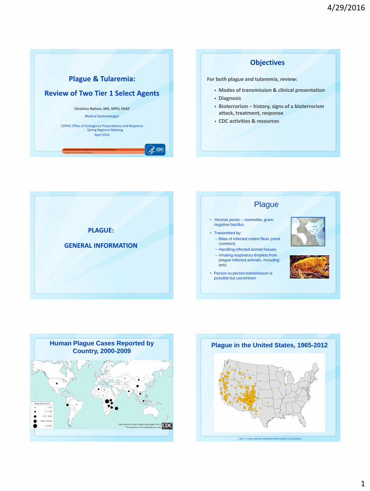
Plague & Tularemia:
Review of Two Tier 1 Select Agents
Christina Nelson, MD, MPH, FAAP
Medical Epidemiologist
CDPHE Office of Emergency Preparedness and ResponseSpring Regional Meeting
April 2016
National Center for Emerging and Zoonotic Infectious Diseases
Division of Vector-Borne Diseases
Objectives
For both plague and tularemia, review:
Modes of transmission & clinical presentation
Diagnosis
Bioterrorism – history, signs of a bioterrorism attack, treatment, response
CDC activities & resources
PLAGUE:
GENERAL INFORMATION
Plague
• Yersinia pestis – nonmotile, gram-
negative bacillus
• Transmitted by:
– Bites of infected rodent fleas (most
common)
– Handling infected animal tissues
– Inhaling respiratory droplets from
plague-infected animals, including
pets
• Person-to-person transmission is
possible but uncommon
Human Plague Cases Reported by
Country, 2000-2009Plague in the United States, 1965-2012
1 dot = 1 case, placed randomly within county of occurrence

4/29/2016
2
Clinical Manifestations of Plague
• Bubonic
• Septicemic
• Pneumonic
• Less common forms –
pharyngeal, meningeal
• 66% mortality prior to
availability of antibiotics
• Mortality now 5-14%
depending on clinical form
Photo credit: CDC Public Health Image Library
Frequency of Primary Plague
Types, United States, 1965-2012
Bubonic
(80%)
Pneumonic (2%)
Septicemic (17%)
Other (1%)
Kugeler et al. Epidemiology of Human Plague in the United States, 1900–2012. EID 2015.
Initial Laboratory Testing for Plague
Collect primary specimen
– Bubo aspirate, blood, or sputum
– Inoculate for culture
• General microbiologic media
(e.g. sheep blood agar)
• Grows slowly
– Staining
• Gram-negative coccobacilli
• Giemsa stain: bipolar staining organismsPhoto credit: CDC
Collect blood for acute and convalescent serology
Peripheral Blood Smear from Patient with
Septicemic Plague
Smear shows characteristic bipolar (safety pin) staining of Y. pestis
Wright-Giemsa stain; magnification x 1000
Laboratory Testing for Plague - Continued
• Culture– Growth ~24-48 hours after inoculation
– Automated bacterial identification systems may misidentify Y. pestis
– Direct fluorescent antibody testing (F1 antigen detection) for rapid presumptive identification of isolate
– Phage lysis for definitive identification of Y. pestis
• PCR
• Serology– Four-fold rise between acute and convalescent samples
– Convalescent at least 2 weeks after acute sample
CDC research and prevention efforts
Uganda
– Rat control projects
– Treatment trial: doxycycline vs. ciprofloxacin
Plague “Dipstick” (Lateral Flow Assay)
– Rapid
– Inexpensive
– Technically simply
– Guide diagnosis and treatment,
reduce the need for culture at
remote sites
4/29/2016
3
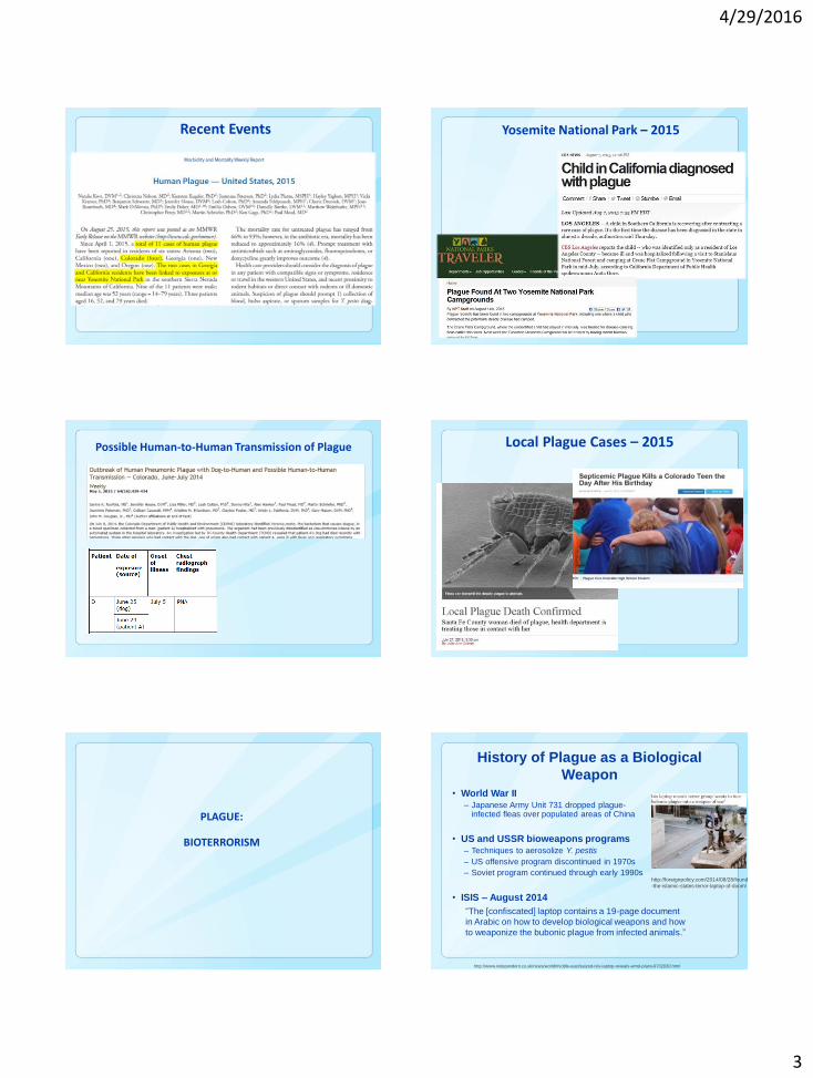
Recent Events Yosemite National Park – 2015
Possible Human-to-Human Transmission of Plague Local Plague Cases – 2015
PLAGUE:
BIOTERRORISM
History of Plague as a Biological
Weapon
• World War II– Japanese Army Unit 731 dropped plague-
infected fleas over populated areas of China
• US and USSR bioweapons programs– Techniques to aerosolize Y. pestis
– US offensive program discontinued in 1970s
– Soviet program continued through early 1990s
• ISIS – August 2014
“The [confiscated] laptop contains a 19-page document
in Arabic on how to develop biological weapons and how
to weaponize the bubonic plague from infected animals.”
http://www.independent.co.uk/news/world/middle-east/seized-isis-laptop-reveals-wmd-plans-9702030.html
http://foreignpolicy.com/2014/08/28/found
-the-islamic-states-terror-laptop-of-doom/

4/29/2016
4
Intentional Release of Y. pestis
• 1970 World Health Organization (WHO) report
–50 kg of Y. pestis released as aerosol
–Metropolitan area of 5 million
• Outcomes
–150,000 pneumonic plague cases
–36,000 deaths
–Y. pestis would remain viable as an aerosol for one hour for a distance of up to 10 km
–Further spread could occur
Intentional Release of Y. pestis
• Factors influencing size of outbreak:– Quantity of biological agent used
– Characteristics of the strain
– Environmental conditions
– Methods of aerosolization
• Indications of artificial dissemination:– Occurrence of disease in areas with no enzoonotic foci
– Occurrence in persons with no known risk factors
– Absence of prior rodent deaths
Working Group on Civilian Biodefense
• Series of articles in JAMA
“Medical and Public Health Management Following the Use of a Biological Weapon: Consensus Statements of
the Working Group on Civilian Biodefense”
• Representatives from academic medical centers, research, government, military, public health, and emergency management institutions and agencies
• Critical biological agents
Inglesby et al. Plague as a biological weapon: medical and public health
management. Working Group on Civilian Biodefense. JAMA. 2000.
Plague as a Biological Weapon –
Clinical Manifestations
• Primary pneumonic plague
• Incubation period: 2-4 days (range 1-6)
• First signs of illness
– Fever with cough and dyspnea
– Bloody, watery, or purulent sputum
– Gastrointestinal symptoms may be present
Specimen cup
containing
purulent, bloody
sputum from a
pneumonic plague
patient, Uganda.
• Absence of buboes (except, rarely, cervical buboes)
• Pulmonary disease with areas of profound lobular exudation & bacillary aggregation
• Consolidation on CXR
• Treat suspect plague patients without waiting for lab confirmation
• Without treatment, death quickly follows onset of symptoms
Plague as a Biological Weapon –
Clinical Manifestations
CXR of patient with
primary pneumonic
plague shows extensive
lobar consolidation in left
lower and left middle lung
fields
Treatment of Pneumonic Plague in the
Contained Casualty Setting
Adults
Preferred Choices• Gentamicin, 5mg/kg IM or IV once daily or
2mg/kg loading dose followed by 1.7 mg/kg IM or IV 3 times daily
• Streptomycin, 1 g IM twice daily
Alternative Choices• Doxycycline, 100 mg IV twice daily or 200 mg
IV once daily
• Ciprofloxacin, 400 mg IV twice daily
• Chloramphenicol, 25 mg/kg IV 4 times daily
For children – same antibiotics but different dosing

4/29/2016
5
Treatment of Pneumonic Plague in the
Contained Casualty Setting
Pregnant
Women
Preferred Choice• Gentamicin, 5mg/kg IM or IV once daily or
2mg/kg loading dose followed by 1.7 mg/kg IM or IV 3 times daily
Alternative Choices• Doxycycline, 100 mg IV twice daily or 200 mg
IV once daily
• Ciprofloxacin, 400 mg IV twice daily
Treatment of Pneumonic Plague in the
Mass Casualty Setting and for
Postexposure Prophylaxis
Adults (including pregnant women)
Preferred Choices
• Doxycycline, 100 mg orally twice daily
• Ciprofloxacin, 500 mg orally twice daily
Alternative Choice
• Chloramphenicol, 25 mg/kg orally 4
times daily
For children – same antibiotics but different dosing
Infection Control Measures for
Pneumonic Plague Patients
• Respiratory droplet precautions until
at least 48 hours of antibiotic therapy
and clinical improvement is seen
– Disposable surgical masks
– Gown, gloves, and eye protection
• Bodies of patients who have died should be handled with strict precautions
– Aerosol-generating procedures during autopsy not recommended
Environmental Decontamination
Recommendations for Plague
• Plague bacilli are sensitive to sunlight and heating
- No spore form in Y. pestis life cycle
- Does not survive long outside the host
• No evidence that plague bacilli pose an environmental threat following dissolution of primary aerosol
• Standard precautions for cleaning patient rooms
and linens
TULAREMIA:
GENERAL INFORMATION
Tularemia - Overview
• Caused by Francisellatularensis – small, nonmotile, aerobic, gram-negative coccobacillus
• Hardy – survives well in water, moist soil, straw, decaying animal carcasses
• First identified in 1911 as plague-like illness of ground squirrels in Tulare County, CA
• Aka “rabbit fever”
4/29/2016
6
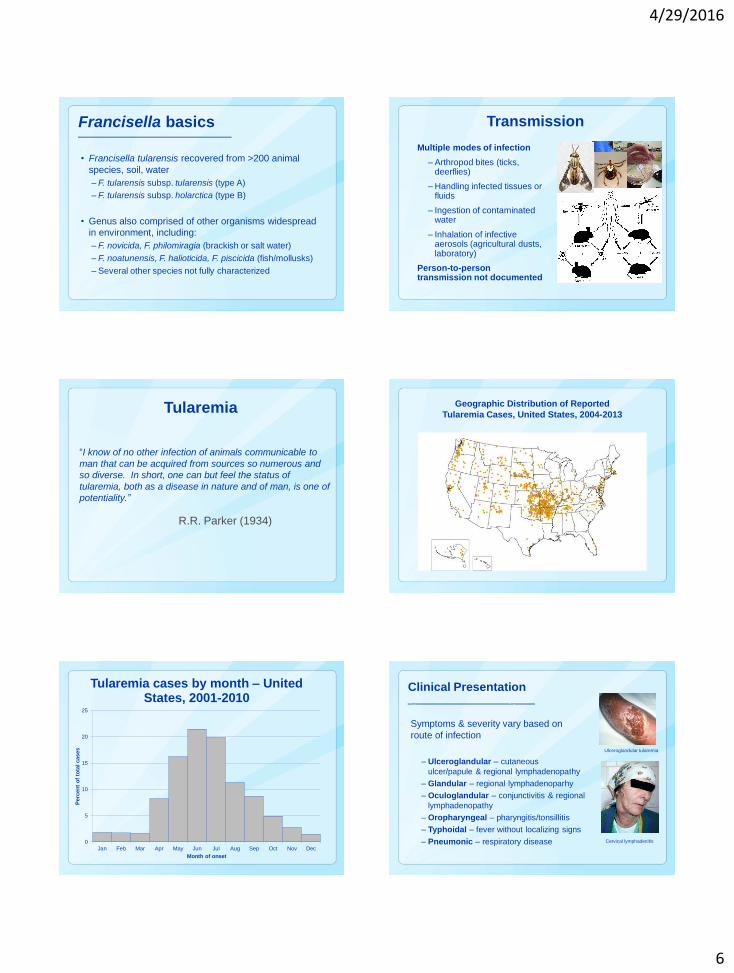
Francisella basics
• Francisella tularensis recovered from >200 animal
species, soil, water
– F. tularensis subsp. tularensis (type A)
– F. tularensis subsp. holarctica (type B)
• Genus also comprised of other organisms widespread
in environment, including:
– F. novicida, F. philomiragia (brackish or salt water)
– F. noatunensis, F. halioticida, F. piscicida (fish/mollusks)
– Several other species not fully characterized
Transmission
Multiple modes of infection
– Arthropod bites (ticks, deerflies)
– Handling infected tissues or fluids
– Ingestion of contaminated water
– Inhalation of infective aerosols (agricultural dusts, laboratory)
Person-to-person transmission not documented
Tularemia
“I know of no other infection of animals communicable to
man that can be acquired from sources so numerous and
so diverse. In short, one can but feel the status of
tularemia, both as a disease in nature and of man, is one of
potentiality.”
R.R. Parker (1934)
Geographic Distribution of Reported
Tularemia Cases, United States, 2004-2013
0
5
10
15
20
25
Jan Feb Mar Apr May Jun Jul Aug Sep Oct Nov Dec
Perc
en
t o
f to
tal cases
Month of onset
Tularemia cases by month – United States, 2001-2010
Clinical Presentation
Symptoms & severity vary based on
route of infection
– Ulceroglandular – cutaneous
ulcer/papule & regional lymphadenopathy
– Glandular – regional lymphadenoparhy
– Oculoglandular – conjunctivitis & regional
lymphadenopathy
– Oropharyngeal – pharyngitis/tonsillitis
– Typhoidal – fever without localizing signs
– Pneumonic – respiratory disease
Ulceroglandular tularemia
Cervical lymphadenitis

4/29/2016
7
Summary of Surveillance Data
• NNDSS data tabulated annually after finalized
• Usually 100-200 cases per year in recent decades
• Most cases (~70%) are ulceroglandular or glandular
– Arthropod bites or other cutaneous exposure
• More than 1/3 of cases diagnosed serologically
Laboratory Diagnosis
• Clinical samples: blood, lymph node aspirate, or respiratory sample (sputum, bronchoalveolar lavage, biopsy)
• Wright, Giemsa or Wayson stain may show tiny, gram-negative coccobacilli
• Culture
• DFA – rapid identification of F. tularensis in primary specimens or isolates
• PCR (does not distinguish between species)
• Serology
Recent Events Public Health Response to Tularemia
• Alerts triggered by confirmed cases in human
accompanied by positive carcasses
• Alerts include signage in affected areas, HANs, MMWR
articles, and public service announcements
TULAREMIA:
BIOTERRORISM
Tularemia in an Age of Bioterrorism
• F. tularensis is one of the most infectious bacteria
known inoculation with only 10 organisms can
cause disease
• Weaponized by US in the 1950s and 1960s,
parallel effort by USSR until early 1990s
• Other countries suspected to have weaponized it
as well
• Can be cultured and engineered for antimicrobial
resistance
• Contamination of the water supply also a concern

4/29/2016
8
Intentional Release of F. tularensis
• Bioterrorist attack would most likely occur via
aerosolization of F. tularensis
• 1970 WHO expert committee report
– 50 kg of virulent F. tularensis released as aerosol
– Metropolitan area of 5 million
• Outcomes
– 250,000 incapacitating casualties
– 100,000 deaths
– Illness expected to persist for several weeks
BioWatch
• Federal government program to detect the release of
pathogens into the air as part of a terrorist attack on major
American cities
• Created in 2001 in response to anthrax attacks
• System of filters located within existing Environmental
Protection Agency air filters that monitor air quality.
• Cross-reactivity with naturally-occurring Francisella spp.
• CDC provides SME support on national calls and on-site
investigations
Inhalational Tularemia
• Incubation: 3-6 days for acute symptoms (range 1-14 days)
– Only 25-50% of patients may have radiologic evidence of pneumonia in early stages of infection
• Acute illness with one or more of the following signs/symptoms:
– Pharyngitis - Bronchiolitis
– Pleuropneumonitis - Hilar lymphadenitis
– Manifestations of systemic illness
• Inhalational exposures can present as systemic illness with
few signs of respiratory disease
Inhalational Tularemia - Continued
• Earliest radiographic findings may be:– Periobronchial infiltrates – typically
advancing to broncho-pneumonia in
one or more lobes
– Accompanied by pleural effusions and
hilar lymphadenopathy
• However, signs may be minimal or absent– Some patients show only one or several small,
discrete pulmonary infiltrates, or
– Scattered granulomatous lesions of parenchyma or pleura
CXR showing
bilateral pneumonitis and
left pleural effusion
Epidemiology of Tularemia Following
Intentional Release
• First indication of clandestine release– Cluster of acute, severe respiratory illness with unusual
epidemiologic features
• Early diagnosis of inhalation tularemia requires a high index of suspicion
• Unlikely that serendipitous lab identification would be sentinel event– Identification of F. tularensis in clinical specimens may
be missed or delayed for days when routine screening procedures for bacterial pathogens are followed
Dispersal after Intentional Release
• Under natural conditions, F. tularensis can survive for extended periods in cold, moist environment
• Survival of intentionally dispersed particles is unknown but expected to be limited– Expect a short half-life due to desiccation, solar
radiation, oxidation, and other environmental factors
– Very limited risk from secondary dispersal

4/29/2016
9
Treatment of Tularemia in the Contained
Casualty Setting
Adults
Preferred Choices
• Streptomycin, 1 g IM twice daily
• Gentamicin, 5mg/kg IM or IV once daily
Alternative Choices
• Doxycycline, 100 mg IV twice daily
• Chloramphenicol, 15 mg/kg IV 4 times daily
• Ciprofloxacin, 400 mg IV twice daily
For children – same antibiotics but different dosing
Treatment of Tularemia in the Contained
Casualty Setting
Pregnant
Women
Preferred Choices
• Gentamicin, 5mg/kg IM or IV once daily
• Streptomycin, 1 g IM twice daily
Alternative Choices
• Doxycycline, 100 mg IV twice daily
• Ciprofloxacin, 400 mg IV twice daily
Treatment of Tularemia in the Mass Casualty
Setting and for Postexposure Prophylaxis
Adults (including pregnant women)
Preferred Choices• Doxycycline, 100 mg orally twice daily
• Ciprofloxacin, 500 mg orally twice daily
Preferred Choices• Doxycycline
If > 45 kg, give adult dose
If < 45 kg, give 2.2 mg/kg orally twice daily
• Ciprofloxacin, 15 mg/kg orally twice daily
Children
Infection Control Measures for
Inhalational Tularemia Patients
• No human-to-human transmission of tularemia documented
• Standard precautions are appropriate
• Isolation of patients is not recommended
• Bodies of patients should be handled using standard precautions– Autopsy procedures likely to produce aerosols should
be avoided
• **Caution for laboratory workers**
IN SUMMARY…
Differential Diagnosis: Inhalational
Tularemia, Plague and Anthrax
• Plague: progress rapidly to severe pneumonia– Copious watery or purulent sputum production
– Hemoptysis, respiratory insufficiency, sepsis and shock
• Tularemia: slower progression of illness and lower case fatality rate
• Anthrax: characteristic findings of prominent mediastinitis– Absence of bronchopneumonia
– Develop fulminating, toxic, fatal illness despite antibiotic treatment

4/29/2016
10
CDC RESOURCES & ACTIVITIES
• Track and report on national epidemiology
• Subject matter expertise for health departments
and clinicians
• Health communications assistance
• Ecologic and entomologic guidance
• Outbreak response
e.g. Devils Tower, Wyoming
CDC Resources & Activities
CDC Laboratory Resources & Activities
• WHO Collaborating Center on Vector-
Borne Bacterial Diseases
• National strain reference collections
2,500 Yp + 1,200 Ft strains
• Reference diagnostic services and training
– Molecular characterization and antimicrobial susceptibility testing
– Laboratory Response Network coordination and reagent distribution
• R&D for new diagnostics
Questions?
Thank you!
AcknowledgmentsCDPHE
Nicole Comstock, MSPH & Korey Bell, MA
Paul Mead, MD, MPH
C. Ben Beard, PhD
Stacie Marshall, MPH
Alison Hinckley, PhD
Kiersten Kugeler, PhD
National Center for Emerging and Zoonotic Infectious Diseases
Division of Vector-Borne Diseases l Bacterial Diseases Branch