Embed Size (px)
Citation preview
7
Periodontal Disease in Dogs
Fábio Alessandro Pieri, Ana Paula Falci Daibert, Elisa Bourguignon and Maria Aparecida Scatamburlo Moreira
Federal University of Viçosa, Brazil
1. Introduction
Periodontics is a science that aims to study the periodontium and the diagnosis, prevention
and treatment of periodontal diseases, in order to promote and restore the periodontal
health (Harvey & Emily, 1993; Roman et al., 1995).
The periodontium is the set of adjacent structures to the teeth that provides them with
support and protection. These structures are: gingiva, cementum, alveolar bone and
periodontal ligament (Harvey & Emily, 1993; Roman et al., 1995; De Marco & Gioso, 1997;
Clarke, 2001).
Periodontal disease is the most common oral disease in dogs with up of 80% of animals
affected (Riggio et al., 2011). This disease is progressive and involves two stages: gingivitis
(reversible) and periodontitis (irreversible, but often controllable). It is caused by plaque
buildup on teeth. The plaque is a smooth membrane, adhesive, contaminated with saliva
bacteria and debris. Bacteria and bacterial products cause inflammation of soft tissue. The
plaque becomes mineralised to form calculus, which migrates into the gingival sulcus,
causing additional inflammation, loss of periodontal ligament, bone loss and ultimately
tooth loss (Ford & Mazzaferro, 2007).
Medical problems that affect the oral cavity should be identified in its early stages, so that
the animals can be treated before showing serious secondary systemic disorders related to
malnutrition and/or infections (Pachaly, 2006). One should also be aware of ways to prevent
the disease, as animal tooth brushing and the use of antimicrobials as an adjunct in
periodontal therapy (De Marco & Gioso, 1997).
2. Dental anatomy of dogs
As in most domestic mammals and in humans, dogs have diphyodont dentition, featuring
two sets of teeth, a deciduous or primary and a permanent, although edentulous at birth
(Harvey, 1992).
The oral anatomy of dogs has subdivisions and similar structures to those of humans
(Figure 1), differing in the shape of the cavity, which also varies between breeds (Roza,
2004), anatomy and quantity of teeth and in the teeth apex (Harvey & Emily, 1993). Dogs
www.intechopen.com
A Bird's-Eye View of Veterinary Medicine
120
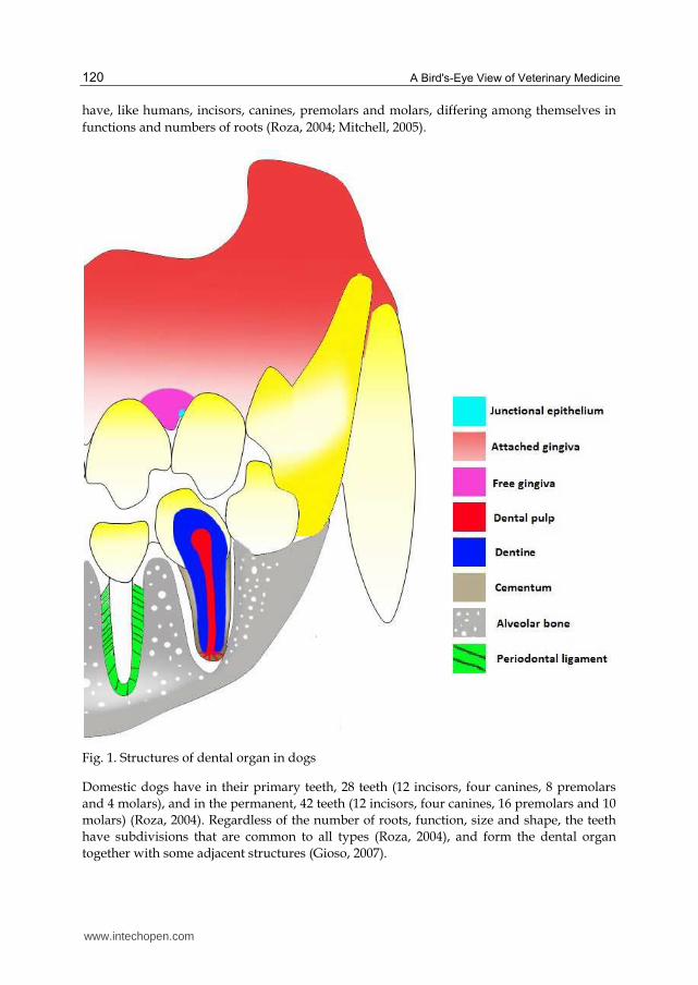
have, like humans, incisors, canines, premolars and molars, differing among themselves in
functions and numbers of roots (Roza, 2004; Mitchell, 2005).
Fig. 1. Structures of dental organ in dogs
Domestic dogs have in their primary teeth, 28 teeth (12 incisors, four canines, 8 premolars
and 4 molars), and in the permanent, 42 teeth (12 incisors, four canines, 16 premolars and 10
molars) (Roza, 2004). Regardless of the number of roots, function, size and shape, the teeth
have subdivisions that are common to all types (Roza, 2004), and form the dental organ
together with some adjacent structures (Gioso, 2007).
www.intechopen.com
Periodontal Disease in Dogs
121
For a better understanding of periodontal disease it is important to have further information about a set of structures that constitute the alveolar-dental joint, the periodontium (Picosse, 1987).
The periodontium (peri, around; dental, tooth) (Lindhe & Karring, 1997) is the set of hard and soft tissues (Mitchell, 2005) that support (Harvey & Emily, 1993, Domingues et al., 1999), by fixing, adhering (Lindhe & Karring, 1997; Roza, 2004) and protecting the tooth in the alveolar bone (Roza, 2004). The structures that comprise the periodontium are the periodontal ligament, cementum, gingiva and alveolar bone (Carranza, 1983; Lindhe & Karring, 1997; Wiggs & Lobprise, 1997). There is a division of these structures according to their functions, so there is a periodontal support formed by the cementum, the periodontal ligament, alveolar bone, and gingiva that besides participating in the support also comprises the protection periodontium (Roza, 2004).
The gingiva (Figure 1) is the part of the masticatory mucosa that surrounds the cervical portion of the tooth and covers the alveolar process (Lindhe & Karring, 1997). Its main function is to protect structures adjacent to the tooth, being the first line of defence against periodontal disease (Harvey & Emily, 1993). Two parts can be distinguished: the free and attached gingiva (Harvey & Emily, 1993; Lindhe & Karring, 1997).
The free gingiva can be pink or pigmented in some breeds, with firm consistency and an opaque surface (Lindhe & Karring, 1997). The margin of the free gingiva is the edge of it. Between the free gingiva and the tooth, a groove is formed (Mitchell, 2005) known as the gingival sulcus, which, in normal conditions in the dogs, varies in depth from one to three millimeters (Harvey & Emily, 1993; Roza, 2004). The sulcus is surrounded by an adhered epithelium that secretes a fluid with inflammation mediatory cells, immunoglobulins and antibacterial substances important in the physical and immunological protection of the junctional epithelium and deeper tissues (Pope, 1993).
The junctional epithelium (Figure 1) is located at the bottom of the sulcus, with flat and elongated cells (Hennet, 1995; Wiggs & Lobprise, 1997) adhering to the enamel through hemidesmosomes, promoting the junction between the gingiva and the tooth (Harvey & Emily, 1993). The junctional epithelium ends in the cementum-enamel junction (Roza, 2004).
In processes such as inflammation, hyperplasia or in both, the junctional epithelium can recede apically or the gingiva can increase, making deeper the gingival sulcus (Harvey & Emily, 1993). In gingival hyperplasia the deepening of the sulcus occurs without loss of periodontal tissue, named false pocket, although when there is a loss of the support tissue and protection of the tooth, the sulcus is called periodontal pockets, which can be of two types: suprabone, when the bottom of the sulcus is coronal to the support alveolar bone, and intrabone, when the bottom is located apically in relation to the adjacent alveolar bone. The pocket depth can vary between regions of the mouth and even between neighbouring teeth (Newman et al., 2004).
The attached gingiva (Figure 1) is the continuation of the free gingiva, that, is firmly attached to the underlying bone, and extends to the mucogingival junction (Carranza, 1983).
Cementum (Figure 1) is a hard tissue with no vascularity (Harvey et al. 1994; Hennet, 1995) that covers the tooth root (Mitchell, 2005), is composed of collagen fibres embedded in an organic matrix, and has in its mineral portion, which is responsible for about 65% of its
www.intechopen.com
A Bird's-Eye View of Veterinary Medicine
122
weight, hydroxyapatite crystals (Lindhe & Karring, 1997). It has as its main functions the insertion of periodontal ligament fibres into the root of the tooth (Harvey & Emily, 1993; Lindhe & Karring, 1997; Roza, 2004), the contribution to the process of repair of the root surface and the maintenance of the periodontal ligament fibres (Picasso, 1987; Lindhe & Karring, 1997).
There are two types of cementum: the first, called primary or acellular cementum, is formed in association with root formation and teeth eruption (Lindhe & Karring, 1997). It occupies the coronal and middle thirds of the tooth root and is constituted mostly of Sharpey's fibres, which are the periodontal ligament collagen fibres that attach at one end to the cementum and at the other to the alveolar bone (Roza, 2004). The other type of cementum is called secondary or cellular cementum, formed after the teeth (Lindhe & Karring, 1997) and usually located in the periapical region. It is secreted by cementocytes or cementoblasts, which are cells that are trapped into the organic matrix of cementum. It does not have vascularization, therefore is nourished from the periodontal ligament. The cells secrete cellular cementum in response to functional demands (Wiggs & Lobprise, 1997). Despite the description of the location of each type of cementum, in some cases they can occur alternately in some areas of the root surface (Lindhe & Karring, 1997).
The alveolar bone (Figure 1) involves the maxilla, incisor bone and jawbone that support the teeth in cavities where they are inserted. These cavities are called alveolus (Picasso, 1987; Harvey & Emily, 1993; Roza, 2004).
Composed of 65% minerals (Wiggs & Lobprise, 1997), this bone has a hard consistency and
is very dense and compact, but differs from the root cementum because it has innervation,
blood and lymphatic vasculature (Lindhe & Karring, 1997).
The interior of the alveolus is where the cribriform plate is located, which radiographically
is known as lamina dura, characterised as a radiopaque line around the alveolus. The fibres
of the periodontal ligament that attach to the tooth are connected to this plaque and it is
where the vessels pass for ligament irrigation and for the nutrition of the cementum organic
matrix (Harvey & Emily, 1993; Roza, 2004)
The alveolar bone can be resorbed or remodelled, according to the stimuli that it may suffer (Harvey & Emily, 1993; Roza, 2004).
The periodontal ligament (Figure 1) is a connective tissue structure that binds the tooth to its alveolus, fixing it (Lindhe & Karring, 1997; Figueiredo & Parra, 2002). It originates from mesenchymal cells of the dental sac (Picasso, 1987; Wiggs & Lobprise, 1997).
The periodontal ligament contains nerves and great vascularity, with vessels emanating
from the maxillary artery in the case of the maxilla and from the inferior alveolar artery in
the case of the jaw, and other cells. It is located between the root cementum and the
cribriform plate. Its height, width, quality and condition are crucial to give the tooth its
characteristic mobility (Harvey & Emily, 1993; Lindhe & Karring, 1997; Roza, 2004).
There are three different categories of fibres in the periodontal ligament: the fibres of the
gingival grouping, composed by dental gingival fibres (connect the cementum to the
gingiva), the alveologingival fibres (connects the cribriform plate to the gingiva), the
circular (surrounding the tooth at the free gingiva), the transseptal fibres (connects the supra
www.intechopen.com
Periodontal Disease in Dogs
123
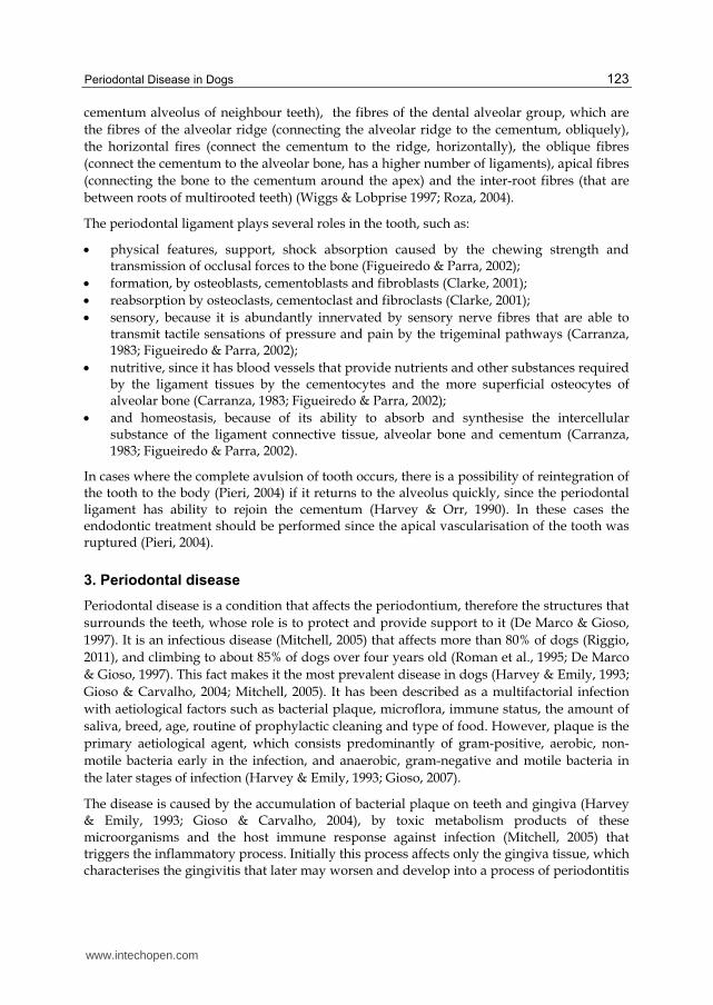
cementum alveolus of neighbour teeth), the fibres of the dental alveolar group, which are
the fibres of the alveolar ridge (connecting the alveolar ridge to the cementum, obliquely),
the horizontal fires (connect the cementum to the ridge, horizontally), the oblique fibres
(connect the cementum to the alveolar bone, has a higher number of ligaments), apical fibres
(connecting the bone to the cementum around the apex) and the inter-root fibres (that are
between roots of multirooted teeth) (Wiggs & Lobprise 1997; Roza, 2004).
The periodontal ligament plays several roles in the tooth, such as:
physical features, support, shock absorption caused by the chewing strength and transmission of occlusal forces to the bone (Figueiredo & Parra, 2002);
formation, by osteoblasts, cementoblasts and fibroblasts (Clarke, 2001);
reabsorption by osteoclasts, cementoclast and fibroclasts (Clarke, 2001);
sensory, because it is abundantly innervated by sensory nerve fibres that are able to transmit tactile sensations of pressure and pain by the trigeminal pathways (Carranza, 1983; Figueiredo & Parra, 2002);
nutritive, since it has blood vessels that provide nutrients and other substances required by the ligament tissues by the cementocytes and the more superficial osteocytes of alveolar bone (Carranza, 1983; Figueiredo & Parra, 2002);
and homeostasis, because of its ability to absorb and synthesise the intercellular substance of the ligament connective tissue, alveolar bone and cementum (Carranza, 1983; Figueiredo & Parra, 2002).
In cases where the complete avulsion of tooth occurs, there is a possibility of reintegration of the tooth to the body (Pieri, 2004) if it returns to the alveolus quickly, since the periodontal ligament has ability to rejoin the cementum (Harvey & Orr, 1990). In these cases the endodontic treatment should be performed since the apical vascularisation of the tooth was ruptured (Pieri, 2004).
3. Periodontal disease
Periodontal disease is a condition that affects the periodontium, therefore the structures that
surrounds the teeth, whose role is to protect and provide support to it (De Marco & Gioso,
1997). It is an infectious disease (Mitchell, 2005) that affects more than 80% of dogs (Riggio,
2011), and climbing to about 85% of dogs over four years old (Roman et al., 1995; De Marco
& Gioso, 1997). This fact makes it the most prevalent disease in dogs (Harvey & Emily, 1993;
Gioso & Carvalho, 2004; Mitchell, 2005). It has been described as a multifactorial infection
with aetiological factors such as bacterial plaque, microflora, immune status, the amount of
saliva, breed, age, routine of prophylactic cleaning and type of food. However, plaque is the
primary aetiological agent, which consists predominantly of gram-positive, aerobic, non-
motile bacteria early in the infection, and anaerobic, gram-negative and motile bacteria in
the later stages of infection (Harvey & Emily, 1993; Gioso, 2007).
The disease is caused by the accumulation of bacterial plaque on teeth and gingiva (Harvey & Emily, 1993; Gioso & Carvalho, 2004), by toxic metabolism products of these microorganisms and the host immune response against infection (Mitchell, 2005) that triggers the inflammatory process. Initially this process affects only the gingiva tissue, which characterises the gingivitis that later may worsen and develop into a process of periodontitis
www.intechopen.com
A Bird's-Eye View of Veterinary Medicine
124
which involves changes in other periodontium tissues and can cause bone, periodontal ligament, and in some cases, cementum or even tooth loss (Harvey & Emily, 1993).
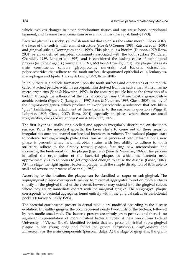
Bacterial plaque is a sticky, yellowish material that colonises the entire mouth (Gioso, 2007), the faces of the teeth in their enamel structure (Slee & O'Connor, 1983; Katsura et al., 2001) and gingival sulcus (Domingues et al., 1999). This plaque is a biofilm (Dupont, 1997; Roza, 2004) or an undefined microbial community associated with the tooth surface (Wilderer; Charaklis, 1989, Lang et al., 1997), and is considered the leading cause of pathological process (aetiologic agent) (Tanzer et al. 1977; McPhee & Cowley, 1981). The plaque has as its main constituents: salivary glycoproteins, minerals, oral bacteria, extracellular polysaccharides that adhere to the tooth surface, desquamated epithelial cells, leukocytes, macrophages and lipids (Harvey & Emily, 1993; Roza, 2004)
Initially there is a pellicle formation upon the tooth surfaces and other areas of the mouth, called attached pellicle, which is an organic film derived from the saliva that, at first, has no micro-organisms (Sans & Newman, 1997). In the acquired pellicle begins the formation of a biofilm through the adhesion of the first microorganisms that are mostly gram-positive aerobic bacteria (Figure 2) (Lang et al. 1997; Sans & Newman, 1997; Gioso, 2007), mainly of the Streptococcus genus, which produce an exopolysaccharide, a substance that acts like a "glue", facilitating the attachment of these bacteria to the surfaces in question (Wiggs & Lobprise, 1997; Gioso, 2007; Roza, 2004) especially in places where there are small irregularities, cracks or roughness (Sans & Newman, 1997).
The first layer is usually single-celled and appears irregularly distributed on the tooth surface. With the microbial growth, the layer starts to come out of these areas of irregularities onto the enamel surface and increases in volume. The isolated plaques start to coalesce, forming a single plate. Over time in the process of plaque formation, a new phase is present, where new microbial strains with less ability to adhere to tooth structure, adhere to the already formed plaque, featuring new microcolonies and increasing the biodiversity of the plaque (Figure 2) (Sans & Newman, 1997). This process is called the organisation of the bacterial plaque, in which the bacteria need approximately 24 to 48 hours to get organised enough to cause the disease (Gioso, 2007). At this stage, the fight against bacterial plaque, with the simple disruption of it, is able to stall and reverse the process (Slee et al., 1983).
According to the location, the plaque can be classified as supra or sub-gingival. The
supragingival plaque corresponds mainly to microbial aggregates found on tooth surfaces
(mostly in the gingival third of the crown), however may extend into the gingival sulcus,
where they are in immediate contact with the marginal gingiva. The subgingival plaque
corresponds to bacterial aggregates found entirely within the gingival sulcus or periodontal
pockets (Harvey & Emily 1993).
The bacterial constituents present in dental plaque are modified according to the disease evolution. In healthy gingiva, the cocci represent nearly two-thirds of the bacteria, followed by non-motile small rods. The bacteria present are mostly gram-positive and there is no significant representation of more virulent bacterial types. A new work from Federal University of Viçosa, Brazil, identified bacteria that are present in initial supra-gingival plaque in ten young dogs and found the genera Streptococcus, Staphylococcus and Enterococcus as the main components (personal data). At the stage of gingivitis, the gram-
www.intechopen.com
Periodontal Disease in Dogs
125
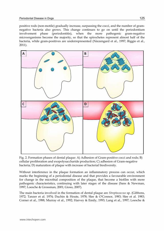
positive rods (non-motile) gradually increase, surpassing the cocci, and the number of gram-negative bacteria also grows. This change continues to go on until the periodontium involvement phase (periodontitis), when the more pathogenic gram-negative microorganisms become the majority, so that the spirochetes represent almost half of the bacteria, while gram-positives are underrepresented (Niezengard et al., 1997; Riggio et al., 2011).
Fig. 2. Formation phases of dental plaque: A) Adhesion of Gram-positive cocci and rods; B) cellular proliferation and exopolysaccharide production; C) adhesion of Gram-negative bacteria; D) maturation of plaque with increase of bacterial biodiversity.
Without interference in the plaque formation an inflammatory process can occur, which marks the beginning of a periodontal disease and that provides a favourable environment for change in the microbial composition of the plaque, that become a biofilm with more pathogenic characteristics, continuing with later stages of the disease (Sans & Newman, 1997; Loesche & Grossman, 2001; Gioso, 2007).
The main bacteria involved in the formation of dental plaque are Streptococcus sp. (Gibbons, 1972; Tanzer et al. 1974; Duchin & Houte, 1978; Slee & O'Connor, 1983; Slee et al. 1983; Corner et al., 1988; Murray et al., 1992; Harvey & Emily, 1993; Lang et al., 1997; Loesche &
www.intechopen.com

A Bird's-Eye View of Veterinary Medicine
126
Grossman, 2001; Katsura et al., 2001; Drummond et al., 2004; Swerts et al., 2005), Actinomyces sp. (Slee & O'Connor, 1983; Katsura et al., 2001), and Lactobacillus sp. (Drummond et al. 2004; Roza, 2004). These colonise initially the adhered film of the enamel and then start to multiply and aggregate. Thereafter, surface receptors on the cocci and gram-positive rods allow adherence of gram-negative bacteria and, over time, they present the greatest biodiversity and pathogenic potential (Lang et al., 1997). In dogs, the most important bacteria of this group are Veillonella, Bacterioides, Prevotella (Domingues et al., 1999), Fusobacterium (Murray et al., 1992; Braga et al., 2005) and Porphyromonas (Domingues et al., 1999; Katsura et al., 2001; Braga et al., 2005; Senhorinho et al., 2011).
The bacteria involved in the periodontal disease process may migrate to other regions of the body by bacteraemia and colonise there, causing various diseases such as endocarditis, nephritis (Harvey & Emily, 1993; Debowes, 1996; Gioso, 2007), hepatitis (Debowes, 1996; Gioso, 2007) and myocarditis (Harvey & Emily, 1993). Another disease that can be caused by the microorganisms involved in periodontal disease, is a pathological process known as periodontic-endodontic lesion. There is an infection and inflammation of the dental pulp caused by the migration of periodontal bacteria at the apex of the tooth and root canal penetration through the foramen, which are small holes through which the vascular supply, lymphatics and nerves of the tooth passes (Pieri, 2004).
In dogs, the progression of periodontal disease can be divided into stages based on the clinical appearance of the gingiva (classified according to an index ranging from zero degree representing healthy gingiva, to four, when there is severe involvement of it (Roza, 2004; Mitchell, 2005) and loss of adherence of the periodontium, which is measured by inserting a millimeter probe into the gingival sulcus. This measure represents how much the junctional epithelium has migrated toward the apex of the tooth. The result in millimeters is the distance between the bottom of the gingival sulcus or periodontal pocket to the cementoenamel junction. The rate of adhesion loss is expressed by the ratio between the loss and the total normal adhesion (Mitchell, 2005).
In the process of disease formation, the healthy periodontium is considered stage 0 (Figure 3) (Harvey & Emily, 1993) when the gingiva presents a uniform colour, the bacterial plaque is imperceptible and there are little pathogenic characteristics, and there is no halitosis (De Marco & Gioso, 1997). With the increase in bacterial plaque and its change regarding the amount and specificity of the present microorganisms, such as its pathogenicity, begins to emerge an inflammation that marks the beginning of periodontal disease itself, which can be divided into stages of gingivitis (stage 1), initial periodontitis (stage 2), moderate periodontitis (stage 3) and severe periodontitis (stage 4) (Harvey & Emily, 1993).
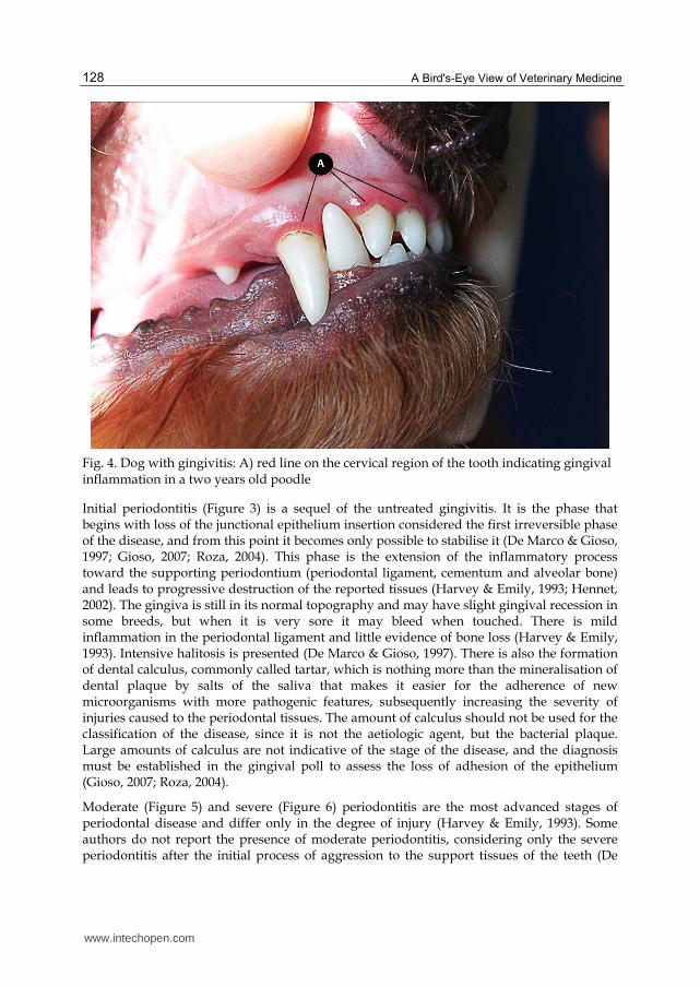
The first stage of periodontal disease begins with the emergence of bacteria which, through its metabolites, cause inflammation of the gingival tissue, called gingivitis (Figure 3 and Figure 4). This is the first defence of the tooth, without much pathogenicity and without damaging the structures of the support periodontium. Such inflammation is similar to that seen in other connective tissues. Vasodilation, leukocyte marginalisation, cell migration, production of prostaglandins and destructive enzymes also occurs (Gioso, 2007), making the gingiva red, swollen and painful, and may cause halitosis (De Marco & Gioso, 1997). At this stage the possibility of regression of the disease remains through the proper management of the oral health of the animal with measures such as brushing, promoting removal of the aetiological agent (bacterial plaque) (Gorrel & Rawlings, 1996; Gioso, 2007). At this moment
www.intechopen.com
Periodontal Disease in Dogs
127
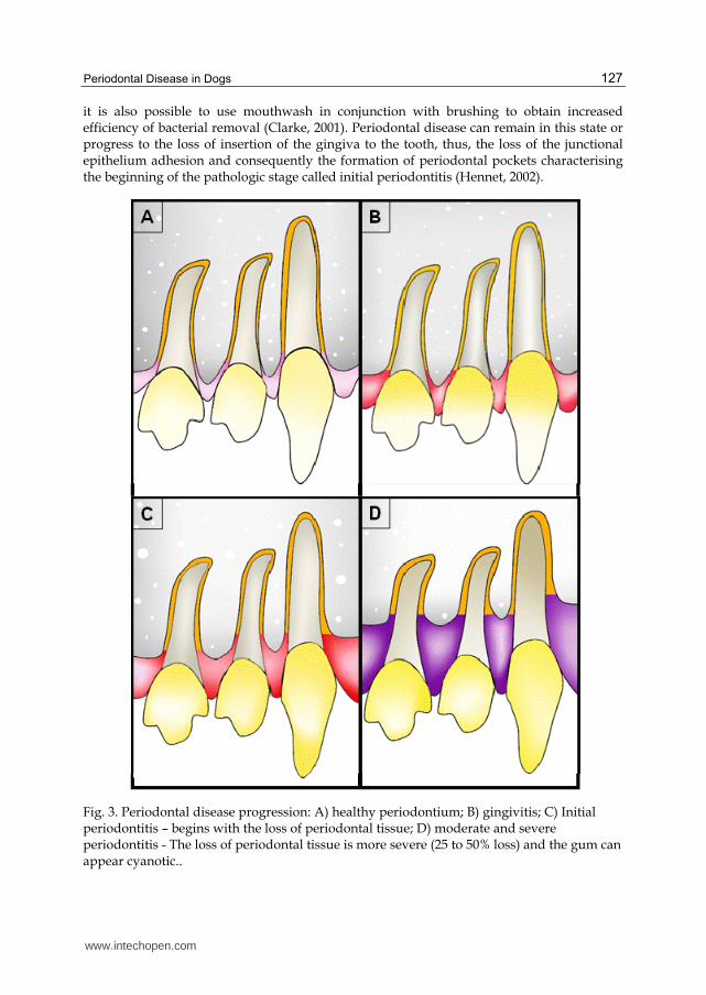
it is also possible to use mouthwash in conjunction with brushing to obtain increased efficiency of bacterial removal (Clarke, 2001). Periodontal disease can remain in this state or progress to the loss of insertion of the gingiva to the tooth, thus, the loss of the junctional epithelium adhesion and consequently the formation of periodontal pockets characterising the beginning of the pathologic stage called initial periodontitis (Hennet, 2002).
Fig. 3. Periodontal disease progression: A) healthy periodontium; B) gingivitis; C) Initial periodontitis – begins with the loss of periodontal tissue; D) moderate and severe periodontitis - The loss of periodontal tissue is more severe (25 to 50% loss) and the gum can appear cyanotic..
www.intechopen.com
A Bird's-Eye View of Veterinary Medicine
128
Fig. 4. Dog with gingivitis: A) red line on the cervical region of the tooth indicating gingival inflammation in a two years old poodle
Initial periodontitis (Figure 3) is a sequel of the untreated gingivitis. It is the phase that begins with loss of the junctional epithelium insertion considered the first irreversible phase of the disease, and from this point it becomes only possible to stabilise it (De Marco & Gioso, 1997; Gioso, 2007; Roza, 2004). This phase is the extension of the inflammatory process toward the supporting periodontium (periodontal ligament, cementum and alveolar bone) and leads to progressive destruction of the reported tissues (Harvey & Emily, 1993; Hennet, 2002). The gingiva is still in its normal topography and may have slight gingival recession in some breeds, but when it is very sore it may bleed when touched. There is mild inflammation in the periodontal ligament and little evidence of bone loss (Harvey & Emily, 1993). Intensive halitosis is presented (De Marco & Gioso, 1997). There is also the formation of dental calculus, commonly called tartar, which is nothing more than the mineralisation of dental plaque by salts of the saliva that makes it easier for the adherence of new microorganisms with more pathogenic features, subsequently increasing the severity of injuries caused to the periodontal tissues. The amount of calculus should not be used for the classification of the disease, since it is not the aetiologic agent, but the bacterial plaque. Large amounts of calculus are not indicative of the stage of the disease, and the diagnosis must be established in the gingival poll to assess the loss of adhesion of the epithelium (Gioso, 2007; Roza, 2004).
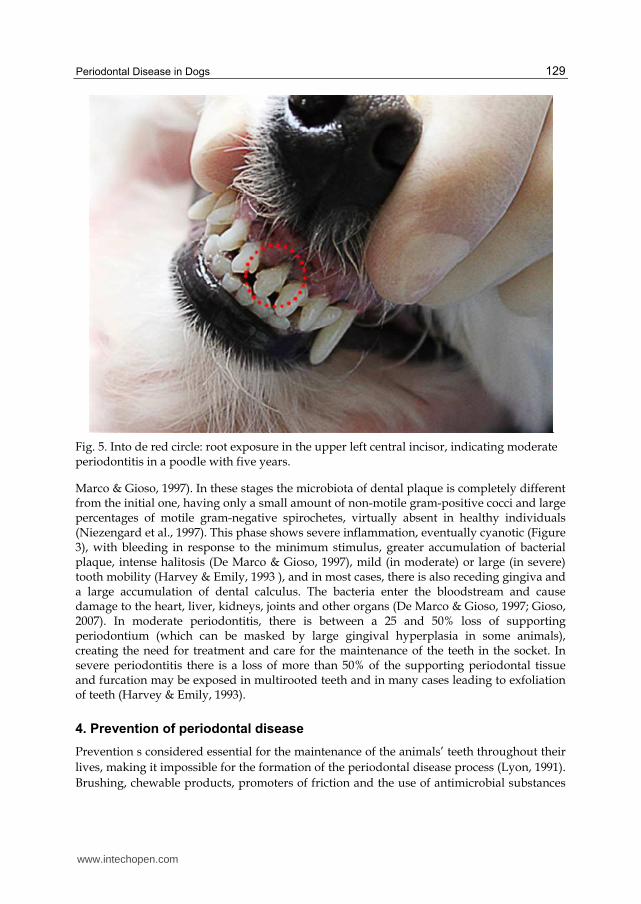
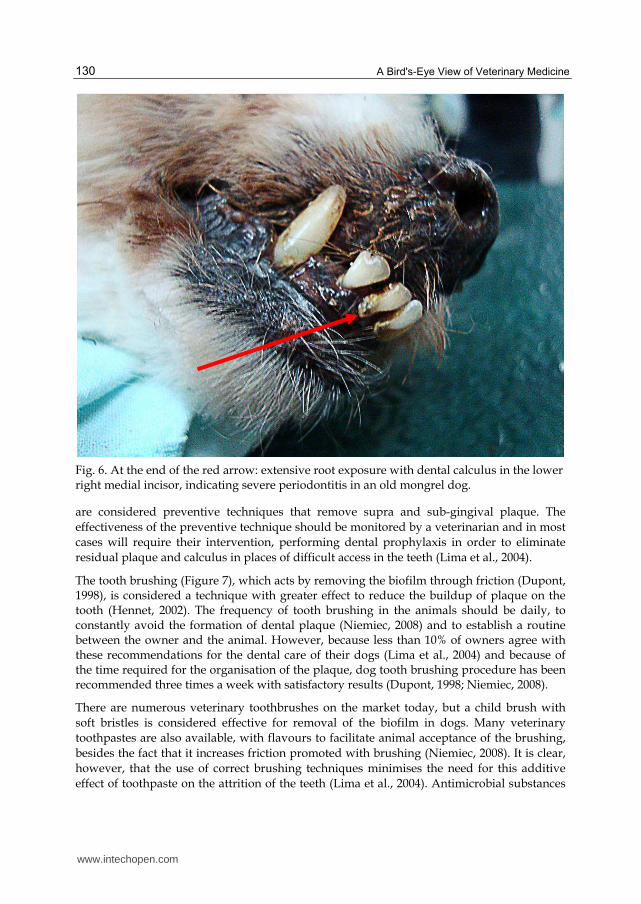
Moderate (Figure 5) and severe (Figure 6) periodontitis are the most advanced stages of periodontal disease and differ only in the degree of injury (Harvey & Emily, 1993). Some authors do not report the presence of moderate periodontitis, considering only the severe periodontitis after the initial process of aggression to the support tissues of the teeth (De
www.intechopen.com
Periodontal Disease in Dogs
129
Fig. 5. Into de red circle: root exposure in the upper left central incisor, indicating moderate periodontitis in a poodle with five years.
Marco & Gioso, 1997). In these stages the microbiota of dental plaque is completely different from the initial one, having only a small amount of non-motile gram-positive cocci and large percentages of motile gram-negative spirochetes, virtually absent in healthy individuals (Niezengard et al., 1997). This phase shows severe inflammation, eventually cyanotic (Figure 3), with bleeding in response to the minimum stimulus, greater accumulation of bacterial plaque, intense halitosis (De Marco & Gioso, 1997), mild (in moderate) or large (in severe) tooth mobility (Harvey & Emily, 1993 ), and in most cases, there is also receding gingiva and a large accumulation of dental calculus. The bacteria enter the bloodstream and cause damage to the heart, liver, kidneys, joints and other organs (De Marco & Gioso, 1997; Gioso, 2007). In moderate periodontitis, there is between a 25 and 50% loss of supporting periodontium (which can be masked by large gingival hyperplasia in some animals), creating the need for treatment and care for the maintenance of the teeth in the socket. In severe periodontitis there is a loss of more than 50% of the supporting periodontal tissue and furcation may be exposed in multirooted teeth and in many cases leading to exfoliation of teeth (Harvey & Emily, 1993).
4. Prevention of periodontal disease
Prevention s considered essential for the maintenance of the animals’ teeth throughout their
lives, making it impossible for the formation of the periodontal disease process (Lyon, 1991).
Brushing, chewable products, promoters of friction and the use of antimicrobial substances
www.intechopen.com
A Bird's-Eye View of Veterinary Medicine
130
Fig. 6. At the end of the red arrow: extensive root exposure with dental calculus in the lower right medial incisor, indicating severe periodontitis in an old mongrel dog.
are considered preventive techniques that remove supra and sub-gingival plaque. The
effectiveness of the preventive technique should be monitored by a veterinarian and in most
cases will require their intervention, performing dental prophylaxis in order to eliminate
residual plaque and calculus in places of difficult access in the teeth (Lima et al., 2004).
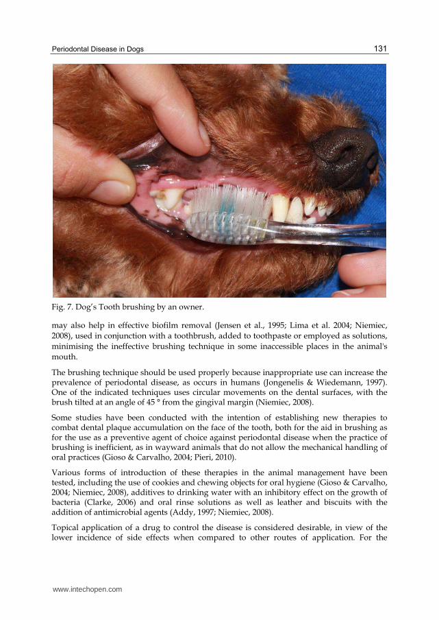
The tooth brushing (Figure 7), which acts by removing the biofilm through friction (Dupont, 1998), is considered a technique with greater effect to reduce the buildup of plaque on the tooth (Hennet, 2002). The frequency of tooth brushing in the animals should be daily, to constantly avoid the formation of dental plaque (Niemiec, 2008) and to establish a routine between the owner and the animal. However, because less than 10% of owners agree with these recommendations for the dental care of their dogs (Lima et al., 2004) and because of the time required for the organisation of the plaque, dog tooth brushing procedure has been recommended three times a week with satisfactory results (Dupont, 1998; Niemiec, 2008).
There are numerous veterinary toothbrushes on the market today, but a child brush with
soft bristles is considered effective for removal of the biofilm in dogs. Many veterinary
toothpastes are also available, with flavours to facilitate animal acceptance of the brushing,
besides the fact that it increases friction promoted with brushing (Niemiec, 2008). It is clear,
however, that the use of correct brushing techniques minimises the need for this additive
effect of toothpaste on the attrition of the teeth (Lima et al., 2004). Antimicrobial substances
www.intechopen.com
Periodontal Disease in Dogs
131
Fig. 7. Dog’s Tooth brushing by an owner.
may also help in effective biofilm removal (Jensen et al., 1995; Lima et al. 2004; Niemiec,
2008), used in conjunction with a toothbrush, added to toothpaste or employed as solutions,
minimising the ineffective brushing technique in some inaccessible places in the animal's
mouth.
The brushing technique should be used properly because inappropriate use can increase the prevalence of periodontal disease, as occurs in humans (Jongenelis & Wiedemann, 1997). One of the indicated techniques uses circular movements on the dental surfaces, with the brush tilted at an angle of 45 ° from the gingival margin (Niemiec, 2008).
Some studies have been conducted with the intention of establishing new therapies to combat dental plaque accumulation on the face of the tooth, both for the aid in brushing as for the use as a preventive agent of choice against periodontal disease when the practice of brushing is inefficient, as in wayward animals that do not allow the mechanical handling of oral practices (Gioso & Carvalho, 2004; Pieri, 2010).
Various forms of introduction of these therapies in the animal management have been tested, including the use of cookies and chewing objects for oral hygiene (Gioso & Carvalho, 2004; Niemiec, 2008), additives to drinking water with an inhibitory effect on the growth of bacteria (Clarke, 2006) and oral rinse solutions as well as leather and biscuits with the addition of antimicrobial agents (Addy, 1997; Niemiec, 2008).
Topical application of a drug to control the disease is considered desirable, in view of the lower incidence of side effects when compared to other routes of application. For the
www.intechopen.com

A Bird's-Eye View of Veterinary Medicine
132
prevention of periodontal disease, taking into account the least harmful nature of bacteria in the onset of the disease and higher prevalence of supragingival plaque, it is recommended that the use of topical oral solutions such as mouthwashes is sufficient to combat bacteria in question with great advantage because of its easy application in most patients (Ciancio & Niezengard, 1997).
Among the chemicals, that can be used this way to reduce the accumulation of plaque on dental surfaces, the bisguanids, quaternary ammonia and phenols have been widely evaluated. Chlorhexidine appears as a substance that has the greatest efficacy in the inhibition of oral plaque (Hennet, 2002) and has good antiseptic activity against all oral pathogens, more directly on the bacterial plaque organisms (Harvey & Emily, 1993). Its main concentration is the commercial use of alcoholic solution at 0.12% and it is also found in alcohol-free solutions and in gel form (Robinson, 1995).
Despite the above indications for the use of chlorhexidine in the fight against dental plaque, it presents a series of unpleasant effects when used for prolonged therapy, such as loss of taste by the patient, pigmentation of the enamel, burning and even ulceration of the buccal mucosa (Zanini et al., 1995). These effects justify the use of this material only for few days (Gioso, 2007), which makes its application not recommended in the prevention of periodontal disease, which requires a prolonged use of the antimicrobial agent chosen for this purpose (Lascala & Moussalli, 1995).
The natural sweetener xylitol has been used in human patients in chewing gum, mouthwashes and toothpastes in order to reduce plaque. There is also one xylitol based product in the veterinary market today, which when added to the animal drinking water has a lowering effect on oral bacterial plaque formation (Dunayer & Gwaltney-Brant, 2006).
Recently, the ozonised sunflower oil was tested, with positive results on microbial reduction in human patients with periodontal disease (Fiorini et al., 2006) and copaiba oil was applied topically on dogs and the results were equal to those obtained with chlorhexidine on the oral microbial population (Pieri, 2010). Additionally, some in vitro tests were performed to analyse the antimicrobial activity of Copaiba oil on plaque-forming bacteria (Simões, 2004; Pieri, 2010; Valdevite et al., 2007) and the evaluation of the inhibition of Streptococcus sp. adherence in glass capillaries caused by the same phytotherapic (Pieri, 2010), obtaining in both cases positive results. A actual work has been conduced to evaluate the copaiba oil as antimicrobial against bacterial isolates from initial dental plaque of dogs, aiming identify a potential drug to prevent the plaque formation and consequently the periodontal disease (personal data).
Many researchers continue their analysis looking for natural drugs as propolis (Swerts et al., 2005), Camellia sinensis (Chang et al., 2009), Mimosa tenuiflora (Macedo-Costa, 2009), Vitis amurensis (Yim et al., 2010), Rhinacanthus nasutus (Puttarak et al., 2010), Murraya koenigii, Allium sativum and Melaleuca alternifolia (Prabhakar et al., 2009) to its use in the prevention of periodontal disease by inhibiting plaque formation.It important that this drug combine properties such as antimicrobial activity that does not induce bacterial resistance, and inhibition of microbial adherence on tooth surfaces that suggest a great potential for use in therapies in the oral cavity and as an aid in oral hygiene (Sudo et al., 1986; Corner et al., 1988; Pieri, 2010). For the use in the treatment of domestic animals it is suggested the inclusion of this antimicrobial and non-adherent agent in formulations containing the base flavours of chicken, beef, fish, etc. (De Marco & Gioso, 1997).
www.intechopen.com
Periodontal Disease in Dogs
133
5. Diagnosis
The diagnosis of periodontal disease is based on history, clinical examination and radiological evaluation. Any changes in apprehension and chewing of food, as well as in general conditions and in the behaviour of animals, can be associated with oral disorders. Certain physical and behavioral changes are highly suggestive of dental disorders, including abnormal ways of eating and drinking, acute reactions to the ingestion of cold water, selective appetite (preference for soft foods), anorexia and weight loss, salivation, bleeding, epitaxy, digging of the ground, behavior of rubbing their feet on the face, shaking of the head, oronasal fistulas, abnormal aggressive behavior (because of pain) and distress and anguish (Emily & Penman, 1994; Pachaly, 2006; Gorrel, 2004). When it comes to periodontal disease, the main complaint of the owner will always be halitosis (Emily & Penman, 1994; Gorrel, 2004; Gioso, 2007) due to tissue decay and bacterial fermentation in the sulcus or periodontal pocket (Gioso, 2007).
Like any other clinical examination, the examination in dentistry should be preceded by thorough history and general physical examination. At the end, the oral cavity should be examined. It is necessary to do a complete oral examination to assess the presence of periodontal disease and other diseases, such as fractures or dental malocclusions. The intra-and extraoral structures should be assessed, including bone surfaces, the jaw muscles, salivary glands and regional cervical lymph nodes (Gorrel., 2004). Ideally, the complete periodontal examination should be performed in anaesthetised dogs (Harvey, 1992; Gorrel, 2004; Gioso, 2007). The evaluation of the tooth must be made with an explorer and periodontal probe (Gioso, 2007). The examination must be careful; incorrect handling of the probe may damage the soft tissues and lead to misdiagnosis of periodontal lesions (Gorrel, 2004). The changes observed should be recorded in an appropriate medical record and serve as the basis for the therapeutic treatment (Pachaly, 2006).
The periodontal examination includes the evaluation of teeth mobility, of injuries or furcation exposure, gingival retracting or hyperplasia, the evaluation of the depth, the presence of dental plaque, of gingivitis and dental calculus The furcation is the area between the roots of teeth that have more than one root. This area is usually filled with alveolar bone. During exploration, a depression can be felt while passing the extremity of a probe perpendicular to the tooth crown and below the gingival margin. In the presence of periodontitis, the furcation bone can be resorbed and probe inserted between the roots. Changes in the furcation are classified on a scale ranging from 0 to 3, where in grade 3 lesions the probe passes freely through the furcation, from the vestibular part to the lingual/palatal tooth (Gorrel, 2004).
The gingival sulcus is the space between the free gingiva and the tooth crown. In dogs, the depth of the gingival sulcus should be less than 3mm, and in giant breed dogs less than 4 mm (Gioso, 2007). When periodontitis is established, the junctional epithelium, the region of the gingival tissue inserted to the tooth surface, migrates apically along the root. If the apical migration is not accompanied by a receding gingiva then the periodontal pocket is formed, which has a depth greater than 3mm (Gorrel, 2004). Values above 3 mm mean loss of clinical attachment of the junctional epithelium with bone destruction (periodontitis) and periodontal pocket formation (Gioso, 2007)
The periodontal probe is essential in the examination and diagnosis of periodontal disease.
This thin probe has a tip calibrated in millimetres, measuring the depth of the gingival
www.intechopen.com

A Bird's-Eye View of Veterinary Medicine
134
sulcus when it is inserted between the gingiva and the tooth (Grove, 1998). The probe depth
is defined as the distance between the coronal margin of the free gingiva and apical
junctional epithelium (Gorrel, 2004). It is measured by positioning the tip of the periodontal
probe parallel to the long axis of the tooth (or following the contour of the crown), and
gently inserting between the teeth and free gingiva until the bottom of the sulcus is felt. In
cases of gingival recession, periodontal destruction usually does not cause the formation of
periodontal pockets. Gingival recession is measured in millimetres from the cementoenamel
junction, where the gingival attachment should be normally at the gingival margin. The
most profound measure for each tooth must be registered in the dental chart. Normally the
junctional epithelium is located near the cementoenamel junction. In cases of gingival
hyperplasia, in other words, in the presence of excessive amounts of soft tissues, the
pseudopocket formed is measured with the probe, defined as the distance between the
junctional epithelium and gingival margin. Since the areas deeper than 5 mm are difficult to
clean mechanically, surgery might be needed to remove the deep pockets and
pseudopockets, depending on the care undertaken by the owner (Gorrel, 2004).
The accumulation of dental deposits (plaque and calculus) and the severity of gingivitis can
be quantified by standardised indices that correspond to the numerical expression of the
presence or absence of disease severity. These indexes are extremely useful when there is a
need for assessment of periodontal disease. The accumulation of plaque and calculus can be
quantified in terms of coverage or thickness for all teeth (Gorrel, 2004).
The plaque is not always visible to the dental inspection, therefore solutions that highlight
the plaque may be used (Gorrel, 2004; Gioso, 2007). The calculus is evident, presenting as a
hard mass on the tooth surface, intra-or extra-sulcular, yellowish, brownish, sometimes
greenish, which is not removed by scraping or brushing with gauze. The calculus most
frequently occurs in fourth premolar and first superior molar teeth, as close to them are the
openings of the parotid ducts and zygomatic glands, however, over time, almost all teeth
can be affected (Gioso, 2007).
Bleeding during the survey, which indicates an inflammatory process in the connective tissues within the junctional epithelium, is a particularly useful method for evaluating an active gingivitis (Grove, 1998). Dogs rarely show signs of pain due to periodontal disease, even when there is loss of many teeth or exposed root dentine, which can cause sensitivity. There may be ulcers on the buccal mucosa (cheek) or on the tongue, because of the direct contact with areas of severe periodontal disease (Gioso, 2007). However, to form a definitive diagnosis, loss of tooth support must be present (Grove, 1998). The full-mouth radiographic examination is mandatory for patients with periodontal disease to get information from bone and periodontal structures (Gorrel, 2004). The results obtained by clinical and radiographic examinations are complementary and the diagnosis requires the completion of both (Harvey & Emily, 1993). Although radiographs provide essential data for determining the state of periodontal disease, this diagnostic test has low sensitivity to assess the progression of periodontitis. This is due to the inability to accurately repeat the positions, exposure and development time. Thus, any comparison between two different radiographs of the same animal becomes limited (Gorrel, 2004).
Radiographs are evaluated for changes in alveolar bone, interdental bone height, presence of lamina dura, trabecular pattern, periodontal ligament and severity of bone loss. X-rays show
www.intechopen.com
Periodontal Disease in Dogs
135
two-dimensional representation of three-dimensional structures. Sometimes, the radiographs do not show adequately the severity of the disease. Early lesions of bone destruction are sometimes not observed radiographically. Buccal and lingual alveolar bones are particularly difficult to assess because of the overlap. In addition to the radiological findings, the clinician must rely on clinical examination, including sulcular depths, tooth mobility, and gingival appearance, in order to decide on the diagnosis and treatment plan (Bellows, 2001).
The earliest radiographic sign of periodontitis is loss of definition of the bone ridge. In healthy animals, the bony ridge appears as a radiopaque line, which follows one or two millimetres in the apical direction, in parallel to an imaginary line drawn between the cementoenamel junction of two adjacent teeth (Harvey, 1992). This loss of definition of the bone ridge is always accompanied by progressive demineralisation of the lamina dura (Harvey & Emily, 1993). Other radiographic signs of periodontal disease include rounding of the alveolar margin, the discontinuity of the lamina dura, widening of the periodontal space and the gradual disappearance of the alveolar bone (Gorrel, 2004).
In some cases, pathological fractures of the jaw are seen as a consequence of severe bone loss. This situation occurs especially in small breed dogs, typically in the inferior first molar, whose roots reach the ventral cortex of the lower jaw (Gorrel, 2004).
Once the diagnosis is established, treatment plan should be developed that will range from just dental curettage and polishing to extraction. In some situations the extraction of the involved tooth is the best treatment option, especially if the loss of adhesion and mobility is very pronounced. However, for moderate disease cases there are a variety of other treatment strategies (Wiggs & Lobprise, 1997).
6. Treatment
Harvey and Emily (1993) described that the goal of periodontal treatment is to control
microorganisms, restore normal anatomy and physiology and avoid new adhesion of
bacterial plaque on tooth surfaces. Furthermore, periodontal pockets should be eliminated
and re-adhesion of tissue to the tooth should be promoted, aiming, wherever possible, to do
this by destroying the minimum of healthy tissue and keeping the gingiva.
The periodontal soft tissues quickly re-adhere to the cementum after the debridement, which removes the dead space of the pocket. However, this union could be weaker than the original depending on the type of tissue that repopulates the root surface; gingival epithelium, gingival connective tissue, alveolar bone and periodontal ligament; the latter being more desirable. When replacement by the alveolar bone occurs, the result is root resorption or ankylosis. Epithelium and gingival connective tissue are not desirable because they are extremely weak. Grafts or barrier materials can be used to delay or exclude the gingival tissue growth, favouring the growth of periodontal ligament growth (Wiggs & Lobprise, 1997).
According to Gioso (2007), treatment is based on the elimination of plaque or calculus, normal gingival depth restoration and monitoring through a preventive program. General anaesthesia is essential to perform the scraping and it is a procedure that can last around 2 to 3 hours in more advanced cases. The main treatment options for periodontal curettage are
www.intechopen.com
A Bird's-Eye View of Veterinary Medicine
136
supragingival curettage, subgingival curettage, root planing, gingivectomy, gingivoplasty and gingival grafts.
Manfra-Marretta et al. (1992) highlighted the importance of subgingival curettage, where the plaques accumulated in the marginal gingiva that cause inflammation and affect the supporting structures of the tooth are removed. A curette to subgingival scrapping may be used. In some cases it is necessary to do a gingivectomy of periodontal pockets.
After removing all of the dental calculus, teeth must undergo a polishing with a rubber cup. It is not necessary to put a lot of pressure on the tooth and polishing should not exceed 15 seconds per tooth (Manfra-Marretta et al., 1992).
If there is severe bone loss, with roots exposure, an elevation of a gingival flap, complete curettage and displacement of the gingival margin closer to the apex of the tooth may be necessary, followed by fixating it with sutures. In the case of failure of this treatment, the extraction is the next step (Gioso, 2007).
Some surgical techniques are also indicated in the treatment of periodontal disease. According to Gioso (2007), when the periodontal pocket has more than 2 mm depth, partial gingivectomy is indicated, eliminating the pocket and re-forming the normal depth of the gingival sulcus (gingivoplasty). Another indication of the gingivectomy is gingival hyperplasia. In this surgery, excess gingiva is excised with a scalpel blade or electrocautery, which controls bleeding and is therefore preferred.
Another surgical technique described is the simple gingiva flap (or retail), which is indicated to obtain access to deeper periodontal structures through the creation of mucogingival flap. For its realisation an incision along the longitudinal axis of the root should be made, preserving the interdental papillae. Subsequently, the gingival flap should be completely translocated with the aid of a periosteum elevator and then root planing and repair of bone defects should be proceeded with. At the end of the procedure the flap should be sutured to its source with separate sutures (Wiggs & Lobprise, 1997).
The sliding flap can also be done in order to cover the root in cases of secondary exposure to gingival defect or periodontal disease. In order to do this an adjacent donor site must be identified and a flap at least 2.5 times wider than the defect to be covered must be created. The flap should contain full thickness of the epithelium and connective tissues. The periosteum should be maintained at the donor site, which will be left exposed. The proceeding finishes with the lateral slip of the flap and its suturing with simple interrupted technique, with stitches 1.5 mm apart, in its receptor place (Wiggs & Lobprise, 1997). This surgical technique is also indicated for closure of oronasal fistulas, which are abnormal communications between the oral and nasal cavities. In these cases it is very important to provide hermetic sealing of the suture without tension (Bolson & Pachaly, 2004).
7. Acknowledgments
The authors would like to thank Pró-Reitoria de Extensão e Cultura from Federal University of Viçosa (UFV), FAPEMIG (Fundação de Amparo à Pesquisa do Estado de Minas Gerais), CAPES (Coordenação de Aperfeiçoamento de Pessoal de Nível Superior) and CNPq (Conselho Nacional de Desenvolvimento Científico e Tecnológico) for financial support8. References
www.intechopen.com
Periodontal Disease in Dogs
137
Addy, M. (1997). Anti-sépticos na terapia periodontal. In: Lindhe J. Tratado de periodontia clínica e implantologia oral. 3ed. p.332-349, Guanabara Koogan, Rio de Janeiro..
Bellows, J. (2001) All pets dental - The why, when, and how of small animal dental radiology. Avaiable from:
http://www.dentalvet.com/vets/basicdentistry/whywhenhow_radiology.htm Bolson, J.; Pachaly, J.R. (2004) Fístula oronasal em cães (Canis familiaris Linnaeus, 1758) –
Revisão de literatura. Arquivos de Ciências Veterinárias e Zoologia, Vol.7 No.1 pp. 53-56.
Braga, C.; Rezende, C.; Pestana, A.; Carmo, L.; Costa, J.; Silva, L.; Assis, L.; Lima, L.; Farias, L.; Carvalho, M. (2005) Isolamento e Identificação da microbiota periodontal de cães da raça pastor alemão. Ciência Rural, Vol.35, pp.385-390.
Carranza, F.A. (1983) Periodontia Clínica de Glickman. Editora interamericana, Rio de Janeiro, Brazil.
Chang, H.; Hwang, H.; Kang, E.; Lee, J.; Chung, D.; Yang, W.; Chung, W.; Kim, H. (2009) The effect of Green tea bag in dogs with periodontal disease. Journal of Veterinary Clinics, Vol.26, pp.41-47.
Ciancio, S.; Niezengard, R. (1997) Controle e prevenção da doença periodontal In: Niezengard, R.; Newman, M. Microbiologia oral e imunologia. 2ed. Guanabara Koogan, pp.309-330, Rio de Janeiro.
Clarke, D.E. (2006) Drinking water additive decreases plaque and calculus accumulation in cats. Journal of Veterinary Dentistry, Vol.23, pp.79-82.
Clarke, D.E. (2001) Clinical and microbiological effects of oral zinc ascorbate gel in dogs. Journal of Veterinary Dentistry, Vol.18, pp.177-183.
Corner, A.; Dolan, M.M.; Yankell, S.L.; Malamud, D. (1988) C31G, a new agent for oral use with potent antimicrobial and antiadherence properties. Antimicrobial Agents and Chemotherapy, Vol.32, pp.350-353.
Debowes, L.; Mosier, D.; Logan, E.; Harvey, C.E.; Lowry, S.; Richardson, D.C. (1996) Association of periodontal disease and histologic lesions in multiple organs from 45 dogs. Journal of Veterinary Dentistry, Vol.13, pp.57-60.
De Marco, V.; Gioso, M.A. (1997) Doença periodontal em cães e gatos: profilaxia e manejo dietético. Clinica Veterinária (São Paulo), Vol.2, pp.24-28.
Domingues, L.M.; Alessi, A.C.; Schoken-Iturrino, R.P.; Dutra LS (1999) Microbiota saprófita associada à doença periodontal em cães. Arquivo Brasileiro de Medicina Veterinária e Zootecnia, Vol.51, pp.329-332.
Drumond, M.R.; Castro, R.D.; Almeida, R.V.; Pereira, M.S.; Padilha, W.W. (2004) Comparative study in vitro of the antibacterial activity from phytotherapeutic products against cariogenical bacteria. Pesquisa Brasileira em Odontopediatria e Clínica Integrada, Vol.4, pp.33-38.
Duchin S; Houte V (1978) Colonization of teeth in humans by Streptococcus mutans as related to its concentration in saliva and host age. Infection and Immunity,Vol.20, pp.120-125.
Dunayer, E.K.; Gwaltney-Brant, S.M. (2006) Acute hepatic failure and coagulopathy associated with xylitol ingestion in eight dogs. Journal of the American Veterinary Medical Association, Vol.229, pp.1113-1117.
Dupont, G.A. (1997) Understanding dental plaque: biofilm dynamics. Journal of Veterinary Dentistry, Vol.14, pp.91-94.
www.intechopen.com

A Bird's-Eye View of Veterinary Medicine
138
Dupont, G.A. (1998) Prevention of periodontal disease. Veterinary Clinics of North American: small animal practice, Vol.28, pp.1129-1145.
Emily PP; Penman S (1994) Handbook of small animal dentistry. Pergamon, p.35-53, Oxford. Figueiredo, M.C.; Parra, S.L. (2002) Aspectos normais da membrana periodontal e osso alveolar.
Avaiable from: http://www.odontologia.com.br. Fiorini, J.M.; Cardoso, C.; Macedo, S.; Schneedorf, J.M.; Fiorini, J.E. (2006) Ação do óleo
ozonizado como coadjuvante na terapia da doença periodontal. Revista Internacional de Periodontia Clínica, Vol.3, pp.55-99.
Ford, R.B.; Mazzaferro, E.M. (2007) Manual de procedimentos veterinários e tratamento emergencial segundo Kirk e Bistner. Editora Roca, pp.279-365, São Paulo.
Gibbons, R.J. (1972) Ecology and cariogenic potential of oral streptococci. In: Wannamaker, L.W.; Matsen, J. Streptococci and streptococcal diseases. Academic press Inc, pp.371-385, New York.
Gioso, M.A. (2007) Odontologia para o clínico de pequenos animais. Ed.Manole, São Paulo. Gioso, M.A.; Carvalho, V.G. (2004) Métodos Preventivos para a manutenção da boa saúde
bucal em cães e gatos. Clínica Veterinária (São Paulo), Vol.9, pp.68-76. Gorrel, C.; Rawlings, J.M. (1996) The role of dental hygiene chew in maintaining periodontal
health in dogs. Journal of Veterinary Dentistry, Vol.13, pp.31-34. Gorrel, C. (2004) Veterinary dentistry for the general practitioner. W.B. Saunders, pp.87- 110,
Philadelphia. Grove, T.K. (1998) Treatment of periodontal disease. Veterinary Clinics North American: small
anim pract, Vol.28, No.5, pp.1147-1164, Harvey, C.E. (1992) Distúrbios orais, faringianos e das glândulas salivares. In: Ettinger, S.J.
Tratado de medicina interna veterinária. Ed. Manole, pp.1265-1290, São Paulo. Harvey, C.; Emily, P. (1993) Small Animal Dentistry. Mosby – year book inc, St. Louis. Harvey, C.E.; Orr, S. (1990) Manual of small animal dentistry. British small animal veterinary
association, Cheltenham; Harvey, C.; Shofer, F.S.; Laster, L. (1994) Association of age and body weight with
periodontal disease in north American dogs. Journal of Veterinary Dentistry, Vol.11, pp.94-105.
Hennet, P. (1995) Dental anatomy and physiology of small carnivores. In: Crossley, D.A.; Penmann, S. Manual of Small Animal Dentistry. British small animal veterinary association pp. 93-104, Cheltenham.
Hennet, P. (2002) Effectiveness of a dental gel to reduce plaque in beagle dogs. Journal of Veterinary Dentistry, Vol.19, pp.11-14.
Jensen, L.; Logan, E.; Finney, O.; Lowry, S.; Smith, M.; Hefferren, J.; Simone, A.; Richardson, D. (1995) Resuction in accumulation of plaque, stain, and calculus in dogs by dietary means. Journal of Veterinary Dentistry, Vol.12, pp.161-163.
Jongenelis, A.P.; Wiedemann, W (1997) A comparison of plaque removal effectiveness of electric versus a manual toothbrush in children. Journal of Dentistry for Children, Vol.64, pp.176-182.
Katsura, H.; Tsukiyama, R.; Suzuky, A.; Kobayashi, M.; (2001) In vitro antimicrobial activities of bakuchiol against oral microorganisms. Antimicrobial agents and chemotherapy, Vol.45, pp.3009-3013.
www.intechopen.com
Periodontal Disease in Dogs
139
Lang, N.; Mombelli, A.; Attström, R. (1997) Placa e cálculo dentais. In: Lindhe, Jan. Tratado de periodontia clínica e implantologia oral. 3.ed. Guanabara Koogan. pp.66-91, Rio de Janeiro.
Lascala, N. T.; Moussalli, N.H. (1995) Compêndio terapêutico periodontal. 2.ed. Editora Artes Médicas, São Paulo.
Lima, T.B.; Eurides, D.; Rezende, R. Milken, V; Silva, L.; Fioravanti, M. (2004) Escova dental e dedeira na remoção da placa bacteriana dental em cães. Ciência Rural, Vol.34, pp.155-158.
Lindhe, J; Karring, T. (1997) Tratado de periodontia clínica e implantologia oral. 3ed. Guanabara Koogan, Rio de Janeiro.
Loesche, W.; Grossman, N. (2001) Periodontal disease as a specific, albeit chronic, infection: diagnosis and treatment. Clinical Microbiologicals Reviews, Vol.14, pp.727-752.
Lyon, K.F. (1991) Dental home care. Journal of Veterinary Dentistry,Vol.8, pp.26-30. Macêdo-Costa, M.; Pereira, M.; Pereira, L.; Pereira, A.; Rodriques, O. (2009) Atividade
Antimicrobiana do Extrato da mimosa tenuiflora. Pesquisa Brasileira de Odontopediatria Clínica Integrada, Vol.9, pp.161-165.
Manfra-Marretta, S.; Cchloss, A.J.; Klippert, L.S. (1992) Classification and prognostic factors of endodontic-periodontic lesions in the dog. Journal of Veterinary Dentistry, Vol.9, No.2, pp.27-30.
McPhee, T.; Cowley, G. (1981) Essentials of periodontology and periodontics. 3ed. Blackwell scientific, Oxford.
Mitchell, P. (2005) Odontologia de Pequenos Animais. Editora Roca, São Paulo. Murray, P.; Prakobphol, A.; Lee, T.; Hoover, C.; Fisher, S. (1992) Adherence of oral
streptococci to salivary glycoproteins. Infection and Immunity, Vol.60, pp.31-38. Newman, M.G.; Carranza, F.A.; Takei, F.A.; Henry, H. (2004) Periodontia Clínica. 9.ed.
Guanabara Koogan, Rio de Janeiro.. Niemiec, B.A. (2008) Periodontal Therapy. Topics in Companion Animal Medicine, Vol.23,
pp.81-90. Niezengard, R.; Newman, M.G.; Zambom, J.J. (1997) Doença Periodontal In: Niezengard, R.;
Newman, M.G. Microbiologia Oral e Imunologia. 2.ed. Guanabara Koogan. pp.309-330, Rio de Janeiro.
Pachaly, J.R. (2006) Odontoestomatologia. In: Cubas, Z.S.; Silva, J.C.R.; Catão-dias, Z.S. Tratado de animais selvagens. Editora Roca, pp. 1068-1091, São Paulo.
Picosse, M. (1987) Anatomia dentária. 4.ed. Sarvier, São Paulo. Pieri, F.A. (2004) Tratamento endodôntico em pequenos animais. Faculdade de Medicina
Veterinária da Universidade Federal Rural de Pernambuco, Recife. 33p. Pieri, F.A. (2010) Clinical and microbiological effects of copaiba oil (Copaifera officinalis) on
dental plaque forming bacteria in dogs. Arquivo Brasileiro de Medicina Veterinária e Zootecnia, Vol 62, No. 3, pp.578-585.
Pope, E.R. (1993) Periodontal and endodontic disease. In: Bojrab, M.J. Disease Mechanisms in Small Animal Surgery. 3ed. Lea Febiger, p.187-190, Philadelphia.
Prabhakar, A.R.; Vipin, A.; Basappa, N. (2009) Effect of curry leaves, garlic and tea tree oil on Streptococcus mutans and Lactobacilli in children. Pesquisa Brasileira de Odontopediatria Clínica Integrada, Vol.9, pp.259-263.
Puttarak, P.; Charoonratana, T. ; Panichayupakaranant, P. (2010) Antimicrobial activity and stability of rhinacanthins-rich Rhinacanthus nasutus extract. Phytomedicine, Vol.17, pp.323-327.
www.intechopen.com
A Bird's-Eye View of Veterinary Medicine
140
Riggio, M.P.; Lennon, A.; Taylor, D.J.; Bennett, D. (2011) Molecular identification of bacteria associated with canine periodontal disease. Veterinary Microbiology, Vol.150, No.3-4, pp. 394-400.
Robinson, J. (1995) Chlorhexidine Gluconate – the solution for dental problems. Journal of Veterinary Dentistry, Vol.12, pp.29-31.
Roman, F.S.; Cancio, S.; Cediel, R.; Garcia, P.; Sanches, M. (1995) Periodoncia. Canis et felis, Vol.16, pp.37-38.
Roza, M.R. (2004) Odontologia em pequenos animais. L.F. Livros de Veterinária, Rio de Janeiro. Sans, M.; Newman, M.G. (1997) Placa dental e cálculo. In: Niezengard, R.J.; Newman, M.G.
(1997) Microbiologia Oral e Imunologia. 2.ed. Guanabara Koogan, pp.275-292, Rio de Janeiro.
Senhorinho, G.N.A., Nakano, V., Liu, C., Song, Y., Finegold, S.M., Avila-Campos, M.J. (2011) Detection of Porphyromonas gulae from subgingival biofilms of dogs with and without periodontitis. Anaerobe, in press.
Simões, C.A. (2004) Brazilian Patent n°PI0404266-2. Instituto Nacional de Propriedade Industrial, Rio de Janeiro.
Slee, A.; O’Connor, J. (1983) In vitro activity of octenidine dihydrochloride (WIN 41464-2) against preformed plaques of selected oral plaque forming microorganisms. Antimicrobial Agents and Chemotherapy, Vol.23, pp.379-384.
Slee, A.; O’Connor, J.; Bailey, D. (1983) Relationship between structure and antiplaque and antimicrobial activities for a series of bispyridines. Antimicrobial Agents and Chemotherapy, Vol.23, pp.531-535.
Sudo, S.; Schotzko, N.K.; Floke, L.E. (1976) Use of hydroxyapatite-coated glass beads for preclinical testing of potential antiplaque agents. Applied and Environmental Microbiology, Vol.32, pp.428-432.
Swerts, M.S.; Costa, A.M.; Fiorini, J.E. (2005) Efeito da associação de clorexidina e própolis na inibição da aderência de Streptococcus spp. Revista Internacional de Periodontia Clínica, Vol.2, pp.10-16.
Tanzer, J.M.; Freedman M.L.; Fitzgerald, R.J.; Larson, R.H. (1974) Diminished virulence of glucan synthesis-defective of Streptococcus mutans. Infection and Immunity. Vol.16, pp.197-203.
Tanzer JM, Slee A, Kamay B ; Scheer E (1977) In vitro evaluation of three iodine-containing compounds as antiplaque agents. Antimicrobial Agents and Chemotherapy, 12:107-113.
Valdevite, L.M.; Leitão, D.P.; Leite, M.F.; Polizello, A.C.; Freitas, O.; Spadaro, A. (2007) Study of the in vitro effect of copaíba oil upon virulence factors of the cariogenic bacterium Streptococcus mutans. In: Anals of 10th IUBMB Conference e 36ª Reunião Anual da SBBq, Salvador, Brazil.
Wiggs, R.B.; Lobprise, H. (1997) Veterinary dentistry principles and practice. Lippincott-Raven, Philadelphia.
Wilderer, P.A.; Charaklis, W.G. (1989) Structure and function of biofilms. John Wiley, Chichester.
Yim, N.; Ha, D.; Trung, T.; Kim, J.; Lee, S.; Na, M.; Jung, H.; Kim, H.; Kim, Y.; Bae, K. (2010) The antimicrobial activity of compounds from the leaf and stem of Vitis amurensis against two oral pathogens. Bioorganic & Medicinal Chemistry Letters, Vol.20, pp.1165-1168
Zanini, A.C.; Basile, A.C.; Martin, M.I.; Oga, S. (1995) Guia de Medicamentos. Atheneu, São Paulo:
www.intechopen.com
A Bird's-Eye View of Veterinary MedicineEdited by Dr. Carlos C. Perez-Marin
ISBN 978-953-51-0031-7Hard cover, 626 pagesPublisher InTechPublished online 22, February, 2012Published in print edition February, 2012
InTech EuropeUniversity Campus STeP Ri Slavka Krautzeka 83/A 51000 Rijeka, Croatia Phone: +385 (51) 770 447 Fax: +385 (51) 686 166www.intechopen.com
InTech ChinaUnit 405, Office Block, Hotel Equatorial Shanghai No.65, Yan An Road (West), Shanghai, 200040, China
Phone: +86-21-62489820 Fax: +86-21-62489821
Veterinary medicine is advancing at a very rapid pace, particularly given the breadth of the discipline. Thisbook examines new developments covering a wide range of issues from health and welfare in livestock, pets,and wild animals to public health supervision and biomedical research. As well as containing reviews offeringfresh insight into specific issues, this book includes a selection of scientific articles which help to chart theadvance of this science. The book is divided into several sections. The opening chapters cover the veterinaryprofession and veterinary science in general, while later chapters look at specific aspects of applied veterinarymedicine in pets and in livestock. Finally, research papers are grouped by specialisms with a view to exploringprogress in areas such as organ transplantation, therapeutic use of natural substances, and the use of newdiagnostic techniques for disease control. This book was produced during World Veterinary Year 2011, whichmarked the 250th anniversary of the veterinary profession. It provides a fittingly concise and enjoyableoverview of the whole science of veterinary medicine.
How to referenceIn order to correctly reference this scholarly work, feel free to copy and paste the following:
Fábio Alessandro Pieri, Ana Paula Falci Daibert,Elisa Bourguignon and Maria Aparecida Scatamburlo Moreira(2012). Periodontal Disease in Dogs, A Bird's-Eye View of Veterinary Medicine, Dr. Carlos C. Perez-Marin(Ed.), ISBN: 978-953-51-0031-7, InTech, Available from: http://www.intechopen.com/books/a-bird-s-eye-view-of-veterinary-medicine/periodontal-disease-in-dogs