Embed Size (px)
Citation preview

111Copyright © 2015 Korean Academy of Periodontology
pISSN 2093-2278eISSN 2093-2286Effect of fibroblast growth factor on
injured periodontal ligament and cementum after tooth replantation in dogsSang-Joun Yu1, Jung-Seok Lee2, Ui-Won Jung2, Joo-Cheol Park3, Byung-Ock Kim1, Seong-Ho Choi2,*1Department of Periodontology, School of Dentistry, Chosun University, Gwangju, Korea2Department of Periodontology, Research Institute for Periodontal Regeneration, College of Dentistry, Yonsei University, Seoul, Korea3Department of Oral Histology-Developmental Biology and Dental Research Institute, School of Dentistry, Seoul National University, Seoul, Korea
Research ArticleJ Periodontal Implant Sci 2015;45:111-119http://dx.doi.org/10.5051/jpis.2015.45.3.111
Purpose: The purpose of this animal study was to perform a histological and histomorpho-metric analysis in order to elucidate the effect of fibroblast growth factor-2 (FGF-2) on in-jured periodontal ligament (PDL) and cementum after tooth replantation in dogs.Methods: The roots of 36 mandibular premolars from six mongrel dogs were used in this study. The roots were randomly divided into three groups: (1) a positive control group (n=12), in which the PDL was retained; (2) a negative control group (n=12), in which the PDL and the cementum between the notches were removed; and (3) an experimental group (n=12), in which the PDL and the cementum between the notches were removed and the roots were soaked in an FGF-2 solution (30 μg/0.1 mL). After treating the root surfaces, the extracted roots were replanted into extraction sockets. The animals were sacrificed four and eight weeks after surgery for histologic and histomorphometric evaluation.Results: At four and eight weeks, normal PDLs covered the roots in the positive control group. In the negative control group, most replanted roots showed signs of replacement resorption. In the experimental group, new PDL-like tissue and cementum-like tissue were observed to partially occupy the region between the root surfaces and the newly formed bone. Histomorphometric analysis showed that the mean length of the newly formed ce-mentum-like tissue on the roots treated with FGF-2 was significantly greater than that of the tissue on the roots in the negative control group (four weeks, P=0.008; eight weeks, P=0.042). However, no significant differences were observed between the roots treated with FGF-2 and the negative control roots with respect to newly formed PDL-like tissue.Conclusions: The results of this study suggest that use of FGF-2 on injured root surfaces promotes cementogenesis after tooth replacement in dogs.
Keywords: Cementogenesis, Growth factor, Periodontium, Regeneration, Tooth replantation.
Received: Mar. 15, 2015Accepted: May 20, 2015
*Correspondence: Seong-Ho ChoiDepartment of Periodontology, Yonsei University College of Dentistry, 50 Yonsei-ro, Seodaemun-gu, Seoul 120-752, KoreaE-mail: [email protected]: +82-2-2228-3189Fax: +82-2-392-0398
INTRODUCTION
Transplantation and replantation of teeth are methods that were developed to resolve tooth loss, especially in situations where dental implants and other prosthetic treatments are contraindicated [1-3]. Nevertheless, due to the frequent development of ankylosis and root resorption, the outcomes of these methods become less favorable over time, and hence, they are not considered the first-line treatment for tooth loss. However, the proportion of pa-tients who want to preserve their own teeth is increasing, and the use of tooth transplanta-tion and replantation is increasing due to the efforts of many clinicians to reduce ankylosis and root resorption.
Andreasen [4] and Löe et al. [5] reported that substantial ankylosis and inflammatory root resorption develop if the periodontal ligament (PDL) is exposed for some time (≥18 min-
This is an Open Access article distributed under the terms of the Creative Commons Attribution Non-Commercial License (http://creativecommons.org/licenses/by-nc/3.0/).
Effect of fibroblast growth factor on replanted teeth
dx.doi.org/10.5051/jpis.2015.45.3.111
www.jpis.org112
utes) prior to tooth transplantation or replantation. This type of root resorption is referred to as replacement resorption (ankylosis), inflammatory resorption, or superficial root resorption, and the pat-tern of root resorption is determined by factors including the ex-tent of injury to the PDLs attached to the root and in the presence of infection in the pulp [6]. Replacement resorption occurs when PDLs are lost in a wide area and results in the fusion of bone tissue and roots. In contrast, inflammatory resorption develops in teeth with an infected pulp cavity. Furthermore, when the PDL is partially lost, even dentin is resorbed.
If the injury to the PDL is minor, it may frequently occur that the area of ankylosis disappears and the affected PDL is restored by ad-jacent PDLs and cells adjacent to the alveolar bone [7]. Neverthe-less, ankylosis and root resorption are unavoidable when the area of PDL loss exceeds approximately 2 mm2 [8] and when some time has passed prior to replantation, which results in the PDL drying out over a wide area. Furthermore, ankylosis can develop when replan-tation is performed in cases where a wide area of damage to the PDL and cementum has occurred due to periodontal disease.
A method of transplanting soft tissue to injured PDL regions has been introduced in order to treat wide areas of PDL damage [9], and good clinical outcomes have been reported for the transplan-tation of PDL tissue directly to areas where the PDL and cementum have been injured [10]. Current research programs have been di-rected towards the development of cell-based techniques for peri-odontal regeneration. Furthermore, the application of tissue engi-neering to periodontal regeneration provides excellent models for study [11]. The choice of the stem cell population is the most criti-cal component of tissue engineering [12]; of the various types of stem cells, mesenchymal stem cells hold special promise for tissue regeneration due to their accessibility, capacity for growth, and multipotentiality. In addition, endogenous mesenchymal stem cells and/or progenitor cells are believed to exist within PDL tissue, sug-gesting that PDL cells might be useful for the treatment of peri-odontal disease [13,14].
Recently, some researchers have tried to develop new techniques to accelerate the regeneration of periodontal tissue by applying hu-man recombinant cytokines locally to stimulate the proliferation and differentiation of undifferentiated mesenchymal cells among PDL cells. To date, the direct local applications of various growth factors, such as platelet-derived growth factor, insulin-like growth factor-1, bone morphogenetic protein-2, transforming growth factor-β, and fibroblast growth factors (FGFs), have been reported to stimulate and promote the regeneration of regional periodontal tissue.
Fibroblast growth factor (FGF)-2 is a heparin-binding protein with several physiological functions. It is produced primarily by fibro-blasts and endothelial cells, enhances PDL cell proliferation, and has a dose-dependent migratory effect on PDL cells and gingival fibro-blasts [15, 16]. FGF-2 also potently stimulates the angiogenic and mitogenic activities of mesenchymal cells. Previous studies have shown that the application of FGF-2 enhances the healing of peri-odontal tissue without ankylosis, root resorption, or epithelial down-
growth in experimental alveolar bone defects in beagle [17] and primate models [18]. In addition, studies employing animal models with artificial periodontal tissue defects or surgically induced peri-odontitis have found that FGF-2 effectively promotes the regenera-tion of periodontal tissues [18,19]. However, the degree to which FGF-2 promotes PDL regeneration after tooth replantation remains unclear.
The purpose of this study was perform a histological and histo-morphometric investigation of the effect of FGF-2 on injured PDL and cementum after tooth replantation in dogs.
MATERIALS AND METHODS
AnimalsThis experimental study was performed according to the guide-
lines approved by the Institutional Animal Care and Use Commit-tee, Yonsei Medical Center, Seoul, Korea (2012-0251-1). Six mon-grel dogs (12 to 16 kg, six to eight months old) were used for this study.
Study designTwenty-four mandibular third and fourth premolars were select-
ed and extracted. Any tooth found to be fused, fractured, or dam-aged on the root surface during hemisection was excluded from the experiment. A total of 36 roots were used in this study. The roots were randomly divided into three groups: (1) a positive con-trol group (n=12), in which the PDL was retained; (2) a negative control group (n=12), in which the PDL and the cementum be-tween the notches were removed; and (3) an experimental group (n=12), in which the PDL and the cementum between the notches were removed and the roots were soaked in FGF-2 (Fig. 1). Each group was subdivided into a four-week group and an eight-week group as the experiment was conducted, with six roots in each subgroup.
Figure 1. Diagram illustrating replantation. CO, coronal part: This area was root-planed. AP, apical part: The periodontal ligament was retained in this area. Extraction sockets were curetted.
Sang-Joun Yu et al.
dx.doi.org/10.5051/jpis.2015.45.3.111
www.jpis.org 113
Surgical protocolBefore the experimental operation, scaling and oral hygiene were
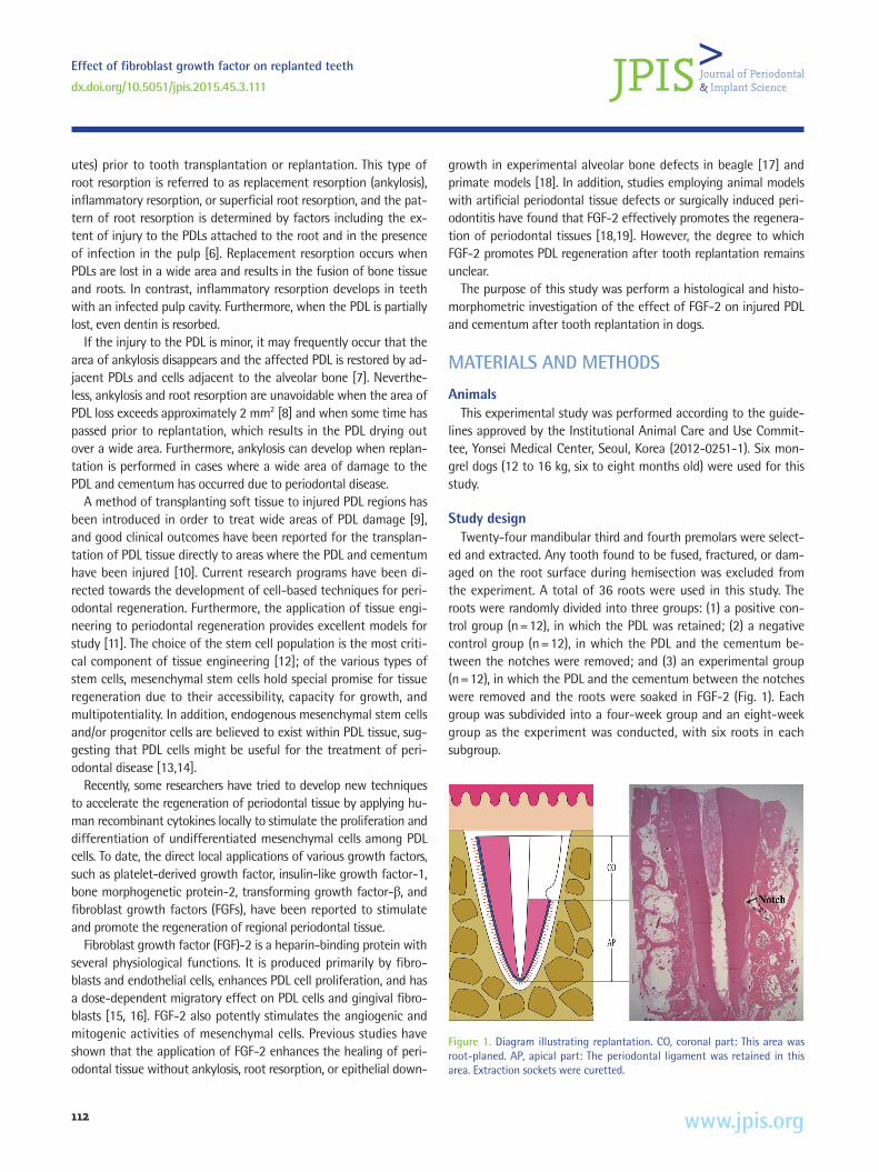
performed. During the surgical procedures, animals were placed under general anesthesia by Gerolan (Choongwae Pharmaceutical, Seoul, Korea) inhalation along with an intravenous injection of Zo-letil (5 mg/kg; Virbac, Carros, France) and xylazine (0.2–0.5 mg/kg; Rompun, Bayer Korea, Seoul, Korea). In addition, lidocaine hydro-chloride containing 1:80,000 epinephrine was administered for lo-cal anesthesia. The third and fourth mandibular premolars were ex-tracted from the mandibles with forceps using rotary movements, after separating the roots (Fig. 2A). Four notches were formed on the coronal parts of the root surfaces at intervals of approximately 5 mm with a #1/2 round bur (Fig. 2B). In the negative control group and the experimental group, the PDL and the cementum between the notches were removed with a sharp-edged Gracey curette (Fig. 2C). The extraction sockets were also curetted. Next, the roots in the experimental group were soaked in 0.1 mL saline containing 30 μg of recombinant human FGF-2 (Genoss Institute, Suwon, Korea) for two minutes (Fig. 2D). The roots in the control group were covered with saline solution for two minutes to prevent dryness. The extrac-tion sockets were curetted. After treating the root surfaces, the ex-tracted roots were replanted into their respective sockets and the tooth crowns were severed using a carbide bur (Fig. 2E). The roots were completely covered with coronally repositioned flaps (Fig. 2F). For three days after surgery, 20 mg/kg of cefazoline sodium (Yuhan, Seoul, Korea) was injected intramuscularly, and soft foods were giv-en. One week after surgery, the sutures were removed. Oral hygiene was maintained through the weekly application of 0.2% chloro-hexidine solution (Hexamedin, Bukwang Pharmaceutical, Seoul, Ko-rea) for infection control.
Histological processingThe dogs were euthanized with an overdose of pentobarbital so-
dium (90–120 mg/kg, intravenously) at four or eight weeks. The
A B C
D E F
Figure 2. Surgical procedures. (A) The crowns and furcation areas of the premolars were severed with an apical-coronal cut using a carbide bur. (B) Notches were prepared with a round bur; (C) The periodontal ligament and cementum were removed between notches by scaling and root planing. (D) The roots were soaked in FGF-2 solution. (E) After replanting the roots, the crowns were removed; (F) The roots were covered using a coronally repositioned flap.
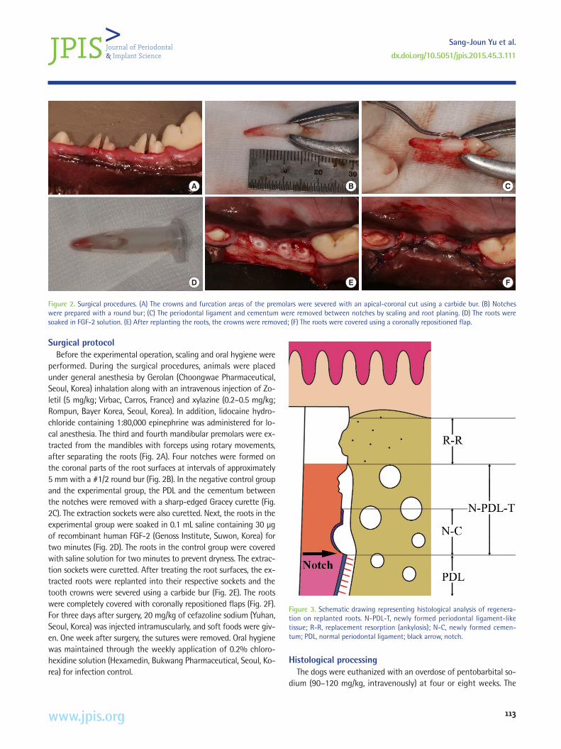
Figure 3. Schematic drawing representing histological analysis of regenera-tion on replanted roots. N-PDL-T, newly formed periodontal ligament-like tissue; R-R, replacement resorption (ankylosis); N-C, newly formed cemen-tum; PDL, normal periodontal ligament; black arrow, notch.

Effect of fibroblast growth factor on replanted teeth
dx.doi.org/10.5051/jpis.2015.45.3.111
www.jpis.org114
jaws were removed and the specimens were placed in 10% neutral buffered formaldehyde, decalcified in 5% formic acid, trimmed, and embedded in paraffin. Sections were cut in the mesiodistal plane, and serial 5-μm sections were stained with hematoxylin and eosin or Masson’s trichrome.
Histological and histomorphometric evaluationsAll measurements were obtained using a light microscope (LEICA
DM750, Wetzlar, Hesse, Germany) equipped with a digital camera (LEICA ICC50, Wetzlar, Hesse, Germany). Measurements were made
using the i-SOLUTION Lite® processing and analysis program (IMT i-Solution Corporation, Burnaby, British Columbia, Canada) on a per-sonal computer. Six randomly chosen sections from each experi-mentally treated tooth were used for morphometric evaluation. The following measurements were obtained (Fig. 3): (1) newly formed PDL-like tissue (N-PDL-T), reflecting the length of the connective tissue attached to the root-planed surface; (2) replacement resorp-tion (ankylosis) (R-R), comprising the combined longitudinal height of areas with ankylosis; and (3) newly formed cementum (N-C), re-flecting the longitudinal length of the regenerated cementum or
A B
C D
Figure 4. Histological observations of specimens retrieved at 4 weeks after surgery (positive control group, A; negative control group, B; experimental group, C-D) (H&E stained). (A) Well-preserved periodontal ligaments were observed. In the coronal root portion, some resorption cavities were found in the root surfaces (original magnification 40×). (B) Replacement resorption at the root surface was observed. Resorbed surfaces were in direct contact with newly formed bone (original magnification 40×). (C) Newly formed peri-odontal ligament (PDL)-like tissue was observed between the root surfaces and newly formed bone in the notches. In regions coronal to the notches, newly formed cementum-like tissue surfaces were observed on the dentin surfaces (original magnification 40×). (D) Newly formed cementum-like tis-sue was observed at the dentin surface. (original magnification 200×). Ab, alveolar bone; De, dentin; Ce, cementum; Pd, periodontal ligament; nCe, newly formed cementum-like tissue; nPd, newly formed periodontal liga-ment-like tissue; black asterisk, surface resorption; white asterisk, ankylosis (replacement resorption).
A B
C D
Figure 5. Histological observations of specimens retrieved at 8 weeks after surgery (positive control group, A; negative control group, B; experimental group, C-D) (H&E stain). (A) The periodontal ligament typically extended to the apical portions of the roots (original magnification ×40). (B) Large areas of replacement resorption and superficial resorption were observed. The peri-odontal ligament was completely replaced by bone (original magnification 40×). (C) Newly formed periodontal ligament (PDL)-like tissue formed be-tween the root surface and new bone in the area coronal to the notch (origi-nal magnification 40×). (D) Spaces in the alveolar bone and root surface were filled with newly formed PDL-like tissue. Root surfaces exhibited new cementum-like tissue formation (original magnification 200×). Ab, alveolar bone; De, dentin; Ce, cementum; Pd, periodontal ligament; nCe, newly formed cementum-like tissue; nPd, newly formed periodontal ligament-like tissue; black asterisk, surface resorption; white asterisk, ankylosis (replace-ment resorption).
Sang-Joun Yu et al.
dx.doi.org/10.5051/jpis.2015.45.3.111
www.jpis.org 115
cementum-like deposits on roots. The values of N-PDL-T and R-R were expressed as percentages of planed root lengths, and values of N-C were expressed in percentages of the N-PDL-T lengths. The re-sults are presented in the format of mean±standard deviation.
Statistical analysisThe mean and standard deviation for each measurement were
calculated for each group. The Kruskall-Wallis test, the Mann-Whit-ney U test, and the Bonferroni correction were used to assess whether the differences between the control groups and the exper-imental groups were statistically significant. P-values < 0.05 were considered to indicate statistical significance, and the analysis was performed using SPSS version 17.0 (SPSS Inc., Chicago, IL, USA).
RESULTS
Clinical observationsEight weeks after surgery, the extraction wounds had healed and
were covered by healthy mucosa, although gingival recession was observed in one root of the positive control group at eight weeks and one root of the experimental group at four weeks. Extensive inflammatory root resorption also occurred in these roots.
Histologic observationsThe positive control group
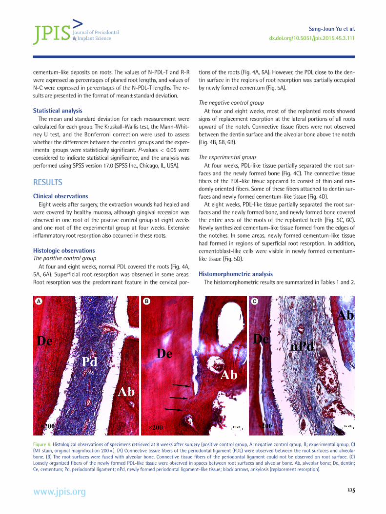
At four and eight weeks, normal PDL covered the roots (Fig. 4A, 5A, 6A). Superficial root resorption was observed in some areas. Root resorption was the predominant feature in the cervical por-
tions of the roots (Fig. 4A, 5A). However, the PDL close to the den-tin surface in the regions of root resorption was partially occupied by newly formed cementum (Fig. 5A).
The negative control groupAt four and eight weeks, most of the replanted roots showed
signs of replacement resorption at the lateral portions of all roots upward of the notch. Connective tissue fibers were not observed between the dentin surface and the alveolar bone above the notch (Fig. 4B, 5B, 6B).
The experimental groupAt four weeks, PDL-like tissue partially separated the root sur-
faces and the newly formed bone (Fig. 4C). The connective tissue fibers of the PDL-like tissue appeared to consist of thin and ran-domly oriented fibers. Some of these fibers attached to dentin sur-faces and newly formed cementum-like tissue (Fig. 4D).
At eight weeks, PDL-like tissue partially separated the root sur-faces and the newly formed bone, and newly formed bone covered the entire area of the roots of the replanted teeth (Fig. 5C, 6C). Newly synthesized cementum-like tissue formed from the edges of the notches. In some areas, newly formed cementum-like tissue had formed in regions of superficial root resorption. In addition, cementoblast-like cells were visible in newly formed cementum-like tissue (Fig. 5D).
Histomorphometric analysisThe histomorphometric results are summarized in Tables 1 and 2.
Figure 6. Histological observations of specimens retrieved at 8 weeks after surgery (positive control group, A; negative control group, B; experimental group, C) (MT stain, original magnification 200×). (A) Connective tissue fibers of the periodontal ligament (PDL) were observed between the root surfaces and alveolar bone. (B) The root surfaces were fused with alveolar bone. Connective tissue fibers of the periodontal ligament could not be observed on root surface. (C) Loosely organized fibers of the newly formed PDL-like tissue were observed in spaces between root surfaces and alveolar bone. Ab, alveolar bone; De, dentin; Ce, cementum; Pd, periodontal ligament; nPd, newly formed periodontal ligament-like tissue; black arrows, ankylosis (replacement resorption).
A B C

Effect of fibroblast growth factor on replanted teeth
dx.doi.org/10.5051/jpis.2015.45.3.111
www.jpis.org116
One root of the positive control group at eight weeks and one root of the experimental group at four weeks were excluded from the analysis because they displayed extensive inflammatory root re-sorption. Ankylosis was observed in part of the positive control group. However, four weeks after surgery, the mean length of the PDL tissue on the roots of the positive control group was signifi-cantly greater than that of the newly formed PDL-like tissue on the roots treated with FGF-2. At four and eight weeks after surgery, the mean length of newly formed cementum-like tissue on the roots treated with FGF-2 was significantly greater than on the pos-itive control roots (four weeks, P=0.010; eight weeks, P=0.027) and the negative control roots (four weeks, P=0.008; eight weeks, P=0.042) (Tables 1, 2). The mean length of newly formed PDL-like tissue on the roots treated with FGF-2 was greater than on the negative control roots, and the mean length of replacement re-sorption on the roots treated with FGF-2 was lower than on the
negative control roots. However, no significant differences were observed between the roots treated with FGF-2 and the negative control roots with respect to newly formed PDL-like tissue and re-placement resorption (Tables 1, 2).
DISCUSSION
Root resorption is the major complication after tooth transplan-tation or replantation. Of the three categories of root resorption, replacement resorption is the most irreversible because it is a com-ponent of the physiological process of normal bone remodeling. Furthermore, ankylosis is largely responsible for the low five-year survival of teeth after tooth injury [20], and for this reason, it is im-portant to prevent ankylosis in replanted teeth. In the present study, the PDL and cementum in the coronal portion of replanted roots were removed to model root surface damage. After healing,
Table 1. Histomorphometric evaluations of periodontal healing patterns at four weeks after surgery (mean±standard deviation).
Groups PDL / N-PDL-T (%) R-R (%) N-C (%)
Control (+) (n=6) 92.1±12.6 8.3±13.5 32.1±21.4
Control (−) (n=6) 34.2±27.8 65.8±27.8 11.5±16.8
Experimental (FGF-2) (n=5) 64.7±10.7 34.7±11.0 81.1±22.5a)
P-value (positive control vs. negative control vs. FGF-2) 0.004b) 0.005b) 0.006b)
P-value (positive control vs. FGF-2) 0.009c), d) 0.015c), d) 0.010c), d)
P-value (negative control vs. FGF-2) 0.100 0.100 0.008c), d)
P-value (positive control vs. negative control) 0.006c), d) 0.006c), d) 0.161
Control (+), positive control roots; control (−), negative control roots; experimental (FGF-2), fibroblast growth factor-2 (FGF-2)-treated roots; PDL, periodontal ligament tissue (in the positive group); N-PDL-T, newly formed periodontal ligament-like tissue (in the negative group and experimental group); R-R, replace-ment resorption (ankylosis); N-C, newly formed cementum.a)Statistically significant difference between the negative control and experimental groups at the same week (P<0.05) (Mann-Whitney U test).b)Statistically significant difference (P<0.05) (Kruskall-Wallis test).c)Statistically significant difference (P<0.05) (Mann-Whitney U test).d)Statistically significant difference (P<0.017) (Bonferroni correction).
Table 2. Histomorphometric evaluations of periodontal healing patterns at eight weeks after surgery (mean ± standard deviation).
Groups PDL / N-PDL-T (%) R-R (%) N-C (%)
Control (+) (n=5) 88.2±26.4 11.8±26.4 11.5±17.7
Control (−) (n=6) 27.9±22.7 72.1±22.7 14.1±27.0
Experimental (FGF-2) (n=6) 63.2±30.0 36.9±30.0 55.0±37.2a)
P-value (positive control vs. negative control vs. FGF-2) 0.020b) 0.020b) 0.037b)
P-value (positive control vs. FGF-2) 0.126 0.126 0.027c)
P-value (negative control vs. FGF-2) 0.055 0.055 0.042c)
P-value (positive control vs. negative control) 0.015c), d) 0.015c), d) 1.000
Control (+), positive control roots; control (−), negative control roots; experimental (FGF-2), fibroblast growth factor-2 (FGF-2)-treated roots; PDL, periodontal ligament tissue (in the positive group); N-PDL-T, newly formed periodontal ligament-like tissue (in the negative group and experimental group); R-R, replace-ment resorption (ankylosis); N-C, newly formed cementum.a)Statistically significant difference between the negative control and experimental groups at the same week (P<0.05) (Mann-Whitney U test).b)Statistically significant difference (P<0.05) (Kruskall-Wallis test).c)Statistically significant difference (P<0.05) (Mann-Whitney U test).d)Statistically significant difference (P<0.017) (Bonferroni correction).
Sang-Joun Yu et al.
dx.doi.org/10.5051/jpis.2015.45.3.111
www.jpis.org 117
roots treated with FGF-2 were found to have shown favorable heal-ing with respect to newly formed cementum-like tissue. Further-more, FGF-2 induced the formation of newly formed PDL-like tissue in previously denuded coronal areas. However, the roots in the neg-ative control group frequently showed ankylosis. This pattern of fa-vorable healing and reduction in replacement resorption (ankylosis) in teeth treated with FGF-2 suggests that FGF-2 effectively pro-motes the regeneration of the injured PDL and cementum.
Many attempts have been made to prevent ankylosis and to en-courage the production of new PDL tissue and newly formed ce-mentum tissue after the transplantation or replantation of teeth. Seshima et al. [21] reported that the incidence of ankylosis in their FGF-2 treatment group was significantly lower than in the control group (3.6%±4.0% of total root length versus 25.4%±1.0% of total root length; P<0.01) at eight weeks after surgery. Shiratani et al. [22] reported that roots treated with FGF-2 exhibited the formation of new PDL-like tissue and new cementum with inserting collagen fi-bers after delayed transplantation, and Sato et al. [23] reported that teeth treated with 1 μɡ of basic FGF in collagen gel exhibited the formation of dense fibers bound to alveolar bone and new cemen-tum at eight weeks. Similarly, the histomorphometric analysis con-ducted in the present study showed that the amount of replacement resorption in the FGF-2-treated group was less than in the negative control group at four and eight weeks (four weeks, 34.7%±11.0% of the denuded surface of the roots versus 65.8%±27.8% of the denuded surface of the roots; eight weeks, 36.9%±30.0% of the denuded surface of the roots versus 72.1%±22.7% of the denuded surface of roots). No significant difference was observed between the FGF-2-treated group and the negative control group. However, the amount of newly formed cementum-like tissue in the FGF-2-treated group was significantly greater than in the negative con-trol group (four weeks, 81.1%±22.5% of the denuded surface of the roots versus 11.5%±16.8% of the denuded surface of the roots; eight weeks, 55.0%±37.2% of the denuded surface of the roots versus 14.1%±27.0% of the denuded surface of the roots, P<0.05). Although the amounts of newly formed cementum-like tissue in the FGF-2-treated group at four and eight weeks were not signifi-cantly different, these findings enable us to confirm that FGF-2 cy-tokine therapy prevents ankylosis and promotes cementogenesis and the regeneration of PDL-like tissues on injured root surfaces.
However, the proportion of roots showing ankylosis in the nega-tive control group and the experimental group of this study was considerably higher than in other studies. Moreover, the width of the newly formed PDL-like tissue on roots treated with FGF-2 was less than that observed in other studies. Three reasons for this dif-ference may be suggested. First, a lower amount of remaining PDL was present, because curettage of the remaining PDL tissue was performed on extraction sockets and on experimental roots. Sec-ond, the distances between the root surfaces and the extraction sockets were small because replantation was performed without further reduction of the extraction sockets. Third, the roots were submerged to prevent exposure to occlusal forces, and thus, the
activities of PDL tissue might have been blocked.The best method for increasing the amount of remaining PDL is
to preserve vital PDL cells on the root surfaces and the extraction socket walls. In order to maintain the viability of the PDL cells, the extra-alveolar time of an avulsed tooth should be less than five minutes prior to replantation [24]. Soder et al. [25] reported that the number of visible PDL cells decreases in proportion to extra-al-veolar time, and Andreasen [4] reported a greater incidence of re-placement resorption in teeth with an extra-alveolar time of 18 minutes versus 0 minutes. However, Iqbal and Bamaas [26] reported no significant observable differences in the incidence of root re-sorption for extra-alveolar times of 15, 30, and 60 minutes, but a decreased incidence of periodontal healing was found correspond-ing to increased extraoral dry time. In the present study, the roots were replanted within five minutes to maintain PDL cell vitality. This explains why intact PDL tissue was found on a large proportion of the root surfaces in the positive control group. However, we also found that short-term replantation had no effect on the regenera-tion of injured PDL and cementum in the negative control group. Although it is important to maintain the viability of PDL cells dur-ing short-term replantation, the histologic results of FGF-2-treated roots showed that a signaling factor, such as FGF-2, was required for the regeneration of injured PDL and cementum.
The ability of storage media to maintain tooth viability during transplantation and replantation is considered to be more impor-tant than extra-alveolar time [27,28]. Different types of wet storage media, such as Hank’s balanced salt solution, minimum essential medium, saline, water, saliva, bovine milk, propolis, and green tea, have been investigated [29]. In the present study, saline was used to avoid tooth dehydration. However, although saline is a physiologi-cal match in terms of osmolality and pH, it does not contain essen-tial ions and glucose, which are fundamental requirements of cells [30, 31]. Moreira-Neto et al. [32] evaluated the viability of cultured cells in saline, finding that 55% of cells remained alive after storage in saline for four hours. Consequently, saline is not a suitable medi-um for long-term storage, but is probably suitable for short periods of time, as demonstrated by the fact that intact PDL tissue was ob-served in many areas in the positive control group of this study.
The distance between recipient bone tissue and the root surface of a transplanted tooth is another important consideration. Optimal contact with the recipient site can improve the supply of blood and nutrients to PDL cells, thereby improving success rates after tooth transplantation [33]. In fact, good blood supply has been shown to be important for wound healing [34]. Furthermore, optimal contact between donor teeth and recipient bone ensures immobilization. However, to our knowledge, no study has been performed to deter-mine the optimal distance between transplanted root surfaces and alveolar bone during tooth transplantation. In an effort to provide better blood supply and make the seating of donor teeth easier, Nethander [35] advocated a two-stage transplantation procedure. Promising results were obtained after a follow-up period of up to five years, and it was found that this two-stage surgical technique
Effect of fibroblast growth factor on replanted teeth
dx.doi.org/10.5051/jpis.2015.45.3.111
www.jpis.org118
made it possible to transplant autogenous teeth with little risk of root resorption or other complications. For transplantation, it has been suggested that the donor site should be made 1–2 mm wider than the transplanted tooth in order to preserve the PDL [36]. Hence, in the present study, a small gap between the root surface and the socket wall promoted higher rates of replacement resorp-tion, because the osteogenic action of granulation tissue on the surface of the extraction socket wall is activated soon after the tooth is extracted.
Furthermore, in the present study, tooth crowns were removed and the roots were submerged for protection. However, several re-ports have suggested that masticatory stimulation during the heal-ing period may activate the function of the periodontal membrane area, promote the proliferation of PDL cells, and thus improve the prognosis of transplantation [37,38]. Yang et al. [39] concluded that the application of orthodontic forces promotes PDL healing and possibly prevents dentoalveolar ankylosis. However, they did recom-mend a rest period of at least two weeks before loading the trans-plants, because the outcomes of regenerative procedures are criti-cally dependent on initial wound stability [24]. This fact shows that complete coverage by a flap may have a beneficial influence on the wound healing of the PDL and cementum during the initial two weeks, while subsequently disrupting the stimulation of PDL cells. This may explain our finding that N-PDL-T did not increase and R-R did not decrease from four to eight weeks.
Moreover, in the present study, the application of FGF-2 did not completely prevent replacement resorption, and more instances of ankylosis were observed in roots treated with FGF-2 than has been reported in other experiments [21,22]. Nonetheless, the results of this study clearly show that FGF-2 stimulates adjacent PDL tissue on the root surface and exerts a positive influence on the regener-ation of the PDL and cementum.
FGF-2 enhances PDL cell proliferation, but inhibits cell differenti-ation by downregulating intracellular alkaline phosphatase activity [17]. However, the effects of FGF-2 on hard tissue formation and mineralization are probably cell-type dependent [40]. In addition, FGF-2 plays a significant role in the migration and proliferation of PDL fibroblasts, and their migration into areas of healing is key dur-ing the initial phase of periodontal regeneration. Murakami et al. [19] reported that FGF-2 expression could be detected in immature granulation tissue one week after flap surgery, and suggested that the aggregates formed by FGF-2 during the early phase of wound healing constituted a favorable environment for periodontal cell proliferation.
In present study, the standard deviation was high because the sample size was small, and healing dynamics vary considerably among individuals. Further studies are required with larger samples.
In summary, the histological and histomorphometric results ob-tained in the present study demonstrate that the use of FGF-2 re-duces ankylosis and promotes the formation of newly formed peri-odontal tissue on injured PDLs and cementum after tooth replanta-tion in dogs.
CONFLICT OF INTEREST
No potential conflict of interest relevant to this article was re-ported.
ACKNOWLEDGEMENTS
This study was supported by a grant of the Korea Health tech-nology R&D Project, Ministry of Health & Welfare, Republic of Ko-rea. (A101578).
ORCID
Sang-Joun Yu http://orcid.org/0000-0001-8818-549XJung-Seok Lee http://orcid.org/0000-0003-1276-5978Ui-Won Jung http://orcid.org/0000-0001-6371-4172Joo-Cheol Park http://orcid.org/0000-0002-3162-7557Byung-Ock Kim http://orcid.org/0000-0001-8952-617XSeong-Ho Choi http://orcid.org/0000-0001-6704-6124
REFERENCES
1. Kim E, Jung JY, Cha IH, Kum KY, Lee SJ. Evaluation of the progno-sis and causes of failure in 182 cases of autogenous tooth trans-plantation. Oral Surg Oral Med Oral Pathol Oral Radiol Endod 2005;100:112-9.
2. Temmerman L, De Pauw GA, Beele H, Dermaut LR. Tooth trans-plantation and cryopreservation: state of the art. Am J Orthod Dentofacial Orthop 2006;129:691-5.
3. Lee EU, Lim HC, Lee JS, Jung UW, Kim US, Lee SJ, et al. Delayed intentional replantation of periodontally hopeless teeth: a retro-spective study. J Periodontal Implant Sci 2014;44:13-9.
4. Andreasen JO. Effect of extra-alveolar period and storage media upon periodontal and pulpal healing after replantation of mature permanent incisors in monkeys. Int J Oral Surg 1981;10:43-53.
5. Löe H, Waerhaug J. Experimental replantation of teeth in dogs and monkeys. Arch Oral Biol 1961;3:176-84.
6. Andreasen JO. Experimental dental traumatology: development of a model for external root resorption. Endod Dent Traumatol 1987;3:269-87.
7. Beertsen W, McCulloch CA, Sodek J. The periodontal ligament: a unique, multifunctional connective tissue. Periodontol 2000 1997; 13:20-40.
8. Andreasen JO. Relationship between cell damage in the peri-odontal ligament after replantation and subsequent develop-ment of root resorption. A time-related study in monkeys. Acta Odontol Scand 1981;39:15-25.
9. Andreasen JO, Kristerson L. Evaluation of different types of auto-transplanted connective tissues as potential periodontal ligament substitutes. An experimental replantation study in monkeys. Int J Oral Surg 1981;10:189-201.
10. Akbay A, Baran C, Günhan O, Ozmeriç N, Baloş K. Periodontal re-

Sang-Joun Yu et al.
dx.doi.org/10.5051/jpis.2015.45.3.111
www.jpis.org 119
generative potential of autogenous periodontal ligament grafts in Class II furcation defects. J Periodontol 2005;76:595-604.
11. Modino SA, Sharpe PT. Tissue engineering of teeth using adult stem cells. Arch Oral Biol 2005;50:255-8.
12. Hynes K, Menicanin D, Gronthos S, Bartold PM. Clinical utility of stem cells for periodontal regeneration. Periodontol 2000 2012; 59:203-27.
13. Melcher AH. On the repair potential of periodontal tissues. J Peri-odontol 1976;47:256-60.
14. Zhou Y, Hutmacher DW, Sae-Lim V, Zhou Z, Woodruff M, Lim TM. Osteogenic and adipogenic induction potential of human peri-odontal cells. J Periodontol 2008;79:525-34.
15. Okamoto T, Yatsuzuka N, Tanaka Y, Kan M, Yamanaka T, Sakamo-to A, et al. Growth and differentiation of periodontal ligament-derived cells in serum-free defined culture. In Vitro Cell Dev Biol Anim 1997;33:302-9.
16. Nishimura F, Terranova VP. Comparative study of the chemotactic responses of periodontal ligament cells and gingival fibroblasts to polypeptide growth factors. J Dent Res 1996;75:986-92.
17. Murakami S, Takayama S, Ikezawa K, Shimabukuro Y, Kitamura M, Nozaki T, et al. Regeneration of periodontal tissues by basic fi-broblast growth factor. J Periodontal Res 1999;34:425-30.
18. Takayama S, Murakami S, Shimabukuro Y, Kitamura M, Okada H. Periodontal regeneration by FGF-2 (bFGF) in primate models. J Dent Res 2001;80:2075-9.
19. Murakami S, Takayama S, Kitamura M, Shimabukuro Y, Yanagi K, Ikezawa K, et al. Recombinant human basic fibroblast growth factor (bFGF) stimulates periodontal regeneration in class II fur-cation defects created in beagle dogs. J Periodontal Res 2003;38: 97-103.
20. Humphrey JM, Kenny DJ, Barrett EJ. Clinical outcomes for perma-nent incisor luxations in a pediatric population. I. Intrusions. Dent Traumatol 2003;19:266-73.
21. Seshima F, Ota M, Kinumatsu T, Shibukawa Y, Yamada S. Effect of recombinant basic fibroblast growth factor on reimplanted teeth in beagle dogs. Oral Surg Oral Med Oral Pathol Oral Radiol Endod 2010;109:142-8.
22. Shiratani S, Ota M, Fujita T, Seshima F, Yamada S, Saito A. Effect of basic fibroblast growth factor on root resorption after delayed autotransplantation of tooth in dogs. Oral Surg Oral Med Oral Pathol Oral Radiol 2012;114:e14-21.
23. Sato Y, Kikuchi M, Ohata N, Tamura M, Kuboki Y. Enhanced cemen-tum formation in experimentally induced cementum defects of the root surface with the application of recombinant basic fibroblast growth factor in collagen gel in vivo. J Periodontol 2004;75:243-8.
24. Schwartz O, Andreasen JO. Allo- and autotransplantation of ma-ture teeth in monkeys: a sequential time-related histoquantitative study of periodontal and pulpal healing. Dent Traumatol 2002;18: 246-61.
25. Söder PO, Otteskog P, Andreasen JO, Modéer T. Effect of drying
on viability of periodontal membrane. Scand J Dent Res 1977;85: 164-8.
26. Iqbal MK, Bamaas N. Effect of enamel matrix derivative (EM-DOGAIN) upon periodontal healing after replantation of perma-nent incisors in beagle dogs. Dent Traumatol 2001;17:36-45.
27. Gopikrishna V, Thomas T, Kandaswamy D. A quantitative analysis of coconut water: a new storage media for avulsed teeth. Oral Surg Oral Med Oral Pathol Oral Radiol Endod 2008;105:e61-5.
28. Thomas T, Gopikrishna V, Kandaswamy D. Comparative evalua-tion of maintenance of cell viability of an experimental trans-port media "coconut water" with Hank's balanced salt solution and milk, for transportation of an avulsed tooth: An in vitro cell culture study. J Conserv Dent 2008;11:22-9.
29. Jung IH, Yun JH, Cho AR, Kim CS, Chung WG, Choi SH. Effect of (-)-epigallocatechin-3-gallate on maintaining the periodontal ligament cell viability of avulsed teeth: a preliminary study. J Periodontal Implant Sci 2011;41:10-6.
30. Goswami M, Chaitra T, Chaudhary S, Manuja N, Sinha A. Strate-gies for periodontal ligament cell viability: An overview. J Con-serv Dent 2011;14:215-20.
31. Malhotra N. Current developments in interim transport (storage) media in dentistry: an update. Br Dent J 2011;211:29-33.
32. Moreira-Neto JJ, Gondim JO, Raddi MS, Pansani CA. Viability of human fibroblasts in coconut water as a storage medium. Int En-dod J 2009;42:827-30.
33. Andreasen JO. Periodontal healing after replantation and auto-transplantation of incisors in monkeys. Int J Oral Surg 1981;10: 54-61.
34. Goerig AC, Nagy WW. Successful intentional reimplantation of mandibular molars. Quintessence Int 1988;19:585-8.
35. Nethander G. Periodontal conditions of teeth autogenously trans-planted by a two-stage technique. J Periodontal Res 1994;29: 250-8.
36. Northway W. Autogenic dental transplants. Am J Orthod Dento-facial Orthop 2002;121:592-3.
37. Andersson L. Dentoalveolar ankylosis and associated root resorp-tion in replanted teeth. Experimental and clinical studies in mon-keys and man. Swed Dent J Suppl 1988;56:1-75.
38. Mine K, Kanno Z, Muramoto T, Soma K. Occlusal forces promote periodontal healing of transplanted teeth and prevent dentoalve-olar ankylosis: an experimental study in rats. Angle Orthod 2005; 75:637-44.
39. Yang Y, Bai Y, Li S, Li J, Gao W, Ru N. Effect of early orthodontic force on periodontal healing after autotransplantation of per-manent incisors in beagle dogs. J Periodontol 2012;83:235-41.
40. Klagsbrun M, Smith S, Sullivan R, Shing Y, Davidson S, Smith JA, et al. Multiple forms of basic fibroblast growth factor: amino-terminal cleavages by tumor cell- and brain cell-derived acid proteinases. Proc Natl Acad Sci USA 1987;84:1839-43.