Embed Size (px)
Citation preview

PARTIAL PURIFICATION AND CHARACTERIZATION OF POLYPHENOL
OXIDASE FROM THERMOPHILIC Bacillus sp.
A Thesis Submitted to the Graduate School of Engineering and Sciences of
İzmir Institute of Technology in Partial Fulfillment of the Requirements for the Degree of
MASTER OF SCIENCE
in Biotechnology
by Melda Zeynep GÜRAY
July 2009 İZMİR
2
We approve the thesis of Melda Zeynep GÜRAY
______________________________ Assist. Prof. Dr. Gülşah ŞANLI Supervisor ______________________________ Assist. Prof. Dr. Çağlar KARAKAYA Co-Supervisor ______________________________ Prof. Dr. Ahmet YEMENİCİOĞLU Committee Member ______________________________ Assoc. Prof. Dr. Talat YALÇIN Committee Member
1 July 2009
______________________________ _______________________ Assoc. Prof. Dr. Ahmet KOÇ Prof. Dr. Hasan BÖKE Head of Biotechnology Department Dean of the Graduate School of Engineering and Sciences

ACKNOWLEDGEMENTS
First of all, I would like to express my sincere appreciation to my supervisor
Assist. Prof. Dr. Gülşah ŞANLI for her professional guidance, valuable help,
encouragement, understanding and endless patience not only throughout this study but
also for other situations.
I would like to thank to Prof. Dr. Ahmet YEMENİCİOĞLU for his beneficial
suggestions and valuable comments for this study. I am also thankful to Assoc. Prof. Dr.
Talat YALÇIN and my co-supervisor Assist. Prof. Dr. Çağlar KARAKAYA for their
helps.
I wish to express my thankfulness to my laboratory friends Erhan BAL and
Hasan Cihad TEKEDAR and my friends Ece YAPAŞAN, İrem ULUIŞIK, A. Banu
DEMİR, Elise HACIOĞLU, Beren ATAÇ, Pınar BAYDARA and G. Ozan BOZDAĞ
for their good friendship, sincere helps and supports.
Finally, I want to express my deepest gratitude and love to my parents İrfan-
Hümeyra GÜRAY and my brother Berk GÜRAY for their understanding and
encouragement that they have showed in every stages of my life, including this thesis
study. My special thanks are for Mert TAŞKINARDA, mainly for his endless love and
also for his supports. I would not be able to finish my study without these people.

iv
ABSTRACT
PARTIAL PURIFICATION AND CHARACTERIZATION OF
POLYPHENOL OXIDASE FROM THERMOPHILIC Bacillus sp.
Polyphenol oxidases are enzymes that catalyze the oxidation of phenolic
compounds using molecular oxygen. The ability of polyphenol oxidases to act on
phenolic compounds makes them highly useful biocatalysts for various biotechnological
applications. They are commonly found in animals, plants and fungi. Recent genome
analysis have shown that polyphenol oxidases are also widespread in bacterial species.
In this study, detection, partial purification and characterization of polyphenol
oxidase from thermophilic Bacillus sp., which was isolated from a geothermal region
was achieved. The samples from bacterial culture were boiled and compared with not
boiled ones in order to prove the existence of enzyme in bacterium. The existence was
also supported with the appearance of dark bands on polyacrylamide gel after staining
with catechol solution. Results of activity staining and activity measurements of
samples from intracellular and extracellular extract revealed that the enzyme was
intracellular. Partial purification was performed by acetone precipitation and gel
filtration chromatography with 35% yield and 1.24 purification fold.
Characterization studies indicated that the enzyme showed highest activity at pH
7.0 and 60ºC, was stable at temperatures between 30 and 60ºC and more than 80% of
activity was retained in the pH range of 5-8. The results of agent and metal ion effect on
enzyme activity revealed that the enzyme was totally inhibited in the presence of DTT
and sodium diethyldithiocarbamate and highly activated with copper ions whereas other
agents or metal ions did not have significant effect on activity. Km and Vmax values for
the enzyme were determined as 91mM and 2.25 ∆abs/min/ml, respectively.

v
ÖZET
POLİFENOL OKSİDAZ ENZİMİNİN TERMOFİLİK Bacillus sp.’ den
KISMİ OLARAK SAFLAŞTIRILMASI VE KARAKTERİZASYONU
Polifenol oksidazlar, moleküler oksijen varlığında fenolik bileşiklerin
oksidasyonunu katalizleyen enzimlerdir. Polifenol oksidazların fenolik bileşikler
üzerine etki edebilme yetenekleri, onları çok sayıda biyoteknolojik uygulamada
kullanışlı birer biyokatalizör yapar. Bu enzimler çoğunlukla hayvanlar, bitkiler ve
funguslarda yaygın olarak bulunurlar. Yakın zamandaki genom analizleri, polifenol
oksidazların bakteriyel türlerde de yaygın olduğunu göstermiştir.
Bu çalışmada, polifenol oksidazın jeotermal bir bölgeden izole edilmiş olan
termofilik Bacillus suşundan bulunması, kısmi saflaştırılması ve karakterizasyonu
gerçekleştirilmiştir. Polifenol oksidaz enziminin termofilik Bacillus suşundaki varlığını
kanıtlamak amacıyla, bakteri kültüründen alınan örnekler kaynatılmış ve kaynatılmamış
olanlar ile karşılaştırılmıştır. Enzimin bakterideki varlığı, poliakrilamit jel üzerinde
katekol solüsyonu ile boyama sonrası koyu renkli bantların belirmesi ile de
desteklenmiştir. Hücre içi ekstraktan ve bakterinin büyüme ortamından alınan
örneklerin spektrofotometrik aktivite ölçüm ve poliakrilamit jelde aktivite boyama
sonuçları, enzimin hücre içi olduğunu ortaya çıkarmıştır. Enzimin kısmi saflaştırılması,
aseton çöktürmesi ve jel filtrasyon kromatografisi ile iki basamakta gerçekleştirilmiştir.
Saflaştırma sonrası verim ve saflık katsayısı sırasıyla %35 ve 1.24’ tür.
Katekol substratı kullanılarak yapılan karakterizasyon çalışmaları enzimin pH
7.0 ve 60ºC’ de en yüksek aktiviteye sahip olduğunu göstermiştir. Enzim, 30 ve 60ºC
arası sıcaklıklarda stabildir ve pH 5-8 aralığında aktivitesinin %80’ inden fazlasını
korumuştur. Enzim aktivitesi üzerine bazı etmenlerin ve metal iyonlarının etkisi
incelendiğinde; sodyum dietilditiyokarbamat ve DTT’nin varlığında aktivite tamamen
inhibe olmuş ve bakır iyonları ile yüksek derecede stimüle olmuşken diğer iyonların ve
etmenlerin aktivite üzerinde önemli bir etkisi görülmemiştir. Enzimin kinetik
parametleri olan Km ve Vmax değerleri sırasıyla 91mM ve 2.25 ∆abs/min/ml olarak
bulunmuştur.

vi
TABLE OF CONTENTS
LIST OF FIGURES ....................................................................................................... viii
LIST OF TABLES........................................................................................................... ix
CHAPTER 1. INTRODUCTION ..................................................................................... 1
1.1. Phenols .................................................................................................. 1
1.2. Polyphenol Oxidases ............................................................................. 2
1.2.1. Tyrosinase........................................................................................ 2
1.2.2. Catechol Oxidase............................................................................. 3
1.2.3. Laccase ............................................................................................ 4
1.3. Biological Function and Distribution of Polyphenol Oxidases ............. 6
1.3.1. Bacterial Polyphenol Oxidases........................................................ 7
1.4. Industrial Applications of Polyphenol Oxidases ................................... 9
1.5. Thermophiles....................................................................................... 10
1.5.1. Thermophilic Enzymes.................................................................. 12
1.5.2. Applications of Thermophiles in Biotechnology and Industry ..... 15
1.5.3. Thermophilic Bacillus ................................................................... 16
CHAPTER 2. MATERIALS AND METHODS ........................................................... 17
2.1. Materials .............................................................................................. 17
2.2. Bacterial Strain and Growth Conditions.............................................. 17
2.3. Determination of Protein Concentration ............................................. 17
2.4. Evidence for Polyphenol Oxidase Activity in
Thermophilic Bacillus sp ..................................................................... 18
2.5. Determination of Enzyme Activity ..................................................... 18
2.6. Preparation of Crude Enzyme Extract ................................................. 19
2.7. Enzyme Purification Procedure........................................................... 19
2.8. Electrophoretic Studies and Activity Staining .................................... 20
2.8.1. SDS-PAGE.................................................................................... 21
2.8.2. Native-PAGE and Activity Staining ............................................. 22
vii
2.9. Characterization Studies...................................................................... 22
2.9.1. Kinetic Analysis ............................................................................ 22
2.9.2. Effect of pH on Enzyme Activity and Stability............................. 23
2.9.3. Effect of Temperature on Enzyme Activity and Stability ............. 23
2.9.4. Effect of Metal Ions on Enzyme Activity...................................... 23
2.9.5. Effect of Various Agents on Enzyme Activity.............................. 24
2.9.6. Substrate Specificity of Enzyme ................................................... 24
CHAPTER 3. RESULTS AND DISCUSSION.............................................................. 25
3.1. Evidence for Polyphenol Oxidase Activity ......................................... 25
3.2. Partial Purification of Polyphenol Oxidase ......................................... 26
3.3. Electrophoretic Studies and Activity Staining .................................... 28
3.3.1. SDS-PAGE.................................................................................... 28
3.3.2. Native-PAGE and Activity Staining ............................................. 29
3.4. Characterization of Polyphenol Oxidase ............................................. 31
3.4.1. Kinetic Analysis ............................................................................ 31
3.4.2. Effect of pH on Polyphenol Oxidase Activity and Stability ......... 32
3.4.3. Effect of Temperature on Polyphenol Oxidase Activity and
Stability.......................................................................................... 34
3.4.4. Effect of Metal Ions on Polyphenol Oxidase Activity .................. 36
3.4.5. Effect of Various Agents on Polyphenol Oxidase Activity........... 37
3.4.6. Substrate Specificity of Polyphenol Oxidase ................................ 38
CHAPTER 4. CONCLUSION ....................................................................................... 39
REFERENCES ............................................................................................................... 41
APPENDICES
APPENDIX A. PREPARATION OF BRADFORD REAGENT AND PROTEIN
STANDARTS FOR BRADFORD ASSAY.......................................... 48
APPENDIX B. STANDART CURVE FOR BRADFORD ASSAY ............................. 50
APPENDIX C. REAGENTS AND GEL PREPARATION FOR SDS-PAGE .............. 51

viii
LIST OF FIGURES
Figure Page
Figure 1.1. Reaction mechanism of tyrosinase ............................................................. 3
Figure 1.2. Reaction mechanism of catechol oxidase ................................................... 3
Figure 1.3. Reaction mechanism of laccase .................................................................. 4
Figure 1.4. Some substrates of polyphenol oxidase ...................................................... 5
Figure 1.5. The universal phylogenetic tree................................................................ 11
Figure 2.1. Low pressure liquid chromatography system ........................................... 20
Figure 2.2. Gel electrophoresis system ....................................................................... 21
Figure 3.1. Activity measurements of boiled and not boiled samples ........................ 25
Figure 3.2. Gel filtration profile of polyphenol oxidase ............................................. 27
Figure 3.3. SDS-PAGE image..................................................................................... 28
Figure 3.4. Activity and colloidal coomassie staining of polyphenol oxidase
on native polyacrylamide gels .................................................................. 30
Figure 3.5. Lineweaver-Burk plot for polyphenol oxidase ......................................... 31
Figure 3.6. Effect of pH on polyphenol oxidase activity ............................................ 33
Figure 3.7. pH stability of polyphenol oxidase ........................................................... 33
Figure 3.8. Effect of temperature on polyphenol oxidase activity .............................. 34
Figure 3.9. Thermal stability of polyphenol oxidase .................................................. 35
Figure 3.10. Effect of metal ions on polyphenol oxidase activity................................. 36

ix
LIST OF TABLES
Table Page
Table 1.1. Biochemical characteristics of polyphenol oxidases from
different bacteria ............................................................................................. 7
Table 1.2. Thermophiles and their environments .......................................................... 12
Table 1.3. Bioconversion reactions and applications of thermostable
enzymes ........................................................................................................ 16
Table 3.1. Purification of polyphenol oxidase from thermophilic
Bacillus sp..................................................................................................... 26
Table 3.2. Effect of various agents on polyphenol oxidase activity .............................. 38
Table 3.3. Substrate specificity of polyphenol oxidase ................................................. 38
x
ABBREVIATIONS
ABTS 2,2’-azino-bis(3-ethylbenzthiazoline-6-sulphobic acid)
APS Ammonium persulfate
BSA Bovine serum albumin
CBB Commasie brilliant blue
DMSO Dimethyl sulfoxide
DTT Dithiothreitol
EDTA Ethylenediamine tetra acetic acid
L-DOPA 3,4-dihydroxyphenylalanine
PAGE Polyacrylamide gel electrophoresis
PPO Polyphenol oxidase
SDS Sodium dodecyl sulfate
SGZ Syringaldazine
TEMED Tetramethylethylenediamine
2xYT Yeast extract tryptone

1
CHAPTER 1
INTRODUCTION
1.1. Phenols
Phenols are a class of aromatic compounds that have a hydroxyl group directly
attached to a benzene or a benzonoid ring and the simplest member of this class,
C6H5OH, is phenol (Carey 1996).
The presence of hydroxyl group highly effects the physical properties of
phenols. The hydroxyl group enables to form strong hydrogen bonds with other phenol
molecules and with water, therefore phenols have higher boiling points and more
solubility in water than hydrocarbons of the same molecular weight (Carey 1996,
Solomons 1990).
Phenols and its derivatives are widespread in nature. Phenol itself was isolated
for the first time from coal tar in the early nineteenth century (Carey 1996). Tyrosine, an
amino acid, is found in proteins. Methyl salicylate is found in oil of wintergreen and
eugenol is found in oil of cloves. Thymol and vanillin are important components of
thyme and vanilla beans. Estradiol is a female sex hormone and tetracylines are
significant antibiotics (Solomons 1990).
Phenols are important medicinal and industrial chemicals. They are used as the
raw material in the production of a large number of commercial products like aspirin,
dyes, adhesives and plastics (Carey 1996). They were found to act as antiseptic and
disinfectant. At the same time they are found in some commercial products such as
deodorants, soaps, disinfectant sprays and oinments, first aid sprays, gargles, lozenges
and muscle rubs because of their antiseptic and anesthetic activities. However the
medical use of phenol is limited due to its skin irritaton and toxic properties. In food and
cosmetics phenols are used as antioxidants since they are oxidized instead of the
protected substance. Also some phenols are used in photography as photographic
developers (Bailey 1995).

2
1.2. Polyphenol Oxidases
Polyphenol oxidases (PPOs) are enzymes, belonging to a group of copper-
containing metalloproteins and are members of oxidoreductases, that catalyze the
oxidation of a wide range of phenolic compounds by utilizing molecular oxygen
(Queiroz, et al. 2008, Simsek and Yemenicioglu 2007). There are mainly three types of
polyphenol oxidases classified according to their substrate specificities and mechanism
of actions. These are; tyrosinase, catechol oxidase and laccase.
1.2.1. Tyrosinase
Tyrosinases (E.C. 1.14.18.1) are copper containing monooxygenases that use
molecular oxygen to catalyze two different reactions: the o-hydroxylation of
monophenols to o-diphenols which is referred to as monophenolase or cresolase activity
and the subsequent oxidation of o-diphenols to o-quinones which is referred to as
diphenolase or catecholase activity (Figure 1.1) (Claus and Decker 2006).
Tyrosinases are mainly involved in the biosynthesis of melanin and are
responsible for the first steps of melanin synthesis from L-tyrosine leading to the
formation of L-dopaquinone and L-dopachrome (Halaouli, et al. 2006).
Chemical and spectroscopic studies of tyrosinases have revealed that their active
site consists of a pair of coupled copper ions (Duran, et al. 2002). These copper ions,
CuA and CuB, each of which are coordinated by three histidine residues (His)
constitutes the site where interaction of tyrosinase with both molecular oxygen and its
phenolic substrate takes place (VanGelder, et al. 1997).

3
OH
R
1/2 O2
R
O
O
R
OH
OH
1/2 O2
H2O
monophenol o-diphenol o-quinone
Cresolase activity Catecholase activity
Figure 1.1. Reaction mechanism of tyrosinase
1.2.2. Catechol Oxidase
Catechol oxidases (E.C. 1.10.3.1), which are also known as o-diphenol oxidases,
catalyze the oxidation of o-diphenols to the corresponding o-quinones by molecular
oxygen (Figure 1.2). Similar to tyrosinases, the active site of catechol oxidases contains
a pair of coupled copper ions and this feature makes catechol oxidases belong to the
group of type-3 copper proteins which also includes tyrosinases and heamocyanins.
Unlike tyrosinases, these enzymes lack hydroxylase activity hence can not use
monophenols (like tyrosine and cresol) as substrate (Guell and Siegbahn 2007).
OH
R
1/2 O2
O
O
R
H2O
o-quinoneo-diphenol
OH
Figure 1.2. Reaction mechanism of catechol oxidase

4
1.2.3. Laccase
Laccases (E.C. 1.10.3.2) are multicopper enzymes that are capable of oxidizing
various aromatic compounds by a radical catalyzed reaction mechanism in the presence
of oxygen (Figure 1.3). They are a family of blue-multicopper oxidases which comprise,
beside laccase, plant ascorbate oxidase, the mammalian plasma protein ceruloplasmin
and bilirubin oxidase (Polaina and MacCabe 2007).
Laccases contain four copper atoms in the active site which play an important
role in catalytic activity. These copper atoms are classified in three types according to
their spectroscopic and functional characteristics (Duran, et al. 2002); type 1 (or blue),
type 2 (or normal) and type 3 (or coupled binuclear) (Alexandre and Zhulin 2000).
OH
HO
R
p-diphenol
1/2 O2
O
O
p-quinone
R
Figure 1.3. Reaction mechanism of laccase
In literature; the name polyphenoloxidase is used to summarize tyrosinases,
catechol oxidases and laccases. Although these three types of enzymes have the ability
to oxidize an overlapping range of phenolic compounds, they are differentiated on the
basis of their substrate specificities, polypeptidic copper binding sites and sensitivity to
inhibitors. Concerning substrate specificity; the main difference between these enzymes
is that only tyrosinase can show cresolase activity and oxidize monophenols like L-
tyrosine and only laccase have the ability to oxidize methoxy-activated phenols such as
syringaldazine and 2,6-dimethoxyphenol (Sanchez-Amat, et al. 2001). Also laccase
shows more affinity for the oxidation of p-diphenols like hydroquinone than o-
diphenols like catechol (Solano, et al. 1997). Concerning copper binding sites; while the
active site of tyrosinase and catechol oxidase contains a pair of coupled copper ions

5
(type-3), the active site of laccase contains three different types of four copper ions
(one type-1, one type-2 and a pair of type-3) (Hernandez-Romero, et al. 2006) .
Concerning inhibition, tyrosinase and catechol oxidase activity is inhibited by
tropolone, cinnamic acid and salicylhydroxamic acid (Fernandez, et al. 1999). However
the laccase activity is not effected much by these chemicals, instead it is strongly
inhibited by azide, cyanide, thiocyanide, flouride and sulfhydryl reagents (Polaina and
MacCabe 2007). Some phenolic substrates that are oxidized by polyphenol oxidases are
shown in Figure 1.4
Figure 1.4. Some substrates of polyphenol oxidase

6
1.3. Biological Function and Distribution of Polyphenol Oxidases
Polyphenol oxidases are widely distributed in nature. They can be found in
almost all living organisms including animals, plants, fungi and bacteria.
In animals including humans, tyrosinase is mostly found and involves in the
pigmentation of skin, hair and eye by melanin synthesis. In humans, the too much or
insufficient activity of this enzyme causes a number of important diseases such as
albinism, vitiligo and melanoma. Polyphenol oxidase activity is also found in insects
where they are responsible for the exoskeleton formation (Halaouli, et al. 2006).
In plants, all types of polyphenol oxidases are found. Tyrosinase activity has
important roles in metabolism of plants including wound healing system (Kong, et al.
2000). When a plant gets a bruise, cut or damage, the enzyme oxidizes some phenolic
compounds to form a polymer structure in order to protect the plant against insects or
microorganisms (VanGelder, et al. 1997) and causes enzymatic browning of fruits and
vegetables which is most cases undesirable as it causes loss of quality. Besides
tyrosinases, also laccases have important roles in plants. They participate in lignification
process of plant cell walls since they are able to cross-link phenolic monomers (Claus
2004).
In fungi polyphenol oxidases, particularly laccases play role in lignin
degradation, fungal spore formation and pigmentation, detoxification of toxic
compounds, pathogenesis (fungal virulence factors) and fungal morphogenesis
(Ruijssenaars and Hartmans 2004).
To date polyphenol oxidases were mostly identified and studied from eukaryotic
sources. Nevertheless, homology searches in protein databases and experimental data
have shown that polyphenol oxidases are also widespread in bacteria (Ruijssenaars and
Hartmans 2004). On the other hand little attention has been paid to bacterial polyphenol
oxidases and the function of the enzyme in these organisms is not fully understood. The
best documented function of polyphenol oxidase in bacteria is the formation of
melanins. Melanin, a polyphenolic pigment, protects the bacterial spores and cells
against oxidants, free radicals and UV radiation (Claus and Decker 2006, Dalfard, et al.
2006).

7
1.3.1. Bacterial Polyphenol Oxidases
With the discovery of polyphenol oxidases in bacterial species, the studies of
them from these new sources have picked up in order to find more species that exhibit
polyphenol oxidase activity.
Until now relevant genes have been found in both gram-positive and gram-
negative bacteria. The first bacterial laccase was reported from Azospirillum lipoferum
(Diamantidis, et al. 2000) as a multimeric enzyme. Laccase was also found in bacterial
species living in extreme environments e.g. Aquifex aeolicus (Deckert, et al. 1998).
Bacterial tyrosinases were first purified from Streptomyces species (Claus and Decker
2006). Streptomyces tyrosinases are well characterized and studied. Besides, polyphenol
oxidases are found in a number of Bacillus species. Some biochemical characteristics
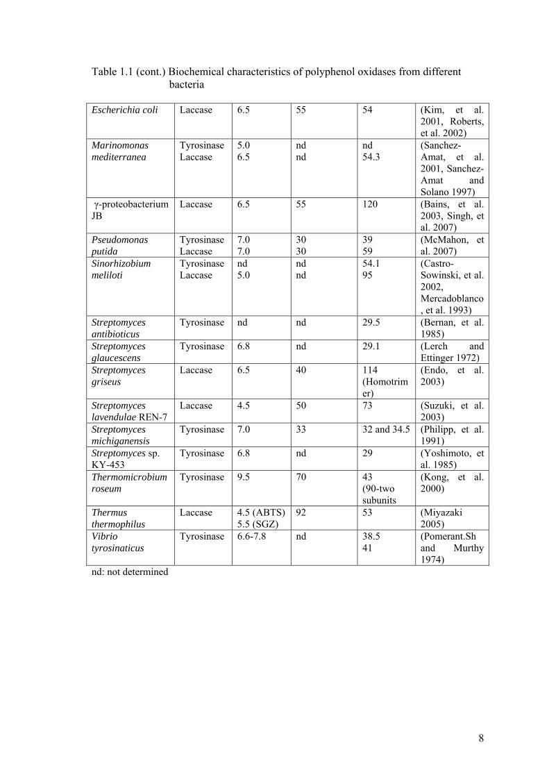
and activity of polyphenol oxidases from different bacteria can be seen in Table 1.1
Table 1.1. Biochemical characteristics of polyphenol oxidases from different bacteria
Species Type of PPO
Optimum pH
Optimum temperature
(ºC)
Molecular mass (kDa)
References
Aquifex aeolicus Laccase nd nd 59.3 (Deckert, et al. 1998)
Azospirillum lipoferum
Laccase 6.0 nd 48.9, 97.8, and 179.3 (Multimeric)
(Diamantidis, et al. 2000)
Bacillus halodurans
Laccase 7.5-8 nd 56 (Ruijssenaars and Hartmans 2004)
Bacillus licheniformis
Laccase 4.2 85 65 (Koschorreck, et al. 2008)
Bacillus subtilis Laccase 3.0 (ABTS) 7.0 (SGZ)
75 65 (Martins, et al. 2002)
Bacillus sphaericus
Laccase nd nd nd (Claus and Filip 1997)
Bacillus spp. Tyrosinase nd 40 nd (Mayende, et al. 2006)
Bacillus sp. HR03 Tyrosinase Laccase
5.0 5.5
55 55
50 50
(Dalfard, et al. 2006)
Bacillus thuringiensis
Tyrosinase 9.0 75 14 (Liu, et al. 2004)
(cont. on next page)
8
Table 1.1 (cont.) Biochemical characteristics of polyphenol oxidases from different bacteria
Escherichia coli Laccase 6.5 55 54 (Kim, et al. 2001, Roberts, et al. 2002)
Marinomonas mediterranea
Tyrosinase Laccase
5.0 6.5
nd nd
nd 54.3
(Sanchez-Amat, et al. 2001, Sanchez-Amat and Solano 1997)
γ-proteobacterium JB
Laccase 6.5 55 120 (Bains, et al. 2003, Singh, et al. 2007)
Pseudomonas putida
Tyrosinase Laccase
7.0 7.0
30 30
39 59
(McMahon, et al. 2007)
Sinorhizobium meliloti
Tyrosinase Laccase
nd 5.0
nd nd
54.1 95
(Castro-Sowinski, et al. 2002, Mercadoblanco, et al. 1993)
Streptomyces antibioticus
Tyrosinase nd nd 29.5 (Bernan, et al. 1985)
Streptomyces glaucescens
Tyrosinase 6.8 nd 29.1 (Lerch and Ettinger 1972)
Streptomyces griseus
Laccase 6.5 40 114 (Homotrimer)
(Endo, et al. 2003)
Streptomyces lavendulae REN-7
Laccase 4.5 50 73 (Suzuki, et al. 2003)
Streptomyces michiganensis
Tyrosinase 7.0 33 32 and 34.5 (Philipp, et al. 1991)
Streptomyces sp. KY-453
Tyrosinase 6.8 nd 29 (Yoshimoto, et al. 1985)
Thermomicrobium roseum
Tyrosinase 9.5 70 43 (90-two subunits
(Kong, et al. 2000)
Thermus thermophilus
Laccase 4.5 (ABTS) 5.5 (SGZ)
92 53 (Miyazaki 2005)
Vibrio tyrosinaticus
Tyrosinase 6.6-7.8 nd 38.5 41
(Pomerant.Sh and Murthy 1974)
nd: not determined

9
1.4. Industrial Applications of Polyphenol Oxidases
In recent years polyphenol oxidases have garnered significant interest because of
their high capacity for oxidizing aromatic compounds. This feature makes the use of
polyphenol oxidases very suitable for some biotechnological applications in food
industry, pulp and paper industry, textile industry, medicine and environmental
technology.
In food industry, although polyphenol oxidases are undesirable for their
browning effects, they can be applied for various beneficial purposes. They can be used
in beverage processing for the elimination of phenolics which are responsible for
browning, haze formation and turbidity development in beer, wine and fruit juice.
Polyphenol oxidases, particularly laccases are currently of interest in baking since they
are able to cross-link biopolymers (Rodriguez Couto and Toca Herrera 2006). They can
be also used for the biosynthesis of antioxidants and food colorants (Simsek and
Yemenicioglu 2007). Applications of polphenol oxidases in different aspects of food
industry includes color formation and flavor enhancement of tea, cocoa and coffee,
ascorbic acid determination, sugar beet pectin gelation and as a biosensor (Polaina and
MacCabe 2007).
In pulp and paper industry, the industrial preparation of paper requires the
removal of lignin from woody tissues and pulp bleaching. Conventionally pulp
bleaching is carried out by using chlorine-based agents which causes environmental
concerns because they lead to the release of toxic contaminants. The applications of
laccases for the purposes of delignification and biobleaching were found to be succesful
since they provide cleaner and milder strategies (Rodriguez Couto and Toca Herrera
2006).
In environmental technology, the presence of hazardous phenolic compounds
and their derivatives in industrial wastewaters from coal conversion, petroleum refining,
wood preservation, textile, paper, food and chemical industries constitues a big
problem. Government legislation is becoming more stringent in developed countries for
the removal of the toxic compounds from wastewaters before they are discharged into
the environment. Recent interest has focused on the use of peroxidases and polyphenol
oxidases as an enzymatic approach for the removal of phenolics from industrial

10
effluents. Peroxidases have the ability to treat phenolic compounds over wide ranges of
pH and temperature but requires stoichiometric amounts of hydrogen peroxide. In this
aspect polyphenol oxidases appear to be more advantageous because they require only
molecular oxygen as oxidant to work (Edwards, et al. 1999).
In medical area, according to a recent research, polyphenol oxidases were found
to inhibit the adhesion of Streptococcus sobrinus, a bacteria responsible from oral cavity
formation, on tooth surface (Cowan, et al. 2000). Moreover, polyphenol oxidases can be
used for the treatment of Parkinson’s disease. By the action of polyphenol oxidase, L-
tyrosine is converted to L-DOPA that is used to supplement the insufficient amount of
dopamine in Parkinson’s disease (Asanuma, et al. 2003, Xu, et al. 1998). Polyphenol
oxidases are also of interest in clinical applications as a marker of vitiligo which is an
autoimmune disease, as a prodrug therapy agent and as a tumor-suppressing (Seo, et al.
2003).
Polyphenol oxidases find additional applications in other fields of industry. They
can be used in the development of biosensors for immunoassays, for the detection of
phenols and phenolic compounds in wastewaters, food and beverage (Duran and
Esposito 2000), for the detection of morphine, codeine and catecholamines. In
cosmetics, some hair dyes and dermatological skin lightning preparations are based on
laccase. In textile industry, they are used for the purposes of denim bleaching and dye
decolorization (Rodriguez Couto and Toca Herrera 2006).
1.5. Thermophiles
Temperature is an important environmental factor and constrains all living
organisms. Thus classification of living organisms according to their relation to
temperature is essential for biological systematics (Kristjansson 1992). Microorganisms
are therefore classified into three groups on the basis of their optimum growth
temperatures; psycrophiles (below 20ºC), mesophiles (moderate temperatures) and
thermophiles (high temperatures) (Turner, et al. 2007). Thermophiles, the
microorganisms that love heat, are further subdivided into three categories according to
their minimal and maximal growth temperatures as follows: moderate thermophiles (35-

11
70ºC) , extreme thermophiles (55-85ºC) and hyperthermophiles (75-113ºC) (Baker, et
al. 2001).
After the discovery of a thermophilic microorganism, Thermus aquaticus from
Yellow Stone National Park by Brock and his colleagues, the research on thermophiles
have gained significant interest (Kristjansson 1989). In this respect the search for new
microorganisms have been carried out and up to date the representatives of these
organisms have been isolated from high temperature water containing terrestrial and
marine habitats. The most common habitats are geothermally and volcanically heated
hydrothermal systems such as solfataric fields, neutral hot springs and submarine saline
hot vents (Horikoshi 1998).
By using 16S rRNA sequence comparisons, the universal phylogenetic tree,
which can be seen in Figure 1.5, has been constructed with a tripartite division of the
living world consisting of the domains Eucarya, Bacteria and Archaea (Andrade, et al.
1999).
Figure 1.5. The universal phylogenetic tree. Bold lines represent thermophiles. (Source: Huber and Stetter 1998)

12
Thermophiles are present within Bacteria and Archaea. While most of the
thermophilic bacteria characterized today grow below the hyperthermophilic boundary,
hyperthermophilic species are primarily archaea (Turner, et al. 2007).
In Table 1.2, bacterial and archaeal thermophiles can be seen.
Surprisingly hyperthermophiles represent all the deep short branches within the
phylogenetic tree, including the genera Aquifex and Thermotoga within the Bacteria and
Pyrodictium, Pyrolobus, Pyrobaculum, Desulfurococcus, Sulfolobus, Methanopyrus,
Thermococcus, Methanothermus and Archaeoglobus within the Archaea with the
conclusion that they are the most primitive organisms still existing (Huber and Stetter
1998).
Table 1.2. Thermophiles and their environments (Source: Hough and Danson 1999)
Phenotype Environment Typical genera
Thermophilic
55-80 ºC Methanobacterium, Thermoplasma, Thermus*, some Bacillus* species
Hyperthermophilic
80-113 ºC
Aquifex*, Archaeoglobus, Hydrogenobacter*, Methanothermus, Pyrococcus, Pyrodictium, Pyrolobus, Sulfolobus, Themococcus, Thermoproteus, Thermotoga*
* Genera of the domain Bacteria; all others are Archaea.
1.5.1. Thermophilic Enzymes
It is known that living organisms and their cell components such as nucleic
acids, proteins/enzymes and lipids are very sensitive to heat. Therefore they are killed or
denatured by heating. On the other hand the discovery of thermophiles, which can live
and survive in extremes of temperature, made them very attractive and interesting field
of study since not only these microbes themselves have the ability to thrive at elevated
temperatures and withstand heat but also their components have.

13
As it is stated, cellular components of thermophilic microorganisms have to be
thermostable in order to maintain viability. It was reported that the DNA of
thermophiles have a reverse DNA gyrase, a unique type I DNA topoisomerase, which
produces positive supercoils in the DNA and increases the melting point of DNA thus
contributes to stability at elevated temperatures. The cell membranes of thermophiles
consist of saturated fatty acids that provide a hydrophobic environment for the cell and
are responsible for the rigidity of the membrane at high temperatures. In contrast,
Archaea, which includes most of the hyperthermophiles, contains lipids linked with
ether on the cell wall. This structure possesses a greater heat resistance than a
membrane composed of fatty acids. These organisms also produce specialized proteins
called chaperonins. Chaperonins help to refold the proteins to their native form after
denaturation and maintain their functions (Haki and Rakshit 2003).
The enzymes synthesized by thermophiles and hyperthermophiles are known as
thermozymes (Li, et al. 2005). The properties of these biocatalysts have adapted to
nonstandart conditions, where their producers live optimally, in order to be active and
maintain cellular functions. Thus it is important to study and investigate the bases of
protein adaptation to high temperature so as to understand protein folding, protein
structure-function relationship, the design of high temperature biocatalysts and the
history of life on this planet (D'Auria, et al. 2000).
The enzymes having similar functions from mesophilic and thermophilic
organisms were compared to figure out the question of how thermozymes differ from
their mesophilic counterparts. According to the results of comparison studies, it was
reported that the homologous thermophilic and mesophilic enzymes are highly similar
with their: (i) amino acid sequences (40 to 85% similarity), (ii) superimposable three-
dimensional structures, and (iii) catalytic mechanisms (Vieille and Zeikus 2001). The
increasing number of three dimensional structures of thermozymes has shed some light
on their thermostability strategies. These strategies include; additional intermolecular
interactions (i.e. hydrogen bonds, electrostatic interactions, hydrophobic interactions,
disulfide bonds, metal binding), good general conformational structure (i.e. more rigid,
compact packing, reduced entropy of unfolding, conformational strain release, stability
of a-helix) and replacement of some amino acids (Iyer and Ananthanarayan 2008).
It was reported that the stability of a protein is enhanced by increasing the
number of hydrogen bonds and salt bridges in thermophilic organisms. Also it was
believed that disulfide bridges stabilize proteins (Vieille and Zeikus 2001) thus the

14
effect of them on stability was examined by mutagenesis studies. In these studies, Cys
residues were introduced in subtilisin E and an increase in the halflife and melting
temperature of the mutant enzyme was observed (Takagi, et al. 1990).
It is known that some metals stabilize and activate enzymes. According to a
study of xylose isomerase from Bacillus licheniforms, the major stabilizing forces were
found to be related with the presence of metal ions and the stability of the enzyme was
improved (Vieille, et al. 2001).
Thermophilic proteins may contain more hydrophobic cores than their
mesophilic counterparts (Gerday 2007). In molecular folding and thermostability,
hydrophobic interactions are the main contributors and thermostability of a protein is
directly correlated with its total hydrophobicity (Li, et al. 2005).
Thermozymes are more rigid than their mesophilic cousins, particularly at room
temperature. Although increased rigidity correlates with the increased thermostability,
for enzyme catalysis flexibility is necessary (Gerday 2007). For that reason
thermozymes, which are highly stable and active at elevated temperatures, are
catalytically inactive at moderate temperatures (D'Auria, et al. 2000). However the
rigidity of those enzymes at room temperature should not be overemphasized since they
may be optimally flexible at the temperatures that their producers live (Gerday 2007).
Thermophilic enzymes require specific amino acid compositions. They generally
prefer charged residues such as Glu, Arg and Lys that have the ability to provide an
increase in the formation of ion pairs and their networks (Gerday 2007). It was reported
that the most frequent amino acid exchange occuring at helical segment is Gly Ala
exchange. Since Gly is the most flexible amino acid residue and Ala is known as the
best helix inducer among all amino acids, it is not surprising that this exchange occurs
frequently as a stabilizing strategy in thermozymes. Also the content of Pro, the most
rigid amino acid, of these unusual enzymes is greater than their mesophilic relatives
(Fontana, et al. 1998).
Other important factors in the stabilization of thermozymes are: (i) increasing
molecular compactness by shortening one or more loops that have increased mobility at
high temperatures; (ii) increasing the number of buried atoms in the molecule and
elimination of unnecessary cavities; (iii) decreasing the entropy of unfolding (Vieille
and Zeikus 2001).

15
1.5.2. Applications of Thermophiles in Biotechnology and Industry
The importance and use of enzymes in many processes has been well established
for a long time. Although to date more than 3000 enzymes have been identified and
many of these are being used in industrial and biotechnological applications, the present
enzymes toolbox is still not sufficient to meet all demands (van den Burg 2003). It is
because, most of the enzymes originate from mesophilic organisms and they can not
withstand the harsh conditions of industrial processes (Hough and Danson 1999, van
den Burg 2003). Thus, characterization of thermophiles and the stable enzymes they
synthesize have been of great scientific and industrial interest for several decades.
Applied interest on thermozymes is related to the fact that their ability of
thermostability enables to operate the processes, in which they are used, at high
temperatures and also these enzymes are resistant to common protein denaturants like
detergents, proteolytic enzymes and organic solvents (Andrade, et al. 1999). The
advantages of operating industrial processes at elevated temperatures are;
• higher reaction rates,
• decrease in viscosity,
• increased diffusion rates,
• better solubility of compounds (especially polymeric substrates but
except gases),
• reduced risk of contamination (very important for food and
pharmaceutical industry) (van den Burg 2003).
Enzymes from thermophiles with specific features have considerable potential
for many industrial applications. The possible use of the thermostable properties of
thermozymes in industry has been recognized for a long time ago with the
characterization and analytical application of Taq DNA polymerase (from Thermus
aquaticus) in polymerase chain reaction, but has recently started to replace their
mesophilic relatives in industrial processes which require high temperature. The
thermophilic enzymes that have found way into several industrial applications are;
proteases, lipases, DNA-processing enzymes, polymer-degrading enzymes such as
amylases, chitinases and cellulases (van den Burg 2003) (Table 1.3).

16
Table 1.3. Bioconversion reactions and applications of thermostable enzymes (Source: Haki and Rakshit 2003)
Enzyme Temperature (ºC) Bioconversions Applications α-Amylase (bacterial) 90-100 Starch→dextrose syrups Starch hydrolysis,brewing,
baking, detergents α-Amylase (fungal) 50-60 Starch→dextrose syrups Production of maltose Pullulanase 50-60 Starch→dextrose syrups Production of glucose
syrups Xylanase 45-65, 105 Craft pulp→xylan+lignin Pulp and paper industry Chitinase 65-75 Chitin→chitobiose
Chitin→N-acetyl glucosamine N-acetyl glucosamine→glucosamine Chitin→chitosan
Food, cosmetics, pharmaceuticals, agrochemicals
Cellulase 45-55, 95 Cellulose→glucose Cellulose hydrolysis, polymer degradation in detergents
Protease 65-85 Protein→amino acids and peptides Baking, brewing, detergents, leather industry
Lipase 30-70 Fat removal, hydrolysis, interesterification, alcholysis, aminolysis
Dairy, oleo chemical, detergent, pulp, pharmaceuticals, cosmetics and leather industry
DNA polymerase 90-95 DNA amplification Genetic engineering/PCR
1.5.3. Thermophilic Bacillus
The genus Bacillus is a large and heterogenius collection of aerobic or
facultatively anaerobic, endospore-forming, rod-shaped, Gram-positive (to Gram
variable) bacteria (Yavuz, et al. 2004). The genus is widely distributed in soil, water and
air and reported to have low incidence of pathogenicity (Harwood 1989). This genus
includes many kinds of species with thermophilic, psycrophilic, acidophilic,
alkalophilic and halophilic properties (Nazina, et al. 2001).
Thermophilic bacilli, which grow optimally at high temperatures ranging from
45 to 70ºC can be isolated from thermophilic and also mesophilic environments. In
1881, the first publication on the characterization of thermophilic bacteria Bacillus
thermophilus; aerobic, spore-forming, and able to grow at 70ºC, was carried out. These
organisms have gained significant biotechnological and industrial interest due to their
ability to synthesize thermostable enzymes such as; lipases, proteases, amylases,
pullulanases, xylanases, glucose-isomerases and DNA restriction endonucleases
(Maugeri, et al. 2001).

17
CHAPTER 2
MATERIALS AND METHODS
2.1. Materials
L-DOPA was purchased from Fluka; L-tyrosine and Coomassie Brilliant Blue
G-250 were purchased from Merck. Catechol, ABTS, Sephadex G-100 gel filtration
resin and all chemicals for electrophoresis studies were purchased from Sigma Chem.
Co.
2.2. Bacterial Strain and Growth Conditions
The bacterium, thermophilic Bacillus sp., that was used in this study, was
isolated from an uncontrolled thermal leak of Balçova Geothermal Region in İzmir
(Yavuz, et al. 2004). Thermophilic Bacillus was cultivated overnight in 2xYT media, at
55ºC and 200 rpm. 2xYT (Yeast Extract Tryptone) medium consisted of 16.0g tryptone,
10.0g yeast extract and 5.0g NaCl per liter with a final pH of 7.0.
2.3. Determination of Protein Concentration
Protein contents of the samples were determined by Bradford method using
bovine serum albumin (BSA) as the standart. Bradford method is used to determine the
total protein concentration of a sample. This method is based on the shift of absorbance
maximum of the dye, Coomassie Brilliant Blue G-250, from 465 to 595 nm upon

18
protein binding. The Coomassie Brilliant Blue G-250 dye binds arginine, lysine and
histidine residues of proteins.
Composition of Bradford reagent, procedure of the assay and standart curve are
given in Appendices A and B.
2.4. Evidence for Polyphenol Oxidase Activity in Thermophilic Bacillus
sp.
In order to find out whether thermophilic Bacillus sp. exhibits polyphenol
oxidase activity or not, two sets of bacterial growth was carried out in a volume of
approximately 5ml 2xYT media as in conditions described in section 2.2. Following
bacterial growth, discontinuous sonication was applied to bacterial cultures for about 6
minutes in ice bath to disrupt cells and release cellular components including enzymes.
Then the homogenate was centrifuged at 5000rpm, 4ºC for 20 minutes. The resulting
supernatants were taken and one set was kept in boiling water for 10 minutes. The
supernatant after heat treatment was centrifuged again and supernatants of both boiled
or not boiled samples were used as enzyme solutions in activity measurements. The
activities were determined by recording the change in absorbance at 420nm with the
assay mixture containing 1.5ml supernatant (boiled or not boiled) and 1.5ml 20mM
catechol, which is a specific substrate for polyphenol oxidase, (prepared with 0.1M
sodium phosphate buffer, pH 7.0) at 55ºC. The reference cuvettes contained 1.5ml
2xYT media and 1.5ml 20mM catechol solution.
2.5. Determination of Enzyme Activity
All the spectrophotometric assays were performed using Shimadzu UV-VIS
spectrophotometer (Model 1700) with a constant temperature water circulator. The
polyphenol oxidase activity of the samples were determined at 55ºC for 20 minutes, by
recording the increase in absorbance at 420nm caused by the oxidation of catechol
19
substrate. The assay mixture (1 ml in all cases) contained 10µl 2M catechol with a final
concentration of 20mM, 955µl 0.1M sodium phosphate buffer at pH 7.0, and the
reaction was initiated by the addition of 35µl enzymatic sample. The reference cuvettes
had the same composition except for the enzyme. The enzyme activities were given as
Unit or percent initial activity and Unit is defined as the amount of enzyme that causes
0.001 absorbance change in one minute.
2.6. Preparation of Crude Enzyme Extract
Bacterial growth was carried out in erlenmeyer flask containing 100ml of 2xYT
media and in conditions as described in section 2.2. Bacterial culture was centrifuged at
5000rpm, 4ºC for 20 minutes to obtain culture supernatant and harvest bacterial cells.
The cell pellet was then resuspended in 10ml of 0.05M sodium phosphate buffer, pH
7.0, and the cells were disrupted by discontinuous sonication in ice bath for 6 minutes.
The homogenate was centrifuged at 7500rpm, 4ºC for 20 minutes in order to remove
cell debris and the supernatant was used as crude intracellular enzyme extract.
2.7. Enzyme Purification Procedure
The supernatant obtained after sonication, which was called as crude
intracellular enzyme extract, was subjected to total protein precipitation with an organic
solvent; acetone. For that purpose, two volumes of cold acetone (-20ºC) was added
slowly to one volume of sample, the mixture was vortexed and then incubated at -20ºC
for an hour. After incubation period, the resulting precipitate was collected by
centrifugation at 15000rpm, 0ºC for 30 minutes and the pellet was resuspended in
10.5ml 0.05M sodium phosphate buffer, pH 7.0.
The enzyme solution was then loaded on to gel filtration (Sephadex G-100)
column (2.5cm x 50cm) which had been equilibrated with sodium phosphate buffer
(0.05M, pH 7.0). The fractions of 3ml were collected by washing the column with the
20
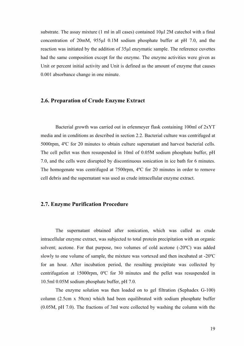
same buffer using ProTeam low pressure liquid chromatography system (Model LC
320, Teledyne ISCO) (Figure 2.1) Collected fractions were assayed for their protein
concentrations at 280nm and for polyphenol oxidase activity. The active fractions were
pooled and stored at -20 ºC until use for further experiments.
Figure 2.1. Low pressure liquid chromatography system
2.8. Electrophoretic Studies and Activity Staining
Electrophoretic studies included sodium dodecyl sulfate polyacrylamide gel
electrophoresis (SDS-PAGE) and activity staining after native-PAGE. Both SDS-PAGE
and native-PAGE were performed using acrylamide concentrations of 12% for
separating and 4% for stacking gels; runned at 65V for 30 minutes and 100V for about
an hour using Thermo Scientific gel electrophoresis device (Figure 2.2) at room
temperature.

21
2.8.1. SDS-PAGE
SDS-PAGE is an electrophoretic method that separates the proteins according to
their molecular sizes in an electrical field. This method is commonly used to estimate
the purity and molecular weight of a protein.
The partially purified sample and the samples from former steps were applied to
SDS-PAGE. While performing SDS-PAGE, samples were diluted by a volume ratio of
1:4 with sample buffer, and kept in boiling water for 4 minutes to denature proteins.
5µl of molecular weight marker and 25µl of sample-sample buffer mixture were loaded
onto gel. After electrophoretic run, at conditions given above, SDS-polyacrylamide gel
was stained using colloidal coomassie staining solution and incubated overnight. Then
the gel was subjected to neutralization, destaining and fixation solutions for 3 minutes, 1
minute and 1 hour, respectively. At the end of these steps, the image of the gel was
taken with a special camera under white light and if needed the gel was kept in 5%
acetic acid solution (v/v) at 4ºC for a long time.
The procedure for preparation of gels and reagents that were used in SDS-PAGE
are given in Appendix C.
Figure 2.2. Gel electrophoresis system

22
2.8.2. Native-PAGE and Activity Staining
Native-PAGE is also an electrophoretic method very similar to SDS-PAGE but
the main difference is that in native-PAGE the proteins are separated in non-denaturing
conditions. The procedure for preparing solutions and gels for native-PAGE is based on
SDS-PAGE’s, but to provide non-denaturing conditions, SDS and 2-mercaptoethanol
were exluded from sample buffer and SDS was also excluded in the preparation of both
separating and stacking gels. Instead of these reagents, same amount of water was
added. The samples were diluted with sample buffer by the ratio of 1:4 and loaded onto
gel skipping the heating step.
To perform specific activity staining after electrophoretic run, native
polyacrylamide gel was equilibrated by immersion in 0.1M sodium phosphate buffer,
pH 7.0, for 10 minutes at room temperature. Then the gel was transferred into a freshly
prepared solution of catechol (25mM, prepared in sodium phosphate buffer) and
incubated overnight at 55ºC in incubator. At the end of incubation period, the gel was
washed with distilled water and stored in 5% acetic acid solution at 4ºC, if needed.
2.9. Characterization Studies
2.9.1. Kinetic Analysis
To perform kinetic analysis, enzyme activity was measured at different
concentrations of catechol varying from 5mM to 60mM, then the kinetic parameters of
the enzyme, Km and Vmax, were determined by Lineweaver-Burk plot method.

23
2.9.2. Effect of pH on Enzyme Activity and Stability
The effect of pH on enzyme activity was investigated using 0.1M sodium
phosphate buffer at different pH values. The optimum pH of the enzyme was
determined under the standart assay conditions by measuring activity in the presence of
buffers at different pH values ranging from 4.0 to 10.0.
To determine the pH stability of the enzyme, 35µl enzyme solution was mixed
with 70µl of buffer at various pHs (pH 5.0, 6.0, 7.0, 8.0, 9.0, 10.0) and then the mixture
was incubated at 55ºC for 1.5 hour. The residual activity was measured under standart
assay conditions using 35µl of enzyme-buffer mixture.
2.9.3. Effect of Temperature on Enzyme Activity and Stability
The effect of temperature on polyphenol oxidase activity was examined under
standart assay conditions at different temperatures ranging from 30 to 90ºC and the
buffer was heated to relevant temperature before the assay.
Temperature stability was assayed by incubating the enzyme solution at a range
of temperatures from 30 to 80ºC for 1.5 hour and then measuring the remaining activity
using standart assay procedure.
2.9.4. Effect of Metal Ions on Enzyme Activity
To examine the effect of metal ions on polyphenol oxidase function, the enzyme
was incubated in the presence of 1mM metal ion at room temperature for 10 minutes. At
the end of the incubation period the reaction was initiated by addition of catechol
solution to give a final substrate concentration of 20mM and change in absorbance was
measured under standart assay conditions. For this purpose, CaCl2, CuSO4, MgSO4,
KCl and ZnCl2 were used.

24
2.9.5. Effect of Various Agents on Enzyme Activity
The effect of some agents such as ethylenediaminetetraacetic acid (EDTA),
sodium flouride, dimethyl sulfoxide (DMSO), dithiothreitol (DTT), sodium
diethyldithiocarbamate, SDS, and Triton X-100 on enzyme activity were also examined.
For this purpose, the enzyme was incubated in the presence of different agents for 10
minutes at room temperature. The concentrations of the agents were; 5% (v/v) of Triton
X-100 or 1mM of other agents in 1ml assay mixture. At the end of incubation period,
the reaction was initiated by addition of catechol and change in absorbance was
measured under standart assay conditions.
2.9.6. Substrate Specificity of Enzyme
The substrate specificity of the enzyme was determined by measuring activity
towards several monohydroxyphenol and dihydroxyphenol compounds like L-tyrosine,
catechol, L-DOPA, ABTS and hydroquinone. The activities of the enzyme for this
purpose were measured using solutions of these compounds prepared in 0.1M sodium
phosphate buffer at concentrations of 20mM for catechol and hydroquinone, 10mM for
L-DOPA, 2mM for L-tyrosine and ABTS.
25
CHAPTER 3
RESULTS AND DISCUSSION
3.1. Evidence for Polyphenol Oxidase Activity
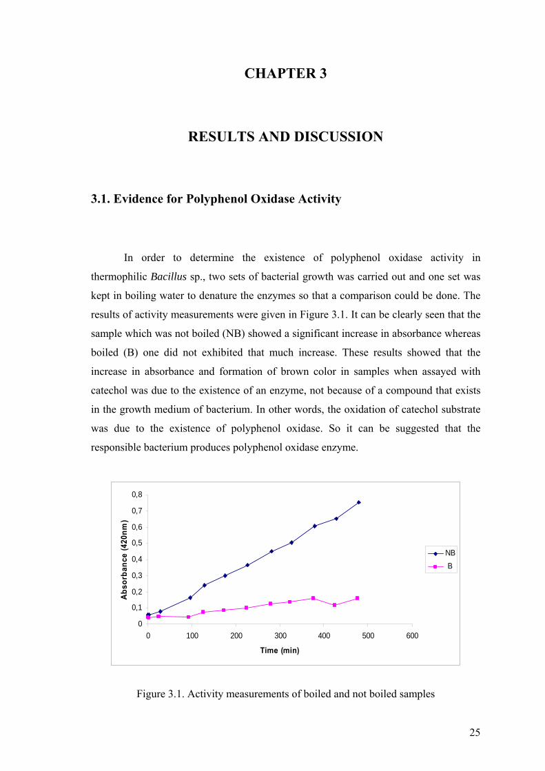
In order to determine the existence of polyphenol oxidase activity in
thermophilic Bacillus sp., two sets of bacterial growth was carried out and one set was
kept in boiling water to denature the enzymes so that a comparison could be done. The
results of activity measurements were given in Figure 3.1. It can be clearly seen that the
sample which was not boiled (NB) showed a significant increase in absorbance whereas
boiled (B) one did not exhibited that much increase. These results showed that the
increase in absorbance and formation of brown color in samples when assayed with
catechol was due to the existence of an enzyme, not because of a compound that exists
in the growth medium of bacterium. In other words, the oxidation of catechol substrate
was due to the existence of polyphenol oxidase. So it can be suggested that the
responsible bacterium produces polyphenol oxidase enzyme.
0
0,1
0,2
0,3
0,4
0,5
0,6
0,7
0,8
0 100 200 300 400 500 600
Time (min)
Abso
rban
ce (4
20nm
)
NB B
Figure 3.1. Activity measurements of boiled and not boiled samples

26
3.2. Partial Purification of Polyphenol Oxidase
In this study, partial purification of polyphenol oxidase from thermophilic
Bacillus sp. was achieved by acetone precipitation and gel filtration chromatography. It
should be noted that the result of activity measurement of growth medium which may
contain extracellular enzymes was poor when compared with the result of intracellular
extract. Also a sample from growth medium of bacterium was loaded onto native-
polyacrylamide gel and subjected to activity staining with catechol solution. Although
intracellular enzyme extract was stained with catechol, no dark band on the lane where
growth medium of bacterium was loaded could be observed (see Figure 3.4.b for result).
These results suggested that polyphenol oxidase from thermophilic Bacillus sp. was
intracellular thus intracellular extract was used as starting material for purification.
An outline of the purification procedure is illustrated and the results are given in
Table 3.1. The intracellular enzyme extract was first subjected to acetone precipitation
to precipitate total protein by changing the dielectric constant of the medium and
increasing the interaction of proteins. The yield and purification fold after this step was
81% and 1.19, respectively. The precipitate was then resuspended in buffer and loaded
onto gel filtration column which acts as a molecular sieve and separates the proteins
according to their molecular sizes. The fractions that were eluted from the column were
tested for their polyphenol oxidase activities and the ones with highest activity were
pooled (Figure 3.2). The resulting enzyme solution which had a specific activity of
134,9 U/mg, was purified 1.24 fold and contained 35% of the activity.
Table 3.1. Purification of polyphenol oxidase from thermophilic Bacillus sp.
Purification
Step
Volume
(ml)
Total
Activity
(U)
Total
Protein
(mg)
Specific
Activity
(U/mg)
Yield
(%)
Purification
(Fold)
Crude Extract 10 7030 64,8 108,5 100 1
Acetone
Precipitation
10,5 5691 43,89 129,7 81 1,19
Gel Filtration 6 2478 18,36 134,9 35 1,24

27
Purification fold and yield values of polyphenol oxidases that were obtained
with other bacterial species are; 27 and 24% after purification of Azospirillum lipoferum
polyphenol oxidase by acetone precipitation and hydroxyapatite chromatography
(Diamantidis, et al. 2000); 21 and 9% after purification of γ-proteobacterium JB
polyphenol oxidase by ammonium sulfate precipitation, ion exchange chromatography
and preparative PAGE (Singh, et al. 2007); 50 and 21% after purification of
Thermomicrobium roseum polyphenol oxidase by ion exhcange chromatography (Kong,
et al. 2000); 261 and 9% after purification of Streptomyces lavendulae polyphenol
oxidase by heat treatment, ammonium sulfate precipitation, ion exchange,
hydroxyapatite and gel filtration chromatography (Suzuki, et al. 2003), respectively.
Also 72% yield was obtained after purification of Bacillus thrungiensis polyphenol
oxidase with one-step purification method using copper sulfate saturated ion exchange
resin (Liu, et al. 2004). Since thermophilic Bacillus sp. polyphenol oxidase was partially
purified, the yield and purification fold values are lower than obtained for other
polyphenol oxidases from different bacterial species.
01234567
0 20 40 60 80
Fraction number
Abs
orba
nce
(280
nm)
0
10
20
30
40
50
Act
ivity
(U/m
l)
A280nmPPO Activity
Figure 3.2. Gel filtration profile of polyphenol oxidase

28
3.3. Electrophoretic Studies and Activity Staining
3.3.1. SDS-PAGE
The partially purified sample and the samples from former steps were applied to
SDS-PAGE. The image of the gel after colloidal coomassie staining is given in Figure
3.3. Supernatant of intracellular extract was loaded on lane 1; sample after acetone
precipitation was loaded on lane 2; the mixture of polyphenol oxidase active fractions
which were pooled after gel filtration column was loaded on lane 3 and a fraction which
was not polyphenol oxidase active was loaded on lane 4. Since the enzyme was
partially purified, the composition of the samples were very complex. Among those
protein bands on gel, which band corresponds to the enzyme of interest could not be
determined so did the molecular weight.
Figure 3.3. SDS-PAGE image. M: Protein marker

29
Molecular weight of polyhenol oxidases from other bacterial species vary from
120 to 14kDa. For example, molecular weight of polyphenol oxidase from γ-
proteobacterium JB is 120kDa (Singh, et al. 2007), Streptomyces griseus is 114kDa
(Endo, et al. 2003), Streptomeyces lavendulae is 73kDa (Suzuki, et al. 2003), Bacillus
subtilis and Bacillus licheniformis are 65kDa (Martins, et al. 2002, Koschorreck, et al.
2008), Thermus thermophilus is 53kDa (Miyazaki 2005), Streptomyces antibioticus and
Streptomyces glaucescens are 29kDa (Bernan, et al. 1985, Lerch and Ettinger 1972) and
Bacillus thuringiensis is 14kDa (Liu, et al. 2004).
3.3.2. Native-PAGE and Activity Staining
Native-PAGE separates the proteins on polyacrylamide gel under non-
denaturing conditions. So the enzymes loaded onto native polyacrylamide gel are not
denatured and retain their catalytic activity. In the light of this knowlegde, native-PAGE
of the samples was performed duplicated in same conditions. Following native-PAGE,
one gel was stained using catechol substrate for the detection of polyphenol oxidase
activity and the other one was stained using CBB dye to visualize all protein bands. The
images of the gels under white light are given in Figure 3.4. The gels on the left side are
stained with catechol solution and the right side are with CBB dye. As it can be seen
from Figure 3.4.a, the appearance of dark bands indicated the existence of polyphenol
oxidase in samples. Also as it was stated in section 3.2, the intracellular nature of the
enzyme was evidenced with the image of the gel that can be seen in Figure 3.4.b.

30
(a)
(b)
Figure 3.4. Activity and colloidal coomassie staining of polyphenol oxidase on native polyacrylamide gels. (a) Lane 1, supernatant of intracellular extract; lane 2, sample after acetone precipitation; lane 3, enzyme solution after column; lane 4 inactive fraction after column. (b) Lane 1, supernatant of intracellular extract; lane 2, growth medium of thermophilic Bacillus sp.

31
3.4. Characterization of Polyphenol Oxidase
3.4.1. Kinetic Analysis
To determine the kinetic constants, Km and Vmax, of thermophilic Bacillus
polyphenol oxidase, initial reaction rates at different catechol concentrations, ranging
from 5 to 60mM were measured. In order to obtain Lineweaver-Burk plot; 1/V
(1/Reaction rate) values were plotted against 1/S (1/Substrate concentration) values and
kinetic constants were calculated using this graph. Km and Vmax values of the enzyme
were determined as 91mM catechol and 2.25 ∆abs/min/ml, respectively.
-0,5
0
0,5
1
1,5
2
2,5
3
-0,02 -0,01 0 0,01 0,02 0,03 0,04 0,05 0,06
1/[S] (1/mM)
1/V
(∆A
bs/m
in/m
l)
Figure 3.5. Lineweaver-Burk plot for polyphenol oxidase
Bacillus thuringiensis polyphenol oxidase has a Km value of 34.05mM catechol
(Liu, et al. 2004). Polyphenol oxidase from γ-proteobacterium JB has a Km value of
0.055mM catechol (Singh, et al. 2007). In plants, polyphenol oxidase from apple (cv
Amasya), Ipomoea batatas and Amanita muscaria have Km values of 34mM, 2.5mM
and 83mM catechol, respectively (Mueller, et al. 1996, Oktay, et al. 1995). When
polyphenol oxidase from thermophilic Bacillus sp. was compared with polyphenol
oxidases from other sources, it was seen that this enzyme has lower affinity.

32
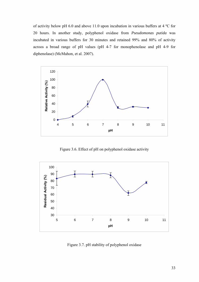
3.4.2. Effect of pH on Polyphenol Oxidase Activity and Stability
The effect of pH on polyphenol oxidase activity was investigated by measuring
enzyme activity at different pH values ranging from 4 to 10. The pH profile of the
enzyme, which can be seen in Figure 3.6, showed a bell shaped curve with the highest
activity at pH 7.0 and it was concluded that pH 7.0 was optimum pH of thermophilic
Bacillus sp. polyphenol oxidase. A significant loss in activity was observed upon
increasing or decreasing the optimum pH value even by one pH unit. The enzyme
exhibited low activity at pH 5.0 and no activity at pH 4.0. On the other hand at alkaline
pH values, the enzyme is not effected much as in acidic conditions and showed 30% of
its activity.
Similar to thermophilic Bacillus sp. polyphenol oxidase, optimum pH value
close to neutrality have been reported for polyphenol oxidases from other bacterial
species such as; Streptomyces michiganensis (pH 7.0) (Philipp, et al. 1991),
Pseudomonas putida (pH 7.0) (McMahon, et al. 2007), Vibrio tyrosinaticus (pH 6.6-
7.8) (Pomerant.Sh and Murthy 1974), Streptomyces glaucescens (pH 6.8) (Lerch and
Ettinger 1972), Streptomyces griseus (Endo, et al. 2003) and γ-proteobacterium JB (pH
6.5) (Bains, et al. 2003). Nevertheless, acidic and alkaline optimum pH values for
polyphenol oxidases from bacterial species such as Thermomicrobium roseum (pH 9.5)
(Kong, et al. 2000), Bacillus thuringiensis (pH 9.0) (Liu, et al. 2004) and Bacillus
licheniformis (pH 4.2) (Koschorreck, et al. 2008) have also been observed.
The pH stability of the enzyme was examined by incubating the enzyme in
various buffers for 1.5 hour. The residual activities were measured under standart assay
conditions. The activity of enzyme which was not subjected to pH treatment was
regarded as hundred percent, then the residual activities were calculated and plotted
against pH values as in Figure 3.7. It can be clearly seen from the figure that the enzyme
retained more than 80% of its activity in the pH range of 5-8, however lost 40% of its
activity at pH 9. This enzyme was found to be stable as it retained most of its activity
through a broad range of pH after 1.5 hour incubation period.
pH stability studies have been carried out with polyphenol oxidases from other
bacteria. Kong et al. (2000) reported that Thermomicrobium roseum polyphenol oxidase
retained more than 70% activity in the pH range of 8.5-10.0 but lost approximately 75%
33
of activity below pH 6.0 and above 11.0 upon incubation in various buffers at 4 ºC for
20 hours. In another study, polyphenol oxidase from Pseudomonas putida was
incubated in various buffers for 30 minutes and retained 99% and 80% of activity
acroos a broad range of pH values (pH 4-7 for monophenolase and pH 4-9 for
diphenolase) (McMahon, et al. 2007).
0
20
40
60
80
100
120
4 5 6 7 8 9 10 11
pH
Rela
tive
Activ
ity (%
)
Figure 3.6. Effect of pH on polyphenol oxidase activity
30
40
50
60
70
80
90
100
5 6 7 8 9 10 11
pH
Resi
dual
Act
ivity
(%)
Figure 3.7. pH stability of polyphenol oxidase

34
3.4.3. Effect of Temperature on Polyphenol Oxidase Activity and
Stability
In order to determine the effect of temperature on enzyme activity, polyphenol
oxidase activities at different temperatures ranging from 30 to 90ºC were measured. The
results of these measurements indicated that the enzyme showed highest activity at
60ºC. As it can be seen in Figure 3.8, the activity of the enzyme was stimulated upon
heating up to 60 and 70ºC. However, at temperatures above 70ºC, a decrease in
polyphenol oxidase activity was observed with 82% and 35% of the activity at 80 and
90ºC, respectively.
Such a high temperature which was determined for thermophilic Bacillus sp.
polyphenol oxidase in this study or even higher temperatures of maximal activity were
also observed for polyphenol oxidases obtained from other bacteria. The temperature
maxima of 92ºC was recorded with Thermus thermophilus polyphenol oxidase
(Miyazaki 2005), 85ºC with Bacillus licheniformis polyphenol oxidase (Koschorreck, et
al. 2008), 75ºC with Bacillus thuringiensis (Liu, et al. 2004) and CotA protein of
Bacillus subtilis (Martins, et al. 2002), 70ºC with Thermomicrobium roseum (Kong, et
al. 2000), and 55ºC with both Bacillus sp. HR03 (Dalfard, et al. 2006) and γ-
proteobacterium JB (Bains, et al 2003).
0
20
40
60
80
100
120
30 40 50 60 70 80 90 100
Temperature (ºC)
Rela
tive
Activ
ity (%
)
Figure 3.8. Effect of temperature on polyphenol oxidase activity

35
Thermal stability of polyphenol oxidase from thermophilic Bacillus sp. was
determined by incubating the enzyme solution at different temperatures for 1.5 hour and
measuring the remaining activity under standart assay conditions. The activity of the
enzyme which was not subjected to temperature treatment was regarded as hundred
percent. The thermal stability profile of polyphenol oxidase can be seen in Figure 3.9.
These results showed that the enzyme was fairly stable for 1.5 hour at temperatures up
to 60ºC. At temperatures above 60ºC, a decline in activity was observed. Although the
enzyme retained nearly 70% of its activity at 70ºC; at 80ºC, the activity was completely
lost upon incubation for 1.5 hour.
A hyperthermophilic polyphenol oxidase from Thermus thermophilus was found
to be resistant to incubation at 85ºC for 10 minutes, also the enzyme retained two-thirds
of its activity at 100ºC for 10 minutes (Miyazaki 2005). Polyphenol oxidase from
Bacillus thuringiensis was most stable at 75ºC (Liu, et al. 2004). Thermomicrobium
roseum polyphenol oxidase was very stable between 30-70ºC with 10 minutes
incubation period (Kong, et al. 2000). On the other hand Streptomyces polyphenol
oxidase had a half-life of 1-5 minutes at 60ºC (Huber and Lerch 1988). According to
these results, thermophilic Bacillus polyphenol oxidase can be considered as
thermostable with an incubation period of 1.5 hour.
0
20
40
60
80
100
120
140
30 40 50 60 70 80 90
Temperature (ºC)
Res
idua
l Act
ivity
(%)
Figure 3.9. Thermal stability of polyphenol oxidase

36
3.4.4. Effect of Metal Ions on Polyphenol Oxidase Activity
In order to determine the effect of various metal ions on polyphenol oxidase
activity, the enzyme was incubated in the presence of an ion for 10 minutes at room
temperature and the activity was measured in a normal manner. The concentrations of
the metal ions used in this study were all 1mM. The sample which did not contain any
metal ion served as control and its activity was regarded as hundred percent. The effects
of the ions on enzyme activity are shown in Figure 3.10.
According to the results, the presence of Zn2+ and K+ did not stimulate the action
of polyphenol oxidase but an increase in enzyme activity was observed in the presence
of Ca2+, Cu2+ and Mg2+. As it can be clearly seen from Figure 3.10, Cu2+ caused a
significant amount of activation on polyphenol oxidase activity. This outcome is not
surprising since polyphenol oxidases are copper containing enzymes and copper is
essential for catalytic activity. Similar activator effect of copper on the activity of
polyphenol oxidase from Thermomicrobium roseum (Kong, et al. 2000) and Bacillus
thuringiensis (Liu, et al. 2004) were also reported. Also addition of copper to the growth
medium of Bacillus (HR03) was found to increase the melanin production (Dalfard, et
al. 2006).
0
50
100
150
200
250
300
350
400
Control Ca Cu K Mg Zn
Rel
ativ
e A
ctiv
ity (%
)
Figure 3.10. Effect of metal ions on polyphenol oxidase activity

37
3.4.5. Effect of Various Agents on Polyphenol Oxidase Activity
The effect of several agents, which act as inhibitor or activator on the action of
polyphenol oxidase were tested. For this purpose, the enzyme was incubated in the
presence of relevant agent for 10 minutes at room temperature and then the activity was
measured spectrophotometrically under standart assay conditions. The activity of the
sample in the absence of agent was regarded as hundred percent and this sample served
as control.
The results, which can be summarized in Table 3.2, clearly indicated that DTT
and sodium diethyldithiocarbamate are strong inhibitors for polyphenol oxidase from
thermophilic Bacillus sp. Even in the presence of 1mM of these agents, the enzyme
exhibited no activity under standart assay conditions. Sodium diethyldithiocarbamate is
a sulfur containing compound and used as a chelating agent for transition metal ions.
This agent is known as potent inhibitor of tyrosinase activity of polyphenol oxidases
and it was suggested that this compound may cause inhibition by forming complexes
with copper atoms in the active site (Kong, et al. 2000). Sodium fluoride, which is
regarded as a typical inhibitor for laccase activity of polyphenol oxidases, did not
exhibit a strong inhibitory action on polyphenol oxidase in this study. Sodium fluoride
with a concentration of 5mM inhibited the polyphenol oxidase activity of Bacillus
thuringiensis (Liu, et al 2004). Thus, higher concentrations of this agent may be
required for the inhibiton of thermophilic Bacillus polyphenol oxidase. DMSO and
some detergents such as SDS and Triton X-100 did not cause much effect on activity
such that the enzyme showed approximately 97% of its activity in the presence of those
detergents and 91% in the presence of DMSO. The effect of a chelating agent, EDTA,
on enzyme activity was also investigated. Since the active site of polyphenol oxidase
contains copper ions and they are involved in catalytic activity, chelating compounds
would inhibit polyphenol oxidase activity by removing copper ions. Interestingly, the
presence of 1mM EDTA barely effected the action of polyphenol oxidase and the
enzyme showed 95% of its activity. However polyphenol oxidases from Streptomyces
griseus and Bacillus thuringiensis showed 67% and 72% of their activity in the presence
of EDTA with same concentration, respectively (Endo, et al. 2003, Liu, et al. 2004). In
contrast to inhibitory effect, the activator effect of EDTA on Bacillus thuringiensis

38
polyphenol oxidase, in the concentration range of 200-400mM, have been reported (Liu,
et al. 2004).
Table 3.2. Effect of various agents on polyphenol oxidase activity
Agent Concentration Relative activity (%)
Control - 100
Sodium diethyldithiocarbamate 1mM 0
Sodium fluoride 1mM 89
DTT 1mM 0
EDTA 1mM 95
SDS 1mM 97
Triton X-100 5% 98
DMSO 1mM 91
3.4.6. Substrate Specificity of Polyphenol Oxidase
The substrate specificity of polyphenol oxidase from thermophilic Bacillus sp. was
determined by measuring enzyme activity using catechol (20mM), hydroquinone
(20mM), L-tyrosine (2mM), ABTS (2mM) and L-DOPA (10mM) at appropriate
wavelengts. The results of this assay can be seen in Table 3.3.
Table 3.3. Substrate specificity of polyphenol oxidase
Substrate Wavelength (nm) Relative Activity (%)
ABTS 420 2,24
Catechol 420 100
L-DOPA 475 20
L-tyrosine 475 0,78
Hydroquinone 420 8,20

39
CHAPTER 4
CONCLUSION
The goal of this work was to study bacterial polyphenol oxidases. In order to do
that, thermophilic Bacillus sp. was choosen and polyphenol oxidase obtained from this
bacterium was characterized.
First of all, the activities of boiled and not boiled cultures were measured. Boiled
sample which contained denatured enzymes, did not exhibit polyphenol oxidase activity
whereas not boiled sample did. This result showed that increase in absorbance when
assayed with catechol was due to the existence of polyphenol oxidase, not because of an
oxidizing compound that exists in the growth medium of bacterium. In addition, the
appearence of dark bands on native polyacrylamide gel after activity staining supported
the existence of polyphenol oxidase in thermophilic Bacillus sp. Besides, the results of
native-PAGE with samples from growth medium of bacterium and intracellular enzyme
extract showed that the related enzyme is an intracellular one.
The enzyme was partially purified by acetone precipitation and gel filtration
chromatograpy. The yield and purification fold after partial purification of the enzyme
were 35% and 1.24, respectively.
The characterization studies indicated that polyphenol oxidase from
thermophilic Bacillus sp. had highest activity at pH 7.0 and 60ºC. The enzyme was
stable at temperatures between 30 and 60ºC and retained approximately 85% of its
activity between pH 5 and 8. Also it retained 67% and 62% of its activity even after 1.5
hour incubation at 70ºC and pH 9.0, respectively. However it could not resist to
incubation for 1.5 hour at 80ºC and lost all its activity. The activity of the related
enzyme was highly stimulated in the presence of copper ion and totally inactivated by
DTT and sodium diethyldithiocarbamate. Other agents or metal ions did not have a
considerable inhibitory or stimulating effect on enzyme activity. Km and Vmax values for
the enzyme determined from Lineweaver-Burk plot were 91mM and 2.25 ∆abs/min/ml.

40
In conclusion, the enzyme was partially purified and some general
characteristics of it were determined. To obtain higher acitivities and be applicable for
industrial purposes, further purification steps may be required. Additionally, the gene
responsible for polyphenol oxidase enzyme can be cloned and expressed in suitable
hosts and some features of the enzyme may be altered by protein engineering
techniques. The enzyme of interest can be used for the development of biosensors to
detect phenolic compounds for various purposes, also the ability of polyphenol oxidase
to act on phenolic compounds can be used for the degradation of phenols in industrial
waste waters.

41
REFERENCES
Alexandre, G., and I. B. Zhulin. 2000. Laccases are widespread in bacteria. Trends in Biotechnology 18 (2):41-42.
Andrade, C. M. M. C., N. Pereira, and G. Antranikian. 1999. Extremely thermophilic microorganisms and their polymer-hydrolytic enzymes. Revista De Microbiologia 30 (4):287-298.
Asanuma, M., I. Miyazaki, and N. Ogawa. 2003. Dopamine- or L-DOPA-induced neurotoxicity: The role of dopamine quinone formation and tyrosinase in a model of Parkinson's disease. Neurotoxicity Research 5 (3):165-176.
Bailey, Philip S. 1995. Organic chemistry: a brief survey of concepts and applications. Englewood Cliff: Prentice Hall
Bains, J., N. Capalash, and P. Sharma. 2003. Laccase from a non-melanogenic, alkalotolerant gamma-proteobacterium JB isolated from industrial wastewater drained soil. Biotechnology Letters 25 (14):1155-1159.
Baker, G. C., S. Gaffar, D. A. Cowan, and A. R. Suharto. 2001. Bacterial community analysis of Indonesian hot springs. Fems Microbiology Letters 200 (1):103-109.
Bernan, V., D. Filpula, W. Herber, M. Bibb, and E. Katz. 1985. The Nucleotide-Sequence of the Tyrosinase Gene from Streptomyces-Antibioticus and Characterization of the Gene-Product. Gene 37 (1-3):101-110.
Carey, Francis A. 1996. Organic chemistry. New York: McGraw Hill.
Castro-Sowinski, S., G. Martinez-Drets, and Y. Okon. 2002. Laccase activity in melanin-producing strains of Sinorhizobium meliloti. FEMS Microbiol Lett 209 (1):119-25.
Claus, H. 2004. Laccases: structure, reactions, distribution. Micron 35 (1-2):93-96.
Claus, H., and H. Decker. 2006. Bacterial tyrosinases. Syst Appl Microbiol 29 (1):3-14.
Claus, H., and Z. Filip. 1997. The evidence of a laccase-like enzyme activity in a Bacillus sphaericus strain. Microbiological Research 152 (2):209-216.
42
Cowan, M. M., E. A. Horst, S. Luengpailin, and R. J. Doyle. 2000. Inhibitory effects of plant polyphenoloxidase on colonization factors of Streptococcus sobrinus 6715. Antimicrobial Agents and Chemotherapy 44 (9):2578-2580.
D'Auria, S., P. Herman, J. R. Lakowicz, F. Tanfani, E. Bertoli, G. Manco, and M. Rossi. 2000. The esterase from the thermophilic eubacterium Bacillus acidocaldarius: Structural-functional relationship and comparison with the esterase from the hyperthermophilic archaeon Archaeoglobus fulgidus. Proteins-Structure Function and Genetics 40 (3):473-481.
Dalfard, A. B., K. Khajeh, M. R. Soudi, H. Naderi-Manesh, B. Ranjbar, and R. H. Sajedi. 2006. Isolation and biochemical characterization of laccase and tyrosinase activities in a novel melanogenic soil bacterium. Enzyme and Microbial Technology 39 (7):1409-1416.
Deckert, G., P. V. Warren, T. Gaasterland, W. G. Young, A. L. Lenox, D. E. Graham, R. Overbeek, M. A. Snead, M. Keller, M. Aujay, R. Huber, R. A. Feldman, J. M. Short, G. J. Olsen, and R. V. Swanson. 1998. The complete genome of the hyperthermophilic bacterium Aquifex aeolicus. Nature 392 (6674):353-358.
Diamantidis, G., A. Effosse, P. Potier, and R. Bally. 2000. Purification and characterization of the first bacterial laccase in the rhizospheric bacterium Azospirillum lipoferum. Soil Biology & Biochemistry 32 (7):919-927.
Duran, N., and E. Esposito. 2000. Potential applications of oxidative enzymes and phenoloxidase-like compounds in wastewater and soil treatment: a review. Applied Catalysis B-Environmental 28 (2):83-99.
Duran, N., M. A. Rosa, A. D'Annibale, and L. Gianfreda. 2002. Applications of laccases and tyrosinases (phenoloxidases) immobilized on different supports: a review. Enzyme and Microbial Technology 31 (7):907-931.
Edwards, W., R. Bownes, W. D. Leukes, E. P. Jacobs, R. Sanderson, P. D. Rose, and S. G. Burton. 1999. A capillary membrane bioreactor using immobilized polyphenol oxidase for the removal of phenols from industrial effluents. Enzyme and Microbial Technology 24 (3-4):209-217.
Eicken, C., F. Zippel, K. Buldt-Karentzopoulos, and B. Krebs. 1998. Biochemical and spectroscopic characterization of catechol oxidase from sweet potatoes (Ipomoea batatas) containing a type-3 dicopper center. FEBS Lett 436 (2):293-9.
Endo, K., Y. Hayashi, T. Hibi, K. Hosono, T. Beppu, and K. Ueda. 2003. Enzymological characterization of EpoA, a laccase-like phenol oxidase produced by Streptomyces griseus. Journal of Biochemistry 133 (5):671-677.

43
Fernandez, E., A. Sanchez-Amat, and F. Solano. 1999. Location and catalytic characteristics of a multipotent bacterial polyphenol oxidase. Pigment Cell Research 12 (5):331-339.
Fontana, A., V. De Filippis, P. P. de Laureto, E. Scaramella, and M. Zambonin. 1998. Rigidity of thermophilic enzymes. Stability and Stabilization of Biocatalysts 15:277-294.
Gerday, Charles. 2007. Physiology and biochemistry of extremophiles. Washington, D.C: ASM Press.
Guell, M., and P. E. M. Siegbahn. 2007. Theoretical study of the catalytic mechanism of catechol oxidase. Journal of Biological Inorganic Chemistry 12 (8):1251-1264.
Haki, G. D., and S. K. Rakshit. 2003. Developments in industrially important thermostable enzymes: a review. Bioresource Technology 89 (1):17-34.
Halaouli, S., M. Asther, J. C. Sigoillot, M. Hamdi, and A. Lomascolo. 2006. Fungal tyrosinases: new prospects in molecular characteristics, bioengineering and biotechnological applications. Journal of Applied Microbiology 100 (2):219-232.
Harwood, Colin R. 1989. Bacillus. New York: Plenum Press.
Hernandez-Romero, D., A. Sanchez-Amat, and F. Solano. 2006. A tyrosinase with an abnormally high tyrosine hydroxylase/dopa oxidase ratio. FEBS J 273 (2):257-70.
Horikoshi, Koki. 1998. Extremophiles: microbial life in extreme environments. New York: John Wiley.
Hough, D. W., and M. J. Danson. 1999. Extremozymes. Current Opinion in Chemical Biology 3 (1):39-46.
Huber, H., and K. O. Stetter. 1998. Hyperthermophiles and their possible potential in biotechnology. Journal of Biotechnology 64 (1):39-52.
Huber, M., and K. Lerch. 1988. Identification of 2 Histidines as Copper Ligands in Streptomyces-Glaucescens Tyrosinase. Biochemistry 27 (15):5610-5615.
Iyer, P. V., and L. Ananthanarayan. 2008. Enzyme stability and stabilization - Aqueous and non-aqueous environment. Process Biochemistry 43 (10):1019-1032.

44
Kim, C., W. W. Lorenz, J. T. Hoopes, and J. F. D. Dean. 2001. Oxidation of phenolate siderophores by the multicopper oxidase encoded by the Escherichia coli yacK gene. Journal of Bacteriology 183 (16):4866-4875.
Kong, K. H., M. P. Hong, S. S. Choi, Y. T. Kim, and S. H. Cho. 2000. Purification and characterization of a highly stable tyrosinase from Thermomicrobium roseum. Biotechnol Appl Biochem 31 ( Pt 2):113-8.
Koschorreck, K., S. M. Richter, A. B. Ene, E. Roduner, R. D. Schmid, and V. B. Urlacher. 2008. Cloning and characterization of a new laccase from Bacillus licheniformis catalyzing dimerization of phenolic acids. Applied Microbiology and Biotechnology 79 (2):217-224.
Kristjansson, J. K. 1989. Thermophilic Organisms as Sources of Thermostable Enzymes. Trends in Biotechnology 7 (12):349-353.
Lerch, K., and L. Ettinger. 1972. Purification and characterization of a tyrosinase from Streptomyces glaucescens. Eur J Biochem 31 (3):427-37.
Li, W. F., X. X. Zhou, and P. Lu. 2005. Structural features of thermozymes. Biotechnology Advances 23 (4):271-281.
Liu, N., T. Zhang, Y. J. Wang, Y. P. Huang, J. H. Ou, and P. Shen. 2004. A heat inducible tyrosinase with distinct properties from Bacillus thuringiensis. Lett Appl Microbiol 39 (5):407-12.
Martins, L. O., C. M. Soares, M. M. Pereira, M. Teixeira, T. Costa, G. H. Jones, and A. O. Henriques. 2002. Molecular and biochemical characterization of a highly stable bacterial laccase that occurs as a structural component of the Bacillus subtilis endospore coat. Journal of Biological Chemistry 277 (21):18849-18859.
Maugeri, T. L., C. Gugliandolo, D. Caccamo, and E. Stackebrandt. 2001. A polyphasic taxonomic study of thermophilic bacilli from shallow, marine vents. Syst Appl Microbiol 24 (4):572-87.
Mayende, L., B. S. Wilhelmi, and B. I. Pletschke. 2006. Cellulases (CMCases) and polyphenol oxidases from thermophilic Bacillus spp. isolated from compost. Soil Biology & Biochemistry 38 (9):2963-2966.
McMahon, A. M., E. M. Doyle, S. Brooks, and K. E. O'Connor. 2007. Biochemical characterisation of the coexisting tyrosinase and laccase in the soil bacterium Pseudomonas putida F6. Enzyme and Microbial Technology 40 (5):1435-1441.

45
Mercadoblanco, J., F. Garcia, M. Fernandezlopez, and J. Olivares. 1993. Melanin Production by Rhizobium-Meliloti Gr4 Is Linked to Nonsymbiotic Plasmid Prmegr4b - Cloning, Sequencing, and Expression of the Tyrosinase Gene Mepa. Journal of Bacteriology 175 (17):5403-5410.
Miyazaki, K. 2005. A hyperthermophilic laccase from Thermus thermophilus HB27. Extremophiles 9 (6):415-25.
Mueller, L. A., U. Hinz, and J. P. Zryd. 1996. Characterization of a tyrosinase from Amanita muscaria involved in betalain biosynthesis. Phytochemistry 42 (6):1511-1515.
Nazina, T. N., T. P. Tourova, A. B. Poltaraus, E. V. Novikova, A. A. Grigoryan, A. E. Ivanova, A. M. Lysenko, V. V. Petrunyaka, G. A. Osipov, S. S. Belyaev, and M. V. Ivanov. 2001. Taxonomic study of aerobic thermophilic bacilli: descriptions of Geobacillus subterraneus gen. nov., sp nov and Geobacillus uzenensis sp nov from petroleum reservoirs and transfer of Bacillus stearothermophilus Bacillus thermocatenulatus, Bacillus thermoleovorans, Bacillus kaustophilus, Bacillus thermoglucosidasius and Bacillus thermodenitrificans to Geobacillus as the new combinations G-stearothermophilus, G-thermocatenulatus, G-thermoleovorans, G-kaustophilus, G-thermoglucosidasius and G-thermodenitrificans. International Journal of Systematic and Evolutionary Microbiology 51:433-446.
Oktay, M., I. Kufrevioglu, I. Kocacaliskan, and H. Sakiroglu. 1995. Polyphenoloxidase from Amasya Apple. Journal of Food Science 60 (3):494-496.
Philipp, S., T. Held, and H. J. Kutzner. 1991. Purification and Characterization of the Tyrosinase of Streptomyces-Michiganensis Dsm-40015. Journal of Basic Microbiology 31 (4):293-300.
Polaina, Julio and MacCabe, Andrew P. 1974. Industrial enzymes: structure, function and applications. The Netherlands: Springer
Pomerant.Sh, and V. V. Murthy. 1974. Purification and Properties of Tyrosinases from Vibrio-Tyrosinaticus. Archives of Biochemistry and Biophysics 160 (1):73-82.
Queiroz, C., M. L. M. Lopes, E. Fialho, and V. L. Valente-Mesquita. 2008. Polyphenol oxidase: Characteristics and mechanisms of browning control. Food Reviews International 24 (4):361-375.
Roberts, S. A., A. Weichsel, G. Grass, K. Thakali, J. T. Hazzard, G. Tollin, C. Rensing, and W. R. Montfort. 2002. Crystal structure and electron transfer kinetics of CueO, a multicopper oxidase required for copper homeostasis in Escherichia coli. Proceedings of the National Academy of Sciences of the United States of America 99 (5):2766-2771.

46
Rodriguez Couto, S., and J. L. Toca Herrera. 2006. Industrial and biotechnological applications of laccases: a review. Biotechnol Adv 24 (5):500-13.
Ruijssenaars, H. J., and S. Hartmans. 2004. A cloned Bacillus halodurans multicopper oxidase exhibiting alkaline laccase activity. Appl Microbiol Biotechnol 65 (2):177-82.
Sanchez-Amat, A., P. Lucas-Elio, E. Fernandez, J. C. Garcia-Borron, and F. Solano. 2001. Molecular cloning and functional characterization of a unique multipotent polyphenol oxidase from Marinomonas mediterranea. Biochimica Et Biophysica Acta-Protein Structure and Molecular Enzymology 1547 (1):104-116.
Sanchez-Amat, A., and F. Solano. 1997. A pluripotent polyphenol oxidase from the melanogenic marine Alteromonas sp shares catalytic capabilities of tyrosinases and laccases. Biochem Biophys Res Commun 240 (3):787-92.
Seo, S. Y., V. K. Sharma, and N. Sharma. 2003. Mushroom tyrosinase: Recent prospects. Journal of Agricultural and Food Chemistry 51 (10):2837-2853.
Simsek, S., and A. Yemenicioglu. 2007. Partial purification and kinetic characterization of mushroom stem polyphenoloxidase and determination of its storage stability in different lyophilized forms. Process Biochemistry 42 (6):943-950.
Singh, G., N. Capalash, R. Goel, and P. Sharma. 2007. A pH-stable laccase from alkali-tolerant gamma-proteobacterium JB: Purification, characterization and indigo carmine degradation. Enzyme and Microbial Technology 41 (6-7):794-799.
Solano, F., E. Garcia, E. P. deEgea, and A. SanchezAmat. 1997. Isolation and characterization of strain MMB-1 (CECT 4803), a novel melanogenic marine bacterium. Applied and Environmental Microbiology 63 (9):3499-3506.
Solomons, T.W. Graham. 1990. Fundamentals of organic chemistry. New York: John Wiley.
Suzuki, T., K. Endo, M. Ito, H. Tsujibo, K. Miyamoto, and Y. Inamori. 2003. A thermostable laccase from Streptomyces lavendulae REN-7: Purification, characterization, nucleotide sequence, and expression. Bioscience Biotechnology and Biochemistry 67 (10):2167-2175.
Takagi, H., T. Takahashi, H. Momose, M. Inouye, Y. Maeda, H. Matsuzawa, and T. Ohta. 1990. Enhancement of the Thermostability of Subtilisin-E by Introduction of a Disulfide Bond Engineered on the Basis of Structural Comparison with a Thermophilic Serine Protease. Journal of Biological Chemistry 265 (12):6874-6878.

47
Turner, P., G. Mamo, and E. N. Karlsson. 2007. Potential and utilization of thermophiles and thermostable enzymes in biorefining. Microbial Cell Factories 6:-.
van den Burg, B. 2003. Extremophiles as a source for novel enzymes. Current Opinion in Microbiology 6 (3):213-218.
VanGelder, C. W. G., W. H. Flurkey, and H. J. Wichers. 1997. Sequence and structural features of plant and fungal tyrosinases. Phytochemistry 45 (7):1309-1323.
Vieille, C., K. L. Epting, R. M. Kelly, and J. G. Zeikus. 2001. Bivalent cations and amino-acid composition contribute to the thermostability of Bacillus licheniformis xylose isomerase. European Journal of Biochemistry 268 (23):6291-6301.
Vieille, C., and G. J. Zeikus. 2001. Hyperthermophilic enzymes: Sources, uses, and molecular mechanisms for thermostability. Microbiology and Molecular Biology Reviews 65 (1):1-+.
Xu, Y. M., A. H. Stokes, R. Roskoski, and K. E. Vrana. 1998. Dopamine, in the presence of tyrosinase, covalently modifies and inactivates tyrosine hydroxylase. Journal of Neuroscience Research 54 (5):691-697.
Yavuz, E., H. Gunes, S. Harsa, and A. F. Yenidunya. 2004. Identification of extracellular enzyme producing thermophilic bacilli from Balcova (Agamemnon) geothermal site by ITS rDNA RFLP. Journal of Applied Microbiology 97 (4):810-817.
Yoshimoto, T., K. Yamamoto, and D. Tsuru. 1985. Extracellular Tyrosinase from Streptomyces Sp Ky-453 - Purification and Some Enzymatic-Properties. Journal of Biochemistry 97 (6):1747-1754.

48
APPENDIX A
PREPARATION OF BRADFORD REAGENT AND PROTEIN
STANDARTS FOR BRADFORD ASSAY
Preparation of Bradford Reagent
• 10.0mg Coomassie Brilliant Blue G-250 (CBB G-250)
• 5ml 95% ethanol
• 10ml 85% phosphoric acid
Dissolve CBB G-250 in ethanol, add 10ml phosphoric acid. Bring to 100ml with
ultra pure water and when the dye has completely dissolved, filter through Whatman
No. 1 paper. Store at 4ºC.
Preparation of Protein Standarts
Bovine serum albumin (BSA) was used as protein standart. To obtain a stock
solution with a concentration of 0.2 mg/ml; 0.02g BSA was dissolved in 1ml dH2O,
then 10 µl was taken from this stock and added 990µl dH2O to give a final
concentration of 0.2mg/ml.
To prepare standarts according to the table, necessary amounts of water, BSA
and bradford reagent were pipetted into cuvettes respectively (Table A.1), then
incubated at room temperature for 5 minutes. At the end of incubation period,
absorbance was measured at 595nm using a spectrophotometer.

49
Table A.1. Preparation of BSA standarts
BSA (µl) dH2O (µl) Bradford Reagent (µl)
Blank 0 800 200 Standart 1- 1 µg/ml 5 795 200 Standart 2- 2 µg/ml 10 790 200 Standart 3- 4 µg/ml 20 780 200 Standart 4- 6 µg/ml 30 770 200 Standart 5- 8 µg/ml 40 760 200

50
APPENDIX B
STANDART CURVE FOR BRADFORD ASSAY
Table B.1. Absorbance values of BSA standarts
Concentration (µg/ml) Absorbance (595nm) 1 0,0782 2 0,1427 4 0,2073 6 0,2806 8 0,3282
Bradford Assay Standart Graph
y = 0,035x + 0,0606R2 = 0,9817
00,050,1
0,150,2
0,250,3
0,350,4
0 2 4 6 8 10
BSA Concentration (µg/ml)
Abso
rban
ce (5
95nm
)
Figure B.1. BSA standart curve for Bradford assay

51
APPENDIX C
REAGENTS AND GEL PREPARATION FOR SDS-PAGE
Stock Solutions
A. 30% Acrylamide Mixture
• 29.2g acrylamide
• 0.8g N’N’-bis-methylene-acrylamide
Make up to 100ml with deionized water. Filter and store at 4ºC in the dark for at
least one month.
B. 1.5M Tris-HCl, pH 8.8
• 18.15g Tris Base
• ~80ml deionized water
Dissolve Tris base in deionized water, adjust to pH 8.8 with HCl. Make up to
100ml with deionized water and store at 4ºC.
C. 0.5M Tris-HCl, pH 6.8
• 6g Tris Base
• ~80ml deionized water
Dissolve Tris base in deionized water, adjust to pH 6.8 with HCl. Make up to
100ml with deionized water and store at 4ºC.

52
D. 10% SDS
Dissolve 10g SDS in 90ml water with gentle stirring and bring to 100 ml with
deionized water.
E. Sample Buffer
• 3.8ml deionized water
• 1.0ml 0.5M Tris-HCl, pH 6.8
• 0.8ml Glycerol
• 1.6ml 10% (w/v) SDS
• 0.4ml 2-mercaptoethanol
• 0.4ml 1% (w/v) bromophenol blue
F. 5X Running Buffer
• 15g Tris Base
• 72g Glycine
• 5g SDS
Dissolve Tris base, glycine and SDS in ~800ml deionized water and make up to
1L with water. Store at 4ºC. For electrophoretic run, dilute 5X stock solution to 1X with
deionized water.
G. 10% Ammonium persulfate (APS)
Dissolve 0.1g APS in 1ml deionized water. This solution should be prepared
fresh daily.

53
H. Colloidal Coomassie Staining Solution
Dissolve 40g ammonium sulfate in ~300ml water, add 8ml 85% o-phosphoric
acid and add 0.5g Coomassie Brilliant Blue G-250. Make up to 400ml with water, add
100ml methanol to 500ml total volume. Store at 4ºC.
I. Neutralization Buffer
0.1M, pH 6.5 Tris-phosphate in deionized water.
J. Destaining Solution
25% (v/v) methanol solution.
K. Fixation Solution
20% (w/v) Ammonium sulfate in deionized water.
Gel Preparation
• Separating Gel
Table C.1. Preparation of 12% SDS-PAGE separating gel (for 5ml)
Deionized water 1.68ml1.5M Tris-HCl, pH 8.8 1.25ml10% SDS 50µl Acrylamide/Bis (30% Stock) 2ml 10% APS 25µl TEMED 2.5µl

54
• Stacking Gel
Table C.2. Preparation of 4% SDS-PAGE stacking gel (for 5ml)
Deionized water 3.05ml0.5M Tris-HCl, pH 6.8 1.25ml10% SDS 50µl Acrylamide/Bis (30% Stock) 665µl 10% APS 25µl TEMED 5µl