Embed Size (px)
Citation preview

PARS Achilles Jig System
Surgical Technique
PAR
S A
chill
es J
ig S
yste
m

PARS Achilles Jig System
Incision planning: The incision is placed approximately 1 cm proximal to the palpable rupture in the Achilles tendon.
The proximal portion of the tendon is grasped with an Alice Clamp or some other grasping device.
Achilles tendon ruptures are common in the elite and recreational athlete and most often occur in the noninsertional region of the tendon complex. Most surgeons will elect to treat these injuries surgically, to lessen the risk for rerupture, while providing the opportunity for a quicker recovery and convenientrehabilitation. Historically, open techniques have been utilized for repair of therupture but can be complicated by wound-healing issues and infection. This percutaneous and minimally invasive technique minimizes this concern.
The PARS provides the opportunity for consistently reliable capture of the proximal and distal aspects of the Achilles tendonand utilizes color-coded FiberWire® suture. The anatomically contoured guide is nondisposable, while the suture and passing needles come packaged in one convenient kit. The system provides the option of transverse or locking sutures, or both. The coloredsutures offer a more organized approach to identifying and securing matched pairs.
Our own experience with this system has been nothing short of outstanding. All patients have healed their rupture uneventfully with no instances of wound dehiscence, infection, rerupture, or sural nerve injury. Anecdotally, the healed tendon appears to achieve a more natural contour, unlike the typical hypertrophic tendon resulting from an open repair. We have found this mini-mally invasive technique ideal for the middle-aged individual where there may be a heightened concern for wound-healing issues. Relative indications include those patients with compro-mised skin and soft tissue, or those with systemic diseases(e.g. diabetes mellitus, rheumatoid arthritis) in which there is a great risk for infection. Robert B. Anderson, M.D.OrthoCarolina Orthopedic GroupCharlotte, NC
1 2
Surgical Technique
PARS Achilles Jig System
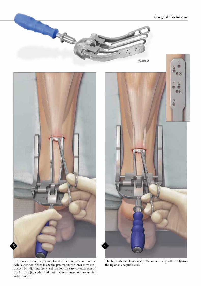
The Jig is advanced proximally. The muscle belly will usually stop the Jig at an adequate level.
The inner arms of the Jig are placed within the paratenon of the Achilles tendon. Once inside the paratenon, the inner arms are opened by adjusting the wheel to allow for easy advancement of the Jig. The Jig is advanced until the inner arms are surrounding viable tendon.
Achilles tendon ruptures are common in the elite and recreational athlete and most often occur in the noninsertional region of the tendon complex. Most surgeons will elect to treat these injuries surgically, to lessen the risk for rerupture, while providing the opportunity for a quicker recovery and convenientrehabilitation. Historically, open techniques have been utilized for repair of therupture but can be complicated by wound-healing issues and infection. This percutaneous and minimally invasive technique minimizes this concern.
The PARS provides the opportunity for consistently reliable capture of the proximal and distal aspects of the Achilles tendonand utilizes color-coded FiberWire® suture. The anatomically contoured guide is nondisposable, while the suture and passing needles come packaged in one convenient kit. The system provides the option of transverse or locking sutures, or both. The coloredsutures offer a more organized approach to identifying and securing matched pairs.
Our own experience with this system has been nothing short of outstanding. All patients have healed their rupture uneventfully with no instances of wound dehiscence, infection, rerupture, or sural nerve injury. Anecdotally, the healed tendon appears to achieve a more natural contour, unlike the typical hypertrophic tendon resulting from an open repair. We have found this mini-mally invasive technique ideal for the middle-aged individual where there may be a heightened concern for wound-healing issues. Relative indications include those patients with compro-mised skin and soft tissue, or those with systemic diseases(e.g. diabetes mellitus, rheumatoid arthritis) in which there is a great risk for infection. Robert B. Anderson, M.D.OrthoCarolina Orthopedic GroupCharlotte, NC
PARS Achilles Jig
3 4
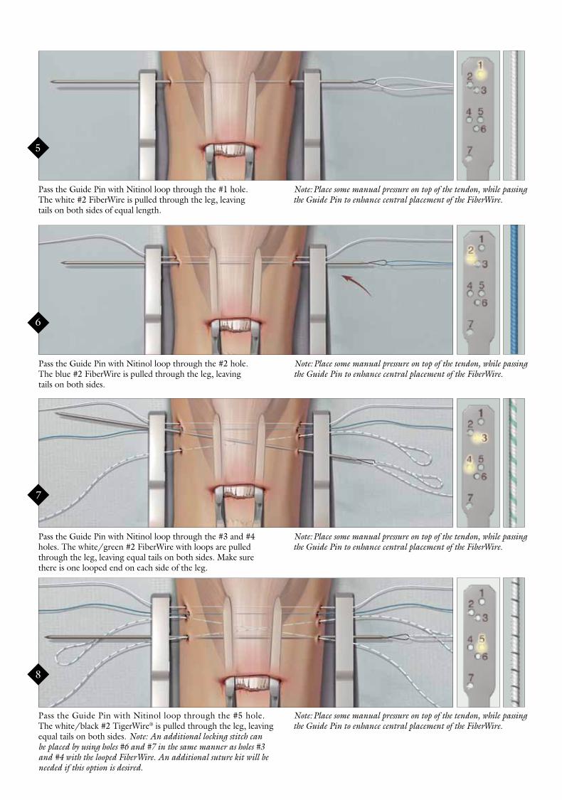
Pass the Guide Pin with Nitinol loop through the #5 hole. The white/black #2 TigerWire® is pulled through the leg, leaving equal tails on both sides. Note: An additional locking stitch can be placed by using holes #6 and #7 in the same manner as holes #3 and #4 with the looped FiberWire. An additional suture kit will be needed if this option is desired.
Note: Place some manual pressure on top of the tendon, while passing the Guide Pin to enhance central placement of the FiberWire.
Pass the Guide Pin with Nitinol loop through the #1 hole. The white #2 FiberWire is pulled through the leg, leaving tails on both sides of equal length.
Pass the Guide Pin with Nitinol loop through the #2 hole. The blue #2 FiberWire is pulled through the leg, leaving tails on both sides.
Pass the Guide Pin with Nitinol loop through the #3 and #4 holes. The white/green #2 FiberWire with loops are pulled through the leg, leaving equal tails on both sides. Make sure there is one looped end on each side of the leg.
Note: Place some manual pressure on top of the tendon, while passing the Guide Pin to enhance central placement of the FiberWire.
Note: Place some manual pressure on top of the tendon, while passing the Guide Pin to enhance central placement of the FiberWire.
Note: Place some manual pressure on top of the tendon, while passing the Guide Pin to enhance central placement of the FiberWire.
8
5
6
7

Final construct prior to removal of the Jig.
9
Pull the Jig slowly out of the operative site. Once the inner arms are out of the incision, pull the suture out of the outer arms so they don’t get stuck in the holes.
10
9

Continue to pull the Jig slowly down until all the suture is out of the wound.
Illustration showing all of the sutures once they have been pulled out of the wound.
11
12

Organize the sutures the way they were originally placed through the Jig.
Pass the #2 blue suture UNDER the #3 and #4 looped sutures and back through the loop of the white/green looped suture.
14
13
1
2
3
4
5
1
2
3
4
5
2
3
4
2
2
3
4
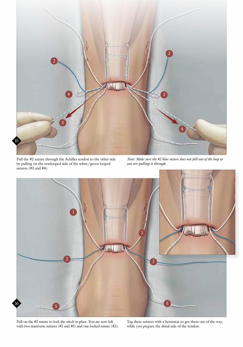
Pull the #2 suture through the Achilles tendon to the other side by pulling on the nonlooped side of the white/green looped sutures (#3 and #4).
Pull on the #2 suture to lock the stitch in place. You are now left with two transverse sutures (#1 and #5) and one locked suture (#2).
Tag these sutures with a hemostat to get them out of the way, while you prepare the distal side of the tendon.
Note: Make sure the #2 blue suture does not fall out of the loop as you are pulling it through.
15
16
2
3
4
2
3
4
5
1
2
5
1
2

Place the Jig in the distal part of the incision and perform the exact same steps as for the proximal side of the tendon.
17

You are left with three sutures proximally and three distally, ready for reapproximation of the tendon.
With the foot in maximum plantarflexion, tie the white/black suture on both sides of the leg first. Three to four surgeon’s knots are recommended.
Note: The first side you tie is the ‘stay’ stitch and will slide. You will lock this knot down when you tie the other side.
18
19

With the foot in maximum plantarflexion, tie the locked blue suture on both sides of the leg. Six to eight surgeon’s knots are recommended.
With the foot in maximum plantarflexion, tie the white suture on both sides of the leg last. Six to eight surgeon’s knots are recommended.Note: The first side you tie is the ‘stay’ stitch and will slide.You will lock this knot down when you tie the other side. At this time, you can close the sheath with an absorbable suture under the incision.
Final Repair. The wound can be closed with the suture of the surgeon’s choice. Postoperative routine is left to the surgeon’s preference.
Note: This suture will not slide since it is locked within the tendon.
20
21 22

This description of technique is provided as an educational tool and clinical aid to assist properly licensed medical professionals in the usage of specific Arthrex products. As part of this professional usage, the medical professional must use
their professional judgment in making any final determinations in product usage and technique. In doing so, the medical professional should rely on their own training and experience and should conduct
a thorough review of pertinent medical literature and the product’s Directions For Use.
U.S. Patent Nos. 6,716,234; 7,029,490 and PATENT PENDING© 2012, Arthrex Inc. All rights reserved. LT0464B
Ordering Information:
PARS Achilles Jig Instrument Set (AR-8860S) includes:
PARS Achilles Jig AR-8860J Cannulated Driver Handle with AO Connection AR-13221AOC
PARS Achilles Jig Suture Kit (AR-8860DS) includes:
Two #2 FiberWire, 38”, whiteTwo #2 FiberWire, 38”, blueTwo #2 TigerWire, 38”, white/blackTwo #2 FiberWire, with loops, 40”, white/greenTwo 1.6 mm Straight Needles with Nitinol loops