Embed Size (px)
Citation preview

Revue Méd. Vét., 2014, 165, 3-4, 77-88
PALAEOPATHOLOGICAL STUDY OF CATTLE AND HORSE OF SIRMIUM 77
Introduction
After the publication of the well-known “Animal Diseases in Archaeology” by Baker and Brothwell in 1980s, animal paleopathology experienced great improvement. However, in some countries, such as Serbia, this scientific discipline is relatively unknown. Paleopathology in general can help us gather more data on health [3, 16, 17, 22, 31, 36, 43, 47, 49, 56, 75, 76, 78], ways of living and the relationships between humans and animals, as well as the history of some diseases [24, 48, 59, 73, 74]. In literature different diagnoses for the same pathological changes can be noticed. Similar observations have been given by archaeozoologist [29], who claim that can be the main reason for the misinterpretation of pathological changes in the bones. Research in animal paleopathology is limited to individual archaeological sites [14, 25, 29, 37, 61, 63] or several sites from different periods [33, 71]. However, our idea was to perform paleopathological analysis of animal remains from several sites of the ancient
city of Sirmium, which is a valuable archaeological location in Serbia and beyond.
It is clear, that pathological changes in domestic animals are in close relations with its use [5, 8, 9, 10, 19]. The cultural practices of communities have a significant role for understanding and interpretation of archaeozoological findings. In other words archaeozoological discoveries must be placed in their sociocultural context eg. the presence of a hippodrome in a Roman city means that the analysis of horse remains should take this fact into account.
Because of the relatively large number of Roman works
about agriculture and animal husbandry, the knowledge about animal use is quite extensive. According to Roman authors Columella (De re rustica 2.2.1.5; 2.2.24; 6.22.1), Varro (Res rusticae 1.20.2, 4–5) and Cato (De agricultura 11.2.4; 14.2; 62), cattle, especially oxen, were used as draught animals in agriculture [60]. Same authors wrote about horses having been used in the military, their care from
SUMMARY
This paper considers the observation of pathological changes in animal skeletal remains from four archaeological sites in Sirmium and one rustic villa (Vranj) located near the city. Before the pathological analysis, skeletal elements, as well as insight of taxonomic and age of animals were determined. 13599 bones or bone fragments were examined. In 72 specimens various abnormal changes have been observed. Most of the pathological changes were present in skeletal remains of cattle and horses. Macroscopic and radiographic analysis of pathological changes in the animal bones established that the lesions had proliferative and chronic character. Proliferative changes in the bones of cattle and horses indicate that these animals were used as draught animals and/or carrying cargo. Considering that most of the horses skeletal remains are from the Hippodrome, it is very possible that they were animals for amusement purposes in ancient city of Sirmium.
Keywords: Archaeozoology, Paleopathology, Roman City Sirmium, cattle (Bos taurus), horse (Equus caballus)
RÉSUMÉ
Etude paléopathologique des ossements de bovins et de chevaux de la ville antique de Sirmium (Pannonie / Serbie)
Cet article traite des altérations pathologiques observées sur les restes de squelettes d’animaux provenant de quatre sites archéologiques de Sirmium (Pannonie / Serbie) et d’une villa rustique (Vranj) située près de cette ville. Avant d’en faire l’analyse pathologique, une diagnose d’espèce et d’âge des éléments de squelettes a été réalisée. 13599 os et fragments ont été éxaminés. Sur 72 spécimens des anomalies osseuses ont été remarquées. La plupart de ces affections osseuses ont été observées parmi les restes de squelettes de bétail et de chevaux. Le caractère prolifératif et chronique des lésions a été montré par des examens macroscopiques et radiographiques des os. Ces modifications osseuses du squelette des bovins et des chevaux indiquent que les animaux ont été utilisés pour traction. La majorité des restes de squelettes ont été retrouvés à l’ hippodrome de Sirmium, c’est pourquoi l’hypothèse que ces animaux aient pu être utilisés pour le sport est possible.
Mots clés : Archéozoologie, Paléopathologie, Ville Romaine Sirmium, Bétail (Bos taurus), Cheval (Equus caballus)
Palaeopathological study of Cattle and Horse bone remains of the Ancient Roman city of Sirmium (Pannonia / Serbia)
N. MARKOVIĆ¹*, O. STEVANOVIĆ², V. NEŠIĆ², D. MARINKOVIĆ², N. KRSTIĆ², D. NEDELJKOVIĆ³, D. RADMANOVIĆ4, M. JANECZEK5
¹Institute of Archaeology, Knez Mihailova 34/IV, 11000, Belgrade, SERBIA.²Faculty of Veterinary Medicine, University of Belgrade, Bulevar oslobođenja 18, 11000, Belgrade, SERBIA.³Museum of Srem, Vuka Karadžića 3, 22000, Sremska Mitrovica, SERBIA.4Museum of Vojvodina, Dunavska 35, 21000, Novi Sad, SERBIA. 5Department of Biostructure and Animal Physiology, Wrocław University of Environmental and Life Sciences, Kozuchowska 1/3, 51-631 Wrocław, POLAND.
*Corresponding author: [email protected]

Revue Méd. Vét., 2014, 165, 3-4, 77-88
MARKOVIĆ (N.) AND COLLABORATORS78
the veterinary perspective, their breeding and use in the chariot racing [44]. The special Roman hippiatric writings by Apsyrtos, Theomnest, Anatolis and others are well known. Above mentioned ancient authors described the great number of various diseases symptoms and healings methods, too [1, 2, 27]. In the Roman Empire horses were used very wide eg. riding, cavalry and chariot racing. Post service (cursus publicus) depended upon horses and mules. The horse veterinarians in Roman army had a large importance including status immunes. Taruttienus Paternus praetorian prefect (parefectus praetorio) wrote about animal hospital (veterinarium) in legionary camp [23, 35]. From the other hand, veterinarians in cursus publicus were slaves.
The aim of this study was to determine the type and age of the animal remains on which paleopathological changes were found, with emphasis on cattle (Bos taurus) and horses (Equus caballus); presence of bones and / or fragments with the changes of the examined bone material; types and localization of the changes in the bones of cattle and horses; possible changes in the etiology of changes and hypotheses about how these animals was used in Sirmium.
Archaeological background
SIRMIUM
Sirmium was the capital of the province of Pannonia Secunda in Late Antiquity. The city was located alongside the ancient road that connected the eastern and western provinces of the Roman Empire (fig. 1). Strategic location of Sirmium enabled its rapid development from a military camp into an imperial city [53].
The history of Sirmium can be traced from the time of Augustus’ conquering of Illyria (35−33 BC) up to 582 AD when the Avars completely destroyed the city. The Romans conquered Sirmium in time of Tiberius wars in Pannonia (13−9 BC). During the Flavia dynasty Sirmium acquired the
status of a colony (Colonia Flavia). During the Tetrarchy, in year 293, Sirmium became one of the capitals of the Roman Empire (fig. 2).
The space occupied by the ancient city was successively colonized for a long time, changing from a medieval village built around the remains of the late antique basilica, in the medieval town Dmitrovica. At the same place the town of Sremska Mitrovica is located today [55].
Systematic archaeological excavations in Sirmium began in 1957. The Regional Institute for Protection of Cultural Monuments of Novi Sad has led research work to Sirmium in 1962 when the Archaeological Institute in Belgrade took over the researching work. During the period from 1968 to 1972 the archaeological excavations were performed at the participating experts the Smithsonian Institute, Washington, Denison University, Ohio and the City University of New York [54].
The Museum of Srem and the Institute for the Protection of Cultural Monuments of Sremska Mitrovica had an important role in the research from the very beginning. During the period between 1957 and 2013 on the territory of Sirmium, today’s Sremska Mitrovica and its surroundings, over a hundred sites have been investigated, but the archaeozoological material was only regularly or systematically collected from 1996 onwards.
SITE 1A – IMPERIAL PALACE
The most important architectural complex in Sirmium was the Imperial Palace, built between the end of the III and the beginning of the IV century. A.D. This complex building was situated in the southern part of the ancient city, close to its south wall and bank of the Sava river. The first rescue excavations, carried out between 1957 and 1960 were the first archaeological research of the ancient Sirmium, marked as site 1a. After a thirty-year break, during 2006 and 2007 the excavations of the Palace continued, at the moment when it was opened for visitors [62].
Figure 1: Map of the provinces of Moesia Prima and Pannonia Secunda in the IVc. AD
Figure 2: Sirmium during 4th century ideal reconstruction (Jeremić 2004: 11, fig.12)

Revue Méd. Vét., 2014, 165, 3-4, 77-88
PALAEOPATHOLOGICAL STUDY OF CATTLE AND HORSE OF SIRMIUM 79
SITE 66 – HIPPODROME
Hippodrome was built in the early IV century as the monumental structure of the Sirmium, primarily designed for chariot races. This complex was positioned close to the Imperial Palace, oriented SE-NW, which deviates from the usual orientation of an ancient hippodrome, the function loses at the end of the IV century, and later used for accommodation of military units [39, 52]. The skeletons of animals were not systematically collected. A large number of bones between hippodrome walls and spaces below the auditorium have been observed [46, 64].
SITE 80
Locality 80 is situated near the western wall of the ancient Sirmium, not far from the beneficial station, in the southern outskirts of the pagan necropolis, close to the main Roman road Cibalae-Singidunum [42]. The results of rescue archaeological excavations showed that this area was one of the many waste areas outside the walls. These landfills were in function from I to VI century. A small part of the site, during the period from II to III c. belonged to the periphery of the major western necropolis [65].
SITE 85
Systematic archaeological excavation of the site 85 began in 2002. The primary objective of the research was to investigate the complex urban core of Sirmium, near the Imperial Palace, and the medieval horizon of Mitrovica [40]. Two major constructions can be distinguished on the site. The first section is a monumental late antique building with massive walls. The function of the structure from IV c. cannot be determined with certainty. The second section was a medieval cathedral church of Mitrovica (Civitas Sancti Demetrii). The church was placed in the center of a necropolis which was in function from X to XVI c. [41].
VRANJ
The Vranj Locality is situated in the village of Hrtkovci (fig. 1), close to the left bank of the Sava River. In the period between 1991 and 2004, a Roman villa rustica was excavated in Vranj by the Museum of Vojvodina. The villa, which was located in the wider region of Sirmium, was an agricultural establishment with well-equipped rooms. The villa was continuously inhabited from the mid-III century, until the mid-IV century [69, 70].
Material and Methods
A total number of identified skeletal parts (NISP) 13599 of whole and fragmented animal bones from 5 archaeological sites, four of which were excavated in Sirmium (Location 1a−Imperial Palace, Location 66 – Hippodrome, Location 80 and Location 85) and one villa rustica (Vranj) near Sirmium were examined. Archaeozoological material from the sites
was previously published. Considering the fact, that material from archaeological sites in Sirmium belong to the layers that were dated from the beginning of III century, until the end of IV century, it will be observed as unique sample (12165 NISP) [28, 57, 58]. Material from villa rustica (Vranj) which belongs to the layers from the mid-III century, until the mid-IV century, is represented with 1434 NISP [15].
All the archaeozoological material was subjected to macroscopic analysis and the following was determined: taxon, age, element, localization and form of pathological changes.
Species determination was carried out by the literature [72] and comparative collection of Laboratory for Bioarcheology, Department of Archaeology, Faculty of Philosophy, and University of Belgrade. Age was determined on the basis coalescence epiphysis of long bones, which was found in the age categories - juvenile, sub adult and adult [72].
The diagnosis of pathological changes was made based on reference literature in paleopathology [4], and veterinary medicine [80, 12]. The representative examples of bones with pathological changes presented in this paper were analyzed radio graphically. X-ray analyses of bones with pathomorphological changes were carried out by X-ray apparatus Siemens Selenos 400.
Results
The results are based on paleopathological changes that were recorded on the bones of animals from four archaeological sites of the Roman city of Sirmium. Out of 12165 bones and bone fragments examined osteopathological alterations were found in 65 (0. 53%) cases. In fauna sample from Vranj (1434 NISP) 7 (0.48%) bones with abnormal changes were noted. The bones with pathological changes originated from the following animal species: cattle (Bos taurus), horse (Equus caballus), sheep/goat (Ovis/Capra), pig (Sus scrofa domestica), dog (Canis familiaris) and the chicken (Gallus domesticus). The results on the distribution of pathological changes are shown in table I.
Bones of cattle and horses with pathological changes were most prevalent in archaeofauna of Sirmium. Therefore, our further analysis is reduced to these animals. On the bones of cattle and horses, native to Sirmium, only acquired pathological changes were recorded. Localization of changes in the skeleton of cattle and horses is shown in table II. Also, from the table II it can be noticed that the pathological changes found in the bones of the appendicular skeleton, in the limbs, are more numerous than the pathological changes observed in the axial skeleton (osteophytes on the thoracic and lumbal vertebrae).
During the macroscopical analysis of appendicular skeleton of cattle different pathological changes were recorded. On the front limbs of cattle pathological changes were noticed mostly on metacarpal bones, metatarsal bones

Revue Méd. Vét., 2014, 165, 3-4, 77-88
MARKOVIĆ (N.) AND COLLABORATORS80
and on phalanges, but also on humerus, radius, pelvis, femur and calcaneus. In one case pathological change was noticed on humerus in form of abnormal deviation of its distal part. Also the lost of bone tissue - osteolysis of radius was noticed in one case. Localized overgrowths of new bone – exostoses were noticed on the proximal, as well on distal part of metacarpal bones and phalanges of cattle (fig. 3). Beside this articular depressions on the articular surfaces of these bones were also noticed.
The pathological changes on the bones of rare limb of cattle were noted on the acetabulum in form of osteoarthritis which was characterized with grooving of articular surface, eburnation and exostoses which overgrown the articular edge. In one case the loss of cancellous bone of femur – osteoporosis was noted. Fracture with formed callus was noted in one case on the proximal part of metatarsal bone. Radiography of the sagital and lateral projection of this bone showed massive spongy bone proliferations which form a mass around the low density zone which contains sequesters. This appearance is characteristic for the osteomyelitic process. In the level of the callus compact bone was expanded in the medullar canal whose structure showed signs of eburnisation. Linear radiolucency, without bony deposits, which expands through proximal part of the bone to the medial diaphysis was noted (fig. 4). Beside this, also in one case signs of osteoarthritis
Animal species Sirmium Vranj
NISP Pathological changes % NISP Pathological
changes %
Cattle (Bos taurus) 4423 30 0.67 507 6 1.1Horse (Equus caballus) 555 13 2.3 9 1 11Sheep/goat (Ovis/ Capra) 3598 8 0.22 369 - -Pig (Sus scrofa domestica) 2486 6 0.24 365 - -Dog (Canis familiaris) 238 6 2.5 6 - -Cat (Felis domestica) 45 - - - - -Hen (Gallus domesticus) 558 2 0.35 123 - -Goose (Anser domesticus) 39 - - - - -Duck (Anas domesticus) 20 - - - - -Red deer (Cervus elaphus) 132 - - 23 - -Roe deer (Capreolus capreolus) 7 - - 13 - -Wild boar (Sus scrofa) 33 - - 15 - -Beaver (Castor fiber) 1 - - - - -Wolf (Canis lupus) 1 - - 2 - -Rabbit (Lepus europaeus) 28 - - - - -Badger (Meles meles) 1 - - - - -Eagle (Aquila heliacal) - - - 2 - -Total 12165 65 100 1434 7 100
Table I: Summary of pathologies present on specimens.
Localization Cattle (Bos taurus) Horse (Equus caballus)Appendicular skeleton 33 13Axial skeleton 3 1
Table II: Localization of changes in the skeleton.
Figure 3: Exostosis of the phalanx of cattle (Bos taurus)

Revue Méd. Vét., 2014, 165, 3-4, 77-88
PALAEOPATHOLOGICAL STUDY OF CATTLE AND HORSE OF SIRMIUM 81
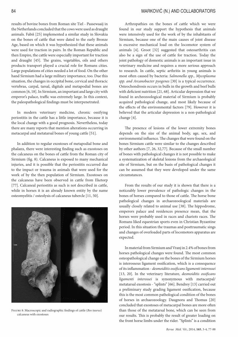
was recorded on the tarso-metatarsal joint of cattle. Radiography of the sagital projection of this joint (with lack of fused second and third tarsal bone) of cattle showed disfigurement of articular plate with cystic radiolucency in the subchondral bone. Massive bone defects, narrowing of the articular cleft, calcification of the cartilaginous tissue and secondary degenerative-deformative changes caused by deposits of new bone formations were recorded. Decreased densities of the bones as well as the patches of the zone of eburnisation, characterized by increase bone density caused by trabecular collapse were noted in the proximal part of the metatarsal bone (fig.5). In four cases pathological changes were noted on the calcaneus in form of exostoses and ankylosis. Radiography of the latero-lateral projection of the cattle calcaneus showed chronic local periosteal bony plaques which extends from the compact bone towards external surface of the bone. These bony proliferations led to the deformation of the size and shape of the tuber calcanei. Bony deposits filled the most part of the talo-calcaneal articular surface. In the cancellous bone of the tuber calcanei large blots of radiolucence and multilocular cystic areas with clearly marked zone of eburnisation were noticed (fig. 8). Most frequent pathological changes on the appendicular skeleton of cattle were on the phalangis in form of proximal and distal exostoses, articular depressions, osteoarthritis and nodular exostoses at ligamentous insertions – enthesophytes. A detailed view of the pathological changes in the bones of cattle is shown in table III.
Pathological changes on the appendicular skeleton of the horse were recorded in a lesser degree (table IV). Grooving of articular surface, eburnation and exostoses which overgrown the articular edge of the tibio-tarsal joint, which are all signs of osteoarthritis was noticed in one case. Latero-medial radiography of the equine tibia revealed iregulary shaped, blurry edged periostal ossification plaques localized in the medial maleolus. Scaley bone deposits in the distal tibial epiphysis as well as blurry and amorphous appearance of the distal metaphysis were recorded. Lateral tibial condilus showed signs of small bony proliferations in form of osteophytes. Total sclerosis of the medullary canal (cancellous bone had an appearance of the compact bone) in the level of the distal diaphysis was noted. Zones of decreased bone density which opens to the articular plate surface were noted in the subchondral bone of the proximal epiphysis. (fig. 6). The most frequent pathological change on the bones of horses were noted on the metacarpal bones in form exostosis, or ossification of interosseus ligament - “splints”, which is a consequence of its inflammation - desmoiditis ossificans ligamenti interossei (fig. 7). Beside these changes, other changes, in form of localized overgrowth of new bone – exostoses on the proximal, as well on distal part of these bones were also noticed. In contrast to cattle, in horses neither fractures nor metabolic bone diseases were not observed.
Discussion
Bones have great reparatory potential and they also play an important role in the metabolism of the animals. Bones, like some other tissues, are prone to change, in the form of atrophy, hypertrophy, hyperplasia, degeneration, necrosis, metaplasia and neoplasia. By macroscopic analysis it is impossible to
Figure 4: Macroscopic and radiographic finding of fracture with well-for-med callus on the metatarsal bone of cattle (Bos taurus)
Figure 5: Macroscopic and radiographic findings of osteoarthritis of tarso-metatarsal joint of cattle (Bos taurus)

Revue Méd. Vét., 2014, 165, 3-4, 77-88
MARKOVIĆ (N.) AND COLLABORATORS82
Archaeological site Element Part of the element Age Pathological findings
Sirmium 85 Radius Proximal part Adult Osteolysis Sirmium 85 Calcaneus Whole Adult ExostosisSirmium 85 Calcaneus whole Adult ExostosisSirmium 85 Calcaneus whole Adult AnkylosisSirmium 85 Metatarsal Distal part Adult FractureSirmium 1a Metacarpal whole Adult Distal exostosisSirmium 1a Metacarpal Distal part Adult Distal exostosisSirmium 85 First phalanx Whole Adult ExostosisSirmium 85 Metacarpal Whole Adult Proximal exostosisSirmium 85 Metatarsal Proximal part Adult OsteoarthritisSirmium 85 Humerus Distal part Adult Abnormal deviationSirmium 85 Metacarpal Whole Adult Distal exostosisSirmium 85 First phalanx Whole Adult Proximal/distal
exostosisSirmium 85 First phalanx Whole Adult Proximal/distal
exostosisSirmium 80 First phalanx Whole Adult Distal exostosisSirmium 80 Metatarsal Proksimal part Adult ExostosisSirmium 80 Acetabulum Whole Adult OsteoarthritisSirmium 80 Calcaneus Whole Adult AnkylosisSirmium 80 First phalanx Whole Adult Proximal/distal
exostosisSirmium 80 First phalanx Whole Adult Distal exostosisSirmium 80 First phalanx Whole Adult Distal exostosisSirmium 80 Femur Distal part Adult OsteoporosisSirmium 80 Third phalanx Whole Adult EnthesophytesSirmium 80 First phalanx Whole Adult Distal exostosisSirmium 80 First phalanx Whole Adult Distal exostosisSirmium 80 Metacarpal Distal part Adult Articular depressionSirmium 80 Metacarpal Distal part Adult Distal exostosisVranj First phalanx Whole Adult Distal exostosisVranj First phalanx Whole Adult Articular depressionVranj First phalanx Whole Adult Articular depressionVranj First phalanx Whole Adult OsteoarthritisVranj Third phalanx Whole Adult EnthesophytesVranj Third phalanx Whole Adult Enthesophytes
Table III: Pathological changes on the bones of appendicular skeleton of cattle (Bos taurus)
Archaeological site Element Part of the element Age Pathological findings
Hipodrom Tibia Whole Adult OsteoarthritisHipodrom Metacarpal Whole Adult Metacarpal exostosisSirmium 85 Metacarpal Whole Adult Metacarpal exostosisSirmium 85 Metacarpal Whole Adult Metacarpal exostosisSirmium 85 Metacarpal Whole Adult Metacarpal exostosisSirmium 85 Metacarpal Whole Adult Metacarpal exostosisSirmium 85 Metacarpal Whole Adult Metacarpal exostosisSirmium 80 First phalanx Whole Adult EnthesophytesSirmium 80 Metacarpal Whole Adult Metacarpal exostosisSirmium 80 First phalanx Whole Adult Articular depressionSirmium 80 Metacarpal Whole Adult Metacarpal exostosisSirmium 80 First phalanx Whole Adult EnthesophytesVranj Second phalanx Whole Adult Enthesophytes
Table IV: Pathological changes on the bones of appendicular skeleton of horse (Equus caballus)

Revue Méd. Vét., 2014, 165, 3-4, 77-88
PALAEOPATHOLOGICAL STUDY OF CATTLE AND HORSE OF SIRMIUM 83
distinguish all of these pathological alterations, and therefore set the valid diagnosis. In addition, the negative findings on the bones do not mean that the animals during the life did not suffer from other diseases of organs and organ systems, which are not reflected in their bones. Paleopathology in archeozoology is still based on the macroscopic analysis of the changes described by Baker and Brothwell [4]. Even today there is no detailed and definitive systematization and nomenclature of bone pathology. For this reason in many papers different terms for the same pathological change are used. The advantage of macroscopic analysis is that it is not expensive or complex to perform. During the analysis of abnormal changes in the bones of animals from Sirmium and Vranj, in addition to basic paleopathological data, latest discoveries in veterinary medicine were used. It is important to compare paleopathological findings with similar changes observed in modern veterinary practice. Known case history can be a useful tool in the potential pathogenesis description in the past [38].
The most pathological changes on the bones of cattle were located on appendicular skeleton. Distal part of limbs (metapodial bones and phalanx) was more susceptible to these changes, but in few cases pathological changes were also recorded on the humerus, radius, pelvis and femur. The cause of abnormal deviation of distal part of humerus was probably as a result of congenital defect, as a result of some metabolic disorders in young age (in example rachitis) or as badly healed fracture. Osteolysis of radius can be caused by some metabolic disorder as osteomalatia or as a result of destruction due to some neoplastic lesion.
The pathological changes on the distal bones of the limbs of cattle were manifested in forms of different exostosis, which are typically the result of a local inflammatory non-infectious periosteal injury [80]. In other words, exostoses are the result of periostitis which is common in paleopathological findings [4], while in other similar papers it is described as periostitis ossificans chronica [26]. Bartosiewicz et al. [7] examined the exostoses on the metapodial bones and phalanges of draught cattle from Romania, and they introduced a new grading system of lesions. Proliferative changes represent a sign of loading of the cattle during exploitation [7]. In study dealing with exostoses on the third phalanges in cattle and water buffalo very interesting results have been found [34]. According to the conclusions of this study, it was suspected that the buffalo in prehistoric Thailand were used for traction. Performing systematic analysis of bones on the lower extremities of cattle from Eketorp (Sweden) it has observed that there are no major changes in the prevalence of pathological lesions between animals from Iron Age and the Middle Ages [78]. However, there is a higher frequency of occurrence of exostoses in the third phalanges which may be related to a larger extent to biomechanical stress of the bone. Groot [32] argues that exostoses on the bones of lower extremities can serve as a sure sign of the use of cattle for traction from the Roman period in the Netherlands. Furthermore, the same author, based on paleopathological
Figure 6: Macroscopic and radiographic findings of osteoarthritis of the horse (Equus caballus) tibia
Figure 7: Metacarpal exostoses - «splints» in the horse (Equus caballus)
Revue Méd. Vét., 2014, 165, 3-4, 77-88
MARKOVIĆ (N.) AND COLLABORATORS84
results of bovine bones from Roman site Tiel - Passewaaij in the Netherlands concluded that the cows were used as draught animals. Fabiš [25] implemented a similar study in Slovakia on the bones of cattle that were dated to the early Bronze Age, based on which it was hypothesized that these animals were used for traction in pairs. In the Roman Republic and then Empire, the cattle were especially important for traction and draught [45]. The grains, vegetables, oils and others products transport played a crucial role for Romans cities. Large populations of cities needed a lot of food. On the other hand Sirmium had a large military importance, too. Due this situation, the changes in occipital bone, cervical and thoracic vertebras, carpal, tarsal, digitals and metapodial bones are common [6, 18]. In Sirmium, an important and large city with emperor’s palace, traffic was extremely large. In this context, the paleopathological findings must be interpenetrated.
In modern veterinary medicine, chronic ossifying periostitis in the cattle has a little importance, because it is the local change with a good prognosis. Nevertheless, today there are many reports that mention alterations occurring in metacarpal and metatarsal bones of young cattle [51].
In addition to regular exostoses of metapodial bone and phalanx, there were interesting finding such as exostoses on the calcaneus on the bones of cattle from the Roman city of Sirmium (fig. 8). Calcaneus is exposed to many mechanical injuries, and it is possible that the periostitis occurred due to the impact or trauma in animals that were used for the work of by the then population of Sirmium. Exostoses on the calcaneus have been observed in cattle from Eketorp [77]. Calcaneal periostitis as such is not described in cattle, while in horses it is an already known entity by the name osteomyelitis / osteolysis of calcaneus tubercle [11, 50].
Arthropathies on the bones of cattle which we were found in our study support the hypothesis that animals were intensively used for the work of by the inhabitants of Sirmium, because one of the main causes of joint disease is excessive mechanical load on the locomotor system of animals [4]. Groot [32] suggested that osteoarthritis can also be a sign of the use of cattle for traction. Today the joint pathology of domestic animals is an important issue in veterinary medicine and requires a more serious approach to research. In cattle, septic arthritis in young animals is most often caused by bacteria: Salmonella spp., Mycoplasma spp. and Arcanobacter pyogenes [30] is a typical occurrence. Osteochondrosis occurs in bulls in the growth and beef bulls with deficient nutrition [21, 68]. Articular depression that we found in archeozoological material of Sirmium is probably acquired pathological change, and most likely because of the effects of the environmental factors [79]. However it is believed that the articular depression is a non-pathological change [4].
The presence of lesions of the lower extremity bones depends on the size of the animal body, age, sex, and environmental influence. The changes that were found on the bones Sirmium cattle were similar to the changes described by other authors [7, 26, 32,77]. Because of the small number of bones with pathological changes it is not possible to make a systematization of skeletal lesions from the archaeological site of Sirmium, but on the basis of pathological changes it can be assumed that they were developed under the same circumstances.
From the results of our study it is shown that there is a noticeably lower prevalence of pathologic changes in the bones of horses compared to those of cattle. The horse bone pathological changes in archaeozoological materials are usually closely related to animal use [38]. The hippodrome, emperors palace and residences presence mean, that the horses were probably used in races and chariots races. The Romans liked equestrian sports even in Christian Byzantine period. In this situation the traumas and posttraumatic sings and changes of overloaded parts of locomotors apparatus are expected.
In material from Sirmium and Vranj in 2.4% of bones from horses pathological changes were found. The most common osteopathological change on the bones of the Sirmium horses is interosseus ligament ossification, which is a consequence of its inflammation - desmoiditis ossificans ligamenti interossei [13, 20]. In the veterinary literature, desmoiditis ossificans ligamenti interossei is synonymous with metacarpal/metatarsal exostosis - “splints” [66]. Bendrey [13] carried out a preliminary study grading ligament ossification, because this is the most common pathological condition of the bones of horses in archaeozoology. Daugnora and Thomas [20] concluded that exostoses of metacarpal bones are more often than those of the metatarsal bone, which can be seen from our results. This is probably the result of greater loading on the front horse limbs under the rider. “Splints” is a condition
Figure 8: Macroscopic and radiographic findings of cattle (Bos taurus) calcaneus with exostoses

Revue Méd. Vét., 2014, 165, 3-4, 77-88
PALAEOPATHOLOGICAL STUDY OF CATTLE AND HORSE OF SIRMIUM 85
of young horses that are subjected to early sport training, and the essence of the situation is tearing ligaments, external trauma and healing of longitudinal fracture of metacarpal/metatarsal bones [12]. In our case, according to the analysis, there were not fractured bones of horses, but we speculate that exostoses are the result of external trauma. Injuries that lead to exostosis are most likely caused by training and/or horse races at the Hippodrome of Sirmium. However, metacarpal / metatarsal exostoses can occur independently of the uses of animals. “Splints” is often seen in 3 − 4 year old horses [12], and the possible reason of lameness of diseased animals. During the Roman period it is unknown if any treatment of „splints“ or similar pathologic changes existed, so that horses with heavy „splints“ are likely excluded from the competition or use. The similar can be said about arthropathy of a horse that is diagnosed in material from the Hippodrome of Sirmium. Enthesophytes of the phalanges found [67] in archaeozoological material that came from wild horses. Arthropathy is probably a severe “bone spavin”, which is a common finding in animal paleopathology [4, 29]. However, for correct diagnosis it is necessary to have an analysis of tarsal bones where the changes are most prominent. Similar pathologies of extremity bones were described by Onar et al. [60] in Theodosius Harbour excavation.
Conclusion
Pathological lesions in the bones of cattle and horses are the result of local mechanical injuries of bones. Obviously, the load of the musculoskeletal system played an important role in the progression of the disease later. A greater representation of bones of cattle in relation to other animals indicates that the cattle had great economic importance for the local population. Based on our results, exostoses on the bones of the lower extremities confirm the possibility that the Sirmium cattle were used for traction. According to the macro-morphological analysis of the bones of horses we can say that the changes seen in the form of metacarpal / metatarsal exostosis are paleopathological evidence that horses from the Roman period were probably subjected to severe training that has led to frequent injuries of horse limbs. If we compare the latest knowledge in the field of veterinary orthopedics and our findings, we can state that the diagnosis of locomotors disorders reported in this study probably represented a serious healthcare problem for sport horses in Sirmium.
To increase the significance of animals’ paleopathology in science it is necessary to introduce a more detailed analysis, systematics and nomenclature that are based on the discoveries of modern veterinary pathology. As shown in the paper all diseases of the bones that we found are listed today in the veterinary literature. No matter when the disease appeared, aspects of pathological characteristics of a disease in an animal organism remain the same.
Acknowledgement
This research is a part of the projects “The process of urbanization and the development of medieval society”, no. OI 177021, funded by the Ministry of Education, Science and Technological Development of the Republic of Serbia.
References
1. - AMANN J.: Ausgewählte Kapitel űber Chirurgie und Pferdezucht im Corpus Hippiatricorum Graecorum, Űbersetzung und Beschprechung, Dissertation, Műnchen 1983.
2. - APPEL J.: Die Kapitel űber die Haut, die Haare, und das Urogenitalsystem in Corpus Hippiatricorum Graecorum, Űbersetzung und Beschprechung, Dissertation, Műnchen 1983.
3. - AUFDERHEIDE A. C., RODRIGUEZ-MARTÍN C.: The Cambridge Encyclopedia of Human Paleopathology, 498 pages, Cambridge University Press, Cambridge, 2011.
4. - BAKER J., BROTHWELL D.: Animal Diseases in Archaeology, 235 pages, Academic Press, London, 1980.
5. - BALASESCU A., MOISE D., RADU V.: Une utilisation des bovins pour la traction pendant le Chalcolithique en Roumanie? In: P. PÉTREQUIN, R. M. ARBOGAST, A. M. PÉTREQUIN, S. VAN WILLIGEN, M. BAILLY (ed.): Premiers chariots, Premiers araires. La diffusion de la traction animale en Europe pendant les IVe et IIIe millénaires avant notre ère, CNRS Editions, Paris, 2006, 269–273.
6. - BARTOSIEWICZ L., DEMEUER R., MOTTET I., VAN NEER W., LENTACKER A.: Magnetic resonance imaging in the study of spavin in recent and subfossil cattle. Anthropozoologica, 1997, 25–26, 57–60.
7. - BARTOSIEWICZ L.,VAN NEER W., LENTACKER A.: Draught cattle: their osteological identification and history, 147 pages, Koninklijk Museum voor Midden-Afrika, Annalen Zoölogische Wetenschappen Vol. 281, 1997.
8. - BARTOSIEWICZ L.: Pathological lesions on prehistoric animal remains from southwest Asia. In: H. BUITENHUIS, A. M. CHOYKE, M. MASHKOUR, A. H. AL-SHIYAB (ed.): Archaeozoology of the Near East V. Proceedings of the fifth international symposium on the archaeozoology of southwestern Asia and adjacent areas, ARC-Publicaties, Groningen, 2002, 320–336.
9. - BARTOSIEWICZ L.: Mettre le chariot avant les boeufs. Anomalies ostéologiques liées à l’utilisation des boeufs pour la traction. In: P. PÉTREQUIN, R. M. ARBOGAST, A. M. PÉTREQUIN, S. VAN WILLIGEN, M. BAILLY (ed.): Premiers chariots, Premiers araires. La diffusion de la traction animale en Europe pendant les IVe et IIIe millénaires avant notre ère, CNRS Editions, Paris, 2006, 259–267.
10. - BARTOSIEWICZ L.: Description, diagnosis and the use of published data in animal palaeopathology: a case study using fractures. Veterinarija ir Zootechnika, 2008, 41, 12–24.
Revue Méd. Vét., 2014, 165, 3-4, 77-88
MARKOVIĆ (N.) AND COLLABORATORS86
11. - BASSAGE L.H., GARCIA-LOPEZ J., CURID E.M.: Osteolytic lesions of the tuber calcanei in two horses. Journal of the American Veterinary Medical Association, 2000, 217, 710–716.
12. - BAXTER G. M., STASHAK T.S., BELKNAP J.K., PARKS A.: Lameness in Extremities. In: G. M. BAXTER (ed.): Adams and Stashaks Lameness in Horses, 6thEdition, Blackwell, 2007, 709–1288.
13. - BENDREY R.: Ossification of the Interosseous Ligaments between the Metapodials in Horses: A New Recording Methodology and Preliminary Study. International Journal of Osteoarcheology, 2007, 17, 207–213.
14. - BENDREY R., TAYLOR G.M., BOUWMAN A. S., CASSIDY J. P.: Suspected bacterial disease in two archaeological horse skeletons from southern England: paleopathological and biomolecular studies. Journal of Archaeological Sciences, 2008, 35, 1581–1590.
15. - BLAŽIĆ S.: Ostaci životinja sa lokaliteta Vranj 1991 (Animal remains from the site Vranj 1991.). Rad Muzeja Vojvodine, 1993, 35, 71–78. (Serbian).
16. - BROTHWELL D.: Avian osteopathology and its evaluation. Archaeofauna, 1993, 2, 33–43.
17. - CHARLIER P.: Apports de la paléopathologie humaine à l’archéozoologie. L’exemple de Monterenzio Vecchio. In: A. CURCI, D. VITALI (ed.): Animali tra uomini e dei. Archeozoologia del mondo preromano. Atti del Convegno Internazionale, Ante Quem, Bologne, 8-9 novembre 2002, 143–162.
18. - CUPERE de B., LENTACKER A., VAN NEER W., WAELKENS M., VERSLYPE L.: Osteological evidence for the draught exploitation of cattle: First application of a new methodology. International Journal of Osteoarchaeology, 2000, 10, 254–267.
19. - CUPERE de B., WAELKENS M.: Draught cattle and its osteological indications: the exemple of Sagalassos. In: H. BUITENHUIS, A. M. CHOYKE, M. MASHKOUR, A. H. AL-SHIYAB (ed.): Archaeozoology of the Near East V. Proceedings of the fifth international symposium on the archaeozoology of southwestern Asia and adjacent areas, ARC-Publicaties, Groningen, 2002, 305–315.
20. - DAUGNORA L., THOMAS R.: Horse burials from Middle Lithuania: a palaeopathological investigation. In: J. DAVIES, M. FABIŠ, I. MAINLAND, M. RICHARDS, R. THOMAS (ed.): Diet and health in past animai populations: current research and future directions, Proceedings of the 9th conference of the ICAZ, Durham, 2005, 68–74.
21. - DAVIES I.H., MUNRO R.: Osteochondrosis in bull beef cattle following lack of dietary mineral and vitamin supplementation. Veterinary Record, 1999, 145, 232–233.
22. - DRIESCH A. von den: La paléopathologie animale. Analyse d’ossements animaux pathologiques pré-et protohistoriques. Revue de Médecine Vétérinaire, 1989, 140, 645–652.
23. - DUNLOP R., H., WILLIAMS D., J.: Veterinary Medicine. An illustrated History, 692 pages, Mosby Year Book, 1996.
24. - ETIER-LAFON V.: Présentation de la paléopathologie animale. Etude de cas. Thèse de Doctorat vétérinaire, Toulouse, 1997.
25. - FABIŠ M.: Palaeopathology of Findings among Archaeofaunal Remains of Small Seminar Site in Nitra. Acta Veteinaria Brno, 2004, 73, 55–58.
26. - FABIŠ M.: Pathological alteration of cattle skeletons - evidence for the draught exploitation of animals? In: J. DAVIES, M. FABIŠ, I. MAINLAND, M. RICHARDS, R. THOMAS (ed.).: Diet and health in past animai populations: current research and future directions, Proceedings of the 9th conference of the ICAZ, Durham, 2005, 58–62.
27. - GÖBEL P.: Ausgewählte Kapitel aus dem Bereich der Inneren Medizin im in Corpus Hippiatricorum Graecorum: Űbersetzung und Beschprechung, Disseration, Műnchen, 1984.
28. - GILIĆ D.: The remains of the horse (Equus caballus L.) from the archaeological site of the Hippodrome in Sirmium (Ostaci konja Equus caballus L. sa arheološkog lokaliteta hipodrom u Sirmijumu). Rad Muzeja Vojvodine, 1994, 36, 81–93. (Serbian).
29. - GRIMM J.: Break a leg: Animal helth and welfare in Medieval Emden, Germany. Veterinarija ir Zootechnik, 2008, 41, 49–59.
30. - GUARD C.: Musculoskeletal Disorders. In: DIVERS T., PEEK S., (ed.) Rebhuns Disease of Dairy Cattle, Elsevier, 2008, 467–507.
31. - GUNITARD C.: L’histoire de la médecine vétérinaire : apport des sources écrites (Moyen Âge et Époque moderne) et réflexions sur l’intérêt et les limites de l’archéozoologie pour l’étude de la paléopathologie animale. In: M. MOUSNIER (ed.): Les animaux malades en Europe occidentale (VIe-XIXe siècle), Actes des XXVes Journées Internationales d’Histoire de l’Abbaye de Flaran, Presses Universitaires du Mirail, Toulouse, 2005, 125–152.
32. - GROOT M.: Palaeopathological evidence for draught cattle on a Roman site in the Netherlands. In: J. DAVIES, M. FABIŠ, I. MAINLAND, M. RICHARDS, R. THOMAS (ed.).: Diet and health in past animai populations: current research and future directions, Proceedings of the 9th conference of the ICAZ, Durham, 2005, 52–57.
33. - HARCOURT R. A.: The paleopathology of animal skeletal remains. The Veterinary Record, 1971, 89, 267-272.
34. - HIGHAM C.F.W., KIJNGA A., MANL B. F. J., MOORE S. J. E.: The bovid third phalanx and prehistoric ploughing. Journal of Archeological Science, 1981, 8, 353–365.
35. - HYLAND A.: The horse in the Ancient World, 210 pages, Sutton Publishing, 2003. m

Revue Méd. Vét., 2014, 165, 3-4, 77-88
PALAEOPATHOLOGICAL STUDY OF CATTLE AND HORSE OF SIRMIUM 87
36. - ISIDRO A., MALGOSA A.: Paleopatología. La enfermedad no escrita, 351 pages, Masson, Barcelona, 2003.
37. - JANECZEK M., CHRÓSZCZ, A., MIKLIKOVA, Z., FABIS, M.: The pathological changes in the hind limb of a horse from the Roman Period. Veterinarni Medicina, 2010, 55, 331–335.
38. - JANECZEK M., CHRÓSZCZ A., ONAR V., HENKLEWSKI R., PIEKALSKI J., DUMA P., CZARSKI A., CAŁKOSIŃSKI I.: Anatomical and Biomechanical Aspects of the Horse Spine: The Interpretation of Vertebral Fusion in a Medieval Horse from Wrocław (Poland). International Journal of Osteoarchaeology, 2013, DOI: 10.1002/oa2248.
39. - JEREMIĆ M.: Sirmijum u periodu tetrarhije - arhitektura, Hipodrom u Rimski carski gradovi i palate u Srbiji (Sirmium during the Tetrarchy- architecture, Hippodrome in Roman imperial cites and palaces in Serbia), 236 pages, Serbian Academy of Sciences and Arts, Belgrade, 1993. (Serbian).
40. - JEREMIĆ M., POPOVIĆ I.: Arheološka istraživanja Sirmijuma u Sremskoj Mitrovici na lokalitetima 79 i 85 (u periodu od 2000. do 2003. godine). Hronika iskopavanja (Archaeological research Sirmium in Sremska Mitrovica in site 79 and 85 (in the period 2000. -2003).Chronicle of excavation.). Starinar, 2003/2004, LIII–LIV, 284–288. (Serbian).
41. - JEREMIĆ M., POPOVIĆ I.: Arheološka istraživanja na lokalitetu 85 u Sremskoj Mitrovici u 2003. godini. (Archaeological research at the site 85 in Sremska Mitrovica in the 2003.). Zbornik Muzeja Srema, 2005, 6, 17–29. (Serbian).
42. - JESRETIĆ M.: Pregled arheoloških istraživanja Sirmijuma od 1991. do 2006. godine. (Overview of archaeological research of Sirmium from 1991. to 2006.). Zbornik Muzeja Srema, 2007, 7, 29–40. (Serbian).
43. - JOHANNSEN N. N.: Palaeopathology and Neolithic cattle traction: methodological issues and archaeological perspectives. In: J. DAVIES, M. FABIŠ, I. MAINLAND, M. RICHARDS, R. THOMAS (ed.).: Diet and health in past animai populations: current research and future directions, Proceedings of the 9th conference of the ICAZ, Durham, 2005, 39–51.
44. - JOHNSTONE. J: A Biometrical Study of Equids in the Roman World, (Thesis submitted for PhD), University of York, Department of Archaeology, UK, 2004.
45. - KOEPKE N., BATEN J.: Agricultural specialization and height in ancient and medieval Europe. Explorations in Economic History, 2008, 45, 127–146.
46. - LAUWERIER R. C. G. M.: Dieren in Sirmium, (Thesis submitted for PhD) Biologisch-Archaeologisch Institut Groningen, Groningen, 1978.
47. - LIGNEREUX Y., PETERS J., ALZIEU J. P., MANESSE M., PAILHAUGUE N.: Lésions et pathologie dentaires et locomotrices des ruminants chassés de la Grotte de la
Vache (Tardiglaciaire, Alliat, Ariège, France). Revue de Médecine Vétérinaire, 1995, 146, 829–846.
48. - LIGNEREUX Y., PETERS J.: Histoire de la tuberculose animale : données écrites et traces archéologiques. Contribution de la paléopathologie animale à l’histoire de la tuberculose. Bulletin C. E. H. M. 1999, 28, 21–36.
49. - LIGNEREUX Y., VAQUER J., COLLONGE J.: Traction animale et lésions osseuses. Quelques cas dans le Néolithique final languedocien (France). In: P. PÉTREQUIN, R. M. ARBOGAST, A. M. PÉTREQUIN, S. VAN WILLIGEN, M. BAILLY (ed.): Premiers chariots, Premiers araires. La diffusion de la traction animale en Europe pendant les IVe et IIIe millénaires avant notre ère, CNRS Editions, Paris, 2006, 31–37.
50. - MacDONALD M. H., HONNAS C. M., MEAGHER D. M.: Osteomyelitis of the calcaneus in horses: 28 cases (1972–1987). Journal of the American Veterinary Medical Association, 1989, 194, 1317–1323.
51. - MENESE R. M., FERRARINI M. C., HAGE N. S., POMPERMAYER L. G., BULOS L.H.S., Radiografic aspects of ossifying periostitis in metatarsus of Holstein cow. Ciência Rural, 2010, 40, 1223–1226.
52. - MILOŠEVIĆ P.: Sirmium - Panorama Panonske prestonice (Sirmium - Panorama of the Pannonian capital), 145 pages, Sremska Mitrovica, 1988. (Serbian).
53. - MILOŠEVIĆ P.: Topografija Sirmijuma, Arheološka građa Srbije I/3. Građa za arheološku kartu Vojvodine 1 (Topography of Sirmium, Archaeological finds Serbia I/3. Records for archaeological map of Vojvodina 1), 57 pages, Serbian Academy of Sciences and Arts, Novi Sad, 1994. (Serbian).
54. - MILOŠEVIĆ P.: Četrdeset godina istraživanja Sirmijuma (Forty years of researching Sirmiuma). Zbornik Muzeja Srema, 1997, 3, 7–15. (Serbian).
55. - MILOŠEVIĆ P.: Arheologija i istorija Sirmijuma (Archaeology and History of Sirmium), 225 pages, Matica Srpska, Novi Sad, 2001. (Serbian).
56. - MURPHY E. M.: Animal palaeopathology in prehistoric and historic Ireland: a review of the evidence. In: J. DAVIES, M. FABIŠ, I. MAINLAND, M. RICHARDS, R. THOMAS (ed.).: Diet and health in past animai populations: current research and future directions, Proceedings of the 9th conference of the ICAZ, Durham, 2005, 8–23.
57. - NEDELJKOVIĆ D.: Ostaci životinjskih kostiju sa lokaliteta 80 Sirmijuma (1996) - preliminarni izveštaj (The remains of animal bones from the site 80 of Sirmium (1996) - Preliminary Report). Zbornik Muzeja Srema, 1997, 3, 37–45. (Serbian).
58. - NEDELJKOVIĆ D.: Pregled arheozooloških istraživanja Sirmijuma - lokalitet 85 (2002-2005) (Overview of archaezoological research of Sirmium - Site 85 (2002-2005)). Zbornik Muzeja Srema, 2009, 8, 7–50. (Serbian).
59. - NODDLE B.: Bone defects in joint surfaces of intensively farmed livestock. In: S. ANDERSON (ed.): Current and
Revue Méd. Vét., 2014, 165, 3-4, 77-88
MARKOVIĆ (N.) AND COLLABORATORS88
Recent Research in Osteoarchaelogy 2, Proceedings of the fourth, fifth and sixth meetings of the Osteoarchaeological Research Group held, York on 27th April 1996, Cardiff on 16th November 1996 and Durham on 7th June 1997, Oxbow Books, Oxford, 1999, 1–3.
60. - ONAR V., ALPAK H., PAZVANT G., ARMUTAK A., CHRÓSZCZ A.: Byzantine horse skeletons of Theodosis harbour: 1. Paleopathology. Revue de Médecine Vétérinaire, 2012, 163, 139–146.
61. - PARAIN C.: Roman and medieval agriculture in the Mediterranean area. In: M. M. POSTAN (ed.): The Agrarian Life of the Middle Ages, The Cambridge Economic History of Europe 1, Cambridge University Press, Cambridge, 1966, 126–179.
62. - PEJOVIĆ Z., LUČIĆ B.: Nekropola iz perioda seobe naroda sa lokaliteta 1a Sirmijuma (Necropolis of the Great Migration period from site 1a Sirmium). Zbornik Narodnog Muzeja, 2011, XX-1, 389–413. (Serbian).
63. - PLUSKOWSKI A., SEETAH K., MALATY M.: Potential osteoarchaeological evidence for riding and military use of horses at Malbok Castle, Poland. International Journal of Osteoarchaeology, 2010, 20, 335–343.
64. - POPOVIĆ V.: Kasnocarski hipodrom u Sirmijumu (Late Imperial Hippodrome in Sirmium). Starinar, 1976, XXVI, 57–70. (Serbian).
65. - PREMK A., JEREMIĆ M.: Sirmijum - Antički grad (Sirmium - The Ancient city). Starinar, 1996, XLVII, 300–303. (Serbian).
66. - RAY C., BAXTER G.M.: Splint bone injuries in horses. Compendium on Continuing Education for the Practicing Veterinarian, 1995, 17, 723–730.
67. - RONEY J. R.: Equid paleopathology. Journal of Equine Veterinary Science, 1997, 17, 430–446.
68. - REILAND S., STROMBERG P., OLSSON S.E.: Osteochondrosis in growing bulls, Acta Radiol, 1978, 358, 179–196.
69. - RUŠEVLJAN V. D.: Vila rustika u Hrtkovcima, Sistematsko-zaštitno iskopavanje (Rustic villa in Hrtkovci, Systematic-protective excavation). Zbornik Matice srpske za klasične studije, 2004, 6, 171–173. (Serbian).
70. - RUŠEVLJAN V. D.: Vranj. Glasnik Srpskog Arheološkog društva, 2005, 21, 239–249. (Serbian).
71. - SAPIR-HEN L., BAR-OZ G., HERSKOVITZ I., RABAN-GERSTEL N., MAROM N., DAYAN T.: Paleopoathology survey of ancient mammal bones in Israel. Veterinarija ir Zootechnik, 2008, 42, 62–70.
72. - SCHMID E.: Atlas of Animal Bones: for prehistorians, archaeologists and quaternary geologists, 159 pages, Elsevier, New York, 1972.
73. - SERVAT S., GUINTARD C., FOREST V., NGUYEN F., JOURDAN L.: Un cas d’actinomycose bovine sur le site de Rougiers (Var) aux XIIe-XIIIe siècles, Revue de Médecine Vétérinaire, 2003, 154, 525–530.
74. - SIEGEL J.: Animal Palaeopathology: Possibilities and Problems. Journal of Archaeological Science, 1976, 3, 349–384.
75. - STEPPAN K.: Les boeufs néolithiques de Seekirch (Lkr. Biberach, Allemagne) et leurs modifications pathologiques. In: P. PÉTREQUIN, R. M. ARBOGAST, A. M. PÉTREQUIN, S. VAN WILLIGEN, M. BAILLY (ed.): Premiers chariots, Premiers araires. La diffusion de la traction animale en Europe pendant les IVe et IIIe millénaires avant notre ère, CNRS Editions, Paris, 2006, 179–185.
76. - TAKACS I.: Evidence of horse use and harnessing on horse skeletons from the Migration Period and the time of the Hungarian Conquest. Archaeozoologia, 1995, 7, 43–54.
77. - TELLDAHL Y.: Skeletal changes in lower limb bones in domestic cattle from Eketorp ringfort on the Öland Island in Sweden. International Journal of Paleopathology, 2012, 2, 208–216.
78. - TELLDAHL Y.: Can palaeopathology be used as evidence for draught animals? In: J. DAVIES, M. FABIŠ, I. MAINLAND, M. RICHARDS, R. THOMAS (ed.).: Diet and health in past animai populations: current research and future directions, Proceedings of the 9th conference of the ICAZ, Durham, 2005, 63–67.
79. - THOMAS R., JOHANSSEN N.: Articular lesions in cattle phalanges and their archaeological relevance. International Journal of Paleopathology, 2011, 1, 43–54.
80. - THOMPSON K.: Bone and Joints, In: M. GRANT (ed.): Jubb, Kennedy Palmers Pathology of Domestic Animals, Elsevier Ltd, 2007, 1–184.