Embed Size (px)
Citation preview

Case ReportPainful Os Peroneum Syndrome: Underdiagnosed Condition inthe Lateral Midfoot Pain
Francisco Abaete Chagas-Neto,1,2,3 Barbara Nogueira Caracas de Souza,1
and Marcello Henrique Nogueira-Barbosa4
1Division of Radiology, Antonio Prudente Hospital, Fortaleza, CE, Brazil2School of Medicine, Division of Radiology, University of Fortaleza, Fortaleza, CE, Brazil3School of Medicine, Division of Radiology, Christus University Center, Fortaleza, CE, Brazil4Division of Radiology, Internal Medicine Department, Ribeirao Preto Medical School, Sao Paulo University, Ribeirao Preto, SP, Brazil
Correspondence should be addressed to Francisco Abaete Chagas-Neto; [email protected]
Received 9 May 2016; Accepted 23 June 2016
Academic Editor: Atsushi Komemushi
Copyright © 2016 Francisco Abaete Chagas-Neto et al. This is an open access article distributed under the Creative CommonsAttribution License, which permits unrestricted use, distribution, and reproduction in any medium, provided the original work isproperly cited.
Os peroneum is an accessory ossicle located within the peroneus longus tendon. The painful os peroneum syndrome (POPS)results from a wide spectrum of conditions, including fractures, diastases, and other causes. POPS can result in tenosynovitis ordiscontinuity of the peroneus longus tendonwith a clinical presentation of pain in the lateral aspect of themidfoot. Authors report atypical case of POPS, illustrating this entity through different imaging methods (radiographs, ultrasound, and magnetic resonanceimaging). We emphasize the prevalence of this ossicle and discuss painful complications.
1. Introduction
Os peroneum is an accessory ossicle located within thesubstance of the peroneus longus tendon. Os peroneum isidentified in 4.7–30% of normal feet [1] and is bipartite inapproximately 30% of cases and unilateral in 40%. Its fullyossified form is found in about 26% of population [2].
Painful os peroneum syndrome (POPS) results from awide spectrum of conditions, including fractures or diastases,and may result in tenosynovitis or even rupture of theperoneal tendon [1].
This syndrome should be considered in patients withpain in the lateral aspect of the midfoot. A positive physicalexamination reveals pain during palpation of the ossicle;however, it is easily overlooked.
Imaging, such as radiographs, ultrasonography, andmag-netic resonance imaging (MRI), plays an important role in thediagnosis and in the other associated conditions.
This report aims to illustrate, using different imagingmethods, a typical case of POPS, to raise the degree ofsuspicion of this entity and highlight possible related com-plications.
2. Case Report
A 60-year-old female patient presented with progressive painin the lateral aspect of the right midfoot. She denied anyhistory of recent trauma, sprain, or high-impact sport activity.She was evaluated by an orthopedist who requested plainfilms of the right foot (Figure 1(a)). The plain film showedthe presence of an accessory ossicle in the lateral aspect ofthe midfoot, located in the path of the peroneal tendonswith cortical discontinuity, fragmentation, irregular margins,and heterogeneous density. Simple contralateral comparativeradiograph of the left foot also showed the same accessorybone; however, there was intact margins and homogeneousdensity (Figure 1(b)).
Following the plain film, an MRI was performed forsoft tissue evaluation. The accessory ossicle was identifiedwithin the peroneus longus tendon in the lateral aspect of themidfoot. It showed diffuse marrow edema, irregular margins,and cortical discontinuity. Also, there was edema and intenseenhancement in the adjacent soft tissues (Figure 2). Theperoneus longus tendon was thickened and heterogeneous,consistent with tendinopathy.
Hindawi Publishing CorporationCase Reports in RadiologyVolume 2016, Article ID 8739362, 4 pageshttp://dx.doi.org/10.1155/2016/8739362
2 Case Reports in Radiology
(a)
(b)
Figure 1: 60-year-old female plain film of the feet in an oblique view. (a) Right foot: complaint side, showing an irregular and fragmented osperoneum with heterogeneous density (arrow). (b) Left foot: comparative contralateral side, showing a regular and complete os peroneumwith regular contours and homogeneous density (arrow).
(a) (b) (c) (d)
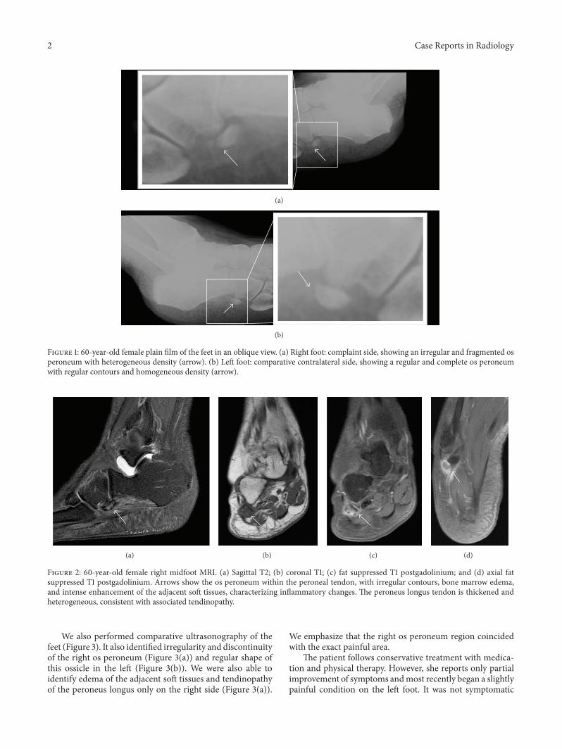
Figure 2: 60-year-old female right midfoot MRI. (a) Sagittal T2; (b) coronal T1; (c) fat suppressed T1 postgadolinium; and (d) axial fatsuppressed T1 postgadolinium. Arrows show the os peroneum within the peroneal tendon, with irregular contours, bone marrow edema,and intense enhancement of the adjacent soft tissues, characterizing inflammatory changes. The peroneus longus tendon is thickened andheterogeneous, consistent with associated tendinopathy.
We also performed comparative ultrasonography of thefeet (Figure 3). It also identified irregularity and discontinuityof the right os peroneum (Figure 3(a)) and regular shape ofthis ossicle in the left (Figure 3(b)). We were also able toidentify edema of the adjacent soft tissues and tendinopathyof the peroneus longus only on the right side (Figure 3(a)).
We emphasize that the right os peroneum region coincidedwith the exact painful area.
The patient follows conservative treatment with medica-tion and physical therapy. However, she reports only partialimprovement of symptoms andmost recently began a slightlypainful condition on the left foot. It was not symptomatic

Case Reports in Radiology 3
∗
(a)
∗
(b)
Figure 3: 60-year-old female ultrasonography of the long axis of the peroneus longus tendon. (a) Right foot: complaint side demonstratinga thickened and heterogeneous peroneus longus tendon (asterisk) and irregular and fragmented os peroneum, associated with swelling ofthe surrounding soft tissues. (b) Left foot: contralateral side for comparison, demonstrating a preserved echotexture of the peroneus longustendon (asterisk) and regular contours of the os peroneum without changes in the surrounding soft tissues.
when the imaging studies presented in this report wereperformed.
3. Discussion
There are different sesamoids and accessory ossicles in theskeleton. Some of them are known to be associated withpainful syndromes, such as os trigonum, os navicular, andfabela.These syndromesmay be caused by different etiologiessuch as trauma, infection, impact, and degenerative changes[3].
The os peroneum is an accessory ossicle, round or oval,within the substance of the peroneal tendon [1], and can beclassified accordingly to Nwawka et al. and Blitz and Nemesas a sesamoid [2, 4]. Its histological structure is composed ofdifferent degrees of ossification and fibrous tissue [5].
The peroneus longus tendon is located proximal andposteriorly to the lateralmalleolus on the lateral surface of thecalcaneus, cuboid (along the midfoot), and distally insertingat the base of the first metatarsal and medial cuneiform [1, 6].
There are several causes for pain in the lateral aspect ofthe foot, including dislocation or subluxation of the peronealtendon, injury, to the talofibular ligament or calcaneofibularligament, or fractures in the fifth metatarsal, anterior processof the calcaneus, or cuboid [1].
The os peroneum fracturemay be complicated by ruptureof the peroneus longus tendon. The most common mecha-nism occurs with a strong contraction of the peroneus longusmuscle in response to a sudden inversion or supination.Such contraction can compress the os peroneum against thecuboid, resulting in fracture and rupture of the peroneuslongus tendon. It has been suggested that the presence ofthis ossicle can predispose to its distal rupture due to poten-tial increased friction with adjacent structures [7]. Physicalexamination can reveal swelling over the cuboid, with painin this area during palpation. The pain is usually exacerbatedby plantar flexion and heel elevation stage during gait [7].
POPS has two main forms: acute and chronic. The acuteform occurs as a result of trauma, commonly with anklesprain or supination movement, resulting in fracture or dias-tasis of the os peroneum, whichmay ormay not be associatedwith peroneus longus tendon rupture. Chronic presentation
is closely linked to a healing process of a fracture withsubsequent calcification, remodeling, or chronic diastasis ofthe os peroneum with a variable frequency of tenosynovitisof the peroneus longus tendon.
With MRI, the ossicle is usually isointense to bonemarrow and presents with increased intrasubstantial signalwithin the peroneus longus tendon, typically close to thecuboid. Under ultrasonography, its identification is easilyappreciated because of its typical bone appearance, as acurved echogenic focus with posterior acoustic shadow [8].
On radiographs, it is better identified in an oblique viewof the foot. Both radiography and computed tomographymaydemonstrate displacement of the os peroneum from its usualposition, fracture, or diastasis of a bipartite sesamoid. Thedisplacement of the os peroneum is an indirect sign of aperoneal tendon rupture [2].
The radiographic differentiation between a fractured orsplit os peroneummay be difficult. In an acute event, fracturemargins seem relatively nonsclerotic and bone fragmentsgenerally fit together, as “pieces of a puzzle.” In the bipartitesesamoid, margins become rounded and sclerotic. It is possi-ble that over time due to remodeling, the edges of the fractureresemble the appearance of a split os peroneum. Brigido et al.suggested that a diastasis between fragments of os peroneum,greater than five millimeters, must indicate the diagnosis offracture. US and MRI can also be used, especially to evaluateother possible associated abnormalities.
In the same study by Brigido et al., all bone fragmentsidentified with US were hyperechogenic [7]. The evaluationof sesamoid fractures with MRI is difficult because of theirsmall size and low signal. Bone marrow swelling can alsocomplicate evaluation of fractures due to abnormal marrowsignal intensity.
Therefore, early diagnosis and correct characterizationof POPS are essential for an adequate management of thesepatients. Knowledge of its presentation through differentimaging methods is very important during training of spe-cialists in Radiology and Diagnostic Imaging.
Competing Interests
The authors declare that they have no conflict of interests.

4 Case Reports in Radiology
References
[1] S. J. Oh, Y. H. Kim, S. K. Kim, and M.-W. Kim, “Painful osperoneum syndrome presenting as lateral plantar foot pain,”Annals of Rehabilitation Medicine, vol. 36, no. 1, pp. 163–166,2012.
[2] O. K. Nwawka, D. Hayashi, L. E. Diaz et al., “Sesamoids andaccessory ossicles of the foot: anatomical variability and relatedpathology,” Insights into Imaging, vol. 4, no. 5, pp. 581–593, 2013.
[3] A. R. Barreto, F. A. Chagas-Neto, M. D. Crema et al., “Fractureof the fabella: a rare injury in knee trauma,” Case Reports inRadiology, vol. 2012, Article ID 390150, 3 pages, 2012.
[4] N. M. Blitz and K. K. Nemes, “Bilateral peroneus longus tendonrupture through a bipartite os peroneum,” The Journal of Footand Ankle Surgery, vol. 46, no. 4, pp. 270–277, 2007.
[5] C. M. Sofka, R. S. Adler, G. R. Saboeiro, and H. Pavlov, “Sono-graphic evaluation and sonographic-guided therapeutic optionsof lateral ankle pain: peroneal tendon pathology associated withthe presence of an os peroneum,” HSS Journal, vol. 6, no. 2, pp.177–181, 2010.
[6] K. L. Moore, A. F. Dalley, and C. L. C. de Araujo, AnatomiaOrientada Para a Clınica, Guanabara Koogan, 2006.
[7] M. K. Brigido, D. P. Fessell, J. A. Jacobson et al., “Radiographyand US of os peroneum fractures and associated peronealtendon injuries: initial experience,” Radiology, vol. 237, no. 1, pp.235–241, 2005.
[8] A. Donovan, Z. S. Rosenberg, J. T. Bencardino et al., “Plantartendons of the foot: MR imaging and US,” Radiographics, vol.33, no. 7, pp. 2065–2085, 2013.

Submit your manuscripts athttp://www.hindawi.com
Stem CellsInternational
Hindawi Publishing Corporationhttp://www.hindawi.com Volume 2014
Hindawi Publishing Corporationhttp://www.hindawi.com Volume 2014
MEDIATORSINFLAMMATION
of
Hindawi Publishing Corporationhttp://www.hindawi.com Volume 2014
Behavioural Neurology
EndocrinologyInternational Journal of
Hindawi Publishing Corporationhttp://www.hindawi.com Volume 2014
Hindawi Publishing Corporationhttp://www.hindawi.com Volume 2014
Disease Markers
Hindawi Publishing Corporationhttp://www.hindawi.com Volume 2014
BioMed Research International
OncologyJournal of
Hindawi Publishing Corporationhttp://www.hindawi.com Volume 2014
Hindawi Publishing Corporationhttp://www.hindawi.com Volume 2014
Oxidative Medicine and Cellular Longevity
Hindawi Publishing Corporationhttp://www.hindawi.com Volume 2014
PPAR Research
The Scientific World JournalHindawi Publishing Corporation http://www.hindawi.com Volume 2014
Immunology ResearchHindawi Publishing Corporationhttp://www.hindawi.com Volume 2014
Journal of
ObesityJournal of
Hindawi Publishing Corporationhttp://www.hindawi.com Volume 2014
Hindawi Publishing Corporationhttp://www.hindawi.com Volume 2014
Computational and Mathematical Methods in Medicine
OphthalmologyJournal of
Hindawi Publishing Corporationhttp://www.hindawi.com Volume 2014
Diabetes ResearchJournal of
Hindawi Publishing Corporationhttp://www.hindawi.com Volume 2014
Hindawi Publishing Corporationhttp://www.hindawi.com Volume 2014
Research and TreatmentAIDS
Hindawi Publishing Corporationhttp://www.hindawi.com Volume 2014
Gastroenterology Research and Practice
Hindawi Publishing Corporationhttp://www.hindawi.com Volume 2014
Parkinson’s Disease
Evidence-Based Complementary and Alternative Medicine
Volume 2014Hindawi Publishing Corporationhttp://www.hindawi.com