Embed Size (px)
Citation preview
This article can be downloaded from www.ijpbs.net
B - 107
International Journal of Pharma and Bio Sciences
P38 MAPK ACTIVATION IN MYOCARDIAL ISCHAEMIA
S. KUMPHUNE, PhD *
Department of Medical Technology, Faculty of Allied Health Sciences, Naresuan University,
Phitsanulok 65000 Thailand
REVIEW ARTICLE BIOCHEMISTRY
ABSTRACT Ischaemic heart disease remains, and is likely to continue to be, the leading life
threatening disease around the world. Signaling pathways have become more
interesting as novel therapeutic targets in ischaemic heart disease. However, one
needs to be very careful in picking the therapeutic target as one signaling molecule
can activate and also cross-talk with other kinases. The activation of the p38-MAPK
during myocardial ischaemia aggravates lethal injury. Recent evidences suggested
the mechanism of p38-MAPK activation may differ by circumstances. Determining the
precise mechanisms is crucial since it may allow prevention of the detrimental, but not
the beneficial, and lead to the identification of the relevant downstream signals.
Therefore, p38 MAPK may be a viable clinical target and form the basis of future
studies designed to further dissect the signaling pathways and discover the
downstream substrates will become hopes as a new frontier of therapeutic approach
in ischaemic heart diseases.

This article can be downloaded from www.ijpbs.net
B - 108
KEY WORDS p38 MAPK; Myocardial Ischaemia; Autophosphorylation; Transphosphorylation
INTRODUCTION Cardiovascular diseases (CVD), principally heart disease and stroke, are a leading cause of global morbidity and mortality. The World Health Organization (WHO) statistic report 2008 predicted that, although non-communicable diseases are considered the leading killers worldwide, currently, ischemic heart disease and cerebrovascular disease (stroke) will become the 2 diseases resulting in the majority of deaths in 2030 1. Most seriously, ischemic heart disease is still ranked on the top of the mortality, by cause, from 2004 to 2030. Although the annual mortality from this disease is decreasing in developed countries 2 it is increasing in the more populace developing countries 3. Ischaemic heart disease, otherwise known as coronary artery disease, is a condition that affects the supply of blood to the heart, which is essential for proper functioning of the heart. This may eventually result in a portion of the heart being suddenly deprived of its blood supply leading to the death of that area of heart tissue, resulting in a heart attack or acute myocardial infarction. Currently, the most efficient method of reducing mortality in such patients is to achieve rapid reperfusion by lysis or mechanical disruption of the occlusive coronary thrombus and plaque. The mortality from acute myocardial infarction under these circumstances is inversely related to the amount of myocardial salvage achieved by reperfusion,4 so any intervention that slow the rate of ischaemic necrosis are likely to save many lives.5 There are many evidences demonstrated that the activation of p38 MAPK that occurs during prolonged ischaemia6 accelerates injury. The information achieved from p38 MAPK inhibition by pharmacological inhibitors7-32 or genetic modified to knock down p38 MAPK or its downstream effectors 33-35 means slows the rate of infarction/death. Although this evidence is based on animal data it seems likely similar mechanisms operate in the
human heart since p38 MAPK is identically activated by ischaemia36, 37 and early clinical trials indicate a potential benefit. Thus, in theory at least, inhibitors of p38 MAPK have therapeutic potential in ischemic heart disease38. GENERAL BACKGROUND OF P38 MAPK The p38 MAPK, (also known as CSBP, mHOG1, RK, and SAPK2)39, 40 is a family of serine/threonine protein kinases that plays an important role in cellular responses to external stress signaling and also function in many cellular processes including inflammation, cell differentiation, cell growth and death. The human p38 MAPK was originally isolated as a 38-kDa protein rapidly tyrosine phosphorylated in response to lipopolysaccharide (LPS)-stimulation in human monocytes 40 . Moreover, it was also identified as the target of a pyridinyl imidazole
drug that blocked production of tumour
necrosis factor-α (TNFα) and as such was called cytokine-suppressive anti-inflammatory drug-binding protein or CSBP 40. There are 4 isoforms of p38 MAPK that have
been identified including p38 α, β, γ, and δ. Of
these, p38α (or MAPK14) is the best characterized and perhaps the most physiologically relevant kinase involved in inflammatory responses, and found that has
more than 70% identity of p38β (or MAPK
11)41, 42. Both α and β are ubiquitously expressed in several tissues, but are thought to have different functions 41, 42. Moreover, these 2 isoforms are sensitive to p38 MAPK inhibition by a pyridinyl imidazole inhibitor.
p38γ (also known as SAPK3 and MAPK12) is largely expressed in skeletal muscle,
whereas p38δ (also known as SAPK4 or MAPK13) 41-45 is expressed more widely in several adult tissues and during development. Sequence comparisons revealed that each p38 isoform has more

This article can be downloaded from www.ijpbs.net
B - 109
than 69% identity within this group, but only 40 to 45% to the other MAP kinase family members 46. These 2 isoform are different
from α and β in term of sensitivity to pyridinyl
imidazole inhibitor, as γ and δ found to insensitive to kinase inhibition by this family of inhibitor.
ACTIVATION OF P38 MAPK AND ITS ACTIVITY Similar to other MAPKs pathway, p38 MAPK is activated as a result of phosphorylation at specific sites by upstream family of MAPKKK and MAPKK, consequently. p38 activation in response to a variety of extracellular stimuli can be observed by the diverse range of MAP kinase kinase kinases (MAP3Ks) that participate in p38 activation. These include TAK1 47, ASK1/MAPKKK5 48, DLK/MUK/ZPK 49, 50, and MEKK4 49, 51, 52. Subsequently activation downstream of MAP3Ks is a selective activation of MAP kinase kinases
(MKKs) or MKKs, which lie upstream of p38 MAPK. This MKKs are selectively dual phosphorylated p38 MAPK at specific phosphorylation sites, Thr180 and Tyr182. There are two main MKKs that are known to activate p38, MKK3 and MKK6 (Figure 1). MKK induced dual phosphorylation of p38 at Thr180 and Tyr182 is thought to cause the activation loop to refold 53 and move out of the peptide-binding channel. This movement is then thought to exert a “crank-handle” effect on the overall tertiary structure of the kinase reorientating, which enabling the cooperation necessary for ATP binding 54. In support of this model Diskin et al., based on a gain of function mutagenesis screen of the yeast p38 homologue, demonstrated p38 MAPK activation can also be achieved in the absence of the dual phosphorylation of Thr180 and Tyr182 by mutations that similarly disrupt their hydrophobic environment 55.
Figure 1 p38 MAPK activators and substrates.
This article can be downloaded from www.ijpbs.net
B - 110
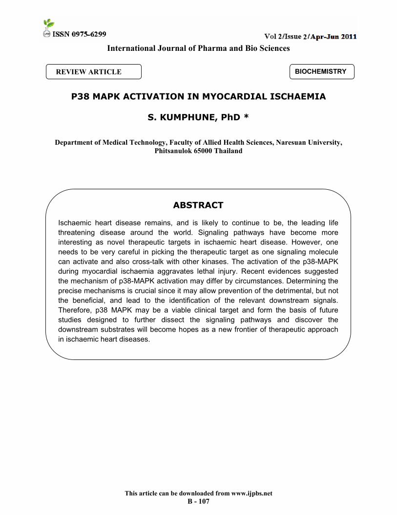
When p38 MAPK is in the non-dual phosphorylated, inactive conformation, the loop is thought to reside in the peptide-binding channel that lies in the cleft between amino- and carboxy-terminal lobes of the kinase. In addition, in p38 MAPK, in contrast to p42/44-MAPK, there is a misalignment of these lobes, that prevents the co-operation between Lys53, in the N-terminal lobe, and Asp168, in the C-terminal lobe, imperative to the binding and stabilization of the alpha-phosphate group and adjacent ribose of ATP, respectively 56. Therefore, it is widely thought that the nondual phospho- form of p38 is inactive as a result of steric obstruction of the peptide-binding channel and low ATP affinity 56. After p38 MAPK dual phosphorylation, the active form of p38 MAPK can access to ATP
and use its kinase activity to phosphorylated specific downstream substrates. The MAPKs have a substrate preference, but not an absolute requirements, for sites containing a serine or threonine followed by a proline residue 57. There are 2 distinct types of p38 MAPK downstream substrate including protein kinases and transcription factors. MAP kinase-activated protein kinase 2 (MAPAP-K2 or M2) was the first identified p38 MAPK substrate 58, 59 (Figure 1). This substrate has kinase activity itself and is shown to phosphorylate various substrates including small heat shock protein 27 (HSP27) 60, lymphocyte-specific protein 1 (LSP1) 61, cAMP responsive element-binding protein (CREB) 62, Serum Responsive Factor (SRF) 63 and tyrosine hydroxylase 64.
Figure 2 Structure of p38 MAPK. A, electron density around the phosphorylation sites Thr180 and Y182. B, Ribbon diagram of p38. C, the specific pocket and phosphorylation lip region of
p38. This figure modified from Wang et al, 1997.65
MECHANISMS OF P38 MAPK Transphosphorylation External stimuli initiate the activation of the serine/threonine MAP kinase kinase kinases (MAPKKKs) that are upstream of p38. Theses kinases phosphorylate and activate the dual-specificity kinases MAPKK (MKK),
which in turn phosphorylate p38 on Thr180 and Tyr182 (Figure 3) Several upstream MAPKKs (MKK) have been identified from in vitro analysis, including MKK3 and MKK6 66, 67. Mice lacking both MKK3 and MKK6 are not viable, dying in midgestation with defects in the placenta and

This article can be downloaded from www.ijpbs.net
B - 111
the embryonic vasculature 68 Moreover, Deacon and Blank 69 showed that MKK4, which is a MAPKK involved in JNK activation, can also activate p38 in vitro.. However, p38 activation is not limited to this traditional phospho-relay signaling cascade. Since SB203580, the most widely used p38 kinase inhibitor occupies the catalytic site, without inhibiting upstream MKK, it should only inhibit the phosphorylation events downstream of p38 without inhibiting the dual-
phosphorylation of p38 itself 70. Nonetheless the literature is replete with examples of SB203580, and structurally related compounds 71, inhibiting the dual phosphorylation of p38 MAPK 72-77. Two possible explanations of these findings are p38 is able to autophosphorylation its activation loop or the inhibitory effect of SB203580 is on a kinase upstream of p38 involved in its activation by transphosphorylation.
Figure 3 Schematic representation of the transphosphorylation mechanism of p38 activation.
Autophosphorylation It has long been known that, as with other MAPKs, p38 can autophosphorylate in vitro to a limited extent 78, 79. The degree of autophosphorylation is influenced by the length of the activation loop containing the TGY phosphorylation motif and the identity of the non-phosphorylated intervening residue (for example, autophosphorylation is more likely to occur if there is an intervening aspartic acid residue rather than a glycine residue) 78, 79.
Ge et al reported the MKK independent activation of p38 MAPK mediated by the interaction of scaffolding protein TAB1 80 (Figure 4). TAB1, or transforming growth
factor-β-activated protein kinase-1 (TAK1) binding protein-1, is a scaffold protein known to associate with TAK1 81 and facilitate TAK1 autophosphorylation 82. TAK1 is also an upstream kinase of MKK3 and MKK6. Through a comprehensive series of experiments in HEK293 cells, Ge et al demonstrated a similar functional interaction

This article can be downloaded from www.ijpbs.net
B - 112
with p38 MAPK 80. For example the ectopic expression of TAB1 caused the dual phosphorylation of wild-type p38 MAPK, but not of p38 mutants rendered kinase dead by substitution of the critical Lys53 or Asp168 residues. This was despite the fact that TAB1 still interacted with these mutants on the basis of co-immunoprecipitation and that the mutants could be transphosphorylated in the presence of constitutively activated MKK3 or MKK6 80. Reciprocally, the TAB1-induced activation of wild-type p38 MAPK could not be prevented by co-expression of dominant negative forms of MKK3, MKK6 and/or TAK1, but could be prevented by SB203580. When
differentially tagged wild-type and kinase dead forms of p38 MAPK were co-incubated with TAB1 only the wildtype form was dual phosphorylated implying that the observations were the result of true, intramolecular, autophosphorylation rather than one p38 MAPK molecule transphosphorylating another 80. The activation of by the autophosphorylation of
p38α after interaction with TAB1 although there is an indication that TAB1-dependent p38 phosphorylation occurs in LPS, TNF, and CpG treated B cell lines.
Figure 4 Schematic representation of the Autophosphorylation mechanism of p38 activation. TAB1
association with p38, induces p38 autophosphorylation in an SB-sensitive manner. Negative Feedback Autophosphorylation In 2003, one year after Ge et al. reported TAB1 mediated autophosphorylation of p38, Cohen’s group independently identified TAB1 as a p38 interacting protein by yeast-two hybrid screening 83. This finding made the interpretation of TAB1-p38 interaction and mediation of p38 activation more complicated as they showed that TAB1 is also phosphorylated on Ser423, Tyr431, and Ser438. Moreover, p38 induced phosphorylation of
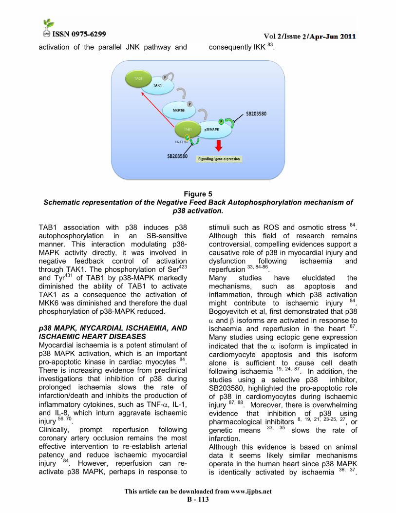
TAB1 on Ser423 and Tyr431 which was inhibited by SB203580 83. Inhibition of p38 mediated phosphorylation of TAB1 (Ser423/Tyr431) by SB203580 enhanced the activity of TAK1, which then activated p38 via MKK3 and MKK6. These findings proposed a feedback control mechanism of TAK1 activity, whereby p38 activity inhibits TAK1, through the phosphorylation of TAB 83 (Figure 5). Inhibition of p38 activity by SB203580 can abolish this feedback control and cause the
This article can be downloaded from www.ijpbs.net
B - 113
activation of the parallel JNK pathway and consequently IKK 83.
Figure 5 Schematic representation of the Negative Feed Back Autophosphorylation mechanism of
p38 activation.
TAB1 association with p38 induces p38 autophosphorylation in an SB-sensitive manner. This interaction modulating p38-MAPK activity directly, it was involved in negative feedback control of activation through TAK1. The phosphorylation of Ser423 and Tyr431 of TAB1 by p38-MAPK markedly diminished the ability of TAB1 to activate TAK1 as a consequence the activation of MKK6 was diminished and therefore the dual phosphorylation of p38-MAPK reduced. p38 MAPK, MYCARDIAL ISCHAEMIA, AND ISCHAEMIC HEART DISEASES Myocardial ischaemia is a potent stimulant of p38 MAPK activation, which is an important pro-apoptotic kinase in cardiac myocytes 84. There is increasing evidence from preclinical investigations that inhibition of p38 during prolonged ischaemia slows the rate of infarction/death and inhibits the production of
inflammatory cytokines, such as TNF-α, IL-1, and IL-8, which inturn aggravate ischaemic injury 56, 70. Clinically, prompt reperfusion following coronary artery occlusion remains the most effective intervention to re-establish arterial patency and reduce ischaemic myocardial injury 84. However, reperfusion can re-activate p38 MAPK, perhaps in response to
stimuli such as ROS and osmotic stress 84. Although this field of research remains controversial, compelling evidences support a causative role of p38 in myocardial injury and dysfunction following ischaemia and reperfusion 33, 84-86. Many studies have elucidated the mechanisms, such as apoptosis and inflammation, through which p38 activation might contribute to ischaemic injury 84. Bogoyevitch et al, first demonstrated that p38
α and β isoforms are activated in response to ischaemia and reperfusion in the heart 87. Many studies using ectopic gene expression
indicated that the α isoform is implicated in cardiomyocyte apoptosis and this isoform alone is sufficient to cause cell death following ischaemia 19, 24, 87. In addition, the studies using a selective p38 inhibitor, SB203580, highlighted the pro-apoptotic role of p38 in cardiomyocytes during ischaemic injury 87, 88. Moreover, there is overwhelming evidence that inhibition of p38 using pharmacological inhibitors 8, 19, 21, 23-25, 27, or genetic means 33, 35 slows the rate of infarction. Although this evidence is based on animal data it seems likely similar mechanisms operate in the human heart since p38 MAPK is identically activated by ischaemia 36, 37.
This article can be downloaded from www.ijpbs.net
B - 114
Thus, superficially at least, inhibitors of p38 MAPK have therapeutic potential in ischaemic heart disease 38. However, a beneficial effect can also follow p38 activation, since under many circumstances its activation leads to protection rather than to injury 89-92. This particularly seems to be the case when myocardial p38 MAPK activation occurs as a consequence of a sub-lethal stress that precedes lethal myocardial ischaemia. For example, in many studies 18, 21, 23 p38 MAPK inhibition during a “conditioning” sub-lethal stress abolishes subsequent ischaemic protection. However in these studies the same inhibitor, at the same concentration, reduces injury if present solely during lethal ischaemic injury 18, 21, 23, 27, 93. The cause of these apparent paradoxical observations is complex but may relate to an attenuation of p38 MAPK activation during lethal ischaemia by prior transient activation 18, 21, 23, 27, 93,
94Furthermore very recently pre-ischaemic activation of p38 MAPK by transgenic overexpression of an upstream kinase led to similar protection 89. However p38 MAPK content was reduced in transgenic hearts possibly resulting in reduced active p38 MAPK during ischaemia and complicating interpretation 89. Whatever the exact underlying mechanism there is ample evidence from the cardiac, 18, 21, 23, 89-92 as well as from others 95, 96 research fields that p38 MAPK activation can have beneficial consequences. However it is also incontrovertible that restricting p38 MAPK inhibition to the activation that accompanies lethal myocardial ischaemia reduces infarction 8, 11, 18, 19, 21, 23-25, 27, 33, 35, 85. Taken together these observations compel a greater understanding of the mechanisms and targets of p38 MAPK activation. Without such an understanding there is a danger of repeating the previous failings made when clinical trials, in the related area of anti-TNF therapy, proceeded without due appreciation of contrary data 95, 97, 98. This has become especially pertinent since a phase 2 clinical trial of p38 MAPK inhibition during acute coronary syndromes has just been completed with a positive result based on a surrogate
primary outcome 38, 99. Again reminiscent of the early experience with anti-TNF therapy 100.
MECHANISMS OF P38 MAPK DURING MYOCARDIAL ISCHAEMIA There are many studies investigated the actually mechanism of p38 MAPK activation occur during myocardial ischaemia. According to studies using pharmacological inhibitor SB203580, there are numerous examples where this has been observed (often inadvertently) during myocardial ischaemia7, 9-11, 28, 31, 101-103. Thus unlike most stresses, myocardial ischaemia seems to reproducibly cause an SB-sensitive form of p38-MAPK dual phosphorylation. Two mutually exclusive explanations for this observation are that p38-MAPK is able to AUTOPHOSPHORYLATION its activation loop or that SB203580 inhibits a kinase upstream of p38-MAPK involved in its activation, by TRANSPHOSPHORYLATION, during ischaemia. In attempt to prove the mechanism of p38 MAPK activation during myocardial ischaemia is achieved by transphosphorylation, Jacquet et al showed that RIP2 Kinase, another upstream kinase of p38 MAPK and known to be inhibited byp38 MAPK inhibitor, SB203580104, does not contribute to p38 MAPK activation in response to myocardial ischaemia105. Although the hypothesis of RIP2 mediated transphosphorylation of p38 in response to ischaemia was not proved, the hypothesis of transphosphorylation p38 MAPK by SB-sensitive upstream kinases is still viable. Thus identification of other cardiac kinases binding to SB203580 is still important for future studies. Recently Kumphune et al, showed the knock-in mice, expressing specific inhibitor resistant
form of p38α (DR), was determined the
biological and physiological function of p38α in myocardial ischaemia 106. The findings
suggested that p38α is the major isoform of p38 MAPK contributing to myocardial ischaemia and support the cardioprotective effect of SB203580 106, and also highlighted that myocardial ischaemia activates p38 MAPK by autophosphorylation.

This article can be downloaded from www.ijpbs.net
B - 115
CONCLUSION Ischaemic heart disease remains, and is likely to continue to be, the leading life threatening disease around the world. Numerous studies from independent groups suggest that the p38-MAPK activation that accompanies myocardial ischaemia aggravates injury. However, under different circumstances activation of this kinase can reduce myocardial injury. The better understand the mechanisms of p38-MAPK activation during myocardial ischaemia is a likely prerequisite to the exploitation of the
wealth of preclinical data that suggests inhibiting this kinase will benefit patients with ischemic heart disease. Using gene manipulation techniques and also transgenic animal models, many studies demonstrated the detrimental role of p38 MAPK activation. Inhibition of p38 activation by specific ATP competitive inhibitor, SB203580, is cardioprotection. However, these findings
need further validate p38α as a viable clinical target and form the basis of future studies designed to further dissect the signaling pathways and discover the downstream substrates responsible for injury.
REFERENCES
1. World Heatlh Organization. World Heath Statistics 2008. http://www who int/whosis/whostat/EN_WHS08_Full pdf, 2009http://www.who.int/entity/whosis/whostat/EN_WHS08_Full.pdf).
2. Unal B, Critchley JA, Capewell S. Explaining the decline in coronary heart disease mortality in England and Wales between 1981 and 2000. Circulation, 109(9):1101-1107, (2004).
3. Levy D, Thom TJ. Death rates from coronary disease--progress and a puzzling paradox. N Engl J Med, 8;339(13):915-917, (1998).
4. Braunwald E. Evolution of the management of acute myocardial infarction: a 20th century saga. Lancet,352(9142):1771-1774, (1998).
5. Braunwald E. Acute myocardial infarction--the value of being prepared. N Engl J Med, 334(1):51-52, (1996).
6. Pombo CM, Bonventre JV, Avruch J, Woodgett JR, Kyriakis JM, Force T. The stress-activated protein kinases are major c-Jun amino-terminal kinases activated by ischemia and reperfusion. J Biol Chem, 269(42):26546-26551, (1994).
7. Aleshin A, Sawa Y, Ono M, Funatsu T, Miyagawa S, Matsuda H. Myocardial protective effect of FR167653; a novel cytokine inhibitor in ischemic-reperfused rat heart. Eur J Cardiothorac Surg, 26(5):974-980, (2004).
8. Barancik M, Htun P, Strohm C, Kilian S, Schaper W. Inhibition of the cardiac p38-MAPK pathway by SB203580 delays ischemic cell death. J Cardiovasc Pharmacol, 35(3):474-483, (2000).
9. Capano M, Crompton M. Bax translocates to mitochondria of heart cells during simulated ischaemia: involvement of AMP-activated and p38 mitogen-activated protein kinases. Biochem J, 395(1):57-64, (2006).
10. Clanachan AS, Jaswal JS, Gandhi M et al. Effects of inhibition of myocardial extracellular-responsive kinase and P38 mitogen-activated protein kinase on mechanical function of rat hearts after prolonged hypothermic ischemia. Transplantation, 75(2):173-180, (2003).
11. Gorog DA, Tanno M, Cao X et al. Inhibition of p38 MAPK activity fails to attenuate contractile dysfunction in a mouse model of low-flow ischemia. Cardiovasc Res, 61(1):123-131, (2004).
12. Gysembergh A, Simkhovich BZ, Kloner RA, Przyklenk K. p38 MAPK activity is not increased early during sustained coronary artery occlusion in preconditioned versus control rabbit heart. J Mol Cell Cardiol, 33(4):681-690, (2001).
13. Kim JK, Pedram A, Razandi M, Levin ER. Estrogen prevents cardiomyocyte apoptosis through inhibition of reactive
This article can be downloaded from www.ijpbs.net
B - 116
oxygen species and differential regulation of p38 kinase isoforms. J Biol Chem, 281(10):6760-6767, (2006).
14. Koike N, Takeyoshi I, Ohki S, Tokumine M, Matsumoto K, Morishita Y. Effects of adding P38 mitogen-activated protein-kinase inhibitor to celsior solution in canine heart transplantation from non-heart-beating donors. Transplantation, 77(2):286-292, (2004).
15. Ma XL, Kumar S, Gao F et al. Inhibition of p38 mitogen-activated protein kinase decreases cardiomyocyte apoptosis and improves cardiac function after myocardial ischemia and reperfusion. Circulation, 99(13):1685-1691, (1999).
16. Mackay K, Mochly-Rosen D. An inhibitor of p38 mitogen-activated protein kinase protects neonatal cardiac myocytes from ischemia. J Biol Chem, 274(10):6272-6279, (1999).
17. Mackay K, Mochly-Rosen D. Involvement of a p38 mitogen-activated protein kinase phosphatase in protecting neonatal rat cardiac myocytes from ischemia. J Mol Cell Cardiol, 32(8):1585-1588, (2000).
18. Marais E, Genade S, Huisamen B, Strijdom JG, Moolman JA, Lochner A. Activation of p38 MAPK induced by a multi-cycle ischaemic preconditioning protocol is associated with attenuated p38 MAPK activity during sustained ischaemia and reperfusion. J Mol Cell Cardiol, 33(4):769-778, (2001).
19. Martin JL, Avkiran M, Quinlan RA, Cohen P, Marber MS. Antiischemic effects of SB203580 are mediated through the inhibition of p38alpha mitogen-activated protein kinase: Evidence from ectopic expression of an inhibition-resistant kinase. Circ Res, 89(9):750-752, (2001).
20. Meldrum DR, Dinarello CA, Cleveland JC, Jr. et al. Hydrogen peroxide induces tumor necrosis factor alpha-mediated cardiac injury by a P38 mitogen-activated protein kinase-dependent mechanism. Surgery, 124(2):291-296, (1998).
21. Nagarkatti DS, Sha'afi RI. Role of p38 MAP kinase in myocardial stress. J Mol Cell Cardiol, 30(8):1651-1664, (1998).
22. Rakhit RD, Kabir AN, Mockridge JW, Saurin A, Marber MS. Role of G proteins and modulation of p38 MAPK activation in the protection by nitric oxide against ischemia-reoxygenation injury. Biochem Biophys Res Commun, 286(5):995-1002, (2001).
23. Sanada S, Kitakaze M, Papst PJ et al. Role of phasic dynamism of p38 mitogen-activated protein kinase activation in ischemic preconditioning of the canine heart. Circ Res, 88(2):175-180, (2001).
24. Saurin AT, Martin JL, Heads RJ et al. The role of differential activation of p38-mitogen-activated protein kinase in preconditioned ventricular myocytes. FASEB J, 14(14):2237-2246, (2000).
25. Schneider S, Chen W, Hou J, Steenbergen C, Murphy E. Inhibition of p38 MAPK alpha/beta reduces ischemic injury and does not block protective effects of preconditioning. Am J Physiol Heart Circ Physiol, 280(2):H499-H508, (2001).
26. Sharov VG, Todor A, Suzuki G, Morita H, Tanhehco EJ, Sabbah HN. Hypoxia, angiotensin-II, and norepinephrine mediated apoptosis is stimulus specific in canine failed cardiomyocytes: a role for p38 MAPK, Fas-L and cyclin D1. Eur J Heart Fail, 5(2):121-129, (2003).
27. Tanno M, Bassi R, Gorog DA et al. Diverse mechanisms of myocardial p38 mitogen-activated protein kinase activation: evidence for MKK-independent activation by a TAB1-associated mechanism contributing to injury during myocardial ischemia. Circ Res, 93(3):254-261, (2003).
28. Wang M, Tsai BM, Turrentine MW, Mahomed Y, Brown JW, Meldrum DR. p38 mitogen activated protein kinase mediates both death signaling and functional depression in the heart. Ann Thorac Surg, 80(6):2235-2241, (2005).
29. Wang M, Tsai BM, Reiger KM, Brown JW, Meldrum DR. 17-beta-Estradiol decreases p38 MAPK-mediated myocardial inflammation and
This article can be downloaded from www.ijpbs.net
B - 117
dysfunction following acute ischemia. J Mol Cell Cardiol, 40(2):205-212, (2006).
30. Xing H, Zhang S, Weinheimer C, Kovacs A, Muslin AJ. 14-3-3 proteins block apoptosis and differentially regulate MAPK cascades. EMBO J, 19(3):349-358, (2000).
31. Yada M, Shimamoto A, Hampton CR et al. FR167653 diminishes infarct size in a murine model of myocardial ischemia-reperfusion injury. J Thorac Cardiovasc Surg, 128(4):588-594, (2004).
32. Yue TL, Wang C, Gu JL et al. Inhibition of extracellular signal-regulated kinase enhances Ischemia/Reoxygenation-induced apoptosis in cultured cardiac myocytes and exaggerates reperfusion injury in isolated perfused heart. Circ Res, 86(6):692-699, (2000).
33. Kaiser RA, Bueno OF, Lips DJ et al. Targeted inhibition of p38 mitogen-activated protein kinase antagonizes cardiac injury and cell death following ischemia-reperfusion in vivo. J Biol Chem, 279(15):15524-15530, (2004).
34. Melendez J, Turner C, Avraham H, Steinberg SF, Schaefer E, Sussman MA. Cardiomyocyte apoptosis triggered by RAFTK/pyk2 via Src kinase is antagonized by paxillin. J Biol Chem, 279(51):53516-53523, (2004).
35. Otsu K, Yamashita N, Nishida K et al. Disruption of a single copy of the p38alpha MAP kinase gene leads to cardioprotection against ischemia-reperfusion. Biochem Biophys Res Commun, 302(1):56-60, (2003).
36. Cook SA, Sugden PH, Clerk A. Activation of c-Jun N-terminal kinases and p38-mitogen-activated protein kinases in human heart failure secondary to ischaemic heart disease. J Mol Cell Cardiol, 31(8):1429-1434, (1999).
37. Corbucci GG, Perrino C, Donato G et al. Transient and reversible deoxyribonucleic acid damage in human left ventricle under controlled ischemia and reperfusion. J Am Coll Cardiol, 43(11):1992-1999, (2004).
38. Force T, Kuida K, Namchuk M, Parang K, Kyriakis JM. Inhibitors of protein kinase
signaling pathways: emerging therapies for cardiovascular disease. Circulation, 109(10):1196-1205, (2004).
39. Han J, Lee JD, Bibbs L, Ulevitch RJ. A MAP kinase targeted by endotoxin and hyperosmolarity in mammalian cells. Science, 265(5173):808-811, (1994).
40. Lee JC, Laydon JT, McDonnell PC et al. A protein kinase involved in the regulation of inflammatory cytokine biosynthesis. Nature, 372(6508):739-746, (1994).
41. Jiang Y, Chen C, Li Z et al. Characterization of the structure and function of a new mitogen-activated protein kinase (p38beta). J Biol Chem, 271(30):17920-17926, (1996).
42. Kumar S, McDonnell PC, Gum RJ, Hand AT, Lee JC, Young PR. Novel homologues of CSBP/p38 MAP kinase: activation, substrate specificity and sensitivity to inhibition by pyridinyl imidazoles. Biochem Biophys Res Commun, 235(3):533-538, (1997).
43. Li Z, Jiang Y, Ulevitch RJ, Han J. The primary structure of p38 gamma: a new member of p38 group of MAP kinases. Biochem Biophys Res Commun, 228(2):334-340, (1996).
44. Mertens S, Craxton M, Goedert M. SAP kinase-3, a new member of the family of mammalian stress-activated protein kinases. FEBS Lett, 383(3):273-276, (1996).
45. Cuenda A, Cohen P, Buee-Scherrer V, Goedert M. Activation of stress-activated protein kinase-3 (SAPK3) by cytokines and cellular stresses is mediated via SAPKK3 (MKK6); comparison of the specificities of SAPK3 and SAPK2 (RK/p38). EMBO J, 16(2):295-305, (1997).
46. Ono K, Han J. The p38 signal transduction pathway: activation and function. Cell Signal, 12(1):1-13, (2000).
47. Moriguchi T, Kuroyanagi N, Yamaguchi K et al. A novel kinase cascade mediated by mitogen-activated protein kinase kinase 6 and MKK3. J Biol Chem, 271(23):13675-13679, (1996).
This article can be downloaded from www.ijpbs.net
B - 118
48. Ichijo H, Nishida E, Irie K et al. Induction of apoptosis by ASK1, a mammalian MAPKKK that activates SAPK/JNK and p38 signaling pathways. Science, 275(5296):90-94, (1997).
49. Hirai S, Katoh M, Terada M et al. MST/MLK2, a member of the mixed lineage kinase family, directly phosphorylates and activates SEK1, an activator of c-Jun N-terminal kinase/stress-activated protein kinase. J Biol Chem, 272(24):15167-15173, (1997).
50. Ubeda M, Wang XZ, Zinszner H, Wu I, Habener JF, Ron D. Stress-induced binding of the transcriptional factor CHOP to a novel DNA control element. Mol Cell Biol, 16(4):1479-1489, (1996).
51. Cuenda A, Dorow DS. Differential activation of stress-activated protein kinase kinases SKK4/MKK7 and SKK1/MKK4 by the mixed-lineage kinase-2 and mitogen-activated protein kinase kinase (MKK) kinase-1. Biochem J, 333 ( Pt 1):11-15, (1998).
52. Takekawa M, Posas F, Saito H. A human homolog of the yeast Ssk2/Ssk22 MAP kinase kinase kinases, MTK1, mediates stress-induced activation of the p38 and JNK pathways. EMBO J, 16(16):4973-4982, (1997).
53. Canagarajah BJ, Khokhlatchev A, Cobb MH, Goldsmith EJ. Activation mechanism of the MAP kinase ERK2 by dual phosphorylation. Cell, 90(5):859-869, (1997).
54. Wilson KP, Fitzgibbon MJ, Caron PR et al. Crystal structure of p38 mitogen-activated protein kinase. J Biol Chem, 271(44):27696-27700, (1996).
55. Diskin R, Askari N, Capone R, Engelberg D, Livnah O. Active mutants of the human p38alpha mitogen-activated protein kinase. J Biol Chem, 279(45):47040-47049, (2004).
56. Clark JE, Sarafraz N, Marber MS. Potential of p38-MAPK inhibitors in the treatment of ischaemic heart disease. Pharmacol Ther, 116(2):192-206, (2007).
57. Songyang Z, Lu KP, Kwon YT et al. A structural basis for substrate specificities of protein Ser/Thr kinases: primary
sequence preference of casein kinases I and II, NIMA, phosphorylase kinase, calmodulin-dependent kinase II, CDK5, and Erk1. Mol Cell Biol, 16(11):6486-6493, (1996).
58. Freshney NW, Rawlinson L, Guesdon F et al. Interleukin-1 activates a novel protein kinase cascade that results in the phosphorylation of Hsp27. Cell, 78(6):1039-1049, (1994).
59. Rouse J, Cohen P, Trigon S et al. A novel kinase cascade triggered by stress and heat shock that stimulates MAPKAP kinase-2 and phosphorylation of the small heat shock proteins. Cell, 78(6):1027-1037, (1994).
60. Stokoe D, Engel K, Campbell DG, Cohen P, Gaestel M. Identification of MAPKAP kinase 2 as a major enzyme responsible for the phosphorylation of the small mammalian heat shock proteins. FEBS Lett, 313(3):307-313, (1992).
61. Huang CK, Zhan L, Ai Y, Jongstra J. LSP1 is the major substrate for mitogen-activated protein kinase-activated protein kinase 2 in human neutrophils. J Biol Chem, 272(1):17-19, (1997).
62. Tan Y, Rouse J, Zhang A, Cariati S, Cohen P, Comb MJ. FGF and stress regulate CREB and ATF-1 via a pathway involving p38 MAP kinase and MAPKAP kinase-2. EMBO J, 15(17):4629-4642, (1996).
63. Heidenreich O, Neininger A, Schratt G et al. MAPKAP kinase 2 phosphorylates serum response factor in vitro and in vivo. J Biol Chem, 274(20):14434-14443, (1999).
64. Thomas GM, Haavik J, Cohen P. A stress-activated kinase cascade can mediate the activation of tyrosine hydroxylase in chromaffin cells. Biochem Soc Trans, 25(4):S571, (1997).
65. Wang Z, Harkins PC, Ulevitch RJ, Han J, Cobb MH, Goldsmith EJ. The structure of mitogen-activated protein kinase p38 at 2.1-A resolution. Proc Natl Acad Sci U S A, 94(6):2327-2332, (1997).
This article can be downloaded from www.ijpbs.net
B - 119
66. Derijard B, Raingeaud J, Barrett T et al. Independent human MAP-kinase signal transduction pathways defined by MEK and MKK isoforms. Science, 267(5198):682-685, (1995).
67. Han J, Lee JD, Jiang Y, Li Z, Feng L, Ulevitch RJ. Characterization of the structure and function of a novel MAP kinase kinase (MKK6). J Biol Chem, 271(6):2886-2891, (1996).
68. Brancho D, Tanaka N, Jaeschke A et al. Mechanism of p38 MAP kinase activation in vivo. Genes Dev, 17(16):1969-1978, (2003).
69. Deacon K, Blank JL. Characterization of the mitogen-activated protein kinase kinase 4 (MKK4)/c-Jun NH2-terminal kinase 1 and MKK3/p38 pathways regulated by MEK kinases 2 and 3. MEK kinase 3 activates MKK3 but does not cause activation of p38 kinase in vivo. J Biol Chem, 272(22):14489-14496, (1997).
70. Young PR, McLaughlin MM, Kumar S et al. Pyridinyl imidazole inhibitors of p38 mitogen-activated protein kinase bind in the ATP site. J Biol Chem, 272(18):12116-12121, (1997).
71. Takahashi S, Keto Y, Fujita T, Uchiyama T, Yamamoto A. FR167653, a p38 mitogen-activated protein kinase inhibitor, prevents Helicobacter pylori-induced gastritis in Mongolian gerbils. J Pharmacol Exp Ther, 296(1):48-56, (2001).
72. Zhuang S, Demirs JT, Kochevar IE. p38 mitogen-activated protein kinase mediates bid cleavage, mitochondrial dysfunction, and caspase-3 activation during apoptosis induced by singlet oxygen but not by hydrogen peroxide. J Biol Chem, 275(34):25939-25948, (2000).
73. Frantz B, Klatt T, Pang M et al. The activation state of p38 mitogen-activated protein kinase determines the efficiency of ATP competition for pyridinylimidazole inhibitor binding. Biochemistry, 37(39):13846-13853, (1998).
74. Galan A, Garcia-Bermejo ML, Troyano A et al. Stimulation of p38 mitogen-activated protein kinase is an early regulatory event for the cadmium-induced
apoptosis in human promonocytic cells. J Biol Chem, 275(15):11418-11424, (2000).
75. Matsuguchi T, Musikacharoen T, Ogawa T, Yoshikai Y. Gene expressions of Toll-like receptor 2, but not Toll-like receptor 4, is induced by LPS and inflammatory cytokines in mouse macrophages. J Immunol, 165(10):5767-5772, (2000).
76. Huang H, Rose JL, Hoyt DG. p38 Mitogen-activated protein kinase mediates synergistic induction of inducible nitric-oxide synthase by lipopolysaccharide and interferon-gamma through signal transducer and activator of transcription 1 Ser727 phosphorylation in murine aortic endothelial cells. Mol Pharmacol, 66(2):302-311, (2004).
77. Hata K, Nishimura R, Ikeda F et al. Differential roles of Smad1 and p38 kinase in regulation of peroxisome proliferator-activating receptor gamma during bone morphogenetic protein 2-induced adipogenesis. Mol Biol Cell, 14(2):545-555, (2003).
78. Ashwell JD. The many paths to p38 mitogen-activated protein kinase activation in the immune system. Nat Rev Immunol, 6(7):532-540, (2006).
79. Jiang Y, Li Z, Schwarz EM et al. Structure-function studies of p38 mitogen-activated protein kinase. Loop 12 influences substrate specificity and autophosphorylation, but not upstream kinase selection. J Biol Chem, 272(17):11096-11102, (1997).
80. Ge B, Gram H, Di PF et al. MAPKK-independent activation of p38alpha mediated by TAB1-dependent autophosphorylation of p38alpha. Science, 295(5558):1291-1294, (2002).
81. Shibuya H, Yamaguchi K, Shirakabe K et al. TAB1: an activator of the TAK1 MAPKKK in TGF-beta signal transduction. Science, 272(5265):1179-1182, (1996).
82. Kishimoto K, Matsumoto K, Ninomiya-Tsuji J. TAK1 mitogen-activated protein kinase kinase kinase is activated by autophosphorylation within its activation
This article can be downloaded from www.ijpbs.net
B - 120
loop. J Biol Chem, 275(10):7359-7364, (2000).
83. Cheung PC, Campbell DG, Nebreda AR, Cohen P. Feedback control of the protein kinase TAK1 by SAPK2a/p38alpha. EMBO J, 22(21):5793-5805, (2003).
84. See F., Kompa A., Krum H. p38 MAP kinase as a therapeutic target in cardiovascular disease. Drug Discovery Today: Therapeutic Strategies, 1(2):149-154, (2004).
85. Kaiser RA, Lyons JM, Duffy JY et al. Inhibition of p38 reduces myocardial infarction injury in the mouse but not pig after ischemia-reperfusion. Am J Physiol Heart Circ Physiol, 289(6):H2747-H2751, (2005).
86. See F, Thomas W, Way K et al. p38 mitogen-activated protein kinase inhibition improves cardiac function and attenuates left ventricular remodeling following myocardial infarction in the rat. J Am Coll Cardiol, 44(8):1679-1689, (2004).
87. Bogoyevitch MA, Gillespie-Brown J, Ketterman AJ et al. Stimulation of the stress-activated mitogen-activated protein kinase subfamilies in perfused heart. p38/RK mitogen-activated protein kinases and c-Jun N-terminal kinases are activated by ischemia/reperfusion. Circ Res, 79(2):162-173, (1996).
88. Wang Y, Huang S, Sah VP et al. Cardiac muscle cell hypertrophy and apoptosis induced by distinct members of the p38 mitogen-activated protein kinase family. J Biol Chem, 273(4):2161-2168, (1998).
89. Martindale JJ, Wall JA, Martinez-Longoria DM et al. Overexpression of mitogen-activated protein kinase kinase 6 in the heart improves functional recovery from ischemia in vitro and protects against myocardial infarction in vivo. J Biol Chem, 280(1):669-676, (2005).
90. Craig R, Larkin A, Mingo AM et al. p38 MAPK and NF-kappa B collaborate to induce interleukin-6 gene expression and release. Evidence for a cytoprotective autocrine signaling pathway in a cardiac myocyte model system. J Biol Chem, 275(31):23814-23824, (2000).
91. Weinbrenner C, Liu GS, Cohen MV, Downey JM. Phosphorylation of tyrosine 182 of p38 mitogen-activated protein kinase correlates with the protection of preconditioning in the rabbit heart. J Mol Cell Cardiol, 29(9):2383-2391, (1997).
92. Communal C, Colucci WS, Singh K. p38 mitogen-activated protein kinase pathway protects adult rat ventricular myocytes against beta -adrenergic receptor-stimulated apoptosis. Evidence for Gi-dependent activation. J Biol Chem, 275(25):19395-19400, (2000).
93. Sanada S, Kitakaze M. Ischemic preconditioning: emerging evidence, controversy, and translational trials. Int J Cardiol, 97(2):263-276, (2004).
94. Marais E, Genade S, Salie R et al. The temporal relationship between p38 MAPK and HSP27 activation in ischaemic and pharmacological preconditioning. Basic Res Cardiol, 100(1):35-47, (2005).
95. Bulavin DV, Fornace AJ, Jr. p38 MAP kinase's emerging role as a tumor suppressor. Adv Cancer Res, 92:95-118, (2004).
96. Zheng S, Zuo Z. Isoflurane preconditioning induces neuroprotection against ischemia via activation of P38 mitogen-activated protein kinases. Mol Pharmacol, 65(5):1172-1180, (2004).
97. Mann DL. Stress-activated cytokines and the heart: from adaptation to maladaptation. Annu Rev Physiol, 65:81-101, (2003).
98. Mann DL, McMurray JJ, Packer M et al. Targeted anticytokine therapy in patients with chronic heart failure: results of the Randomized Etanercept Worldwide Evaluation (RENEWAL). Circulation, 109(13):1594-1602, (2004).
99. Preliminary Phase IIa Data for VX-702 Demonstrate Tolerability and Reduction in C-Reactive Protein in Cardiovascular Patients. http://investors vrtx com/releasedetail cfm?ReleaseID=233083, 2004http://investors.vrtx.com/releasedetail.cfm?ReleaseID=233083).
This article can be downloaded from www.ijpbs.net
B - 121
100. Bozkurt B, Torre-Amione G, Warren MS et al. Results of targeted anti-tumor necrosis factor therapy with etanercept (ENBREL) in patients with advanced heart failure. Circulation 2001;103(8):1044-1047.
101. House SL, Branch K, Newman G, Doetschman T, Schultz JJ. Cardioprotection induced by cardiac-specific overexpression of fibroblast growth factor-2 is mediated by the MAPK cascade. Am J Physiol Heart Circ Physiol, 289(5):H2167-H2175, (2005).
102. Lau JM, Jin X, Ren J et al. The 14-3-3tau phosphoserine-binding protein is required for cardiomyocyte survival. Mol Cell Biol, 27(4):1455-1466, (2007).
103. Maulik N, Yoshida T, Zu YL, Sato M, Banerjee A, Das DK. Ischemic preconditioning triggers tyrosine kinase
signaling: a potential role for MAPKAP kinase 2. Am J Physiol, 275(5 Pt 2):H1857-H1864, (1998).
104. Godl K, Wissing J, Kurtenbach A et al. An efficient proteomics method to identify the cellular targets of protein kinase inhibitors. Proc Natl Acad Sci U S A, 100(26):15434-15439, (2003).
105. Jacquet S, Nishino Y, Kumphune S et al. The role of RIP2 in p38 MAPK activation in the stressed heart. J Biol Chem, 283(18):11964-11971, (2008).
106. Kumphune S, Bassi R, Jacquet S et al. A chemical genetic approach reveals that p38alpha MAPK activation by diphosphorylation aggravates myocardial infarction and is prevented by the direct binding of SB203580. J Biol Chem, 285(5):2968-2975, (2010).


![Mechanisms and functions of p38 MAPK signalling and functions of p38 MAPK signalling 405 Both MKK3 and MKK6 are highly specific for p38 MAPKs [14,23].Inaddition,p38αcanbealsophophorylatedbyMKK4,an](https://img.dokumen.tips/doc/110x75/5ae2800d7f8b9a097a8d0b79/mechanisms-and-functions-of-p38-mapk-signalling-and-functions-of-p38-mapk-signalling.jpg)


![Bigendothelin-1 via p38-MAPK-dependent mechanism regulates ... · failure in humans [15] and animal models [16]. We have also observed increased phosphorylation of myocardial p38-MAPK](https://img.dokumen.tips/doc/110x75/5f0992017e708231d4277623/bigendothelin-1-via-p38-mapk-dependent-mechanism-regulates-failure-in-humans.jpg)