Embed Size (px)
Citation preview

216 Research Article
IntroductionThe mitogen-activated protein kinases (MAPKs) work infundamental cellular processes and participate in signalingnetworks composing four distinct pathways: the extracellular signal-regulated kinases (ERK) pathway, the Jun N-terminal or stress-activated protein kinase (JNK–SAPK) pathway, the ERK5–BMK1(big MAP kinase 1) pathway, and the p38 MAPK pathway. Thep38 MAPK cascade is involved in several crucial cellular signalingevents, including inflammatory and stress responses, cell growthand differentiation, and regulation of cell cycle and cell death(Aouadi et al., 2006; Thornton and Rincon, 2009). Four p38MAPKs, namely p38 (MAPK14), p38 (MAPK11), p38(MAPK12/ERK6/SAPK3) and p38 (MAPK13/SAPK4) have beencharacterized in mammals (Martín-Blanco, 2000).
The role of the p38 MAPK pathway in the regulation of mitoticentry has been well established in nonstress conditions (Cha et al.,2007) and in response to various genotoxic stresses such as DNAdamage (Bulavin et al., 2002; Mikhailov et al., 2005). However,evidence for direct roles of the p38 MAPKs in mitotic progressionafter the G2–M transition has remained controversial and is verylimited. Takenaka and co-workers (Takenaka et al., 1998) presentedevidence that the p38 MAPK cascade is activated in NIH3T3 cellsin response to spindle damage induced by the microtubule drug,nocodazole, but not during unperturbed mitosis. Moreover, theauthors reported that the nocodazole-induced mitotic block wasalleviated by SB203580, a putative inhibitor of p38 MAPK. Incontrast to these results, p38 MAPK pathway activation was foundto be essential for unperturbed mitosis in HeLa and NIH3T3 cells(Tang et al., 2008) and in rat retinal cells (Campos et al., 2002).Moreover, treatment with SB203580 led to mitotic arrest in bothstudies. Last, p38 but not p38 or p38 promoted the G2–M
transition in Xenopus oocytes by activating XCdc25C (Perdigueroet al., 2003). To get new insights into p38 MAPK signaling duringmitosis we have individually silenced each of the four p38 MAPKisoforms in HeLa cells and monitored the cell progression throughmitosis. We find that depletion of p38 results in severe mitoticdefects including multipolar spindle formation, chromosomemisalignment and massive cell death from mitosis. This paperrepresents the first comprehensive analysis of p38 function duringmitosis and identifies a new link between the p38 MAPK pathwayand the mitotic signaling network.
Resultsp38-depleted cells exhibit a transient M phase arrestfollowed by massive cell deathTo address how the lack of p38 MAPK affects mitotic progressionin cells, we depleted individual p38 MAPK isoforms from humanHeLa cells using RNAi and followed their progression through thecell cycle. First, the relative amounts of mRNA encoding theindividual p38 MAPK isoforms were determined using RT-PCR;mRNAs encoding p38 and p38 were much more abundant inHeLa cells compared with those encoding p38 and p38 (Fig.1A,B). Transfection of 100 nM siRNA targeting p38 or p38reduced the mRNA levels by 89.4±12.6% and 82.5±10.4%,respectively, compared with mock-transfected cells (Fig. 1B). Thesilencing of p38 (66.2±23.0% reduction) and p38 (27±14.0%reduction) was not as successful. The results were confirmed withquantitative PCR, which indicated reduction of mRNA encodingp38 and p38 by 87.4±6.3% and 89.5±4.1%, whereas p38 andp38 mRNAs were down by 59.3±9.1% and 76.9±5.2%,respectively (Fig. 1C). We also tested two other siRNAs targetingp38 and p38. The second p38-targeting siRNA (-siRNA2)
Journal of Cell Science 124, 216-227 © 2011. Published by The Company of Biologists Ltddoi:10.1242/jcs.068254
SummaryThe p38 mitogen-activated protein kinase (p38 MAPK) family, which is comprised of four protein isoforms, p38, p38, p38 andp38, forms one of the key MAPK pathways. The p38 MAPKs are implicated in many cellular processes including inflammation,differentiation, cell growth, cell cycle and cell death. The function of p38 MAPKs in mitotic entry has been well established, but theirrole in mitotic progression has remained controversial. We identify p38 MAPK as a modulator of mitotic progression and mitotic celldeath. In HeLa cells, loss of p38 results in multipolar spindle formation and chromosome misalignment, which induce a transient Mphase arrest. The majority of p38-depleted cells die at mitotic arrest or soon after abnormal exit from M-phase. We show that p38MAPKs are activated at the kinetochores and spindle poles throughout mitosis by kinase(s) that are stably bound to these structures.Finally, p38 is required for the normal kinetochore localization of polo-like kinase 1 (Plk1), and this contributes to the activity of thep38 MAPK pathway. Our data suggest a link between mitotic regulation and the p38 MAPK pathway, in which p38 preventschromosomal instability and supports mitotic cell viability.
Key words: p38 MAPK, p38, Mitosis, Cell death, Kinetochore
Loss of p38 MAPK induces pleiotropic mitotic defectsand massive cell deathAnu Kukkonen-Macchi1,2, Oana Sicora1, Katarzyna Kaczynska1, Christina Oetken-Lindholm1,Jeroen Pouwels1,2, Leena Laine2 and Marko J. Kallio1,2,*1Turku Centre for Biotechnology, University of Turku, 20521 Turku, Finland2VTT Technical Research Centre of Finland, Medical Biotechnology, 20521 Turku, Finland*Author for correspondence ([email protected])Accepted 12 September 2010
Jour
nal o
f Cel
l Sci
ence

did not markedly reduce the p38 mRNA levels, whereas thesecond p38-targeting siRNA (-siRNA2) resulted in a 58.8±4.1%reduction of p38 mRNA (Fig. 1C). Therefore, the silencing of p38(-siRNA1) and p38 (-siRNA1) was effective and these isotypeswere selected for further analyses. The specificity of the siRNAsselected for further analysis were confirmed by Q-PCR analysis,demonstrating that siRNAs targeting p38 or p38 did not affectthe quantity of mRNA encoding the other p38 MAPKs (Fig. 1D).
Next, we monitored the cell cycle progression in HeLa cellstransfected with the potent p38 siRNA (-siRNA1) or with thetwo individual p38 siRNAs (-siRNA1 and -siRNA2) and
217p38 MAPK in mitosis
compared results with those from cells transfected withnontargeting control siRNA (Fig. 2A,B). Both p38-targetingsiRNAs produced the same phenotype described below. However,the more potent one (-siRNA1) was selected for further analyses.Live cell imaging (Fig. 2A–C) and FACS (Fig. 2E) analysesshowed normal proliferation rates (mitotic indices 4.1±1.8% and6.2±2.3%), cell cycle progression and low cell death indices(4.8% and 4.0% at 72 hours) after transfections with control orp38-targeted siRNA, respectively. By sharp contrast, p38silencing resulted in a significant accumulation of cells in mitosis(27.4±8.3%, P<0.001) and an increase in the cell death rate(31.6% at 72 hours, Fig. 2E). Transfection of HeLa cells with thep38-targeting -siRNA1 resulted in a notable reduction (39%) inthe p38 protein levels 30 hours after transfection (Fig. 2D),which most likely is an underestimation due to the induced celldeath. Since FACS might underestimate cell death because ofloss of fragmented nuclei, we confirmed p38 siRNA-inducedcell death using transmitted light microscopy. Various cellpopulations were filmed using time-lapse imaging for 72 hoursstarting immediately after transfection. The cell death index wasdetermined by classification of cells into living and dead cellcategories using the criteria defined by Häcker (Häcker, 2000) byscoring a total of 500 cells per sample and time point indicated.Significantly more of the p38-depleted cells (69.0±13.0%) diedby 72 hours after transfection compared with p38-depleted cells(19±3%) and cells transfected with control siRNA (16.4±1.0%)(Fig. 2F, P<0.001). Apo-ONE® assay indicated rapid andstatistically significant elevation of caspase-3 and caspase-7activity in the p38-depleted cells and in the All Death® controlcells at 48 hours and 72 hours compared with control and p38-depleted cells (Fig. 2G, P<0.01 at 48 hours and P<0.001 at 72hours after transfection for p38-depleted cells). In addition, 20M pan-caspase inhibitor, zVAD.fmk, was used to inhibit caspaseactivity in the p38-depleted cell population to examine the modeof cell death. Inhibitor was added 40 hours after siRNAtransfection and cells were followed by time-lapse imaging. Thecell death index was determined as described above. Significantlyfewer p38-depleted cells died in zVAD.fmk treated populationcompared with controls; 26±7% vs 44±9% at 24 hours and 67±5%vs 79±4% at 48 hours after drug addition, respectively (P<0.05,supplementary material Fig. S1). These data suggest that depletionof p38 induces caspase-dependent apoptotic cell death. Duringthe course of the time-lapse filming, it became evident that themajority of p38-depleted cells die at M phase. Therefore, weexamined the fate of mitotic cells using separate sets of siRNA-transfected cells that were subjected to time-lapse filming starting48 hours after transfection. The mitotic cells were classified intoliving and dead cell categories using the same morphologicalcriteria as above. The viability of mitotic cells in the p38-depleted population declined dramatically during the siRNA-induced M phase arrest. An average of 50% of the cells (n100)were dead 7 hours after they had entered M phase and themaximum death index of mitotic cells (96%) was achieved 12hours after entry into M phase (Fig. 2H). This is significantlydifferent (P<0.001) from p38 and control siRNA populations,in which fewer than 5% of the mitotic cells died during thefilming session (Fig. 2H). The majority of mitotic cells (72%,n100) analyzed in the p38-depleted population died withoutclear signs of exit from mitosis whereas 24% of the cells exitedM-phase with cytokinesis, but all daughter cells died within 4hours of the mitotic exit.
Fig. 1. p38 MAPK silencing efficacy and siRNA specificity in HeLa cells.Quantification of p38 MAPK mRNAs from control (mock transfection) andisoform-specific siRNA transfected HeLa cell populations 48 hours aftertransfection determined using RT-PCR (A,B) and Q-PCR (C). (D)p38 MAPKisoform mRNA levels 48 hours after silencing of p38 or p38. All data arefrom three separate assays.
Jour
nal o
f Cel
l Sci
ence

218 Journal of Cell Science 124 (2)
Fig. 2. See next page for legend.
Jour
nal o
f Cel
l Sci
ence
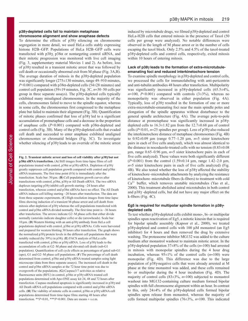
p38-depleted cells fail to maintain metaphasechromosome alignment and show anaphase defectsTo determine the effects of p38 silencing on chromosomesegregation in more detail, we used HeLa cells stably expressinghistone H2B–GFP. Populations of HeLa H2B–GFP cells weretransfected with p38, p38 or nontargeting control siRNA, andtheir mitotic progression was monitored with live cell imaging(Fig. 3, supplementary material Movies 1 and 2). As before, lossof p38 resulted in a transient mitotic arrest that was followed bycell death or occasionally abnormal exit from M phase (Fig. 3A,B).The average duration of mitosis in the p38-depleted populationwas significantly longer (275±130 minutes, range 49–910 minutes,P<0.001) compared with p38-depleted cells (54±28 minutes) andcontrol cell population (59±19 minutes, Fig. 3C, n30–50 cells pergroup in three separate assays). The p38-depleted cells typicallyexhibited many misaligned chromosomes. In the majority of thecells, chromosomes failed to move to the spindle equator, whereasin some cells, the chromosomes first congressed to the metaphaseplate but failed to maintain their alignment (Fig. 3B). Quantificationof mitotic phases confirmed that loss of p38 led to a significantaccumulation of prometaphase cells and a decrease in the proportionof anaphase cells (P<0.001) compared with p38-depleted andcontrol cells (Fig. 3B). Many of the p38-depleted cells that evadedcell death and succeeded to enter anaphase exhibited unalignedchromosomes or chromatid bridges (Fig. 3C). To investigatewhether silencing of p38 leads to an override of the mitotic arrest
219p38 MAPK in mitosis
induced by microtubule drugs, we filmed p38-depleted and controlHeLa-H2B cells that entered mitosis in the presence of Taxol (50cells per group were analyzed). No notable differences wereobserved in the length of M phase arrest or in the number of cellsescaping the taxol block. Only 2.5% and 4.5% of the taxol-treatedp38-depleted cells and control cells, respectively, exited mitosiswithin 10 hours of entering mitosis.
Lack of p38 leads to the formation of extra-microtubule-emanating foci and reduced interkinetochore tensionTo examine spindle morphology in p38-depleted and control cells,we processed the cells for immunolabeling with anti-pericentrinand anti-tubulin antibodies 48 hours after transfection. Multipolaritywas significantly increased in p38-depleted cells (65.5±4%,n100, P<0.001) compared with controls (3±3%), whereas nomonopolarity was observed in either population (Fig. 4A).Typically, loss of p38 resulted in the formation of one or moreextra-microtubule-emanating foci near the main spindle poles andelongation of the spindle apparatus without disturbance to thegeneral spindle architecture (Fig. 4A). The average pole-to-poledistance at prometaphase was significantly increased in p38-depleted cells to 16.3±1.82 m from the 12.7±0.68 m of controlcells (P<0.01, n25 spindles per group). Loss of p38 also reducedthe interkinetochore distance of metaphase chromosomes (Fig. 4B)to 0.92±0.09 m (range 0.77–1.20 m, n5 sister kinetochorepairs in each of five cells analyzed), which was almost identical tothe distance in nocodazole-treated cells with no tension (0.83±0.08m, range 0.65–0.96 m, n5 sister kinetochore pairs in each offive cells analyzed). These values were both significantly different(P<0.001) from the control (1.59±0.14 m, range 1.42–2.0 m,n5 sister kinetochore pairs in each of five cells analyzed) (Fig.4B). We also tested whether the loss of p38 affected the stabilityof kinetochore–microtubule attachments by analyzing the resistanceof kinetochore–microtubule fibers (k-fibers) to treatment with coldCa2+ buffer, which removes unstable microtubules (Yao et al.,2000). This treatment abolished astral microtubules in both controland p38 -depleted cells, but did not have any major effect on thek-fibers (Fig. 4C).
Eg5 is required for multipolar spindle formation in p38-depleted cellsTo test whether p38-depleted cells exhibit mono-, bi- or multipolarspindles upon reactivation of Eg5, a mitotic kinesin that is requiredfor bipolar spindle assembly (Kapoor et al., 2000), we treatedp38-depleted and control cells with 100 M monastrol (an Eg5inhibitor) for 4 hours and then removed the drug by extensivewashing. The proteasome inhibitor MG132 was added to the culturemedium after monastrol washout to maintain mitotic arrest. In thep38-depleted population 37±8% of the cells (n100) had arrestedin mitosis with monopolar spindles during the 4 hour drugincubation, whereas 95±1% of the control cells (n100) weremonopolar (Fig. 4D). This difference was due to the largepopulation of p38-negative cells that were already arrested at Mphase at the time monastrol was added, and these cells remainedbi- or multipolar during the 4 hour incubation (Fig. 4D). Themajority of control cells (83±3%, n100) subjected to monastrolwashout into MG132-containing culture medium formed bipolarspindles with full chromosome alignment within an hour. In contrastto this, only 24±4% of the p38-depleted cells formed bipolarspindles upon release from monastrol, whereas the majority ofcells formed multipolar spindles (76±3%, n100. This indicated
Fig. 2. Transient mitotic arrest and loss of cell viability after p38 but notp38 siRNA transfection. (A)Still images from time-lapse films of cellpopulations treated with control, p38 or p38 siRNA. Depletion of p38greatly suppresses cell population growth compared with control and p38siRNA treatments. The first time point (0 h) is immediately after thetransfection. Scale bar: 50m. (B)Cell population growth curves aftertransfections with control, p38, p38 or All Death siRNA. The two siRNAduplexes targeting p38 inhibit cell growth starting ~24 hours aftertransfection, whereas control and p38 siRNAs have no effect. The All DeathsiRNA induces cell killing starting ~20 hours after transfection. The data arefrom three separate experiments. (C)High-resolution images from time-lapsefilms showing induction of a transient M-phase arrest and cell death frommitosis after depletion of p38 whereas the cell populations transfected withcontrol and p38 siRNA divided normally. The first time point is ~36 hoursafter transfection. The arrows indicate G2–M phase cells that either dividenormally (asterisks indicate daughter cells) or die (arrowheads). Scale bar:10m. (D)Western blotting with an anti-p38 antibody from HeLa cellpopulations depleted with control, p38 or p38 siRNAs. Cells were harvestedand prepared for western blotting 30 hours after transfection. The graph showsthe normalized p38 protein levels in the different cell populations that werenotably reduced (by 39%) in p38. (E) FACS analysis of HeLa cellstransfected with control, p38 or p38 siRNA. Loss of p38 leads to theaccumulation of cells at G2–M phase and elevated cell death (sub-G1population). Quantification of cell cycle effects as percentages of gated sub-G1(apo), G1 and G2–M phase cell populations. (F)The percentage of cell deathdetermined from control, p38 and p38 siRNA treated samples using lightmicroscope (data from three separate assays). The increased cell death in thecontrol and p38 siRNA samples at the 72 hour time point was due toovergrowth of the populations. (G)Caspase3/7 activities as relativefluorescence units (RFU) in control, p38 or p38 siRNA treated cellpopulations determined with Apo-ONE assay kit 24, 48 and 72 hours aftertransfection. Caspase-mediated apoptosis is significantly increased in p38 andAll Death siRNA cell populations compared with control and p38 siRNAcells. (H)The viability of mitotic cells in control, p38 or p38 siRNA cellpopulations determined from time-lapse films starting 48 hours aftertransfection. **P<0.01, ***P<0.001. Data are means ± s.e.m.
Jour
nal o
f Cel
l Sci
ence

that p38-negative monopolar cells became multipolar upon Eg5reactivation (Fig. 4D). We also observed that most of the p38-depleted cells with bipolar spindles exhibited several unalignedchromosomes after monastrol washout (Fig. 4E,F, yellow arrowsin bottom panel).
Active p38 MAPK localizes to kinetochores and spindlepoles and is created by bound mitotic kinasesTo investigate localization of active MAPKs in mitotic cells, westained cells with an antibody that recognizes the duallyphosphorylated p38 MAPKs (Thr180,Tyr182; phosphorylated p38)(Fig. 5A-E). Upon staining cells in different mitotic phases, wedetected activated p38 MAPK at the kinetochores from prophaseto late telophase, at the spindle poles from prometaphase to latetelophase and at the mid-body in telophase. Also, a notablecytoplasmic fraction of phosphorylated p38 was present throughoutmitosis. To confirm that the antibody against phosphorylated p38recognized the p38 isoform, we immunoprecipitated p38 fromtaxol-arrested HeLa cells and performed western blotting. A strongprotein band corresponding to the expected size of thephosphorylated p38 MAPK was present below the heavy chain inthe p38 pull-down fraction, whereas no detectable amount ofphosphorylated p38 remained in the supernatant (Fig. 5F). Theimmunoprecipitation assay was also performed reciprocally withsimilar results (data not shown). To exclude the possibility that the
220 Journal of Cell Science 124 (2)
anti-p38 antibody either recognizes or pulls down the p38isoform we blotted the p38 immunoprecipitate with an antibodyspecific to p38 (ms p38). There were no detectable levels ofp38 present in the p38 immunoprecipitate, but instead all of thep38 isoform remained in the supernatant fraction. We concludethat the antibody against phosphorylated p38 also recognizes thep38 isoform in mitotic cells and that the p38 does not interactwith the p38 isoform.
To investigate which kinases are involved in p38 MAPKactivation in early mitosis, we used the lysed cell assay (Ahonenet al., 2005; Daum and Gorbsky, 2006). In this assay, cellsgrowing on coverglasses are extracted with a detergent andincubated in a buffer without the phosphatase inhibitormicrocystin-LR (MC) or PhosSTOP phosphatase inhibitor cocktailallowing removal of the soluble cytoplasm and dephosphorylationof phosphoepitopes at kinetochores and other subcellularstructures by the active phosphatases. Supplementation of thelysed cells with a buffer containing MC or PhosSTOP andATP allows the re-creation of those phosphoepitopes that aretargeted by adjacent stably bound kinase(s) (endogenousrephosphorylation). As expected, phosphorylated p38 signal wasabolished to background levels after dephosphorylation (Fig. 6A).However, bound endogenous kinase(s) were able to re-createthe epitope on kinetochores and spindle poles duringrephosphorylation without any external source of kinase activity
Fig. 3. Impaired chromosome alignment and anaphase defects in cells depleted of p38. (A)Still images from time-lapse films of p38-depleted HeLa H2B–GFP cells. Typically, p38-depleted cells exhibited either multiple misaligned chromosomes (upper cell, arrowheads) that never achieved metaphase alignment orfirst showed normal congression of chromosomes to the spindle equator but later failure to maintain the metaphase chromosome alignment (lower cell, arrows).Numbers indicate hours and minutes. (B)Quantification of the duration and phases of mitosis in control, p38- and p38-depleted HeLa H2B–GFP populations.(C)A few p38-depleted cells enter anaphase in the presence of unaligned chromosomes (arrows) or exhibit the cut phenotype (arrowhead) more frequentlycompared with the control and p38-depleted cells. *P<0.05, **P<0.01, ***P<0.001. Data are means ± s.e.m. Scale bars: 10m.
Jour
nal o
f Cel
l Sci
ence

221p38 MAPK in mitosis
Fig. 4. Spindle abnormalities in cells depleted of p38. (A)Loss of p38 results in mitosis with multipolar spindles. Immunolocalization of pericentrin foci(arrows) and -tubulin in HeLa cells 36 hours after transfection with non-targeting control siRNA or siRNA to knockdown p38. The numbers indicate the averagepole-to-pole distance (n25 spindles per group). In the merge image, DAPI-stained chromosomes (blue), microtubules (red) and spindle poles (green/yellow) areshown. (B)Loss of p38 reduces interkinetochore distance. In the graph, the black, white and grey lines indicate the mean, s.e.m. and range of the interkinetochoredistance, respectively. The micrographs show cells transfected with control (±3 hours nocodazole treatment) or p38 siRNA after staining of the kinetochores(Crest) and DNA. The insets show pairs of sister kinetochores near the spindle equator. (C)Representative images of cells from control and p38 siRNA transfectedpopulations fixed either directly (control) or subjected to cold calcium buffer lysis before immunostaining for -tubulin. The non-stable astral microtubules (insets)are preserved in the control conditions (arrows) and lost after cold lysis (arrowheads). In the merge, DAPI-stained chromosomes (red) and microtubules (green) areshown. (D)In p38-depleted cells, Eg5 is required for multipolar spindle formation. The flowchart of the monastrol assay and percentages of mono-, bi- ormultipolar spindles in control and p38 siRNA transfected cell populations before and during monastrol treatment, and after drug washout is shown. (E)Controland p38 siRNA transfected cells fixed and labelled with anti-tubulin (red) and anti-pericentrin (green) antibodies 4 hours after the addition of monastrol or 1 hourafter release into MG132-containing medium. The yellow and white arrows indicate pericentrin foci in bipolar and multipolar cells, respectively. The arrowheadsindicate unaligned chromosomes in DAPI (blue)-stained cells. (F)The lower graph shows percentages of cells with bipolar spindles with either normal or perturbedchromosome alignment. ***P<0.001; means ± s.e.m. Scale bars: 10m.
Jour
nal o
f Cel
l Sci
ence

than the ATP in rephosphorylation buffer (Fig. 6B). Therefore,kinases that are responsible for creating phosphorylated p38 werestably bound to these subcellular structures. To investigate whichmitotic kinases control phosphorylation of p38, we tested anumber of mitotic kinase inhibitors in the rephosphorylationassay. Interestingly, the inhibition of Polo-like kinase 1 (Plk1) byZK-thiazolidinone (TAL) (Santamaria et al., 2007) during therephosphorylation step significantly reduced phosphorylated p38signal intensity at kinetochores and spindle poles (Fig. 6B)whereas inhibition of Aurora B by ZM447439 (Ditchfield et al.,2003) did not affect the phosphorylated p38 epitope. Forquantification of phosphorylated p38 at the kinetochores, wemeasured the integrated fluorescence intensities minus thebackground per 10 kinetochores in each of seven cells analyzedper group as described elsewhere (Beardmore et al., 2004). Whenthe activities of the bound enzymes were abolished with N-ethylmaleimide (NEM) and the lyzed cells were supplementedwith exogenous kinases present in M phase extract prepared from
222 Journal of Cell Science 124 (2)
nocodazole-blocked cells, the phosphorylated p38 epitopereappeared at kinetochores and spindle poles (Fig. 7A,C). Bycontrast, the addition of S phase extract to the lyzed cells afterNEM treatment did not lead to the re-phosphorylation ofphosphorylated p38. This indicates that kinase activity requiredfor creating phosphorylated p38 was present in mitotic but not ininterphase cells. When M phase extract depleted of Plk1 wasadded together with ATP or when Plk1-depleted M phase extractwas washed out before addition of ATP on NEM pretreated cells,the phosphorylated p38 signal was not regenerated (Fig. 7B,C).This strengthens the notion that Plk1 is required for creation ofthe Thr180, Tyr182 epitope of p38 MAPK.
p38 is needed to target Plk1 to the kinetochoreA recently reported mitotic substrate of the p38 MAPK pathwayis Plk1, which is phosphorylated at Ser326 by MAPK-activatedprotein kinase 2 (MK2) in vitro and in vivo (Tang et al., 2008).To analyze the effect of p38 depletion on Plk1 levels at poles and
Fig. 5. Localization of active p38 MAPK in mitosis. (A)Antibody recognizing the dually phosphorylated p38 MAPK (Thr180/Tyr182) stains kinetochores fromprophase to late telophase, the spindle poles from prometaphase to late anaphase and the mid-body at telophase (red in the merge) in HeLa cells. Removal ofmicrotubules with short (1 hour) nocodazole treatment does not markedly change the phosphorylated p38 (p-p38) signal intensities whereas treatment with theblocking peptide totally abolishes the signals. The arrows indicate the spindle poles, the arrowhead the mid-body and the insets show higher magnification views ofthe kinetochores at prophase and telophase. DNA is stained with DAPI (green in the merge). (B)Line graphs showing the colocalization of active p38 MAPK (red)with Hec1 (green, left-hand panel) at outer kinetochores but not with Aurora B (green, right-hand panel) at inner centromeres. (C)Western blotting with the anti-p38-P antibody detects the active p38 MAPK (arrowhead) in mitotic HeLa cell extracts. (D)The anti-p38-P antibody also recognizes the active p38 MAPK atkinetochore and spindle poles of U2OS cells and Ptk1 cells. The micrographs show representative prometaphase cells. The merge shows overlay of DNA (grey),Crest (green) and phosphorylated p38 (red). (E)The phosphorylated p38 epitope is conserved between the four p38 MAPK isoforms. (F)Western blots fromimmunoprecipitation assay using a rabbit anti-p38 antibody. In the upper panel, the immunoprecipitated sample is blotted with anti-p38-P antibody, which detectsa strong protein band corresponding to the anticipated size of the phosphorylated p38 MAPK. The heavy chain (Hc) is detected because of use of rabbit antibodyboth in the immunoprecipitation and western blotting. In the lower panel, the same sample is blotted with an antibody detecting the p38 isoform. No apparentband is present in the immunoprecipitate whereas a strong band is detected in the supernatant (Sup) indicating that p38 does not coprecipitate with p38.
Jour
nal o
f Cel
l Sci
ence

kinetochores, we stained p38-depleted and control cells withantibodies against Plk1. A dramatic reduction was observed in thekinetochore level of Plk1 (down by 93±9%, P<0.001) after p38depletion in comparison with control cells (Fig. 8A). In thesecells, the loss of kinetochore-bound Plk1 was accompanied withan increase in the cytosolic fraction of Plk1 (Fig. 8A). Plk1 levelsat the main spindle poles were also decreased by 41±7% in thep38-depleted cells compared with levels in the control (P<0.01,Fig. 8A), but unexpectedly the signal intensity of phosphorylatedPlk1 (Ser326) was not affected (Fig. 8B). This suggests that p38MAPK isoforms other than p38 are responsible forphosphorylation of Plk1 at Ser326 and that only a proportion ofpole-located Plk1 is phosphorylated at this residue. Further supportfor this notion was provided by the observation that thephosphorylation of MK2 at Thr334 was not reduced in the p38-depleted cells (data not shown).
223p38 MAPK in mitosis
DiscussionThe p38 MAPK pathway consists of four kinase isoforms (p38,p38, p38, and p38) that have been implicated in inflammation,cell differentiation, cell growth, cell death, senescence andtumorigenesis (Aouadi et al., 2006; Thornton and Rincon, 2009).The pathway is also an interesting therapeutic opportunity for thetreatment of various diseases and over 20 different p38 MAPKinhibitors have entered clinical trials (Goldstein and Gabriel, 2005).p38 was the first p38 MAPK isolated (Han et al., 1994) and isnow the most thoroughly studied isoform, whereas much less isknown about the other three family members. In addition to thefunctions mentioned above, the p38 MAPK pathway also controlsthe cell cycle by regulating both G1–S and G2–M transitions(MacCorkle and Tan, 2005). A role for the pathway in mitotic entryhas been well established (Cha et al., 2007; Bulavin et al., 2002;Mikhailov et al., 2005), but data regarding the control of mitotic
Fig. 6. The phosphorylated p38 epitope is created by bound mitotic kinases. (A)The phosphorylated p38 epitope is preserved at kinetochores in lyzed HeLacells that were fixed in the presence of the phosphatase inhibitor microcystin-LR (MC) but is lost from lyzed cells that were subjected to dephosphorylation byendogenous phosphatases in the absence of MC. The same applies for phosphorylation of CenpA, a centromeric histone H3 homologue and target of Aurora Bkinase, detected with an antibody recognizing the phosphorylation at Ser7 (CenpA-P). (B)In the cells that were first allowed to undergo dephosphorylation byextraction in the absence of MC, the endogenous kinetochore and spindle-pole-bound kinases can regenerate the p38-P and phosphorylated CenpA epitopes uponincubation in buffer containing ATP and MC (upper row). When Plk1 inhibitor ZK-thiazolidinone (TAL) is added to the rephosphorylation buffer, thephosphorylation of phosphorylated p38 epitope is prevented. Inhibition of Aurora B kinase activity by ZM477439 has no effect on p38 phosphorylation. Additionof ZM477439 to the rephosphorylation buffer abolishes signals from the Aurora B target residue of CenpA (Ser7). Lambda phosphatase treatment abolishes p38-Psignals to background levels. The graphs show the average p38-P and CenpA-P signal intensities at the kinetochores (n10–15 in each of the 5–10 analyzed cells).**P<0.01, ***P<0.001. Data are means ± s.e.m. Scale bars: 10m.
Jour
nal o
f Cel
l Sci
ence

events by p38 MAPKs is very limited and controversial. Here, wepresent evidence that p38 acts as important modulator of mitoticspindle architecture and that its loss of function has an impact onmitotic cell survival.
Role of p38 in mitotic progression and mitotic cellsurvivalOur studies revealed that p38 and p38 were the most abundantp38 MAPK isoforms expressed in HeLa cells. The presence ofp38 in cycling HeLa cells was an unexpected finding becauseearlier work demonstrated that this isoform is predominantlyexpressed in skeletal muscle (Lechner et al., 1996; Li et al., 1996),whereas p38 is more ubiquitously expressed (Jiang et al., 1996).Analyses after silencing individual p38 MAPKs revealed isoform-specific mitotic effects. In sharp contrast to p38 siRNA treatment,which did not yield any notable cell cycle defects or induction ofapoptosis, the specific depletion of p38 led to a transientaccumulation of mitotic cells starting 24 hours after transfection.The p38-depleted mitotic cells exhibited spindle anomalies thatprevented normal chromosome alignment and exit from mitosis.The spindle checkpoint remained activated because these cellsspend several hours in mitosis and no notable escape from taxol-induced block was observed. The viability of the mitotic cells wasdramatically reduced in the p38-depleted populations and thecells were eliminated via caspase-dependent apoptosis.
224 Journal of Cell Science 124 (2)
Our live cell assays showed that the p38-depleted cells hadseveral unaligned chromosomes. This could be explained by thespindle anomalies observed in the p38-depleted cells. The cellsexhibited several microtubule-nucleating foci that perturbed thenormal spindle architecture. The multipolar phenotype wasdependent on the normal function of Eg5. Furthermore, the Eg5reactivation assays demonstrated that loss of p38 impaired normalchromosome congression, even in cells with bipolar spindles,suggesting a role for p38 in the correction of erroneousmicrotubule–kinetochore attachments or motor functions that movethe chromosomes. However, as the k-fiber attachments in p38-depleted cells were stable according to their resistance to the coldCa2+ buffer treatment, chromosome alignment defects were probablynot due to mistakes in microtubule–kinetochore attachments.Interestingly, when p38 was depleted, the interkinetochore tensionwas reduced to the same level as that seen in the absence ofmicrotubules. Together, these phenotypes resemble the situation incells after treatment with low doses of microtubule drugs thatdiminish interkinetochore tension but not the k-fiber occupancy(Waters et al., 1998; Skoufias et al., 2001) and therefore suggestthat loss of p38 interferes with normal microtubule dynamics.
Mitotic substrates of the p38 MAPK pathwayIn the literature, only a few mitotic targets of the p38 MAPKcascade, such as Plk1 (Tang et al., 2008) and cytoplasmic dynein
Fig. 7. Plk1 is required for theregeneration of thephosphorylated p38 epitope.(A)HeLa cell M phase (panel 5)but not S phase extract (panel 6)contains kinase activity that canregenerate the p38-Pphosphoepitope at kinetochoresand spindle poles ofpermeabilized, dephosphorylated,and N-ethylmaleimide (NEM)-treated cells (panel 4). (B)HeLacell M phase extract depleted ofPlk1 by RNAi cannot regeneratethe p38-P phosphoepitope (panels5 and 7). (C)The graph on theright corresponds to A and showsthe average p38-P signalintensities at kinetochores (n10in each of the seven analyzed cellsper sample). The graph on the leftcorresponds to B and shows theaverage p38-P intensities at thekinetochore in the analyzed cells(n5). **P<0.01, ***P<0.001.Data are means ± s.e.m. Scalebars: 10m.
Jour
nal o
f Cel
l Sci
ence

(Whyte et al., 2008), have been described. Both Plk1 and dyneinare implicated in several mitotic processes including spindleorganisation, chromosome motion and spindle checkpoint signaling(Karki and Holzbaur, 1999; Petronczki et al., 2008). MK2colocalizes with activated p38 MAPK and Plk1 at spindle poles,where it phosphorylates Plk1 at Ser326 to promote normal mitoticprogression (Tang et al., 2008). The expression of a Plk1 phospho-mutant (S326A) causes spindle pole defects and the accumulationof cells at M phase (Tang et al., 2008).
Another phenotypic link between Plk1 and p38 is theelimination of the M-phase-arrested cells after RNAi. Depletion ofPlk1 causes mitotic arrests, albeit with monopolar spindle, and adramatic decrease in cell viability (Spänkuch-Schmitt et al., 2002;Liu and Eriksson, 2003; Reagan-Shaw and Ahmad, 2005; Guan etal., 2005; Bu et al., 2008). Moreover, induction of apoptosis byPlk1 RNAi is associated with p53 malfunction (Guan et al., 2005),but whether this is linked to p38 function remains to be determined.Interestingly, mislocalization of kinetochore-associated Plk1 byRNAi of Polo-box domain-binding protein PBIP1 has been reportedto impair kinetochore–microtubule attachment and the generationof tension, thus causing severe chromosome misalignment (Kanget al., 2006). This is a reminiscent of the p38-depleted phenotype,although PBIP1 RNAi also caused other mitotic defects and
225p38 MAPK in mitosis
compromised the spindle assembly checkpoint. Nevertheless, theobserved requirement of p38 for the normal kinetochorelocalization of Plk1 suggests a potential connection between thep38 MAPK cascade and Plk1 as determinants of cell fate at Mphase.
Control of p38 MAPK activity during mitosisIn earlier studies, the active form of p38 MAPK was found in thecytosol of mitotic cells (Fan et al., 2005) or localized to the mitoticspindle poles (Tang et al., 2008). Here, we extend these findingsby reporting kinetochore signals after immunofluorescent detectionwith an antibody recognizing the conserved Thr180,Tyr182phosphoepitope present in all p38 MAPK isoforms. Activated p38MAPK localized on kinetochores and spindle poles during mitosis.Silencing of p38 alone was not sufficient to abolish the phosho-epitope signals, suggesting that p38 is not the only active p38isoform present at these subcellular foci (unpublished data). Usingthe lyzed cell model (Ahonen et al., 2005; Daum and Gorbsky,2006), we tested the conditions that are required for creatingphosphorylated p38 in mitotic cells. First, kinetochore and spindle-pole-bound kinases were able to re-phosphorylate the epitopepointing to the presence of immediate upstream kinases of p38MAPK at these subcellular locations, or TAB1-dependentautophosphorylation mechanism of p38 MAPKs (Ge et al., 2002;Salvador et al., 2005). Second, the capability of the M phase butnot the S phase extract to re-create phosphorylated p38 epitopedemonstrated the mitotic origin of the elements required for p38MAPK activation. Plk1 was implicated in the process by theobservation that chemical perturbation of Plk1 but not Aurora Bkinase or loss of Plk1 from the mitotic cell extracts during therephosphorylation reaction was sufficient to suppressphosphorylated p38 epitope phosphorylation by the bound kinases.Together, the data strengthen the notion that Plk1 communicateswith p38 MAPK to control its mitotic activity. Alternatively, Plk1might stimulate the autophosphorylation of the Thr180,Tyr182phosphoepitope of p38 MAPKs in the TAB1-dependent reaction.
In summary, the pleiotropic mitotic errors and massive celldeath caused by loss of p38 suggest that the kinase has severalmitotic substrates whose perturbed function contributes to theobserved phenotypes. Moreover, our results provide the firstevidence for functional links between the p38 MAPK cascade andPlk1 in the control of mitotic progression and in promotion of cellviability at M phase. Details of such mitotic signaling reactions aresubject to further experiments in various model systems.
Materials and MethodsCell cultureHeLa and U2OS (human osteosarcoma) cells obtained from the American TypeCulture Collection (Manassas, VA) were maintained in DMEM (Invitrogen, Carlsbad,CA) and Ptk1 (rat kangaroo kidney epithelial) cells (a gift from Gary Gorbsky,OMRF, Oklahoma City, OK) in MEM (Invitrogen) supplemented with 10% fetalbovine serum, 2 mM L-glutamine, 0.1 mg/ml penicillin-streptomycin, 1 mM sodiumpyruvate, 0.1 mM non-essential amino acids and 20 mM HEPES. The medium ofstable H2B–GFP HeLa cell line (a gift from Geoffrey Wahl, Scripps ResearchInstitute, La Jolla, CA) included 2 ng/ml blasticidin. Cells were grown in +37°C with5% CO2.
Antibodies and reagentsIn the study we used antibody against phosphorylated p38 (Cell Signaling Technology,Danvers, MA, 1:500), CREST autoimmune serum (Antibodies Incorporated, Davis,CA, 1:200), anti-Plk1 (Abcam, Cambridge, UK, 1:100), antibody againstphosphorylated CenpA (Upstate Biotechnology Incorporated, Lake Placid, NY,1:200), antibody against phosphorylated Plk1 (S326, 1:200), anti--tubulin (DM1A,Abcam, 1:200), anti-pericentrin (Abcam, 1:2000), anti-p38 (Cell SignalingTechnology, Danvers, MA, 1:100 in IF or 1:1000 in WB) and anti-p38 (1:1000 inWB) antibodies. The secondary antibodies anti-Cy3 (1:1000), anti-FITC (1:600) and
Fig. 8. Loss of p38 modulates subcellular localization of Plk1. (A)Plk1 isabolished from kinetochores and reduced at spindle poles in p38-depletedcells. (B)Phosphorylation of Plk1 at S326 by MK2 is not affected by p38depletion. The graphs show kinetochore (n10 kinetochores per five cells perassay) and/or spindle pole (two main poles from five cells per assay) signalintensities of Plk1 and phosphorylated Plk1 (p-Plk1) (Ser326). **P<0.01,***P<0.001. Data are mean ± s.e.m. from three separate assays. Scale bars:10m.
Jour
nal o
f Cel
l Sci
ence

226 Journal of Cell Science 124 (2)
anti-Cy5 (1:400) were from Jackson ImmunoResearch (Newmarket, UK) and AlexaFluor antibodies from Invitrogen. Other reagents used in this study were purchasedfrom Sigma if not stated otherwise.
siRNA transfectionsOligofectamine (Invitrogen) was used according to the manufacturer’s instructionsusing the following siRNA targeting sequences: AAC TGC GGT TAC TTA AACATA (p38-oligo 1), CAG AGA ACT GCG GTT ACT TAA (p38-oligo 2), CAGGAT GGA GCT GAT CCA GTA (p38), CTG GAC GTA TTC ACT CCT GAT(p38-oligo 1), TGG AAG CGT GTT ACT TAC AAA (p38-oligo 2), CCG GAGTGG CAT GAA GCT GTA (p38) and AAGATCACCCTCCTTAAATAT (Plk1). AllDeath (AllStars Cell Death Control siRNA) was purchased from Qiagen (Stanford,CA).
ImmunofluorescenceHeLa, U2OS and Ptk1 cells growing on coverslips were fixed and permeabilized in2% paraformaldehyde in 60 mM PIPES, 25 mM HEPES (pH 7.0), 10 mM EGTA,4 mM MgSO4 (PHEM) containing 0.5% Triton X-100 and 400 nM microcystin-LRfor 15 minutes. For microtubule staining, 0.2% glutaraldehyde was added to thefixative. Cells on coverslips were rinsed in 10 mM MOPS (pH 7.4), 150 mM NaCland 0.05% Tween 20 (MBST) and blocked with 20% boiled normal goat serum(BNGS) for 1 hour at room temperature (RT). Cells were stained with primaryantibodies for 1 hour at RT. Cells on coverslips were washed with MBST and treatedwith secondary antibodies for 1 hour at RT. Antibodies were diluted to MBSTcontaining 5% BNGS. Cells on coverslips were washed with MBST and DNA wasstained with DAPI (4�,6-diamidino-2-phenylindole, 10 ng/ml in water). Cells oncoverslips were washed with distilled water and mounted on microscope slides withVectashield mounting medium (Vector Laboratories).
Lyzed cell assayHeLa cells on coverslips were rinsed briefly in rinse buffer (50 mM Tris-HCl, pH7.5, 4 mM MgSO4, 5 g/ml protease inhibitor cocktail) and extracted in extractionbuffer (50 mM Tris-HCl, pH 7.5, 4 mM MgSO4, 0.5% Triton X-100, 1 mM DTT, 5g/ml protease inhibitor cocktail) for 5 minutes. Samples were dephosphorylated indephosphorylation buffer (50 mM Tris-HCl, pH 7.5, 4 mM MgSO4, 1 mM DTT, 5g/ml protease inhibitor cocktail) for 7 minutes (in some samples -phosphatase wasadded to dephosphorylation buffer), rinsed twice in rinse buffer and treated witheither rephosphorylation buffer [50 mM Tris-HCl, pH 7.5, 4 mM MgSO4, 1 mMDTT, 400 nM Microcystin-LR or PhosSTOP (Roche), 1 mM ATP, 5 g/ml proteaseinhibitor cocktail] for 20 minutes or with buffer without ATP (50 mM Tris-HCl, pH7.5, 4 mM MgSO4, 1 mM DTT, 400 nM Microcystin-LR or PhosSTOP, 5 g/mlprotease inhibitor cocktail). In some assays 10 M ZK-Thiazolidinone (TAL) or 20M ZM447439 was added to the rephosphorylation buffer before incubation of thesamples for 20 minutes. In exogenous rephosphorylation assay cells were treatedwith N-ethylmaleimide (NEM) buffer (5 nM NEM in 50 mM Tris-HCl, pH 7.5, 4mM MgSO4, 5 g/ml protease inhibitor cocktail) for 10 minutes and washed in rinsebuffer. Rephosphorylation buffer ± ATP was added together with mitotic or S phaseHeLa cell extract or mitotic Plk1-depleted extract (5�106 cells/ml in extractionbuffer; 1:20 in rephosphorylation buffer) for 40 minutes at RT. In some samplesextracts were added in rephosphorylation buffer without ATP for 40 minutes, washedout in presence of phosphatase inhibitor followed by ATP addition for 40 minutes.Samples were fixed and subjected for immunofluorescence as described above.
Cold calcium lysisCells on coverslips were rinsed in 0.1 M PIPES (pH 6.95) and placed in ice cold (0–2°C) 0.1 M PIPES, pH 6.95, 80 M CaCl2, 1% Triton X-100 for 5 minutes on icebath. After the lysis, cells were rinsed in PIPES before fixation andimmunofluorescence as described above.
Monastrol washout experimentHeLa cells transfected with scrambled siRNA or siRNA to knockdown p38 or p38were treated with 100 M monastrol for 4 hours. The cells were washed and releasedinto fresh growth medium containing 20 M MG132 for 1 hour before fixation andstaining for tubulin and pericentrin. Some samples were also fixed before monastroltreatment or directly after the drug treatment before washout.
Microscopy and image analysisFixed samples were imaged with a Zeiss Axiovert microscope equipped with a 63�objective, Hamamatsu Orca-ER camera (Hamamatsu Photonics Norden AB, Solna,Sweden) and Metamorph imaging software (Molecular Devices, Downingtown,PA). Quantification of signal intensities was done using Metamorph imaging software.All values were corrected with background deduction.
Live cell imagingAsynchronous or double thymidine synchronized HeLa and HeLa H2B–GFP cellsgrowing on a well plate or -Slide eight-well plates (Ibidi, Martinsried, Germany)were imaged 48 hours after siRNA transfection for 16 hours. Images were capturedusing a Zeiss Axiovert microscope equipped with a 40� or 63� objective,Hamamatsu Orca-ER camera (Hamamatsu Photonics Norden) and Metamorph
imaging software (Molecular Devices) or with the 3I-spinning disc confocalmicroscope (Intelligent Imaging Innovations, Goettingen, Germany) equipped with63� objective, Hamamatsu Orca-ER camera (Hamamatsu Photonics Norden) andSlidebook imaging software (Intelligent Imaging Innovations). We also time-lapseimaged asynchronous HeLa cell populations on 384-well plates immediately afterthe siRNA transfection using incucyte imaging system (Essen Instruments, WelwynGarden City, UK) for a total of 66 hours. In this study, duration of mitosis wasdefined as the time from nuclear envelope breakdown to end of telophase, or to thetime of mitotic cell death. To calculate the cell death indices we used themorphological criteria of apoptosis described by Hacker (Hacker, 2000) and scored1000 cells per each time point from three replicate assays.
FACSHeLa cells transfected with scrambled, p38 or p38 siRNA were harvested after 48or 72 hours after transfection. Cells were fixed with –20°C ethanol followed by briefincubation in –20°C. Cells were centrifuged at 1300 rpm for 4 minutes. Cell pelletswere washed and resuspended in PBS in round-bottom 96-well plates. RNase (100g/ml) and propidium iodide (20 g/ml) diluted in PBS were added to the resuspendedcells and the cells were incubated for 30 minutes with agitation at RT in a light-protected box. The samples were measured with LSR II system (Becton Dickinson,Franklin Lakes, NJ) and results were analyzed with FCS Express program.
RT-PCRNondepleted HeLa cells or cells depleted for p38 MAPK isoforms were harvested48 hours after the siRNA transfection. RNA was isolated with the RNeasy Midi Kit(Qiagen) and cDNA was prepared according to the manufacturer’s instructions usingiScriptTM cDNA Synthesis Kit (Bio-Rad, Hercules, CA). Different p38 MAPKisoforms were amplified from cDNAs using specific primers for different isoforms(see below). PCR products were separated on 1% agarose gel including 0.5 g/mlethidium bromide and imaged with UV (Gene Genius Bio Imaging System bySyngene, Cambridge, UK). Band intensities were quantified using Metamorphimaging software (Molecular Devices).
Q-PCRRNA was isolated from depleted HeLa cells and treated with DNaseI (Invitrogen)followed by cDNA isolation. p38 MAPK isoforms were amplified using isoformspecific primers with q-PCR (TaqMan®, Applied Biosystems, Foster City, CA).
Statistical analysisStatistical analysis was performed with GraphPad Prism and Excel software usingStudent’s t-test or two-factor ANOVA test. The significance threshold accepted wasP<0.05.
Caspase activity assay and inhibitionThe Apo-ONE homogeneous caspase-3/7 assay (Promega, Madison, WI) was usedto evaluate the activities of caspase-3 and caspase-7. HeLa cells were depleted withnegative control, All Death control, p38 and p38 siRNAs in 384-well format usingoligofectamine reagent in a reverse transfection according to the manufacturer’sinstructions. After 24, 48 or 72 hours of incubation at 37°C, part of the medium wasaspirated and Apo-ONE substrate buffer mix was added to each well. The plate wasincubated for 30–60 minutes with agitation and was protected from light. EnVision2100 multilabel reader (Perkin Elmer) was used to measure fluorescence (excitationwavelength 485 nm, emission wavelength 535 nm). Caspase activity was inhibitedwith 20 M pan-caspase inhibitor zVAD.fmk. Inhibitor was added 40 hours aftertransfection to cells treated with siRNA against p38 in 96-well plates.
Immunoprecipitation and western blotDynabeads (protein G) were washed twice with citrate-phosphate buffer (pH 5)containing 0.01% Tween-20 and left in citrate-phosphate buffer (pH 5). p38 antibodywas added on beads and incubated for 40 minutes at RT in rotation. Bead–antibodycomplexes were washed three times with citrate-phosphate buffer (pH 5) containing0.01% Tween-20. Mitotic cell extract (5�106 cells/ml in PBS-based IP buffer; 1�PBS, 1 mM EDTA, 2 mM MgCl2, 0.2 mM CaCl2, 0.5% Triton X-100, 150 mMNaCl) arrested for 16 hours was then incubated with 0.6 M Taxol at +4°C for 3hours. IPs were washed three times with PBS-based IP buffer and bound proteinswere eluted by adding SDS buffer and boiling for 5 minutes. Proteins from IPsupernatant were precipitated with acetone and dried pellet was dissolved in SDSbuffer and boiled for 5 minutes. SDS-PAGE was carried out using 4–12% gradientgels (Lonza) and proteins transferred to nitrocellulose membrane. Membrane wasblocked in 10 mM Tris-HCl (pH 8.0), 150 mM NaCl, and 0.05% Tween 20 (TBST)containing 5% nonfat dry milk for 1 hour. Membranes were incubated with primaryantibodies overnight at 4°C, washed three times with TBST and incubated withsecondary antibodies for 1 hour at RT protected from light. After washing, theOdyssey Infrared Imaging System (LI-COR Biotechnology) was used for detectionof proteins.
We thank Bayer Schering Pharma AG, Xiaoqi Liu and Geoff Wahlfor providing reagents for the study and Gary Gorbsky for discussions
Jour
nal o
f Cel
l Sci
ence

227p38 MAPK in mitosis
and critical reading of the manuscript. This study was supported bygrants to M.J.K. from the Academy of Finland (120804), EUFP6(Marie Curie EXT grant 002697), the Centre of Excellence forTranslational Genome-Scale Biology, the Finnish Cancer Organisationsand the Foundation for the Finnish Cancer Institute.
Supplementary material available online athttp://jcs.biologists.org/cgi/content/full/124/2/216/DC1
ReferencesAhonen, L. J., Kallio, M. J., Daum, J. R., Bolton, M., Manke, I. A., Yaffe, M. B.,
Stukenberg, P. T. and Gorbsky, G. J. (2005). Polo-like kinase 1 creates the tension-sensing 3F3/2 phosphoepitope and modulates the association of spindle-checkpointproteins at kinetochores. Curr. Biol. 15, 1078-1089.
Aouadi, M., Binetruy, B., Caron, L., Le Marchand-Brustel, Y. and Bost, F. (2006).Role of MAPKs in development and differentiation lessons from knockout mice.Biochemie 88, 1091-1098.
Beardmore, V. A., Ahonen, L. J., Gorbsky, G. J. and Kallio, M. J. (2004). Survivindynamics increases at centromeres during G2/M phase transition and is regulated bymicrotubule-attachment and Aurora B kinase activity. J. Cell Sci. 117, 4033-4042.
Bu, Y., Yang, Z., Li, Q. and Song, F. (2008). Silencing of polo-like kinase (Plk) 1 viasiRNA causes inhibition of growth and induction of apoptosis in human esophagealcancer cells. Oncology 74, 198-206.
Bulavin, D. V., Amundson, S. A. and Fornace, A. J. (2002). p38 and Chk1 kinases:different conductors for the G(2)/M checkpoint symphony. Curr. Opin. Genet. Dev. 12,92-97.
Campos, C. B., Bédard, P. A. and Linden, R. (2002). Activation of p38 mitogen-activated protein kinase during normal mitosis in the developing retina. Neuroscience112, 583-591.
Cha, H., Wang, X., Li, H. and Fornace, A. J., Jr (2007). A functional role for p38MAPK in modulating mitotic transit in the absence of stress. J. Biol. Chem. 282, 22984-22992.
Daum, J. R. and Gorbsky, G. J. (2006). Lysed cell models and isolated chromosomesfor the study of kinetochore/centromere biochemistry in vitro. Methods 38, 52-59.
Ditchfield, C., Johnson, V. L., Tighe, A., Ellston, R., Haworth, C., Johnson, T.,Mortlock, A., Keen, N. and Taylor, S. S. (2003). Aurora B couples chromosomealignment with anaphase by targeting BubR1, Mad2, and Cenp-E to kinetochores. J.Cell Biol. 161, 267-280.
Fan, L., Yang, X., Du, J., Marshall, M., Blanchard, K. and Ye, X. (2005). A novel roleof p38 alpha MAPK in mitotic progression independent x of its kinase activity. CellCycle 4, 1616-1624.
Ge, B., Gram, H., Di Padova, F., Huang, B., New, L., Ulevitch, R. J., Luo, Y. and Han,J. (2002). MAPKK-independent activation of p38alpha mediated by TAB1-dependentautophosphorylation of p38alpha. Science 295, 1291-1294.
Goldstein, D. M. and Gabriel, T. (2005). Pathway to the clinic: inhibition of P38 MAPkinase. a review of ten chemotypes selected for development. Curr. Top. Med. Chem.5, 1017-1029.
Guan, R., Tapang, P., Leverson, J. D., Albert, D., Giranda, V. L. and Luo, Y. (2005).Small interfering RNA-mediated Polo-like kinase 1 depletion preferentially reduces thesurvival of p53-defective, oncogenic transformed cells and inhibits tumor growth inanimals. Cancer Res. 65, 2698-2704.
Häcker, G. (2000). The morphology of apoptosis. Cell Tissue Res. 301, 5-17.Han, J., Lee, J. D., Bibbs, L. and Ulevitch, R. J. (1994). A MAP kinase targeted by
endotoxin and hyperosmolarity in mammalian cells. Science 265, 808-811.Jiang, Y., Chen, C., Li, Z., Guo, W., Gegner, J. A., Lin, S. and Han, J. (1996).
Characterization of the structure and function of a new mitogen-activated protein kinase(p38beta). J. Biol. Chem. 271, 17920-17926.
Kang, Y. H., Park, J. E., Yu, L. R., Soung, N. K., Yun, S. M., Bang, J. K., Seong, Y.S., Yu, H., Garfield, S., Veenstra, T. D. et al. (2006). Self-regulated Plk1 recruitment
to kinetochores by the Plk1-PBIP1 interaction is critical for proper chromosomesegregation. Mol. Cell 24, 409-422.
Kapoor, T. M., Mayer, T. U., Coughlin, M. L. and Mitchison, T. J. (2000). Probingspindle assembly mechanisms with monastrol, a small molecule inhibitor of the mitotickinesin, Eg5. J. Cell Biol. 150, 975-988.
Karki, S. and Holzbaur, E. L. (1999). Cytoplasmic dynein and dynactin in cell divisionand intracellular transport. Curr. Opin. Cell Biol. 11, 45-53.
Lechner, C., Zahalka, M. A., Giot, J.-F., Moler, N. P. and Ullrich, A. (1996). ERK6, amitogen-activated protein kinase involved in C2C12 myoblast differentiation. Proc.Natl. Acad. Sci. USA 93, 4355-4359.
Li, Z., Jiang, Y., Ulevitch, R. J. and Han, J. (1996). The primary structure of p38gamma: a new member of p38 group of MAP kinases. Biochem. Biophys. Res. Commun.228, 334-340.
Liu, X. and Erikson, R. L. (2003). Polo-like kinase (Plk)1 depletion induces apoptosis incancer cells. Proc. Natl. Acad. Sci. USA 100, 5789-5794.
MacCorkle, R. A. and Tan, T. H. (2005). Mitogen-activated protein kinases in cell-cyclecontrol. Cell Biochem. Biophys. 43, 451-461.
Martín-Blanco, E. (2000). p38 MAPK signalling cascades: ancient roles and new functions.Bioessays 22, 637-645.
Mikhailov, A., Shinohara, M. and Rieder, C. L. (2005). The p38-mediated stress-activated checkpoint. a rapid response system for delaying progression through antephaseand entry into mitosis. Cell Cycle 4, 57-62.
Perdiguero, E., Pillaire, M. J., Bodart, J. F., Hennersdorf, F., Frödin, M., Duesbery,N. S., Alonso, G. and Nebreda, A. R. (2003). Xp38gamma/SAPK3 promotes meioticG(2)/M transition in Xenopus oocytes and activates Cdc25C. EMBO J. 22, 5746-5756.
Petronczki, M., Lénárt, P. and Peters, J. M. (2008). Polo on the rise-from mitotic entryto cytokinesis with Plk1. Dev. Cell 14, 646-659.
Reagan-Shaw, S. and Ahmad, N. (2005). Silencing of polo-like kinase (Plk) 1 via siRNAcauses induction of apoptosis and impairment of mitosis machinery in human prostatecancer cells: implications for the treatment of prostate cancer. FASEB J. 19, 611-613.
Salvador, J. M., Mittelstadt, P. R., Guszczynski, T., Copeland, T. D., Yamaguchi, H.,Appella, E., Fornace, A. J., Jr and Ashwell, J. D. (2005). Alternative p38 activationpathway mediated by T cell receptor-proximal tyrosine kinases. Nat. Immunol. 6, 390-395.
Santamaria, A., Neef, R., Eberspächer, U., Eis, K., Husemann, M., Mumberg, D.,Prechtl, S., Schulze, V., Siemeister, G., Wortmann, L. et al. (2007). Use of the novelPlk1 inhibitor ZK-thiazolidinone to elucidate functions of Plk1 in early and late stagesof mitosis. Mol. Biol. Cell 18, 4024-4036.
Skoufias, D. A., Andreassen, P. R., Lacroix, F. B., Wilson, L. and Margolis, R. L.(2001). Mammalian mad2 and bub1/bubR1 recognize distinct spindle-attachment andkinetochore-tension checkpoints. Proc. Natl. Acad. Sci. USA 98, 4492-4497.
Spänkuch-Schmitt, B., Bereiter-Hahn, J., Kaufmann, M. and Strebhardt, K. (2002).Effect of RNA silencing of polo-like kinase-1 (PLK1) on apoptosis and spindle formationin human cancer cells. J. Natl. Cancer Inst. 18, 1863-1877.
Takenaka, K., Moriguchi, T. and Nishida, E. (1998). Activation of the protein kinasep38 in the spindle assembly checkpoint and mitotic arrest. Science 280, 599-602.
Tang, J., Yang, X. and Liu, X. (2008). Phoshorylation of Plk1 at Ser326 regulates itsfunctions during mitotic progression. Oncogene 27, 6635-6645.
Thornton, T. M. and Rincon, M. (2009). Non-classical p38 map kinase functions: cellcycle checkpoints and survival. Int. J. Biol. Sci. 5, 44-52.
Waters, J. C., Chen, R. H., Murray, A. W. and Salmon, E. D. (1998). Localization ofMad2 to kinetochores depends on microtubule attachment, not tension. J. Cell Biol.141, 1181-1191.
Whyte, J., Bader, J. R., Tauhata, S. B., Raycroft, M., Hornick, J., Pfister, K. K., Lane,W. S., Chan, G. K., Hinchcliffe, E. H., Vaughan, P. S. et al. (2008). Phosphorylationregulates targeting of cytoplasmic dynein to kinetochores during mitosis. J. Cell Biol.183, 819-834.
Yao, X., Abrieu, A., Zheng, Y., Sullivan, K. F. and Cleveland, D. W. (2000). CENP-Eforms a link between attachment of spindle microtubules to kinetochores and themitotic checkpoint. Nat. Cell Biol. 2, 484-491.
Jour
nal o
f Cel
l Sci
ence