Embed Size (px)
Citation preview
nature CHeMICaL BIOLOGY | vol 8 | february 2012 | www.nature.com/naturechemicalbiology 211
articlepuBLIsHed OnLIne: 8 januarY 2012 | dOI: 10.1038/nCHeMBIO.765
Embryonic development is directed by a core set of signaling pathways such as the Hedgehog, Wnt or Notch pathways. As their roles in both development and adult life have been unrav-
eled, these pathways have become important therapeutic targets in oncology and regenerative medicine. The Hedgehog pathway, the focus of our work, has been implicated in human birth defects as well as in a variety of familial and sporadic cancers1. In fact, a recent survey of http://clinicaltrials.gov/ revealed that over 20 clinical tri-als are testing small-molecule inhibitors of the Hedgehog pathway against tumors of the lung, skin, brain, pancreas and prostate.
Despite extensive studies in vertebrate and invertebrate systems, the biochemical mechanisms regulating many steps in Hedgehog signaling remain incompletely understood. The initiating step in Hedgehog signaling is controlled by a poorly characterized inter-action between two multipass transmembrane proteins, Patched1 (Ptc1, a tumor suppressor protein) and Smoothened (Smo, an oncoprotein). In the absence of Hedgehog ligands, the receptor Ptc1 inhibits the activity of Smo2. Upon binding of a Hedgehog ligand (Sonic, Indian or Desert Hedgehog; Shh, Ihh and Dhh, respectively), Ptc1 is inactivated, unleashing Smo and allowing the Gli transcription factors to initiate target gene transcription3,4. Despite the fact that the interaction between Ptc1 and Smo is the most commonly damaged step in Hedgehog-related diseases5, the biochemical basis of this step remains unknown. The plant alkaloid cyclopamine, which is to our knowledge the first small-molecule inhibitor of Hedgehog signaling to be discovered6, directly binds Smo7,8. In addition, a large number of cell-based screens against the Hedgehog pathway have identified small molecules that directly bind Smo, as either agonists such as SAG9 and purmorphamine10 or antagonists such as the SANTs9 and GDC-0449 (ref. 11). All of these small molecules are assumed to interact with the same site on Smo because they compete with cyclopamine for binding to Smo9–11. The presence of this seemingly easily ‘druggable’ site has incited a search for an endogenous small-molecule ligand that may regulate the activity of Smo.
Oxysterols, naturally occurring molecules derived from the enzy-matic and nonenzymatic oxidation of cholesterol, have emerged as useful probes of this critical step in signaling. In cultured fibro-blasts, mesenchymal stem cells and medulloblastoma cells, specific oxysterols can activate Hedgehog signaling, trigger the transcrip-tion of target genes and drive the differentiation of osteogenic cells in culture and in animals12–14. Oxysterols can induce the marked accumulation of Smo in the primary cilium15, a key change in sub-cellular localization that allows Smo to activate downstream signal-ing16. The elimination of Smo abrogates the effects of oxysterols on Hedgehog signaling, suggesting that they function at the level of Smo, Ptc1 or an undiscovered intermediate step13. However, unlike most synthetic Hedgehog effectors, oxysterols do not compete with cyclopamine for binding to Smo, a property that has been used to argue that Smo is not a direct target for oxysterols13. How oxysterols activate Hedgehog signaling remains an open question.
Oxysterols, in addition to having a role in the Hedgehog path-way, are a class of understudied endogenous small molecules that most likely have important roles in signaling pathways. They have been implicated in vesicle and lipid trafficking17, cholesterol homeo-stasis signaling18, activation of nuclear receptors19,20 and leukocyte chemotaxis21,22. However, they have many other effects, such as induction of apoptosis23 and regulation of macrophage function24, that remain to be understood at a molecular level. Although their cellular concentrations are orders of magnitude below those of cholesterol, their higher hydrophilicity allows them to rapidly move between membrane compartments25. The study of molecules such as oxysterols is complicated by their ability to influence cellular processes in two distinct ways that are often difficult to disentan-gle. First, oxysterols can bind directly to proteins and affect their activity, exemplified by their interactions with the liver X receptor (LXR)19,20 and the INSIG protein18. Second, because of their lipo-philicity, oxysterols can incorporate into host membranes and alter their physical properties, thereby indirectly influencing membrane proteins such as Ptc1 or Smo26,27.
1Department of biochemistry, Stanford university School of Medicine, Stanford, California, uSa. 2Department of Developmental biology, Washington university School of Medicine, St. louis, Missouri, uSa. 3Department of Cell biology and Physiology, Washington university School of Medicine, St. louis, Missouri, uSa. 4Department of Medicine, Stanford university School of Medicine, Stanford, California, uSa. *e-mail: [email protected] or [email protected]
Oxysterols are allosteric activators of the oncoprotein smoothenedsigrid nachtergaele1, Laurel K Mydock2, Kathiresan Krishnan2, jayan rammohan3, paul H schlesinger3, douglas F Covey2* & rajat rohatgi4*
Oxysterols are a class of endogenous signaling molecules that can activate the Hedgehog pathway, which has critical roles in development, regeneration and cancer. However, it has been unclear how oxysterols influence Hedgehog signaling, including whether their effects are mediated through a protein target or indirectly through effects on membrane properties. To answer this question, we synthesized the enantiomer and an epimer of the most potent oxysterol, 20(S)-hydroxycholesterol. Using these molecules, we show that the effects of oxysterols on Hedgehog signaling are exquisitely stereoselective, consistent with the hypothesis that they function through a specific protein target. We present several lines of evidence that this protein target is the seven-pass transmembrane protein Smoothened, a major drug target in oncology. Our work suggests that these enig-matic sterols, which have multiple effects on cell physiology, may act as ligands for signaling receptors and provides a generally applicable framework for probing sterol signaling mechanisms.
npg
© 2
012
Nat
ure
Am
eric
a, In
c. A
ll rig
hts
rese
rved
.
212 nature CHeMICaL BIOLOGY | vol 8 | february 2012 | www.nature.com/naturechemicalbiology
article NaTUre cHemical biOlOgy dOI: 10.1038/nCHeMBIO.765
Using a series of structure-activity studies that exploit the funda-mental properties of regio- and stereochemical isomerism, we show that oxysterols influence the Hedgehog pathway through a protein target. Using pharmacological analysis and a new affinity reagent, we provide evidence that oxysterols activate Smo by an allosteric mechanism and act at a site on Smo distinct from the canonical cyclopamine binding site.
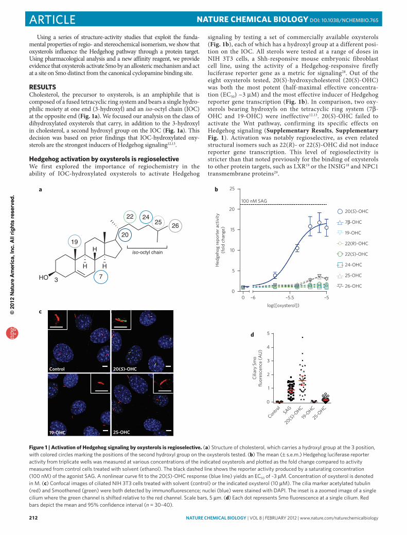
reSUlTSCholesterol, the precursor to oxysterols, is an amphiphile that is composed of a fused tetracyclic ring system and bears a single hydro-philic moiety at one end (3-hydroxyl) and an iso-octyl chain (IOC) at the opposite end (Fig. 1a). We focused our analysis on the class of dihydroxylated oxysterols that carry, in addition to the 3-hydroxyl in cholesterol, a second hydroxyl group on the IOC (Fig. 1a). This decision was based on prior findings that IOC-hydroxylated oxy-sterols are the strongest inducers of Hedgehog signaling12,13.
Hedgehog activation by oxysterols is regioselectiveWe first explored the importance of regiochemistry in the ability of IOC-hydroxylated oxysterols to activate Hedgehog
signaling by testing a set of commercially available oxysterols (Fig. 1b), each of which has a hydroxyl group at a different posi-tion on the IOC. All sterols were tested at a range of doses in NIH 3T3 cells, a Shh-responsive mouse embryonic fibroblast cell line, using the activity of a Hedgehog-responsive firefly luciferase reporter gene as a metric for signaling28. Out of the eight oxysterols tested, 20(S)-hydroxycholesterol (20(S)-OHC) was both the most potent (half-maximal effective concentra-tion (EC50) ~3 μM) and the most effective inducer of Hedgehog reporter gene transcription (Fig. 1b). In comparison, two oxy-sterols bearing hydroxyls on the tetracyclic ring system (7β-OHC and 19-OHC) were ineffective12,13. 20(S)-OHC failed to activate the Wnt pathway, confirming its specific effects on Hedgehog signaling (Supplementary Results, Supplementary Fig. 1). Activation was notably regioselective, as even related structural isomers such as 22(R)- or 22(S)-OHC did not induce reporter gene transcription. This level of regioselectivity is stricter than that noted previously for the binding of oxy sterols to other protein targets, such as LXR19 or the INSIG18 and NPC1 transmembrane proteins29.
c
19-OHC 25-OHC
Control 20(S)-OHC
d
Control
20(S)-O
HC
19-O
HC
25-OHC
Cili
ary
Smo
fluor
esce
nce
(AU
)
0
1
2
3
4
5
SAG
b
0 –6 –5.5 –50
5
10
15
20
25
Hed
geho
g re
port
er a
ctiv
ity(f
old
chan
ge)
100 nM SAG
log([oxysterol])
20(S)-OHC
7β-OHC
19-OHC
22(R)-OHC
22(S)-OHC
24-OHC
25-OHC
26-OHC
a
HO
H H
H iso-octyl chain
3
19
7
20
22 2425 26
Figure 1 | activation of Hedgehog signaling by oxysterols is regioselective. (a) Structure of cholesterol, which carries a hydroxyl group at the 3 position, with colored circles marking the positions of the second hydroxyl group on the oxysterols tested. (b) The mean (± s.e.m.) Hedgehog luciferase reporter activity from triplicate wells was measured at various concentrations of the indicated oxysterols and plotted as the fold change compared to activity measured from control cells treated with solvent (ethanol). The black dashed line shows the reporter activity produced by a saturating concentration (100 nM) of the agonist SaG. a nonlinear curve fit to the 20(S)-oHC response (blue line) yields an eC50 of ~3 μM. Concentration of oxysterol is denoted in M. (c) Confocal images of ciliated NIH 3T3 cells treated with solvent (control) or the indicated oxysterol (10 μM). The cilia marker acetylated tubulin (red) and Smoothened (green) were both detected by immunofluorescence; nuclei (blue) were stained with DaPI. The inset is a zoomed image of a single cilium where the green channel is shifted relative to the red channel. Scale bars, 5 μm. (d) each dot represents Smo fluorescence at a single cilium. red bars depict the mean and 95% confidence interval (n = 30–40).
npg
© 2
012
Nat
ure
Am
eric
a, In
c. A
ll rig
hts
rese
rved
.
nature CHeMICaL BIOLOGY | vol 8 | february 2012 | www.nature.com/naturechemicalbiology 213
articleNaTUre cHemical biOlOgy dOI: 10.1038/nCHeMBIO.765
We assayed this same set of oxysterols for the ability to drive the accumulation of Smo in the primary cilium, a hallmark of Hedgehog pathway activation15,16. A representative subset of this group is shown in Figure 1c,d. This cilia localization assay, which measures the ciliary concentrations of Smo using quantita-tive fluorescence microscopy, is an important complement to the Hedgehog reporter activity assay described above because it moni-tors an early, nontranscriptional step in signaling. Consistent with the results from the reporter assay, 20(S)-OHC induced Smo pro-tein accumulation in the primary cilium to a much greater extent than the other oxy sterols tested (Fig. 1c,d). These results also con-firmed that 20(S)-OHC likely regulates the subcellular localization of Smo by acting on the pathway at the level of Ptc1, Smo or an intermediate step.
Hedgehog activation by oxysterols is stereoselectiveThe degree of regioselectivity observed for Hedgehog pathway acti-vation suggests that oxysterols act on a protein target, as ligand bind-ing sites of proteins have specific structural requirements. However, regioisomers of IOC-hydroxylated oxysterols have dramatically different effects on the physical properties of membranes, such as membrane thickness and area or the distributions of ordered and disordered domains30. Thus, regioselectivity cannot be interpreted as definitive evidence for a protein-oxysterol interaction.
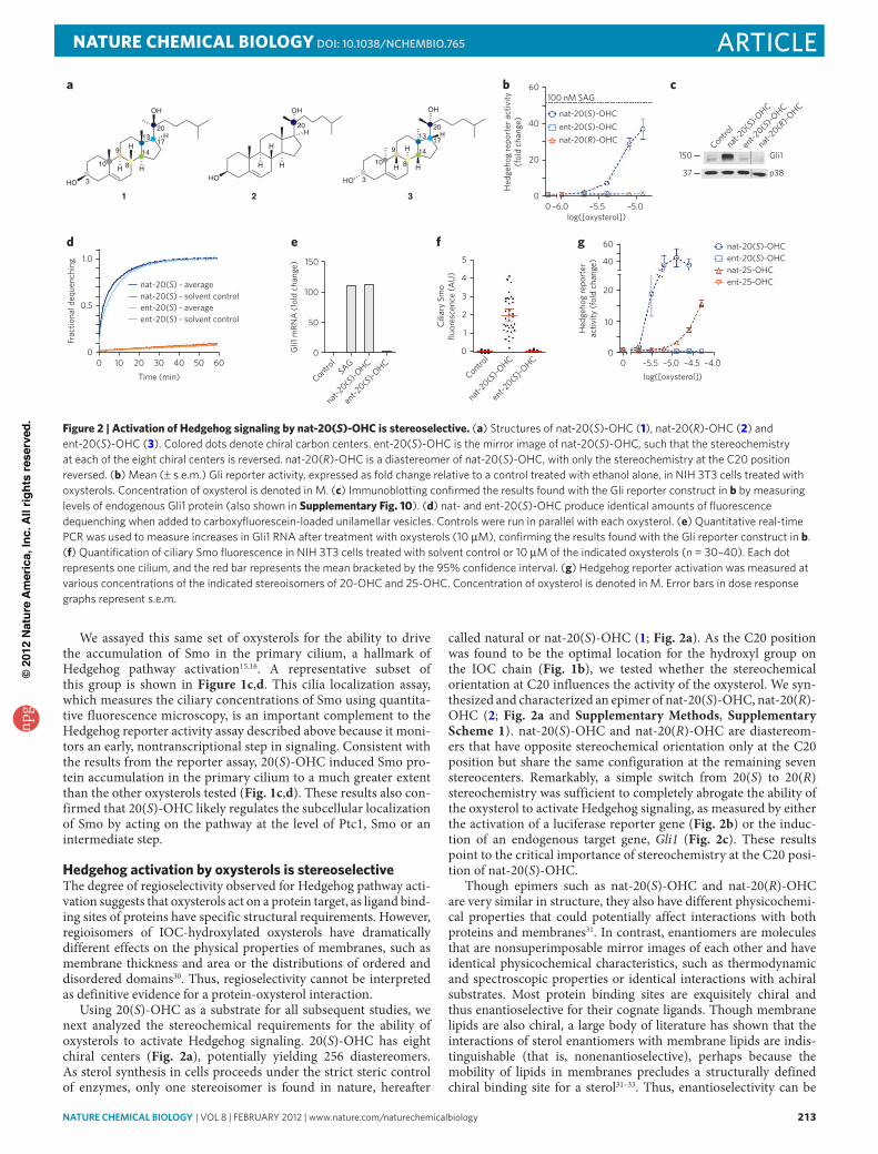
Using 20(S)-OHC as a substrate for all subsequent studies, we next analyzed the stereochemical requirements for the ability of oxysterols to activate Hedgehog signaling. 20(S)-OHC has eight chiral centers (Fig. 2a), potentially yielding 256 diastereomers. As sterol synthesis in cells proceeds under the strict steric control of enzymes, only one stereoisomer is found in nature, hereafter
called natural or nat-20(S)-OHC (1; Fig. 2a). As the C20 position was found to be the optimal location for the hydroxyl group on the IOC chain (Fig. 1b), we tested whether the stereochemical orientation at C20 influences the activity of the oxysterol. We syn-thesized and characterized an epimer of nat-20(S)-OHC, nat-20(R)-OHC (2; Fig. 2a and Supplementary Methods, Supplementary Scheme 1). nat-20(S)-OHC and nat-20(R)-OHC are diastereom-ers that have opposite stereochemical orientation only at the C20 position but share the same configuration at the remaining seven stereocenters. Remarkably, a simple switch from 20(S) to 20(R) stereochemistry was sufficient to completely abrogate the ability of the oxysterol to activate Hedgehog signaling, as measured by either the activation of a luciferase reporter gene (Fig. 2b) or the induc-tion of an endogenous target gene, Gli1 (Fig. 2c). These results point to the critical importance of stereochemistry at the C20 posi-tion of nat-20(S)-OHC.
Though epimers such as nat-20(S)-OHC and nat-20(R)-OHC are very similar in structure, they also have different physicochemi-cal properties that could potentially affect interactions with both proteins and membranes31. In contrast, enantiomers are molecules that are nonsuperimposable mirror images of each other and have identical physicochemical characteristics, such as thermodynamic and spectroscopic properties or identical interactions with achiral substrates. Most protein binding sites are exquisitely chiral and thus enantioselective for their cognate ligands. Though membrane lipids are also chiral, a large body of literature has shown that the inter actions of sterol enantiomers with membrane lipids are indis-tinguishable (that is, nonenantioselective), perhaps because the mobility of lipids in membranes precludes a structurally defined chiral binding site for a sterol31–33. Thus, enantioselectivity can be
ba c
e fd g
Frac
tiona
l deq
uenc
hing
Time (min)0 10 20 30 40 50 60
0
1.0
0.5
nat-20(S) - average
ent-20(S) - averagenat-20(S) - solvent control
ent-20(S) - solvent control
Control
nat-20(S)
-OHC
ent-20(S)
-OHC
Cili
ary
Smo
fluor
esce
nce
(AU
)
0
1
2
3
4
5
Gli1
mRN
A (f
old
chan
ge)
Control
nat-20(S)
-OHC
ent-20(S)
-OHC
SAG0
50
100
150
log([oxysterol])
Hed
geho
g re
port
erac
tivity
(fol
d ch
ange
)
0 –5.0–5.5 –4.5 –4.0
nat-20(S)-OHCent-20(S)-OHC
ent-25-OHCnat-25-OHC
0
20
40
60
10
150
37
Gli1
p38
Control
nat-20(S)
-OHC
nat-20(R
)-OHC
ent-20(S)
-OHC
HO
H
H H
OH
20
3
1713
14
8
9
10
H
1
HO
H
H H
OH
20H
2
Hed
geho
g re
port
er a
ctiv
ity(f
old
chan
ge)
0 –5.0–5.50
20
40
60
nat-20(S)-OHCent-20(S)-OHCnat-20(R)-OHC
log([oxysterol])
100 nM SAG
–6.0
HO
H
H H
OH
20
1713
149
8
3
10
H
3
Figure 2 | activation of Hedgehog signaling by nat-20(S)-OHc is stereoselective. (a) Structures of nat-20(S)-oHC (1), nat-20(R)-oHC (2) and ent-20(S)-oHC (3). Colored dots denote chiral carbon centers. ent-20(S)-oHC is the mirror image of nat-20(S)-oHC, such that the stereochemistry at each of the eight chiral centers is reversed. nat-20(R)-oHC is a diastereomer of nat-20(S)-oHC, with only the stereochemistry at the C20 position reversed. (b) Mean (± s.e.m.) Gli reporter activity, expressed as fold change relative to a control treated with ethanol alone, in NIH 3T3 cells treated with oxysterols. Concentration of oxysterol is denoted in M. (c) Immunoblotting confirmed the results found with the Gli reporter construct in b by measuring levels of endogenous Gli1 protein (also shown in Supplementary Fig. 10). (d) nat- and ent-20(S)-oHC produce identical amounts of fluorescence dequenching when added to carboxyfluorescein-loaded unilamellar vesicles. Controls were run in parallel with each oxysterol. (e) Quantitative real-time PCr was used to measure increases in Gli1 rNa after treatment with oxysterols (10 μM), confirming the results found with the Gli reporter construct in b. (f) Quantification of ciliary Smo fluorescence in NIH 3T3 cells treated with solvent control or 10 μM of the indicated oxysterols (n = 30–40). each dot represents one cilium, and the red bar represents the mean bracketed by the 95% confidence interval. (g) Hedgehog reporter activation was measured at various concentrations of the indicated stereoisomers of 20-oHC and 25-oHC. Concentration of oxysterol is denoted in M. error bars in dose response graphs represent s.e.m.
npg
© 2
012
Nat
ure
Am
eric
a, In
c. A
ll rig
hts
rese
rved
.
214 nature CHeMICaL BIOLOGY | vol 8 | february 2012 | www.nature.com/naturechemicalbiology
article NaTUre cHemical biOlOgy dOI: 10.1038/nCHeMBIO.765
used as a simple but powerful criterion to distinguish between the effects of sterols on proteins or on membranes.
To this end, we synthesized the enantiomer of nat-20(S)-OHC, ent-20(S)-OHC (3; Fig. 2a and Supplementary Scheme 2). To assess the effects of these enantiomers on membranes, we used a flu-orescence dequenching assay using unilamellar vesicles composed of a bilayer of dioleoylphosphatidylcholine (DOPC). This assay has been previously used to show that the oxysterols nat-25-OHC and ent-25-OHC induce similar degrees of membrane expansion in DOPC vesicles34. nat-20(S)-OHC and ent-20(S)-OHC pro-duced similar degrees of fluorescence dequenching when added to carboxyfluorescein-loaded DOPC liposomes, suggesting that both members of this enantiomeric pair are equally capable of expanding a lipid bilayer (Fig. 2d and Supplementary Fig. 2).
In contrast to their identical behavior when added to lipo-somes, nat- and ent-20(S)-OHC had drastically different effects
on Hedgehog signaling, which was assayed both by the transcrip-tion of target genes (Fig. 2b,c,e) and by the accumulation of Smo in primary cilia (Fig. 2f and Supplementary Fig. 3). ent-20(S)-OHC was completely inactive in all assays, suggesting exquisite enantioselectivity in the ability of nat-20(S)-OHC to influence Hedgehog signaling. We confirmed that nat- and ent-20(S)-OHC did not affect cell viability at the concentrations used in these experiments (Supplementary Fig. 4).
Although this is to our knowledge the first reported study of ent-20(S)-OHC, a prior study used a battery of assays with phos-pholipid bilayers and monolayers to rigorously demonstrate that nat-25-OHC and ent-25-OHC have identical effects on model membranes34. Because nat-25-OHC produced a small degree of Hedgehog pathway activation at higher concentrations (Fig. 1b), we tested whether the activation of Hedgehog signaling by 25-OHC is enantioselective. Consistent with the 20(S)-OHC
a b c
Shh Itraconazole
CyclopamineSANT-1SANT-2
nat-20(S)-OHC
? ?
Ptc1 Smo
?
Transcription oftarget genes
Hed
geho
g re
port
er a
ctiv
ity (%
of m
ax)
Hed
geho
g re
port
er
activ
ity (A
U)
0 –9 –8 –7 –6
0
20
40
60
80
100
1208 µM nat-20(S)-OHC1/4 Shh
log([cyclopamine])
–8
1.0
0
0.5
1.5
0 –9 –7 –6
Hed
geho
g re
port
er a
ctiv
ity (%
of m
ax)
0
20
40
60
80
100
120
0 –9 –8 –7 –6log([cyclopamine])
Concentrationof nat-20(S)
8 µM
2 µM
4 µM
1/4 Shh
d e
f g
0 –5.8 –5.4 –5.0 –4.60.01
0.1
1.0 0 nM
100 nM
250 nM
500 nM
log([nat-20(S)-OHC])
Hed
geho
gre
port
er a
ctiv
ity (A
U)
Concentrationof cyclopamine
0 –8.0 –7.0–8.5 –7.5
log([SANT-2])
Concentrationof nat-20(S)
12 µM 10 µM 6 µM
0
20
40
60
80
100
120
Hed
geho
gre
port
er a
ctiv
ity (%
of m
ax)
log([itraconazole])
0 –6.5 –6.0 –5.5 –5.0
Concentrationof nat-20(S)
3 µM
10 µM 6 µM
0
20
40
60
80
100
120
Hed
geho
gre
port
er a
ctiv
ity (%
of m
ax)
log([SANT-1])
0 –9.0 –8.0–8.5 –7.50
20
40
60
80
100
120
Hed
geho
gre
port
er a
ctiv
ity (%
of m
ax)
Concentrationof nat-20(S)
3 µM
12 µM6 µM
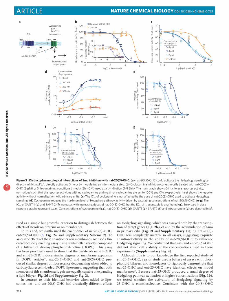
Figure 3 | Distinct pharmacological interactions of Smo inhibitors with nat-20(S)-OHc. (a) nat-20(S)-oHC could activate the Hedgehog signaling by directly inhibiting Ptc1, directly activating Smo or by modulating an intermediate step. (b) Cyclopamine inhibition curves in cells treated with nat-20(S)-oHC (8 μM) or Shh-containing conditioned media (Shh-CM) used at a 1/4 dilution (1/4 Shh). The main graph shows Gli luciferase reporter activity, normalized such that the reporter activities with no cyclopamine and maximal cyclopamine are set to 100% and 0%, respectively. Inset shows the reporter activity without normalization. au, arbitrary units. (c) The IC50 of cyclopamine is not affected by the dose of nat-20(S)-oHC used to activate Hedgehog signaling. (d) Cyclopamine reduces the maximum level of Hedgehog pathway activity driven by saturating concentrations of nat-20(S)-oHC. (e–g) The IC50 of SaNT-1 (e) and SaNT-2 (f) increases with increasing doses of nat-20(S)-oHC, but the IC50 of itraconazole is unaffected (g). error bars in dose response graphs represent s.e.m. Concentrations of cyclopamine (b,c), nat-20(S)-oHC (d), SaNT1 (e), SaNT2 (f) and intraconazole (g) are denoted in M.
npg
© 2
012
Nat
ure
Am
eric
a, In
c. A
ll rig
hts
rese
rved
.
nature CHeMICaL BIOLOGY | vol 8 | february 2012 | www.nature.com/naturechemicalbiology 215
articleNaTUre cHemical biOlOgy dOI: 10.1038/nCHeMBIO.765
results, only nat-25-OHC was able to activate Hedgehog reporter genes; ent-25-OHC was completely inactive (Fig. 2g).
Taken together, the regioselectivity, enantioselectivity and diaste-reoselectivity observed at the C20 position provided a compelling case for the existence of a protein receptor for nat-20(S)-OHC that has an important role in Hedgehog signaling.
cyclopamine noncompetitively inhibits nat-20(S)-OHcWhat protein target mediates the effects of nat-20(S)-OHC on Hedgehog signaling? Previous studies have clearly established that oxysterols act at the level of Ptc1, Smo or at an intermediate step between Ptc1 and Smo13 (Fig. 3a). These findings are consis-tent with the observation that nat-20(S)-OHC does not influence target gene transcription in cells lacking Ptc1, which demonstrate maximum levels of signaling driven by constitutive Smo activity13 (Supplementary Fig. 5). However, the study of nat-20(S)-OHC– interacting proteins has been difficult because of the lack of radiola-beled or otherwise modified oxysterol analogs.
The availability of many Hedgehog pathway agonists and antagonists with defined targets motivated us to analyze the pharmacological interactions between these molecules and nat-20-(S)-OHC. We measured the half-maximal inhibitory concentration (IC50) of cyclopamine, a direct Smo inhibitor, under conditions in which Hedgehog target genes were activated to similar levels by either Shh or nat-20(S)-OHC. Cyclopamine was chosen for the ini-tial analysis because the majority of Smo agonists and antagonists have been shown to compete with cyclopamine for binding to Smo-containing membranes, thus defining the cyclopamine-binding site as an important regulatory site on Smo35. We reasoned that if nat-20(S)-OHC functions by inactivating Ptc1 in a manner analo-gous to Shh or by influencing a step between Ptc1 and Smo, the IC50 of cyclopamine should be the same in the presence of equal activation by either agonist because the cyclopamine target, Smo, is downstream of these steps. Contrary to this prediction, the IC50 of cyclopamine was lower by a factor of nine when the pathway was activated with nat-20(S)-OHC (IC50 = 50 nM) compared to when it
was activated with Shh (IC50 = 460 nM) (Fig. 3b). Importantly, we used doses of Shh and nat-20(S)-OHC that produced near-equiv-alent amounts of target gene transcription, ensuring that this dif-ference did not occur simply because nat-20(S)-OHC activated the pathway to a lesser extent (Fig. 3b). Our findings suggest that the mechanism of activation of Hedgehog signaling by nat-20(S)-OHC is different from that of Shh. Smo is more sensitive to inhibition by cyclopamine when cells are activated by nat-20(S)-OHC com-pared to when they are activated by Shh, suggesting that Smo is in different conformations under these two conditions. These data make Ptc1 an unlikely candidate for the nat-20(S)-OHC receptor, motivating us to question the prevailing model and ask whether nat-20(S)-OHC binds Smo after all.
We further characterized the antagonistic interaction between nat-20(S)-OHC and cyclopamine by determining the IC50 of cyclo-pamine at different doses of nat-20(S)-OHC. In the case of competi-tive inhibition, increasing doses of nat-20(S)-OHC should produce a progressively higher IC50 for cyclopamine36. This is exactly the interaction observed between cyclopamine and the small mol-ecule SAG, which activates Smo by competing for the cyclopamine binding site9 (Supplementary Fig. 6). In contrast, we found that the IC50 of cyclopamine did not change with increasing doses of nat-20(S)-OHC (Fig. 3c), indicating that this is not a competitive interaction and making it unlikely that nat-20(S)-OHC and cyclo-pamine bind the same site on Smo. A prior study reached a similar conclusion regarding the lack of a competitive interaction between nat-20(S)-OHC and cyclopamine, both at the level of Smo binding and Hedgehog pathway activation, leading the authors to suggest that Smo is not a target of oxysterols13.
However, an alternative possibility is that cyclopamine and nat-20(S)-OHC bind Smo at distinct sites and that cyclopamine is a noncompetitive, ‘insurmountable’ inhibitor of nat-20(S)-OHC– induced Hedgehog signaling36. Formally, this would imply an allo-steric interaction between the two molecules because cyclopamine binding at one site would influence either the binding or effi-cacy of nat-20(S)-OHC at a different site. The kinetic signature of
f
b c
SAG (nM)00 0
0081.5 1.5
0.3 0.3150
37
Gli1
p38
Hed
geho
g re
port
er a
ctiv
ity(%
of m
ax)
020406080
100120
0 –10.0 –9.5 –9.0 –8.0–8.5
Concentration ofnat-20(S)-OHC
0.25 µM 0.5 µM
log([SAG])
–7.5 –6.5 –5.5–7.0 –6.0 –5.0Hed
geho
g re
port
er a
ctiv
ity(%
of m
ax)
0
020406080
100120
Concentrationof Shh
1/128 1/64
log([nat-20(S)-OHC]) log([nat-20(S)-OHC])0 –7.5 –6.5 –5.5
Frac
tiona
l ind
uctio
n
0.2
0.4
0.6
0.8
1.0
1.2 Shh observedShh predicted
SAG predictedSAG observed
e
–5.0–6.0–7.00
nat-20(S) (µM)
0
20
406080
100
120
–6.5 –5.5–6.0 –5.00log([nat-20(S)-OHC])
Concentration ofpurmorphamine
0.05 µM0.1 µM
Hed
geho
g re
port
er a
ctiv
ity(%
of m
ax)
g
0 µM
0 0 µM
Frac
tiona
l ind
uctio
n
0.20.40.60.81.01.2
0
1.4
0 –6.5 –5.5 –5.0–6.0log([nat-20(S)-OHC])
0.05 µM observed0.05 µM predicted
0.1 µM predicted0.1 µM observed
h
2.0
Cili
ary
Smo
fluor
esce
nce
(AU
)
0
0.5
1.5
1.0
nat-20(S) (µM)SAG (nM)
00 0
00.50.3
0.50.3
daH
edge
hog
repo
rter
act
ivity
(% o
f max
)
0 –7.5 –6.5 –5.5–7.0 –6.0 –5.0
020406080
100120
0.3 nM
Concentrationof SAG
0.03 nM
log([nat-20(S)-OHC])
0 nM
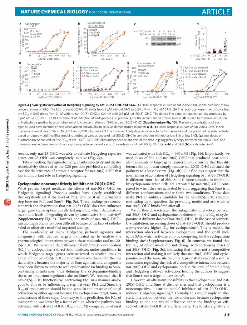
Figure 4 | Synergistic activation of Hedgehog signaling by nat-20(S)-OHc and Sag. (a) Dose-response curves of nat-20(S)-oHC in the presence of low concentrations of SaG. The eC50 of nat-20(S)-oHC shifts from 3 μM without SaG to 0.24 μM with 0.3 nM SaG. (b) The reciprocal experiment shows that the eC50 of SaG drops from 2 nM with no nat-20(S)-oHC to 0.4 nM with 0.5 μM nat-20(S)-oHC. The dotted line denotes reporter activity produced by 8 μM nat-20(S)-oHC. (c,d) The amount of induction of endogenous Gli1 protein (c) or the accumulation of Smo in cilia (d) is used to measure activation of Hedgehog signaling by a combination of low concentrations of SaG and nat-20(S)-oHC (Supplementary Fig. 10). The low concentrations of each agonist used have minimal effects when added individually to cells, as demonstrated in panels a–d. (e) Dose-response curves of nat-20(S)-oHC in the presence of low doses of Shh-CM (1/64 and 1/128 dilutions). (f) The observed Hedgehog reporter activity from a and e and the predicted reporter activity based on a purely additive bliss model is plotted at various doses of nat-20(S)-oHC in combination with either low Shh or low SaG. (g) low doses of purmorphamine can reduce the eC50 of nat-20(S)-oHC. (h) bliss independence analysis of the data in g suggests synergy between nat-20(S)-oHC and purmorphamine. error bars in dose-response graphs represent s.e.m. Concentrations of nat-20(S)-oHC (a, e–h) and SaG (b) are denoted in M.
npg
© 2
012
Nat
ure
Am
eric
a, In
c. A
ll rig
hts
rese
rved
.
216 nature CHeMICaL BIOLOGY | vol 8 | february 2012 | www.nature.com/naturechemicalbiology
article NaTUre cHemical biOlOgy dOI: 10.1038/nCHeMBIO.765
noncompetitive antagonism is a reduction in the maximum achiev-able level of activation by the agonist36. Consistent with noncom-petitive inhibition, cyclopamine reduced the maximum extent of activation produced by saturating doses of nat-20(S)-OHC (Fig. 3d). The data presented to this point are consistent with the model that nat-20(S)-OHC binds and activates Smo, albeit through a site different from the canonical cyclopamine binding site.
nat-20(S)-OHc interactions with other Smo antagonistsSANT-1 and SANT-2 are two direct Smo antagonists that compete with cyclopamine for binding to Smo9,35. Unlike cyclopamine, both antagonists show a competitive relationship with nat-20(S)-OHC (Fig. 3e,f). Increasing doses of nat-20(S)-OHC led to a progressive rightward shift in the SANT-1 and SANT-2 inhibition curves and an approximately three- to four-fold increase in their IC50 values (Fig. 3e,f). As the SANT molecules inhibit cyclopamine binding9 and nat-20(S)-OHC has no effect on cyclopamine binding13, it is unlikely that the SANTs occupy the same binding site as nat-20(S)-OHC; instead, the interaction between them is most likely mediated by an allosteric mechanism.
Among the Smo antagonists described to date, the antifungal itraconazole is the only one that does not compete with cyclo-pamine for Smo binding37. Itraconazole was previously inferred to be a direct Smo antagonist on the basis of its ability to synergize with cyclopamine to inhibit Hedgehog signaling37. Given that nat-20(S)-OHC also does not inhibit the cyclopamine-Smo interac-tion13 (Fig. 3c), we considered the possibility that itraconazole and
nat-20(S)-OHC bind the same site on Smo, a mechanism that predicts a competitive interaction between these molecules. However, itra-conazole and nat-20(S)-OHC most likely bind different sites because increasing doses of nat-20(S)-OHC did not cause a rightward shift in the itraconazole inhibition curve or alter its IC50 (Fig. 3g).
Taken together, the interaction of nat-20(S)-OHC with a panel of direct Smo antagonists demonstrates the property of ‘probe speci-ficity’: nat-20(S)-OHC has an impact on the IC50 values of SANT-1 and SANT-2 but has no effect on those of cyclopamine and itracon-azole. Probe specificity is considered a hallmark of allosteric ligands because their ability to stabilize an ensemble of protein conforma-tions can have a large impact on one receptor probe but little impact on others36. In summary, the distinct pharmacological interactions between nat-20(S)-OHC and various Smo ligands provide addi-tional evidence for its direct interaction with Smo.
Smo agonists synergize with nat-20(S)-OHcThe model in which nat-20(S)-OHC and cyclopamine bind dis-tinct sites on Smo predicts that nat-20(S)-OHC should show allosteric interactions with Smo activators, such as SAG or pur-morphamine, that compete with cyclopamine9,10. In contrast, if nat-20(S)-OHC activated signaling in a completely different way, without directly interacting with Smo, it would be unlikely to show an allosteric interaction with these agonists. We mea-sured the dose-response curve for activation by nat-20(S)-OHC in the absence or presence of low concentrations of SAG (Fig. 4a). Notably, the concentration of SAG used here (0.3 nM) was lower
0 –6.5 –6 –5.5 –5–7.5 –7log([sterol])
Hed
geho
g re
port
er a
ctiv
ity
(fol
d ch
ange
)
0
20
10
30
40
50
nat-20(S)-OHCnat-20(S)-yne
a b c
WB: αGFP
100
Smo–/–:YFP-SmoSmo–/–
Dark exposure
Control beads+
++
+
Inputs Eluates Inputs Eluates
++
++
e 100
80
00 –6 –5 –4
log[nat-20(S)-yne]
YFP-
Smo
sign
al(%
spe
cific
bin
ding
)
60
20
40 Am
ount
elu
ted
(fol
d ch
ange
ove
rso
lven
t con
trol
)
0
Solvent
nat-20(S)
-yne
nat-20(S)
-OHC
ent-20(S)
-OHC
22(S)-O
HC
4
2
6fd
20
0YFP-Smo SSTR3-GFP HTR6-GFP
40
60
GFP
sig
nal
(fol
d ch
ange
ove
r con
trol
bea
ds) Control beads
nat-20(S)-yne beads
nat-20(S)-yne beads
HO
H
H H
OH
H
HO
H
H H
OH
H
N
NN
O
O
OH4
HO
H
H H
OH
H
N
NN
O
O
NH4
4nat-20(S)-yne
5
nat-20(S)-yne beads
bead
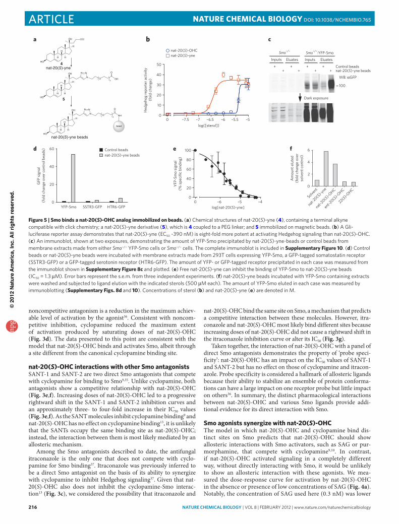
Figure 5 | Smo binds a nat-20(S)-OHc analog immobilized on beads. (a) Chemical structures of nat-20(S)-yne (4), containing a terminal alkyne compatible with click chemistry; a nat-20(S)-yne derivative (5), which is 4 coupled to a PeG linker; and 5 immobilized on magnetic beads. (b) a Gli-luciferase reporter assay demonstrates that nat-20(S)-yne (eC50 ~390 nM) is eight-fold more potent at activating Hedgehog signaling than nat-20(S)-oHC. (c) an immunoblot, shown at two exposures, demonstrating the amount of yfP-Smo precipitated by nat-20(S)-yne-beads or control beads from membrane extracts made from either Smo−/− yfP-Smo cells or Smo−/− cells. The complete immunoblot is included in Supplementary Figure 10. (d) Control beads or nat-20(S)-yne beads were incubated with membrane extracts made from 293T cells expressing yfP-Smo, a GfP-tagged somatostatin receptor (SSTr3-GfP) or a GfP-tagged serotonin receptor (HTr6-GfP). The amount of yfP- or GfP-tagged receptor precipitated in each case was measured from the immunoblot shown in Supplementary Figure 8c and plotted. (e) free nat-20(S)-yne can inhibit the binding of yfP-Smo to nat-20(S)-yne beads (IC50 = 1.3 μM). error bars represent the s.e.m. from three independent experiments. (f) nat-20(S)-yne beads incubated with yfP-Smo containing extracts were washed and subjected to ligand elution with the indicated sterols (500 μM each). The amount of yfP-Smo eluted in each case was measured by immunoblotting (Supplementary Figs. 8d and 10). Concentrations of sterol (b) and nat-20(S)-yne (e) are denoted in M.
npg
© 2
012
Nat
ure
Am
eric
a, In
c. A
ll rig
hts
rese
rved
.
nature CHeMICaL BIOLOGY | vol 8 | february 2012 | www.nature.com/naturechemicalbiology 217
articleNaTUre cHemical biOlOgy dOI: 10.1038/nCHeMBIO.765
than its EC50 by a factor of ten9 and resulted in less than 10% of the maximum degree of Hedgehog pathway activation. In the presence of this miniscule concentration of SAG, the EC50 of nat-20(S)-OHC sharply declined by a factor of over ten from 3.2 μM to 0.24 μM (Fig. 4a), providing evidence for a synergistic interaction between these agonists. In the reciprocal experiment, low concentrations of nat-20(S)-OHC also decreased the EC50 of SAG from 2 nM to 0.4 nM (Fig. 4b). We confirmed the synergy between SAG and nat-20(S)-OHC by measuring the amount of endogenous Gli1 protein (Fig. 4c) and the accumulation of Smo in primary cilia (Fig. 4d and Supplementary Fig. 7).
The simplest explanation for the synergy observed between SAG and nat-20(S)-OHC is that both agonists bind Smo at dif-ferent sites and show a positive allosteric interaction. In compari-son, the combination of nat-20(S)-OHC with Shh, which does not bind Smo but indirectly leads to its activation by inactivating Ptc1, did not show evidence of synergy (Fig. 4e). The EC50 of nat-20(S)-OHC did not change drastically when combined with low concen-trations of Shh (EC50 without Shh: 3.2 μM; EC50 with Shh: 2.5 μM). We used Bliss independence analysis38 to compare the interaction between nat-20(S)-OHC and SAG with the interaction between nat-20(S)-OHC and Shh (Fig. 4f). The Bliss score uses the activi-ties of each agonist to predict the response resulting from purely additive behavior with respect to each other, as might be expected if the two molecules influence different proteins. The responses observed when nat-20(S)-OHC and SAG were combined were much stronger than the Bliss prediction (Fig. 4f), consistent with synergy. In contrast, the Bliss prediction was virtually superim-posable with the observed response when Shh and nat-20(S)-OHC were combined (Fig. 4f).
Dose-response data and Bliss analysis showed that nat-20(S)-OHC also shows a synergistic interaction with purmorphamine, a direct Smo agonist that is less potent than SAG by a factor of approx-imately 100 (Fig. 4g,h)10. Trace doses of purmorphamine (below its EC50 by a factor of ten) decreased the EC50 of nat-20(S)-OHC by a factor of approximately three (Fig. 4g). Thus, two different direct Smo agonists, varying in potency over two orders of magnitude, showed positive allosteric interactions with nat-20(S)-OHC.
Smo binds to nat-20(S)-OHc immobilized on beadsOur pharmacological analyses suggested that Smo is the receptor for nat-20(S)-OHC. To test this prediction, we synthesized an analog of nat-20(S)-OHC with a terminal alkyne group (4; nat-20(S)-yne) com-patible with click chemistry techniques for coupling the molecule to a matrix (Fig. 5a and Supplementary Scheme 3). Remarkably, this ana-log was eight-fold more potent than nat-20(S)-OHC itself (Fig. 5b) when tested in the Hedgehog reporter assay, making it an ideal mol-ecule for ligand affinity chromatography. nat-20(S)-yne induced Smo accumulation in cilia, confirming that its mechanism of action is the same as that of nat-20(S)-OHC (Supplementary Fig. 8a). Using click chemistry, nat-20(S)-yne was covalently coupled to magnetic beads through an intermediate molecule (5) that contained a polyethyl-ene glycol (PEG) linker interposed to minimize steric inhibition of receptor binding (Fig. 5a and Supplementary Schemes 4 and 5). As a source of Smo protein, detergent extracts were prepared from membranes isolated from Smo−/− fibroblasts stably expressing func-tional, YFP-tagged Smo (Smo−/− YFP-Smo cells)39. Consistent with a physical interaction between nat-20(S)-OHC and Smo, YFP-Smo was captured on nat-20(S)-yne bearing magnetic beads (Fig. 5c). Binding was proportional to the amount of YFP-Smo added (Supplementary Fig. 8b), and control beads (Supplementary Scheme 5) captured amounts of YFP-Smo that were lower by a factor of ten (Fig. 5c). To provide more precise relative quantification of YFP-Smo captured in these affinity chromatography experiments, all immunoblots shown were developed and quantified using the LiCor infrared detection system rather than chemiluminescence.
Several controls were performed to establish the specificity of this interaction. A band at the position corresponding to YFP-Smo was not observed on immunoblots when affinity chromatography was performed using Smo−/− cells (lacking YFP-Smo), confirming the specificity of the signal detected with the antibody to GFP (Fig. 5c). Notably, the pattern of background bands captured from Smo−/− extracts by control beads and nat-20(S)-yne beads was similar (Fig. 5c), highlighting the relevance of the specific interaction between nat-20(S)-yne beads and YFP-Smo. In addition, two other seven-pass transmembrane receptors that localize in primary cilia did not show a specific interaction with nat-20(S)-yne beads (Fig. 5d and Supplementary Fig. 8c).
The binding of YFP-Smo to nat-20(S)-yne beads was inhibited by free nat-20(S)-yne added to the extract in a dose-dependent fash-ion (IC50 = 1.3 μM), demonstrating that this interaction is mediated through the sterol itself and not through the PEG linker (Fig. 5e). Three Smo ligands, cyclopamine, SANT-1 and itraconazole, which inhibited nat-20(S)-OHC–induced Hedgehog signaling (Fig. 3), did not inhibit the interaction between YFP-Smo and nat-20(S)-yne beads when used at concentrations up to five-fold higher than their respective IC50 values (Supplementary Fig. 9). Finally, to demon-strate the enantioselectivity of the interaction between Smo and nat-20(S)-OHC, we tested the ability of various ligands to elute YFP-Smo captured on nat-20(S)-yne beads. Both nat-20(S)-yne and nat-20(S)-OHC eluted YFP-Smo from the beads. However, ent-20(S)-OHC and an inactive, structurally related oxysterol, 22(S)-OHC, were both unable to elute YFP-Smo (Fig. 5f and Supplementary Fig. 8d). As nat- and ent-20(S)-OHC have identical hydrophobic character-istics evident from their identical interactions with lipid bilayers (Fig. 2d), the selective elution with one enantiomer supports a specific interaction between nat-20(S)-OHC and Smo. Taken together, these biochemical data corroborate the pharmacological data and demon-strate a physical interaction between nat-20(S)-OHC and Smo.
SAG
nat-20(S)-OHC
Cyclopamine
Active Smo
Inactive Smo
Competitive
Noncompetitive
Allosteric
Allosteric
Allosteric connectivity
Smo
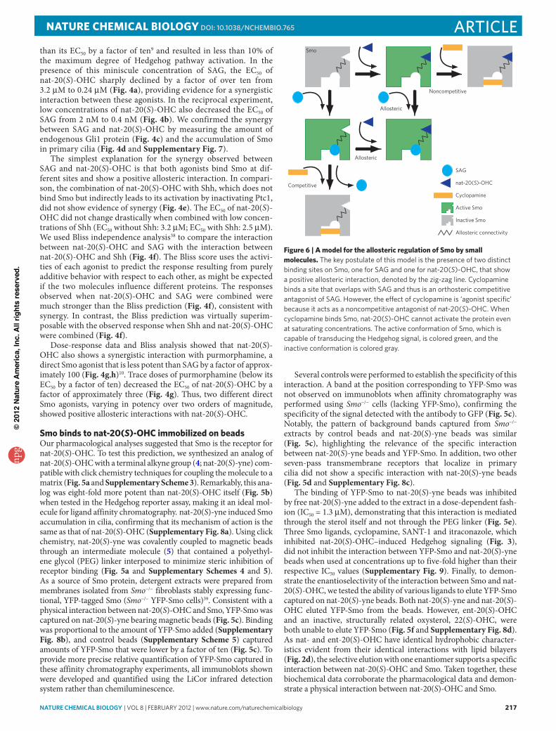
Figure 6 | a model for the allosteric regulation of Smo by small molecules. The key postulate of this model is the presence of two distinct binding sites on Smo, one for SaG and one for nat-20(S)-oHC, that show a positive allosteric interaction, denoted by the zig-zag line. Cyclopamine binds a site that overlaps with SaG and thus is an orthosteric competitive antagonist of SaG. However, the effect of cyclopamine is ‘agonist specific’ because it acts as a noncompetitive antagonist of nat-20(S)-oHC. When cyclopamine binds Smo, nat-20(S)-oHC cannot activate the protein even at saturating concentrations. The active conformation of Smo, which is capable of transducing the Hedgehog signal, is colored green, and the inactive conformation is colored gray.
npg
© 2
012
Nat
ure
Am
eric
a, In
c. A
ll rig
hts
rese
rved
.
218 nature CHeMICaL BIOLOGY | vol 8 | february 2012 | www.nature.com/naturechemicalbiology
article NaTUre cHemical biOlOgy dOI: 10.1038/nCHeMBIO.765
DiScUSSiONIn addition to their roles in regulating the local structure and dynam-ics of membranes, sterols can affect a broad range of physiologi-cal and pathological processes, ranging from lipid metabolism17–20 and atherosclerosis40 to apoptosis23, inflammation24 and cancer susceptibility41. In many of these cases, the specific mechanisms and molecular pathways by which sterols exert their effects on cells remain unknown. One possibility is that endogenous oxy sterols function as second messengers in cellular signaling pathways. A prominent example supporting this idea is the Hedgehog sig-naling pathway, which has long been postulated to be regulated by endogenous sterols12,42.
Although the molecular basis of this sterol dependency remains to be understood, oxysterols have recently emerged as candidates for regulatory small molecules in Hedgehog signaling. nat-20(S)-OHC is an effective activator of Hedgehog signaling but is a par-ticularly enigmatic and poorly understood molecule. Biochemical pathways for the synthesis and degradation of this molecule remain unknown, even though it is detectable in tissues43. Herein, we used a combination of chemical and pharmacological methods to understand the mechanism by which nat-20(S)-OHC influences Hedgehog signaling.
A classical problem in the study of sterols (and most amphiphiles) comes from the difficulty in distinguishing their effects on mem-brane properties from their direct effects on proteins31. The use of enantiomers represents an incisive and generally applicable meth-odology to make this critical distinction; thus, we report what is to our knowledge the first synthesis of ent-20(S)-OHC (3). This enantiomer was completely inactive in Hedgehog pathway assays, suggesting that nat-20(S)-OHC activates Hedgehog signaling by binding to a highly chiral protein-binding pocket rather than by incorporating into a dynamic lipid membrane wherein even ‘ordered’ domains are most likely rapidly assembling and disassem-bling and are therefore insensitive to stereochemical changes31. In addition, we have also reported an improved synthesis of the C20 epimer of nat-20(S)-OHC, nat-20(R)-OHC44 (2). nat-20(R)-OHC did not activate Hedgehog signaling, further exemplifying the struc-tural discrimination associated with a specific protein interaction.
Our pharmacological and biochemical studies provide evidence for the direct effect of nat-20(S)-OHC on Smo, an oncoprotein and cancer drug target. The high degree of synergy seen between nat-20(S)-OHC and the direct Smo agonist SAG is most parsimo-niously explained by a positive allosteric interaction between the two molecules mediated through distinct sites on Smo. As SAG and cyclopamine show a competitive interaction9 both in binding and activation assays, nat-20(S)-OHC most likely binds a site distinct from the canonical cyclopamine binding site that has been the tar-get for most anti-Smo drugs9–11. Taken together, our data are con-sistent with the presence of at least two binding sites on Smo, one that binds nat-20(S)-OHC and a second that binds SAG and cyclo-pamine (Fig. 6). SAG and nat-20(S)-OHC show a positive allosteric interaction, SAG and cyclopamine show a competitive interaction, and cyclopamine and nat-20(S)-OHC show a noncompetitive inter-action (Fig. 6). Given the similarity of Smo to G protein–coupled receptors (GPCRs), our discovery of a Smo-oxysterol interaction is reminiscent of recent evidence showing that metarhodopsin I (ref. 45) and the β2-adrenergic receptor46 bind cholesterol through their transmembrane regions and that oxysterols can func-tion in chemotaxis of leukocytes by acting on a GPCR21,22. Thus, it is possible that sterols represent a class of ligands that have an important role in modulating the activity of many GPCR-initiated signaling pathways.
Like GPCRs, Smo most likely adopts a range of conformations with different signaling properties39. Different Smo ligands clearly stabi-lize distinct ensembles of these conformations. One striking example comes from the differences between SANT-1 and cyclopamine, two
Smo ligands that show a competitive binding interaction. Although both molecules inhibit Hedgehog target-gene transcription, SANT-1 inhibits Smo accumulation in cilia, whereas cyclopamine drives it, proving that they stabilize distinct conformations39. This is consistent with our finding that nat-20(S)-OHC interacts noncompetitively with cyclopamine but does so competitively with SANT-1. From a thera-peutic perspective, Smo may be susceptible to allosteric regulation, and future drug discovery efforts should focus on targeting of such allosteric sites. In addition, it will be important to determine whether different Smo ligands can favor the coupling of Smo to distinct sets of downstream signaling complexes.
Although our results provide evidence for the allosteric acti-vation of Smo by nat-20(S)-OHC, further studies are needed to address the effects of endogenous nat-20(S)-OHC on Hedgehog pathway activity in cells and animals. The EC50 of nat-20(S)-OHC for Hedgehog target gene induction (~3 μM) is in the same range as EC50 values (4–7 μM) reported for the activation of LXRα recep-tors by the endogenous ligands 24-OHC and 22-OHC19,20. Though the concentration of nat-20(S)-OHC in Hedgehog-responsive cells or embryos has not been carefully measured, concentrations of oxysterols in tissues have been estimated to be in the 0.1–10 μM range47,48. Although this seems lower than our measured EC50 for nat-20(S)-OHC, it is noteworthy that the affinity constants (Kd values) for oxysterol-protein interaction are often more than an order of magnitude lower than the EC50 values, presumably because much of the oxysterol is not available to the receptor when its added to cell culture19. In addition, the local concentration of a lipophilic molecule in a cellular compartment can be substantially higher than the bulk concentration measured in a tissue. Future progress in this area will require new methods to reliably measure nat-20(S)-OHC concentrations in cells and an understanding of how this molecule is synthesized, transported and degraded such that its concentrations can be perturbed. nat-20(S)-yne (4), a potent, click chemistry–compatible analog of nat-20(S)-OHC, provides an invaluable bio-orthogonal reporter to dissect nat-20(S)-OHC func-tion in cells and animals. A rigorous understanding of how sterols influence Smo and other GPCRs is certain to provide new avenues for the modulation of these key therapeutic targets in a variety of human diseases.
meTHODSCells and reagents. NIH 3T3 and 293T cells were obtained from the American Type Culture Collecton, Wnt-L cells from E. Lee (Vanderbilt University), SAG (≥ 95%) from Enzo Life Sciences, cyclopamine from Toronto Research Chemicals (≥ 98%), itraconazole (≥ 98%) from Sigma, the SANTs (≥ 95%) from EMD Chemicals and purmorphamine (≥ 98%) from Cayman Chemicals. All sterols except ent-20(S)-OHC, nat-20(R)-OHC and nat-20(S)-yne were purchased from Steraloids (purity ≥ 98%).
Chemical synthesis. Schemes for nat-20(R)-OHC, ent-20(S)-OHC, nat-20(S)-yne and nat-20(S)-yne beads are fully described in Supplementary Schemes 1–5.
Hedgehog reporter assays. NIH 3T3 cells in a 10-cm plate were transfected with 8 μg of a 19:1 ratio of firefly luciferase reporter driven by a Gli-responsive promoter28 and a constitutive Renilla reniformis luciferase reporter. The next day, transfected cells were seeded into a 96-well plate, grown to confluence and treated overnight with drugs diluted in medium containing 0.5% FBS. Activity of both reporters was measured using the Dual-Luciferase Reporter kit (Promega) and read on a Berthold LB 96-V luminometer or a Tecan Infinite M1000 plate reader. The Gli luciferase/Renilla luciferase ratio was taken as a metric for Hedgehog signaling.
Immunoblotting. Immunoblotting was performed as described previously39, and detailed methods are provided in Supplementary Methods.
Ligand affinity chromatography. Smo−/− cells and Smo−/− YFP-Smo cells39 were lysed by hypotonic lysis in SEAT buffer (250 mM sucrose, 1 mM EDTA, 10 mM acetic acid, 10 mM triethanolamine, 10 μg ml−1 leupeptin-pepstatin-chymostatin (LPC) protease inhibitor mix and the SigmaFast EDTA-free protease cocktail). After removal of nuclei by centrifugation (500g, 5 min), membranes were pel-leted by centrifugation at 95,000g for 30 min. Membranes were extracted in an n-dodecyl-β-D-maltopyranoside (DDM) extraction buffer (50 mM Tris pH 7.4,
npg
© 2
012
Nat
ure
Am
eric
a, In
c. A
ll rig
hts
rese
rved
.
nature CHeMICaL BIOLOGY | vol 8 | february 2012 | www.nature.com/naturechemicalbiology 219
articleNaTUre cHemical biOlOgy dOI: 10.1038/nCHeMBIO.765
500 mM NaCl, 10% v/v glycerol, 0.1% w/v DDM and the SigmaFast EDTA-Free protease cocktail) for 4 h at 4 °C, followed by removal of insoluble material by centrifugation (100,000g, 30 min). This DDM extract was incubated with nat-20(S)-yne–coupled magnetic beads or control beads for 12 h at 4 °C to allow binding. In experiments in which soluble ligands were included as competitors, the extracts were incubated with free ligand for 1 h at 4 °C before the addition of beads. In all experiments, the amount of solvent was carefully equalized across samples. After extensive washing, proteins captured on the beads were eluted with free ligand or with reducing SDS sample buffer. The presence of YFP-Smo in these eluates was determined by quantitative immunoblotting with a GFP-specific antibody (Novus, NB600-308, 1:5000) and infrared imaging (LiCor Odyssey system).
Quantitative real-time PCR. After drug treatment (described above), RNA was harvested from cells in TRIzol (Invitrogen) and converted to cDNA using the iScript cDNA synthesis kit (BioRad) with a mixture of oligo(dT) and random hexamer primers. This cDNA was used as a substrate for quantitative real-time PCR using primers for Gli1 (forward: 5′-CCAAGCCAACTTTATGTCAGGG-3′; reverse: 5′-AGCCCGCTTCTTTGTTAATTTGA-3′) and the loading control GAPDH (forward 5′-AGTGGCAAAGTGGAGATT-3′; reverse: 5′-GTGGAG TCATACTGGAACA-3′). Quantitative real-time PCR assays were performed with IQ SYBR Green Supermix (Bio-Rad) on a Sequence Detector 7900HT (Applied Biosystems).
Immunofluorescence, microscopy and image analysis. Immunofluorescence, microscopy and image analysis were performed as described previously15, and detailed methods are included in the Supplementary Methods.
Data analysis. All statistical analysis and curve fitting was done in GraphPad Prism. For microscopy data, the Smo fluorescence for each cilium was individually plotted, generating a scatter plot that represents variability in the data. To compare the amount of Smo between different conditions, the mean and 95% confidence interval are provided (n = 30–40).
For Hedgehog reporter assays, each point represents the mean of measure-ments from triplicate wells, with error bars representing the s.e.m. Each experi-ment in the paper was repeated at least three times with similar outcomes. Relative luciferase activity was calculated by dividing the luminescence of Gli luciferase by that of Renilla luciferase. Fold change in reporter activity was calculated by divid-ing the activity measured in each replicate by the mean reporter activity of the solvent-treated control. Normalized (percentage of maximum) Hedgehog reporter activity was calculated by setting the minimum and maximum values of a curve to 0% and 100%, respectively, using the ‘normalize’ function of GraphPad Prism. For the binding inhibition curves, percentage specific binding refers to the amount of binding detected above nonspecific background binding (defined as the bind-ing observed even at maximal inhibitor concentrations). The maximum binding detected in the absence of any inhibitor was set to 100%. In all graphs, dotted lines are straight connectors between points, and solid lines represent nonlinear curve fits of the data to a sigmoidal (variable-slope) equation. Curve fits were calculated in GraphPad Prism using the log(agonist/inhibitor) versus response or normalized response as appropriate.
Bliss independence analysis. For Bliss independence analysis38, we calculated the fractional activation (F) produced by each drug dose by dividing its reporter activ-ity by the maximal Hedgehog reporter activity obtained with that drug. The Bliss score for a combination of two drugs (A and B) was calculated with the equation FA + FB (1 − FA), representing the activation predicted if the two drugs act inde-pendently. This score was compared to the observed response when the two drugs were combined at the same doses. The Bliss analysis presented in Figure 4f uses data from Figures 4a,e, and the analysis in Figure 4h uses data from Figure 4g.
Liposome expansion assay. Carboxyfluorescein-loaded unilamellar liposomes were prepared and used in the dequenching assay as described previously33, and detailed methods are outlined in the Supplementary Methods.
received 15 July 2011; accepted 3 November 2011; Published online 8 January 2012
references1. Varjosalo, M. & Taipale, J. Hedgehog: functions and mechanisms. Genes Dev. 22,
2454–2472 (2008).2. Murone, M., Rosenthal, A. & de Sauvage, F.J. Sonic hedgehog signaling by the
patched-smoothened receptor complex. Curr. Biol. 9, 76–84 (1999).3. Stone, D.M. et al. The tumour-suppressor gene patched encodes a candidate
receptor for Sonic hedgehog. Nature 384, 129–134 (1996).4. Marigo, V. et al. Biochemical evidence that patched is the Hedgehog receptor.
Nature 384, 176–179 (1996).5. Barakat, M.T., Humke, E.W. & Scott, M.P. Learning from Jekyll to control
Hyde: Hedgehog signaling in development and cancer. Trends Mol. Med. 16, 337–348 (2010).
6. Cooper, M.K., Porter, J.A., Young, K.E. & Beachy, P.A. Teratogen-mediated inhibited of target tissue response to Shh signaling. Science 280, 1603–1607 (1998).
7. Chen, J.K. et al. Inhibition of Hedgehog signaling by direct binding of cyclopamine to Smoothened. Genes Dev. 16, 2743–2748 (2002).
8. Heretsch, P., Tzagkaroulaki, L. & Giannis, A. Cyclopamine and hedgehog signaling: chemistry, biology, medical perspectives. Angew. Chem. Int. Ed. Engl. 49, 3418–3427 (2010).
9. Chen, J.K. et al. Small molecule modulation of Smoothened activity. Proc. Natl. Acad. Sci. USA 99, 14071–14076 (2002).
10. Sinha, S. & Chen, J.K. Purmorphamine activates the Hedgehog pathway by targeting Smoothened. Nat. Chem. Biol. 2, 29–30 (2006).
11. Romer, J.T. et al. Suppression of the Shh pathway using a small molecule inhibitor eliminates medulloblastoma in Ptc1+/−p53−/− mice. Cancer Cell 6, 229–240 (2004).
12. Corcoran, R.B. & Scott, M.P. Oxysterols stimulate Sonic hedgehog signal transduction and proliferation of medulloblastoma cells. Proc. Natl. Acad. Sci. USA 103, 8408–8413 (2006).
13. Dwyer, J.R. et al. Oxysterols are novel activators of the hedgehog signaling pathway in pluripotent mesenchymal cells. J. Biol. Chem. 282, 8959–8968 (2007).
14. Johnson, J.S. et al. Novel oxysterols have pro-osteogenic and anti-adipogenic effects in vitro and induce spinal fusion in vivo. J. Cell Biochem. 112, 1673–1684 (2011).
15. Rohatgi, R., Milenkovic, L. & Scott, M.P. Patched1 regulates hedgehog signaling at the primary cilium. Science 317, 372–376 (2007).
16. Corbit, K.C. et al. Vertebrate Smoothened functions at the primary cilium. Nature 437, 1018–1021 (2005).
17. LeBlanc, M.A. & McMaster, C.R. Lipid binding requirements for oxysterol-binding protein Kes1 inhibition of autophagy and endosome-trans-Golgi trafficking pathways. J. Biol. Chem. 285, 33875–33884 (2010).
18. Radhakrishnan, A. et al. Sterol-regulated transport of SREBPs from endoplasmic reticulum to Golgi: oxysterols block transport by binding to Insig. Proc. Natl. Acad. Sci. USA 104, 6511–6518 (2007).
19. Janowski, B.A. et al. Structural requirements of ligands for the oxysterol liver X receptors LXRα and LXRβ. Proc. Natl. Acad. Sci. USA 96, 266–271 (1999).
20. Chen, W. et al. Enzymatic reduction of oxysterols impairs LXR signaling in cultured cells and the livers of mice. Cell Metab. 5, 73–79 (2007).
21. Hannedouche, S. et al. Oxysterols direct immune cell migration via EBI2. Nature 475, 524–527 (2011).
22. Liu, C. et al. Oxysterols direct B-cell migration through EBI2. Nature 475, 519–523 (2011).
23. Panini, S.R. & Sinensky, M.S. Mechanisms of oxysterol-induced apoptosis. Curr. Opin. Lipidol. 12, 529–533 (2001).
24. Park, K. & Scott, A.L. Cholesterol 25-hydroxylase production by dendritic cells and macrophages is regulated by type I interferons. J. Leukoc. Biol. 88, 1081–1087 (2010).
25. Theunissen, J.J. et al. Membrane properties of oxysterols. Interfacial orientation, influence on membrane permeability and redistribution between membranes. Biochim. Biophys. Acta 860, 66–74 (1986).
26. Rentero, C. et al. Functional implications of plasma membrane condensation for T cell activation. PLoS ONE 3, e2262 (2008).
27. Olkkonen, V.M. & Hynynen, R. Interactions of oxysterols with membranes and proteins. Mol. Aspects Med. 30, 123–133 (2009).
28. Sasaki, H. et al. A binding site for Gli proteins is essential for HNF-3β floor plate enhancer activity in transgenics and can respond to Shh in vitro. Development 1997, 1313–1322 (1997).
29. Infante, R.E. et al. Purified NPC1 protein. I. Binding of cholesterol and oxysterols to a 1278-amino acid membrane protein. J. Biol. Chem. 283, 1052–1063 (2008).
30. Massey, J.B. & Pownall, H.J. Structures of biologically active oxysterols determine their differential effects on phospholipid membranes. Biochemistry 45, 10747–10758 (2006).
31. Covey, D.F. ent-Steroids: novel tools for studies of signaling pathways. Steroids 74, 577–585 (2009).
32. Mannock, D.A. et al. Effects of natural and enantiomeric cholesterol on the thermotropic phase behavior and structure of egg sphingomyelin bilayer membranes. Biophys. J. 84, 1038–1046 (2003).
33. Westover, E.J. et al. Cholesterol depletion results in site-specific increases in epidermal growth factor receptor phosphorylation due to membrane level effects. Studies with cholesterol enantiomers. J. Biol. Chem. 278, 51125–51133 (2003).
34. Gale, S.E. et al. Side chain oxygenated cholesterol regulates cellular cholesterol homeostasis through direct sterol-membrane interactions. J. Biol. Chem. 284, 1755–1764 (2009).
35. Rominger, C.M. et al. Evidence for allosteric interactions of antagonist binding to the smoothened receptor. J. Pharmacol. Exp. Ther. 329, 995–1005 (2009).
36. Kenakin, T.P. A Pharmacology Primer. 3rd edn. 101–147 (Elsevier, 2009).37. Kim, J. et al. Itraconazole, a commonly used antifungal that inhibits Hedgehog
pathway activity and cancer growth. Cancer Cell 17, 388–399 (2010).
npg
© 2
012
Nat
ure
Am
eric
a, In
c. A
ll rig
hts
rese
rved
.
220 nature CHeMICaL BIOLOGY | vol 8 | february 2012 | www.nature.com/naturechemicalbiology
article NaTUre cHemical biOlOgy dOI: 10.1038/nCHeMBIO.765
acknowledgmentsWe thank members of the Rohatgi lab for helpful discussions, G. Luchetti for help with ligand affinity chromatography, P. Niewiadomski and A. Lebensohn for critical reading of the manuscript, E. Lee (Vanderbilt University) for Wnt-L cells, K. Mykytyn (Ohio State University) for the SSTR3-GFP and HTR6-GFP constructs, A. Sweet-Cordero for use of a Li-Cor Odyssey imager and M. Scott for support and use of a confocal microscope. MS analysis was conducted at the NIH – National Center for Research Resources MS facility at Washington University, supported by the NIH (RR00954, DK020579, DK056341). This work was supported by a Pew Scholar Award and an Innovation Research Grant from the Stand Up to Cancer – American Association for Cancer Research Foundation to R.R., by NIH grants to D.F.C. (GM47969 and HL67773) and P.H.S. (HL67773), and by NIH training grants to S.N. (5 T32 GM007276) and L.K.M. (5 T32 HL007275).
author contributionsL.K.M., K.K. and D.F.C. designed and synthesized the oxysterol analogs. S.N. performed cellular experiments with oxysterols. J.R. and P.H.S. designed and performed vesicle expansion experiments. All authors analyzed the data and contributed to the manuscript. S.N. and R.R. wrote the paper with input from L.K.M., D.F.C., J.R. and P.H.S.
Competing financial interestsThe authors declare no competing financial interests.
additional informationSupplementary information and chemical compound information is available online at http://www.nature.com/naturechemicalbiology/. Reprints and permissions information is available online at http://www.nature.com/reprints/index.html. Correspondence and requests for materials should be addressed to R.R. or D.F.C.
38. Fitzgerald, J.B. et al. Systems biology and combination therapy in the quest for clinical efficacy. Nat. Chem. Biol. 2, 458–466 (2006).
39. Rohatgi, R. et al. Hedgehog signal transduction by Smoothened: pharmacologic evidence for a 2-step activation process. Proc. Natl. Acad. Sci. USA 106, 3196–3201 (2009).
40. Töröcsik, D., Szanto, A. & Nagy, L. Oxysterol signaling links cholesterol metabolism and inflammation via the liver X receptor in macrophages. Mol. Aspects Med. 30, 134–152 (2009).
41. Brown, A.J. Cholesterol, statins and cancer. Clin. Exp. Pharmacol. Physiol. 34, 135–141 (2007).
42. Cooper, M.K. et al. A defective response to Hedgehog signaling in disorders of cholesterol biosynthesis. Nat. Genet. 33, 508–513 (2003).
43. Lin, Y.Y., Welch, M. & Lieberman, S. The detection of 20S-hydroxycholesterol in extracts of rat brains and human placenta by a gas chromatograph/mass spectrometry technique. J. Steroid Biochem. Mol. Biol. 85, 57–61 (2003).
44. Mijares, A. et al. Studies on the C20 epimers of 20-hydroxycholesterol. J. Org. Chem. 32, 810–812 (1967).
45. Ruprecht, J.J. et al. Electron crystallography reveals the structure of metarhodopsin I. EMBO J. 23, 3609–3620 (2004).
46. Cherezov, V. et al. High-resolution crystal structure of an engineered human beta2-adrenergic G protein-coupled receptor. Science 318, 1258–1265 (2007).
47. Roberts, K.D., Bandy, L. & Lieberman, S. The occurrence and metabolism of 20 alpha-hydroxycholesterol in bovine adrenal preparations. Biochemistry 8, 1259–1270 (1969).
48. Lütjohann, D. et al. Cholesterol homeostasis in human brain: evidence for an age-dependent flux of 24S-hydroxycholesterol from the brain into the circulation. Proc. Natl. Acad. Sci. USA 93, 9799–9804 (1996).
npg
© 2
012
Nat
ure
Am
eric
a, In
c. A
ll rig
hts
rese
rved
.