Embed Size (px)
Citation preview

Ovarian Tumour in a Bitch: Diagnosis, Surgery and Recovery
Isfendiyar DARBAZ 1 Osman ERGENE 1 Gursel SONMEZ 2 Selim ASLAN 1
1 Department of Obstetrics and Gynecology, Faculty of Veterinary Medicine, Near East University, 99138, Nicosia - NORTHERN CYPRUS2 Department of Pathology, Faculty of Veterinary Medicine, Uludağ University, TR-16059 Bursa - TURKEY
Article Code: KVFD-2017-17718 Received: 08.03.2017 Accepted: 02.06.2017 Published Online: 02.06.2017
Citation of This Article
Darbaz I, Ergene O, Sonmez G, Aslan S: Ovarian tumour in a bitch: Diagnosis, surgery and recovery. Kafkas Univ Vet Fak Derg, 23 (5): 839-842, 2017. DOI: 10.9775/kvfd.2017.17718
AbstractA 12 year old dog was submitted for a routine pregnancy examination. The information received from the owner was that the animal had been mated 45 days previously. During abdominal palpation, a hard, round mobile structure was detected. Extension of the tumour from the right ovary into the abdomen was determined by ultrasonography. Hemogram, blood biochemistry, estradiol and serum progesterone analyzes were performed before surgery. An ovariohysterectomy was performed and 1.6 L of ascites fluid aspirated. A tumour in the right ovary weighing 1.3 kg was detected. Histopathological examination revealed ovarian papillary cystadenoma. One month after the operation, the animal showed good general condition, however, five months after the operation, the state of health deteriorated. Euthanasia followed this, because the metastases were detected in the repeated laparotomy operation. As a result, Increased serum E2 and E2/P4 ratio (3.15), and sonographically detectable abdominal mass and ascites could be useful for the dectection of the ovarian tumour in bitch. In case of rapidly growing papillary adenomas, frequent post-operative controls should be recommendable.
Keywords: Bitch, Ovarian tumour, Cystadenoma
Bir Köpekte Ovaryum Tümörü: Teşhis, Cerrahi ve İyileşme
ÖzetOniki yaşında köpek gebelik kontrolü için getirildi. Alınan anamnezde köpeğin 45 gün önce çiftleştiği bilgisi verildi. Abdominal palpasyonda yuvarlak, sert kıvamda hareketli bir yapının olduğu hissedildi. Ultrasonografik muayenede sağ ovaryumla bağlantılı tümöral yapının varlığı saptandı. Cerrahi operasyon öncesi hemogram, kan biyokimya, estradiol ve serum progesteron analizleri yapıldı. Yapılan Ovariohysterektomi operasyonu ile abdomendeki asites aspire edildi (1.6 L). Sağ ovaryumdaki tümörün 1.3 kg ağırlığında olduğu tespit edildi. Histopatolojik kontroller sonucunda ovaryum papillar kist adenom tanısını konuldu. Ameliyattan bir ay sonra, hayvanın genel durumunun iyi olduğu, ancak ameliyattan beş ay sonra sağlık durumu kötüleştiği tespit edildi. Bu durum ardından tekrardan yapılan laparatomi operasyonunda metastazlar tespit edildiği için ötenazi uygulandı. Sonuç olarak, artmış serum E2 ve E2/P4 oranı (3.15) ile ultrasonografik olarak saptanabilen abdominal kitle ve asides varlığı ovaryum tümörünün tanısı bakımından önem taşımaktadır. Hızla büyüyen papiller adenomlar durumunda sık post-operatif kontroller yapılması önerilebilir.
Anahtar sözcükler: Köpek, Ovaryum tümörü, Kist adenom
INTRODUCTIONOvarian tumours are rarely found in dogs. They constitute 0.5-1.2% of all tumours detected in dogs. Papillary adenoma and adenoma carcinoma constitute 40-50% of these phenomena. The most frequently occuring sex cord stromal tumours are granulosa cell tumours which comprise 50% of all ovarian neoplasms [1].
Clinical symptoms in dogs are: slow moving, lethargy
and especially long-term enlargement of the abdomen [2]. They can cause symptoms like anoestrus, nymphomania, masculinization, hyperadrenocorticism, alopecia and occasionaly with mammary complex carcinoma but may also be asymptomatic [3,4].
In this case report, clinical, ultrasonographic, vagino-scopic, radiographic and laboratory findings along with post-operative and pathological results are presented. Additionally post-operative recovery and monitoring of
İletişim (Correspondence) +90 533 8663500 [email protected]
Case ReportKafkas Univ Vet Fak Derg23 (5): 839-842, 2017DOI: 10.9775/kvfd.2017.17718
KafKas Universitesi veteriner faKUltesi Dergisi
JoUrnal Home-Page: http://vetdergikafkas.orgonline sUbmission: http://submit.vetdergikafkas.org
840Ovarian Tumour in a Bitch ...
a dog suffering from this type of tumour is described.
CASE HISTORY
Medical History and Clinical Findings
A 12 year old Labrador Retriever dog weighing 30 kg was brought to Near East University Animal Hospital with a history of abdominal distension and suspected pregnancy. In the medical history obtained from the owner, mating of the dog took place 45 days previously, soon afterwards, the dog calmed down, and her activity declined despite a normal appetite.
During clinical examination, abdominal extension was observed and a large structure with hard consistency was palpated on the right side of the abdomen. The mammary glands were not enlarged and no secretion was assessed. Body temperature was 38.6°C.
In the vagina, during vaginoscopical examination, hemorrhagic and petechial areas were seen. Furthermore, ulceration areas and local bleeding were observed towards the longitudinal folds of the vagina.
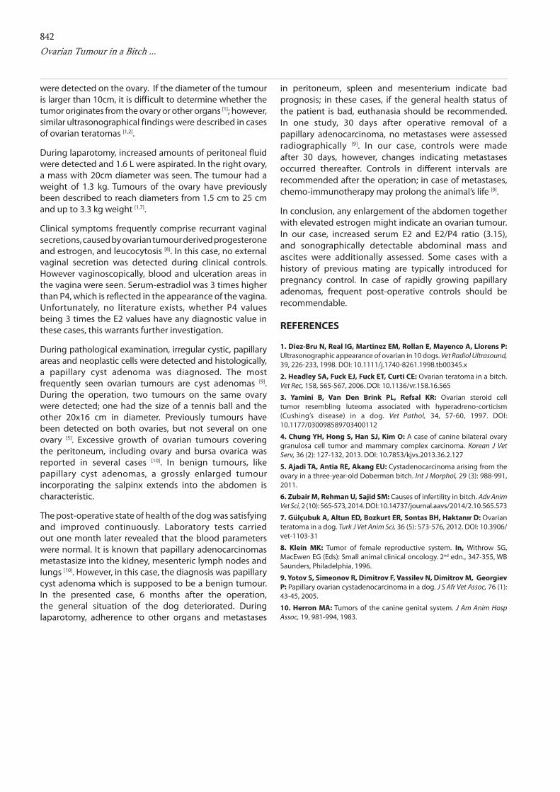
During ultrasonographic examination, the animal was found not to be pregnant, and the uterus showed physio-logical structure and dimension (Fig. 1). In contrast, a structure with knotty-wavy-cauliflower like appearance, most probably related to the right ovary, filled the abdomen (Fig. 2). Connection to other organs could not be excluded sono-graphically. The presence of anechogenic areas indicated fluid accumulation in the abdomen. A hyperechogenic structure and distinct boundaries of the intestine were determined free floating in the abdominal fluid.
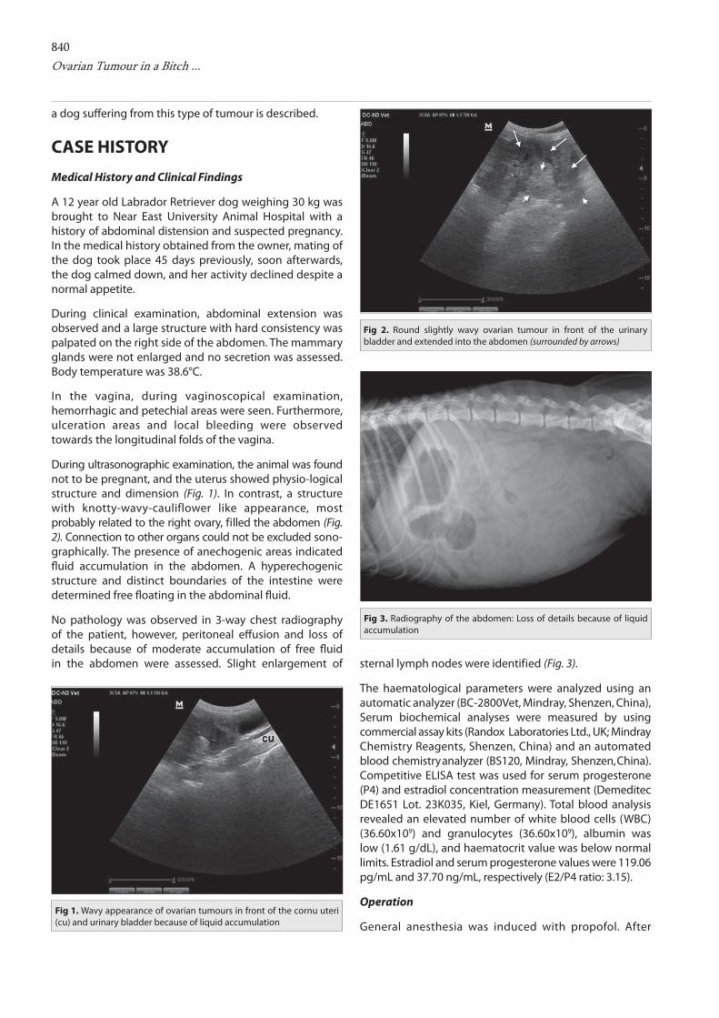
No pathology was observed in 3-way chest radiography of the patient, however, peritoneal effusion and loss of details because of moderate accumulation of free fluid in the abdomen were assessed. Slight enlargement of sternal lymph nodes were identified (Fig. 3).
The haematological parameters were analyzed using an automatic analyzer (BC-2800Vet, Mindray, Shenzen, China), Serum biochemical analyses were measured by using commercial assay kits (Randox Laboratories Ltd., UK; Mindray Chemistry Reagents, Shenzen, China) and an automated blood chemistry analyzer (BS120, Mindray, Shenzen, China). Competitive ELISA test was used for serum progesterone (P4) and estradiol concentration measurement (Demeditec DE1651 Lot. 23K035, Kiel, Germany). Total blood analysis revealed an elevated number of white blood cells (WBC) (36.60x10⁹) and granulocytes (36.60x10⁹), albumin was low (1.61 g/dL), and haematocrit value was below normal limits. Estradiol and serum progesterone values were 119.06 pg/mL and 37.70 ng/mL, respectively (E2/P4 ratio: 3.15).
Operation
General anesthesia was induced with propofol. After Fig 1. Wavy appearance of ovarian tumours in front of the cornu uteri (cu) and urinary bladder because of liquid accumulation
Fig 2. Round slightly wavy ovarian tumour in front of the urinary bladder and extended into the abdomen (surrounded by arrows)
Fig 3. Radiography of the abdomen: Loss of details because of liquid accumulation

841
DARBAZ, ERGENESONMEZ, ASLAN
intubation, anesthesia was maintained by means of inhalation of sevoflurane. The operation was performed by routine ovariohysterectomy method. First free abdominal fluid was aspirated (1.6 L). Then two tumours were detected, one which is a tennis ball-sized mass in the right ovary was removed by ligaturing (Fig. 4a). The uterus did not show pathological changes (Fig. 4b). Adhesions between mesometrium, mesovary and the ovary were determined (Fig. 4c). The second tumour which completely removed from in the same ovary weighed 1.3 kg (20x16 cm).
Histopathology
Histopathological examination revealed ovarian papillary cystadenoma. The tumour was characterized by the presence of cysts and proliferation of papillae, both lined by single- or multi-layered cuboidal to columnar epithelial cells. The neoplastic epithelial cells with pale eosinophilic cytoplasm, distinct cell margins, round to ovoid nuclei, and prominent nucleoli were arranged mainly around
the cystic and papillary structures, and surrounded by a fibrovascular stroma (Fig. 5).
Patient Monitoring and Postoperative Findings
No problems were encountered during the post-operative week, the patient’s general condition and appetite returned to normal. One month post-operative control of the blood profile, revealed an increased. Five months after the last clinical examination and blood testing, the owner brought his animal to the clinic, since the animals state of health had suddenly deteriorated. Blood analysis showed that BUN (8.27 mg/dL) and UREA (17.50 mg/dL) were decreased. The lymphocytes were (8.10%) decreased, and WBC (21.20x10⁹/L), granulocyte (19.30x10⁹/L-89.40%) and eosinophiles (1.70%) increased. In ultrasonographic examination, metastasis-like formations were detected in the spleen, kidney and other organs. Laparotomy was performed, and tumours were found in the spleen, kidney and all intestines. The animal was euthanized on request of the owner of the animal, however.
DISCUSSIONIn bitch, mated 40-45 days ago, recent enlargement of the abdomen and since the general situation of the dog is normal, dogs are usually examined for pregnancy. Similarly, in some articles [2,5,6] it is mentioned that no deterioration was seen in the general situation of the patient and dogs were brought to controls only due to excess enlargement of the abdomen.
During the abdominal palpation, at the right side a bulk, rigid, round and large structure was assessed. In some reports it is stated that ovarian teratomas can be localized by abdominal palpation [2]. In the presented case, additional soft structures inside the large rigid mass prevented the definitive diagnosis of ovarian tumors.
In the presented case, sonographically round, hetero-geneous, hypoechogenic and locally anechogenic regions
Fig 4. a- The tennis ball-sized tumour in the right ovary (arrows), b- No pathological changes or thickening in the uterus (arrows), c- Adhesions between mesometrium, mesovary and the ovary
Fig 5. Cystic (arrows) and papillary structures (arrowheads) surrounded by a fibrovascular stroma. H&Ex100
842Ovarian Tumour in a Bitch ...
were detected on the ovary. If the diameter of the tumour is larger than 10cm, it is difficult to determine whether the tumor originates from the ovary or other organs [1]; however, similar ultrasonographical findings were described in cases of ovarian teratomas [1,2].
During laparotomy, increased amounts of peritoneal fluid were detected and 1.6 L were aspirated. In the right ovary, a mass with 20cm diameter was seen. The tumour had a weight of 1.3 kg. Tumours of the ovary have previously been described to reach diameters from 1.5 cm to 25 cm and up to 3.3 kg weight [1,7].
Clinical symptoms frequently comprise recurrant vaginal secretions, caused by ovarian tumour derived progesterone and estrogen, and leucocytosis [8]. In this case, no external vaginal secretion was detected during clinical controls. However vaginoscopically, blood and ulceration areas in the vagina were seen. Serum-estradiol was 3 times higher than P4, which is reflected in the appearance of the vagina. Unfortunately, no literature exists, whether P4 values being 3 times the E2 values have any diagnostic value in these cases, this warrants further investigation.
During pathological examination, irregular cystic, papillary areas and neoplastic cells were detected and histologically, a papillary cyst adenoma was diagnosed. The most frequently seen ovarian tumours are cyst adenomas [9]. During the operation, two tumours on the same ovary were detected; one had the size of a tennis ball and the other 20x16 cm in diameter. Previously tumours have been detected on both ovaries, but not several on one ovary [5]. Excessive growth of ovarian tumours covering the peritoneum, including ovary and bursa ovarica was reported in several cases [10]. In benign tumours, like papillary cyst adenomas, a grossly enlarged tumour incorporating the salpinx extends into the abdomen is characteristic.
The post-operative state of health of the dog was satisfying and improved continuously. Laboratory tests carried out one month later revealed that the blood parameters were normal. It is known that papillary adenocarcinomas metastasize into the kidney, mesenteric lymph nodes and lungs [10]. However, in this case, the diagnosis was papillary cyst adenoma which is supposed to be a benign tumour. In the presented case, 6 months after the operation, the general situation of the dog deteriorated. During laparotomy, adherence to other organs and metastases
in peritoneum, spleen and mesenterium indicate bad prognosis; in these cases, if the general health status of the patient is bad, euthanasia should be recommended. In one study, 30 days after operative removal of a papillary adenocarcinoma, no metastases were assessed radiographically [9]. In our case, controls were made after 30 days, however, changes indicating metastases occurred thereafter. Controls in different intervals are recommended after the operation; in case of metastases, chemo-immunotherapy may prolong the animal’s life [9].
In conclusion, any enlargement of the abdomen together with elevated estrogen might indicate an ovarian tumour. In our case, increased serum E2 and E2/P4 ratio (3.15), and sonographically detectable abdominal mass and ascites were additionally assessed. Some cases with a history of previous mating are typically introduced for pregnancy control. In case of rapidly growing papillary adenomas, frequent post-operative controls should be recommendable.
REFERENCES
1. Diez-Bru N, Real IG, Martinez EM, Rollan E, Mayenco A, Llorens P: Ultrasonographic appearance of ovarian in 10 dogs. Vet Radiol Ultrasound, 39, 226-233, 1998. DOI: 10.1111/j.1740-8261.1998.tb00345.x
2. Headley SA, Fuck EJ, Fuck ET, Curti CE: Ovarian teratoma in a bitch. Vet Rec, 158, 565-567, 2006. DOI: 10.1136/vr.158.16.565
3. Yamini B, Van Den Brink PL, Refsal KR: Ovarian steroid cell tumor resembling luteoma associated with hyperadreno-corticism (Cushing’s disease) in a dog. Vet Pathol, 34, 57-60, 1997. DOI: 10.1177/030098589703400112
4. Chung YH, Hong S, Han SJ, Kim O: A case of canine bilateral ovary granulosa cell tumor and mammary complex carcinoma. Korean J Vet Serv, 36 (2): 127-132, 2013. DOI: 10.7853/kjvs.2013.36.2.127
5. Ajadi TA, Antia RE, Akang EU: Cystadenocarcinoma arising from the ovary in a three-year-old Doberman bitch. Int J Morphol, 29 (3): 988-991, 2011.
6. Zubair M, Rehman U, Sajid SM: Causes of infertility in bitch. Adv Anim Vet Sci, 2 (10): 565-573, 2014. DOI: 10.14737/journal.aavs/2014/2.10.565.573
7. Gülçubuk A, Altun ED, Bozkurt ER, Sontas BH, Haktanır D: Ovarian teratoma in a dog. Turk J Vet Anim Sci, 36 (5): 573-576, 2012. DOI: 10.3906/vet-1103-31
8. Klein MK: Tumor of female reproductive system. In, Withrow SG, MacEwen EG (Eds): Small animal clinical oncology. 2nd edn., 347-355, WB Saunders, Philadelphia, 1996.
9. Yotov S, Simeonov R, Dimitrov F, Vassilev N, Dimitrov M, Georgiev P: Papillary ovarian cystadenocarcinoma in a dog. J S Afr Vet Assoc, 76 (1): 43-45, 2005.
10. Herron MA: Tumors of the canine genital system. J Am Anim Hosp Assoc, 19, 981-994, 1983.