Embed Size (px)
Citation preview
www.scireproject.com Version 5.0
Orthostatic Hypotension Following Spinal Cord Injury
Andrei Krassioukov, MD, PhD, FRCPC Jill Maria Wecht, EdD
Robert W Teasell, MD, FRCPC Janice J Eng, PhD, BSc (PT/OT)
Key Points
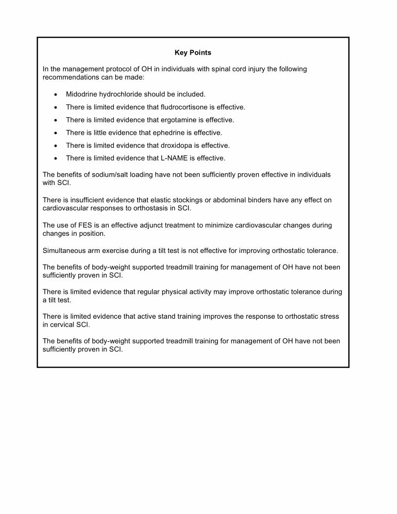
In the management protocol of OH in individuals with spinal cord injury the following recommendations can be made:
• Midodrine hydrochloride should be included.
• There is limited evidence that fludrocortisone is effective.
• There is limited evidence that ergotamine is effective.
• There is little evidence that ephedrine is effective.
• There is limited evidence that droxidopa is effective.
• There is limited evidence that L-NAME is effective. The benefits of sodium/salt loading have not been sufficiently proven effective in individuals with SCI. There is insufficient evidence that elastic stockings or abdominal binders have any effect on cardiovascular responses to orthostasis in SCI. The use of FES is an effective adjunct treatment to minimize cardiovascular changes during changes in position. Simultaneous arm exercise during a tilt test is not effective for improving orthostatic tolerance. The benefits of body-weight supported treadmill training for management of OH have not been sufficiently proven in SCI. There is limited evidence that regular physical activity may improve orthostatic tolerance during a tilt test. There is limited evidence that active stand training improves the response to orthostatic stress in cervical SCI. The benefits of body-weight supported treadmill training for management of OH have not been sufficiently proven in SCI.
This review has been prepared based on the scientific and professional information available in 2013. The SCIRE information (print, CD or web site www.scireproject.com) is provided for informational and educational purposes only. If you have or suspect you have a health problem, you should consult your health care provider. The SCIRE editors, contributors and supporting partners shall not be liable for any damages, claims, liabilities, costs or obligations arising from the use or misuse of this material. Krassioukov A, Wecht JM, Teasell RW, Eng JJ (2014). Orthostatic Hypotension Following Spinal Cord Injury. In Eng JJ, Teasell RW, Miller WC, Wolfe DL, Townson AF, Hsieh JTC, Connolly SJ, Noonan VK, Loh E, McIntyre A, editors. Spinal Cord Injury Rehabilitation Evidence. Version 5.0. Vancouver: p 1- 26. www.scireproject.com
Table of Contents
Abbreviations............................................................................................................................... i1.0 Executive Summary ............................................................................................................. 12.0 Introduction ........................................................................................................................... 23.0 Orthostatic Hypotension Systematic Review .................................................................... 54.0 Pharmacological Management of OH in SCI ...................................................................... 65.0 Non-pharmacological Management of OH in SCI ............................................................ 145.1 Fluid and Sodium Intake in Management of OH in SCI ........................................................ 145.2 Blood Pooling Prevention in Management of OH in SCI ...................................................... 155.3 Whole-Body Vibration in Management of OH in SCI ............................................................ 185.4 Effect of Functional Electrical Stimulation (FES) on OH in SCI ............................................ 195.5 Effect of Exercise on OH in SCI ........................................................................................... 235.6 Effect of Standing on OH in SCI ........................................................................................... 245.7 General Discussion .............................................................................................................. 26
6.0 References .......................................................................................................................... 27
i
Abbreviations
AB Abdominal Binder BP Blood Pressure BWSTT Body Weight Supported Treadmill Training ELC External Leg Compression FES Functional Electrical Stimulation FNS Functional Neuromuscular Stimulation GCS Graduated Compression Stockings HR Heart Rate HUT Head-up Tilt LBNP Lower Body Negative Pressure L-DOPS, Droxidopa L-threo-3,4-dihydroxyphenylserine L-NAME L-arginine-N-methyl-ester MAP Mean Arterial Pressure OH Orthostatic Hypotension RATT Robotic Assisted Tilt-Table RER Respiratory Exchange Ratio WBV Whole Body Vibration
1
Orthostatic Hypotension Following SCI
1.0 Executive Summary
What is orthostatic hypotension? Orthostatic hypotension (OH) is defined as a decrease in systolic blood pressure of at least 20mmHg, or a reduction in diastolic blood pressure of at least 10mmHg, upon the change in body position from a supine (lying) to an upright position, regardless of the presence of symptoms.1 Normally, the nervous system automatically constricts or dilates the blood vessels to balance blood pressure. After a SCI, this ability may become compromised, and orthostatic hypotension may be experienced. Several studies have documented the presence of OH following SCI, particularly during the acute period of injury, but it can persist for many years.2,3,4,5,6,7,8 Sitting or Standing in physiotherapy is reported to trigger blood pressure decreases that are diagnostic of OH in 74% of SCI patients, and cause symptoms of OH (such as light-headedness or dizziness) in 59% of SCI individuals.9 Thus, this may discourage individuals with SCI from participating in rehabilitation. Management of OH consists of pharmacological and non-pharmacological interventions. What are the risk factors of orthostatic hypotension? Many factors can contribute to OH. The low level of efferent sympathetic nervous activity and the loss of the reflex vasoconstriction following SCI are the two major causes of OH. Decreases in blood pressure (BP) following the change to an upright position in individuals with SCI may be related to excessive pooling of blood in the abdominal viscera and lower extremities.5,7,10 Additionally, regular movement of muscles pushes against blood vessels which help guide blood back to the heart. Hence, loss of muscle function in the lower extremities can cause blood to accumulate there. These mechanisms lead to a reduction of blood flow back to the heart and the rest of the body and present as orthostatic hypotension. Other causes of orthostatic hypotension include low blood volume, low sodium levels in the blood, and deconditioning of the heart and blood vessels from extended bedrest.5,7,9 Those with a traumatic SCI may also be at a greater risk than those with a non-traumatic SCI (McKinley et al. 1999). The prevalence of OH is greater in patients with higher spinal cord lesions, and thus it is more common in tetraplegia.7,8,11 Some evidence show that nitric oxide, a chemical that widens the blood vessels, is produced more in SCI individuals.12 This can result in a further decrease in blood pressure. What are the signs and symptoms of orthostatic hypotension? Orthostatic hypotension may occur with or without the presence of symptoms. Common signs and symptoms include:
• Temporary loss of consciousness • Fainting • Dizziness • Light-headedness • Fatigue • Blurry vision • Muscle weakness
How do I manage my patients with OH? Although a wide array of physical and pharmacological measures are recommended for the general management of OH,13 very few have been evaluated for use in SCI. The general approach to
2
management of OH is that the therapeutic interventions should be implemented in stages dependent upon the severity of symptoms.13 Non-pharmacologic measures alone are often insufficient to prevent symptoms of OH. Pharmacological interventions are often needed, particularly in SCI patients with moderate to severe OH symptoms. Pharmacological Options Only Midodrine (a drug that constricts the blood vessels to bring up blood pressure) has some evidence supporting its use. There is evidence that this drug can elevate blood pressure and improve exercise performance.14,15 Even so, the use of midrodrine should be monitored carefully as 2 males reported urinary bladder dysreflexia with its use.16 Unfortunately, the number of studies addressing the pharmacological management of OH following SCI is few. It is often difficult to determine the effects of individual medications when they are used in combination therapies. Non-Pharmacological Options Functional Electrical Stimulation (FES) is one of the only treatments having limited evidence to support efficacy. During functional electrical stimulation, electrical impulses are sent to weak or paralyzed muscles, usually within the legs. This causes muscles to contract and helps move blood back to the heart and around the body. Functional electrical stimulation has been shown to be effective and can be used to supplement other forms of therapy.17-19 Studies demonstrated that leg muscle contraction by functional electrical stimulation allowed tetraplegics with orthostatic hypotension to stand more often and for longer periods of time.18-19 Non-pharmacological options include fluid and salt intake, pressure binders or stockings, whole-body vibration, electrical stimulation, and physical activities. There is currently not enough evidence to support the efficacy of non-pharmacological interventions. Currently, only positive evidence on the effect of fluid and salt intake combined with other pharmacological interventions exist.8,20
2.0 Introduction
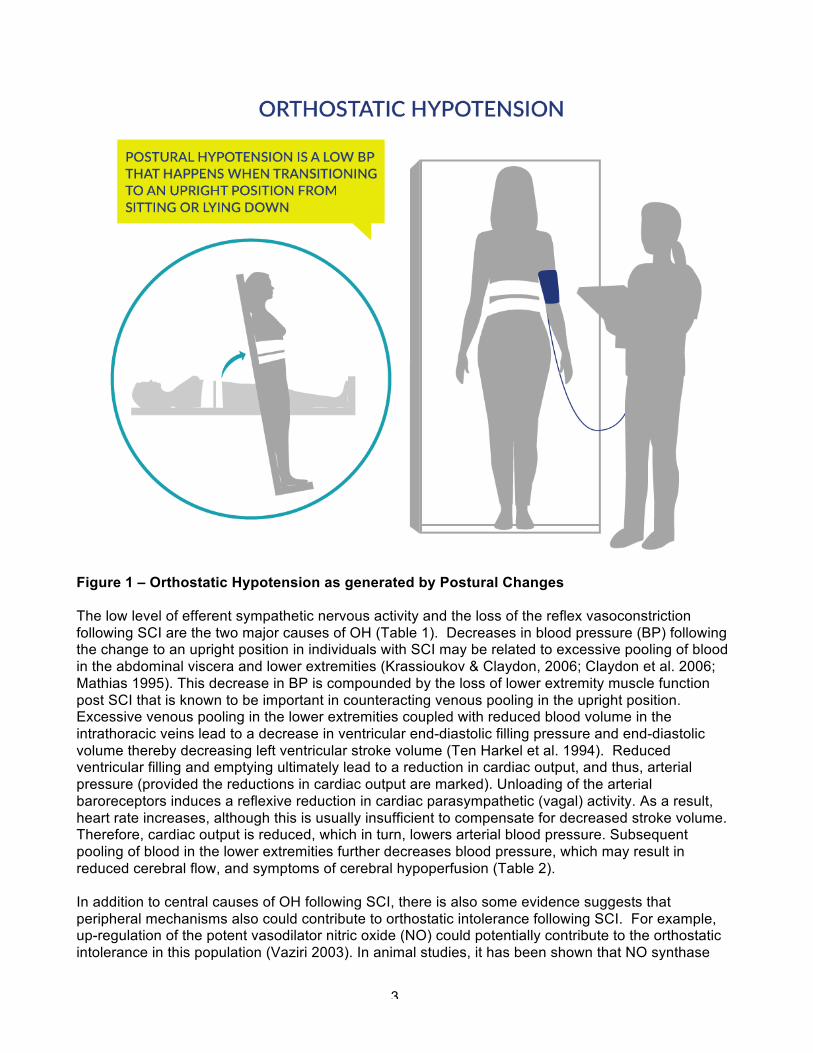
The Consensus Committee of the American Autonomic Society and the American Academy of Neurology (CCAAS & AAN 1996) defined orthostatic hypotension (OH) as a decrease in systolic blood pressure of at least 20mmHg, or a reduction in diastolic blood pressure of at least 10mmHg, upon the change in body position from a supine to an upright position, regardless of the presence of symptoms. Several studies have documented the presence of OH following SCI (Chelvarajah, 2009, Cariga et al. 2002, Faghri et al. 2001; Mathias 1995). This condition occurs during the acute period of injury and persists in a significant number of individuals for many years (Sidorov et al. 2008; Claydon et al. 2006; Frisbie & Steele 1997). Standard mobilization treatment during physiotherapy (e.g. sitting or standing) is reported to trigger blood pressure decreases that are diagnostic of OH in 74% of SCI patients, and cause symptoms of OH (such as light-headedness or dizziness) in 59% of SCI individuals (Illman et al. 2000). Thus, this may discourage individuals with SCI from participating in rehabilitation. Management of OH consists of pharmacological and non-pharmacological interventions.
3
Figure 1 – Orthostatic Hypotension as generated by Postural Changes The low level of efferent sympathetic nervous activity and the loss of the reflex vasoconstriction following SCI are the two major causes of OH (Table 1). Decreases in blood pressure (BP) following the change to an upright position in individuals with SCI may be related to excessive pooling of blood in the abdominal viscera and lower extremities (Krassioukov & Claydon, 2006; Claydon et al. 2006; Mathias 1995). This decrease in BP is compounded by the loss of lower extremity muscle function post SCI that is known to be important in counteracting venous pooling in the upright position. Excessive venous pooling in the lower extremities coupled with reduced blood volume in the intrathoracic veins lead to a decrease in ventricular end-diastolic filling pressure and end-diastolic volume thereby decreasing left ventricular stroke volume (Ten Harkel et al. 1994). Reduced ventricular filling and emptying ultimately lead to a reduction in cardiac output, and thus, arterial pressure (provided the reductions in cardiac output are marked). Unloading of the arterial baroreceptors induces a reflexive reduction in cardiac parasympathetic (vagal) activity. As a result, heart rate increases, although this is usually insufficient to compensate for decreased stroke volume. Therefore, cardiac output is reduced, which in turn, lowers arterial blood pressure. Subsequent pooling of blood in the lower extremities further decreases blood pressure, which may result in reduced cerebral flow, and symptoms of cerebral hypoperfusion (Table 2). In addition to central causes of OH following SCI, there is also some evidence suggests that peripheral mechanisms also could contribute to orthostatic intolerance following SCI. For example, up-regulation of the potent vasodilator nitric oxide (NO) could potentially contribute to the orthostatic intolerance in this population (Vaziri 2003). In animal studies, it has been shown that NO synthase
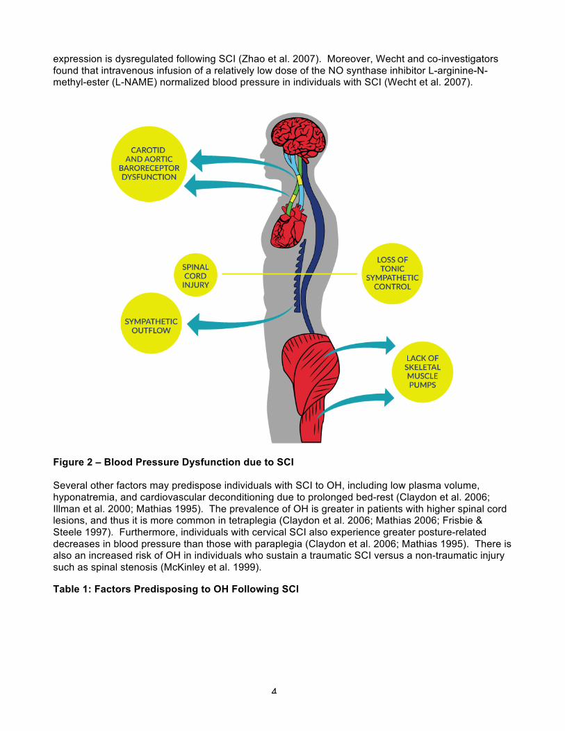
4
expression is dysregulated following SCI (Zhao et al. 2007). Moreover, Wecht and co-investigators found that intravenous infusion of a relatively low dose of the NO synthase inhibitor L-arginine-N-methyl-ester (L-NAME) normalized blood pressure in individuals with SCI (Wecht et al. 2007).
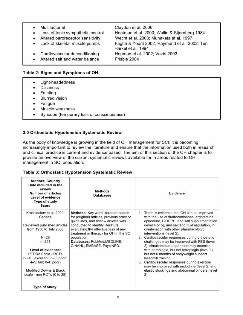
Figure 2 – Blood Pressure Dysfunction due to SCI Several other factors may predispose individuals with SCI to OH, including low plasma volume, hyponatremia, and cardiovascular deconditioning due to prolonged bed-rest (Claydon et al. 2006; Illman et al. 2000; Mathias 1995). The prevalence of OH is greater in patients with higher spinal cord lesions, and thus it is more common in tetraplegia (Claydon et al. 2006; Mathias 2006; Frisbie & Steele 1997). Furthermore, individuals with cervical SCI also experience greater posture-related decreases in blood pressure than those with paraplegia (Claydon et al. 2006; Mathias 1995). There is also an increased risk of OH in individuals who sustain a traumatic SCI versus a non-traumatic injury such as spinal stenosis (McKinley et al. 1999).
Table 1: Factors Predisposing to OH Following SCI
5
Table 2: Signs and Symptoms of OH
3.0 Orthostatic Hypotension Systematic Review
As the body of knowledge is growing in the field of OH management for SCI, it is becoming increasingly important to review the literature and ensure that the information used both in research and clinical practice is current and evidence based. The aim of this section of the OH chapter is to provide an overview of the current systematic reviews available for in areas related to OH management in SCI population.
Table 3: Orthostatic Hypotension Systematic Review
Authors; Country Date included in the
review Number of articles Level of evidence
Type of study Score
Methods Databases Evidence
Krassioukov et al. 2009; Canada
Reviewed published articles
from 1950 to July 2008
N=26 n=251
Level of evidence:
PEDRo Scale - RCTs (9–10: excellent; 6–8: good;
4–5: fair; 0-4: poor)
Modified Downs & Black scale - non RCTs (0 to 28)
Type of study:
Methods: Key word literature search for (original) articles, previous practice guidelines, and review articles was conducted to identify literature evaluating the effectiveness of any treatment or therapy for OH in the SCI population. Databases: PubMed/MEDLINE, CINAHL, EMBASE, PsycINFO.
1. There is evidence that OH can be improved with the use of fludrocortisones, ergotamine, ephedrine, L-DOPS, and salt supplementation (level 4 or 5), and salt and fluid regulation, in combination with other pharmacologic interventions (level 5).
2. Cardiovascular responses during orthostatic challenges may be improved with FES (level 2), simultaneous upper extremity exercise with paraplegia, but not tetraplegia (level 2), but not 6 months of bodyweight support treadmill training.
3. Cardiovascular responses during exercise may be improved with midodrine (level 2) and elastic stockings and abdominal binders (level 2).
• Multifactorial Claydon et al. 2006 • Loss of tonic sympathetic control Houtman et al. 2000; Wallin & Stjernberg 1984 • Altered baroreceptor sensitivity Wecht et al. 2003; Munakata et al. 1997 • Lack of skeletal muscle pumps Faghri & Yount 2002; Raymond et al. 2002; Ten
Harkel et al. 1994 • Cardiovascular deconditioning Hopman et al. 2002; Vaziri 2003 • Altered salt and water balance Frisbie 2004
• Light-headedness • Dizziness • Fainting • Blurred vision • Fatigue • Muscle weakness • Syncope (temporary loss of consciousness)
6
Authors; Country Date included in the
review Number of articles Level of evidence
Type of study Score
Methods Databases Evidence
2 case reports, 1 case series, 2 observational, 1
pre-post, 1 RCT
AMSTAR: 6
Gillis et al. 2008; Belgium
Reviewed published articles
from 1966 to April 2007
N=13 n=138
Level of evidence:
Downs & Black scale
Type of study: Parallel group, cross-over, quasi-random assignment
AMSTAR: 5
Methods: Key word literature search for non-pharmacological management of OH during early rehab in SCI. Databases: PubMed/MEDLINE, OVID/EMBASE, CENTRAL
1. The evidence is inconclusive whether compression/pressure, upper body exercise and biofeedback therapies are able to control OH.
2. Upper body exercise may be more relevant to lower-level paraplegia where sympathetic outflow is intact and motor functionality is present.
3. FES can attenuate the drop in BP by 8/4 mm Hg during an orthostatic challenge and is promising technology. However, few studies utilized patients in the acute stage.
Discussion We found only one systematic review on OH management for individuals with SCI by Krassioukov et al (2009). Although the authors found that the overall quality of the literature was poor and that higher quality research assessing the treatments for OH in the SCI population is needed, there is level 2 evidence that pressure from elastic stockings and abdominal binders may improve cardiovascular physiologic responses during submaximal upper-extremity exercises. In addition, FES is an important adjunct treatment to minimize cardiovascular changes during postural orthostatic stress and that simultaneous upper-extremity exercises may increase orthostatic tolerance during a progressive tilt exercise in subjects with paraplegia. 4.0 Pharmacological Management of OH in SCI
The majority of our knowledge in managing OH has been obtained from patients with neurological causes other than SCI (e.g. diabetic neuropathy, heart disease, multiple system atrophy, pure autonomic failure, Parkinson’s disease, dysautonomia). Numerous medications, including midodrine hydrochloride, fludrocortisone, and ephedrine, have been successful in managing OH in these chronic conditions. However, as the mechanisms underlying the development of OH in the SCI population differ from those in these non-SCI populations, it is important to assess the effectiveness of these medications specifically in people with SCI.
Table 4: Pharmacological Management of OH in SCI
7
Author Year; Country Score
Research Design Total Sample Size
Methods Outcome
Nieshoff et al. 2004; USA
PEDro=6 RCT N=4
Population: Chronic motor complete tetraplegia Treatment: Midodrine 5mg, 10 mg, or placebo (unmarked capsule), double blind, placebo-controlled cross-over design. Outcome Measures: Measure of cardiovascular parameters during wheelchair ergometer test.
1. Midodrine, 10 mg elevated systolic blood pressure during exercise in 3 participants. Peak systolic BPs ranged from 90 to 126 mmHg under baseline and placebo conditions, 114-148 after 5 mg of midodrine, and 104 to 200 mmHg after 10 mg.
2. Two participants showed reduced perceived exertion and increased VO2 following midodrine 10 mg.
3. No adverse effects of midodrine were noted.
Phillips et al. 2014a Canada
Prospective controlled trial
N=16
Population: 8 persons with SCI (1 female) and 8 age-and-sex matched able bodied controls Participants with SCI: Mean (SD) age: 30 (11) years DOI: 7 subjects <1 year post injury, 1 subject >1 year post injury All motor complete cervical spinal cord injuries AIS grade A: 6; AIS grade B: 2 Treatment: Subjects tested supine and during upright tilt. SCI group had 2 treatment sessions, one with midodrine and one without. AB group had one session without midodrine. Outcome Measures: Beat-by-beat BP and HR, common carotid artery (CCA) diameter. Calculated arterial distensibility and arterial stiffness (β-stiffness index)
1. Systolic BP was lower in SCI while supine vs. AB; BP, CCA diameter and diameter difference were reduced in SCI while upright vs. AB; β-stiffness index was elevated in SCI when upright (+12%) and relative decrease in baroreflex sensitivity (BRS) was greater in SCI vs. AB
2. Negative relationship between BRS and β-stiffness in SCI; no relationship in AB
3. Midodrine led to increased BP and decreased HR in both supine and upright positions; no change in BRS or CCA parameters
4. Reduced BRS is closely related to increased arterial stiffness in the SCI population
Phillips et al. 2014b Canada
Prospective controlled trial
N=20
Population: 10 persons with SCI (3 females) and 10 age-and-sex matched able bodied controls Participants with SCI: Mean (SD) age: 29 (10) years DOI: 8 subjects <1 year post injury, 2 subject >1 year post injury 8 cervical injuries, 2 thoracic injuries AIS grade A: 8; AIS grade B: 2 Treatment: Subjects tested supine and during progressive upright tilt. SCI group had 2 treatment sessions, one with midodrine and one without. AB group had one session without midodrine. Outcome Measures: beat-by-beat BP, middle and posterior cerebral artery blood velocity (MCAv, PCAv, respectively)
1. Coherence increased in SCI between BP-MCAv and BP-PCAv by 35% and 22% respectively compared to AB.
2. SCI BP-PCAv gain was reduced 30% compared to AB.
3. The acute (0–30 s after tilt) MCAv and PCAv responses were similar between groups.
4. In SCI, midodrine led to improved PCAv responses 30 – 60 s following tilt (10+/- 3% vs. 4+/- 2% decline)
5. In SCI, midodrine led to a 59% improvement in orthostatic tolerance
Phillips et al. 2014c Canada
Prospective controlled trial
N=20
Population: 10 persons with SCI (3 females) and 10 age-and-sex matched able bodied controls Participants with SCI: Mean (SD) age: 29 (10) years DOI: 8 subjects <1 year post injury, 2 subject >1 year post injury
1. At rest: mean BP was lower in SCI (70±10 versus 92±14 mm Hg); PCAv conductance was higher in SCI (0.56±0.13 versus 0.39±0.15 cm/second/mm Hg)
2. AB had a 20% increase in PCAv during cognition while this response was absent in SCI
8
Author Year; Country Score
Research Design Total Sample Size
Methods Outcome
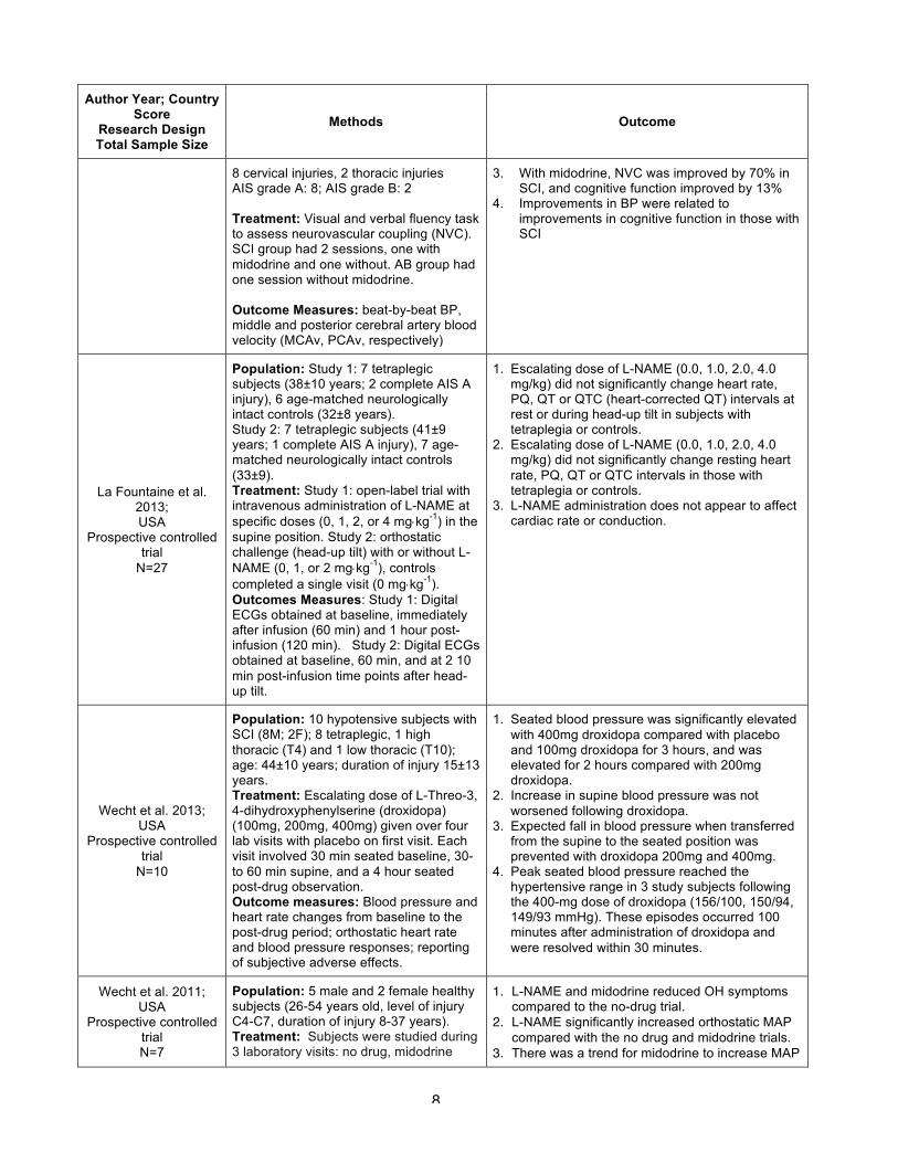
8 cervical injuries, 2 thoracic injuries AIS grade A: 8; AIS grade B: 2 Treatment: Visual and verbal fluency task to assess neurovascular coupling (NVC). SCI group had 2 sessions, one with midodrine and one without. AB group had one session without midodrine. Outcome Measures: beat-by-beat BP, middle and posterior cerebral artery blood velocity (MCAv, PCAv, respectively)
3. With midodrine, NVC was improved by 70% in SCI, and cognitive function improved by 13%
4. Improvements in BP were related to improvements in cognitive function in those with SCI
La Fountaine et al. 2013; USA
Prospective controlled trial
N=27
Population: Study 1: 7 tetraplegic subjects (38±10 years; 2 complete AIS A injury), 6 age-matched neurologically intact controls (32±8 years). Study 2: 7 tetraplegic subjects (41±9 years; 1 complete AIS A injury), 7 age-matched neurologically intact controls (33±9). Treatment: Study 1: open-label trial with intravenous administration of L-NAME at specific doses (0, 1, 2, or 4 mg×kg-1) in the supine position. Study 2: orthostatic challenge (head-up tilt) with or without L-NAME (0, 1, or 2 mg×kg-1), controls completed a single visit (0 mg×kg-1). Outcomes Measures: Study 1: Digital ECGs obtained at baseline, immediately after infusion (60 min) and 1 hour post-infusion (120 min). Study 2: Digital ECGs obtained at baseline, 60 min, and at 2 10 min post-infusion time points after head-up tilt.
1. Escalating dose of L-NAME (0.0, 1.0, 2.0, 4.0 mg/kg) did not significantly change heart rate, PQ, QT or QTC (heart-corrected QT) intervals at rest or during head-up tilt in subjects with tetraplegia or controls.
2. Escalating dose of L-NAME (0.0, 1.0, 2.0, 4.0 mg/kg) did not significantly change resting heart rate, PQ, QT or QTC intervals in those with tetraplegia or controls.
3. L-NAME administration does not appear to affect cardiac rate or conduction.
Wecht et al. 2013; USA
Prospective controlled trial
N=10
Population: 10 hypotensive subjects with SCI (8M; 2F); 8 tetraplegic, 1 high thoracic (T4) and 1 low thoracic (T10); age: 44±10 years; duration of injury 15±13 years. Treatment: Escalating dose of L-Threo-3, 4-dihydroxyphenylserine (droxidopa) (100mg, 200mg, 400mg) given over four lab visits with placebo on first visit. Each visit involved 30 min seated baseline, 30- to 60 min supine, and a 4 hour seated post-drug observation. Outcome measures: Blood pressure and heart rate changes from baseline to the post-drug period; orthostatic heart rate and blood pressure responses; reporting of subjective adverse effects.
1. Seated blood pressure was significantly elevated with 400mg droxidopa compared with placebo and 100mg droxidopa for 3 hours, and was elevated for 2 hours compared with 200mg droxidopa.
2. Increase in supine blood pressure was not worsened following droxidopa.
3. Expected fall in blood pressure when transferred from the supine to the seated position was prevented with droxidopa 200mg and 400mg.
4. Peak seated blood pressure reached the hypertensive range in 3 study subjects following the 400-mg dose of droxidopa (156/100, 150/94, 149/93 mmHg). These episodes occurred 100 minutes after administration of droxidopa and were resolved within 30 minutes.
Wecht et al. 2011; USA
Prospective controlled trial N=7
Population: 5 male and 2 female healthy subjects (26-54 years old, level of injury C4-C7, duration of injury 8-37 years). Treatment: Subjects were studied during 3 laboratory visits: no drug, midodrine
1. L-NAME and midodrine reduced OH symptoms compared to the no-drug trial.
2. L-NAME significantly increased orthostatic MAP compared with the no drug and midodrine trials.
3. There was a trend for midodrine to increase MAP
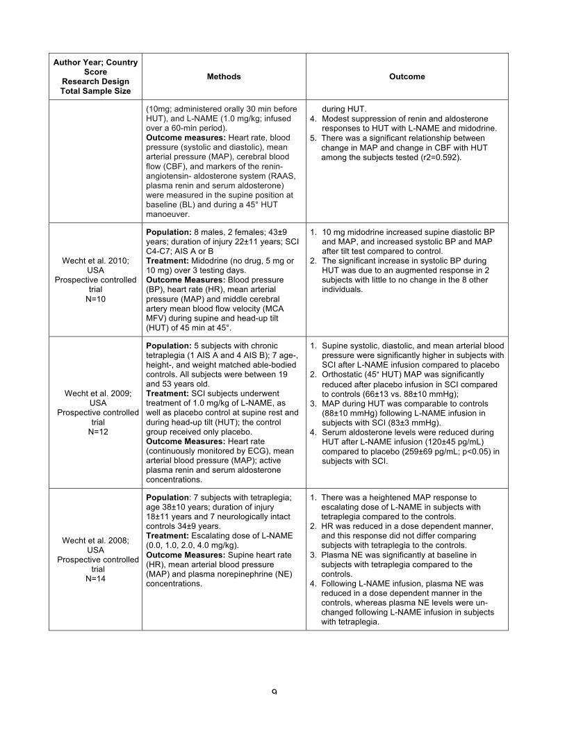
9
Author Year; Country Score
Research Design Total Sample Size
Methods Outcome
(10mg; administered orally 30 min before HUT), and L-NAME (1.0 mg/kg; infused over a 60-min period). Outcome measures: Heart rate, blood pressure (systolic and diastolic), mean arterial pressure (MAP), cerebral blood flow (CBF), and markers of the renin-angiotensin- aldosterone system (RAAS, plasma renin and serum aldosterone) were measured in the supine position at baseline (BL) and during a 45° HUT manoeuver.
during HUT. 4. Modest suppression of renin and aldosterone
responses to HUT with L-NAME and midodrine. 5. There was a significant relationship between
change in MAP and change in CBF with HUT among the subjects tested (r2=0.592).
Wecht et al. 2010; USA
Prospective controlled trial
N=10
Population: 8 males, 2 females; 43±9 years; duration of injury 22±11 years; SCI C4-C7; AIS A or B Treatment: Midodrine (no drug, 5 mg or 10 mg) over 3 testing days. Outcome Measures: Blood pressure (BP), heart rate (HR), mean arterial pressure (MAP) and middle cerebral artery mean blood flow velocity (MCA MFV) during supine and head-up tilt (HUT) of 45 min at 45°.
1. 10 mg midodrine increased supine diastolic BP and MAP, and increased systolic BP and MAP after tilt test compared to control.
2. The significant increase in systolic BP during HUT was due to an augmented response in 2 subjects with little to no change in the 8 other individuals.
Wecht et al. 2009; USA
Prospective controlled trial
N=12
Population: 5 subjects with chronic tetraplegia (1 AIS A and 4 AIS B); 7 age-, height-, and weight matched able-bodied controls. All subjects were between 19 and 53 years old. Treatment: SCI subjects underwent treatment of 1.0 mg/kg of L-NAME, as well as placebo control at supine rest and during head-up tilt (HUT); the control group received only placebo. Outcome Measures: Heart rate (continuously monitored by ECG), mean arterial blood pressure (MAP); active plasma renin and serum aldosterone concentrations.
1. Supine systolic, diastolic, and mean arterial blood pressure were significantly higher in subjects with SCI after L-NAME infusion compared to placebo
2. Orthostatic (45° HUT) MAP was significantly reduced after placebo infusion in SCI compared to controls (66±13 vs. 88±10 mmHg);
3. MAP during HUT was comparable to controls (88±10 mmHg) following L-NAME infusion in subjects with SCI (83±3 mmHg).
4. Serum aldosterone levels were reduced during HUT after L-NAME infusion (120±45 pg/mL) compared to placebo (259±69 pg/mL; p<0.05) in subjects with SCI.
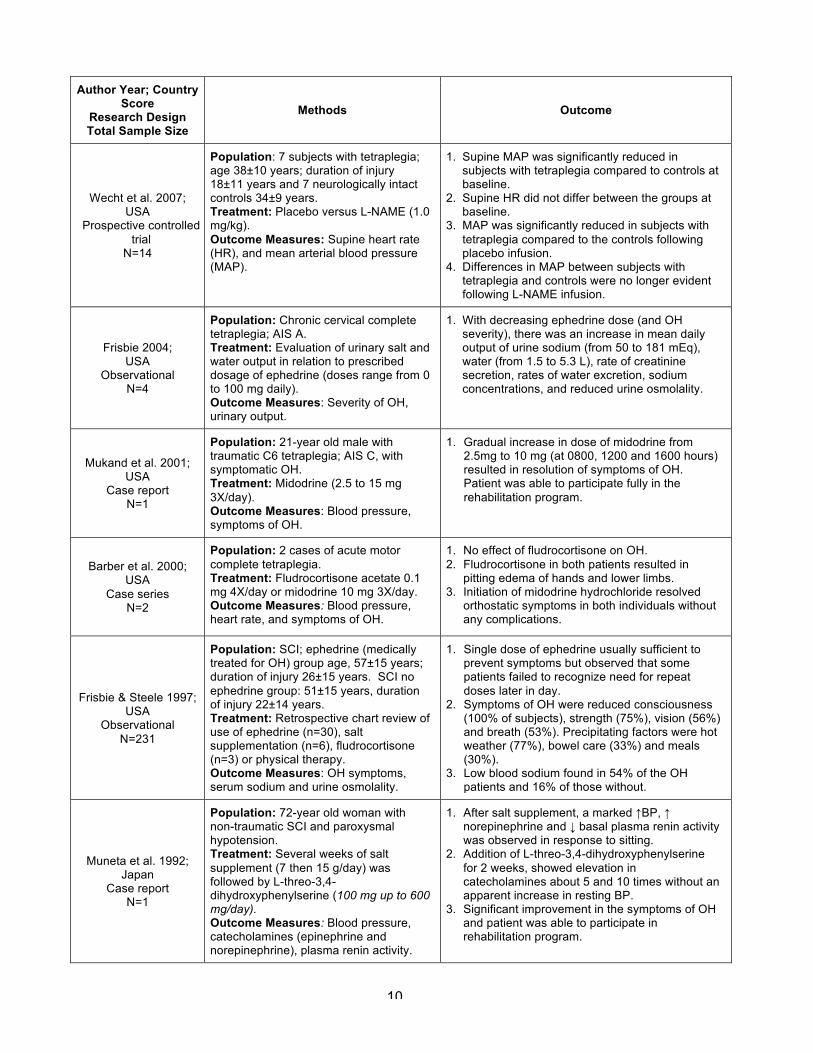
Wecht et al. 2008; USA
Prospective controlled trial
N=14
Population: 7 subjects with tetraplegia; age 38±10 years; duration of injury 18±11 years and 7 neurologically intact controls 34±9 years. Treatment: Escalating dose of L-NAME (0.0, 1.0, 2.0, 4.0 mg/kg). Outcome Measures: Supine heart rate (HR), mean arterial blood pressure (MAP) and plasma norepinephrine (NE) concentrations.
1. There was a heightened MAP response to escalating dose of L-NAME in subjects with tetraplegia compared to the controls.
2. HR was reduced in a dose dependent manner, and this response did not differ comparing subjects with tetraplegia to the controls.
3. Plasma NE was significantly at baseline in subjects with tetraplegia compared to the controls.
4. Following L-NAME infusion, plasma NE was reduced in a dose dependent manner in the controls, whereas plasma NE levels were un-changed following L-NAME infusion in subjects with tetraplegia.
10
Author Year; Country Score
Research Design Total Sample Size
Methods Outcome
Wecht et al. 2007; USA
Prospective controlled trial
N=14
Population: 7 subjects with tetraplegia; age 38±10 years; duration of injury 18±11 years and 7 neurologically intact controls 34±9 years. Treatment: Placebo versus L-NAME (1.0 mg/kg). Outcome Measures: Supine heart rate (HR), and mean arterial blood pressure (MAP).
1. Supine MAP was significantly reduced in subjects with tetraplegia compared to controls at baseline.
2. Supine HR did not differ between the groups at baseline.
3. MAP was significantly reduced in subjects with tetraplegia compared to the controls following placebo infusion.
4. Differences in MAP between subjects with tetraplegia and controls were no longer evident following L-NAME infusion.
Frisbie 2004; USA
Observational N=4
Population: Chronic cervical complete tetraplegia; AIS A. Treatment: Evaluation of urinary salt and water output in relation to prescribed dosage of ephedrine (doses range from 0 to 100 mg daily). Outcome Measures: Severity of OH, urinary output.
1. With decreasing ephedrine dose (and OH severity), there was an increase in mean daily output of urine sodium (from 50 to 181 mEq), water (from 1.5 to 5.3 L), rate of creatinine secretion, rates of water excretion, sodium concentrations, and reduced urine osmolality.
Mukand et al. 2001; USA
Case report N=1
Population: 21-year old male with traumatic C6 tetraplegia; AIS C, with symptomatic OH. Treatment: Midodrine (2.5 to 15 mg 3X/day). Outcome Measures: Blood pressure, symptoms of OH.
1. Gradual increase in dose of midodrine from 2.5mg to 10 mg (at 0800, 1200 and 1600 hours) resulted in resolution of symptoms of OH. Patient was able to participate fully in the rehabilitation program.
Barber et al. 2000; USA
Case series N=2
Population: 2 cases of acute motor complete tetraplegia. Treatment: Fludrocortisone acetate 0.1 mg 4X/day or midodrine 10 mg 3X/day. Outcome Measures: Blood pressure, heart rate, and symptoms of OH.
1. No effect of fludrocortisone on OH. 2. Fludrocortisone in both patients resulted in
pitting edema of hands and lower limbs. 3. Initiation of midodrine hydrochloride resolved
orthostatic symptoms in both individuals without any complications.
Frisbie & Steele 1997; USA
Observational N=231
Population: SCI; ephedrine (medically treated for OH) group age, 57±15 years; duration of injury 26±15 years. SCI no ephedrine group: 51±15 years, duration of injury 22±14 years. Treatment: Retrospective chart review of use of ephedrine (n=30), salt supplementation (n=6), fludrocortisone (n=3) or physical therapy. Outcome Measures: OH symptoms, serum sodium and urine osmolality.
1. Single dose of ephedrine usually sufficient to prevent symptoms but observed that some patients failed to recognize need for repeat doses later in day.
2. Symptoms of OH were reduced consciousness (100% of subjects), strength (75%), vision (56%) and breath (53%). Precipitating factors were hot weather (77%), bowel care (33%) and meals (30%).
3. Low blood sodium found in 54% of the OH patients and 16% of those without.
Muneta et al. 1992; Japan
Case report N=1
Population: 72-year old woman with non-traumatic SCI and paroxysmal hypotension. Treatment: Several weeks of salt supplement (7 then 15 g/day) was followed by L-threo-3,4-dihydroxyphenylserine (100 mg up to 600 mg/day). Outcome Measures: Blood pressure, catecholamines (epinephrine and norepinephrine), plasma renin activity.
1. After salt supplement, a marked ↑BP, ↑ norepinephrine and ↓ basal plasma renin activity was observed in response to sitting.
2. Addition of L-threo-3,4-dihydroxyphenylserine for 2 weeks, showed elevation in catecholamines about 5 and 10 times without an apparent increase in resting BP.
3. Significant improvement in the symptoms of OH and patient was able to participate in rehabilitation program.

11
Author Year; Country Score
Research Design Total Sample Size
Methods Outcome
Senard et al. 1991; France
Pre-post N=7
Population: 45-year-old subject with chronic complete traumatic paraplegia; 6 non-SCI male controls. Treatment: Clonidine (150 µg, 2X/day) and midodrine (specific alpha 1-agonist) (10 mg, 2X daily). Heart rate assessed by blinded tester. Outcome Measures: Blood pressure, heart parameters, plasma catecholamine, alpha-adrenoceptor sensitivity.
1. The increase in systolic blood pressure induced by midodrine (10 mg) was significantly higher in the tetraplegic patient (change of 56 mmHg) compared to controls (change of 15 mmHg).
2. Midodrine and clonidine alone or the two drugs in combination led to an increase in resting BP and decrease severity of OH.
Groomes & Huang 1991; USA Case report
N=1
Population: 28-year-old with chronic C5 tetraplegia. Treatment: Ergotamine (2 mg), daily combined with fludrocortisone (0.1- .05 mg). Outcome Measures: Blood pressure.
1. Following 10 days with fludrocortisone patient able to tolerate sitting. Following additional ergotamine, the patient was able to tolerate an upright position without symptoms.
Discussion Midodrine (ProAmatine) Midodrine, a selective alpha1 adrenergic agonist, exerts its actions by activating the alpha-adrenergic receptors of the arteriolar and venous vasculature, thus producing an increase in vascular tone and blood pressure. Midodrine has a half-life of approximately 25 minutes. Specifically, plasma levels of Midodrine peak approximately half an hour after oral ingestion, with this amount halved every 25 minutes. However, the primary metabolite reaches peak blood concentrations about 1 to 2 hours after a dose of Midodrine and has a half-life of about 3 to 4 hours. Usual starting dose is 2.5mg two or three times daily. Doses are increased quickly until a response occurs or a maximum recommended dose of 10 mg dose, 2-3 times per day (total 30 mg/day) is attained (Wright et al. 1998). Midodrine does not cross the blood-brain barrier and is not associated with CNS effects. Benefits of Midodrine in the management of OH in individuals with SCI were reported in a level 2 RCT (Nieshoff et al. 2004), in four level 2 prospective controlled trials (Phillips et al. 2014a; Phillips et al. 2014b; Phillips et al. 2014c; Wecht et al. 2010) and three level 4 studies (Mukand et al. 2001; Barber et al. 2000; Senard et al. 1991) and one level 5 study (Mukand et al. 1992). Of note, a recent case report on 2 male subjects demonstrated urinary bladder dysreflexia with the use of midodrine (Vaidyanathan et al. 2007) which suggests Midodrine should be employed cautiously. Although the only randomized control trial consisted of 4 subjects (Nieshoff et al. 2004), this study used a rigorous double-blind placebo-controlled, randomized, within-subjects cross-over trial. Not only was systolic blood pressure increased during peak exercise in 3 of the 4 subjects tested, but exercise performance was also enhanced. Thus, there is level 2 evidence (Nieshoff et al. 2004) that Midodrine may increase blood pressure and enhance exercise performance in some (75%) individuals with SCI, similar to other clinical populations with cardiovascular autonomic dysfunction. Furthermore, there are four additional prospective controlled trials (n=10-20), which support the positive effect of midodrine on orthostatic tolerance (Phillips et al. 2014a; Phillips et al. 2014b; Phillips et al. 2014c; Wecht et al. 2010). Nevertheless, it would be important to confirm this evidence with a larger trial. Fludrocortisone (Florinef) Fludrocortisone is a mineralocorticoid that induces more sodium to be released into the bloodstream. Because water follows the movement of sodium, fludrocortisone increases blood volume. Furthermore, fludrocortisone may enhance the sensitivity of blood vessels to circulating
12
catecholamines (Van Lieshout et al. 2000; Schatz 1984). The starting dose is generally 0.1 mg daily. Blood pressure rises gradually over several days with maximum effect at 1-2 weeks. Doses should be adjusted at weekly or biweekly intervals. Adverse effects include hypokalemia (low potassium), which occurs in 50% of individuals, and hypomagnesemia, which occurs in 5%. Both may need to be corrected with supplements. Fludrocortisone should not be used in persons with congestive heart failure due to its effect on sodium retention. Headache is a common side effect. The benefit of Fludrocortisone has not been sufficiently proven in individuals with SCI. One level 4 case series (Barber et al. 2000), one level 5 case report (n=1) (Groomes & Huang 1991), and one level 5 observational (Frisbie & Steele 1997) study have described the use of Fludrocortisone for the management of OH in a SCI population. Barber et al. (2000) studied two patients and did not observe an effect of fludrocortisone. Frisbie and Steele (1997) combined fludrocortisone with other pharmacological and physical agents in three patients; unfortunately, since outcomes specific to this group were not described, the specific effects of fludrocortisone could not be discerned. Therefore, there is level 4 evidence (Barber et al. 2000) from one case series of two patients that fludrocortisone is not effective for OH in SCI. Dihydroergotamine Dihydroergotamine, or Ergotamine, is an ergot alkaloid that interacts with alpha adrenergic receptors and has selective vasoconstrictive effects on peripheral and cranial blood vessels. Plasma levels peak around 2 hours after ingestion. One case report combined Ergotamine with fludrocortisone to successfully prevent symptomatic OH in one individual with SCI (Groomes & Huang 1991). Hence, there is level 5 (Groomes & Huang 1991) evidence that Ergotamine, taken daily combined with fludrocortisone, successfully prevents OH in one individual with SCI. Ephedrine Ephedrine, a non-selective, alpha and beta receptor agonist, acts centrally and peripherally. Its peripheral actions are attributed partly to norepinephrine release and partly to direct effects on receptors. Ephedrine is usually given at a dosage of 12.5-25 mg, administered orally, three times a day. Side effects may include tachycardia, tremor and supine hypertension. Ephedrine raises blood pressure both by increasing cardiac output and inducing peripheral vasoconstriction. Its plasma half-life ranges from 3 to 6 hours (Kobayashi et al. 2003). Systematic review of the literature found level 5 evidence based on one retrospective chart review (Frisbie & Steele 1997) and a cross-sectional observation study (Frisbie 2004). Frisbie (2004) reported that daily urinary output of sodium and fluid was inversely related to the prescribed dose of Ephedrine in 4 patients with OH. While results suggest that Ephedrine resulted in an improvement in hyponatremia, renal conservation of water still exceeded that of sodium in 3 of the 4 cases. Frisbie and Steele (1997) report in their retrospective review of 30 patients taking Ephedrine that one dose in the morning is usually sufficient to reduce symptoms of OH; however, some patients failed to recognize the need for a repeated dose later in the day. Hence, there is level 5 evidence (Frisbie & Steele 1997) that Ephedrine may reduce symptoms of OH. L-threo-3,4-dihydroxyphenylserine (L-DOPS, Droxidopa) L-DOPS is an exogenous, neutral amino acid that is also a precursor of noradrenalin. Two published studies (Wecht et al. 2013; Muneta et al. 1992) evaluate the effects of L-DOPS on OH. Wecht et al. (2013) in a pre-post study found that the use of increased doses of L-threo-3,4-dihydroxyphenylserine (droxidopa 100 mg, 200 mg, 400 mg) in hypotensive subjects did not cause excessive increases in supine blood pressure. Additionally, the 400-mg dose of droxidopa was found to be effective for increasing seated blood pressure for up to 3 hours in study subjects. The expected fall in blood pressure when transferred to the seated position from supine was prevented with droxidopa 200 and 400mg. There is level 4 evidence based on one pre-post study (Wecht et al. 2013) that L-threo-3,4-dihydroxyphenylserine at the doses tested is safe and moderately effective for the treatment of hypotension and OH.
13
Muneta et al. (1992) conducted a level 5 study involving one person with nontraumatic SCI. They showed that treatment with sodium supplementation in combination with L-threo-3,4-dihydroxyphenylserine, markedly improved the syncope and drowsiness associated with hypotension and increased the patient's daily activity. There is level 5 evidence based on one case study (Muneta et al. 1992) that L-DOPS, in conjunction with sodium supplementation may be effective for reducing OH. Nitro-L-arginine methyl ester (L-NAME) L-NAME decreases the production of the vasodilator nitric oxide by inhibiting the expression of its enzyme, nitric oxide synthase. Increased nitric oxide release has been associated with orthostatic intolerance after cardiovascular deconditioning and has been proposed to play a role in OH after SCI (Wecht et al. 2007). Three studies(La Fountaine et al. 2013; Wecht et al. 2009; Wecht 2011) examined the use of L-NAME in the treatment of OH following SCI. These studies found that after infusion of 1.0 or 2.0 mg/kg of L-NAME, individuals with tetraplegia had a higher mean arterial pressure in response to orthostatic challenge (a head tilt procedure) compared with those individuals who received a placebo. It should be noted that the increase in mean arterial pressure in the treatment group was not maintained over the entire head tilt procedure for all 3 studies. In La Fountaine et al. (2013), the effect was maintained for 1 additional hour post-infusion. In summary, there is level 2 evidence that L-NAME increases the blood pressure of SCI subjects following a head up tilt procedure.
General Discussion In summary, the studies addressing the pharmacological management of OH following SCI are limited by a small number of trials with low numbers of subjects and numerous case reports. Furthermore, it is often difficult to determine the effects of individual medications when used as combination therapies. There is sufficient evidence to suggest that Midodrine hydrochloride should be included in the management protocol of OH. Further research needs to quantify the effects of the many pharmacological interventions which have been shown to be effective in conditions other than spinal cord injury.
Conclusion There is level 2 evidence (from 1 RCT and 4 prospective controlled trials) (Nieshoff et al. 2004; Wecht 2010; Phillips et al. 2014a; Phillips et al. 2014b; Phillips et al. 2014c) that Midodrine may be effective in reducing OH in individuals with SCI. There is level 2 evidence (from 4 prospective controlled trials) (Wecht et al. 2011, 2009, 2008, 2007) that L-NAME may be effective for reducing OH. There is level 4 evidence (from 1 case series) (Barber et al. 2000) that fludrocortisone is not effective for OH in SCI. There is level 5 evidence (from 1 case report) (Groomes & Huang 1991) that Ergotamine, combined daily with fludrocortisone, may successfully prevent symptomatic OH. There is level 5 evidence (from 1 observational study) (Frisbie & Steele 1997) that Ephedrine may prevent some symptoms of OH. There is level 4 evidence (from 1 pre-pos study) (Wecht et al. 2013) that droxidopa may be effective for reducing OH. There is level 5 evidence (from 1 case report) (Muneta et al. 1992) that L-DOPS, in conjunction with salt supplementation may be effective for reducing OH.
14
5.0 Non-pharmacological Management of OH in SCI
Of the non-pharmacological studies, three involved the regulation of fluid and sodium intake while others investigated physical modalities such as abdominal binders, whole-body vibration, physical activities, and electrical muscle stimulation. 5.1 Fluid and Sodium Intake in Management of OH in SCI
OH is common among patients with higher levels of SCI, may be present without symptoms, and often coexists with abnormal sodium and fluid metabolism. Increases in fluid intake and a diet high in salt/sodium can expand extracellular fluid volume and improve orthostatic responses. This simple dietary intervention appears to be effective in patients with idiopathic OH without SCI (Claydon & Hainsworth 2004; Davidson et al. 1976).
Table 5: Fluid and Sodium Intake for Management of OH in SCI
Author Year; Country Score
Research Design Total Sample Size
Methods Outcome
Frisbie 2004; USA
Observational N=4
Population: Chronic cervical complete tetraplegia; AIS A Treatment: Evaluation of urinary salt and water output in relation to prescribed dosage of ephedrine (doses range from 0 to 100 mg daily) Outcome Measures: Severity of OH, urinary output.
1. With decreasing ephedrine dose (and OH severity), there was a mean increase in daily output of urine sodium (from 50 to 181 mEq), water (from 1.5 to 5.3 L), rate of creatinine secretion sodium concentrations, and rates of water excretion, and a decrease in urine osmolality.
Frisbie & Steele 1997; USA
Observational N=231
Population: SCI; Ephedrine (medically treated for OH) group: mean(SD) age 57(15) yrs, mean(SD) duration of paralysis 26(15) yrs; No ephedrine group: mean(SD) age 51(15.2) yrs, mean(SD) YPI 22(13.5). Treatment: Retrospective chart review of use of ephedrine (n=30), sodium/salt supplementation (n=6), fludrocortisone (n=3) or physical therapy. Outcome Measures: OH symptoms, serum sodium and urine osmolality.
1. 3/4 patients on ephedrine who started sodium/salt supplementation with meals became independent of ephedrine use.
2. Symptoms of OH were reduced consciousness (100% of subjects), strength (75%), vision (56%) and breathe (53%). Precipitating factors were hot weather (77%) bowel care (33%) and meals (30%).
3. Low blood sodium found in 54% of the ephedrine (OH) patients and 16% of those without.
Muneta et al. 1992; Japan
Case report N=1
Population: 72-year old woman with non-traumatic SCI and paroxysmal hypotension. Treatment: Several weeks of salt/sodium supplement (7 then 15 g/day) was followed by L-threo-3,4-
1. After sodium supplement, a marked increase in BP and norepinephrine were observed in response to sitting, along with a decrease in basal plasma renin activity.
2. Addition of L-threo-3,4-dihydroxyphenylserine for 2 weeks, showed elevation in
Midodrine hydrochloride should be included in the management protocol of OH in individuals with spinal cord injury.
There is limited evidence that the following agents are effective for the management of OH in SCI – ergotamine, fludrocortisone, ephedrine, L-DOPS, L-NAME.
15
Author Year; Country Score
Research Design Total Sample Size
Methods Outcome
dihydroxyphenylserine (100 mg up to 600 mg/day). Outcome Measures: Blood pressure, catecholamines (epinephrine & non-epinephrine), plasma renin activity.
catecholamines about 5 and 10 times without an apparent increase in resting BP level.
3. Significant improvement in the symptoms of the paroxysmal hypotension and patient able to participate in rehabilitation program.
Discussion Three out of 4 subjects taking sodium/salt supplementation with meals in Frisbie and Steele’s (1997) study became independent of their use of Ephedrine. In 4 patients with OH, Frisbie (2004) demonstrated that the estimated daily intake of sodium and water was inversely related to their Ephedrine requirements and suggested that greater sodium and water intake may lead to a more balanced renal action. Thus, level 5 evidence from two observation studies (Frisbie & Steele 1997; Muneta et al. 1992) suggest that sodium and fluid regulation in conjunction with other pharmacological interventions may reduce symptoms of OH. However, as no evidence exists on the effect of sodium/salt or fluid regulation alone for OH management in SCI, these conclusions should be interpreted with caution. As of now, there are no guidelines suggesting appropriate water and sodium intake specific to individuals with SCI.
Conclusion There is no evidence on the effect of sodium or fluid regulation alone for OH management in SCI. Sodium and fluid regulation was evaluated in combination with other pharmacological interventions and thus, the effects of sodium and fluid regulation cannot be determined.
5.2 Blood Pooling Prevention in Management of OH in SCI
The application of external counter pressure through devices such as abdominal binders or pressure stockings is thought to decrease capacitance of the vasculature beds in the legs and abdominal cavity, both major areas of blood pooling during seating or standing.
Table 6: External Pressure Interventions for Management of OH in SCI
Author Year; Country Score
Research Design Total Sample Size
Methods Outcome
Wadsworth et al. 2012; Australia PEDro=4
RCT N=14
Population: 14 adults with recent complete SCI (C3-T1; mean (SD) age: 32(16), range 18-73. Treatment: Abdominal binder (AB) on/off while seated in an upright wheelchair, with three repeated measures at 6 weeks, 3 months, 6 months after commencing daily use of an upright wheelchair. Outcome measures: Forced vital capacity, forced expiratory volume, peak expiratory flow, max inspiratory and
1. No statistically significant improvement in mean arterial pressure (MAP) with use of the abdominal binder.
2. Variable responses: MAP greater with the AB at the 1st and 3rd time points; MAP was less with the AB at the 2nd time point.
3. Measures of supine and seated blood pressure were taken (allowing diagnosis of OH) but this was not a key outcome. 7 occasions of OH found across subjects as indicated by systolic blood pressure changes;
The benefits of sodium/salt loading have not been sufficiently proven effective in individuals with SCI.
16
Author Year; Country Score
Research Design Total Sample Size
Methods Outcome
expiratory pressures, mean arterial pressure (MAP), max sustained vowel time, sound pressure level.
4 had OH regardless of AB application and 3 had OH without the AB only.
Hopman et al. 1998a; The Netherlands
PEDro=5 RCT N=9
Population: 9 males, 5 with tetraplegia, 4 with paraplegia; 8 complete, 1 incomplete. Treatment: 5 discontinuous submaximal arm ergometer exercise tests on different days at 20, 40 and 60% of maximum power output while: 1) sitting, 2) supine, 3) sitting plus an anti-G suit, 4) sitting plus stockings and abdominal binder, and 5) sitting plus FES of the leg muscles. Outcome measures: Oxygen uptake (VO2), carbon dioxide output, respiratory parameters, HR, BP, stroke volume, cardiac output.
1. Both FES and anti-G suit increased BP in subjects with tetraplegia whereas binders and stockings reduced HR in those with tetraplegia
2. The interventions did not improve BP responses in subjects with paraplegia however FES and anti-G suit lowered HR.
Hopman et al. 1998b; USA
PEDro=4 RCT N=9
Population: same subjects as above study. Treatment: 5 conditions as above except at maximal power output. Outcome Measures: VO2, carbon dioxide output, respiratory parameters, HR, BP, stroke volume, cardiac output.
1. The supine posture increased peak VO2 in subjects with tetraplegia, but reduced HR in subjects with paraplegia compared to sitting.
2. The relatively low pressure generated by stockings and bindings did not improve the venous system or cardiovascular responses during exercise. The positive circulatory benefits from FES and the anti-G suite observed in submaximal exercise (Hopman et al. 1998a) was not found for maximal exercise.
Helmi et al. 2013; The Netherlands
Case report N=1
Population: 61-year-old male with C3/C4 traumatic SCI with symptoms of presyncope as a result of severe OH after 60° head-up tilt. Treatment: inflatable external leg compression (ELC); minimal ELC pressure to prevent OH (15 mmHg) found via tolerability test then applied in different positions (supine, 45°, and 60° head-up tilt). Outcome measures: external leg compression (ELC) pressure, mean arterial pressure (MAP), cardiac index, stroke volume index, heart rate, perfusion index (PI), peripheral tissue oxygen saturation (StO).
1. A 28% decrease in MAP when pressure decreased to 7 mmHg, below this level, dizziness rapidly occurred.
2. With the application of ELC 15 mmHg pressure during 45° and 60° head-up tilt: a. stroke volume index and heart rate were
maintained with no presyncopal symptoms. b. global and peripheral perfusion parameters
improved.
Rimaud et al. 2012; France
Pre-post N=9
Population: 9 SCI men (8 were highly-trained athletes who competed regularly at the national or international level); Level of lesion: >T6 (n=4), <T6 (n=5); age in yrs: 34±12 years; range 24-53; duration of injury: 10±10 years; range 2-34. Treatment: Two maximal wheelchair exercise tests with and without graduated compression stockings (GCS). Outcome measures: Heart rate variability (HRV): high frequency (HF), low frequency (LF), and LF/HF ratio; Norepinephrine (NOR) and epinephrine (EPI); BP, heart
1. Increase in sympathetic activity and decrease in parasympathetic activity after maximal exercise in subjects when wearing GCS as shown by the increase in LF and decrease in HF components; results further supported by an enhanced sympathetic activity at rest in SCI, as demonstrated by a significant increase in noradrenergic response when wearing GCS.
2. When wearing GCS: LF increased significantly and HFpost decreased significantly leading to an enhanced LF/HF ratio and a significant increase in resting NOR.
17
Author Year; Country Score
Research Design Total Sample Size
Methods Outcome
rate, max power output, oxygen uptake, stroke volume, cardiac output.
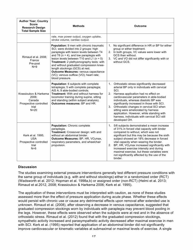
Rimaud et al. 2008; France
Pre-post N=9
Population: 9 men with chronic traumatic SCI, were divided into 2 groups: high paraplegia with lesion levels between T4 and T6 (n = 4), and low paraplegia with lesion levels between T10 and L1 (n = 5) Treatment: 2 plethysmography tests: with and without graduated compression knee-length stockings (GCS) at rest. Outcome Measures: venous capacitance (VC); venous outflow (VO); heart rate; blood pressure.
1. No significant difference in HR or BP for either group or either treatment.
2. In both groups, VC values were lower with GCS than without.
3. VC and VO did not differ significantly with or without GCS.
Krassioukov & Harkema 2006;
Canada Prospective controlled
trial N=20
Population: 6 subjects with complete tetraplegia; 5 with complete paraplegia; AIS A; 9 able-bodied controls. Treatment: With and without harness for locomotor training during supine, sitting and standing (within subject analysis). Outcomes measures: BP and HR.
1. Orthostatic stress significantly decreased arterial BP only in individuals with cervical SCI.
2. Harness application had no effect on cardiovascular parameters in able-bodied individuals, whereas diastolic BP was significantly increased in those with SCI.
3. Orthostatic changes in cervical SCI when sitting were ameliorated by harness application. However, while standing with harness, individuals with cervical SCI still developed OH.
Kerk et al. 1995; USA
Prospective controlled trial N=6
Population: Chronic complete paraplegia. Treatment: Crossover design: with and without an abdominal binder. Outcome Measures: BP, HR, VO2max, respiratory parameters, and wheelchair propulsion.
1. 5/6 subjects demonstrated a mean increase of 31% in forced vital capacity with binder compared to without, which was not significant but this may be because the sixth subject showed an 18% decrease in forced vital capacity when wearing the binder.
2. BP, HR, VO2max increased significantly with increased exercise intensity and during maximal exercise, but these variables were not significantly affected by the use of the binder.
Discussion The studies examining external pressure interventions generally test different pressure conditions with the same group of individuals (e.g. with and without stockings) either in a randomized order (RCT) (Wadsworth et al. 2012; Hopman et al. 1998a,b) or assigned order (non-RCT) (Helmi et al. 2013; Rimaud et al.2012, 2008; Krassioukov & Harkema 2006; Kerk et al. 1995). The application of these interventions must be interpreted with caution, as none of these studies assessed more than the effect of pressure application during acute phase. Whether these effects would persist with chronic use or cause any detrimental effects upon removal after extended use is unknown. Rimaud et al. (2008), after observing a decrease in venous capacitance, suggested that graduated compression stockings worn by individuals with paraplegia may prevent blood pooling in the legs. However, these effects were observed when the subjects were at rest and in the absence of orthostatic stress. Rimaud et al. (2012) found that with the graduated compression stockings, sympathetic activity increased and parasympathetic activity decreased after maximal exercise in men with SCI. Kerk et al. (1995) reported that application of an abdominal binder did not significantly improve cardiovascular or kinematic variables at submaximal or maximal levels of exercise. A single
18
RCT (n=14) by Wadsworth et al. (2012) found that abdominal binders did not significantly affect mean arterial pressure. In his review, Bhambhani (2002) concluded that the use of abdominal binders does not influence cardiovascular responses. Conversely, in another RCT (Hopman et al. 1998b), demonstrated in a small group of subjects with SCI (n=9) that stockings and an abdominal binder have an effect on cardiovascular responses during submaximal exercises, but not during maximal exercises (Hopman et al. 1998a). Krassioukov & Harkema (2006) found that the use of a harness (which applies abdominal pressure) during locomotor training increased diastolic BP in those with SCI, but not in able-bodied individuals. Therefore, there is level 2 evidence (from 1 RCT) that pressure from elastic stocking and abdominal binders may improve cardiovascular responses during submaximal, but not maximal, arm exercise.
Conclusion
There is conflicting evidence based on limited research that elastic stockings/abdominal binders have any effect on cardiovascular responses in individuals with SCI. There is level 2 evidence (Krassioukov & Harkema 2006) that application of a harness in individuals with SCI could alter baseline cardiovascular parameters and orthostatic responses.
5.3 Whole-Body Vibration in Management of OH in SCI
Whole-body vibration (WBV) exercise is performed on a platform that generates vertical sinusoidal vibrations, stimulating muscle spindles and resulting in muscle contractions. The effect of WBV exercise on muscle activity is elicited through reflex muscle activation (Bongiovanni et al. 1990) and muscle twitch potentiation (Cochrane et al. 2010).
Table 7: Whole-Body Vibration in Management of OH in SCI
Author Year; Country Score
Research Design Total Sample Size
Methods Outcome
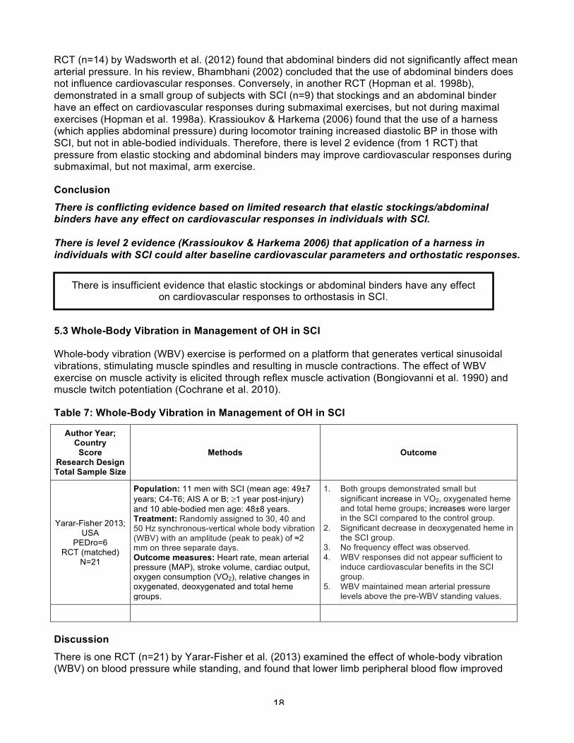
Yarar-Fisher 2013; USA
PEDro=6 RCT (matched)
N=21
Population: 11 men with SCI (mean age: 49±7 years; C4-T6; AIS A or B; ³1 year post-injury) and 10 able-bodied men age: 48±8 years. Treatment: Randomly assigned to 30, 40 and 50 Hz synchronous-vertical whole body vibration (WBV) with an amplitude (peak to peak) of ≈2 mm on three separate days. Outcome measures: Heart rate, mean arterial pressure (MAP), stroke volume, cardiac output, oxygen consumption (VO2), relative changes in oxygenated, deoxygenated and total heme groups.
1. Both groups demonstrated small but significant increase in VO2, oxygenated heme and total heme groups; increases were larger in the SCI compared to the control group.
2. Significant decrease in deoxygenated heme in the SCI group.
3. No frequency effect was observed. 4. WBV responses did not appear sufficient to
induce cardiovascular benefits in the SCI group.
5. WBV maintained mean arterial pressure levels above the pre-WBV standing values.
Discussion There is one RCT (n=21) by Yarar-Fisher et al. (2013) examined the effect of whole-body vibration (WBV) on blood pressure while standing, and found that lower limb peripheral blood flow improved
There is insufficient evidence that elastic stockings or abdominal binders have any effect on cardiovascular responses to orthostasis in SCI.
19
post-WBV. However, the clinical application of WBV in preventing orthostatic hypotension has not yet been studied.
Conclusion There is level 1 evidence (Yarar-Fisher et al. 2013) that whole-body vibration increases standing mean arterial pressure in individuals with SCI. 5.4 Effect of Functional Electrical Stimulation (FES) on OH in SCI
The application of FES triggers intermittent muscle contractions that activate the physiologic muscle pump. The physiologic muscle pump facilitates venous return via compression of the superficial and deep veins of the legs.
Table 8: FES on OH in SCI
Author Year; Country Score
Research Design
Total Sample Size
Methods Outcome
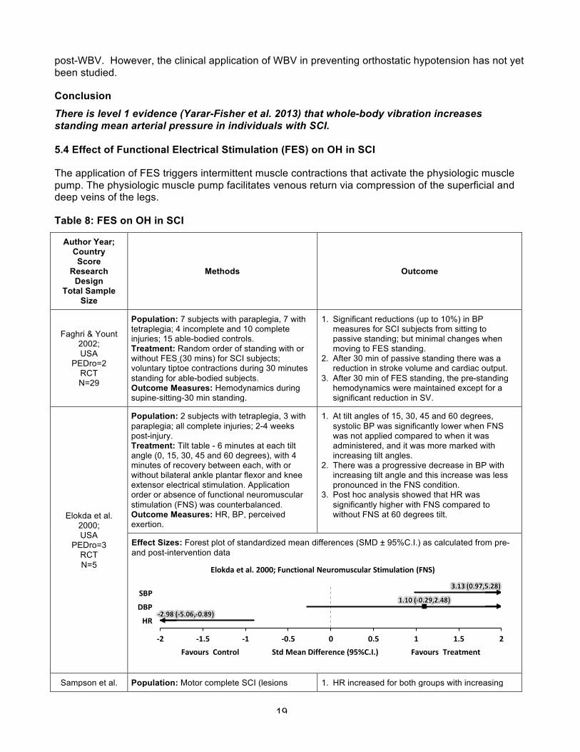
Faghri & Yount 2002; USA
PEDro=2 RCT N=29
Population: 7 subjects with paraplegia, 7 with tetraplegia; 4 incomplete and 10 complete injuries; 15 able-bodied controls. Treatment: Random order of standing with or without FES (30 mins) for SCI subjects; voluntary tiptoe contractions during 30 minutes standing for able-bodied subjects. Outcome Measures: Hemodynamics during supine-sitting-30 min standing.
1. Significant reductions (up to 10%) in BP measures for SCI subjects from sitting to passive standing; but minimal changes when moving to FES standing.
2. After 30 min of passive standing there was a reduction in stroke volume and cardiac output.
3. After 30 min of FES standing, the pre-standing hemodynamics were maintained except for a significant reduction in SV.
Elokda et al. 2000; USA
PEDro=3 RCT N=5
Population: 2 subjects with tetraplegia, 3 with paraplegia; all complete injuries; 2-4 weeks post-injury. Treatment: Tilt table - 6 minutes at each tilt angle (0, 15, 30, 45 and 60 degrees), with 4 minutes of recovery between each, with or without bilateral ankle plantar flexor and knee extensor electrical stimulation. Application order or absence of functional neuromuscular stimulation (FNS) was counterbalanced. Outcome Measures: HR, BP, perceived exertion.
1. At tilt angles of 15, 30, 45 and 60 degrees, systolic BP was significantly lower when FNS was not applied compared to when it was administered, and it was more marked with increasing tilt angles.
2. There was a progressive decrease in BP with increasing tilt angle and this increase was less pronounced in the FNS condition.
3. Post hoc analysis showed that HR was significantly higher with FNS compared to without FNS at 60 degrees tilt.
Effect Sizes: Forest plot of standardized mean differences (SMD ± 95%C.I.) as calculated from pre- and post-intervention data
Sampson et al. Population: Motor complete SCI (lesions 1. HR increased for both groups with increasing
3.13(0.97,5.28)
1.10(-0.29,2.48)
-2.98(-5.06,-0.89)
-2 -1.5 -1 -0.5 0 0.5 1 1.5 2
SBP
DBP
HR
FavoursControlStdMeanDifference(95%C.I.)FavoursTreatment
Elokdaetal.2000;FunctionalNeuromuscularStimulation(FNS)
20
Author Year; Country Score
Research Design
Total Sample Size
Methods Outcome
2000; USA
PEDro=3 RCT N=6
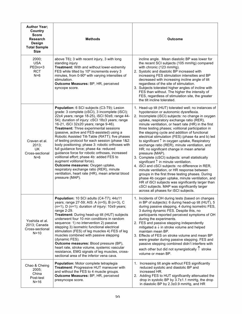
above T6); 3 with recent injury, 3 with long standing injury Treatment: With and without lower-extremity FES while tilted by 10º increments every 3 minutes, from 0-90º with varying intensities of stimulation. Outcome Measures: BP, HR, perceived syncope score.
incline angle. Mean diastolic BP was lower for the recent SCI subjects (105 mmHg) compared with chronic (123 mmHg).
2. Systolic and diastolic BP increased with increasing FES stimulation intensities and BP decreased with increasing incline angle of tilt regardless of the site of stimulation.
3. Subjects tolerated higher angles of incline with FES than without. The higher the intensity of FES, regardless of stimulation site, the greater the tilt incline tolerated.
Craven et al. 2013;
UK Cross-sectional
N=6
Population: 6 SCI subjects (C3-T9); Lesion grade: 3 complete (cSCI), 3 incomplete (iSCI); 22±4 years; range 18-25), iSCI 50±6; range 44-54); duration of injury: cSCI 18±3 years; range 16-21, iSCI 32±20 years; range 9-46). Treatment: Three experimental sessions (passive, active and FES-assisted) using a Robotic Assisted Tilt-Table (RATT); five phases of testing protocol for each session (phase 1&2: body positioning; phase 3: robotic orthoses with full guidance force; phase 4a: reduced guidance force for robotic orthoses, increased volitional effort; phase 4b: added FES to augment volitional force). Outcome measures: Oxygen uptake, respiratory exchange ratio (RER), minute ventilation, heart rate (HR), mean arterial blood pressure (MAP).
1. Head-up tilt (HUT) tolerated well; no instances of hypotension or autonomic dysreflexia.
2. Incomplete (iSCI) subjects: no change in oxygen uptake, respiratory exchange ratio (RER), minute ventilation, or heart rate (HR) in the first three testing phases; volitional participation in the stepping cycle and addition of functional electrical stimulation (FES) (phase 4a and b) led to significant in oxygen uptake, Respiratory exchange ratio (RER), minute ventilation, and HR; no significant change in mean arterial pressure (MAP).
3. Complete (cSCI) subjects: small statistically significant in minute ventilation.
4. iSCI and cSCI subjects: no difference in RER, minute ventilation, or HR response between groups in the first three testing phases. During phase 4b oxygen uptake, minute ventilation, and HR of iSCI subjects was significantly larger than cSCI subjects. MAP was significantly larger across all phases for iSCI subjects.
Yoshida et al. 2013; Canada
Cross-sectional N=10
Population: 10 SCI adults (C4-T7); 44±11 years; range 27-59; AIS: A (n=5), B (n=3), C (n=1), D (n=1); duration of injury: 10±9 years; range 2-29. Treatment: During head-up tilt (HUT) subjects underwent four 10 min conditions in random sequence: 1) no intervention 2) passive stepping 3) isometric functional electrical stimulation (FES) of leg muscles 4) FES of leg muscles combined with passive stepping (dynamic FES). Outcome measures: Blood pressure (BP), heart rate, stroke volume, systemic vascular resistance, EMG signals of leg muscles, cross-sectional area of the inferior vena cava.
1. Incidents of OH during tests (based on changes in BP of subjects): 6 during head-up tilt (HUT), 5 during passive stepping, 4 during isometric FES, 3 during dynamic FES. Despite this, no participants reported perceived symptoms of OH during the experiments.
2. FES and passive stepping independently mitigated a ¯ in stroke volume and helped maintain mean BP.
3. Effects of FES on stroke volume and mean BP were greater during passive stepping. FES and passive stepping combined didn’t interfere with each other but did not synergistically stroke volume or mean BP.
Chao & Cheing 2005; China
Post-test N=16
Population: Motor complete tetraplegia Treatment: Progressive HUT maneuver with and without the FES to 4 muscle groups. Outcome Measures: BP, HR, perceived presyncope score.
1. Increasing tilt angle without FES significantly reduced systolic and diastolic BP and increased HR.
2. Adding FES to HUT significantly attenuated the drop in systolic BP by 3.7±1.1 mmHg, the drop in diastolic BP by 2.3±0.9 mmHg, and HR
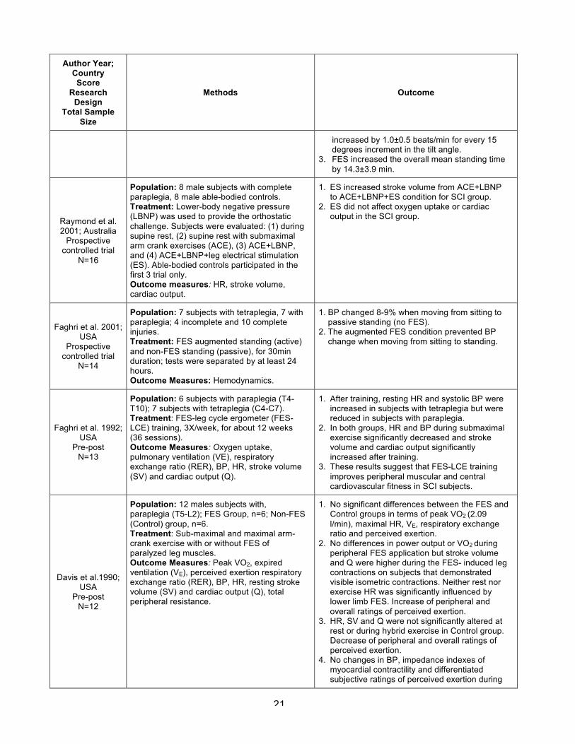
21
Author Year; Country Score
Research Design
Total Sample Size
Methods Outcome
increased by 1.0±0.5 beats/min for every 15 degrees increment in the tilt angle.
3. FES increased the overall mean standing time by 14.3±3.9 min.
Raymond et al. 2001; Australia
Prospective controlled trial
N=16
Population: 8 male subjects with complete paraplegia, 8 male able-bodied controls. Treatment: Lower-body negative pressure (LBNP) was used to provide the orthostatic challenge. Subjects were evaluated: (1) during supine rest, (2) supine rest with submaximal arm crank exercises (ACE), (3) ACE+LBNP, and (4) ACE+LBNP+leg electrical stimulation (ES). Able-bodied controls participated in the first 3 trial only. Outcome measures: HR, stroke volume, cardiac output.
1. ES increased stroke volume from ACE+LBNP to ACE+LBNP+ES condition for SCI group.
2. ES did not affect oxygen uptake or cardiac output in the SCI group.
Faghri et al. 2001; USA
Prospective controlled trial
N=14
Population: 7 subjects with tetraplegia, 7 with paraplegia; 4 incomplete and 10 complete injuries. Treatment: FES augmented standing (active) and non-FES standing (passive), for 30min duration; tests were separated by at least 24 hours. Outcome Measures: Hemodynamics.
1. BP changed 8-9% when moving from sitting to passive standing (no FES).
2. The augmented FES condition prevented BP change when moving from sitting to standing.
Faghri et al. 1992; USA
Pre-post N=13
Population: 6 subjects with paraplegia (T4-T10); 7 subjects with tetraplegia (C4-C7). Treatment: FES-leg cycle ergometer (FES-LCE) training, 3X/week, for about 12 weeks (36 sessions). Outcome Measures: Oxygen uptake, pulmonary ventilation (VE), respiratory exchange ratio (RER), BP, HR, stroke volume (SV) and cardiac output (Q).
1. After training, resting HR and systolic BP were increased in subjects with tetraplegia but were reduced in subjects with paraplegia.
2. In both groups, HR and BP during submaximal exercise significantly decreased and stroke volume and cardiac output significantly increased after training.
3. These results suggest that FES-LCE training improves peripheral muscular and central cardiovascular fitness in SCI subjects.
Davis et al.1990; USA
Pre-post N=12
Population: 12 males subjects with, paraplegia (T5-L2); FES Group, n=6; Non-FES (Control) group, n=6. Treatment: Sub-maximal and maximal arm-crank exercise with or without FES of paralyzed leg muscles. Outcome Measures: Peak VO2, expired ventilation (VE), perceived exertion respiratory exchange ratio (RER), BP, HR, resting stroke volume (SV) and cardiac output (Q), total peripheral resistance.
1. No significant differences between the FES and Control groups in terms of peak VO2 (2.09 l/min), maximal HR, VE, respiratory exchange ratio and perceived exertion.
2. No differences in power output or VO2 during peripheral FES application but stroke volume and Q were higher during the FES- induced leg contractions on subjects that demonstrated visible isometric contractions. Neither rest nor exercise HR was significantly influenced by lower limb FES. Increase of peripheral and overall ratings of perceived exertion.
3. HR, SV and Q were not significantly altered at rest or during hybrid exercise in Control group. Decrease of peripheral and overall ratings of perceived exertion.
4. No changes in BP, impedance indexes of myocardial contractility and differentiated subjective ratings of perceived exertion during
22
Author Year; Country Score
Research Design
Total Sample Size
Methods Outcome
hybrid exercise compared with non-FES conditions.
Discussion FES may be an important treatment adjunct to minimize cardiovascular changes during postural orthostatic stress in individuals with SCI. Several studies have suggested that FES-induced contractions of the leg muscles increases cardiac output and stroke volume, which increases venous return (Raymond et al. 2001). Subsequently, this increases ventricular filling and left ventricular end-diastolic volume (i.e., enhanced cardiac preload). According to the Frank-Starling effect, an increase in ventricular preload will lead to greater stretch of the myocytes and a concomitant increase in left ventricular stroke volume. The increased stroke volume may produce greater cardiac output and in turn, greater arterial blood pressure. In this manner, FES-induced contraction of the leg muscles may attenuate the drop in systolic BP in response to an orthostatic challenge. FES-induced contraction of the leg muscles may also restore the body’s ability to redistribute blood from below the level of the lesion back to the heart. In fact, it is through this means that Davis et al. (1990) attributes FES’s effectiveness during an orthostatic challenge. In their study, Davis et al. found FES of leg muscles resulted in increased cardiac output and stroke volume in 6 males with paraplegia performing maximal arm-crank exercise. These results suggest that FES of leg muscles could alleviate the lower limb pooling effect during the orthostatic challenge. Chi et al. (2008) suggest that alleviation of the pooling effect could be further enhanced when FES of leg muscles is combined with passive mobilization. The clinical utility of this combination must be examined further in subjects with SCI because subjects in Chi et al. (2008) were able-bodied. A cross-sectional study by Yoshida et al. (2013) compares isometric FES of leg muscles vs. passive stepping vs. isometric FES + passive stepping. They found that both FES and passive stepping increased stroke volume and mean BP and that the highest increase in these two resulted from combined FES + stepping; however, the two interventions did not interact to synergistically increase stroke volume and mean aBP.
FES results in a dose-dependent increase in BP independent of the stimulation site that may be useful in treating OH (Sampson et al. 2000) and may be an important treatment adjunct to minimize cardiovascular changes during postural orthostatic stress in individuals with acute SCI. Three level 2 RCTs (Faghri & Yount 2002; Elokda et al. 2000; Sampson et al. 2000) and five non-randomized controlled trials (Chao & Cheing 2005; Raymond et al. 2001; Faghri et al. 2001; Faghri et al. 1992; Davis et al. 1990) with small sample sizes provide support for use of FES in individuals with SCI. FES of the lower extremity could be used by persons with SCI as an adjunct during standing to prevent OH and circulatory hypokinesis. An FES-induced leg muscle contraction is an effective adjunct treatment to delay OH caused by tilting; it allows people with tetraplegia to stand up more frequently and for longer durations (Elokda et al. 2000; Sampson et al. 2000).This effect may be more beneficial to those with tetraplegia who have a greater degree of decentralized cardiovascular autonomic control and may not be able to adjust their hemodynamics to the change in position (Faghri et al. 2001).
Current protocols predominantly evaluate BP after a single application of FES with a single change in position. The feasibility and practicality of implementing FES to influence orthostatic BP over time needs to be further explored.
Conclusion
23
There is level 2 evidence (from small, lower quality RCTs) (Faghri & Yount 2002; Elokda et al. 2000; Sampson et al. 2000) that FES is an important treatment adjunct to minimize cardiovascular changes during postural orthostatic stress in individuals with SCI.
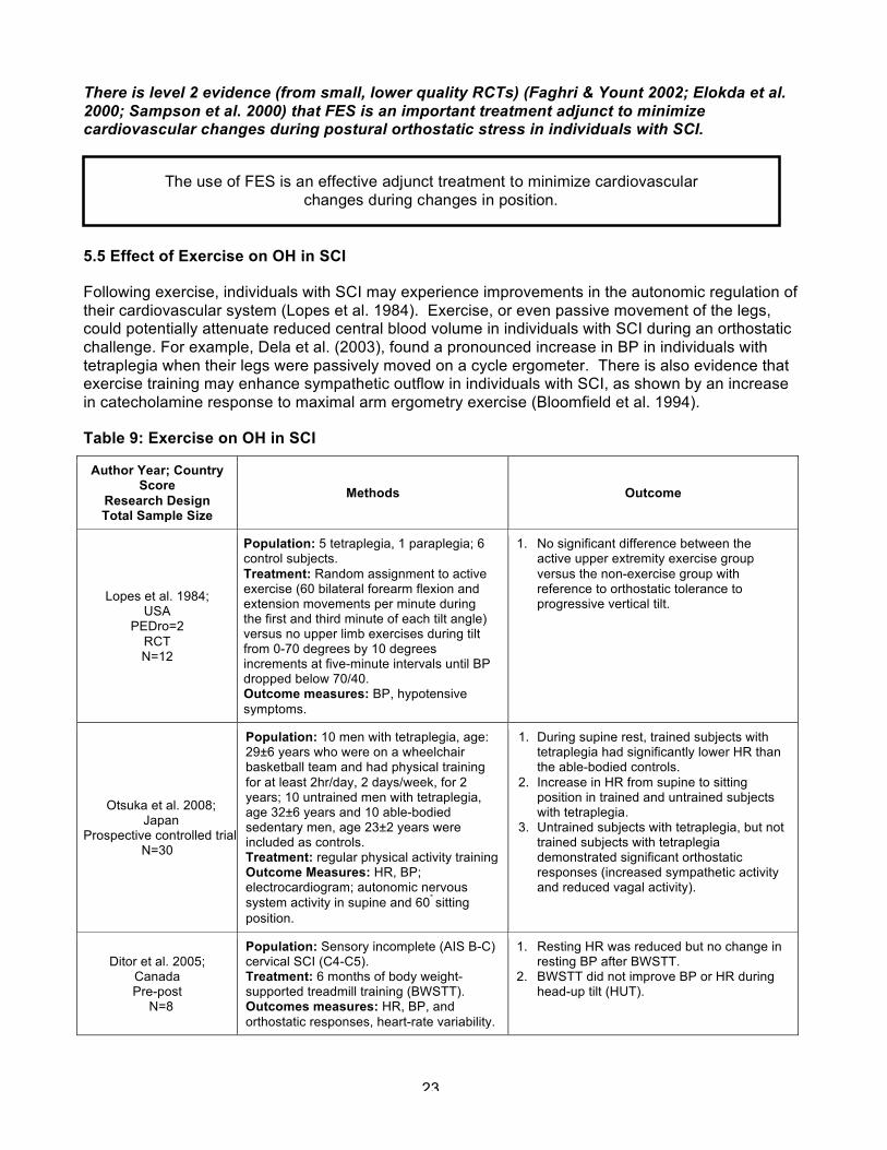
5.5 Effect of Exercise on OH in SCI
Following exercise, individuals with SCI may experience improvements in the autonomic regulation of their cardiovascular system (Lopes et al. 1984). Exercise, or even passive movement of the legs, could potentially attenuate reduced central blood volume in individuals with SCI during an orthostatic challenge. For example, Dela et al. (2003), found a pronounced increase in BP in individuals with tetraplegia when their legs were passively moved on a cycle ergometer. There is also evidence that exercise training may enhance sympathetic outflow in individuals with SCI, as shown by an increase in catecholamine response to maximal arm ergometry exercise (Bloomfield et al. 1994).
Table 9: Exercise on OH in SCI
Author Year; Country Score
Research Design Total Sample Size
Methods Outcome
Lopes et al. 1984; USA
PEDro=2 RCT N=12
Population: 5 tetraplegia, 1 paraplegia; 6 control subjects. Treatment: Random assignment to active exercise (60 bilateral forearm flexion and extension movements per minute during the first and third minute of each tilt angle) versus no upper limb exercises during tilt from 0-70 degrees by 10 degrees increments at five-minute intervals until BP dropped below 70/40. Outcome measures: BP, hypotensive symptoms.
1. No significant difference between the active upper extremity exercise group versus the non-exercise group with reference to orthostatic tolerance to progressive vertical tilt.
Otsuka et al. 2008; Japan
Prospective controlled trial N=30
Population: 10 men with tetraplegia, age: 29±6 years who were on a wheelchair basketball team and had physical training for at least 2hr/day, 2 days/week, for 2 years; 10 untrained men with tetraplegia, age 32±6 years and 10 able-bodied sedentary men, age 23±2 years were included as controls. Treatment: regular physical activity training Outcome Measures: HR, BP; electrocardiogram; autonomic nervous system activity in supine and 60° sitting position.
1. During supine rest, trained subjects with tetraplegia had significantly lower HR than the able-bodied controls.
2. Increase in HR from supine to sitting position in trained and untrained subjects with tetraplegia.
3. Untrained subjects with tetraplegia, but not trained subjects with tetraplegia demonstrated significant orthostatic responses (increased sympathetic activity and reduced vagal activity).
Ditor et al. 2005; Canada Pre-post
N=8
Population: Sensory incomplete (AIS B-C) cervical SCI (C4-C5). Treatment: 6 months of body weight-supported treadmill training (BWSTT). Outcomes measures: HR, BP, and orthostatic responses, heart-rate variability.
1. Resting HR was reduced but no change in resting BP after BWSTT.
2. BWSTT did not improve BP or HR during head-up tilt (HUT).
The use of FES is an effective adjunct treatment to minimize cardiovascular changes during changes in position.
24
Discussion Only three exercise studies have attempted to assess the effect of exercise on orthostatic tolerance in subjects with SCI and protocol differed in each. Lopes et al. (1984) found no effects on orthostatic tolerance with the addition of upper extremity exercises during a progressive head-up tilt (HUT) protocol. Such findings are not surprising given the small muscle mass involved in the upper limbs and the fact that venous pooling occurs primarily in the lower limbs. Ditor et al. (2005) demonstrated that individuals with incomplete tetraplegia retain the ability to make positive changes in cardiovascular autonomic regulation with BWSTT. However, six months of BWSTT did not adversely affect the orthostatic tolerance in subjects with SCI. The authors found this encouraging as it suggests that orthostatic tolerance is retained after exercise training, even though this intervention probably reduced peripheral vascular resistance. Otsuka et al. (2008) found that individuals with complete tetraplegia who were involved in regular physical activity training (2 hrs/day, 2 days/wk, ≥2 yrs) demonstrated greater orthostatic tolerance than inactive individuals with SCI (<30 mins/wk).
Conclusion There is level 2 evidence (from 1 RCT) (Lopes et al. 1984) that simultaneous upper extremity exercises does not improve orthostatic tolerance during a progressive tilt exercise. There is level 4 evidence (from 1 pre-post study) (Ditor et al. 2005) that 6 months of BWSTT does not significantly improve orthostatic tolerance during a tilt test. There is level 4 evidence (from 1 post-test study) (Otsuka et al. 2008) that regular physical activity (2hrs/day, 2x/wk, ≥ 2yrs) may improve orthostatic tolerance during a tilt test.
5.6 Effect of Standing on OH in SCI
Active stand training that emphasizes weight bearing is thought to stimulate the neuromuscular system below the level of injury in individuals with SCI and may affect the response to orthostatic stress by increasing venous return (Harkema et al. 2008).
Table 10: Standing on OH in SCI
Author Year; Country Score
Research Design Total Sample Size
Methods Outcome
Simultaneous arm exercise during a tilt test is not effective for improving orthostatic tolerance.
The benefits of body-weight supported treadmill training for management of OH have not been sufficiently proven in SCI.
There is limited evidence that regular physical activity may improve orthostatic tolerance during a tilt test.
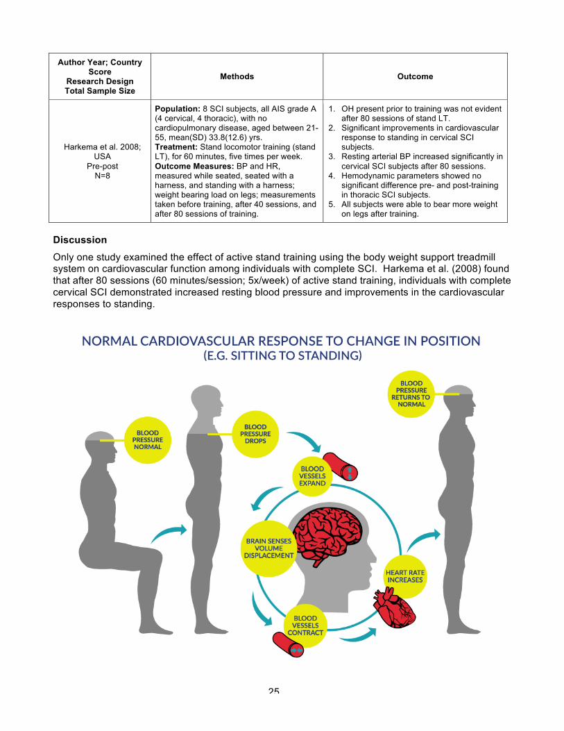
25
Author Year; Country Score
Research Design Total Sample Size
Methods Outcome
Harkema et al. 2008; USA
Pre-post N=8
Population: 8 SCI subjects, all AIS grade A (4 cervical, 4 thoracic), with no cardiopulmonary disease, aged between 21-55, mean(SD) 33.8(12.6) yrs. Treatment: Stand locomotor training (stand LT), for 60 minutes, five times per week. Outcome Measures: BP and HR, measured while seated, seated with a harness, and standing with a harness; weight bearing load on legs; measurements taken before training, after 40 sessions, and after 80 sessions of training.
1. OH present prior to training was not evident after 80 sessions of stand LT.
2. Significant improvements in cardiovascular response to standing in cervical SCI subjects.
3. Resting arterial BP increased significantly in cervical SCI subjects after 80 sessions.
4. Hemodynamic parameters showed no significant difference pre- and post-training in thoracic SCI subjects.
5. All subjects were able to bear more weight on legs after training.
Discussion Only one study examined the effect of active stand training using the body weight support treadmill system on cardiovascular function among individuals with complete SCI. Harkema et al. (2008) found that after 80 sessions (60 minutes/session; 5x/week) of active stand training, individuals with complete cervical SCI demonstrated increased resting blood pressure and improvements in the cardiovascular responses to standing.
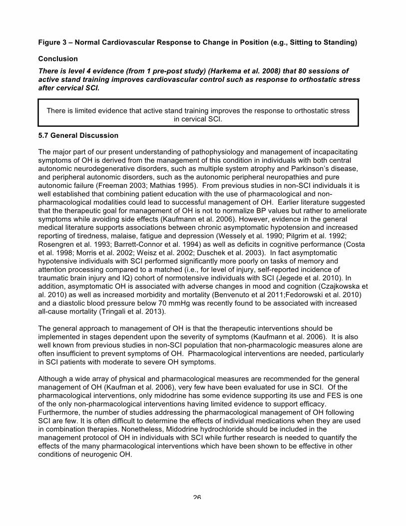
26
Figure 3 – Normal Cardiovascular Response to Change in Position (e.g., Sitting to Standing)
Conclusion There is level 4 evidence (from 1 pre-post study) (Harkema et al. 2008) that 80 sessions of active stand training improves cardiovascular control such as response to orthostatic stress after cervical SCI.
5.7 General Discussion
The major part of our present understanding of pathophysiology and management of incapacitating symptoms of OH is derived from the management of this condition in individuals with both central autonomic neurodegenerative disorders, such as multiple system atrophy and Parkinson’s disease, and peripheral autonomic disorders, such as the autonomic peripheral neuropathies and pure autonomic failure (Freeman 2003; Mathias 1995). From previous studies in non-SCI individuals it is well established that combining patient education with the use of pharmacological and non-pharmacological modalities could lead to successful management of OH. Earlier literature suggested that the therapeutic goal for management of OH is not to normalize BP values but rather to ameliorate symptoms while avoiding side effects (Kaufmann et al. 2006). However, evidence in the general medical literature supports associations between chronic asymptomatic hypotension and increased reporting of tiredness, malaise, fatigue and depression (Wessely et al. 1990; Pilgrim et al. 1992; Rosengren et al. 1993; Barrett-Connor et al. 1994) as well as deficits in cognitive performance (Costa et al. 1998; Morris et al. 2002; Weisz et al. 2002; Duschek et al. 2003). In fact asymptomatic hypotensive individuals with SCI performed significantly more poorly on tasks of memory and attention processing compared to a matched (i.e., for level of injury, self-reported incidence of traumatic brain injury and IQ) cohort of normotensive individuals with SCI (Jegede et al. 2010). In addition, asymptomatic OH is associated with adverse changes in mood and cognition (Czajkowska et al. 2010) as well as increased morbidity and mortality (Benvenuto et al 2011;Fedorowski et al. 2010) and a diastolic blood pressure below 70 mmHg was recently found to be associated with increased all-cause mortality (Tringali et al. 2013). The general approach to management of OH is that the therapeutic interventions should be implemented in stages dependent upon the severity of symptoms (Kaufmann et al. 2006). It is also well known from previous studies in non-SCI population that non-pharmacologic measures alone are often insufficient to prevent symptoms of OH. Pharmacological interventions are needed, particularly in SCI patients with moderate to severe OH symptoms. Although a wide array of physical and pharmacological measures are recommended for the general management of OH (Kaufman et al. 2006), very few have been evaluated for use in SCI. Of the pharmacological interventions, only midodrine has some evidence supporting its use and FES is one of the only non-pharmacological interventions having limited evidence to support efficacy. Furthermore, the number of studies addressing the pharmacological management of OH following SCI are few. It is often difficult to determine the effects of individual medications when they are used in combination therapies. Nonetheless, Midodrine hydrochloride should be included in the management protocol of OH in individuals with SCI while further research is needed to quantify the effects of the many pharmacological interventions which have been shown to be effective in other conditions of neurogenic OH.
There is limited evidence that active stand training improves the response to orthostatic stress in cervical SCI.
27
6.0 References
Barber DB, Rogers SJ, Fredrickson MD, Able AC. Midodrine hydrochloride and the treatment of orthostatic hypotension in tetraplegia: two cases and a review of the literature. Spinal Cord 2000; 38: 109-111.
Barrett-Connor E, Palinkas LA. Low blood pressure and depression in older men: a population based study. Bmj 1994;308:446-449.
Benvenuto LJ, Krakoff LR. Morbidity and mortality of orthostatic hypotension: implications for management of cardiovascular disease. Am J Hypertens. 2011;24:135-144.
Bhambhani Y. Physiology of wheelchair racing in athletes with spinal cord injury. Sports Med 2002; 32: 23-51.
Bloomfield SA, Jackson RD, Mysiw WJ. Catecholamine response to exercise and training in individuals with spinal cord injury. Med Sci Sports Exerc 1994; 26: 1213-1219.
Bongiovanni LG, Hagbarth KE. Tonic vibration reflexes elicited during fatigue from maximal voluntary contractions in man. J Physiol 1990;423:1–14.
Cariga P, Ahmed S, Mathias CJ, Gardner BP. The prevalence and association of neck (coat-hanger) pain and orthostatic (postural) hypotension in human spinal cord injury. Spinal Cord 2002; 40: 77-82.
Chao CY, Cheing GL. The effects of lower-extremity functional electric stimulation on the orthostatic responses of people with tetraplegia. Arch Phys Med Rehabil 2005; 86: 1427-1433.
Chelvarajah R. Orthostatic hypotension following spinal cord injury: impact on the use of standing apparatus. NeuroRehabilitation 2009; 24:237-242.
Chi L, Masani K, Miyatani M, Adam TT, Wayne JK, Mardimae A, Kessler C, Fisher JA. Cardiovascular response to functional electrical stimulation and dynamic tilt table therapy to improve orthostatic tolerance. J Electromyogr Kinesiol 2008; 18: 900-907.
Claydon VE, Hainsworth R. Salt supplementation improves orthostatic cerebral and peripheral vascular control in patients with syncope. Hypertension 2004; 43: 809-813.
Claydon VE, Steeves JD, Krassioukov A. Orthostatic hypotension following spinal cord injury: understanding clinical pathophysiology. Spinal Cord 2006; 44: 341-351.
Cochrane DJ, Stannard SR, Firth EC, Rittweger J. Acute whole-body vibration elicits post-activation potentiation. Eur J Appl Physiol 2010;108:311–319.
Costa M, Stegagno L, Schandry R, Bitti PE. Contingent negative variation and cognitive performance in hypotension. Psychophysiology. 1998;35:737-744.
Craven CTD, Gollee H, Coupaud S, Purcell MA, Allan DB. Investigation of robotic-assisted tilt-table therapy for early-stage spinal cord injury rehabilitation. J Rehabil Res Dev 2013; 50: 367-378.
Czajkowska J, Ozhog S, Smith E, Perlmuter LC. Cognition and hopelessness in association with subsyndromal orthostatic hypotension. J Gerontol A Biol Sci Med Sci. 2010;65:873-879.
Davidson C, Smith D, Morgan DB. Diurnal pattern of water and electrolyte excretion and body weight in idiopathic orthostatic hypotension. The effect of three treatments. Am J Med 1976; 61: 709-715.
Davis GM, Servedio FJ, Glaser RM, Gupta SC, Suryaprasad AG. Cardiovascular responses to arm cranking and FNS-induced leg exercise in paraplegics. J Appl Physiol 1990; 69: 671-677.
Dela F, Mohr T, Jensen CM, Haahr HL, Secher NH, Biering-Sorensen F, Kjaer M. Cardiovascular control during exercise: insights from spinal cord-injured humans. Circulation 2003; 107: 2127-2133.
Ditor DS, Kamath MV, MacDonald MJ. Bugaresti J, McCartney N, Hicks AL. Effects of body weight-supported treadmill training on heart rate variability and blood pressure variability in individuals with spinal cord injury. Journal of Applied Physiology 2005; 98:1519-1525.
Duschek S, Weisz N, Schandry R. Reduced cognitive performance and prolonged reaction time accompany moderate hypotension. Clin Auton Res 2003; 13: 427–432.
Elokda AS, Nielsen DH, Shields RK. Effect of functional neuromuscular stimulation on postural related orthostatic stress in individuals with acute spinal cord injury. J Rehabil Res Dev 2000; 37: 535-542.
28
Faghri PD, Glaser RM, Figoni SF. Functional electrical stimulation leg cycle ergometer exercise: training effects on cardiorespiratory responses of spinal cord injured subjects at rest and during submaximal exercise. Arch Phys Med Rehabil 1992; 73: 1085-1093.
Faghri PD, Yount J. Electrically induced and voluntary activation of physiologic muscle pump: a comparison between spinal cord-injured and able-bodied individuals. Clin Rehabil 2002; 16: 878-885.
Faghri PD, Yount JP, Pesce WJ, Seetharama S, Votto JJ. Circulatory hypokinesis and functional electric stimulation during standing in persons with spinal cord injury. Arch Phys Med Rehabil 2001; 82:1587-1595.
Fedorowski A, Stavenow L, Hedblad B, Berglund G, Nilsson PM, Melander O. Orthostatic hypotension predicts all-cause mortality and coronary events in middle-aged individuals (The Malmo Preventive Project). Eur Heart J. 2010;31:85-91.
Freeman R. Treatment of orthostatic hypotension. Semin Neurol 2003; 23: 435-442. Frisbie JH, Steele DJ. Postural hypotension and abnormalities of salt and water metabolism in
myelopathy patients. Spinal Cord 1997; 35: 303-307. Frisbie JH. Postural hypotension, hyponatremia, and salt and water intake: case reports. J Spinal
Cord Med 2004; 27: 133-137. Gillis DJ, Wouda M, Hjeltnes N. Non-pharmacological management of orthostatic hypotension after
spinal cord injury: a critical review of the literature. Spinal Cord 2008; 46: 652-659. Groomes TE, Huang CT. Orthostatic hypotension after spinal cord injury: treatment with
fludrocortisone and ergotamine. Arch Phys Med Rehabil 1991; 72: 56-58. Harkema SJ, Ferreira CK, van den Brand RJ, Krassioukov AV. Improvements in orthostatic instability
with stand locomotor training in individuals with spinal cord injury. J Neurotrauma 2008; 25: 1467-1475.
Helmi M, Lima A, Gommers D, van Bommel J, Bakker J. Inflatable external leg compression prevents orthostatic hypotension in a patient with a traumatic cervical spinal cord injury. Future Cardiol 2013; 9: 645-648.
Hopman MT, Dueck C, Monroe M, Philips WT, Skinner JS. Limits to maximal performance in individuals with spinal cord injury. Int J Sports Med 1998b; 19: 98-103.
Hopman MT, Groothuis JT, Flendrie M, Gerrits KH, Houtman S. Increased vascular resistance in paralyzed legs after spinal cord injury is reversible by training. J Appl Physiol 2002; 93: 1966-1972.
Hopman MT, Monroe M, Dueck C, Phillips WT, Skinner JS. Blood redistribution and circulatory responses to submaximal arm exercise in persons with spinal cord injury. Scand J Rehabil Med 1998a; 30: 167-174.
Houtman S, Colier WN, Oeseburg B, Hopman MT. Systemic circulation and cerebral oxygenation during head-up tilt in spinal cord injured individuals. Spinal Cord 2000; 38: 158-163.
Illman A, Stiller K, Williams M. The prevalence of orthostatic hypotension during physiotherapy treatment in patients with an acute spinal cord injury. Spinal Cord 2000; 38: 741-747.
Jegede AB, Rosado-Rivera D, Bauman WA, Cardozo CP, Sano M, Moyer JM, et al. Cognitive performance in hypotensive persons with spinal cord injury. Clin Auton Res. 2010;20:3-9.
Kaufmann HC, Freeman RH, Kaplan NM. Treatment of orthostatic and postprandial hypotension. Waltham, MA, 2006.
Kerk JK, Clifford PS, Snyder AC, Prieto TE, O'Hagan KP, Schot PK, Myklebust JB, Myklebust BM. Effect of an abdominal binder during wheelchair exercise. Med Sci Sports Exerc 1995; 27: 913-919.
Kobayashi S, Endou M, Sakuraya F, Matsuda N, Zhang XH, Azuma M, Echigo N, Kemmotsu O, Hattori Y, Gando S. The sympathomimetic actions of l-ephedrine and d-pseudoephedrine: direct receptor activation or norepinephrine release? Anesth Analg 2003; 97:1239-1245.
Krassioukov A, Claydon VE. The clinical problems in cardiovascular control following spinal cord injury: an overview. Prog Brain Res 2006;152:223-229.
Krassioukov AV, Harkema SJ. Effect of harness application and postural changes on cardiovascular parameters of individuals with spinal cord injury. Spinal Cord 2006;44:780-786.
29
Krassioukov, A, Eng JJ, Warburton DE, & Teasell R. A systematic review of the management of orthostatic hypotension after spinal cord injury. Arch Phy Med Rehabil 2009; 90: 876-885.
La Fountaine MF, Wecht JM, Bauman WA. Acute nitric oxide synthase inhibition and cardiac conduction in persons with spinal cord injury: a short report. Pharmazie 2013; 68: 245-250.
Lopes P, Figoni SF, Perkash I. Upper limb exercise effect on tilt tolerance during orthostatic training of patients with spinal cord injury. Arch Phys Med Rehabil 1984; 65: 251-253.
Mathias CJ. Orthostatic hypotension and paroxysmal hypertension in humans with high spinal cord injury. Prog Brain Res 2006; 152: 231-243.
Mathias CJ. Orthostatic hypotension: causes, mechanisms, and influencing factors. Neurology 1995; 45: S6-11.
McKinley WO, Jackson AB, Cardenas DD, DeVivo MJ. Long-term medical complications after traumatic spinal cord injury: a regional model systems analysis. Arch Phys Med Rehabil 1999; 80: 1402-1410.
Morris MC, Scherr PA, Hebert LE, Bennett DA, Wilson RS, Glynn RJ, et al. Association between blood pressure and cognitive function in a biracial community population of older persons. Neuroepidemiology. 2002;21:123-130.
Mukand J, Karlin L, Barrs K, Lublin P. Midodrine for the management of orthostatic hypotension in patients with spinal cord injury: A case report. Arch Phys Med Rehabil 2001; 82: 694-696.
Munakata M, Kameyama J, Kanazawa M, Nunokawa T, Moriai N, Yoshinaga K. Circadian blood pressure rhythm in patients with higher and lower spinal cord injury: simultaneous evaluation of autonomic nervous activity and physical activity. J Hypertens 1997; 15: 1745-1749.
Muneta S, Iwata T, Hiwada K, Murakami E, Sato Y, Imamura Y. Effect of L-threo-3, 4-dihydroxyphenylserine on orthostatic hypotension in a patient with spinal cord injury. Jpn Circ J 1992; 56: 243-247.
Nieshoff EC, Birk TJ, Birk CA, Hinderer SR, Yavuzer G. Double-blinded, placebo-controlled trial of midodrine for exercise performance enhancement in tetraplegia: a pilot study. J Spinal Cord Med 2004; 27: 219-225.
Otsuka Y, Shima N, Moritani T, Okuda K, Yabe K. Orthostatic influence on heart rate and blood pressure variability in trained persons with tetraplegia. Eur J Appl Physiol 2008; 104: 75-78.
Phillips AA (a), Krassioukov AV, Ainslie PN, Cote AT, Warburton DE. Increased central arterial stiffness explains baroreflex dysfunction in spinal cord injury. Journal of neurotrauma. 2014 Jun 15;31(12):1122-8.
Phillips AA (b), Krassioukov AV, Ainslie PN, Warburton DE. Perturbed and spontaneous regional cerebral blood flow responses to changes in blood pressure after high-level spinal cord injury: the effect of midodrine. Journal of Applied Physiology. 2014 Mar 15;116(6):645-53.
Phillips AA (c), Warburton DE, Ainslie PN, Krassioukov AV. Regional neurovascular coupling and cognitive performance in those with low blood pressure secondary to high-level spinal cord injury: improved by alpha-1 agonist midodrine hydrochloride. Journal of Cerebral Blood Flow & Metabolism. 2014 May 1;34(5):794-801.
Pilgrim JA, Stansfeld S, Marmot M. Low blood pressure, low mood? Bmj. 1992;304:75-78. Raymond J, Davis GM, Clarke J, Bryant G. Cardiovascular responses during arm exercise and
orthostatic challenge in individuals with paraplegia. Eur J Appl Physiol. 2001;85:89-95. Rimaud D, Boissier C, Calmels, P. Evaluation of the effects of compression stockings using venous
plethysmography in persons with spinal cord injury. J Spinal Cord Med 2008; 31: 202-207. Rimaud D, Calmels P, Pichot V., Bethoux F, Roche F. Effects of compression stockings on
sympathetic activity and heart rate variability in individuals with spinal cord injury. J Spinal Cord Med 2012; 35: 81-88.
Rosengren A, Tibblin G, Wilhelmsen L. Low systolic blood pressure and self perceived wellbeing in middle aged men. Bmj. 1993;306:243-246.
Sampson EE, Burnham RS, Andrews BJ. Functional electrical stimulation effect on orthostatic hypotension after spinal cord injury. Arch Phys Med Rehabil 2000; 81: 139-143.
Schatz IJ. Orthostatic hypotension. II. Clinical diagnosis, testing, and treatment. Arch Intern Med 1984; 144: 1037-1041.
30
Senard JM, Arias A, Berlan M, Tran MA, Rascol A, Montastruc JL. Pharmacological evidence of alpha 1- and alpha 2-adrenergic supersensitivity in orthostatic hypotension due to spinal cord injury: a case report. Eur J Clin Pharmacol 1991; 41: 593-596.
Sidorov EV, Townson AF, Dvorak MF, Kwon BK, Steeves J, Krassioukov A. Orthostatic hypotension in the first month following acute spinal cord injury. Spinal Cord. 2008;46:65-69.
Ten Harkel AD, van Lieshout JJ, Wieling W. Effects of leg muscle pumping and tensing on orthostatic arterial pressure: a study in normal subjects and patients with autonomic failure. Clin Sci (Lond) 1994; 87: 553-558.
The Consensus Committee of the American Autonomic Society and the American Academy of Neurology. Consensus statement on the definition of orthostatic hypotension, pure autonomic failure, and multiple system atrophy. Neurol 1996; 46: 1470.
Tringali S, Oberer CW, Huang J. Low Diastolic Blood Pressure as a Risk for All-Cause Mortality in VA Patients. Int J Hypertens. 2013;2013:178780.
Vaidyanathan S, Soni SBM, Hughes PL. Midodrine: Insidious development of urologic adverse effects in patients with spinal cord injury: A report of 2 cases. Advances in Therapy 2007; 24: 712-720.
Van Lieshout JJ, ten Harkel AD, Wieling W. Fludrocortisone and sleeping in the head-up position limit the postural decrease in cardiac output in autonomic failure. Clin Auton Res 2000; 10: 35-42.
Vaziri ND. Nitric oxide in microgravity-induced orthostatic intolerance: relevance to spinal cord injury. J Spinal Cord Med 2003; 26: 5-11.
Wadsworth BM, Haines TP, Cornwell PL, Rodwell LT, Paratz JD. Abdominal binder improves lung volumes and voice in people with tetraplegic spinal cord injury. Arch Phys Med Rehabil 2012; 93: 2189-2197.
Wallin BG, Stjernberg L. Sympathetic activity in man after spinal cord injury. Outflow to skin below the lesion. Brain 1984; 107: 183-198.
Wecht JM, De Meersman RE, Weir JP, Spungen AM, Bauman WA. Cardiac autonomic responses to progressive head-up tilt in individuals with paraplegia. Clin Auton Res 2003; 13: 433-438.
Wecht JM, Radulovic M, La Fountaine M, Rosado-Rivera D, Zhang RL, Bauman W. Orthostatic responses to nitric oxide synthase inhibition in persons with tetraplegia. Arch Phys Med Rehabil; 2009:1428-1434.
Wecht JM, Radulovic M, Rosado-Rivera D, Zhang RL, La Fountaine MF, Bauman WA. Orthostatic effects of midodrine versus L-NAME on cerebral blood flow and the renin-angiotensin-aldosterone system in tetraplegia. Arch Phys Med Rehabil 2011; 92: 1789-1795.
Wecht JM, Rosado-Rivera D, Handrakis JP, Radulovic M, Bauman WA. Effects of midodrine hydrochloride on blood pressure and cerebral blood flow during orthostasis in persons with chronic tetraplegia. Arch Phys Med Rehabil 2010; 91: 1429-1435.
Wecht JM, Rosado-Rivera D, Weir JP, Ivan A, Yen C, Bauman WA. Hemodynamic effects of L-Threo-3,4-dihydroxyphenylserine (droxidopa) in hypotensive individuals with spinal cord injury. Arch Phys Med Rehabil 2013; 94: 2006-2012.
Wecht JM, Weir JP, Goldstein DS, Krothe-Petroff A, Spungen AM, Holmes C, et al. Direct and reflexive effects of nitric oxide synthase inhibition on blood pressure. Am J Physiol Heart Circ Physiol 2008;294:H190-197.
Wecht JM, Weir JP, Krothe AH, Spungen AM, Bauman WA. Normalization of supine blood pressure after nitric oxide synthase inhibition in persons with tetraplegia. J Spinal Cord Med 2007; 30: 5-9.
Weisz N, Schandry R, Jacobs AM, Mialet JP, Duschek S. Early contingent negative variation of the EEG and attentional flexibility are reduced in hypotension. Int J Psychophysiol. 2002;45:253-260.
Wessely S, Nickson J, Cox B. Symptoms of low blood pressure: a population study. Bmj. 1990;301:362-365.
Wright RA, Kaufmann HC, Perera R, Opfer-Gehrking TL, McElligott MA, Sheng KN, Low PA. A double-blind, dose-response study of midodrine in neurogenic orthostatic hypotension. Neurology 1998; 51: 120-124.
Yarar-Fisher C, Pascoe DD, Gladden LB, Quindry JC, Hudson J, Sefton J. Acute physiological effects of whole body vibration (WBV) on central hemodynamics, muscle oxygenation and oxygen consumption in individuals with chronic spinal cord injury. Disabil Rehabil 2013; early online: 1-10.

31
Yoshida T, Masani K, Sayenko DG, Miyatani M, Fisher JA, Popovic MR. Cardiovascular response of individuals with spinal cord injury to dynamic functional electrical stimulation under orthostatic stress. IEEE Trans Neural Syst Rehabil Eng 2013; 21: 37-46.
Zhao J, Wecht JM, Zhang Y, Wen X, Zeman R, Bauman WA, Cardozo C. iNOS expression in rat aorta is increased after spinal cord transaction: a possible cause of orthostatic hypotension in man. Neurosci Lett 2007; 415: 210-214.